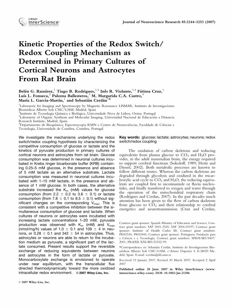
Kinetic Properties of the Redox Switch/ Redox Coupling Mechanism as Determined in Primary Cultures of Cortical Neurons and Astrocytes From Rat Brain Bele ´n G. Ramı ´rez, 1 Tiago B. Rodrigues, 1,4 Ine ˆs R. Violante, 1,4 Fa ´tima Cruz, 1 Luı ´s L. Fonseca, 2 Paloma Ballesteros, 3 M. Margarida C.A. Castro, 4 Marı ´a L. Garcı ´a-Martı ´n, 1 and Sebastia ´n Cerda ´n 1 * 1 Laboratory for Imaging and Spectroscopy by Magnetic Resonance LISMAR, Instituto de Investigaciones Biome ´dicas Alberto Sols CSIC/UAM, Madrid, Spain 2 Instituto de Tecnologia Quı ´mica e Biolo ´gica, Universidade Nova de Lisboa, Oeiras, Portugal 3 Laboratory of Organic Synthesis and Molecular Imaging, Universidad Nacional de Educacio ´n a Distancia Research Institute, Madrid, Spain 4 Departamento de Bioquı ´mica, Espectroscopia RMN e Centro de Neurocie ˆncias, Faculdade de Cie ˆncias e Tecnologia, Universidade de Coimbra, Coimbra, Portugal We investigate the mechanisms underlying the redox switch/redox coupling hypothesis by characterizing the competitive consumption of glucose or lactate and the kinetics of pyruvate production in primary cultures of cortical neurons and astrocytes from rat brain. Glucose consumption was determined in neuronal cultures incu- bated in Krebs ringer bicarbonate buffer (KRB) contain- ing 0.25–5 mM glucose, in the presence and absence of 5 mM lactate as an alternative substrate. Lactate consumption was measured in neuronal cultures incu- bated with 1–15 mM lactate, in the presence and ab- sence of 1 mM glucose. In both cases, the alternative substrate increased the K m (mM) values for glucose consumption (from 2.2 6 0.2 to 3.6 6 0.1) or lactate consumption (from 7.8 6 0.1 to 8.5 6 0.1) without sig- nificant changes on the corresponding V max . This is consistent with a competitive inhibition between the si- multaneous consumption of glucose and lactate. When cultures of neurons or astrocytes were incubated with increasing lactate concentrations 1–20 mM, pyruvate production was observed with K m (mM) and V max (nmol/mg/h) values of 1.0 6 0.1 and 109 6 4 in neu- rons, or 0.28 6 0.1 and 342 6 54 in astrocytes. Thus, astrocytes or neurons are able to return to the incuba- tion medium as pyruvate, a significant part of the lac- tate consumed. Present results support the reversible exchange of reducing equivalents between neurons and astrocytes in the form of lactate or pyruvate. Monocarboxylate exchange is envisioned to operate under near equilibrium, with the transcellular flux directed thermodynamically toward the more oxidized intracellular redox environment. V V C 2007 Wiley-Liss, Inc. Key words: glucose; lactate; astrocytes; neurons; redox switch/redox coupling The oxidation of carbon skeletons and reducing equivalents from plasma glucose to CO 2 and H 2 O pro- vides, in the adult mammalian brain, the energy required to support cerebral functions (Sokoloff, 1989; Hertz and Dienel, 2002). Both metabolic processes are known to follow different routes. Whereas the carbon skeletons are degraded through glycolysis and oxidized in the tricar- boxylic acid cycle to CO 2 and H 2 O, the reducing equiva- lents are coupled first to nicotinamide or flavin nucleo- tides, and finally transferred to oxygen and water through the operation of the mitochondrial respiratory chain (Rodrigues and Cerdan, 2007). In the past decades much attention has been given to the flow of carbon skeletons from glucose to CO 2 and their relationship to cerebral energetics and neurotransmission (Cruz and Cerdan, Contract grant sponsor: Spanish Ministry of Education and Science; Con- tract grant numbers: SAF 2001-2245, SAF 2004-03197; Contract grant sponsor: Institute of Health Carlos III; Contract grant numbers: PI051530, PI051845; Contract grant sponsor: Portuguese Fundac ¸a ˜o para a Cie ˆncia e a Tecnologia; Contract grant numbers: SFRH/BD/5407/ 2001, PRAXIS XXI/BD/21532/99. *Correspondence to: Sebastia ´n Cerda ´n, Instituto de Investigaciones Bio- me ´dicas Alberto Sols CSIC/UAM, c/Arturo Duperier 4, E-28029 Ma- drid, Spain. E-mail: [email protected] Received 15 January 2007; Revised 30 March 2007; Accepted 2 April 2007 Published online 28 June 2007 in Wiley InterScience (www. interscience.wiley.com). DOI: 10.1002/jnr.21386 Journal of Neuroscience Research 85:3244–3253 (2007) ' 2007 Wiley-Liss, Inc.

Welcome message from author
This document is posted to help you gain knowledge. Please leave a comment to let me know what you think about it! Share it to your friends and learn new things together.
Transcript
Kinetic Properties of the Redox Switch/Redox Coupling Mechanism asDetermined in Primary Cultures ofCortical Neurons and AstrocytesFrom Rat Brain
Belen G. Ramırez,1 Tiago B. Rodrigues,1,4 Ines R. Violante,1,4 Fatima Cruz,1
Luıs L. Fonseca,2 Paloma Ballesteros,3 M. Margarida C.A. Castro,4
Marıa L. Garcıa-Martın,1 and Sebastian Cerdan1*1Laboratory for Imaging and Spectroscopy by Magnetic Resonance LISMAR, Instituto de InvestigacionesBiomedicas Alberto Sols CSIC/UAM, Madrid, Spain2Instituto de Tecnologia Quımica e Biologica, Universidade Nova de Lisboa, Oeiras, Portugal3Laboratory of Organic Synthesis and Molecular Imaging, Universidad Nacional de Educacion a DistanciaResearch Institute, Madrid, Spain4Departamento de Bioquımica, Espectroscopia RMN e Centro de Neurociencias, Faculdade de Ciencias eTecnologia, Universidade de Coimbra, Coimbra, Portugal
We investigate the mechanisms underlying the redoxswitch/redox coupling hypothesis by characterizing thecompetitive consumption of glucose or lactate and thekinetics of pyruvate production in primary cultures ofcortical neurons and astrocytes from rat brain. Glucoseconsumption was determined in neuronal cultures incu-bated in Krebs ringer bicarbonate buffer (KRB) contain-ing 0.25–5 mM glucose, in the presence and absenceof 5 mM lactate as an alternative substrate. Lactateconsumption was measured in neuronal cultures incu-bated with 1–15 mM lactate, in the presence and ab-sence of 1 mM glucose. In both cases, the alternativesubstrate increased the Km (mM) values for glucoseconsumption (from 2.2 6 0.2 to 3.6 6 0.1) or lactateconsumption (from 7.8 6 0.1 to 8.5 6 0.1) without sig-nificant changes on the corresponding Vmax. This isconsistent with a competitive inhibition between the si-multaneous consumption of glucose and lactate. Whencultures of neurons or astrocytes were incubated withincreasing lactate concentrations 1–20 mM, pyruvateproduction was observed with Km (mM) and Vmax
(nmol/mg/h) values of 1.0 6 0.1 and 109 6 4 in neu-rons, or 0.28 6 0.1 and 342 6 54 in astrocytes. Thus,astrocytes or neurons are able to return to the incuba-tion medium as pyruvate, a significant part of the lac-tate consumed. Present results support the reversibleexchange of reducing equivalents between neuronsand astrocytes in the form of lactate or pyruvate.Monocarboxylate exchange is envisioned to operateunder near equilibrium, with the transcellular fluxdirected thermodynamically toward the more oxidizedintracellular redox environment. VVC 2007 Wiley-Liss, Inc.
Key words: glucose; lactate; astrocytes; neurons; redoxswitch/redox coupling
The oxidation of carbon skeletons and reducingequivalents from plasma glucose to CO2 and H2O pro-vides, in the adult mammalian brain, the energy requiredto support cerebral functions (Sokoloff, 1989; Hertz andDienel, 2002). Both metabolic processes are known tofollow different routes. Whereas the carbon skeletons aredegraded through glycolysis and oxidized in the tricar-boxylic acid cycle to CO2 and H2O, the reducing equiva-lents are coupled first to nicotinamide or flavin nucleo-tides, and finally transferred to oxygen and water throughthe operation of the mitochondrial respiratory chain(Rodrigues and Cerdan, 2007). In the past decades muchattention has been given to the flow of carbon skeletonsfrom glucose to CO2 and their relationship to cerebralenergetics and neurotransmission (Cruz and Cerdan,
Contract grant sponsor: Spanish Ministry of Education and Science; Con-
tract grant numbers: SAF 2001-2245, SAF 2004-03197; Contract grant
sponsor: Institute of Health Carlos III; Contract grant numbers:
PI051530, PI051845; Contract grant sponsor: Portuguese Fundacao para
a Ciencia e a Tecnologia; Contract grant numbers: SFRH/BD/5407/
2001, PRAXIS XXI/BD/21532/99.
*Correspondence to: Sebastian Cerdan, Instituto de Investigaciones Bio-
medicas Alberto Sols CSIC/UAM, c/Arturo Duperier 4, E-28029 Ma-
drid, Spain. E-mail: [email protected]
Received 15 January 2007; Revised 30 March 2007; Accepted 2 April
2007
Published online 28 June 2007 in Wiley InterScience (www.
interscience.wiley.com). DOI: 10.1002/jnr.21386
Journal of Neuroscience Research 85:3244–3253 (2007)
' 2007 Wiley-Liss, Inc.
1999; Rothman et al., 2003). The pathways for the transferof reducing equivalents to O2 and H2O remained muchless explored. In particular, the highly heterogeneous cel-lular composition of the cerebral tissue led to considerableuncertainties concerning the roles, relative contributions,subcellular compartmentation, and coordination of themetabolisms of carbon skeletons and reducing equivalents,in neurons and glial cells.
The flow of carbon skeletons from glucose to CO2,in neurons and astrocytes during cerebral activation is cur-rently understood to proceed as outlined in the astrocyteto neuron lactate shuttle (ANLS) hypothesis (Pellerin andMagistretti, 1994; Tsacopoulos and Magistretti, 1996;Magistretti et al., 1999). According to this proposal, gluta-matergic stimulation induces glutamate and Na1 influxinto the astrocyte, resulting in partial depolarization and areactive increase of plasma glucose consumption and lac-tate production. Extracellular lactate is then thought to beconsumed selectively by the neurons and oxidized toCO2 in the tricarboxylic acid cycle. Glucose derived fromplasma is, however, also available in the extracellular fluidand can be transported and glycolytically degraded in theneurons. Consequently, both lactate and glucose are avail-able as substrates to support the energetic demands ofneuronal activation, but the mechanisms underlying theselection between the consumption of these two sub-strates and their relative contributions to neuronal or glialmetabolism remain insufficiently understood.
Although the intracellular exchange of reducingequivalents between cytosol and mitochondria of neuronsand astrocytes is receiving increasing attention (Arco andSatrustegui, 2005; McKenna et al., 2006), their transcellu-lar exchange between these cells has not been, to ourknowledge, explored previously. In the original ANLShypothesis, a flux of lactate was proposed to operate fromastrocyte to neuron. The reversibility of the lactate dehy-drogenase isozymes and the monocarboxylate transportersof neurons and astrocytes allows these neural cells to pro-duce and consume lactate or pyruvate, resulting in recy-cling of carbon and reducing equivalents through the cor-responding plasma membranes (Rodrigues and Cerdan,2005b; Rodrigues et al., 2005). Moreover, because bothlactate and pyruvate can exchange reversibly between thecytosol of each cell and the extracellular space, it followsthat these metabolites can also be exchanged transcellu-larly between both cells in a lactate/pyruvate shuttle, anaspect previously not envisioned. The competitive metab-olism of glucose or lactate and the transcellular exchangeof the monocarboxylates lactate and pyruvate betweenneurons and astrocytes, provided the basis for the redoxswitch/redox coupling hypothesis (Cerdan et al., 2006b).However, despite a number of studies on the consump-tion of pyruvate by neurons and astrocytes (Matsumotoet al., 1994; Desagher et al., 1997; Cruz et al., 2001;Zwingmann and Leibfritz, 2003), no evidence had beenpresented to our knowledge on the crucial aspect of pyru-vate production by these cells.
We explore the kinetics of the redox switch/redoxcoupling mechanisms by investigating the consumption of
glucose and lactate in primary cultures of cortical neuronsand show that pyruvate can be produced from lactate,both by neurons and astrocytes. Our results show that lac-tate and glucose are consumed competitively and simulta-neously, the dominant substrate being determined bytheir relative extracellular concentrations, the specific ki-netic constants for substrate consumption, and the cyto-solic redox state. In addition, we show that pyruvate isproduced from lactate in primary cultures of neurons andglial cells, establishing a crucial link for the operation ofthe redox coupling mechanism. A preliminary account ofthis work has been presented (Cerdan et al., 2006a).
MATERIALS AND METHODS
Materials
Dulbecco’s modified Eagle’s medium (DMEM), fetalbovine serum (FBS), horse serum (HS), and phosphate-buf-fered saline (PBS) were purchased from GIBCO (Glasgow,UK). Kits for analytical determinations of glucose, lactate, andpyruvate were from Roche (Mannheim, Germany). Sodium(3-13C)-L-lactate (99% 13C) was purchased from CambridgeIsotope Laboratories (Andover, MA). 2H2O (99.9% 2H) waspurchased from Apollo Scientific (Bradbury, Stockport, UK).All other chemicals were of the purest grade available fromcommercial sources.
Preparation and Characterization of Primary CulturesFrom Neural Cells and Incubation Conditions
Primary cultures of cortical neurons were prepared fromembryos of Wistar rats with 17 days of gestation, essentially asdescribed previously (Cruz et al., 2001). Briefly, the brainswere isolated from the embryos, the meninges removed, andthe cortical areas collected in a PBS solution with 6 mM glu-cose and 1% BSA. The cerebral tissue was minced and resus-pended with a pipette. The suspension was then passedthrough an 80-lm filter and the cells seeded at 1.3 3 106
cells/ml in poly-lysine-coated 8-cm plates with 10 ml of cul-ture medium (DMEM with 15 mM HEPES, 2.5 lg/ml offungizone, 100 U/ml of penicillin/streptomycin, 5 ng/mlNa2SeO4, 5 lg/ml insulin, 5 lg/ml transferrin, 6.3 ng/mlprogesterone, and 100 lM putrescine) with 5% FBS, 5% HS.Plates were maintained in a humidified incubator chamber at378C with 5% CO2. After 24 hr, the cells were transferred toculture medium without serum and maintained for 6 moredays in culture. Forty-eight hours before the experiments,10 lM cytosine b-D-arabinofuranoside was added to the cul-ture medium.
On the day of the experiment, the DMEM mediumwas removed and substituted by Krebs ringer bicarbonatebuffer (KRB) containing glucose concentrations of 0.25, 0.5,1, 2.5, and 5 mM in the absence and presence of 5 mM lac-tate, or lactate concentrations of 1, 2.5, 5, 10, and 15 mM inthe absence and presence of 1 mM glucose. Two aliquotsfrom the incubation medium (0.5 ml each) were withdrawnafter 3 and 6 hr of incubation to determine glucose or lactateconsumption and investigate the kinetic constants of theseprocesses. An independent set of experiments was carried outto evaluate neuronal pyruvate production from lactate. Pri-
Kinetics of Redox Switching and Redox Coupling 3245
Journal of Neuroscience Research DOI 10.1002/jnr
mary cultures of neurons, prepared as described above, wereincubated in KRB containing increasing lactate concentrationsin the same range. In some experiments, the neuronal cultureswere incubated with (3-13C) lactate, to monitor (3-13C) pyru-vate production by 13C NMR. Two aliquots (0.5 ml) fromthe incubation medium were withdrawn at 3 and 6 hr andthe pyruvate concentration determined spectrophotometri-cally. The incubation was continued up to 24 hr, the mediumcollected and concentrated for high resolution 13C NMRanalysis. At least three primary cultures from neurons preparedfrom fetuses derived from different animals were used for ev-ery experimental condition.
Primary cultures of cortical astrocytes were preparedfrom newborn (up to 2-day-old) Wistar rats. Briefly, the cor-tices were removed and cleaned from the meninges. Cerebraltissue was then minced, resuspended with a pipette and vor-texed for 1 min. The resulting suspension was passed throughan 80-lm filter and the cells seeded in poly-lysine-coated 8-cm plate with 10 ml DMEM with 10% FBS, 10% HS,10 mM HEPES, 2.5 lg/ml fungizone, and 100 U/ml penicillin/streptomycin. Plates were maintained in a humidified incuba-tor chamber at 378C with 5% CO2. After 1 week in culture,cells were transferred to DMEM with 5% FBS, 5% HS,10 mM HEPES, 2.5 lg/ml fungizone, and 100 U/ml penicillin/streptomycin. Confluent cultures were obtained and used forexperiments after 2 weeks in culture. In the day of the experi-ment, the DMEM medium was removed and substituted byKRB containing increasing concentrations of lactate in therange 1–15 mM in the absence and presence of 1 mM glu-cose. In some experiments, astrocyte cultures were incubatedwith (3-13C) lactate, to evaluate (3-13C) pyruvate productionunder similar conditions to those described above for neurons.At least three primary astrocyte cultures prepared from differ-ent animals were used for every experimental condition.
Cell culture purity was assessed using standard immuno-labeling techniques. Cells were washed three times with PBSfor 10 min, fixed in 4% (w/v) paraformaldehyde in PBS for5 min, and permeabilized with 0.2% (v/v) Triton in PBS. Neu-rons were visualized (negative staining) using rabbit anti-glialfibrillary acidic protein (GFAP, 1:200) and (positive staining)rat anti-b-III-tubulin (1:200) as primary antibodies and biotinconjugated anti-rat immuno gamma globulin (IgG,1:200),streptavidin Alexa Fluor 488 (1:2,000), and Alexa Fluor 594goat anti-rabbit IgG (1:2,000) as secondary antibodies. Astro-cytes were visualized with rabbit anti-GFAP (1:200) and AlexaFluor 594 goat anti-rabbit IgG (1:2,000). Cellular nuclei werelabeled with 40-6-diamidino-2-phenylindol (DAPI). The puri-ties of neuron or astrocyte cultures determined in this waywere >95%.
Analytical Determinations
Glucose, lactate, or pyruvate in the incubation mediumof neurons or astrocytes, were determined spectrophotometri-cally (340 nm, 378C) using a vertical spectrophotometer(Spectramax TM 340 PC; Molecular Devices, Sunnyvale, CA)in 96-well polypropylene plates. To this end, we adapted con-ventional enzymatic end point methods coupled to NAD(P)Hproduction or consumption (Bergemeyer, 1983). Briefly, glu-
cose was determined through the NADPH production byhexokinase and glucose-6-phosphate dehydrogenase, lactate,and pyruvate through NADH production or consumption bylactate dehydrogenase. Total protein was determined in cellpellets obtained after the incubation using the Bradford assay(Bradford, 1976).
Determination of Kinetic Constants
The apparent Km and Vmax values for glucose consump-tion in the absence and presence of lactate, lactate consump-tion in the presence and absence of glucose, and pyruvate pro-duction, were determined by non-linear least squares fit of themeasured rates to the Michaelis-Menten expression v 5 Vmax
3 [S]/Km 1 [S] where v or [S] represent the initial rates ofsubstrate consumption or the initial concentrations of substrateand Km and Vmax, the corresponding apparent kinetic con-stants. The apparent inhibition constant Ki was determinedusing the expression for competitive inhibition KmI 5 Km (11 [I]/Ki) where KmI and Km represent the Km values in thepresence and absence of the inhibitor and [I] the inhibitorconcentration. Non-linear fits were carried out using theMathematica v4.0 software (Wolfram Research, Champaign,IL) on an Intel Centrino Pentium M 1.5 GHz platform.
13C NMR
High resolution 13C NMR spectra (48C, pH 5 7.2)from aliquots of the incubation medium after incubation with(3-13C) lactate were acquired in a Bruker AVANCE 500 WBNMR spectrometer using a commercial 5-mm high resolutiontriple probe (13C,1H,2H). Conditions were: p/3 pulses, 1.09sec acquisition time, 7.0 sec recycle time, 30 kHz sweepwidth, 64,000 points zero-filled to 256,000 points before Fou-rier transformation and 20,000 scans. Broadband protondecoupling was gated on only during acquisition. Chemicalshifts were measured with respect to the resonance of a 10%dioxane solution in water at 67.4 ppm, placed in a coaxialcapillary. Assignments were carried out using literature valuesand confirmed through the addition of the authentic com-pounds (Cruz and Cerdan, 1999; Rodrigues and Cerdan,2005a).
Statistics
Kinetic constants obtained under different incubationconditions were compared using Student’s t-test, as imple-mented in the SAS package (Statistical Analysis System, Cary,NC) running on an Intel Centrino Pentium M 1.5 GHz Plat-form. Results with P < 0.05 were considered statistically sig-nificant.
RESULTS
Competitive Consumption of Glucose and Lactate
Table I summarizes the kinetic constants for glucoseconsumption in the absence and presence of 5 mM lactateor those for lactate consumption in the presence and ab-sence of 1 mM glucose. Aliquots withdrawn at 3 and 6 hrof incubation showed a linear decrease in the initial glu-cose or lactate concentrations allowing the determinationof the initial rates. Glucose or lactate consumption rates
3246 Ramırez et al.
Journal of Neuroscience Research DOI 10.1002/jnr
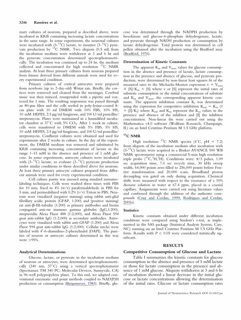
depicted a clear hyperbolic pattern, consistent withMichaelis-Menten kinetics. These experiments werereproduced with neuronal cultures from fetuses of threedifferent litters and the combined results fitted to theMichaelis-Menten expression to determine the apparentKm and Vmax values. The Km for glucose consumptionincreased from 2.2 6 0.2 mM in the absence of lactate, to3.6 6 0.1 mM (P < 0.001) in the presence of 5 mM lac-tate, with no significant effects on the Vmax value. This isconsistent with a competitive inhibition mechanism, witha Ki value of 3.6 mM for the inhibition of glucose con-sumption by extracellular lactate. The Km and Vmax valuesfor lactate consumption increased from 7.8 6 0.1 mMand 440 6 3 nmol/mg/hr in the absence of glucose to8.5 6 0.1 and 451 6 3 nmol/mg/hr (P < 0.05) in thepresence of 1 mM glucose, also consistent with a compet-itive inhibition mechanism with a Ki value of 11.1 mM.
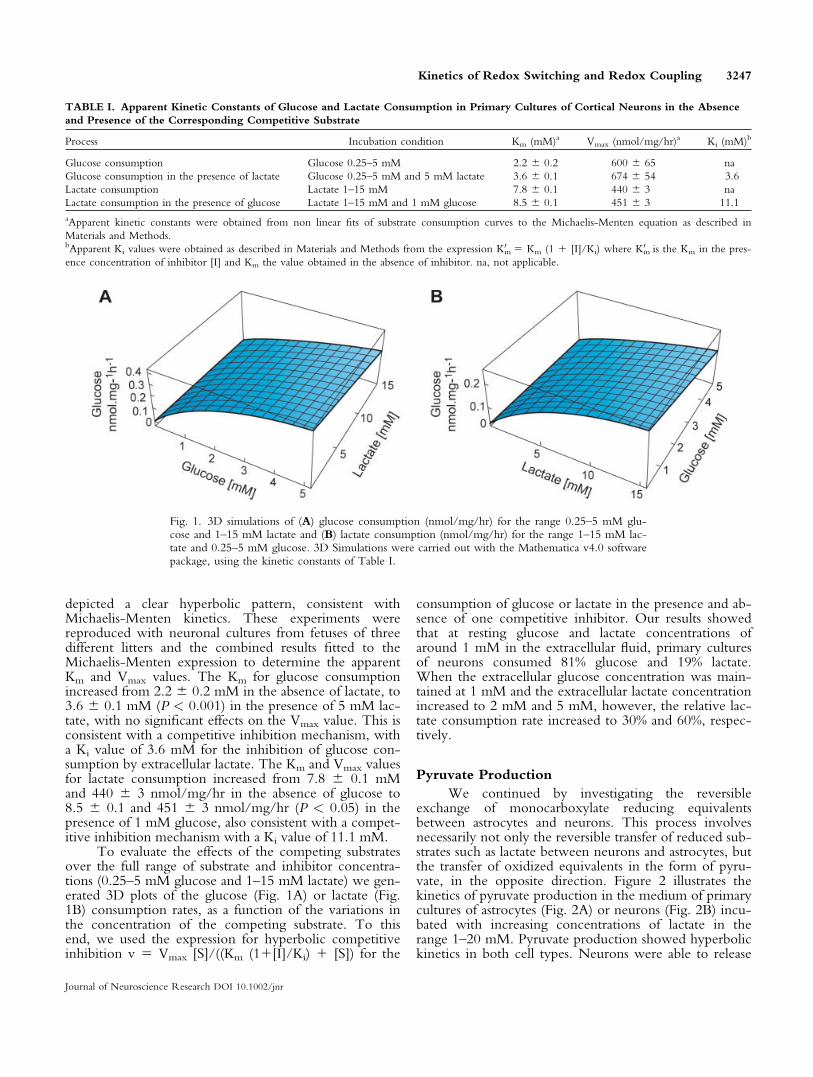
To evaluate the effects of the competing substratesover the full range of substrate and inhibitor concentra-tions (0.25–5 mM glucose and 1–15 mM lactate) we gen-erated 3D plots of the glucose (Fig. 1A) or lactate (Fig.1B) consumption rates, as a function of the variations inthe concentration of the competing substrate. To thisend, we used the expression for hyperbolic competitiveinhibition v 5 Vmax [S]/((Km (11[I]/Ki) 1 [S]) for the
consumption of glucose or lactate in the presence and ab-sence of one competitive inhibitor. Our results showedthat at resting glucose and lactate concentrations ofaround 1 mM in the extracellular fluid, primary culturesof neurons consumed 81% glucose and 19% lactate.When the extracellular glucose concentration was main-tained at 1 mM and the extracellular lactate concentrationincreased to 2 mM and 5 mM, however, the relative lac-tate consumption rate increased to 30% and 60%, respec-tively.
Pyruvate Production
We continued by investigating the reversibleexchange of monocarboxylate reducing equivalentsbetween astrocytes and neurons. This process involvesnecessarily not only the reversible transfer of reduced sub-strates such as lactate between neurons and astrocytes, butthe transfer of oxidized equivalents in the form of pyru-vate, in the opposite direction. Figure 2 illustrates thekinetics of pyruvate production in the medium of primarycultures of astrocytes (Fig. 2A) or neurons (Fig. 2B) incu-bated with increasing concentrations of lactate in therange 1–20 mM. Pyruvate production showed hyperbolickinetics in both cell types. Neurons were able to release
Fig. 1. 3D simulations of (A) glucose consumption (nmol/mg/hr) for the range 0.25–5 mM glu-cose and 1–15 mM lactate and (B) lactate consumption (nmol/mg/hr) for the range 1–15 mM lac-tate and 0.25–5 mM glucose. 3D Simulations were carried out with the Mathematica v4.0 softwarepackage, using the kinetic constants of Table I.
TABLE I. Apparent Kinetic Constants of Glucose and Lactate Consumption in Primary Cultures of Cortical Neurons in the Absence
and Presence of the Corresponding Competitive Substrate
Process Incubation condition Km (mM)a Vmax (nmol/mg/hr)a Ki (mM)b
Glucose consumption Glucose 0.25–5 mM 2.2 6 0.2 600 6 65 na
Glucose consumption in the presence of lactate Glucose 0.25–5 mM and 5 mM lactate 3.6 6 0.1 674 6 54 3.6
Lactate consumption Lactate 1–15 mM 7.8 6 0.1 440 6 3 na
Lactate consumption in the presence of glucose Lactate 1–15 mM and 1 mM glucose 8.5 6 0.1 451 6 3 11.1
aApparent kinetic constants were obtained from non linear fits of substrate consumption curves to the Michaelis-Menten equation as described in
Materials and Methods.bApparent Ki values were obtained as described in Materials and Methods from the expression Km
0 5 Km (1 1 [I]/Ki) where Km0 is the Km in the pres-
ence concentration of inhibitor [I] and Km the value obtained in the absence of inhibitor. na, not applicable.
Kinetics of Redox Switching and Redox Coupling 3247
Journal of Neuroscience Research DOI 10.1002/jnr
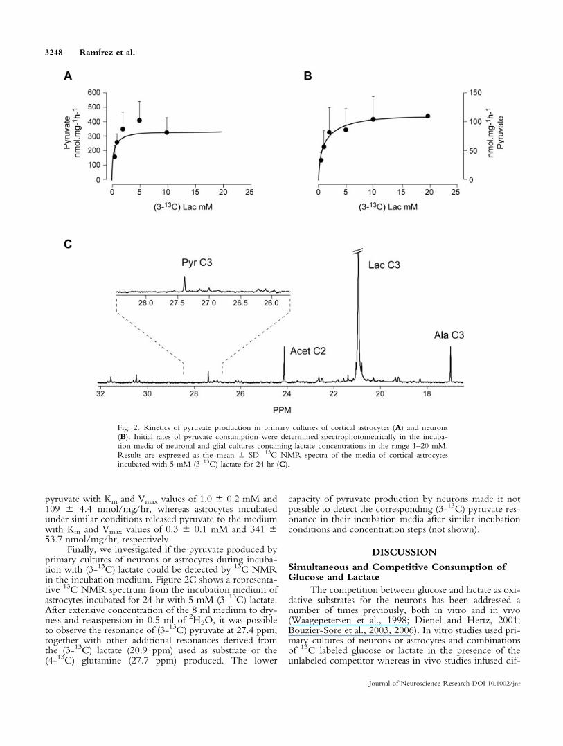
pyruvate with Km and Vmax values of 1.0 6 0.2 mM and109 6 4.4 nmol/mg/hr, whereas astrocytes incubatedunder similar conditions released pyruvate to the mediumwith Km and Vmax values of 0.3 6 0.1 mM and 341 653.7 nmol/mg/hr, respectively.
Finally, we investigated if the pyruvate produced byprimary cultures of neurons or astrocytes during incuba-tion with (3-13C) lactate could be detected by 13C NMRin the incubation medium. Figure 2C shows a representa-tive 13C NMR spectrum from the incubation medium ofastrocytes incubated for 24 hr with 5 mM (3-13C) lactate.After extensive concentration of the 8 ml medium to dry-ness and resuspension in 0.5 ml of 2H2O, it was possibleto observe the resonance of (3-13C) pyruvate at 27.4 ppm,together with other additional resonances derived fromthe (3-13C) lactate (20.9 ppm) used as substrate or the(4-13C) glutamine (27.7 ppm) produced. The lower
capacity of pyruvate production by neurons made it notpossible to detect the corresponding (3-13C) pyruvate res-onance in their incubation media after similar incubationconditions and concentration steps (not shown).
DISCUSSION
Simultaneous and Competitive Consumption ofGlucose and Lactate
The competition between glucose and lactate as oxi-dative substrates for the neurons has been addressed anumber of times previously, both in vitro and in vivo(Waagepetersen et al., 1998; Dienel and Hertz, 2001;Bouzier-Sore et al., 2003, 2006). In vitro studies used pri-mary cultures of neurons or astrocytes and combinationsof 13C labeled glucose or lactate in the presence of theunlabeled competitor whereas in vivo studies infused dif-
Fig. 2. Kinetics of pyruvate production in primary cultures of cortical astrocytes (A) and neurons(B). Initial rates of pyruvate consumption were determined spectrophotometrically in the incuba-tion media of neuronal and glial cultures containing lactate concentrations in the range 1–20 mM.Results are expressed as the mean 6 SD. 13C NMR spectra of the media of cortical astrocytesincubated with 5 mM (3-13C) lactate for 24 hr (C).
3248 Ramırez et al.
Journal of Neuroscience Research DOI 10.1002/jnr
ferent combinations of labeled and unlabeled glucose andlactate (Bouzier-Sore et al., 2003, 2006). The in vitroresults showed a preferential oxidation of lactate by neu-rons as shown by increased 13C labeling of the aminoacids glutamate and glutamine. Similarly, the in vivo datawere consistent with a preferential oxidation of lactate ina compartment devoid of pyruvate carboxylase activity,presumably the neuronal environment (Bouzier et al.,2000).
In contrast, the inhibitory effects of glucose orlactate on the consumption of each other received lessattention and no kinetic studies are available to ourknowledge. Primary cultures of cortical neurons incu-bated with glucose or lactate only, consumed preferen-tially glucose because of its more favorable kinetic con-stants Km and Vmax. Lactate increased the Km for glucoseconsumption without a significant effect on Vmax and glu-cose increased the Km for lactate consumption, withoutapparent effects on Vmax. These results showed that glu-cose and lactate are consumed competitively and simulta-neously, rather than alternatively as thought previously.Moreover, the kinetics for glucose and lactate consump-tion in the presence of each other are consistent with aone-site inhibition by the competing substrate in bothcases. We hypothesize that the common inhibition siteoccurs at the glyceraldehyde 3-phosphate dehydrogenasestep, the inhibition mechanism relying in the competitionfor cytosolic NAD1 between glycolysis and lactate oxida-tion (Cruz et al., 2001; Garcia-Espinosa et al., 2004). Theapparent Ki values indicate that relatively high lactate con-centrations (�3.6 mM) are required to inhibit by 50%glucose consumption. This concentration is in the rangeof the high Km value of the dominant lactate dehydrogen-ase activity of synaptosol (O’Brien et al., 2007), suggestingthat this isoform is the main participant in the regulationof the redox switch.
The relevance of the present results to the in vivosituation in the adult brain remains difficult to evaluate atpresent. Lactate concentrations in the in vivo brain havebeen measured previously by 1H NMR spectroscopy andin vivo microdialysis methods, with values in the range of0.5 lmol/g or 0.4 mM, respectively (Gruetter et al.,1998; Darbin et al., 2006). Moderate increases ranging50–150% have been reported on cerebral activation(Giove et al., 2003). These concentrations seem to be toosmall to elicit a significant inhibition of glucose consump-tion or an important relative rate of lactate consumption,in our cultures. Under these in vivo conditions, presentresults indicate that neurons would consume preferentiallyglucose. This conclusion agrees with the in vivo accumu-lation of fluorescently labeled deoxyglucose observed inneurons from the hippocampus or cerebellar Purkinjecells (Itoh et al., 2004). It is also possible, however, thatthe actual concentrations of lactate in the highly con-strained space of the synaptic cleft are significantly higherthan the average concentrations measured by NMR overlarge voxel volumes or determined by microdialysis in theextracellular space. Under these conditions, synaptic lac-tate could operate effectively the redox switch, provided
that its concentration approaches or exceeds significantlythe Ki value. Extracellular lactate can also approach acomplete switch off of glycolysis under hypoxic or ische-mic conditions, when lactate concentrations reach the10–15 mM range. This confirms previous proposals oflactate becoming a predominant substrate under ischemicconditions (Schurr et al., 1988; Schurr, 2006).
In addition to these considerations, it should benoted that cerebral activation can induce changes in therates of lactate production and consumption by the neu-ronal and glial compartments in vivo (Serres et al., 2003,2004, 2005). Increased glutamate concentrations obtainedduring activation have been shown to increase the uptakeof fluorescently labeled glucose by astrocytes and reduce itin neurons (Loaiza et al., 2003; Porras et al., 2004). To-gether, this evidence indicates that the cerebral activationprocess in vivo could involve changes in the kinetic pa-rameters investigated in this study under resting condi-tions. In summary, our results suggest that glucose andlactate can be consumed simultaneously, the consumptionof one substituting partially the need for the other. This isa useful bioenergetic circumstance because both substratesseem to be equally effective energizing activity dependentsynaptic vesicle turnover (Morgenthaler et al., 2006).
It should be noted that the kinetics of relative glu-cose or lactate consumption reported here are differentfrom the relative glucose or lactate oxidation ratesdescribed previously (Bouzier-Sore et al., 2003, 2006).For the combination of 1 mM glucose and 1 mM lactate,it was calculated that lactate contributed 75% of the car-bons oxidized in the neuronal cycle, whereas the lactateconsumption values reported in the present study wouldindicate that lactate consumption represents only 20% ofthe glucose consumed. These results indicate that eventhough lactate consumption may be smaller than glucoseconsumption, the lactate consumed may be preferentiallyoxidized. This situation is consistent with our previousfindings of two kinetically different pyruvate pools inneurons, one derived from glucose (the Pyrg pool) andthe other from monocarboxylates (Pyrp) pool, able to beoxidized preferentially under different substrate and redoxconditions (Cruz et al., 2001; Garcia-Espinosa et al.,2004). Thus, glucose or lactate consumption rates maynot be linearly related to the respective oxidation rates.
Transcellular Monocarboxylate Redox Shuttle
A number of studies have reported previously lactateand pyruvate consumption by primary cultures of neuralcells (Cruz et al., 2001; Dienel and Hertz, 2001; Zwing-mann and Leibfritz, 2003; Bouzier-Sore et al., 2006). Inaddition, the presence of pyruvate in the extracellularfluid has been classically reported in a variety of micro-dialysis experiments (Ronne-Engstrom et al., 1995; Val-tysson et al., 1998). The origin of the cerebral pyruvatepresent in the extracellular fluid and consumed by neuralcells has not, however, been addressed sufficiently. Thepresent study provides an advance in this direction byshowing pyruvate production and its kinetics in primary
Kinetics of Redox Switching and Redox Coupling 3249
Journal of Neuroscience Research DOI 10.1002/jnr
cultures of neurons and glial cells. Interestingly, our resultsindicate that both neural cells are able to produce pyru-vate from lactate but with different kinetics. For the sameextracellular lactate concentration, astrocytes present alarger capacity of pyruvate production than neurons. Thisoccurs, most probably, because of the different kineticproperties of the lactate dehydrogenase isozymes presentin both cells (O’Brien et al., 2007). Although both neu-rons and astrocytes have been shown recently to containthe five isoforms of LDH, the muscle isoenzyme LDH5seems to be predominant in astrocytes whereas the heartisoenzyme LDH1 is predominant in neurons (Bittar et al.,1996; Pellerin et al., 1998). The kinetic constants ofLDH5 favor lactate production whereas those of LDH1favor pyruvate formation. The fact that from the sameamount of lactate neurons produce less pyruvate thanastrocytes indicates that the pyruvate produced by neu-rons may be, as expected, preferentially oxidized ratherthan exported to the extracellular medium. It should benoted that the capacity for pyruvate production in neu-rons may account for a very large proportion of the lactateconsumed. Considering the kinetic constants of lactateconsumption (Table I) and pyruvate production for a 1mM extracellular lactate concentration, it can be calcu-lated that both lactate and pyruvate can be consumed andproduced virtually at the same rate of 50 nmol/mg/hr.This represents an important observation because manyprevious studies considered that all lactate consumed byneural cells was completely oxidized. On this basis, ourresults suggest that the rates of lactate oxidation reportedpreviously, based on metabolic balances not consideringpyruvate production, could be overestimated.
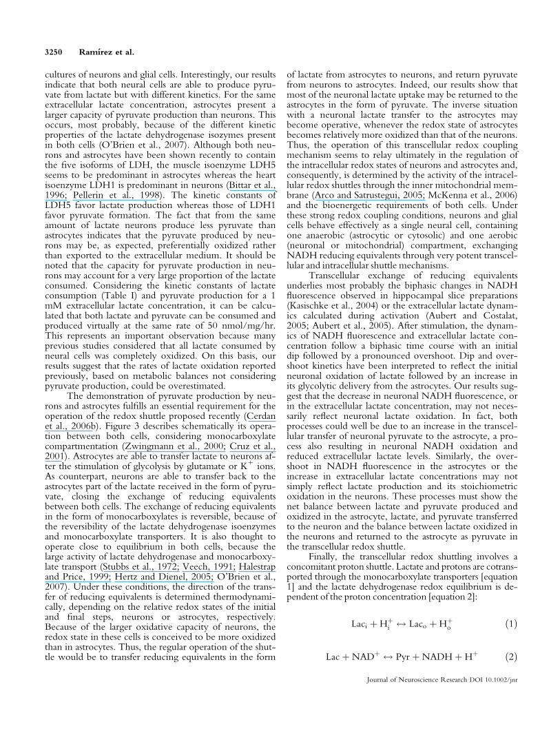
The demonstration of pyruvate production by neu-rons and astrocytes fulfills an essential requirement for theoperation of the redox shuttle proposed recently (Cerdanet al., 2006b). Figure 3 describes schematically its opera-tion between both cells, considering monocarboxylatecompartmentation (Zwingmann et al., 2000; Cruz et al.,2001). Astrocytes are able to transfer lactate to neurons af-ter the stimulation of glycolysis by glutamate or K1 ions.As counterpart, neurons are able to transfer back to theastrocytes part of the lactate received in the form of pyru-vate, closing the exchange of reducing equivalentsbetween both cells. The exchange of reducing equivalentsin the form of monocarboxylates is reversible, because ofthe reversibility of the lactate dehydrogenase isoenzymesand monocarboxylate transporters. It is also thought tooperate close to equilibrium in both cells, because thelarge activity of lactate dehydrogenase and monocarboxy-late transport (Stubbs et al., 1972; Veech, 1991; Halestrapand Price, 1999; Hertz and Dienel, 2005; O’Brien et al.,2007). Under these conditions, the direction of the trans-fer of reducing equivalents is determined thermodynami-cally, depending on the relative redox states of the initialand final steps, neurons or astrocytes, respectively.Because of the larger oxidative capacity of neurons, theredox state in these cells is conceived to be more oxidizedthan in astrocytes. Thus, the regular operation of the shut-tle would be to transfer reducing equivalents in the form
of lactate from astrocytes to neurons, and return pyruvatefrom neurons to astrocytes. Indeed, our results show thatmost of the neuronal lactate uptake may be returned to theastrocytes in the form of pyruvate. The inverse situationwith a neuronal lactate transfer to the astrocytes maybecome operative, whenever the redox state of astrocytesbecomes relatively more oxidized than that of the neurons.Thus, the operation of this transcellular redox couplingmechanism seems to relay ultimately in the regulation ofthe intracellular redox states of neurons and astrocytes and,consequently, is determined by the activity of the intracel-lular redox shuttles through the inner mitochondrial mem-brane (Arco and Satrustegui, 2005; McKenna et al., 2006)and the bioenergetic requirements of both cells. Underthese strong redox coupling conditions, neurons and glialcells behave effectively as a single neural cell, containingone anaerobic (astrocytic or cytosolic) and one aerobic(neuronal or mitochondrial) compartment, exchangingNADH reducing equivalents through very potent transcel-lular and intracellular shuttle mechanisms.
Transcellular exchange of reducing equivalentsunderlies most probably the biphasic changes in NADHfluorescence observed in hippocampal slice preparations(Kasischke et al., 2004) or the extracellular lactate dynam-ics calculated during activation (Aubert and Costalat,2005; Aubert et al., 2005). After stimulation, the dynam-ics of NADH fluorescence and extracellular lactate con-centration follow a biphasic time course with an initialdip followed by a pronounced overshoot. Dip and over-shoot kinetics have been interpreted to reflect the initialneuronal oxidation of lactate followed by an increase inits glycolytic delivery from the astrocytes. Our results sug-gest that the decrease in neuronal NADH fluorescence, orin the extracellular lactate concentration, may not neces-sarily reflect neuronal lactate oxidation. In fact, bothprocesses could well be due to an increase in the transcel-lular transfer of neuronal pyruvate to the astrocyte, a pro-cess also resulting in neuronal NADH oxidation andreduced extracellular lactate levels. Similarly, the over-shoot in NADH fluorescence in the astrocytes or theincrease in extracellular lactate concentrations may notsimply reflect lactate production and its stoichiometricoxidation in the neurons. These processes must show thenet balance between lactate and pyruvate produced andoxidized in the astrocyte, lactate, and pyruvate transferredto the neuron and the balance between lactate oxidized inthe neurons and returned to the astrocyte as pyruvate inthe transcellular redox shuttle.
Finally, the transcellular redox shuttling involves aconcomitant proton shuttle. Lactate and protons are cotrans-ported through the monocarboxylate transporters [equation1] and the lactate dehydrogenase redox equilibrium is de-pendent of the proton concentration [equation 2]:
Laci þHþi $ Laco þHþ
o ð1Þ
LacþNADþ $ Pyr þNADHþHþ ð2Þ
3250 Ramırez et al.
Journal of Neuroscience Research DOI 10.1002/jnr
Consequently, the transcellular redox shuttle seems to beintimately related to intra- and extracellular pH regulationand, because of the proton involvement in many monova-lent and divalent cation transport processes, linked to theoverall ionic homeostasis.
In this context, the transcellular monocarboxylateshuttle investigated in this study is envisioned to play acentral role in the integration of redox processes, pH reg-ulation, and ion homeostasis in the central nervous sys-tem. Redox shuttles based on monocarboxylate exchangemay present a more general biologic significance, cou-pling hypoxic and oxidative environments in many otherheterogeneous tissues including muscle and tumors.
ACKNOWLEDGMENTS
The authors are deeply indebted to the reviewers ofour previous contributions who expressed the importanceof addressing pyruvate production in neural cells. In addi-tion, they wish to express their gratitude to S. Arroyo forexpert technical assistance and the drafting shop of theInstitute of Biomedical Research Alberto Sols for profes-sional preparation of the illustrations. B.G.R. was a post-doctoral fellow of C.S.I.C. and I.R.V. received an Eras-mus fellowship from the University of Coimbra.M.M.C.A.C. was on a sabbatical visit from the Universityof Coimbra (Portugal). T.B.R. and L.L.F. received pre-
Fig. 3. The transcellular monocarboxylate redox shuttle betweenneurons and astrocytes. Reducing equivalents are exchanged trans-cellularly, under reversible, and close to equilibrium conditions, inthe form of the monocarboxylates lactate and pyruvate. The netdirection of the flow occurs toward the more oxidized cytosolic re-dox environment, as determined mainly by the local oxygen avail-ability, the oxidative capacity of the corresponding respiratory chainand the activity of the mitochondrial redox shuttles (dotted line) cou-pling cytosolic and mitochondrial redox environments. The exchangeof monocarboxylates involves primarily the fast turnover pool of
cytosolic monocarboxylates (Lacp and Pyrp), eventually inhibitingglycolytic pyruvate production (Pyrg pool) by NAD1 competition.Lacg, slow turnover glycolytic lactate pool; Laco, extracellular lactate;Lacp, fast turnover intracellular lactate pool derived from extracellularlactate; LDH, lactate dehydrogenase; Glucosei, intracellular glucose;Glucoseo, extracellular glucose; MCT, monocarboxylate transporters;Pyrg, slow turnover glycolytic pyruvate pool; Pyro, extracellular pyru-vate pool; Pyrp, fast turnover intracellular pyruvate pool derived fromextracellular pyruvate.
Kinetics of Redox Switching and Redox Coupling 3251
Journal of Neuroscience Research DOI 10.1002/jnr
doctoral fellowships from the Portuguese FCT.M.L.G.M. holds a tenure track ‘‘Ramon y Cajal’’ contractfrom CSIC. S.C. receives a grant from Institute of HealthCarlos III, as does M.L.G.M. Portuguese Fundacao para aCiencia e a Tecnologia provides funding for T.B.R. andL.L.F.
REFERENCES
Arco AD, Satrustegui J. 2005. New mitochondrial carriers: an overview.
Cell Mol Life Sci 62:2204–2227.
Aubert A, Costalat R. 2005. Interaction between astrocytes and neurons
studied using a mathematical model of compartmentalized energy me-
tabolism. J Cereb Blood Flow Metab 25:1476–1490.
Aubert A, Costalat R, Magistretti PJ, Pellerin L. 2005. Brain lactate
kinetics: modeling evidence for neuronal lactate uptake upon activation.
Proc Natl Acad Sci U S A 102:16448–16453.
Bergemeyer HU. 1983. Methods of enzymatic analysis. Weinheim: Ver-
lag Chemie.
Bittar PG, Charnay Y, Pellerin L, Bouras C, Magistretti PJ. 1996. Selec-
tive distribution of lactate dehydrogenase isoenzymes in neurons and
astrocytes of human brain. J Cereb Blood Flow Metab 16:1079–1089.
Bouzier-Sore AK, Voisin P, Bouchaud V, Bezancon E, Franconi JM,
Pellerin L. 2006. Competition between glucose and lactate as oxidative
energy substrates in both neurons and astrocytes: a comparative NMR
study. Eur J Neurosci 24:1687–1694.
Bouzier-Sore AK, Voisin P, Canioni P, Magistretti PJ, Pellerin L. 2003.
Lactate is a preferential oxidative energy substrate over glucose for neu-
rons in culture. J Cereb Blood Flow Metab 23:1298–1306.
Bouzier AK, Thiaudiere E, Biran M, Rouland R, Canioni P, Merle M.
2000. The metabolism of [3-13C]lactate in the rat brain is specific of a
pyruvate carboxylase-deprived compartment. J Neurochem 75:480–486.
Bradford MM. 1976. A rapid and sensitive method for the quantitation
of microgram quantities of protein utilizing the principle of protein-dye
binding. Anal Biochem 72:248–254.
Cerdan S, Rodrigues TB, Garcıa-Martın ML. 2006a. Redox switching:
modulation of glucose and pyruvate/lactate metabolism. 7th Interna-
tional Conference of Brain Energy Metabolism, Lausanne, Switzerland.
Cerdan S, Rodrigues TB, Sierra A, Benito M, Fonseca LL, Fonseca CP,
Garcia-Martin ML. 2006b. The redox switch/redox coupling hypothe-
sis. Neurochem Int 48:523–530.
Cruz F, Cerdan S. 1999. Quantitative 13C NMR studies of metabolic
compartmentation in the adult mammalian brain. NMR Biomed 12:
451–462.
Cruz F, Villalba M, Garcia-Espinosa MA, Ballesteros P, Bogonez E,
Satrustegui J, Cerdan S. 2001. Intracellular compartmentation of pyru-
vate in primary cultures of cortical neurons as detected by 13C NMR
spectroscopy with multiple 13C labels. J Neurosci Res 66:771–781.
Darbin O, Carre E, Naritoku D, Risso JJ, Lonjon M, Patrylo PR. 2006.
Glucose metabolites in the striatum of freely behaving rats following
infusion of elevated potassium. Brain Res 1116:127–131.
Desagher S, Glowinski J, Premont J. 1997. Pyruvate protects neurons
against hydrogen peroxide-induced toxicity. J Neurosci 17:9060–9067.
Dienel GA, Hertz L. 2001. Glucose and lactate metabolism during brain
activation. J Neurosci Res 66:824–838.
Garcia-Espinosa MA, Rodrigues TB, Sierra A, Benito M, Fonseca C,
Gray HL, Bartnik BL, Garcia-Martin ML, Ballesteros P, Cerdan S.
2004. Cerebral glucose metabolism and the glutamine cycle as detected
by in vivo and in vitro 13C NMR spectroscopy. Neurochem Int
45:297–303.
Giove F, Mangia S, Bianciardi M, Garreffa G, Di Salle F, Morrone R,
Maraviglia B. 2003. The physiology and metabolism of neuronal activa-
tion: in vivo studies by NMR and other methods. Magn Reson Imag-
ing 21:1283–1293.
Gruetter R, Seaquist ER, Kim S, Ugurbil K. 1998. Localized in vivo13C-NMR of glutamate metabolism in the human brain: initial results
at 4 tesla. Dev Neurosci 20:380–388.
Halestrap AP, Price NT. 1999. The proton-linked monocarboxylate
transporter (MCT) family: structure, function and regulation. Biochem
J 343 Pt 2:281–299.
Hertz L, Dienel GA. 2002. Energy metabolism in the brain. Int Rev
Neurobiol 51:1–102.
Hertz L, Dienel GA. 2005. Lactate transport and transporters: general
principles and functional roles in brain cells. J Neurosci Res 79:11–18.
Itoh Y, Abe T, Takaoka R, Tanahashi N. 2004. Fluorometric determina-
tion of glucose utilization in neurons in vitro and in vivo. J Cereb
Blood Flow Metab 24:993–1003.
Kasischke KA, Vishwasrao HD, Fisher PJ, Zipfel WR, Webb WW.
2004. Neural activity triggers neuronal oxidative metabolism followed
by astrocytic glycolysis. Science 305:99–103.
Loaiza A, Porras OH, Barros LF. 2003. Glutamate triggers rapid glucose
transport stimulation in astrocytes as evidenced by real-time confocal
microscopy. J Neurosci 23:7337–7342.
Magistretti PJ, Pellerin L, Rothman DL, Shulman RG. 1999. Energy on
demand. Science 283:496–497.
Matsumoto K, Yamada K, Kohmura E, Kinoshita A, Hayakawa T. 1994.
Role of pyruvate in ischaemia-like conditions on cultured neurons.
Neurol Res 16:460–464.
McKenna MC, Waagepetersen HS, Schousboe A, Sonnewald U. 2006.
Neuronal and astrocytic shuttle mechanisms for cytosolic-mitochondrial
transfer of reducing equivalents: current evidence and pharmacological
tools. Biochem Pharmacol 71:399–407.
Morgenthaler FD, Kraftsik R, Catsicas S, Magistretti PJ, Chatton JY.
2006. Glucose and lactate are equally effective in energizing activity-de-
pendent synaptic vesicle turnover in purified cortical neurons. Neuro-
science 141:157–165.
O’Brien J, Kla KM, Hopkins IB, Malecki EA, McKenna MC. 2007. Ki-
netic parameters and lactate dehydrogenase isozyme activities support
possible lactate utilization by neurons. Neurochem Res 32:597–607.
Pellerin L, Magistretti PJ. 1994. Glutamate uptake into astrocytes stimu-
lates aerobic glycolysis: a mechanism coupling neuronal activity to glu-
cose utilization. Proc Natl Acad Sci U S A 91:10625–10629.
Pellerin L, Pellegri G, Bittar PG, Charnay Y, Bouras C, Martin JL, Stella
N, Magistretti PJ. 1998. Evidence supporting the existence of an activ-
ity-dependent astrocyte-neuron lactate shuttle. Dev Neurosci 20:291–
299.
Porras OH, Loaiza A, Barros LF. 2004. Glutamate mediates acute glucose
transport inhibition in hippocampal neurons. J Neurosci 24:9669–9673.
Rodrigues TB, Cerdan S. 2005a. 13C MRS: an outstanding tool for met-
abolic studies. Concepts Magn Reson Part A 27A:1–16.
Rodrigues TB, Cerdan S. 2005b. A fast and sensitive 1H NMR method
to measure the turnover of the H2 hydrogen of lactate. Magn Reson
Med 54:1014–1019.
Rodrigues TB, Cerdan S. 2007. The cerebral tricarboxylic acid cycles.
In: Lajhta A, Gibson GE, Dienel G, editors. Brain energetics integra-
tion of molecular and cellular processes. New York: Springer.
Rodrigues TB, Gray HL, Benito M, Garrido S, Sierra A, Geraldes CF,
Ballesteros P, Cerdan S. 2005. Futile cycling of lactate through the
plasma membrane of C6 glioma cells as detected by (13C, 2H) NMR.
J Neurosci Res 79:119–127.
Ronne-Engstrom E, Carlson H, Liu Y, Ungerstedt U, Hillered L. 1995.
Influence of perfusate glucose concentration on dialysate lactate, pyru-
vate, aspartate, and glutamate levels under basal and hypoxic conditions:
a microdialysis study in rat brain. J Neurochem 65:257–262.
Rothman DL, Behar KL, Hyder F, Shulman RG. 2003. In vivo NMR
studies of the glutamate neurotransmitter flux and neuroenergetics:
implications for brain function. Annu Rev Physiol 65:401–427.
3252 Ramırez et al.
Journal of Neuroscience Research DOI 10.1002/jnr
Schurr A. 2006. Lactate: the ultimate cerebral oxidative energy substrate?
J Cereb Blood Flow Metab 26:142–152.
Schurr A, West CA, Rigor BM. 1988. Lactate-supported synaptic func-
tion in the rat hippocampal slice preparation. Science 240:1326–1328.
Serres S, Bezancon E, Franconi JM, Merle M. 2004. Ex vivo analysis of
lactate and glucose metabolism in the rat brain under different states of
depressed activity. J Biol Chem 279:47881–47889.
Serres S, Bezancon E, Franconi JM, Merle M. 2005. Ex vivo NMR
study of lactate metabolism in rat brain under various depressed states. J
Neurosci Res 79:19–25.
Serres S, Bouyer JJ, Bezancon E, Canioni P, Merle M. 2003. Involve-
ment of brain lactate in neuronal metabolism. NMR Biomed 16:430–
439.
Sokoloff L. 1989. Circulation and energy metabolism of the brain. In:
Siegel G, Agranoff BW, Albers RW, Molinoff P, editors. Basic neuro-
chemistry. 4th ed. New York: Raven Press. p 565–590.
Stubbs M, Veech RL, Krebs HA. 1972. Control of the redox state of the
nicotinamide-adenine dinucleotide couple in rat liver cytoplasm. Bio-
chem J 126:59–65.
Tsacopoulos M, Magistretti PJ. 1996. Metabolic coupling between glia
and neurons. J Neurosci 16:877–885.
Valtysson J, Persson L, Hillered L. 1998. Extracellular ischaemia markers
in repeated global ischaemia and secondary hypoxemia monitored by
microdialysis in rat brain. Acta Neurochir (Wien) 140:387–395.
Veech RL. 1991. The metabolism of lactate. NMR Biomed 4:53–58.
Waagepetersen HS, Bakken IJ, Larsson OM, Sonnewald U, Schousboe
A. 1998. Comparison of lactate and glucose metabolism in cultured
neocortical neurons and astrocytes using 13C-NMR spectroscopy. Dev
Neurosci 20:310–320.
Zwingmann C, Leibfritz D. 2003. Regulation of glial metabolism studied
by 13C NMR. NMR Biomed 16:370–399.
Zwingmann C, Richter-Landsberg C, Brand A, Leibfritz D. 2000. NMR
spectroscopic study on the metabolic fate of [3-13C] alanine in astro-
cytes, neurons, and cocultures: implications for glia-neuron interactions
in neurotransmitter metabolism. Glia 32:286–303.
Kinetics of Redox Switching and Redox Coupling 3253
Journal of Neuroscience Research DOI 10.1002/jnr
Related Documents