5858 Biochemistry 1994, 33, 5858-5866 Kinetic Characterization of the Chemotactic Protein from Escherichia coli, CheY. Kinetic Analysis of the Inverse Hydrophobic Effect7 Victor Muiioz,'J Eva M. Lopez,* Markus Jager,s and Luis Serranot European Molecular Biology Laboratory (EMBL),Meyerhofstrasse 1, Postfach 10.2209, 0-6901 2 Heidelberg, Germany, and Laboratorium fur Biochimie, Universitat Bayreuth, Universitatsstrasse 30, 0-8580 Bayreuth, Germany Received December 27, 1993; Revised Manuscript Received March 10, 1994' ABSTRACT: CheY, the 129 amino acid chemotactic protein from Escherichia coli, is a good model for studying the folding process of the parallel a/P family of proteins. A study of the folding kinetics of CheY using fluorescence and far-UV circular dichroism (CD) stopped-flow measurements is reported. CheY has three prolines, two of them in the trans conformation and one, Prol 10, with a cis Lys-Pro peptide bond. This protein presents a unimolecular, but complex, kinetic mechanism that is dominated by a slow phase compatible with a trans-cis isomerization. Mutation of Prol 10 to Gly results in the disappearance of this slow phase, indicating that this cis prolyl bond is responsible for it. The slow phase is catalyzed in a very inefficient way by prolyl isomerase, indicating that the cis bond is poorly accessible to the enzyme during refolding. In agreement with this is the fact that the isomerization of the Lys109-Pro110 bond occurs in an intermediate which contains 96% of the native far-UV CD signal and 80% of the native fluorescence signal. Analysis of the unfolded protein with all its prolines in the native conformation shows the existence of a very stable intermediate in the folding reaction. Mutation of a hyperexposed hydrophobic residue, Phel4, to Asn results in an increase in the free energy of unfolding of the protein of - 3 kcal mol-'. Kinetic analysis of the unfolding and refolding reactions of this mutant indicates that the major stabilization effect comes from the relative destabilization of the unfolded state and the kinetic intermediate with respect to the transition state, providing kinetic evidence for the inverse hydrophobic effect. This could also indicate the existence of nonnative interactions in folding intermediates. All of the information necessary for the formation of three- dimensional structures in proteins is contained in the amino acid sequence (Anfinsen & Scheraga, 1975). However, the mechanism by which a three-dimensional structure is formed from the primary structure is not well understood. It is clear that the number of possible conformations available to a polypeptide is so enormous that a random search mechanism for protein folding is excluded (Levinthal, 1968; Wetlaufer, 1973). Consequently, the most interesting aspect of folding studies is to characterize the possible intermediate conforma- tions that lie between theunfolded and folded states. In several cases a structural description of the intermediate has been obtained using quench-flow N M R techniques [for a review see Baldwin (1993)]. For the small ribonuclease from Bacillus amiloliquefaciens, barnase, there is a molecular description of the intermediate structure and of the folding transition state, using quench-flow NMR techniques (Matouschek et al., 1992a) and a method based on a combination of protein engineering and fast kinetics (Matouschek et al., 1992b; Serrano et af., 1992a). All of these studies indicate that late folding intermediates have almost native-like secondary structure but lack some tertiary interactions. One of the difficulties that arises in the kinetic studies of protein folding is the presence of slow folding phases, corresponding to Xaa-Pro peptide bond cis-trans isomer- ization. It is postulated that this is due to the existence of molecules in the unfolded state containing prolines in the cis and trans conformations (Brands et al., 1975; Hagerman & Baldwin, 1976; Hagerman, 1977; Goto & Hamaguchi, 1982). These slow phases become the rate-limiting step of the folding 7 V.M. is a predoctoral fellow of the EMBL predoctoral program. t European Molecular Biology Laboratory. 8 Universitat Bayreuth. e Abstract published in Advance ACS Abstracts, April 15, 1994. 0006-2960/94/0433-5858$04.50/0 reaction, masking the faster rate constants and making the analysis more complex (Creighton, 1978). Recently, a theoretical study on the prolyl isomerization effect has been published, which addresses the quantitative evaluation of its influence on the macroscopic rate constants and amplitudes (Kiefhaber et al., 1992). Mutants in which the prolines have been mutated to other residues have been used as probes to elucidate the kinetic complexityof proline-rich proteins (Kelley & Richards, 1987; Kiefhaber e? al., 1990; Herning et af., 1992; Hurle et al., 1991; Chen et of., 1992; Schultz et al., 1992; Mayr & Schmid, 1993; Mayr e? al., 1993). Our group has decided to characterize the folding pathway, or pathways, present in the a/@ parallel family of proteins, using one of the smallest members of the family, the chemotactic protein from Escherichia coli, CheY, as a model case (Stock et af., 1990). This protein functions as a phosphorylation-activated response regulator that controls bacterial chemotaxis [for a review see Stock et al. (1990)l. The three-dimensional X-ray structures of CheY from E. coli (Volz & Matsumura, 1991; L. Belsollel, J. Prieto, L. Serrano, and M. Coll, submitted for publication) and Salmonella typhimurium (Stock et af., 1989), as well as the nuclear magnetic resonance (NMR)' solution structure from E. coli (Bruix et af., 1993; Bruix et af., unpublished results), have recently been determined at high resolution. Also, a ther- modynamic study in equilibrium of wild-type CheY has been recently published (Filimonov et al., 1993). CheY is composed of a central five-stranded parallel fl-sheet surrounded by five a-helices. There are two hydrophobic cores on each side of the &sheet that result from the packing of the helices against Abbreviations: CD, circular dichroism; PPI, peptidylprolyl cis-trans isomerase; NMR, nuclear magnetic resonance. 0 1994 American Chemical Society

Welcome message from author
This document is posted to help you gain knowledge. Please leave a comment to let me know what you think about it! Share it to your friends and learn new things together.
Transcript
5858 Biochemistry 1994, 33, 5858-5866
Kinetic Characterization of the Chemotactic Protein from Escherichia coli, CheY. Kinetic Analysis of the Inverse Hydrophobic Effect7
Victor Muiioz,'J Eva M. Lopez,* Markus Jager,s and Luis Serranot
European Molecular Biology Laboratory (EMBL), Meyerhofstrasse 1 , Postfach 10.2209, 0-6901 2 Heidelberg, Germany, and Laboratorium fur Biochimie, Universitat Bayreuth, Universitatsstrasse 30, 0-8580 Bayreuth, Germany
Received December 27, 1993; Revised Manuscript Received March 10, 1994'
ABSTRACT: CheY, the 129 amino acid chemotactic protein from Escherichia coli, is a good model for studying the folding process of the parallel a/P family of proteins. A study of the folding kinetics of CheY using fluorescence and far-UV circular dichroism (CD) stopped-flow measurements is reported. CheY has three prolines, two of them in the trans conformation and one, Prol 10, with a cis Lys-Pro peptide bond. This protein presents a unimolecular, but complex, kinetic mechanism that is dominated by a slow phase compatible with a trans-cis isomerization. Mutation of Prol 10 to Gly results in the disappearance of this slow phase, indicating that this cis prolyl bond is responsible for it. The slow phase is catalyzed in a very inefficient way by prolyl isomerase, indicating that the cis bond is poorly accessible to the enzyme during refolding. In agreement with this is the fact that the isomerization of the Lys109-Pro110 bond occurs in an intermediate which contains 96% of the native far-UV C D signal and 80% of the native fluorescence signal. Analysis of the unfolded protein with all its prolines in the native conformation shows the existence of a very stable intermediate in the folding reaction. Mutation of a hyperexposed hydrophobic residue, Phel4, to Asn results in an increase in the free energy of unfolding of the protein of - 3 kcal mol-'. Kinetic analysis of the unfolding and refolding reactions of this mutant indicates that the major stabilization effect comes from the relative destabilization of the unfolded state and the kinetic intermediate with respect to the transition state, providing kinetic evidence for the inverse hydrophobic effect. This could also indicate the existence of nonnative interactions in folding intermediates.
All of the information necessary for the formation of three- dimensional structures in proteins is contained in the amino acid sequence (Anfinsen & Scheraga, 1975). However, the mechanism by which a three-dimensional structure is formed from the primary structure is not well understood. It is clear that the number of possible conformations available to a polypeptide is so enormous that a random search mechanism for protein folding is excluded (Levinthal, 1968; Wetlaufer, 1973). Consequently, the most interesting aspect of folding studies is to characterize the possible intermediate conforma- tions that lie between theunfolded and folded states. In several cases a structural description of the intermediate has been obtained using quench-flow NMR techniques [for a review see Baldwin (1 993)]. For the small ribonuclease from Bacillus amiloliquefaciens, barnase, there is a molecular description of the intermediate structure and of the folding transition state, using quench-flow NMR techniques (Matouschek et al., 1992a) and a method based on a combination of protein engineering and fast kinetics (Matouschek et al., 1992b; Serrano et a f . , 1992a). All of these studies indicate that late folding intermediates have almost native-like secondary structure but lack some tertiary interactions.
One of the difficulties that arises in the kinetic studies of protein folding is the presence of slow folding phases, corresponding to Xaa-Pro peptide bond cis-trans isomer- ization. It is postulated that this is due to the existence of molecules in the unfolded state containing prolines in the cis and trans conformations (Brands et al., 1975; Hagerman & Baldwin, 1976; Hagerman, 1977; Goto & Hamaguchi, 1982). These slow phases become the rate-limiting step of the folding
7 V.M. is a predoctoral fellow of the EMBL predoctoral program. t European Molecular Biology Laboratory. 8 Universitat Bayreuth. e Abstract published in Advance ACS Abstracts, April 15, 1994.
0006-2960/94/0433-5858$04.50/0
reaction, masking the faster rate constants and making the analysis more complex (Creighton, 1978). Recently, a theoretical study on the prolyl isomerization effect has been published, which addresses the quantitative evaluation of its influence on the macroscopic rate constants and amplitudes (Kiefhaber et al., 1992). Mutants in which the prolines have been mutated to other residues have been used as probes to elucidate the kinetic complexity of proline-rich proteins (Kelley & Richards, 1987; Kiefhaber e? al., 1990; Herning et a f . , 1992; Hurle et al., 1991; Chen et o f . , 1992; Schultz et al., 1992; Mayr & Schmid, 1993; Mayr e? al., 1993).
Our group has decided to characterize the folding pathway, or pathways, present in the a /@ parallel family of proteins, using one of the smallest members of the family, the chemotactic protein from Escherichia coli, CheY, as a model case (Stock et af . , 1990). This protein functions as a phosphorylation-activated response regulator that controls bacterial chemotaxis [for a review see Stock et al. (1990)l. The three-dimensional X-ray structures of CheY from E. coli (Volz & Matsumura, 1991; L. Belsollel, J . Prieto, L. Serrano, and M. Coll, submitted for publication) and Salmonella typhimurium (Stock et af . , 1989), as well as the nuclear magnetic resonance (NMR)' solution structure from E. coli (Bruix et af . , 1993; Bruix et af . , unpublished results), have recently been determined at high resolution. Also, a ther- modynamic study in equilibrium of wild-type CheY has been recently published (Filimonov et al., 1993). CheY is composed of a central five-stranded parallel fl-sheet surrounded by five a-helices. There are two hydrophobic cores on each side of the &sheet that result from the packing of the helices against
Abbreviations: CD, circular dichroism; PPI, peptidylprolyl cis-trans isomerase; NMR, nuclear magnetic resonance.
0 1994 American Chemical Society
CheY Folding Analysis
the @-sheet. It has two trans prolyl bonds, one cis prolyl bond (Lysl09-Pro1 lo), and no cysteine residues (Matsumura et al., 1984). There is a single Trp residue (Trp58) which is half-exposed to the solvent (48 A2) and is used to monitor the denaturation of the protein.
Here we report the kinetic characterization of the folding pathway of CheY from E. coli. The studies were carried out using a stopped-flow machine following the reaction by far- UV CD and fluorescence. To determine the role of proline isomerization in the folding reaction, we carried out stopped- flow experiments for CheY wild type as suggested by Kiefhaber et al. (1992), as well as for CheY containing a Gly residue instead of Prol 10. The catalytic effect of PPI on the folding reaction of CheY has also been studied. Finally, taking advantage of the existence of a hyperexposed hydrophobic residue, Phel4, in the loop connecting the first @-strand with the first a-helix, we have investigated the inverse hydrophobic effect kinetically (Bowler et al., 1993; Pakula & Sauer, 1990).
Biochemistry, Vol. 33, No. 19, 1994 5859
EXPERIMENTAL PROCEDURES
Chemicals. Tris[hydroxymethyl]aminomethane (Tris), ethylenediaminetetraacetic acid (EDTA), and 3-(cyclohexyl- amino)- 1 -propanesulfonic acid were obtained from Sigma. Double-distilled water was used in all experiments. Ultrapure urea was from BRL.
Cloning, Protein Expression, and Purification of CheY. The wild-type CheY clone from E. coli was obtained using the polymerase chain reaction and cloned as previously described (Bruix et al., 1993). The plasmid was transformed by electroporation into E. coli XL1 blue (recA-) to avoid recombination problems with the genomic CheY gene. The protein expression and purification protocol for wild type and mutants is identical with that previously published (Bruix et al., 1993).
Site-Directed Mutagenesis. The mutants Prol 1 0-Gly and Phel4-Asn and the double-mutant Prol lO-Gly/Phel4-Asn were obtained by site-directed mutagenesis using a method based on the polymerase chain reaction (Landt et al., 1990). In the case of the double mutant, the Phel4-Asn mutant was done on a Prol 10-Gly mutant template.
Protein Concentration Determination. The molar extinction coefficient of CheY was determined following the method of Gill and von Hippel (1989), as previously indicated (Filimonov et al., 1993).
Chemical Denaturation Experiments. These were carried out as previously indicated (Filimonov et al., 1993).
Kinetic Measurements. All experiments were performed at 25 OC in 5 mM sodium phosphate, pH 7. Slow refolding reactions (more than 300 s) were monitored in a SLM Aminco Bowman Series 2 fluorometer using 1 cm path length cuvettes with excitation at 290 nm and emission at 348 nm or in a Jasco 5700 dichrograph using a 2 mm path length cuvette and monitoring the change in ellipticity at 220 nm. The mixing was done by hand with an average dead time of 15 s.
Fast refolding and unfolding reactions were carried out in a Bio-Logic stopped-flow machine (SFM-3) provided with three syringes coupled to a Bio-Logic modular optical system and to a photoelastic modulator PEM-90 (Hinds Instruments) for CD measurements. A cell of 150 pL and an aging loop of 10 pL were used. The average dead time of the experiments was 40 ms due to artifacts arising from mixing water and high urea concentrations. In the fluorescence experiments we used excitation at 290 nm and used a 305-nm cutoff filter. Changes in ellipticity at 220 nm were monitored in the CD experiments.
v I 1
1.5 I 0.8 z 0 1 2 3 4 5 6 7 8 a
[Urea] M
FIGURE 1: Equilibrium unfolding of CheY protein by urea. The excitation wavelength was 290 nm, and the fluorescence emission was recorded at 315 nm. CheY concentration was 10 rM, and the measurements were taken at 25 O C . (Open circles) F14N mutant in 5 mM phosphate buffer, pH 7.0; (solid circles) PI IOG/F14N in 5 mM phosphate buffer, pH 7.0.
For the double jump, an aging loop of 200 p L was used. We mixed the protein sample in the aging loop with 9 M urea using syringes 1 and 2, to obtain a final urea concentration of 6 M. After 3 or 4 s, the 200 pL of the sample in the aging loop was mixed with different ratios of refolding buffer, and the refolding reaction was monitored by fluorescence or CD.
For the analysis of the slow refolding reactions we utilized the program Kaleidagraph (Adelbeck Software U.S.A.) as described in Matouschek et al. (1992). The analysis of fast reactions (unfolding and double jump) was performed using the Bio-Kine V3.2 software.
Refoldingin the Presence of Prolyl Isomerase (PPI). The refolding reaction was followed by fluorescence in a Hitachi F4010 fluorometer. We used an excitation wavelength of 290 nm, and the emission was recorded at 340 nm (IO-nm bandwidth). The response time was 0.5 s. Cuvettes were thermostat4 at 15 "C. CheY (4.5 mg/mL in 5 mM potassium phosphate buffer, pH 7.0) was diluted 6-fold with 8.2 M urea, 5 mM potassium phosphate, pH 7.0, and 1 mM EDTA to give 6 M urea final concentration and then incubated for 1 h at room temperature to reach equilibrium. Before using it, the sample was centrifuged to remove dust particles.
The refolding solution (1.365 mL) (5 mM potassium phosphate, pH 7.0,l mM EDTA) withvarying concentrations of prolyl isomerase (PPI) was equilibrated for 10 min in the cuvette at 15 OC. Refolding of CheY was initiated by adding 35 pL of the unfolded protein to the refolding solution. Mixing was achieved in 2-3 s by a magnetic stirrer in the cuvette. The refolding reaction was followed by fluorescence for 1 h. The kinetics of the reaction were analyzed by using the program Grafit. PPI was used in the range 0-2400 PU/mL; 1 PU of PPI leads to a doubling of the rate of prolyl isomerization in the assay peptide, suc-Ala-Ala-Pro-Phe-4-nitroanilide.
RESULTS
Urea Denaturation. As has been previously published for wild-type protein (Filimonov et al., 1993), the change in fluorescence of the wild-type Phel4-Asn and the double- mutant Prol lO-Gly/Phe-14-Asnupon titration withurea could be fitted to a two-state transition (Figure 1). In Table 1 we show theaverage free energy of denaturation in water (AG%o), the average slope m that is related to the average fractional change in degree of exposure of residues on denaturation (Foss & Schellman, 1959), and the average urea concentration at which half of the protein is denatured, [urea]50%, for three
5860 Biochemistry, Vol. 33, No. 19, 1994 Mufioz et al.
A Table 1: Free Energy of Denaturationa of the Wild Type, F14N, and Double-Mutant P1 lOG/F14N 8.0
- 'in 4.0 Y
- - e.. A -Qu 1 T T r 1 - 1 r 1
0 1 2 3 4 5 6 7 8
[Urea] M
B
1 I
UJ 4.0 Y
I iL -2.0
-4.0
-
-6.0 1 i
0 1 2 3 4 5 6 7 8
[urea] M FIGURE 2: (A) Natural logarithm of the unfolding and refolding apparent rate constants of CheY, monitored by fluorescence and circular dichroism. All of the experiments were performed in 5 mM sodium phosphate buffer, pH 7.0, at 25 "C. Refolding was initiated by dilution of the urea-unfolded protein (7 or 9 M, for at least 2 h). The mixing ratios werevariable, as was the final protein concentration. In the fluorescence experiments the excitation wavelength was 290 nm and the emission above 305 nm. For CD experiments the wavelength was 220 nm. (Open squares) Refolding reaction monitored by fluorescence; (solid triangles) refolding reaction monitored by CD; (crosses) unfolding reaction monitored by fluorescence; (solid squares) unfolding reaction monitored by CD. (B) Natural logarithm of the unfolding and double-jump reactions of CheY, monitored by fluorescence and circular dichroism. The same experimental conditions described for (A) were employed here. The only difference was that the native protein was denatured for only 2 s prior to the refolding reaction (see Experimental Procedures). (Open squares) Refolding reaction monitored by fluorescence; (solid triangles) refolding reaction monitored by CD; (crosses) unfolding reaction monitored by fluorescence; (solid squares) unfolding reaction monitored by CD. different urea denaturation experiments involving the wild- type protein and the two mutants. In the three experiments the slope m for the mutant Phel4-Asn was found to be higher than that of the wild type (1.8 f 0.1 vs 1.6 f 0.1 kcal mol-' M-l) and the double muntant (1.8 f 0.1 vs 1.6 f 0.1 kcal mol-' M-l), although always within the limits of the experi- mental error. This difference, if real, could indicate that upon mutation of Phel4 to Asn there is a larger difference in solvent exposure between the two states, as compared to the wild- type protein.
b mb [ u r e a l d protein (kcal mol-') (kcal mol-] M-1) (MI
PllOG/F14N 6.3 f 0.4 1.6 f 0.1 3.94
WT 5.2 f 0.3 1.6 f 0.1 3.25 F14N 8.1 i 0.4 1.8 f 0.1 4.50
@ Denaturation was performed as described under Experimental Procedures. All of the buffers were prepared at 50 mM. CheY concentration was 10 1M. AGHzo and m are calculated by assuming that the fluorescence yield of the folded state is independent of [urea], while that of the unfolded state is dependent on [urea]. e [ureallm is unaffected by this model. The [urea]5m value is less sensitive to the changes in the slope m and is a better reporter for the changes in protein stability.
Table 2: Kinetic Parameters of the Unfolding Reaction of the Wild Type and of the Phel4-Asn and Pro1 lO-Ala/Phel4-Asn Mutants
protein In kui.Hlo@ Slope h i b In kuz.H20C wild type 4 . 4 f 0.2 0.80 f 0.02 -3.0 f 0.3
P1 lOG/F14N -2.0 f 0.3 0.74 i 0.04 F14N -5.8 f 0.2 0.83 i 0.03 ndd
@Natural logarithm of the fast rate constant of unfolding, in water, obtained from the linear extrapolation to 0 M urea of the unfolding data obtained under strong denaturant conditions. Slope of the linear extrapolation. e Natural logarithm of the slow rate constant of unfolding, in water, obtained from the linear extrapolation to 0 M urea of the unfolding data obtained under strong denaturant conditions. d Not determined.
Concentration Dependence of the Folding Reactions. In principle, the folding reaction could be considered to be a unimolecular reaction. This assumption has been normally used for this kind of analysis, and mathematical methods based on it have been developed (Tanford, 1968; Matthews, 1987; Matouschek et al., 1989, 1990; Jackson & Fersht, 1991; Kiefhaber et al., 1992; Kiefhaber & Schmid, 1992). Ther- modynamic analysis has shown that in CheY a concentration- dependent reversible dimerization process takes place during thermal unfolding (Filimonov et al., 1993). This process was not observed in urea denaturation equilibrium analysis, when using the same range of concentrations that we used in the kinetic analysis (data not shown). However, to eliminate the possibility of a dependence on the reactant concentration in the unfolding or refolding kinetics, we varied the protein concentration in the kinetic experiments. The rate constants of unfolding and refolding were concentration-independent, within experimental error (data not shown), thus indicating that if a dimer is produced during the reaction, its formation is not rate-limiting.
Wild- Type Unfolding and Refolding Kinetics. The de- pendence of the natural logarithm of the unfolding and refolding rate constants with the final urea concentration is shown in Figure 2A. Under strongly denaturing conditions there is a single-exponential reaction and the natural logarithm of the apparent rate constant increases linearly with increasing concentration of denaturant. The apparent rate constant may be approximated by the microscopic unfolding rate constant ( k ~ 1 ) (Matouscheket al., 1989,1990;Kiefhaber et al., 1992). The linear extrapolation of In kul (Table 2) to 0 M urea (Table 2) results in a value of -4.4 f 0.2. Identical results were obtained regardless of whether the reaction was followed by CD or by fluorescence. In the transition region two phases were detected. The amplitudes of the two phases changed with urea concentration. At higher urea concentrations the faster phase is predominant but its amplitude decreases at lower urea concentrations; the opposite behavior is observed
CheY Folding Analysis
4 5 i O ' -
4 O 1 O a -
3510'-
3 0 1 0 ' -
2510'-
- e
m a:
20103 +
1 5 1 0 ' 7
. !
8
. e
0 2 4 6 8 10 [Urea] M
FIGURE 3: Changes of the fluorescence amplitudes with urea concentration. To calculate the reduced amplitudes, we have given an arbitrary value of 1 to the total change in fluorescence found in the refolding or unfolding reactions. Then the reduced amplitudes represent the relative changes in amplitude of the two kinetically resolved phases with respect to 1 (unfolding reaction) or -1 (refolding reaction). (Open circles) Reduced amplitude of the faster refolding phase; (solid circles) reduced amplitudeof the slower refolding phase.
Table 3: Kinetic Parameters' of the Refolding Reaction of the Wild Tv~e . Pro1 IO-Glv. Phel4-Asn. and Double-Mutant P1 lOG/F14N
WTb 1.0f 0.1 4.8 f 0.1 nde nd F14N 4 .0 i 0.1 nde nd nd P110G/F14N 2.2 f 0.1 0.44 f 0.12 -2.21 f 0.4
a All of the kinetic parameters are the results of the extrapolation to 0 M urea. Natural logarithm of the refolding rateconstant corresponding to the conversion of the intermediate, with the prolyl bonds in the native conformation, to the native state. The rate constant for the trans-cis isomerization of the cis prolyl bond was obtained from the analysis of the refolding reaction of the wild-type protein. d The rate constants for the isomerization of the two truns proline residues. e Not determined.
for the amplitude of the slow phase which becomes predomin- ant at lower urea concentrations (Figure 3).
In the refolding experiments we found two phases (Figure 2A). Identical results were obtained when the refolding reaction was monitored either by CD or by fluorescence. Under strong native conditions (0-2.5 M urea) the slower phase, k ~ 2 , contains around 87% of the observed fluorescence signal (Figure 3) but only around 17% of the native fluorescence. Linear fitting of In k F 2 indicates that there is a slight dependence on urea concentration (slope 0.38 f 0.06) and results in a value at 0 M urea of -4.8 f 0.1 (Table 3). This rate is consistent with a prolyl isomerization reaction from the trans to the cis conformation (Kiefhaber et al., 1992) and could correspond to the isomerization of the cis K109-P110 peptide bond. At urea concentrations higher than 2.5 M urea, we found a decrease in the amplitude of the slower phase, concomitant with an increase in the amplitude of the fast phase (Figure 3). There is also an acceleration of the slower rate and a deceleration of the faster phase.
Quantification of the total signal recovered in the refolding reaction was done by establishing the baseline intensities for the native and denatured proteins. On average, only ap- proximately 20% of the native fluorescence signal and 6% of the far-UV CD signal from the native protein before denaturation are recovered (data not shown). There are two possible explanations for this phenomenon: either the unfold- ing of CheY is not completely reversible, or we are missing a very fast phase that accounts for most of the fluorescence and CD signals. The first possibility can be discarded since it is known by chemical denaturation at equilibrium and
UT + IT + N c (1)
UT is the denatured state with the cis prolyl bond in the trans conformation, IT is the folding intermediate with the trans prolyl bond, and N c is the native conformation.
Measurement of CheYRefolding Rates, Starting with All of the Prolyl Bonds in the Native Conformation, Indicates the Existence of a Folding Intermediate. The double-jump experiment was specifically designed to overcome the slow phases due to proline isomerization (Schmid & Baldwin, 1978; Kiefhaber & Schmid, 1992). The basic idea consists of a rapid unfolding pulse (2-3 s), where protein in native conditions is mixed with urea at very high concentration to unfold it, followed by a refolding reaction in which the sample is mixed with aqueous buffer. After only 2 s of unfolding in very strong unfolding conditions, more than 90% of CheY is already denatured, but only a small proportion of the prolines have isomerized (Kiefhaber & Schmid, 1992). In this way it is possible to enrich the protein preparation in unfolded molecules with all of the proline residues in the native conformation.
5862 Biochemistry, Vol. 33, No. 19, 1994
The refolding transitions obtained in the double-jump reactions are shown in Figure 2B. In these experiments there is a single phase under strong native conditions or in the transition region. This single phase is identical, within experimental error, to the fast phase of the simple refolding experiments and also in the transition region to the fast phase of the unfolding experiments. Quantification of the total fluorescence signal recovered in the reaction indicates that an average of only 20% of the fluorescence signal and 6% of the CD signal, lost in unfolding, are recovered (data not shown). These data indicate that within the dead time of the apparatus there is the formation of some folding intermediates, with all the prolines in the native conformation, and then the observed changes correspond to a small rearrangement of CheY to produce the final native structure.
Under very strongly native conditions we find, as in the refolding experiments, a deviation from linearity in the representation of the natural logarithm of the apparent rate constant versus urea concentration. Deviations from linearity under strongly native conditions have been reported before (Crisanti & Matthews, 1981,1986; Matouscheket al., 1990), and the presence of a metastable intermediate seemed to be the explanation for such behavior. Recently, Kiefhaber and Schmid (1992) indicated that in the presence of a certain number of trans proline residues this interpretation is danger- ous unless the folding reaction is faster than prolyl isomer- ization, which is the case in CheY. Also, the fact that the same rate constants are observed in the double-jump experi- ments indicates that there exists a refolding intermediate since in this case the two trans prolines will not isomerize to cis in the 2-s denaturation pulse. Then the basic folding scheme of the protein starting with all of the prolyl bonds in the native conformation leads to a linear three-state model:
Muiioz et al.
U, -, I, -, N, (2)
Uc is the denatured state with the cis prolyl bond, IC is the folding intermediate with the cis prolyl bond, and Nc is the native conformation.
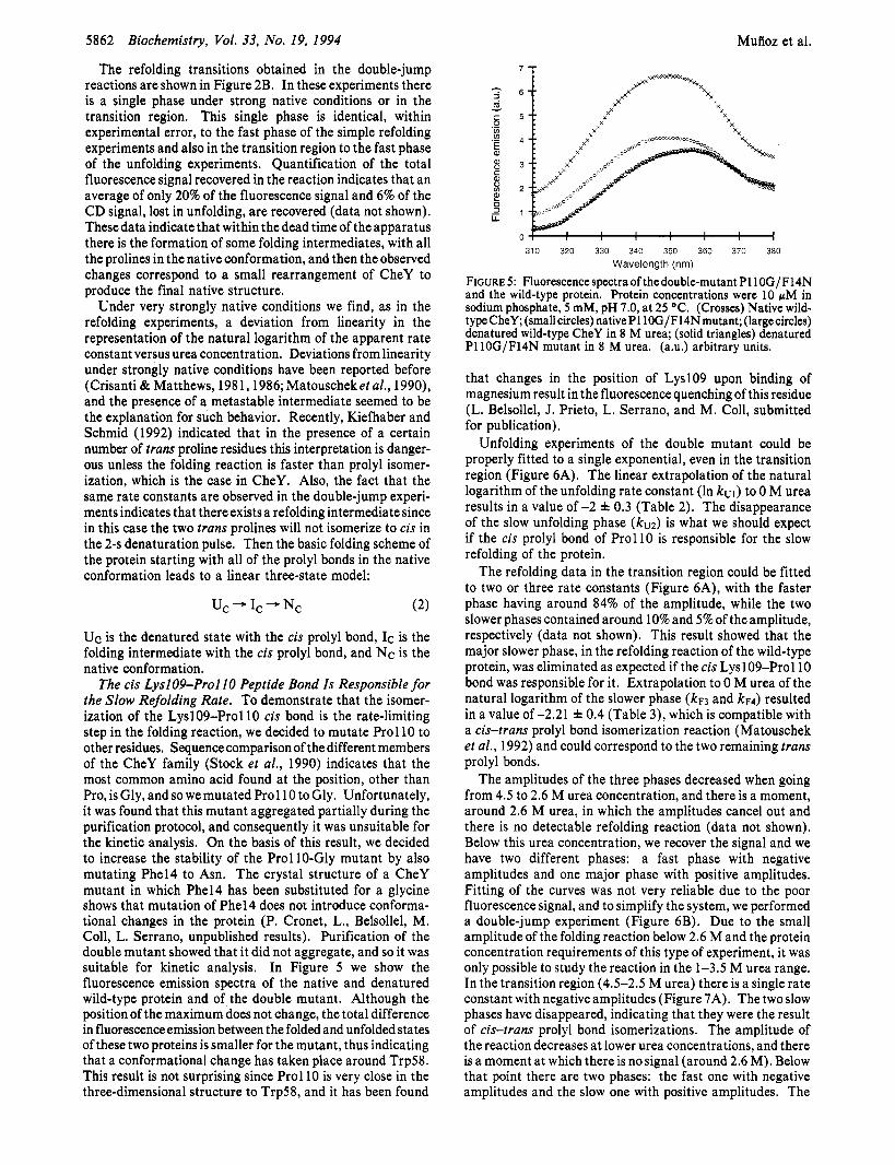
The cis LyslO9-Pro1 IO Peptide Bond Is Responsible for the Slow Refolding Rate. To demonstrate that the isomer- ization of the Lysl09-Pro110 cis bond is the rate-limiting step in the folding reaction, we decided to mutate Prol 10 to other residues. Sequencecomparison of the different members of the CheY family (Stock et al., 1990) indicates that the most common amino acid found at the position, other than Pro, is Gly, and so we mutated Prol 10 to Gly. Unfortunately, it was found that this mutant aggregated partially during the purification protocol, and consequently it was unsuitable for the kinetic analysis. On the basis of this result, we decided to increase the stability of the Prol 10-Gly mutant by also mutating Phel4 to Asn. The crystal structure of a CheY mutant in which Phel4 has been substituted for a glycine shows that mutation of Phel4 does not introduce conforma- tional changes in the protein (P. Cronet, L., Belsollel, M. Coll, L. Serrano, unpublished results). Purification of the double mutant showed that it did not aggregate, and so it was suitable for kinetic analysis. In Figure 5 we show the fluorescence emission spectra of the native and denatured wild-type protein and of the double mutant. Although the position of the maximum does not change, the total difference in fluorescence emission between the folded and unfolded states of these two proteins is smaller for the mutant, thus indicating that a conformational change has taken place around Trp58. This result is not surprising since Prol 10 is very close in the three-dimensional structure to Trp58, and it has been found
310 320 330 340 350 360 370 380
Wavelength (nm)
FIGURE 5: Fluorescence spectra of thedouble-mutant P110G/F14N and the wild-type protein. Protein concentrations were 10 pM in sodium phosphate, 5 mM, pH 7.0, at 25 OC. (Crosses) Native wild- type CheY; (small circles) nativePllOG/F14N mutant; (largecircles) denatured wild-type CheY in 8 M urea; (solid triangles) denatured P110G/F14N mutant in 8 M urea. (a.u.) arbitrary units.
that changes in the position of LyslO9 upon binding of magnesium result in the fluorescence quenching of this residue (L. Belsollel, J. Prieto, L. Serrano, and M. Coll, submitted for publication).
Unfolding experiments of the double mutant could be properly fitted to a single exponential, even in the transition region (Figure 6A). The linear extrapolation of the natural logarithm of the unfolding rate constant (In kul) to 0 M urea results in a value of -2 f 0.3 (Table 2). The disappearance of the slow unfolding phase (ku2) is what we should expect if the cis prolyl bond of Prol 10 is responsible for the slow refolding of the protein.
The refolding data in the transition region could be fitted to two or three rate constants (Figure 6A), with the faster phase having around 84% of the amplitude, while the two slower phases contained around 10% and 5% of the amplitude, respectively (data not shown). This result showed that the major slower phase, in the refolding reaction of the wild-type protein, was eliminated as expected if the cis Lysl09-Pro110 bond was responsible for it. Extrapolation to 0 M urea of the natural logarithm of the slower phase (kF3 and kF4) resulted in a value of -2.21 f 0.4 (Table 3), which is compatible with a cis-trans prolyl bond isomerization reaction (Matouschek et al., 1992) and could correspond to the two remaining trans prolyl bonds.
The amplitudes of the three phases decreased when going from 4.5 to 2.6 M urea concentration, and there is a moment, around 2.6 M urea, in which the amplitudes cancel out and there is no detectable refolding reaction (data not shown). Below this urea concentration, we recover the signal and we have two different phases: a fast phase with negative amplitudes and one major phase with positive amplitudes. Fitting of the curves was not very reliable due to the poor fluorescence signal, and to simplify the system, we performed a double-jump experiment (Figure 6B). Due to the small amplitude of the folding reaction below 2.6 M and the protein concentration requirements of this type of experiment, it was only possible to study the reaction in the 1-3.5 M urea range. In the transition region (4.5-2.5 M urea) there is a single rate constant with negative amplitudes (Figure 7A). The two slow phases have disappeared, indicating that they were the result of cis-trans prolyl bond isomerizations. The amplitude of the reaction decreases at lower urea concentrations, and there is a moment at which there is no signal (around 2.6 M). Below that point there are two phases: the fast one with negative amplitudes and the slow one with positive amplitudes. The
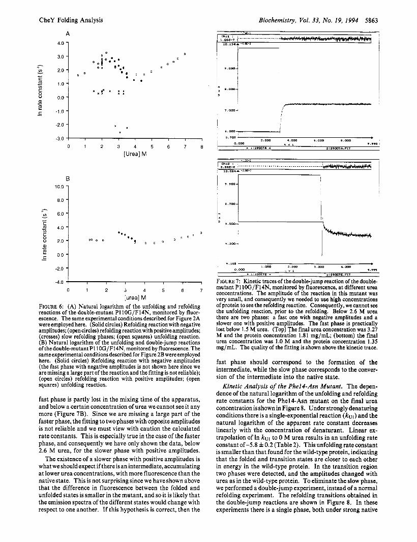
CheY Folding Analysis
A
4.0 1
-2.0 - -3.0
Biochemistry, Vol. 33, No. 19, 1994 5863
, 1 I ! I I I 1
*E-- ~
h - ln
e . 1.0 4
-1 .o O'O 1
B
1 8.0
- 6.0 1 2 4.0
h
ln
C
v -
-2.0 1 -4.0 I I I I I I I
0 1 2 3 4 5 6 7
[urea] M FIGURE 6: (A) Natural logarithm of the unfolding and refolding reactions of the double-mutant P110G/F14N, monitored by fluor- escence. The same experimental conditions described for Figure 2A were employed here. (Solid circles) Refolding reaction with negative amplitudes; (open circles) refolding reaction with positive amplitudes; (crosses) slow refolding phases; (open squares) unfolding reaction. (B) Natural logarithm of the unfolding and double-jump reactions of the double-mutant P110G/F14N, monitored by fluorescence. The same experimental conditions described for Figure 2B were employed here. (Solid circles) Refolding reaction with negative amplitudes (the fast phase with negative amplitudes is not shown here since we are missing a large part of the reaction and the fitting is not reliable); (open circles) refolding reaction with positive amplitudes; (open squares) unfolding reaction.
fast phase is partly lost in the mixing time of the apparatus, and below a certain concentration of urea we cannot see it any more (Figure 7B). Since we are missing a large part of the faster phase, the fitting to two phases with opposite amplitudes is not reliable and we must view with caution the calculated rate constants. This is especially true in the case of the faster phase, and consequently we have only shown the data, below 2.6 M urea, for the slower phase with positive amplitudes.
The existence of a slower phase with positive amplitudes is what we should expect if there is an intermediate, accumulating at lower urea concentrations, with more fluorescence than the native state. This is not surprising since we have shown above that the difference in fluorescence between the folded and unfolded states is smaller in the mutant, and so it is likely that the emission spectra of the different states would change with respect to one another. If this hypothesis is correct, then the
I 1 I
6 . 0 0 0 7 /
5.70'3 I F 2 . 0 0 0 4.000 6 .000 a. 000 . I : P ~ O C T ~ 4 2: 29OCla. FIT
0.000 , X I 9.990 I
! 9.900
~ 9 . 7 0 0
' 9.300-
I. 000 2 , 0 0 0 3 . 0 0 0 4 .000 I 0.000 5 x 1 4.99s I . I.29OCTE f 3:29OCTE.fIT I
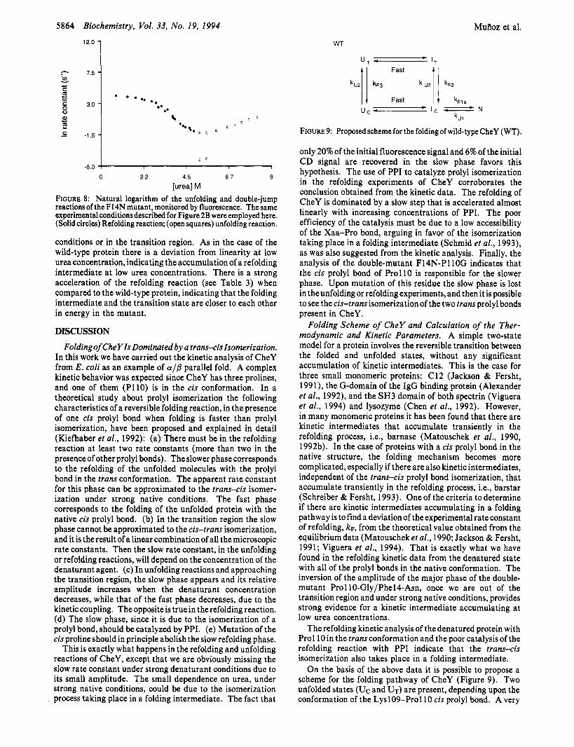
FIGURE 7: Kinetic traces of the double-jump reaction of the double- mutant P110G/F14N, monitored by fluorescence, at different urea concentrations. The amplitude of the reaction in this mutant was very small, and consequently we needed to use high concentrations of protein to see the refolding reaction. Consequently, we cannot see the unfolding reaction, prior to the refolding. Below 2.6 M urea there are two phases: a fast one with negative amplitudes and a slower one with positive amplitudes. The fast phase is practically lost below 1.5 M urea. (Top) The final urea concentration was 3.27 M and the protein concentration 1.81 mg/mL; (bottom) the final urea concentration was 1.0 M and the protein concentration 1.35 mg/mL. The quality of the fitting is shown above the kinetic trace.
fast phase should correspond to the formation of the intermediate, while the slow phase corresponds to the conver- sion of the intermediate into the native state.
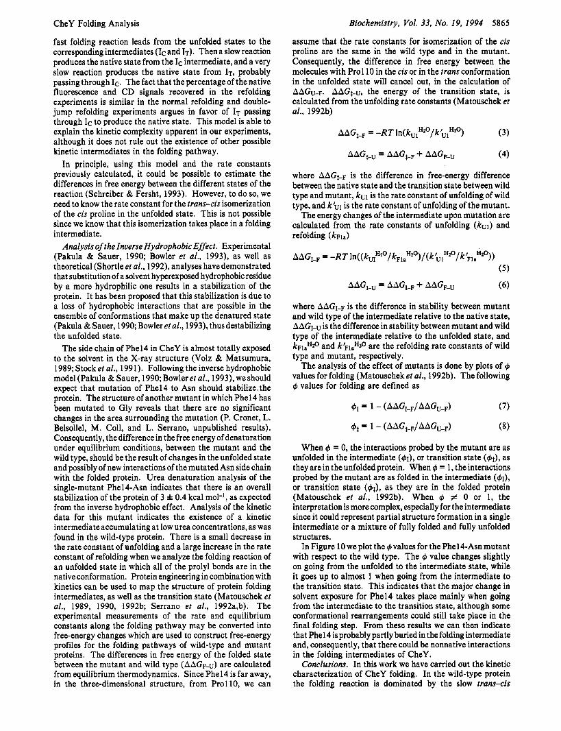
Kinetic Analysis of the Phel4-Asn Mutant. The depen- dence of the natural logarithm of the unfolding and refolding rate constants for the PhelCAsn mutant on the final urea concentration is shown in Figure 8. Under strongly denaturing conditions there is a single-exponential reaction (k"1) and the natural logarithm of the apparent rate constant decreases linearly with the concentration of denaturant. Linear ex- trapolation of In kul to 0 M urea results in an unfolding rate constant of -5.8 f 0.2 (Table 2). This unfolding rate constant is smaller than that found for the wild-type protein, indicating that the folded and transition states are closer to each other in energy in the wild-type protein. In the transition region two phases were detected, and the amplitudes changed with urea as in the wild-type protein. To eliminate the slow phase, we performed a double-jump experiment, instead of a normal refolding experiment. The refolding transitions obtained in the double-jump reactions are shown in Figure 8. In these experiments there is a single phase, both under strong native
5064 Biochemistry, Vol. 33, No. 19, 1994
12.0 1 Fast A
k ~ z k ~ 3 U3
MuAoz et al.
kFZ
wr
7 Fast t kFla
-6.0 f I , I 1
0 22 4 5 6 7 9 [urea] M
FIGURE 8: Natural logarithm of the unfolding and double-jump reactions of the F14N mutant, monitored by fluorescence. The same experimental conditions described for Figure 2B were employed here. (Solid circles) Refolding reaction; (open squares) unfolding reaction.
conditions or in the transition region. As in the case of the wild-type protein there is a deviation from linearity at low urea concentration, indicating the accumulation of a refolding intermediate at low urea concentrations. There is a strong acceleration of the refolding reaction (see Table 3) when compared to the wild-type protein, indicating that the folding intermediate and the transition state are closer to each other in energy in the mutant.
DISCUSSION
Folding of CheYIs Dominated by a trans-cis Isomerization. In this work we have carried out the kinetic analysis of CheY from E. coli as an example of a/@ parallel fold. A complex kinetic behavior was expected since CheY has three prolines, and one of them (P110) is in the cis conformation. In a theoretical study about prolyl isomerization the following characteristics of a reversible folding reaction, in the presence of one cis prolyl bond when folding is faster than prolyl isomerization, have been proposed and explained in detail (Kiefhaber et al., 1992): (a) There must be in the refolding reaction at least two rate constants (more than two in the presence of other prolyl bonds). The slower phasecorresponds to the refolding of the unfolded molecules with the prolyl bond in the trans conformation. The apparent rate constant for this phase can be approximated to the trans-cis isomer- ization under strong native conditions. The fast phase corresponds to the folding of the unfolded protein with the native cis prolyl bond. (b) In the transition region the slow phase cannot be approximated to the cis-trans isomerization, and it is the result of a linear combination of all the microscopic rate constants. Then the slow rate constant, in the unfolding or refolding reactions, will depend on the concentration of the denaturant agent. (c) In unfolding reactions and approaching the transition region, the slow phase appears and its relative amplitude increases when the denaturant concentration decreases, while that of the fast phase decreases, due to the kinetic coupling. The opposite is true in the refolding reaction. (d) The slow phase, since it is due to the isomerization of a prolyl bond, should be catalyzed by PPI. (e) Mutation of the cis proline should in principle abolish the slow refolding phase.
This is exactly what happens in the refolding and unfolding reactions of CheY, except that we are obviously missing the slow rate constant under strong denaturant conditions due to its small amplitude. The small dependence on urea, under strong native conditions, could be due to the isomerization process taking place in a folding intermediate. The fact that
CheY Folding Analysis
fast folding reaction leads from the unfolded states to the corresponding intermediates (IC and IT). Then a slow reaction produces the native state from the IC intermediate, and a very slow reaction produces the native state from IT, probably passing through IC. The fact that the percentage of the native fluorescence and CD signals recovered in the refolding experiments is similar in the normal refolding and double- jump refolding experiments argues in favor of IT passing through IC to produce the native state. This model is able to explain the kinetic complexity apparent in our experiments, although it does not rule out the existence of other possible kinetic intermediates in the folding pathway.
In principle, using this model and the rate constants previously calculated, it could be possible to estimate the differences in free energy between the different states of the reaction (Schreiber & Fersht, 1993). However, to do so, we need to know the rate constant for the trans-cis isomerization of the cis proline in the unfolded state. This is not possible since we know that this isomerization takes place in a folding intermediate.
Analysis of theznverse Hydrophobic Effect. Experimental (Pakula & Sauer, 1990; Bowler et al., 1993), as well as theoretical (Shortle et al., 1992), analyses have demonstrated that substitution of a solvent hyperexposed hydrophobic residue by a more hydrophilic one results in a stabilization of the protein. It has been proposed that this stabilization is due to a loss of hydrophobic interactions that are possible in the ensemble of conformations that make up the denatured state (Pakula & Sauer, 1990; Bowler et al., 1993), thus destabilizing the unfolded state.
The side chain of Phel4 in CheY is almost totally exposed to the solvent in the X-ray structure (Volz & Matsumura, 1989; Stock et al., 1991). Following the inverse hydrophobic model (Pakula & Sauer, 1990; Bowler et ai., 1993), we should expect that mutation of Phel4 to Asn should stabilize,the protein. The structure of another mutant in which Phel4 has been mutated to Gly reveals that there are no significant changes in the area surrounding the mutation (P. Cronet, L. Belsollel, M. Coll, and L. Serrano, unpublished results). Consequently, the difference in the free energy of denaturation under equilibrium conditions, between the mutant and the wild type, should be the result of changes in the unfolded state and possibly of new interactions of the mutated Asn side chain with the folded protein. Urea denaturation analysis of the single-mutant Phel4-Asn indicates that there is an overall stabilization of the protein of 3 f 0.4 kcal mol-', as expected from the inverse hydrophobic effect. Analysis of the kinetic data for this mutant indicates the existence of a kinetic intermediate accumulating at low urea concentrations, as was found in the wild-type protein. There is a small decrease in the rate constant of unfolding and a large increase in the rate constant of refolding when we analyze the folding reaction of an unfolded state in which all of the prolyl bonds are in the native conformation. Protein engineering in combination with kinetics can be used to map the structure of protein folding intermediates, as well as the transition state (Matouschek et al., 1989, 1990, 1992b; Serrano et al., 1992a,b). The experimental measurements of the rate and equilibrium constants along the folding pathway may be converted into free-energy changes which are used to construct free-energy profiles for the folding pathways of wild-type and mutant proteins. The differences in free energy of the folded state between the mutant and wild type (AAGF-u) are calculated from equilibrium thermodynamics. Since Phel4 is far away, in the three-dimensional structure, from Prol 10, we can
Biochemistry, Vol. 33, No. 19, 1994 5865
assume that the rate constants for isomerization of the cis proline are the same in the wild type and in the mutant. Consequently, the difference in free energy between the molecules with Prol 10 in the cis or in the trans conformation in the unfolded state will cancel out, in the calculation of AAGu-F. AAGt-u, the energy of the transition state, is calculated from the unfolding rate constants (Matouschek et al., 1992b)
AAGt-F = -R T In( kUl H20/k h1 Hzo) (3)
where AAG~-F is the difference in free-energy difference between the native state and the transition state between wild type and mutant, kul is the rate constant of unfolding of wild type, and k'"1 is the rate constant of unfolding of the mutant.
The energy changes of the intermediate upon mutation are calculated from the rate constants of unfolding (ku1) and refolding (kFla)
where AAGI-F is the difference in stability between mutant and wild type of the intermediate relative to the native state, AAG1-v is the difference in stability between mutant and wild type of the intermediate relative to the unfolded state, and k ~ l ~ ~ z ~ and k'FlaHZO are the refolding rate constants of wild type and mutant, respectively.
The analysis of the effect of mutants is done by plots of + values for folding (Matouschek et al., 1992b). The following + values for folding are defined as
+I = 1 - (AAGI-F/AAGu-F)
+t = 1 - (AAG*-F/AAGu-F)
(7)
(8)
When + = 0, the interactions probed by the mutant are as unfolded in the intermediate (+I), or transition state (+t), as they are in the unfolded protein. When + = 1, the interactions probed by the mutant are as folded in the intermediate (+I), or transition state (+t), as they are in the folded protein (Matouschek et al., 1992b). When + # 0 or 1, the interpretation is more complex, especially for the intermediate since it could represent partial structure formation in a single intermediate or a mixture of fully folded and fully unfolded structures.
In Figure 10 we plot the + values for the Phel4-Asn mutant with respect to 'the wild type. The + value changes slightly on going from the unfolded to the intermediate state, while it goes up to almost 1 when going from the intermediate to the transition state. This indicates that the major change in solvent exposure for Phel4 takes place mainly when going from the intermediate to the transition state, although some conformational rearrangements could still take place in the final folding step. From these results we can then indicate that Phel4 is probably partly buried in the folding intermediate and, consequently, that there could be nonnative interactions in the folding intermediates of CheY.
Conclusions. In this work we have carried out the kinetic characterization of CheY folding. In the wild-type protein the folding reaction is dominated by the slow trans-cis
5866 Biochemistry, Vol. 33, No. 19, 1994 Muiioz et al.
Foss, J. G., & Schellman, J. A. (1959) J . Phys. Chem. 63,2007. Gill, S . C., & von Hippel, P. H. (1989) Anal. Biochem. 182,319. Goto, Y., & Hamaguchi, K. (1982) J . Mol. Biol. 156,891-910. Hagerman, P. J. (1977) Biopolymers 16, 731-747. Hagerman, P. J., & Baldwin, R. L. (1976) Biochemistry 15,
Hazelbauer, G. L., Berg, H. C., & Matsumura, P. (1993) Cell
Herning, T., Yutani, K., Taniyama, Y., & Kikuchi, M. (1991)
Herning, T., Yutani, K., Inaka, K., Kuroki, R., Taniyama, Y.,
Hurle, M. R., Anderson, S., & Kuntz, I. D. (1991) Protein Eng.
Jackson, S . , & Fersht, A. R. (1991) Biochemistry 30, 10428-
Kelley, R. F., & Richards, F. M. (1987) Biochemistry 26,6765-
Kiefhaber, T., & Schmid, F. X. (1992) J . Mol. Biol. 224, 231-
Kiefhaber, T., Grunert, H. P., Hahn, U., & Schmid, F. X. (1990)
Kiefhaber, T., Kohler, H. H., & Schmid, F. X. (1992) J. Mol.
Landt, O., Grunert, H. P., & Hahn, U. (1 990) Gene 96,125-1 28. Levinthal, C. (1968) Chim. Phys. 65, 44-45. Matouschek, A., Kellis, J. T., Jr., Serrano, L., & Fersht, A. R.
Matouschek, A,, Kellis, J. T., Serrano, L., Bycroft, M., & Fersht,
Matouschek, A,, Serrano, L., Meiering, E., Bycroft, M., & Fersht,
Matouschek, A., Serrano, L., & Fersht, A. R. (1992b) J . Mol.
Matsumura, P., Rydel, J. J., Linzmeier, R., & Vacante, D. (1984)
Matthews, B. W., Nicholson, H., & Becktel, W. J. (1 987) Proc.
Matthews, C. R. (1987) Methods Enzymol. 154, 499-511. Mayr, L. M., & Schmid, F. X. (1993) J . Mol. Biol. 231, 913-
Mayr, L. M., Landt, O., Hahn, U., & Schmid, F. X. (1993) J .
Pakula, A. A., & Sauer, R. T. (1990) Nature 344, 363-364. Schellman, J. A. (1955) C. R. Trav. Lab. Carlsberg. Ser. Chim.
Schmid, F. X . , & Baldwin, R. L. (1978) Proc. Natl. Acad. Sci.
Schmid, F. X . , Lorenz, M. M., Mucke, M., & Schonbrunner, E.
Schreiber, G., & Fersht, A. R. (1993) Biochemistry 32, 1 1 195-
Schultz,D.A., & Baldwin,R.L. (1992) ProteinSci. 1,910-916. Schultz, D. A., Schmid, F. X., & Baldwin, R. L. (1992) Protein
Serrano, L., Matouschek, A., & Fersht, A. R. (1992a) J . Mol.
Serrano, L., Matouschek, A., & Fersht, A. R. (1992b) J. Mol.
Shortle, D., Chan, H. S., & Dill, K. A. (1992) Protein Sci. 1,
Stock, A. M., Motonen, J. M., Stock, J. B., & Schutt, C. E.
Stock, J . B., Stock, A. M., & Mottonen, J. M. (1990) Nature
Tanford, C. (1968) Adv. Protein Chem. 23, 121-282. Viguera, A. R., Martlnez, J. C., Filimonov, V., Mateo, P. L., &
Serrano, L. (1994) Biochemistry 33, 2142-2150. Volz, K., & Matsumura, P. (1991) J . Biol. Chem. 266, 15511- 15519.
Wetlaufer, D. B. (1973) Proc. Natl. Acad. Sci. U.S.A. 70,697- 701.
1 462-1 47 3.
73, 15-22.
J . Mol. Biol. 30, 9882-9891.
& Kikuchi, M. (1992) Biochemistry 31, 7077-7085.
4, 451-455.
10435.
6774.
240.
Biochemistry 29, 6475-6480.
Biol. 224, 217-229.
(1989) Nature 340, 122-126.
A. R. (1990) Nature 346, 440-445.
A. R. (1992a) J . Mol. Biol. 224, 837-845.
Biol. 224, 819-835.
J . Bacetriol. 160, 36.
Natl. Acd. Sci. U.S.A. 84, 6663-6667.
926.
Mol. Biol. 231, 897-912.
29, 230-259.
U.S.A. 75, 4764-4768.
F. (1993) Adv. Protein Chem. 44, 25-66.
11203.
Sci. 1, 917-924.
Biol. 224, 805-818.
Biol. 224, 847-859.
201-21 5.
(1989) Nature 337, 745-749.
344, 395.
1.2 1
0.8 '.O 4 ///i
/' /
0.2 -I /
-0.2 I I I I i
Unfolded Intermediate Transition Folded
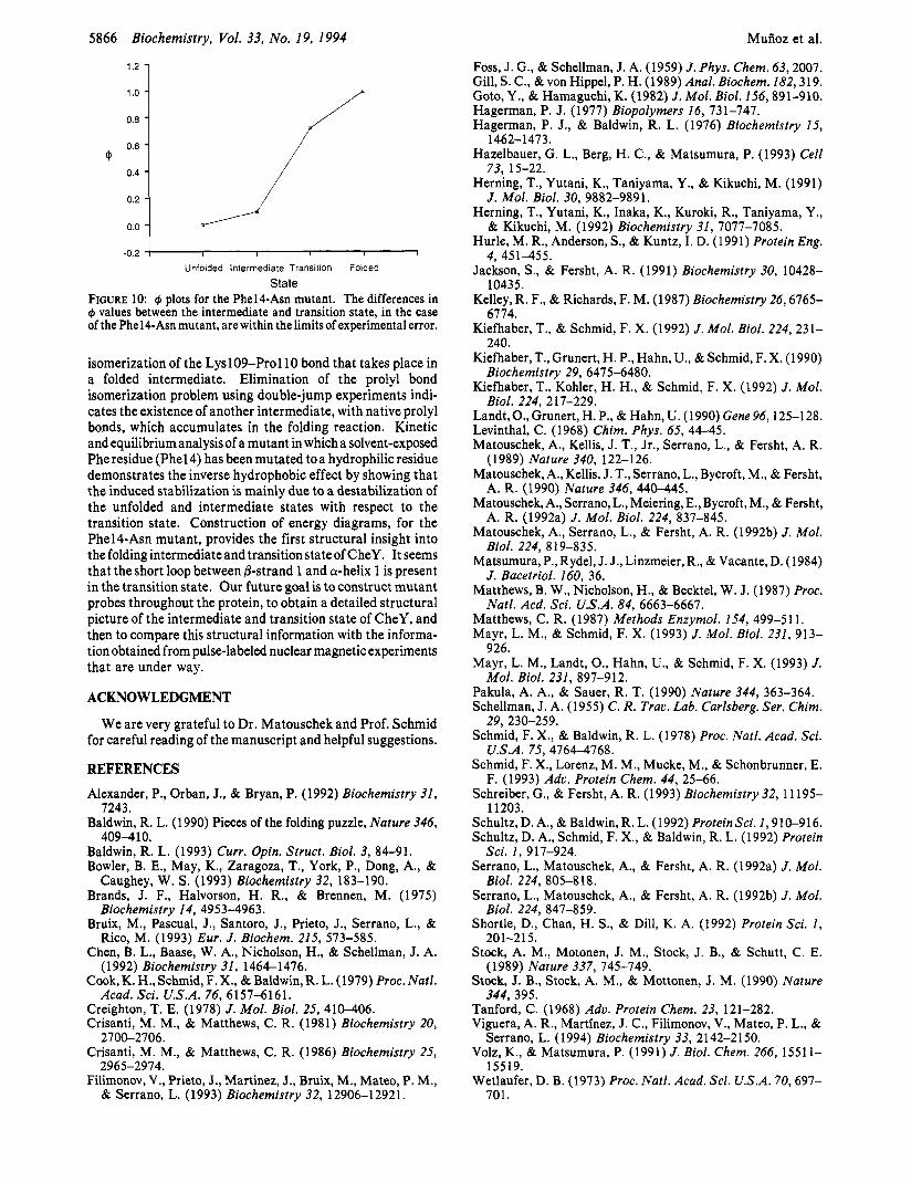
State FIGURE 10: C$ plots for the Phel4-Asn mutant. The differences in C$ values between the intermediate and transition state, in the case of the Phel4-Asn mutant, are within the limits of experimental error.
isomerization of the Lysl09-Pro110 bond that takes place in a folded intermediate. Elimination of the prolyl bond isomerization problem using double-jump experiments indi- cates the existence of another intermediate, with native prolyl bonds, which accumulates in the folding reaction. Kinetic and equilibrium analysis of a mutant in which a solvent-exposed Phe residue (Phel4) has been mutated to a hydrophilic residue demonstrates the inverse hydrophobic effect by showing that the induced stabilization is mainly due to a destabilization of the unfolded and intermediate states with respect to the transition state. Construction of energy diagrams, for the Phel4-Asn mutant, provides the first structural insight into the folding intermediate and transitionstateof CheY. It seems that the short loop between @-strand 1 and a-helix 1 is present in the transition state. Our future goal is to construct mutant probes throughout the protein, to obtain a detailed structural picture of the intermediate and transition state of CheY, and then to compare this structural information with the informa- tion obtained from pulse-labeled nuclear magnetic experiments that are under way.
ACKNOWLEDGMENT
We are very grateful to Dr. Matouschek and Prof. Schmid for careful reading of the manuscript and helpful suggestions.
REFERENCES
Alexander, P., Orban, J., & Bryan, P. (1992) Biochemistry 31,
Baldwin, R. L. (1 990) Pieces of the folding puzzle, Nature 346,
Baldwin, R. L. (1993) Curr. Opin. Struct. Biol. 3, 84-91. Bowler, B. E., May, K., Zaragoza, T., York, P., Dong, A., &
Brands, J. F., Halvorson, H. R., & Brennen, M. (1975)
Bruix, M., Pascual, J., Santoro, J., Prieto, J., Serrano, L., &
Chen, B. L., Baase, W. A., Nicholson, H., & Schellman, J. A.
Cook, K. H., Schmid, F. X., & Baldwin, R. L. (1979) Proc. Natl.
Creighton, T. E. (1978) J . Mol. Biol. 25, 410-406. Crisanti, M. M., & Matthews, C. R. (1981) Biochemistry 20,
Crisanti, M. M., & Matthews, C. R. (1986) Biochemistry 25,
Filimonov, V., Prieto, J., Martinez, J., Bruix, M., Mateo, P. M.,
7243.
409-410.
Caughey, W. S. (1993) Biochemistry 32, 183-190.
Biochemistry 14, 4953-4963.
Rico, M. (1993) Eur. J . Biochem. 215, 573-585.
(1992) Biochemistry 31, 1464-1476.
Acad. Sci. U.S.A. 76, 6157-6161.
2700-2706.
2965-2974.
& Serrano, L. (1993) Biochemistry 32, 12906-12921.
Related Documents