The Journal of Experimental Medicine JEM © The Rockefeller University Press $8.00 ARTICLE www.jem.org/cgi/doi/10.1084/jem.20041277 Cite by DOI: 10.1084/jem.20041277 1 of 11 Identification and characterization of an endogenous chemotactic ligand specific for FPRL2 Isabelle Migeotte, 1 Elena Riboldi, 3 Jean-Denis Franssen, 4 Françoise Grégoire, 2 Cécile Loison, 4 Valérie Wittamer, 1 Michel Detheux, 4 Patrick Robberecht, 2 Sabine Costagliola, 1 Gilbert Vassart, 1 Silvano Sozzani, 3,5 Marc Parmentier, 1 and David Communi 1 1 Institut de Recherche en Biologie Humaine et Moléculaire and 2 Laboratoire de Chimie Biologique et de la Nutrition, Université Libre de Bruxelles Campus Erasme, B-1070 Brussels, Belgium 3 Section of General Pathology and Immunology, University of Brescia, 25121 Brescia, Italy 4 Euroscreen s.a., B-6041 Gosselies, Belgium 5 Istituto di Ricerche Farmacologiche Mario Negri, 20157 Milano, Italy Chemotaxis of dendritic cells (DCs) and monocytes is a key step in the initiation of an adequate immune response. Formyl peptide receptor (FPR) and FPR-like receptor (FPRL)1, two G protein–coupled receptors belonging to the FPR family, play an essential role in host defense mechanisms against bacterial infection and in the regulation of inflammatory reactions. FPRL2, the third member of this structural family of chemoattractant receptors, is characterized by its specific expression on monocytes and DCs. Here, we present the isolation from a spleen extract and the functional characterization of F2L, a novel chemoattractant peptide acting specifically through FPRL2. F2L is an acetylated amino- terminal peptide derived from the cleavage of the human heme-binding protein, an intracellular tetrapyrolle-binding protein. The peptide binds and activates FPRL2 in the low nanomolar range, which triggers intracellular calcium release, inhibition of cAMP accumulation, and phosphorylation of extracellular signal–regulated kinase 1/2 mitogen- activated protein kinases through the G i class of heterotrimeric G proteins. When tested on monocytes and monocyte-derived DCs, F2L promotes calcium mobilization and chemotaxis. Therefore, F2L appears as a new natural chemoattractant peptide for DCs and monocytes, and the first potent and specific agonist of FPRL2. DCs and monocytes are critical players in host immune mechanisms. They are able to sense danger signals of diverse origins (various path- ogen classes and tissular disturbances such as necrosis and tumor development; reference 1). They are attracted during early phases of inflammatory reactions by a diverse set of chemoattractant molecules, including chemo- kines, the complement factor C5a, the bacte- rial peptide FMLP, the lipid metabolite PAF, and the recently identified chemerin (1, 2). Locally, these cells participate to innate immu- nity through the phagocytosis of microbes or diseased cells and the secretion of a range of bioactive mediators, including cytokines and chemokines. Immature DCs take up and pro- cess available antigens and then, under ap- propriate stimuli, undergo maturation, switch their expression of chemokine receptors from inflammatory to lymphoid homing receptors, and migrate to the draining lymph nodes where they interact with T cells to initiate an appropriate immune response. These various populations of antigen-presenting cells are ex- tremely heterogeneous in terms of morphology and function. These functional differences are partly related to the set of chemoattractant seven-transmembrane, G protein–coupled re- ceptors (GPCRs) they express. In the 1970’s, synthetic FMLP was one of the first identified potent leukocyte chemoattrac- tants. Natural FMLP was later purified from Gram-negative bacteria (3, 4). Its human re- ceptor, formyl peptide receptor (FPR), was cloned in 1990 from a differentiated HL-60 myeloid leukemia cell cDNA library (5). Two human genes encoding the structurally related receptors FPR-like receptor (FPRL)1 CORRESPONDENCE Marc Parmentier: [email protected] Abbreviations used: ERK, extra- cellular signal–regulated kinase; FPR, formyl peptide receptor; FPRL, FPR-like receptor; GPCR, G protein–coupled re- ceptor; HBP, heme-binding protein; MAP, mitogen-activated protein; SEC, size-exclusion column. on October 2, 2016 Downloaded from Published December 28, 2004

Welcome message from author
This document is posted to help you gain knowledge. Please leave a comment to let me know what you think about it! Share it to your friends and learn new things together.
Transcript
The
Journ
al o
f Exp
erim
enta
l M
edic
ine
JEM © The Rockefeller University Press $8.00
ARTICLE
www.jem.org/cgi/doi/10.1084/jem.20041277
Cite by DOI: 10.1084/jem.20041277
1 of 11
Identification and characterization of an endogenous chemotactic ligand specific for FPRL2
Isabelle Migeotte,
1
Elena Riboldi,
3
Jean-Denis Franssen,
4
Françoise Grégoire,
2
Cécile Loison,
4
Valérie Wittamer,
1
Michel Detheux,
4
Patrick Robberecht,
2
Sabine Costagliola,
1
Gilbert Vassart,
1
Silvano Sozzani,
3,5
Marc Parmentier,
1
and David Communi
1
1
Institut de Recherche en Biologie Humaine et Moléculaire and
2
Laboratoire de Chimie Biologique et de la Nutrition, Université Libre de Bruxelles Campus Erasme, B-1070 Brussels, Belgium
3
Section of General Pathology and Immunology, University of Brescia, 25121 Brescia, Italy
4
Euroscreen s.a., B-6041 Gosselies, Belgium
5
Istituto di Ricerche Farmacologiche Mario Negri, 20157 Milano, Italy
Chemotaxis of dendritic cells (DCs) and monocytes is a key step in the initiation of an adequate immune response. Formyl peptide receptor (FPR) and FPR-like receptor (FPRL)1, two G protein–coupled receptors belonging to the FPR family, play an essential role in host defense mechanisms against bacterial infection and in the regulation of inflammatory reactions. FPRL2, the third member of this structural family of chemoattractant receptors, is characterized by its specific expression on monocytes and DCs. Here, we present the isolation from a spleen extract and the functional characterization of F2L, a novel chemoattractant peptide acting specifically through FPRL2. F2L is an acetylated amino-terminal peptide derived from the cleavage of the human heme-binding protein, an intracellular tetrapyrolle-binding protein. The peptide binds and activates FPRL2 in the low nanomolar range, which triggers intracellular calcium release, inhibition of cAMP accumulation, and phosphorylation of extracellular signal–regulated kinase 1/2 mitogen-activated protein kinases through the G
i
class of heterotrimeric G proteins. When tested on monocytes and monocyte-derived DCs, F2L promotes calcium mobilization and chemotaxis. Therefore, F2L appears as a new natural chemoattractant peptide for DCs and monocytes, and the first potent and specific agonist of FPRL2.
DCs and monocytes are critical players in hostimmune mechanisms. They are able to sensedanger signals of diverse origins (various path-ogen classes and tissular disturbances such asnecrosis and tumor development; reference 1).They are attracted during early phases ofinflammatory reactions by a diverse set ofchemoattractant molecules, including chemo-kines, the complement factor C5a, the bacte-rial peptide FMLP, the lipid metabolite PAF,and the recently identified chemerin (1, 2).Locally, these cells participate to innate immu-nity through the phagocytosis of microbes ordiseased cells and the secretion of a range ofbioactive mediators, including cytokines andchemokines. Immature DCs take up and pro-cess available antigens and then, under ap-propriate stimuli, undergo maturation, switchtheir expression of chemokine receptors from
inflammatory to lymphoid homing receptors,and migrate to the draining lymph nodeswhere they interact with T cells to initiate anappropriate immune response. These variouspopulations of antigen-presenting cells are ex-tremely heterogeneous in terms of morphologyand function. These functional differences arepartly related to the set of chemoattractantseven-transmembrane, G protein–coupled re-ceptors (GPCRs) they express.
In the 1970’s, synthetic FMLP was one ofthe first identified potent leukocyte chemoattrac-tants. Natural FMLP was later purified fromGram-negative bacteria (3, 4). Its human re-ceptor, formyl peptide receptor (FPR), wascloned in 1990 from a differentiated HL-60myeloid leukemia cell cDNA library (5).Two human genes encoding the structurallyrelated receptors FPR-like receptor (FPRL)1
CORRESPONDENCEMarc Parmentier: [email protected]
Abbreviations used: ERK, extra-cellular signal–regulated kinase; FPR, formyl peptide receptor; FPRL, FPR-like receptor; GPCR, G protein–coupled re-ceptor; HBP, heme-binding protein; MAP, mitogen-activated protein; SEC, size-exclusion column.
on October 2, 2016
Dow
nloaded from
Published December 28, 2004

A NOVEL ENDOGENOUS-SPECIFIC LIGAND FOR FPRL2 | Migeotte et al.
2 of 11
and FPRL2 were subsequently cloned by low stringencyhybridization using FPR cDNA as a probe, and shown tocluster with FPR on human chromosome 19q13.3 (6, 7). Incontrast, the murine FPR gene family comprises at leasteight distinct genes (Fpr1 and Fpr-rs 1–7), of which six areclustered on chromosome 17, in a region of conserved syn-teny with human chromosome 19 (8, 9). Fpr1 is consideredthe murine orthologue of FPR, whereas Fpr-rs1 and Fpr-rs2share structural and functional similarity with FPRL1. Noobvious orthologue of FPRL2 can be determined from astructural viewpoint. Therefore, it appears that the FPRgene cluster has undergone recent and independent gene du-plications in various mammalian species, which may also im-ply functional divergence of the resulting receptor subtypes.
FPR is a high affinity receptor for formyl peptides. Syn-thetic W hexapeptides were described as efficient surrogate ag-onists of FPR, whereas the receptor was also reported to re-spond to HIV-derived peptides, amino-terminal peptides ofannexin I, and cathepsin G, although with relatively low po-tencies (10–12). FPRL1 shares 69% amino acid identity withFPR and displays low affinity for FMLP. However, FPRL1appears as a remarkably promiscuous receptor, as it is activatedby numerous and chemically unrelated ligands. These includethe following: the synthetic peptides MMK1 and W hexapep-tides; pathogen-derived peptides such as the Hp 2–20 peptidefrom
Helicobacter pylori
and peptides derived from HIV gp41and gp120; the lipid lipoxin A4; as well as a variety of hostproteins and peptides such as cathelicidin LL37, annexin I andthe Ac1-25 and Ac1-11 peptides derived from this protein, the88–274 fragment of the urokinase receptor, the serum amyloidA protein, the amyloid
�
peptide A
�
42, the prion proteinfragment P106-126, a fragment from the mitochondrialNADH dehydrogenase subunit I, a truncated form of the che-mokine CK
�
8-1, and the neuroprotective peptide humanin(3, 4, 13, 14). However, the affinity of FPRL1 for these vari-ous ligands is often low, and the functional relevance of theseinteractions is therefore questionable. FPRL2 shares 56 and83% amino acid identity with FPR and FPRL1, respectively.FPRL2 does not respond to formyl peptides, but it was de-scribed as a low affinity receptor for several FPRL1 agonists,namely the annexin I amino-terminal peptide Ac1-25 (15) andthe
H. pylori
–derived peptide Hp 2–20 (16, 17). The synthetichexapeptides WKYMVM and WKYMVm were also de-scribed to act partly through FPRL2 on leukocyte populationsexpressing various combinations of the FPRs and on recombi-nant cells expressing FPRL2 (18, 19). Therefore, no high af-finity endogenous ligand has so far been identified for FPRL2,which can be considered essentially an orphan receptor.
Interestingly, the three FPRs display a quite different ex-pression profile on phagocytic leukocytes. Neutrophils expressfunctional FPR and FPRL1. Monocytes express the three re-ceptors at their surface, whereas monocyte-derived DCs ex-press FPR and FPRL2 when immature and only retain FPRL2after maturation (19, 20). Thus, the three receptors might playkey roles in the differential migration pattern of these antigen-presenting cells. According to the conserved expression of
FPRL2 in monocytes and immature and mature DCs, its cog-nate ligand could be generated all along the trafficking route ofthese cells from the sites of inflammation and antigen uptake tothe secondary lymphoid organs. Thus, we searched for poten-tial agonist(s) in various sources by reverse pharmacology. Weidentified in the spleen a biological activity specific for FPRL2-expressing cells and characterized it as the first natural liganddisplaying both a high affinity and a high specificity forFPRL2. This new acetylated peptide, called F2L (FPRL2ligand), is a potent chemoattractant for DCs and monocytes.
RESULTSIsolation and identification of the F2L peptide as an endogenous ligand of FPRL2
As a screening assay, we developed CHO-K1 cell lines co-expressing human FPRL2, apoaequorin, and G
�
16
. This al-
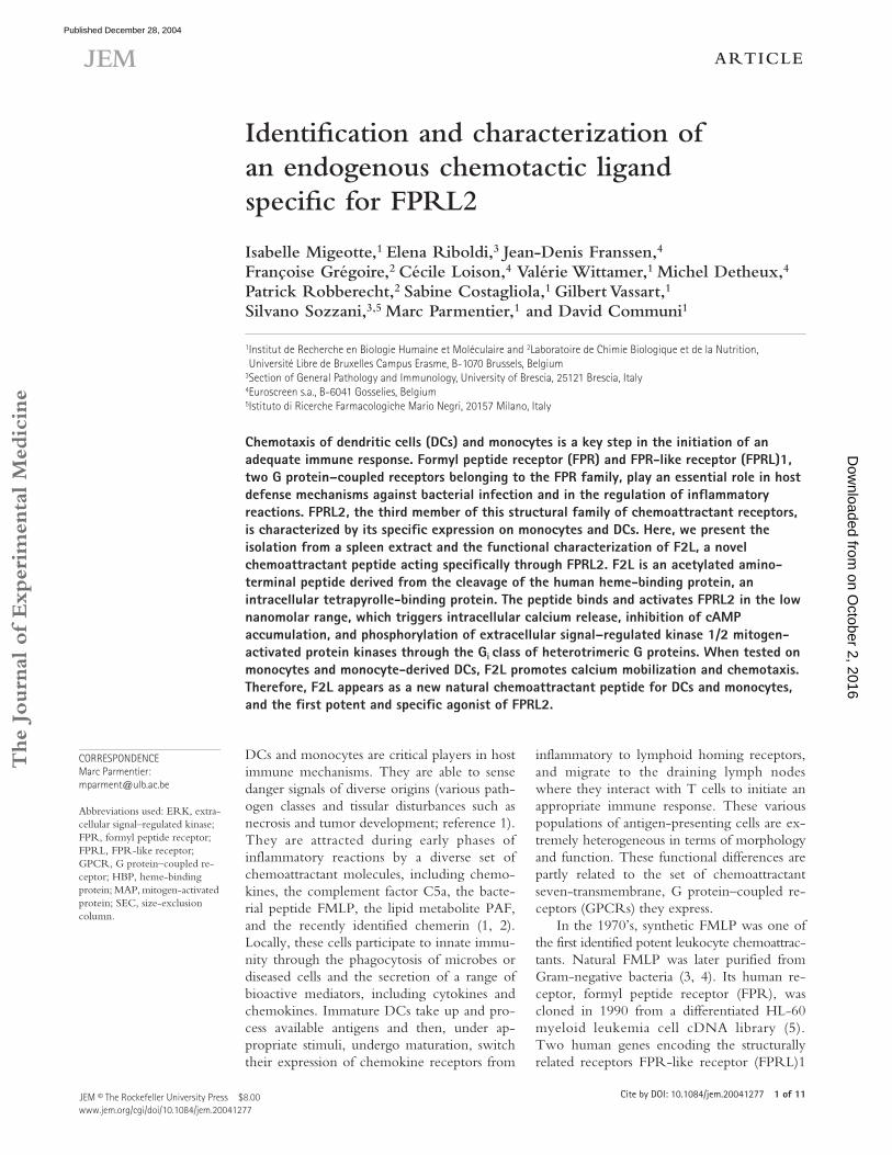
Figure 1. Purification from porcine spleen of the natural ligand of FPRL2. A porcine spleen homogenate was first fractionated by HPLC onto a Poros column (Step 1). The absorbance (AU) and biological activity on FPRL2-expressing CHO-K1 cells are shown. The luminescence measured in an aequorin-based assay (black bars) was normalized to the response ob-tained for 20 �M ATP. A1 (activity 1) and A2 (activity 2) represent the two active regions on the HPLC profile. They were processed together onto a C18 column (Step 2). Thereafter, A1 and A2 were purified separately onto an SEC (Step 3), a C4 column (Step 4), and for A1, a last C18 column (Step 5). The x axis is magnified to focus on the region of interest.
on October 2, 2016
Dow
nloaded from
Published December 28, 2004
JEM VOL. 201, January 3, 2005
ARTICLE
3 of 11
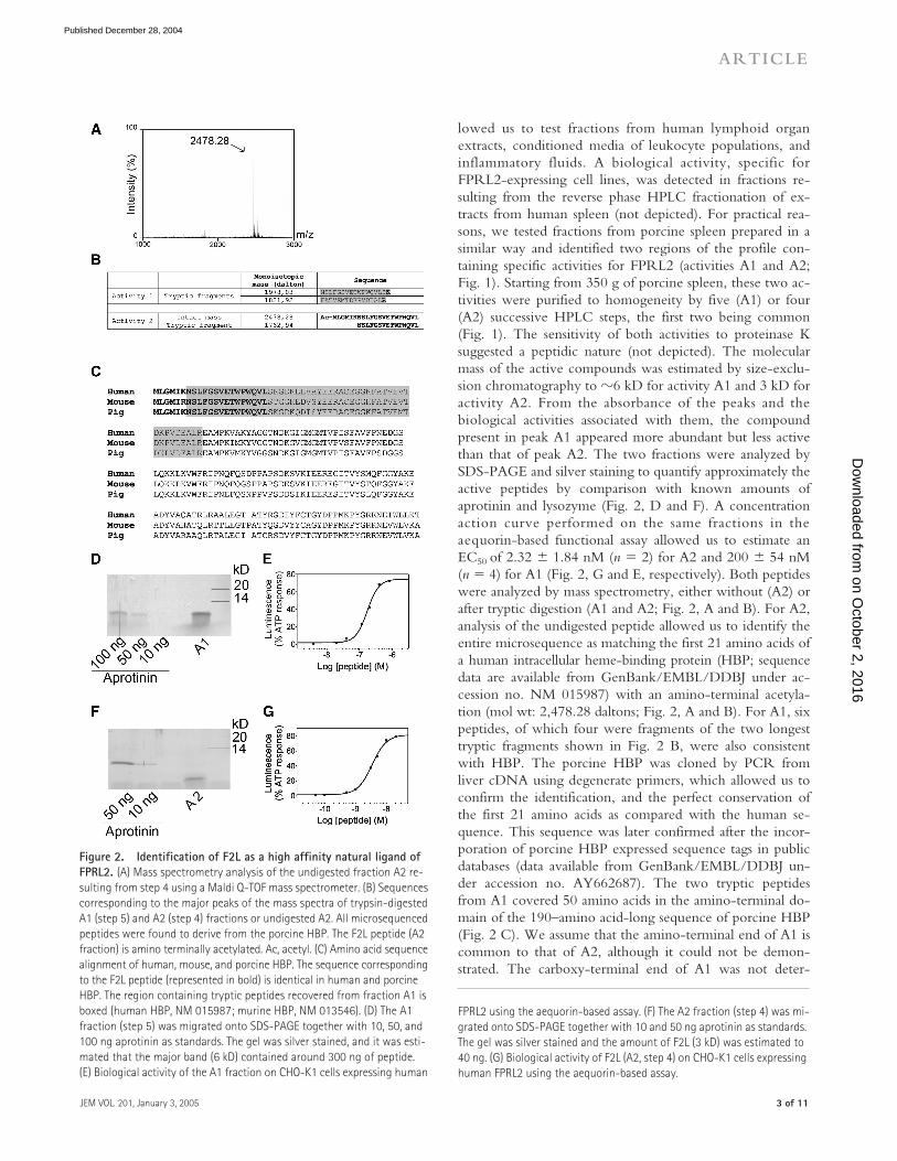
lowed us to test fractions from human lymphoid organextracts, conditioned media of leukocyte populations, andinflammatory fluids. A biological activity, specific forFPRL2-expressing cell lines, was detected in fractions re-sulting from the reverse phase HPLC fractionation of ex-tracts from human spleen (not depicted). For practical rea-sons, we tested fractions from porcine spleen prepared in asimilar way and identified two regions of the profile con-taining specific activities for FPRL2 (activities A1 and A2;Fig. 1). Starting from 350 g of porcine spleen, these two ac-tivities were purified to homogeneity by five (A1) or four(A2) successive HPLC steps, the first two being common(Fig. 1). The sensitivity of both activities to proteinase Ksuggested a peptidic nature (not depicted). The molecularmass of the active compounds was estimated by size-exclu-sion chromatography to
�
6 kD for activity A1 and 3 kD foractivity A2. From the absorbance of the peaks and thebiological activities associated with them, the compoundpresent in peak A1 appeared more abundant but less activethan that of peak A2. The two fractions were analyzed bySDS-PAGE and silver staining to quantify approximately theactive peptides by comparison with known amounts ofaprotinin and lysozyme (Fig. 2, D and F). A concentrationaction curve performed on the same fractions in theaequorin-based functional assay allowed us to estimate anEC
50
of 2.32
�
1.84 nM (
n
�
2) for A2 and 200
�
54 nM(
n
�
4) for A1 (Fig. 2, G and E, respectively). Both peptideswere analyzed by mass spectrometry, either without (A2) orafter tryptic digestion (A1 and A2; Fig. 2, A and B). For A2,analysis of the undigested peptide allowed us to identify theentire microsequence as matching the first 21 amino acids ofa human intracellular heme-binding protein (HBP; sequencedata are available from GenBank/EMBL/DDBJ under ac-cession no. NM 015987) with an amino-terminal acetyla-tion (mol wt: 2,478.28 daltons; Fig. 2, A and B). For A1, sixpeptides, of which four were fragments of the two longesttryptic fragments shown in Fig. 2 B, were also consistentwith HBP. The porcine HBP was cloned by PCR fromliver cDNA using degenerate primers, which allowed us toconfirm the identification, and the perfect conservation ofthe first 21 amino acids as compared with the human se-quence. This sequence was later confirmed after the incor-poration of porcine HBP expressed sequence tags in publicdatabases (data available from GenBank/EMBL/DDBJ un-der accession no. AY662687). The two tryptic peptidesfrom A1 covered 50 amino acids in the amino-terminal do-main of the 190–amino acid-long sequence of porcine HBP(Fig. 2 C). We assume that the amino-terminal end of A1 iscommon to that of A2, although it could not be demon-strated. The carboxy-terminal end of A1 was not deter-
Figure 2. Identification of F2L as a high affinity natural ligand of FPRL2.
(A) Mass spectrometry analysis of the undigested fraction A2 re-sulting from step 4 using a Maldi Q-TOF mass spectrometer. (B) Sequences corresponding to the major peaks of the mass spectra of trypsin-digested A1 (step 5) and A2 (step 4) fractions or undigested A2. All microsequenced peptides were found to derive from the porcine HBP. The F2L peptide (A2 fraction) is amino terminally acetylated. Ac, acetyl. (C) Amino acid sequence alignment of human, mouse, and porcine HBP. The sequence corresponding to the F2L peptide (represented in bold) is identical in human and porcine HBP. The region containing tryptic peptides recovered from fraction A1 is boxed (human HBP, NM 015987; murine HBP, NM 013546). (D) The A1 fraction (step 5) was migrated onto SDS-PAGE together with 10, 50, and 100 ng aprotinin as standards. The gel was silver stained, and it was esti-mated that the major band (6 kD) contained around 300 ng of peptide. (E) Biological activity of the A1 fraction on CHO-K1 cells expressing human
FPRL2 using the aequorin-based assay. (F) The A2 fraction (step 4) was mi-grated onto SDS-PAGE together with 10 and 50 ng aprotinin as standards. The gel was silver stained and the amount of F2L (3 kD) was estimated to 40 ng. (G) Biological activity of F2L (A2, step 4) on CHO-K1 cells expressing human FPRL2 using the aequorin-based assay.
on October 2, 2016
Dow
nloaded from
Published December 28, 2004

A NOVEL ENDOGENOUS-SPECIFIC LIGAND FOR FPRL2 | Migeotte et al.
4 of 11
The comparative pharmacology of the three FPRs wasthen studied in more detail, using F2L and four referenceagonists of FPR and FPRL1 (FMLP, the hexapeptidesWKYMVM and WKYMVm, and the CCL23-derivedSHAAG peptide). Concentration action curves and the re-sulting functional parameters were established both in theaequorin-based assay and the cAMP accumulation assay afterstimulation by 10
�
M forskolin (Fig. 3, A and B, and TableI). Among the tested peptides, F2L was by far the most po-tent on FPRL2, with an EC
50
of 10 nM in the aequorin assayand 5 nM in the cAMP assay. F2L also appeared as highlyspecific, as a weak activity was obtained on FPRL1 (EC
50
of567 and 234 nM in the aequorin and cAMP assays, respec-tively), whereas on FPR, only partial inhibition of cAMP ac-cumulation was obtained for 1
�
M F2L, and no activity wasdetected in the aequorin assay up to 5
�
M. For the otherpeptides, the EC
50
values obtained for FPR and FPRL1 (Ta-ble I) were essentially as described in the literature (3, 4, 13).However, significant differences with published data wereobserved when testing the two W hexapeptides on theFPRL2-expressing cells. Indeed, micromolar concentrationsof these peptides were required to activate FPRL2 (while ac-tive at low nanomolar concentrations on FPR and FPRL1).As described, FMLP and SHAAG were inactive on FPRL2.
To further confirm that F2L is a specific high affinityligand for FPRL2, we performed binding experiments. Amodified F2L peptide bearing a carboxy-terminal tyrosinewas found to display a potency similar to that of wild-typeF2L in the aequorin assay (not depicted). This peptide wasiodinated with
125
I and used as a tracer. Saturation bindingassays, performed on FPRL2-expressing CHO-K1 cells, al-lowed us to determine a K
D
of 11.7
�
4.9 nM (
n
�
3) and a
mined precisely either, due to the large number of trypticsites after Arg
56
of HBP. The purification was performedthree times with distinct protocols, and the same peptideswere identified by mass spectrometry in each case.
Comparative pharmacology and intracellular signaling of FPRs
The pharmacology and signaling pathways activated by thethree members of the human FMLP receptor family were in-vestigated in CHO-K1 cells expressing the receptors with orwithout G
�
16
and apoaequorin (Fig. 3). The acetylated 21–amino acid peptide, named F2L (for FPRL2 ligand), was syn-thesized and tested in the aequorin-based assay on these threecell lines as well as on wild-type CHO-K1 cells and CHO-K1cells expressing chemerinR and other GPCRs. The syntheticF2L peptide was shown to activate the FPRL2-expressingcells with a potency similar to that of the native peptide puri-fied from the spleen and, with a much lower efficiency,FPRL1 and FPR (see below), but it was completely inactiveon all other cell lines tested (not depicted). F2L was also testedin a cAMP accumulation assay on CHO-K1 cells expressingFPRL2 but not G
�
16
. The synthetic peptide was found to in-hibit the cAMP accumulation promoted by forskolin and wasunable to stimulate cAMP production by itself. In the samecells, F2L also promoted intracellular calcium release at lownanomolar concentrations (not depicted) and induced at pico-molar concentrations the phosphorylation of the extracellularsignal–regulated kinase (ERK)1/2 mitogen-activated protein(MAP) kinases (Fig. 3 I). Kinetics study of MAP kinase activa-tion showed a maximal phosphorylation at 15 min (Fig. 3 J).Calcium signaling was totally inhibited by pertussis toxin pre-treatment, demonstrating the coupling of the FPRL2 receptorto the G
i
family of heterotrimeric G proteins (Fig. 3 G).
Table I.
Pharmacology of FPRs
Receptor LigandpEC
50
(aequorin assay)pEC
50
(cAMP assay)pIC
50
(binding assay)
FPRL2
F2L 8.02
�
0.13 (
n
�
9) 8.24
�
0.06 (
n
�
4) 7.48
�
0.003 (
n
�
3)WKYMVm
�
6 NT
�
6WKYMVM
�
6
�
6 NTSHAAG
�
6
�
6
�
6FMLP
�
6
�
6
�
6
FPRL1
F2L 6.26
�
0.12 (
n
�
3) 6.65
�
0.16 (
n
�
8)
�
6WKYMVm 10.57
�
0.10 (
n
�
3) NT 7.66
�
0.15 (
n
�
3)WKYMVM 10.04
�
0.18 (
n
�
3) 10.27
�
0.27 (
n
�
4) NTSHAAG 9.27
�
0.06 (
n
�
3) 9.23
�
0.17 (
n
�
6) NTFMLP 5.94
�
0.03 (
n
�
3)
�
6 NT
FPR
F2L
�
6
�
6
�
6WKYMVm 9.18
�
0.16 (
n
�
3) NT 7.03
�
0.16 (
n
�
3)WKYMVM 7.48
� 0.08 (n � 3) 8.23 � 0.13 (n � 4) NTSHAAG �6 �6 NTFMLP 9.39 � 0.33 (n � 3) 10.15 � 0.08 (n � 4) NT
Binding and activation of CHO-K1 cells expressing FPRL2, FPRL1, or FPR by F2L, FMLP, WKYMVm, WKYMVM, and SHAAG were studied using a binding assay, an aequorin-based assay, and an assay measuring the inhibition of cAMP accumulation. The EC50 and IC50 parameters of the dose-response curves were determined by nonlinear regression using Graphpad Prism software. The results represent the mean pEC50 or pIC50 (-log values of EC50 or IC50 ) � SEM for at least three independent experiments (n).NT, not tested.
on October 2, 2016
Dow
nloaded from
Published December 28, 2004
JEM VOL. 201, January 3, 2005
ARTICLE
5 of 11
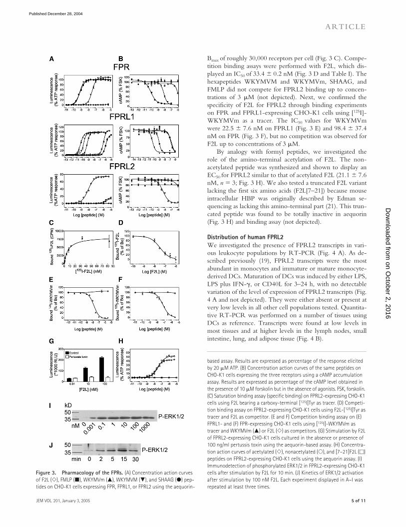
Bmax of roughly 30,000 receptors per cell (Fig. 3 C). Compe-tition binding assays were performed with F2L, which dis-played an IC50 of 33.4 � 0.2 nM (Fig. 3 D and Table I). Thehexapeptides WKYMVM and WKYMVm, SHAAG, andFMLP did not compete for FPRL2 binding up to concen-trations of 3 �M (not depicted). Next, we confirmed thespecificity of F2L for FPRL2 through binding experimentson FPR and FPRL1-expressing CHO-K1 cells using [125I]-WKYMVm as a tracer. The IC50 values for WKYMVmwere 22.5 � 7.6 nM on FPRL1 (Fig. 3 E) and 98.4 � 37.4nM on FPR (Fig. 3 F), but no competition was observed forF2L up to concentrations of 3 �M.
By analogy with formyl peptides, we investigated therole of the amino-terminal acetylation of F2L. The non-acetylated peptide was synthesized and shown to display anEC50 for FPRL2 similar to that of acetylated F2L (21.1 � 7.6nM, n � 3; Fig. 3 H). We also tested a truncated F2L variantlacking the first six amino acids (F2L[7–21]) because mouseintracellular HBP was originally described by Edman se-quencing as lacking this amino-terminal part (21). This trun-cated peptide was found to be totally inactive in aequorin(Fig. 3 H) and binding assay (not depicted).
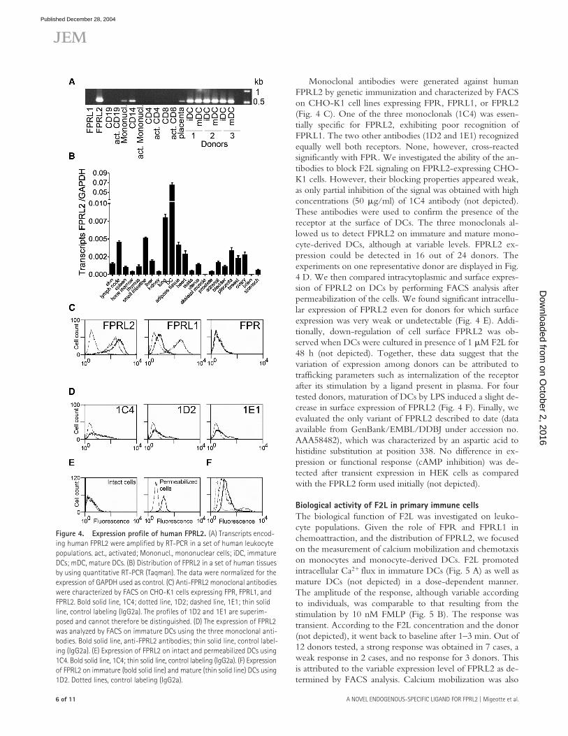
Distribution of human FPRL2We investigated the presence of FPRL2 transcripts in vari-ous leukocyte populations by RT-PCR (Fig. 4 A). As de-scribed previously (19), FPRL2 transcripts were the mostabundant in monocytes and immature or mature monocyte-derived DCs. Maturation of DCs was induced by either LPS,LPS plus IFN-�, or CD40L for 3–24 h, with no detectablevariation of the level of expression of FPRL2 transcripts (Fig.4 A and not depicted). They were either absent or present atvery low levels in all other cell populations tested. Quantita-tive RT-PCR was performed on a number of tissues usingDCs as reference. Transcripts were found at low levels inmost tissues and at higher levels in the lymph nodes, smallintestine, lung, and adipose tissue (Fig. 4 B).
Figure 3. Pharmacology of the FPRs. (A) Concentration action curves of F2L (�), FMLP (�), WKYMVm (�), WKYMVM (�), and SHAAG (�) pep-tides on CHO-K1 cells expressing FPR, FPRL1, or FPRL2 using the aequorin-
based assay. Results are expressed as percentage of the response elicited by 20 �M ATP. (B) Concentration action curves of the same peptides on CHO-K1 cells expressing the three receptors using a cAMP accumulation assay. Results are expressed as percentage of the cAMP level obtained in the presence of 10 �M forskolin but in the absence of agonists. FSK, forskolin. (C) Saturation binding assay (specific binding) on FPRL2-expressing CHO-K1 cells using F2L bearing a carboxy-terminal [125I]Tyr as tracer. (D) Competi-tion binding assay on FPRL2-expressing CHO-K1 cells using F2L-[125I]Tyr as tracer and F2L as competitor. (E and F) Competition binding assay on (E) FPRL1- and (F) FPR-expressing CHO-K1 cells using [125I]-WKYMVm as tracer and WKYMVm (�) or F2L (�) as competitors. (G) Stimulation by F2L of FPRL2-expressing CHO-K1 cells cultured in the absence or presence of 100 ng/ml pertussis toxin using the aequorin-based assay. (H) Concentra-tion action curves of acetylated (�), nonacetylated (�), and [7–21]F2L (�) peptides on FPRL2-expressing CHO-K1 cells using the aequorin assay. (I) Immunodetection of phosphorylated ERK1/2 in FPRL2-expressing CHO-K1 cells after stimulation by F2L for 10 min. (J) Kinetics of ERK1/2 activation after stimulation by 100 nM F2L. Each experiment displayed in A–I was repeated at least three times.
on October 2, 2016
Dow
nloaded from
Published December 28, 2004
A NOVEL ENDOGENOUS-SPECIFIC LIGAND FOR FPRL2 | Migeotte et al.6 of 11
Monoclonal antibodies were generated against humanFPRL2 by genetic immunization and characterized by FACSon CHO-K1 cell lines expressing FPR, FPRL1, or FPRL2(Fig. 4 C). One of the three monoclonals (1C4) was essen-tially specific for FPRL2, exhibiting poor recognition ofFPRL1. The two other antibodies (1D2 and 1E1) recognizedequally well both receptors. None, however, cross-reactedsignificantly with FPR. We investigated the ability of the an-tibodies to block F2L signaling on FPRL2-expressing CHO-K1 cells. However, their blocking properties appeared weak,as only partial inhibition of the signal was obtained with highconcentrations (50 �g/ml) of 1C4 antibody (not depicted).These antibodies were used to confirm the presence of thereceptor at the surface of DCs. The three monoclonals al-lowed us to detect FPRL2 on immature and mature mono-cyte-derived DCs, although at variable levels. FPRL2 ex-pression could be detected in 16 out of 24 donors. Theexperiments on one representative donor are displayed in Fig.4 D. We then compared intracytoplasmic and surface expres-sion of FPRL2 on DCs by performing FACS analysis afterpermeabilization of the cells. We found significant intracellu-lar expression of FPRL2 even for donors for which surfaceexpression was very weak or undetectable (Fig. 4 E). Addi-tionally, down-regulation of cell surface FPRL2 was ob-served when DCs were cultured in presence of 1 �M F2L for48 h (not depicted). Together, these data suggest that thevariation of expression among donors can be attributed totrafficking parameters such as internalization of the receptorafter its stimulation by a ligand present in plasma. For fourtested donors, maturation of DCs by LPS induced a slight de-crease in surface expression of FPRL2 (Fig. 4 F). Finally, weevaluated the only variant of FPRL2 described to date (dataavailable from GenBank/EMBL/DDBJ under accession no.AAA58482), which was characterized by an aspartic acid tohistidine substitution at position 338. No difference in ex-pression or functional response (cAMP inhibition) was de-tected after transient expression in HEK cells as comparedwith the FPRL2 form used initially (not depicted).
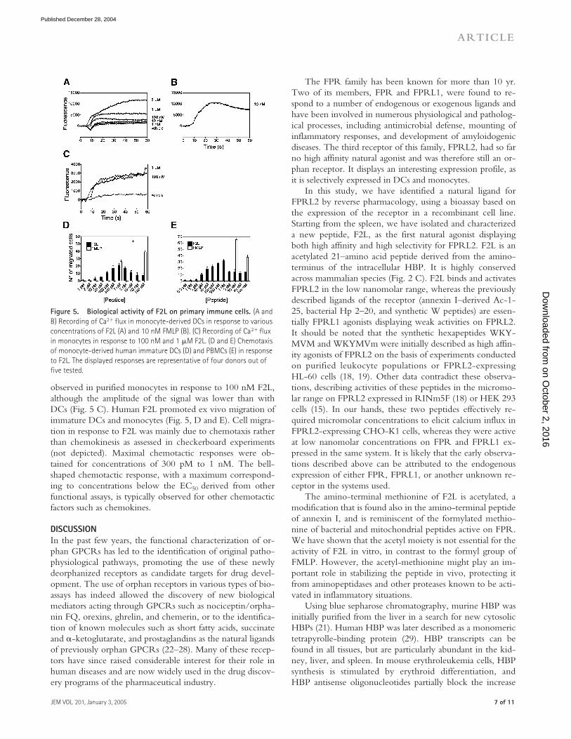
Biological activity of F2L in primary immune cellsThe biological function of F2L was investigated on leuko-cyte populations. Given the role of FPR and FPRL1 inchemoattraction, and the distribution of FPRL2, we focusedon the measurement of calcium mobilization and chemotaxison monocytes and monocyte-derived DCs. F2L promotedintracellular Ca2� flux in immature DCs (Fig. 5 A) as well asmature DCs (not depicted) in a dose-dependent manner.The amplitude of the response, although variable accordingto individuals, was comparable to that resulting from thestimulation by 10 nM FMLP (Fig. 5 B). The response wastransient. According to the F2L concentration and the donor(not depicted), it went back to baseline after 1–3 min. Out of12 donors tested, a strong response was obtained in 7 cases, aweak response in 2 cases, and no response for 3 donors. Thisis attributed to the variable expression level of FPRL2 as de-termined by FACS analysis. Calcium mobilization was also
Figure 4. Expression profile of human FPRL2. (A) Transcripts encod-ing human FPRL2 were amplified by RT-PCR in a set of human leukocyte populations. act., activated; Mononucl., mononuclear cells; iDC, immature DCs; mDC, mature DCs. (B) Distribution of FPRL2 in a set of human tissues by using quantitative RT-PCR (Taqman). The data were normalized for the expression of GAPDH used as control. (C) Anti-FPRL2 monoclonal antibodies were characterized by FACS on CHO-K1 cells expressing FPR, FPRL1, and FPRL2. Bold solid line, 1C4; dotted line, 1D2; dashed line, 1E1; thin solid line, control labeling (IgG2a). The profiles of 1D2 and 1E1 are superim-posed and cannot therefore be distinguished. (D) The expression of FPRL2 was analyzed by FACS on immature DCs using the three monoclonal anti-bodies. Bold solid line, anti-FPRL2 antibodies; thin solid line, control label-ing (IgG2a). (E) Expression of FPRL2 on intact and permeabilized DCs using 1C4. Bold solid line, 1C4; thin solid line, control labeling (IgG2a). (F) Expression of FPRL2 on immature (bold solid line) and mature (thin solid line) DCs using 1D2. Dotted lines, control labeling (IgG2a).
on October 2, 2016
Dow
nloaded from
Published December 28, 2004
JEM VOL. 201, January 3, 2005
ARTICLE
7 of 11
The FPR family has been known for more than 10 yr.Two of its members, FPR and FPRL1, were found to re-spond to a number of endogenous or exogenous ligands andhave been involved in numerous physiological and patholog-ical processes, including antimicrobial defense, mounting ofinflammatory responses, and development of amyloidogenicdiseases. The third receptor of this family, FPRL2, had so farno high affinity natural agonist and was therefore still an or-phan receptor. It displays an interesting expression profile, asit is selectively expressed in DCs and monocytes.
In this study, we have identified a natural ligand forFPRL2 by reverse pharmacology, using a bioassay based onthe expression of the receptor in a recombinant cell line.Starting from the spleen, we have isolated and characterizeda new peptide, F2L, as the first natural agonist displayingboth high affinity and high selectivity for FPRL2. F2L is anacetylated 21–amino acid peptide derived from the amino-terminus of the intracellular HBP. It is highly conservedacross mammalian species (Fig. 2 C). F2L binds and activatesFPRL2 in the low nanomolar range, whereas the previouslydescribed ligands of the receptor (annexin I–derived Ac-1-25, bacterial Hp 2–20, and synthetic W peptides) are essen-tially FPRL1 agonists displaying weak activities on FPRL2.It should be noted that the synthetic hexapeptides WKY-MVM and WKYMVm were initially described as high affin-ity agonists of FPRL2 on the basis of experiments conductedon purified leukocyte populations or FPRL2-expressingHL-60 cells (18, 19). Other data contradict these observa-tions, describing activities of these peptides in the micromo-lar range on FPRL2 expressed in RINm5F (18) or HEK 293cells (15). In our hands, these two peptides effectively re-quired micromolar concentrations to elicit calcium influx inFPRL2-expressing CHO-K1 cells, whereas they were activeat low nanomolar concentrations on FPR and FPRL1 ex-pressed in the same system. It is likely that the early observa-tions described above can be attributed to the endogenousexpression of either FPR, FPRL1, or another unknown re-ceptor in the systems used.
The amino-terminal methionine of F2L is acetylated, amodification that is found also in the amino-terminal peptideof annexin I, and is reminiscent of the formylated methio-nine of bacterial and mitochondrial peptides active on FPR.We have shown that the acetyl moiety is not essential for theactivity of F2L in vitro, in contrast to the formyl group ofFMLP. However, the acetyl-methionine might play an im-portant role in stabilizing the peptide in vivo, protecting itfrom aminopeptidases and other proteases known to be acti-vated in inflammatory situations.
Using blue sepharose chromatography, murine HBP wasinitially purified from the liver in a search for new cytosolicHBPs (21). Human HBP was later described as a monomerictetrapyrolle-binding protein (29). HBP transcripts can befound in all tissues, but are particularly abundant in the kid-ney, liver, and spleen. In mouse erythroleukemia cells, HBPsynthesis is stimulated by erythroid differentiation, andHBP antisense oligonucleotides partially block the increase
Figure 5. Biological activity of F2L on primary immune cells. (A and B) Recording of Ca2� flux in monocyte-derived DCs in response to various concentrations of F2L (A) and 10 nM FMLP (B). (C) Recording of Ca2� flux in monocytes in response to 100 nM and 1 �M F2L. (D and E) Chemotaxis of monocyte-derived human immature DCs (D) and PBMCs (E) in response to F2L. The displayed responses are representative of four donors out of five tested.
observed in purified monocytes in response to 100 nM F2L,although the amplitude of the signal was lower than withDCs (Fig. 5 C). Human F2L promoted ex vivo migration ofimmature DCs and monocytes (Fig. 5, D and E). Cell migra-tion in response to F2L was mainly due to chemotaxis ratherthan chemokinesis as assessed in checkerboard experiments(not depicted). Maximal chemotactic responses were ob-tained for concentrations of 300 pM to 1 nM. The bell-shaped chemotactic response, with a maximum correspond-ing to concentrations below the EC50 derived from otherfunctional assays, is typically observed for other chemotacticfactors such as chemokines.
DISCUSSIONIn the past few years, the functional characterization of or-phan GPCRs has led to the identification of original patho-physiological pathways, promoting the use of these newlydeorphanized receptors as candidate targets for drug devel-opment. The use of orphan receptors in various types of bio-assays has indeed allowed the discovery of new biologicalmediators acting through GPCRs such as nociceptin/orpha-nin FQ, orexins, ghrelin, and chemerin, or to the identifica-tion of known molecules such as short fatty acids, succinateand �-ketoglutarate, and prostaglandins as the natural ligandsof previously orphan GPCRs (22–28). Many of these recep-tors have since raised considerable interest for their role inhuman diseases and are now widely used in the drug discov-ery programs of the pharmaceutical industry.
on October 2, 2016
Dow
nloaded from
Published December 28, 2004
A NOVEL ENDOGENOUS-SPECIFIC LIGAND FOR FPRL2 | Migeotte et al.8 of 11
of heme content associated with this differentiation process(21). The putative function of HBP is to buffer heme andporphyrins synthesized in excess. As HBP is a cytosolic pro-tein, its amino-terminal peptide fragments acting on FPRL2could either be generated intracellularly by proteolytic pro-cessing and then released, or alternatively, the whole proteincould be secreted and the cleavage could take place in theextracellular compartment. The longer, less active HBP-derived peptide isolated from the spleen could represent anintermediate step in the generation of the final, fully activeagonist. HBP and/or its active fragments could be releasedeither through a nonconventional mode of secretion or sec-ondary to cell death. We did not observe biological activityon FPRL2-expressing cells in supernatants from CHO-K1cells transfected with an expression vector containing thefull-size human HBP cDNA (not depicted). This suggeststhat the processing leading to the active F2L peptide does notoccur in CHO-K1 cells and probably requires a specific en-vironment. An increasing number of proteins devoid of sig-nal peptide were shown to be released from the cells by un-known mechanisms. IL-1� is generated from an intracellularprecursor by proteolytic cleavage by the caspase ICE, and theunconventional mechanism by which it is released from thecells is not known (30). Two intracellular peptides, Sun Aand B, were recently described as the ligands of the Dro-sophila receptor Methuselah (31). Annexin I, a ligand ofFPR, FPRL1, and to a lower extent FPRL2, is also an intra-cellular protein of 346 amino acids, which is externalized bya still unclear mechanism when neutrophils adhere to the en-dothelium. The acetylated amino-terminal domain of theprotein, which is exposed only in the presence of calciumions (32), is cleaved by proteolysis, and both the full-sizeprotein and the amino-terminal peptides bind the receptors(33, 34). Another recently identified ligand of FPRL1, hu-manin, is also an intracellular protein synthesized withoutsignal peptide and shown to be released from cells in a non-conventional way. The humanin sequence was further shownto induce the secretion of other proteins when fused tothem, thereby playing the role of an export signal (35).
F2L is a new member of the chemoattractant factor fam-ily. By binding on FPRL2, it triggers the classical intracellu-lar cascades stimulated by chemoattractants through the Gi
class of G proteins: stimulation of intracellular calcium re-lease, inhibition of cAMP production, and activation of theMAP kinases ERK1/2. The peptide is able to chemoattractmonocytes and immature DCs. It could therefore be in-volved in the response of these cells to infection, inflamma-tion, or cell death. DCs are known to process and presentantigens derived from infected, malignant, or allogeneiccells. Recent evidence indicates that these cells are attractedby dead or dying cells (36). The first self-molecules shown torepresent danger signals were the heat shock proteins gp96or hsp70. They are released from necrotic cells and are ableto potently activate antigen-presenting cells (37). Intracellu-lar nucleotides released under conditions of hypoxia, ische-mia, inflammation, or mechanical stress, and crystalline uric
acid deriving from dead cells, were also shown to stimulateDCs (38, 39). Apoptotic cells, in addition to engulfment sig-nals, release chemotactic factors for phagocytes (40). Thus, itis conceivable that F2L, released from HBP by proteolysis af-ter cell suffering or cell death, contributes to the chemoat-traction of monocytes and DCs through FPRL2. Therefore,F2L and FPRL2 might be involved in the development of anumber of inflammatory diseases associated with cell death.In this context, FPRL2 could represent an attractive targetfor therapeutic approaches. However, the characterization ofthe precise contribution of this system in specific human dis-eases will require additional investigations.
MATERIALS AND METHODSExpression of human FPRL2, FPRL1, and FPR. The human codingsequences (data available from GenBank/EMBL/DDBJ accession nosAC005946, M84562, and M60626, respectively) were amplified by PCRfrom human genomic DNA, cloned into the pcDNA3 (Invitrogen) andpEFIN3 (Euroscreen) vectors, and sequenced. The pEFIN3 constructs weretransfected using Fugene 6 into CHO-K1 cells expressing or not G�16 andapoaequorin. G418-resistant clones were characterized for receptor expres-sion by Northern blotting. A functional assay based on the luminescence ofmitochondrial aequorin was performed as described previously (41). Resultswere expressed as relative light units or as the percentage of the response to20 �M ATP.
Purification of bioactive peptides. 350 g of frozen porcine spleen washomogenized in four volumes of ice-cold 20% CH3CN in water. The homog-enate was centrifuged at 10,000 g for 30 min at 4C and snap-frozen in liquidnitrogen. Aliquots of 200 ml of supernatant were diluted fourfold in 0.1%TFA and loaded on a Poros R2 bead 4.6 150–mm column (Applied Bio-systems) at 5 ml/min. A 5–70% CH3CN gradient (6%/min) in 0.1% TFA wasapplied, and 1.25 ml fractions were collected and tested for functional activityon FPRL2-expressing CHO-K1 cells in an aequorin assay. Two regions ofactivity (A1 and A2) were detected on the HPLC profile. The correspondingfractions were pooled, diluted fourfold in 0.1% TFA, and loaded at 1 ml/minon a C18 4.6 250–mm column (Vydac), which was submitted to a 30–50%CH3CN gradient in 0.1% TFA. Two regions of activity were detected andsubsequently treated separately. The 1-ml fractions corresponding to the first(A1, lower CH3CN concentration) and the second (A2, higher CH3CN con-centration) regions from two runs were vacuum concentrated to 50 �l. A1and A2 were diluted threefold in 30% CH3CN/0.05% TFA and 30%CH3CN/0.1% TFA, respectively, and loaded on size-exclusion columns(SECs; A1: TSK-gel Alpha-4000 [7.8 300 mm; Tosoh Biosep]; A2: Super-dex peptide PE [7.5 300 mm; Amersham Biosciences) submitted to a 0.5ml/min flow rate of dilution medium. The active 0.25-ml fractions from oneSEC were diluted fourfold in 0.1% TFA and loaded at 0.2 ml/min on a C42.1 250–mm column (Vydac), which was submitted to a 25–45% (A1) or30–50% (A2) CH3CN gradient at 0.3%/min in 0.1% TFA. The fractionswere collected manually according to the absorbance profile. For A1, the ac-tive fractions from one run were pooled, diluted fivefold in 0.1% TFA, andloaded at 0.05 ml/min on a C18 1 250–mm column (Vydac), which wassubmitted to a 23–50% CH3CN gradient at 0.45%/min in 0.1% TFA. Thefractions were collected manually. For A2, the purity of the final active frac-tion was checked by loading an aliquot on a C18 1 250–mm column. Thepurification was repeated three times with different protocols (i.e., the SECstep was replaced by a reverse phase step on a C18 2.1 250–mm columnsubmitted to a CH3CN gradient in 0.1% H3PO4 as ion-pairing agent). Theprotein concentration in active fractions was determined after SDS/PAGE bycomparison with aprotinin and lysozyme standards after silver staining.
Mass spectrometry analysis. The active fractions were vacuum dried,resuspended in 20 mM ammonium bicarbonate, heated to 100C for 5 min,
on October 2, 2016
Dow
nloaded from
Published December 28, 2004

JEM VOL. 201, January 3, 2005
ARTICLE
9 of 11
digested by 50 ng trypsin overnight or left intact, and purified by solid phaseextraction (C18 ZipTip; Millipore). The peptides were eluted in 1.5 �l of70% CH3CN/0.1% TFA onto a metallic MALDI target, dried, and thenmixed with 1.5 �l of matrix mix (2 mg/ml 2,5-dihydroxybenzoic acid and10 mg/ml �-cyano-4-hydroxycinnamic acid, 2 mM fucose, 5 mM ammo-nium acetate). Mass spectrometry analysis was performed on a Q-TOFUltima Global mass spectrometer equipped with a MALDI source (Micro-mass) and calibrated using the monoisotopic masses of tryptic and chymo-tryptic peptides from bovine serum albumin. Ionization was achieved usinga nitrogen laser (337-nm beam, 10 Hz), and acquisitions were performed ina V mode reflectron position. Microsequencing was performed by argon-induced fragmentation after selection of the parent ion.
Synthetic peptides. Acetylated or nonacetylated MLGMIKNSLFGS-VETWPWQVL (HBP[1–21], renamed F2L), NSLFGSVETWPWQVL(F2L[7–21]), WKYMVM, and MLWRRKIGPQMTLSHAAG (SHAAGpeptide derived from CCL23 amino-terminus) were synthesized locally byusing the solid phase Fmoc strategy (42) or were custom made by Eurogen-tec. WKYMVM and WKYMVm were purchased from Phoenix Pharma-ceuticals and FMLP was purchased from Neosystem. Monoisotopic massesand sequences of all peptides were verified by mass spectrometry. F2L andWKYMVM from different origins displayed the same properties. At highconcentrations, HBP-derived peptides were dissolved in DMSO and heatedat 50C for 10 min due to their high hydrophobicity. Intermediate dilutionswere made in 50% CH3CN and were further diluted 40-fold in assay bufferto reach working concentration.
Quantitative RT-PCR. FPRL2 transcripts were detected by RT-PCR incDNA from human blood cell populations obtained commercially (CLON-TECH Laboratories, Inc.) or prepared locally as described previously (43).Primers for FPRL2 were 5�-CTGGCCACACCGTTCTGT-3� as forwardand 5�-GGCCATGGTAATGAACACGTT-3� as reverse. Amplification ofGAPDH transcripts was performed as a control of the quality of cDNA (notdepicted). FPRL2 transcripts were detected by quantitative RT-PCR (Taq-Man) in total or polyA� RNA samples from human tissues obtained com-mercially (CLONTECH Laboratories, Inc. and Ambion) or prepared locally(DCs). Primers for FPRL2 were 5�-TTACCATGGCCAAGGTCTTTCT-3� as forward, 5�-GCAGACTGTGATGATGGACATAGG-3� as reverse,and 5�FAM-TCCTCCACTTCATTATTGGCTTCAGCGT-3�DABSYLas probe, and for the reference housekeeping gene (GAPDH) were 5�-GAAGGTGAAGGTCGGAGTC-3� as forward, 5�-GAAGATGGT-GATGGGATTTC-3� as reverse, and 5�FAM-CAAGCTTCCCGTTCT-CAGCC-3�DABSYL as probe. Primers were used at 900 nM and probeswere used at 200 nM. Standard curves were run systematically for the twogenes, and the transcript copy number of FPRL2 was normalized to theGAPDH transcript copy number for each sample.
Monoclonal antibodies and flow cytometry. BALB/c mice were in-jected with 100 �g pcDNA3-FPRL2 as described previously (44). Serawere tested by FACS on the CHO-K1-FPRL2 cell line, and immune micewere used to generate monoclonal antibodies by standard hybridoma tech-nology using the NSO myeloma cell line. The Ig class of selected hybrid-omas was determined with a mouse monoclonal antibody isotyping kit(IsoStrip; Boehringer). Flow cytometry was performed using anti-FPRL2antibodies or control IgG2a at 1 �g/ml (for CHO-K1 cells) or 5 �g/ml (forprimary cells) in PBS containing 0.1% BSA, 0.1% sodium azide, and FITC-conjugated � chain–specific goat anti–mouse IgG (Sigma-Aldrich) as sec-ondary antibody. Fluorescence of 10,000 cells was assayed using a FACScanflow cytofluorimeter (Becton Dickinson). Intracytoplasmic staining wasperformed using the Cytoperm/Cytowash kit (Becton Dickinson) accord-ing to the manufacturer’s instructions.
Intracellular cascade assays. The cAMP concentrations were deter-mined using a homogeneous time-resolved fluorescence kit (Cis Bio Inter-national). In brief, cells were detached, resuspended in Krebs-Ringer hepes
(KRH) buffer containing 1 mM 3-isobutyl-1-methylxanthine, and submit-ted to 10 �M forskolin alone or together with increasing concentrations ofagonists for 30 min at room temperature. The reaction was stopped by thesuccessive addition of cAMP-XL665 and anti-cAMP cryptate diluted in ly-sis buffer. The plates were incubated for 60 min at room temperature andread on a Rubystar fluorimeter (Labtech). Results were calculated from the665:620 nm ratio and expressed in delta F (%). A calibration curve was ob-tained by plotting delta F% versus cAMP concentrations. ERK1/2 activa-tion was assayed by Western blotting using an anti–phospho-p42/44 mono-clonal antibody (E10; Cell Signaling Technology) as described previously(45). The aequorin-based assay was performed with or without overnightpretreatment with 100 ng/ml pertussis toxin. It was shown that such pertus-sis toxin pretreatment did not inhibit the functional response to ATP inthese cells. For the analysis of FPRL2 polymorphism, HEK cells were trans-fected with a pcDNA3 vector containing the wild-type or Asp338HisFPRL2 cDNA by the calcium phosphate method. Cells were recovered48 h later and used for FACS or cAMP accumulation experiments.
Binding assays. F2L with an additional carboxy-terminal tyrosine wassynthesized and found to display an EC50 similar to that of wild-type F2L inthe aequorin assay. 5 �g of peptide was labeled with 2 mCi of 125I using theIodogen method (42). After separation of unbound 125I, the resultingspecific activity of the peptide was estimated to 900 Ci/mmole. [125I]-WKYMVm (2,200 Ci/mmole) was purchased from PerkinElmer. FPRL2,FPRL1, and FPR-expressing CHO-K1 cells were plated in 24-well plates(200,000 cells per well for FPRL2 and 100,000 cells per well for the twoother receptors). The next day, the cells were washed twice with a KRHbuffer without NaCl containing 280 mM saccharose and, for FPRL1 andFPR, 0.1% NaN3. For saturation binding assays, cells were incubated withvarious amounts of F2L-[125I]Tyr and nonspecific binding was determinedby using 1 �M F2L as competitor. For competition binding assays, cellswere incubated with 100,000 cpm of F2L-[125I]Tyr or 10,000 cpm of [125I]-WKYMVm and various amounts of F2L or other peptides as competitors inKRH buffer supplemented with 5% BSA for 90 min at room temperature.Cells were washed twice with ice cold buffer, and total radioactivity was re-covered with 1 M NaOH and counted in a gamma counter for 2 min.
Chemotaxis and Ca2� mobilization assays on primary cells. Mono-cyte-derived DCs were generated either from the adherent fraction of PB-MCs cultured with 800 U/ml GM-CSF and 500 U/ml Il-4, or from Per-coll-purified monocytes cultured with 50 ng/ml GM-CSF and 20 ng/mlIL-13 for 5–7 d. For Ca2� mobilization assay, monocytes were obtained bynegative selection with the Monocyte Isolation Kit II (Miltenyi Biotec).Cell migration in response to F2L and FMLP and MIP-1� (CCL3) used ascontrols was evaluated by using a 48-well microchemotaxis chamber tech-nique as described previously (2). For Ca2� mobilization assays, monocyte-derived DCs or monocytes (5 105 cells/ml in HBSS without phenol redbut containing 0.1% BSA and 1 mM Probenecid; Sigma-Aldrich) wereloaded with 4 �M Fluo 4 (Molecular Probes) for 1 h at 20C in the dark.The loaded cells were washed twice, resuspended at 1–2 106 cells/ml, and50 �l of cell suspension was distributed per well in a 96-well plate (View-plate; Packard Instrument Co.). Reading was performed in a Fluostar fluo-rimeter (Labtech) at 25C. 50 �l of ligand-containing medium was injected,and the fluorescence at 520 nm was recorded every second for 1–3 min.Each condition was performed in triplicate, the mean fluorescence for eachtime point was calculated, and the curves were normalized by subtractingthe mean value of the five measurements preceding the injection.
We thank Dominique Revets, Naïma Alaouï, and Nadia Tazir for their expert technical assistance, and Drs. Valérie Panneels, Frédéric Sallman, Stanislas Goriely, and Sophie Noël for expert advice. We are grateful to Dr. Luc De Pauw for providing human clinical samples.
This work was supported by the Belgian program on Interuniversity Poles of Attraction initiated by the Belgian State, Prime Minister’s Office, Science Policy Programming, the LifeSciHealth program of the European Community (grant LSHB-CT-2003-503337), the Fonds de la Recherche Scientifique Médicale of Belgium,
on October 2, 2016
Dow
nloaded from
Published December 28, 2004
A NOVEL ENDOGENOUS-SPECIFIC LIGAND FOR FPRL2 | Migeotte et al.10 of 11
Fortis, and the Fondation Médicale Reine Elisabeth to M. Parmentier. This work was partially supported by the Associazone Italiana per la Ricerca sul Cancro (AIRC) and by the Ministero dell’Istruzione, Università e Ricerca (MIUR). The scientific responsibility is assumed by the authors. I. Migeotte is Aspirant and D. Communi is Research Associate of the Belgian Fonds National de la Recherche Scientifique. V. Wittamer was recipient of grants from the FIRST-Industrie program of the Walloon Region and Télévie. J.-D. Franssen, C. Loison, and M. Detheux are employees of Euroscreen, which has the exploitation right regarding the work described.
The authors have no other conflicting financial interests.
Submitted: 25 June 2004Accepted: 22 November 2004
Note added in proof. After acceptance of this manuscript, Harada et al. reported humanin as a high affinity ligand for FPRL2 (Harada, M., Y. Habata, M. Hosoya, K. Nishi, R. Fujii, M. Kobayashi, and S. Hinuma. 2004. Biochem. Biophys. Res. Commun. 324:255–261.)
REFERENCES1. Lipscomb, M.F., and B.J. Masten. 2002. Dendritic cells: immune regu-
lators in health and disease. Physiol. Rev. 82:97–130.2. Wittamer, V., J.D. Franssen, M. Vulcano, J.F. Mirjolet, E. Le Poul, I.
Migeotte, S. Brezillon, R. Tyldesley, C. Blanpain, M. Detheux, et al.2003. Specific recruitment of antigen-presenting cells by chemerin, anovel processed ligand from human inflammatory fluids. J. Exp. Med.198:977–985.
3. Le, Y., J.J. Oppenheim, and J.M. Wang. 2001. Pleiotropic roles offormyl peptide receptors. Cytokine Growth Factor Rev. 12:91–105.
4. Le, Y., P.M. Murphy, and J.M. Wang. 2002. Formyl-peptide receptorsrevisited. Trends Immunol. 23:541–548.
5. Boulay, F., M. Tardif, L. Brouchon, and P. Vignais. 1990. Synthesisand use of a novel N-formyl peptide derivative to isolate a humanN-formyl peptide receptor cDNA. Biochem. Biophys. Res. Commun.168:1103–1109.
6. Bao, L., N.P. Gerard, R.L. Eddy Jr., T.B. Shows, and C. Gerard. 1992.Mapping of genes for the human C5a receptor (C5AR), human FMLPreceptor (FPR), and two FMLP receptor homologue orphan receptors(FPRH1, FPRH2) to chromosome 19. Genomics. 13:437–440.
7. Murphy, P.M., T. Ozcelik, R.T. Kenney, H.L. Tiffany, D. McDer-mott, and U. Francke. 1992. A structural homologue of the N-formylpeptide receptor. Characterization and chromosome mapping of a pep-tide chemoattractant receptor family. J. Biol. Chem. 267:7637–7643.
8. Gao, J.L., H. Chen, J.D. Filie, C.A. Kozak, and P.M. Murphy. 1998.Differential expansion of the N-formylpeptide receptor gene cluster inhuman and mouse. Genomics. 51:270–276.
9. Wang, Z.G., and R.D. Ye. 2002. Characterization of two new mem-bers of the formyl peptide receptor gene family from 129S6 mice.Gene. 299:57–63.
10. Walther, A., K. Riehemann, and V. Gerke. 2000. A novel ligand ofthe formyl peptide receptor: annexin I regulates neutrophil extravasa-tion by interacting with the FPR. Mol. Cell. 5:831–840.
11. Le, Y.Y., W.H. Gong, B.Q. Li, N.M. Dunlop, W.P. Shen, S.B. Su,R.D. Ye, and J.M. Wang. 1999. Utilization of two seven-transmem-brane, G protein-coupled receptors, formyl peptide receptor-like 1 andformyl peptide receptor, by the synthetic hexapeptide WKYMVm forhuman phagocyte activation. J. Immunol. 163:6777–6784.
12. Sun, R., P. Iribarren, N. Zhang, Y. Zhou, W. Gong, E.H. Cho, S.Lockett, O. Chertov, F. Bednar, T.J. Rogers, et al. 2004. Identificationof neutrophil granule protein cathepsin G as a novel chemotactic ago-nist for the G protein-coupled formyl peptide receptor. J. Immunol.173:428–436.
13. Elagoz, A., D. Henderson, P.S. Babu, S. Salter, C. Grahames, L. Bow-ers, M.O. Roy, P. Laplante, E. Grazzini, S. Ahmad, and P.M. Lembo.2004. A truncated form of CKbeta8-1 is a potent agonist for humanformyl peptide-receptor-like 1 receptor. Br. J. Pharmacol. 141:37–46.
14. Ying, G., P. Iribarren, Y. Zhou, W. Gong, N. Zhang, Z.X. Yu, Y. Le,Y. Cui, and J.M. Wang. 2004. Humanin, a newly identified neuropro-tective factor, uses the G protein-coupled formylpeptide receptor-like-1
as a functional receptor. J. Immunol. 172:7078–7085.15. Ernst, S., C. Lange, A. Wilbers, V. Goebeler, V. Gerke, and U. Rescher.
2004. An annexin 1 N-terminal peptide activates leukocytes by trig-gering different members of the formyl peptide receptor family. J. Im-munol. 172:7669–7676.
16. Betten, A., J. Bylund, T. Cristophe, F. Boulay, A. Romero, K. Hell-strand, and C. Dahlgren. 2001. A proinflammatory peptide from Heli-cobacter pylori activates monocytes to induce lymphocyte dysfunctionand apoptosis. J. Clin. Invest. 108:1221–1228.
17. De Paulis, A., N. Prevete, I. Fiorentino, A.F. Walls, M. Curto, A. Pet-raroli, V. Castaldo, P. Ceppa, R. Fiocca, and G. Marone. 2004. Baso-phils infiltrate human gastric mucosa at sites of Helicobacter pylori infec-tion, and exhibit chemotaxis in response to H. pylori-derived PeptideHp(2-20). J. Immunol. 172:7734–7743.
18. Christophe, T., A. Karlsson, C. Dugave, M.J. Rabiet, F. Boulay, andC. Dahlgren. 2001. The synthetic peptide Trp-Lys-Tyr-Met-Val-Met-NH2 specifically activates neutrophils through FPRL1/lipoxin A4 re-ceptors and is an agonist for the orphan monocyte-expressed chemoat-tractant receptor FPRL2. J. Biol. Chem. 276:21585–21593.
19. Yang, D., Q. Chen, B. Gertz, R. He, M. Phulsuksombati, R.D. Ye,and J.J. Oppenheim. 2002. Human dendritic cells express functionalformyl peptide receptor-like-2 (FPRL2) throughout maturation. J.Leukoc. Biol. 72:598–607.
20. Yang, D., Q. Chen, Y. Le, J.M. Wang, and J.J. Oppenheim. 2001.Differential regulation of formyl peptide receptor-like 1 expressionduring the differentiation of monocytes to dendritic cells and macro-phages. J. Immunol. 166:4092–4098.
21. Taketani, S., Y. Adachi, H. Kohno, S. Ikehara, R. Tokunaga, and T.Ishii. 1998. Molecular characterization of a newly identified heme-binding protein induced during differentiation of murine erythroleuke-mia cells. J. Biol. Chem. 273:31388–31394.
22. He, W., F.J. Miao, D.C. Lin, R.T. Schwandner, Z. Wang, J. Gao, J.L.Chen, H. Tian, and L. Ling. 2004. Citric acid cycle intermediates asligands for orphan G protein-coupled receptors. Nature. 429:188–193.
23. Hirai, H., K. Tanaka, O. Yoshie, K. Ogawa, K. Kenmotsu, Y. Taka-mori, M. Ichimasa, K. Sugamura, M. Nakamura, S. Takano, and K.Nagata. 2001. Prostaglandin D2 selectively induces chemotaxis in Thelper type 2 cells, eosinophils, and basophils via seven-transmembranereceptor CRTH2. J. Exp. Med. 193:255–261.
24. Im, D.S. 2002. Orphan G protein-coupled receptors and beyond. Jpn.J. Pharmacol. 90:101–106.
25. Itoh, Y., Y. Kawamata, M. Harada, M. Kobayashi, R. Fujii, S. Fuku-sumi, K. Ogi, M. Hosoya, Y. Tanaka, H. Uejima, et al. 2003. Freefatty acids regulate insulin secretion from pancreatic beta cells throughGPR40. Nature. 422:173–176.
26. Joost, P., and A. Methner. 2002. Phylogenetic analysis of 277 human Gprotein-coupled receptors as a tool for the prediction of orphan recep-tor ligands. Genome Biol. 3:RESEARCH0063.1–0063.16.
27. Lee, D.K., S.R. George, and B.F. O’Dowd. 2003. Continued discov-ery of ligands for G protein-coupled receptors. Life Sci. 74:293–297.
28. Meunier, J.C., C. Mollereau, L. Toll, C. Suaudeau, C. Moisand, P.Alvinerie, J.L. Butour, J.C. Guillemot, P. Ferrara, B. Monsarrat, et al.1995. Isolation and structure of the endogenous agonist of opioid re-ceptor-like ORL1 receptor. Nature. 377:532–535.
29. Jacob, B.B., T.A. Dailey, X. Lianchun, and H.A. Dailey. 2002. Char-acterization of a human and mouse tetrapyrrole-binding protein. Arch.Biochem. Biophys. 407:196–201.
30. Nickel, W. 2003. The mystery of nonclassical protein secretion. A cur-rent view on cargo proteins and potential export routes. Eur. J. Bio-chem. 270:2109–2119.
31. Cvejic, S., Z. Zhu, S.J. Felice, Y. Berman, and X.Y. Huang. 2004.The endogenous ligand Stunted of the GPCR Methuselah extendslifespan in Drosophila. Nat. Cell Biol. 6:540–546.
32. Rosengarth, A., and H. Luecke. 2003. A calcium-driven conforma-tional switch of the N-terminal and core domains of annexin A1. J.Mol. Biol. 326:1317–1325.
33. Perretti, M., N. Chiang, M. La, I.M. Fierro, S. Marullo, S.J. Getting, E.Solito, and C.N. Serhan. 2002. Endogenous lipid- and peptide-derived
on October 2, 2016
Dow
nloaded from
Published December 28, 2004
JEM VOL. 201, January 3, 2005
ARTICLE
11 of 11
anti-inflammatory pathways generated with glucocorticoid and aspirintreatment activate the lipoxin A4 receptor. Nat. Med. 8:1296–1302.
34. Gavins, F.N., S. Yona, A.M. Kamal, R.J. Flower, and M. Perretti. 2003.Leukocyte antiadhesive actions of annexin 1. Blood. 101:4140–4147.
35. Yamagishi, Y., Y. Hashimoto, T. Niikura, and I. Nishimoto. 2003.Identification of essential amino acids in Humanin, a neuroprotectivefactor against Alzheimer’s disease-relevant insults. Peptides. 24:585–595.
36. Pulendran, B. 2004. Immune activation: death, danger and dendriticcells. Curr. Biol. 14:R30–R32.
37. Binder, R.J., K.M. Anderson, S. Basu, and P.K. Srivastava. 2000. Cut-ting edge: heat shock protein gp96 induces maturation and migrationof CD11c� cells in vivo. J. Immunol. 165:6029–6035.
38. la Sala, A., D. Ferrari, F. Di Virgilio, M. Idzko, J. Norgauer, and G.Girolomoni. 2003. Alerting and tuning the immune response by extra-cellular nucleotides. J. Leukoc. Biol. 73:339–343.
39. Shi, Y., J.E. Evans, and K.L. Rock. 2003. Molecular identification of adanger signal that alerts the immune system to dying cells. Nature. 425:516–521.
40. Lauber, K., E. Bohn, S.M. Krober, Y.J. Xiao, S.G. Blumenthal, R.K.Lindemann, P. Marini, C. Wiedig, A. Zobywalski, S. Baksh, et al.2003. Apoptotic cells induce migration of phagocytes via caspase3-mediated release of a lipid attraction signal. Cell. 113:717–730.
41. Stables, J., A. Green, F. Marshall, N. Fraser, E. Knight, M. Sautel, G.Milligan, M. Lee, and S. Rees. 1997. A bioluminescent assay for ago-nist activity at potentially any G protein-coupled receptor. Anal. Bio-chem. 252:115–126.
42. Gourlet, P., J. Rathe, P. De Neef, J. Cnudde, M.C. Vandermeers-Piret, M. Waelbroeck, and P. Robberecht. 1998. Interaction of lipo-philic VIP derivatives with recombinant VIP1/PACAP and VIP2/PACAP receptors. Eur. J. Pharmacol. 354:105–111.
43. Migeotte, I., J.D. Franssen, S. Goriely, F. Willems, and M. Parmentier.2002. Distribution and regulation of expression of the putative humanchemokine receptor HCR in leukocyte populations. Eur. J. Immunol.32:494–501.
44. Costagliola, S., P. Rodien, M.C. Many, M. Ludgate, and G. Vassart.1998. Genetic immunization against the human thyrotropin receptorcauses thyroiditis and allows production of monoclonal antibodies rec-ognizing the native receptor. J. Immunol. 160:1458–1465.
45. Kotani, M., M. Detheux, A. Vandenbogaerde, D. Communi, J.M.Vanderwinden, E. Le Poul, S. Brezillon, R. Tyldesley, N. Suarez-Huerta, F. Vandeput, et al. 2001. The metastasis suppressor gene KiSS-1encodes kisspeptins, the natural ligands of the orphan G protein-cou-pled receptor GPR54. J. Biol. Chem. 276:34631–34636.
on October 2, 2016
Dow
nloaded from
Published December 28, 2004
Related Documents


