Maternal Fenvalerate Exposure Induces Fetal Intrauterine Growth Restriction Through Disrupting Placental Thyroid Hormone Receptor Signaling Bo Wang,* ,1 Ji-Jie Liu,* ,1 Yan Wang, † Lin Fu,* Ru Shen, † Zhen Yu,* Hua Wang, † Yuan-Hua Chen, ‡ Cheng Zhang, † Xiu-Hong Meng,* ,2 and De-Xiang Xu †,2 *Department of Maternal, Child, & Adolescent Health, Anhui Medical University, Hefei 230032, China; † Department of Toxicology, Anhui Medical University, Hefei 230032, China; and ‡ Department of Histology and Embryology, Anhui Medical University, Hefei 230032, China 1These authors contributed equally to this study. 2 To whom correspondence should be addressed. E-mails: [email protected]; [email protected]. Tel.: +86 551 65167923. Fax: +86 551 65161179. ABSTRACT Fenvalerate is an environmental endocrine disruptor that disrupts testosterone and estradiol synthesis. Nevertheless, whether fenvalerate disturbs placental TR signaling remains unclear. The aim of this study was to investigate whether maternal fenvalerate exposure causes fetal intrauterine growth restriction (IUGR) and to explore the role of placental thyroid hormone receptor (TR) signaling. Pregnant mice except controls were orally administered to fenvalerate (0.2, 2.0, or 20 mg/kg) daily throughout pregnancy. As expected, fetal weight was lowered in dams that were administered with 20.0 mg/kg of fenvalerate. Moreover, the rate of IUGR was elevated not only in male fetuses but also in female fetuses of dams exposed to 20.0 mg/kg of fenvalerate. Histopathology showed that the internal space of blood vessels in the labyrinth layer was smaller in placentas of mice exposed to fenvalerate. Mechanistic study found no significant difference on TT4 level in maternal serum, although TT3 level in maternal serum was slightly reduced in dams exposed to 2.0 mg/kg of fenvalerate. Interestingly, placental TRa1 and TRb1 mRNAs were reduced in mice exposed to fenvalerate. Moreover, nuclear translocation of placental TRb1 was suppressed in fenvalerate-exposed mice. Further analysis showed that placental Vegfa and Igf2, several target genes of TR signaling, were down-regulated in fenvalerate-exposed mice. In addition, mRNA level of placental CD36, Snat1, and Snat2, 3 nutrient transporters, were reduced in fenvalerate-exposed mice. These results suggest that maternal fenvalerate exposure induces fetal IUGR through disrupting placental TR signaling. These results provide a novel mechanistic explanation for fenvalerate-induced fetal IUGR. Key words: fenvalerate; endocrine disruption; intrauterine growth restriction (IUGR); thyroid hormone receptors (TRs). Fenvalerate, one of type II pyrethoid pesticides, is a class of neurotoxic chemical widely used for agricultural and residential pesticides. Due to its wide insecticidal range, superior insecti- cidal activity and a low toxicity, the consumption of fenvalerate has continuously increased in China (Li et al., 2016). According to a recent report, fenvalerate and its metabolites were detected in bovine milk (Bedi et al., 2015). In addition, fenvalerate and its metabolites were also detected in human samples, such as breast milk and urine (Corcellas et al., 2012; Qi et al., 2012). Fenvalerate is a reproductive toxicant. Several reports from ro- dent animals showed that fenvalerate induced germ cell apop- tosis and permanently impaired spermatogenesis (Zhang et al., 2009; Zhao et al., 2011). According to an early epidemiological in- vestigation, an increase in sperm DNA damage was observed among fenvalerate-exposed workers (Bian et al., 2004). On the other hand, fenvalerate is also a developmental toxicant. An early report from our laboratory showed that prenatal fenvaler- ate exposure impaired testicular development in mice V C The Author 2017. Published by Oxford University Press on behalf of the Society of Toxicology. All rights reserved. For Permissions, please e-mail: [email protected] 377 TOXICOLOGICAL SCIENCES, 157(2), 2017, 377–386 doi: 10.1093/toxsci/kfx052 Advance Access Publication Date: March 23, 2017 Research article Downloaded from https://academic.oup.com/toxsci/article/157/2/377/3078979 by guest on 10 July 2022

Welcome message from author
This document is posted to help you gain knowledge. Please leave a comment to let me know what you think about it! Share it to your friends and learn new things together.
Transcript
Maternal Fenvalerate Exposure Induces Fetal
Intrauterine Growth Restriction Through Disrupting
Placental Thyroid Hormone Receptor SignalingBo Wang,*,1 Ji-Jie Liu,*,1 Yan Wang,† Lin Fu,* Ru Shen,† Zhen Yu,* Hua Wang,†
Yuan-Hua Chen,‡ Cheng Zhang,† Xiu-Hong Meng,*,2 and De-Xiang Xu†,2
*Department of Maternal, Child, & Adolescent Health, Anhui Medical University, Hefei 230032, China;†Department of Toxicology, Anhui Medical University, Hefei 230032, China; and ‡Department of Histology andEmbryology, Anhui Medical University, Hefei 230032, China
1These authors contributed equally to this study.2To whom correspondence should be addressed. E-mails: [email protected]; [email protected]. Tel.: +86 551 65167923. Fax: +86 551 65161179.
ABSTRACT
Fenvalerate is an environmental endocrine disruptor that disrupts testosterone and estradiol synthesis. Nevertheless,whether fenvalerate disturbs placental TR signaling remains unclear. The aim of this study was to investigate whethermaternal fenvalerate exposure causes fetal intrauterine growth restriction (IUGR) and to explore the role of placentalthyroid hormone receptor (TR) signaling. Pregnant mice except controls were orally administered to fenvalerate (0.2, 2.0,or 20 mg/kg) daily throughout pregnancy. As expected, fetal weight was lowered in dams that were administered with20.0 mg/kg of fenvalerate. Moreover, the rate of IUGR was elevated not only in male fetuses but also in female fetuses ofdams exposed to 20.0 mg/kg of fenvalerate. Histopathology showed that the internal space of blood vessels in the labyrinthlayer was smaller in placentas of mice exposed to fenvalerate. Mechanistic study found no significant difference on TT4level in maternal serum, although TT3 level in maternal serum was slightly reduced in dams exposed to 2.0 mg/kg offenvalerate. Interestingly, placental TRa1 and TRb1 mRNAs were reduced in mice exposed to fenvalerate. Moreover, nucleartranslocation of placental TRb1 was suppressed in fenvalerate-exposed mice. Further analysis showed that placental Vegfaand Igf2, several target genes of TR signaling, were down-regulated in fenvalerate-exposed mice. In addition, mRNA level ofplacental CD36, Snat1, and Snat2, 3 nutrient transporters, were reduced in fenvalerate-exposed mice. These results suggestthat maternal fenvalerate exposure induces fetal IUGR through disrupting placental TR signaling. These results provide anovel mechanistic explanation for fenvalerate-induced fetal IUGR.
Key words: fenvalerate; endocrine disruption; intrauterine growth restriction (IUGR); thyroid hormone receptors (TRs).
Fenvalerate, one of type II pyrethoid pesticides, is a class ofneurotoxic chemical widely used for agricultural and residentialpesticides. Due to its wide insecticidal range, superior insecti-cidal activity and a low toxicity, the consumption of fenvaleratehas continuously increased in China (Li et al., 2016). Accordingto a recent report, fenvalerate and its metabolites were detectedin bovine milk (Bedi et al., 2015). In addition, fenvalerate and itsmetabolites were also detected in human samples, such asbreast milk and urine (Corcellas et al., 2012; Qi et al., 2012).
Fenvalerate is a reproductive toxicant. Several reports from ro-dent animals showed that fenvalerate induced germ cell apop-tosis and permanently impaired spermatogenesis (Zhang et al.,2009; Zhao et al., 2011). According to an early epidemiological in-vestigation, an increase in sperm DNA damage was observedamong fenvalerate-exposed workers (Bian et al., 2004). On theother hand, fenvalerate is also a developmental toxicant. Anearly report from our laboratory showed that prenatal fenvaler-ate exposure impaired testicular development in mice
VC The Author 2017. Published by Oxford University Press on behalf of the Society of Toxicology.All rights reserved. For Permissions, please e-mail: [email protected]
377
TOXICOLOGICAL SCIENCES, 157(2), 2017, 377–386
doi: 10.1093/toxsci/kfx052Advance Access Publication Date: March 23, 2017Research article
Dow
nloaded from https://academ
ic.oup.com/toxsci/article/157/2/377/3078979 by guest on 10 July 2022

(Zhang et al., 2010). Moreover, pubertal fenvalerate exposure im-paired cognitive and behavioral development (Meng et al., 2011).
Increasing evidence demonstrates that fenvalerate is an envir-onmental endocrine disruptor (EED). Two in vitro studies showedthat fenvalerate inhibited release of steroid hormones in primarycultured rat ovarian follicles and mouse Leydig tumor cells (Feiet al., 2010; Qu et al., 2008). Several in vivo reports found that fenval-erate exposure reduced plasma steroid hormones and delayed sex-ual maturation in male and female animals (Moniz et al., 1999a,2005b). In addition, pubertal fenvalerate exposure disrupted thesynthesis of testosterone and estradiol in the developing brain (Liuet al., 2011). Thyroid hormone (TH), one of the most important hor-mones, is essential for fetal growth and development (Forhead andFowden, 2014). Recently, several epidemiological reports foundthat maternal hyperthyroidism or hypothyroidism was associatedwith fetal intrauterine growth retardation (IUGR) (Aggarawal et al.,2014; Chen et al., 2014; Pearce et al., 2016). The actions of TH aremediated by thyroid hormone receptors (TRs) (Onigata andSzinnai, 2014; Ortiga-Carvalho et al., 2014). Indeed, TRs are highlyexpressed in human and rodent placentas (Kilby et al., 1998;Leonard et al., 2001). Nevertheless, whether maternal fenvalerateexposure disturbs placental TR signaling remains unclear.
The aim of this study was to investigate the effects of mater-nal fenvalerate exposure during pregnancy on fetal IUGR and toexplore the role of placental TR signaling on fenvalerate-inducedfetal IUGR. Our results showed that maternal fenvalerate expos-ure down-regulated the expression of placental TRa1 and TRb1. Inaddition, maternal fenvalerate exposure repressed nuclear trans-location of placental TRb1. The present study provides a novelmechanistic explanation for fenvalerate-induced fetal IUGR.
MATERIALS AND METHODS
Chemicals and reagents. Fenvalerate was purchased from SigmaChemical Co. (St. Louis, MO). Anti-thyroid hormone receptor(TR) a1 and b1 antibodies were from Abcam (Cambtidge, MA,USA). TRI reagent was from Molecular Research Center, Inc(Cincinnati, OH, USA). RNase-free DNase, and real time RT andpolymerase chain reaction (PCR) kits were from PromegaCorporation (Madison, WI, USA). All the other reagents werefrom Sigma or as indicated in the specified methods.
Animals and treatments. The ICR mice (8–10 weeks old; male mice:32–34 g; female mice: 28–30 g) were purchased from Beijing VitalRiver whose foundation colonies were all introduced fromCharles River Laboratories, Inc. The animals were allowed freeaccess to food (Beijing Keao Xieli Feed Co, LTD, Beijing 100107)and water at all times and were housed in a room with controlledlighting (12 h light/12 h dark cycle) and temperature (20–25 �C) for1 week before use. For mating purposes, 4 females were housedovernight with 2 males starting at 9:00 PM. Females were checkedby 7:00 AM the next morning, and the presence of a vaginal plugwas designated as gestational day (GD) 0. To investigate theeffects of maternal fenvalerate exposure during pregnancy onfetal development, 56 pregnant mice were divided into 4 groupsrandomly. In fenvalerate pregnant mice, pregnant mice weredaily administered with fenvalerate (0.2, 2.0, and 20 mg/kg, dis-solved in corn oil) by gavage from GD0 to GD17. The control preg-nant mice were daily administered with corn oil by gavage fromGD0 to GD17. According to our previous study (Zhang et al., 2010),the dose of 20 mg/kg, about 1/10 LD50 of fenvalerate, was chosenas the highest dose. Our preliminary data showed that no signs ofmaternal toxicity were observed in dams that were administeredwith fenvalerate (20 mg/kg) during pregnancy. All dams were
sacrificed on GD18. Gravid uterine weights and the number ofimplantation, live fetuses, dead fetuses, and resorption sites werecounted. Male and female live fetuses were weighed respectively.Anal reproductive distance is used to distinguish the sex of fetalmice. If this way could not clearly identify the sex of the fetus, weidentify fetal sex through the uterus and testicles. In this study,the threshold of IUGR was further determined through evaluatingthe distribution of male and female fetal weights in control group.Fetuses with weights <10th percentile were designated as IUGR(Cotechini et al., 2014; Chen et al., 2016). In our present study,fetuses with weight below a value (male< 1.229 g;female< 1.186 g) were designed as IUGR. Placentas from maleand female live fetuses were weighted respectively. For eachgroup, 6 placentas from 6 different pregnant mice were asepti-cally removed for RT-PCR. Fetal serum was collected for measure-ment of TT4 and TT3. To investigate the effects of fenvalerate onplacental thyroid hormone receptor signaling, 24 pregnant micewere divided into 4 groups randomly. In fenvalerate group, preg-nant mice were daily administered with fenvalerate (0.2, 2.0, and20 mg/kg, dissolved in corn oil) by gavage from GD0 to GD14. Thecontrol pregnant mice were daily administered with corn oil bygavage from GD0 to GD14. All dams were sacrificed on GD15.Maternal serum was collected for measurement of TT4 and TT3.For each group, 6 placentas from 6 different pregnant mice wereaseptically removed for RT-PCR. For each group, 30 placentasfrom 6 different pregnant mice were aseptically removed forimmunoblots. For each group, 6 placentas from 6 different preg-nant mice were collected for placental histopathology.
This study was approved by the Association of LaboratoryAnimal Sciences and the Center for Laboratory Animal Sciencesat Anhui Medical University (Permit Number: 13-0012). All pro-cedures on animals followed the guidelines for humane treat-ment set by the Association of Laboratory Animal Sciences andthe Center for Laboratory Animal Sciences at Anhui MedicalUniversity (Hefei, China).
Electrochemiluminescence immunoassay (ECLIA). Maternal bloodand fetal blood were collected respectively. The blood was thencentrifuged at 3000 r for 10 min and the serum was collected for�80 �C. Serum TT4 and TT3 were measured by ECLIA (Iwakuet al., 2013) on Cobas Elecsys 601 (Roche Diagnostics, Germany).TT4 and TT3 ECLI kits were purchased from Roche AppliedScience. Results were determined via a calibration curve that isinstrument-specifically generated by 2-point calibration and amaster curve provided via the reagent barcode.
Histology in labyrinth. Freshly collected placentas were fixed in 4%paraformaldehyde and embedded in paraffin. Paraffin-embeddedplacentas were serially sectioned. Hematoxylin and eosin (H&E)stained placental sections were analyzed for blood sinusoidal areaquantification according to previous study (Neres et al., 2008). Ineach section, at least 8 fields were randomly selected in the laby-rinthine region in each placenta. We performed an image analysisusing the public domain NIH Image J Program. Briefly, the imageswere given a color threshold to cover the internal space of mater-nal and fetal blood vessels in the labyrinthine layer after noiseremoval. The percentage of blood sinusoidal area was calculatedas the ratio between the number of pixels covered by the areadefined by the threshold and the overall number of pixels in theimage. The results in the present study represent the averageresults for 6 placentas from 6 pregnant mice in each group.
Isolation of total RNA and real-time RT-PCR. Total RNA in placentatissue was extracted using TRI reagent. RNase-free DNase-
378 | TOXICOLOGICAL SCIENCES, 2017, Vol. 157, No. 2
Dow
nloaded from https://academ
ic.oup.com/toxsci/article/157/2/377/3078979 by guest on 10 July 2022
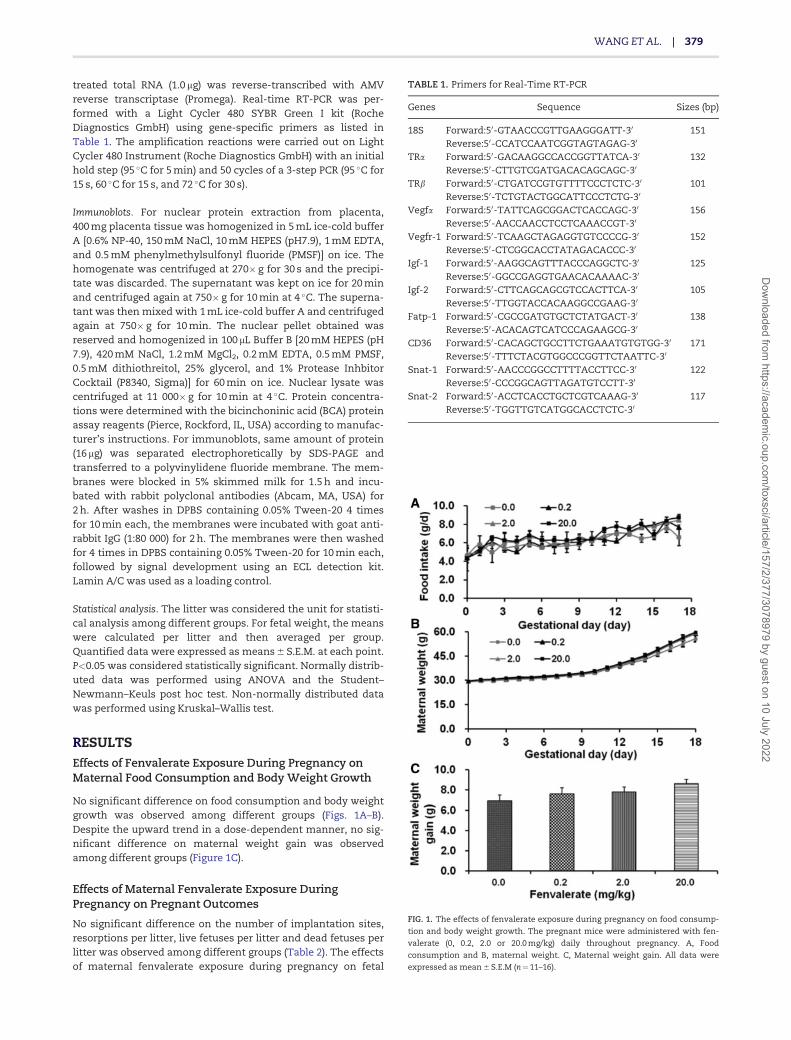
treated total RNA (1.0 lg) was reverse-transcribed with AMVreverse transcriptase (Promega). Real-time RT-PCR was per-formed with a Light Cycler 480 SYBR Green I kit (RocheDiagnostics GmbH) using gene-specific primers as listed inTable 1. The amplification reactions were carried out on LightCycler 480 Instrument (Roche Diagnostics GmbH) with an initialhold step (95 �C for 5 min) and 50 cycles of a 3-step PCR (95 �C for15 s, 60 �C for 15 s, and 72 �C for 30 s).
Immunoblots. For nuclear protein extraction from placenta,400 mg placenta tissue was homogenized in 5 mL ice-cold bufferA [0.6% NP-40, 150 mM NaCl, 10 mM HEPES (pH7.9), 1 mM EDTA,and 0.5 mM phenylmethylsulfonyl fluoride (PMSF)] on ice. Thehomogenate was centrifuged at 270� g for 30 s and the precipi-tate was discarded. The supernatant was kept on ice for 20 minand centrifuged again at 750� g for 10 min at 4 �C. The superna-tant was then mixed with 1 mL ice-cold buffer A and centrifugedagain at 750� g for 10 min. The nuclear pellet obtained wasreserved and homogenized in 100 lL Buffer B [20 mM HEPES (pH7.9), 420 mM NaCl, 1.2 mM MgCl2, 0.2 mM EDTA, 0.5 mM PMSF,0.5 mM dithiothreitol, 25% glycerol, and 1% Protease InhbitorCocktail (P8340, Sigma)] for 60 min on ice. Nuclear lysate wascentrifuged at 11 000� g for 10 min at 4 �C. Protein concentra-tions were determined with the bicinchoninic acid (BCA) proteinassay reagents (Pierce, Rockford, IL, USA) according to manufac-turer’s instructions. For immunoblots, same amount of protein(16 lg) was separated electrophoretically by SDS-PAGE andtransferred to a polyvinylidene fluoride membrane. The mem-branes were blocked in 5% skimmed milk for 1.5 h and incu-bated with rabbit polyclonal antibodies (Abcam, MA, USA) for2 h. After washes in DPBS containing 0.05% Tween-20 4 timesfor 10 min each, the membranes were incubated with goat anti-rabbit IgG (1:80 000) for 2 h. The membranes were then washedfor 4 times in DPBS containing 0.05% Tween-20 for 10 min each,followed by signal development using an ECL detection kit.Lamin A/C was used as a loading control.
Statistical analysis. The litter was considered the unit for statisti-cal analysis among different groups. For fetal weight, the meanswere calculated per litter and then averaged per group.Quantified data were expressed as means 6 S.E.M. at each point.P<0.05 was considered statistically significant. Normally distrib-uted data was performed using ANOVA and the Student–Newmann–Keuls post hoc test. Non-normally distributed datawas performed using Kruskal–Wallis test.
RESULTSEffects of Fenvalerate Exposure During Pregnancy onMaternal Food Consumption and Body Weight Growth
No significant difference on food consumption and body weightgrowth was observed among different groups (Figs. 1A–B).Despite the upward trend in a dose-dependent manner, no sig-nificant difference on maternal weight gain was observedamong different groups (Figure 1C).
Effects of Maternal Fenvalerate Exposure DuringPregnancy on Pregnant Outcomes
No significant difference on the number of implantation sites,resorptions per litter, live fetuses per litter and dead fetuses perlitter was observed among different groups (Table 2). The effectsof maternal fenvalerate exposure during pregnancy on fetal
TABLE 1. Primers for Real-Time RT-PCR
Genes Sequence Sizes (bp)
18S Forward:50-GTAACCCGTTGAAGGGATT-30 151Reverse:50-CCATCCAATCGGTAGTAGAG-30
TRa Forward:50-GACAAGGCCACCGGTTATCA-30 132Reverse:50-CTTGTCGATGACACAGCAGC-30
TRb Forward:50-CTGATCCGTGTTTTCCCTCTC-30 101Reverse:50-TCTGTACTGGCATTCCCTCTG-30
Vegfa Forward:50-TATTCAGCGGACTCACCAGC-30 156Reverse:50-AACCAACCTCCTCAAACCGT-30
Vegfr-1 Forward:50-TCAAGCTAGAGGTGTCCCCG-30 152Reverse:50-CTCGGCACCTATAGACACCC-30
Igf-1 Forward:50-AAGGCAGTTTACCCAGGCTC-30 125Reverse:50-GGCCGAGGTGAACACAAAAC-30
Igf-2 Forward:50-CTTCAGCAGCGTCCACTTCA-30 105Reverse:50-TTGGTACCACAAGGCCGAAG-30
Fatp-1 Forward:50-CGCCGATGTGCTCTATGACT-30 138Reverse:50-ACACAGTCATCCCAGAAGCG-30
CD36 Forward:50-CACAGCTGCCTTCTGAAATGTGTGG-30 171Reverse:50-TTTCTACGTGGCCCGGTTCTAATTC-30
Snat-1 Forward:50-AACCCGGCCTTTTACCTTCC-30 122Reverse:50-CCCGGCAGTTAGATGTCCTT-30
Snat-2 Forward:50-ACCTCACCTGCTCGTCAAAG-30 117Reverse:50-TGGTTGTCATGGCACCTCTC-30
FIG. 1. The effects of fenvalerate exposure during pregnancy on food consump-
tion and body weight growth. The pregnant mice were administered with fen-
valerate (0, 0.2, 2.0 or 20.0 mg/kg) daily throughout pregnancy. A, Food
consumption and B, maternal weight. C, Maternal weight gain. All data were
expressed as mean 6 S.E.M (n¼11–16).
WANG ET AL. | 379
Dow
nloaded from https://academ
ic.oup.com/toxsci/article/157/2/377/3078979 by guest on 10 July 2022
weight were analyzed. As shown in Figure 2A, body weight wassignificantly reduced in fetuses whose mothers were dailyexposed to 20.0 mg/kg fenvalerate from GD0 to GD17. Furtheranalysis showed that body weight of male fetuses was signifi-cantly reduced in dams that were daily exposed to 20.0 mg/kgfenvalerate from GD0 to GD17 (Figure 2B). Despite no statisti-cally significant difference on body weight among differentgroups, there was a downward trend on body weight of femalefetuses in fenvalerate-exposed mice as compared with controls(Figure 2C). The effects of maternal fenvalerate exposure duringpregnancy on the rate of IUGR per litter were analyzed. The
results showed that the rate of IUGR was significantly increasedin dams treated with 20.0 mg/kg of fenvalerate (Figure 2D).Further analysis showed that the rate of IUGR was significantlyincreased not only in male fetus but also in female fetus of miceexposed to 20.0 mg/kg of fenvalerate (Figs. 2E–F).
Effects of Fenvalerate Exposure During Pregnancy onThyroid Hormone Levels in Maternal and Fetal Sera
No significant difference on the level of TT4 in maternal serumwas observed (Figure 3A), despite the level of TT3 in maternal
FIG. 2. The effects of maternal fenvalerate exposure during pregnancy on fetal weight. The pregnant mice were administered with fenvalerate (0, 0.2, 2.0, or 20.0 mg/kg)
daily throughout pregnancy. All dams were sacrificed on GD18. Male and female fetuses were identified and weighted. A, Body weight of all fetuses. B, Body weight of
male fetuses. C, Body weight of female fetuses. D, Rate of IUGR in all fetuses. E, Rate of IUGR in male fetuses. F, Rate of IUGR in female fetuses. All data were expressed as
mean 6 S.E.M (n¼11–16). *P<0.05.
TABLE 2. The Effects of Fenvalerate Exposure During the Whole Pregnancy on Fetal Outcomes
Fenvalerate (mg/kg/day) F/H P
0.0 0.2 2.0 20.0
Number of pregnancy mice 11 16 15 14Implantation sites per litter 12.36 6 0.88 13.25 6 0.40 13.47 6 0.36 13.93 6 0.56 1.382 .259Live fetuses per litter 11.73 6 0.90 12.75 6 0.46 12.80 6 0.44 13.29 6 0.61 1.141 .341Resorptions per litter 0.27 6 0.20 0.31 6 0.12 0.53 6 0.24 0.14 6 0.10 2.084 .555Dead fetuses per litter 0.36 6 0.20 0.19 6 0.10 0.13 6 0.09 0.50 6 0.23 4.081 .253
The data are expressed as the means 6 SEM. The results of implantation sites per litter and live fetuses per litter were analyzed by using one-way ANOVA followed by
Student–Newman–Keuls post hoc test. Resorptions per litter, dead fetuses per litter were analyzed using Kruskal–Wallis test.
380 | TOXICOLOGICAL SCIENCES, 2017, Vol. 157, No. 2
Dow
nloaded from https://academ
ic.oup.com/toxsci/article/157/2/377/3078979 by guest on 10 July 2022
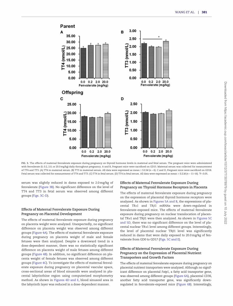
serum was slightly reduced in dams exposed to 2.0 mg/kg of
fenvalerate (Figure 3B). No significant difference on the level ofTT4 and TT3 in fetal serum was observed among differentgroups (Figs. 3C–D).
Effects of Maternal Fenvalerate Exposure DuringPregnancy on Placental Development
The effects of maternal fenvalerate exposure during pregnancyon placenta weight were analyzed. Unexpectedly, no significantdifference on placenta weight was observed among differentgroups (Figure 4A). The effects of maternal fenvalerate exposureduring pregnancy on placenta weight of male and femalefetuses were then analyzed. Despite a downward trend in adose-dependent manner, there was no statistically significantdifference on placenta weight of male fetuses among differentgroups (Figure 4B). In addition, no significant difference on pla-centa weight of female fetuses was observed among differentgroups (Figure 4C). To investigate the effects of maternal fenval-erate exposure during pregnancy on placental vascular space,cross-sectional areas of blood sinusoids were analyzed in pla-cental labyrinthine region using computerized morphometrymethod. As shown in Figures 4D and E, blood sinusoid area inthe labyrinth layer was reduced in a dose-dependent manner.
Effects of Maternal Fenvalerate Exposure DuringPregnancy on Thyroid Hormone Receptors in Placenta
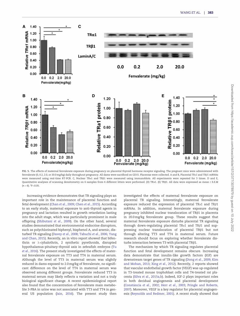
The effects of maternal fenvalerate exposure during pregnancyon the expression of placental thyroid hormone receptors wereanalyzed. As shown in Figures 5A and B, the expressions of pla-cental TRa1 and TRb1 mRNAs were down-regulated infenvalerate-exposed mice. The effects of maternal fenvalerateexposure during pregnancy on nuclear translocation of placen-tal TRa1 and TRb1 were then analyzed. As shown in Figures 5Cand 5D, there was no significant difference on the level of pla-cental nuclear TRa1 level among different groups. Interestingly,the level of placental nuclear TRb1 level was significantlyreduced in dams that were daily exposed to 20.0 mg/kg of fen-valerate from GD0 to GD17 (Figs. 5C and E).
Effects of Maternal Fenvalerate Exposure DuringPregnancy on the Expression of Placental NutrientTransporters and Growth Factors
The effects of maternal fenvalerate exposure during pregnancy onplacental nutrient transporters were analyzed. Although no signif-icant difference on placental Fatp1, a fatty acid transporter gene,was observed among different groups (Figure 6A), placental CD36,another fatty acid transporter gene, was significantly down-regulated in fenvalerate-exposed mice (Figure 6B). Interestingly,
FIG. 3. The effects of maternal fenvalerate exposure during pregnancy on thyroid hormone levels in maternal and fetal serum. The pregnant mice were administered
with fenvalerate (0, 0.2, 2.0, or 20.0 mg/kg) daily throughout pregnancy. A and B, Pregnant mice were sacrificed on GD15. Maternal serum was collected for measurement
of TT4 and TT3. (A) TT4 in maternal serum. (B) TT3 in maternal serum. All data were expressed as mean 6 S.E.M (n¼6). C and D, Pregnant mice were sacrificed on GD18.
Fetal serum was collected for measurement of TT4 and TT3. (C) TT4 in fetal serum. (D) TT4 in fetal serum. All data were expressed as mean 6 S.E.M (n¼11–16). *P<0.05.
WANG ET AL. | 381
Dow
nloaded from https://academ
ic.oup.com/toxsci/article/157/2/377/3078979 by guest on 10 July 2022

placental Snat1 and Snat2, 2 amino acid transporter genes, weresignificantly down-regulated in fenvalerate-exposed mice (Figs.6C and D). The effects of maternal fenvalerate exposure duringpregnancy on placental growth factors were then analyzed. Asshown in Figures 6E and F, placental Vegfa and its receptor Vegfr1
were significantly down-regulated in fenvalerate-exposed mice.Although no significant difference on placental Igf1 was observedamong different groups (Figure 6G), the level of placental Igf2
mRNA was significantly reduced in dams that were daily exposedto 20.0 mg/kg fenvalerate from GD0 to GD17 (Figure 6H).
DISCUSSION
In the present study, we investigated the effects of maternalfenvalerate exposure during pregnancy on fetal IUGR. Ourresults showed that fetal weight was reduced when dams wereexposed to fenvalerate throughout pregnancy. We further ana-lyzed whether maternal fenvalerate exposure induced fetalIUGR in a gender-dependent manner. We found that bodyweight of male fetuses was reduced in dams that were exposedto fenvalerate throughout pregnancy. Despite no statisticallysignificant difference on body weight among different groups,
there was a downward trend on body weight of female fetusesin fenvalerate-exposed mice as compared with controls. Furtheranalysis found that the rate of IUGR was significantly increasednot only in male fetuses but also in female fetuses of damsexposed to 20.0 mg/kg of fenvalerate. This results suggestedfenvalerate-induced IUGR is not gender specific.
The placenta is essential for sustaining the growth anddevelopment of fetuses. Placental labyrinth is the site of oxygenand nutrient exchange between the mother and the fetus and isa highly developed tissue of blood vessels. It is well known thatplacenta insufficiency is a major cause of fetal IUGR (Cetin andAlvino, 2009; Scifres and Nelson, 2009). In addition, a reductionin placenta size can directly reduce the size of the fetus, due toplacenta’s inability to transfer nutrients from the mother to thefetus (Barker and Thornburg, 2013). In the present study, wemeasured the effects of maternal fenvalerate exposure on pla-cental development. Although maternal fenvalerate exposurehad little effect on placenta size, the internal space of blood ves-sels in the labyrinth layer was smaller in placentas of damswith fenvalerate throughout pregnancy. These results suggestthat fenvalerate-induced IUGR may be partially attributed toreduction of placental transport capacity.
FIG. 4. The effects of maternal fenvalerate exposure during pregnancy on placental development. The pregnant mice were administered with fenvalerate (0, 0.2, 2.0, or
20.0 mg/kg) daily throughout pregnancy. A–C, Pregnant mice were sacrificed on GD18. Male and female fetuses were identified. Placenta weights of male and female
fetuses were measured. (A) Placenta weight of all fetuses. (B) Placenta weight of male fetuses. (C) Placenta weight of female fetuses. All data were expressed as
mean 6 S.E.M (n¼11–16). D and E Some pregnant mice were sacrificed on GD15. (D) Placentas were collected and placental cross sections were stained with H&E.
Original magnification: 400�. Scale bar: 50 lm. (E) Vascular area in the labyrinthine region was estimated from at least 8 non-consecutive sections in each placenta
using the public domain NIH Image J Program. The rate of blood sinusoid area was calculated as the ratio between the number of pixels covered by the area defined by
the threshold and the overall number of pixels in the image. Bar size: 50 lm. All data were expressed as mean 6 S.E.M (n¼6). * P<0.05.
382 | TOXICOLOGICAL SCIENCES, 2017, Vol. 157, No. 2
Dow
nloaded from https://academ
ic.oup.com/toxsci/article/157/2/377/3078979 by guest on 10 July 2022
Increasing evidence demonstrates that TR signaling plays animportant role in the maintenance of placental function andfetal development (Chan et al., 2009; Chen et al., 2015). Accordingto an early study, maternal exposure to anti-thyroid agents inpregnancy and lactation resulted in growth retardation lastinginto the adult stage, which was particularly prominent in maleoffspring (Shibutani et al., 2009). On the other hand, severalstudies demonstrated that environmental endocrine disruptors,such as polychlorinated biphenyl, bisphenol A, and arsenic, dis-turbed TR signaling (Davey et al., 2008; Tabuchi et al., 2006; Yangand Chan, 2015). Recently, an in vitro report showed that bifen-thrin or k-cyhalothrin, 2 synthetic pyrethroids, disruptedhypothalamus-pituitary-thyroid axis in zebrafish embryos (Tuet al., 2016). The present study investigated the effects of mater-nal fenvalerate exposure on TT3 and TT4 in maternal serum.Although the level of TT3 in maternal serum was slightlyreduced in dams exposed to 2.0 mg/kg of fenvalerate, no signifi-cant difference on the level of TT4 in maternal serum wasobserved among different groups. Fenvalerate reduced TT3 inmaternal serum may likely reflects a variation and not a trulybiological significant change. A recent epidemiological reportalso found that the concentration of fenvalerate main metabo-lite 3-PBA in urine was not associated with TT3 and TT4 in gen-eral US population (Jain, 2016). The present study then
investigated the effects of maternal fenvalerate exposure onplacental TR signaling. Interestingly, maternal fenvalerateexposure reduced the expression of placental TRa1 and TRb1mRNAs. In addition, maternal fenvalerate exposure duringpregnancy inhibited nuclear translocation of TRb1 in placentain 20.0 mg/kg fenvalerate group. These results suggest thatmaternal fenvalerate exposure disturbs placental TR signalingthrough down-regulating placental TRa1 and TRb1 and sup-pressing nuclear translocation of placental TRb1 but notthrough altering TT3 and TT4 in maternal serum. Futureresearch should focus on exploring whether fenvalerate dis-turbs interaction between T3 with placental TRb1.
The mechanism by which TR signaling regulates placentalfunction and fetal development remains obscure. Increasingdata demonstrate that insulin-like growth factors (IGF) aredownstream target genes of TR signaling (Dong et al., 2009; Kimand Mohan, 2013; Xing et al., 2012). Recently, 2 reports showedthat vascular endothelial growth factor (VEGF) was up-regulatedin T3-treated mouse trophoblast cells and T4-treated rat pla-centa (Silva et al., 2015a,b). Indeed, IGF-2 plays important rolesin both decidual angiogenesis and placental development(Constancia et al., 2002; Herr et al., 2003; Pringle and Roberts,2007). Moreover, VEGF is a key regulator for placental angiogen-esis (Reynolds and Redmer, 2001). A recent study showed that
FIG. 5. The effects of maternal fenvalerate exposure during pregnancy on placental thyroid hormone receptor signaling. The pregnant mice were administered with
fenvalerate (0, 0.2, 2.0, or 20.0 mg/kg) daily throughout pregnancy. All dams were sacrificed on GD15. Placentas were collected. A and B, Placental TRa1 and TRb1 mRNAs
were measured using real-time RT-PCR. C, Nuclear TRa1 and TRb1 were measured using immunoblots. All experiments were repeated for 3 times. D and E,
Quantitative analyses of scanning densitometry on 6 samples from 6 different litters were performed. (D) TRa1. (E) TRb1. All data were expressed as mean 6 S.E.M
(n¼6). *P<0.05.
WANG ET AL. | 383
Dow
nloaded from https://academ
ic.oup.com/toxsci/article/157/2/377/3078979 by guest on 10 July 2022

fetal IUGR in hypothyroidism was associated with down-regulation of placental VEGF and alteration of vascular develop-ment of placental labyrinth in rats (Silva et al., 2012). Thepresent study investigated the effects of maternal fenvalerateexposure during pregnancy on placental IGFs and VEGF. Weshowed that placental Igf2 and Vegfa mRNAs were down-regulated in fenvalerate-exposed mice. These results suggestthat maternal fenvalerate exposure impairs placental functionand fetal development, at least partially, through inhibiting TR-mediated placental IGF-2 and VEGF expression. A recent studyfound that T4-treated rats showed reduced Vegfr1 expression(Silva et al., 2015b). The present study showed that placentalVegfr1 were down-regulated in fenvalerate-treated mice. Theseresults suggest that other mechanism is involved infenvalerate-induced fetal IUGR. Additional experiment isrequired to explore the mechanism through which maternalfenvalerate exposure down-regulates placental Vegfr1 gene.
The present study showed that blood sinusoid area in thelabyrinth layer was reduced not only in high-dose group butalso in middle-dose group, whereas IUGR fetuses were observedonly in high-dose group. These results suggest that fenvalerate-induced IUGR cannot be completely attributed to reduction ofthe internal space of blood vessels in the labyrinth layer.Indeed, placenta exerts its nutrient transport function bynutrient transporters. The density of nutrient transporters oftendetermines the efficiency of nutrient transport across the pla-centa. Fatty acid transport proteins (FATP) and fatty acid trans-locase (FAT/CD36) are the key transporters for fatty acid (Brasset al., 2013; Dube et al., 2012; Duttaroy 2009). Sodium-dependentneutral amino acid transporter (SNAT) transfers neutral aminoacid from maternal circulation to the fetus (Kavitha et al., 2014).
The present study analyzed the effects of maternal fenvalerateexposure on placental nutrient transporters. Although it had lit-tle effect on placental Fatp1, maternal fenvalerate exposure dur-ing pregnancy reduced expression of placental CD36, Snat1, andSnat2 in a dose-dependent manner. These results suggest thatfenvalerate-induced fetal IUGR is partially attributed to reducedexpression of placental nutrient transporters.
The aim of the present study was to explore the role of pla-cental TR signaling on fenvalerate-induced IUGR. The presentstudy also investigated the effects of maternal fenvalerate expo-sure during pregnancy on TH level in fetal serum. Our resultsshowed that no significant difference on the level of TT4 andTT3 in fetal serum was observed among different groups.However, our present study has several deficiencies. First, thepresent study did not investigate the effects of maternal fenval-erate exposure during pregnancy on neurobehavioral develop-ment in fetuses. Second, the present study did not investigatethe effects of maternal fenvalerate exposure during pregnancyon TR target genes in fetal brain. Additional experiment isrequired to explore the effects of maternal fenvalerate exposureon abnormal brain development and TH target gene expression.
In summary, the present study investigated the effects ofmaternal fenvalerate exposure on placental function and fetaldevelopment. Our results showed that maternal fenvalerateexposure throughout pregnancy caused fetal IUGR. Moreover,maternal fenvalerate exposure disturbed placental TR signaling.We demonstrate that maternal fenvalerate exposure impairsplacental function and fetal development, at least partially,through inhibiting TR-mediated placental IGF-2 and VEGFexpression. These results provide a novel mechanistic explana-tion for fenvalerate-induced IUGR.
FIG. 6. The effects of maternal fenvalerate exposure during pregnancy on the expression of placental nutrient transporters and growth factors. The pregnant mice
were administered with fenvalerate (0, 0.2, 2.0, or 20.0 mg/kg) daily throughout pregnancy. A–D and G–H, Pregnant mice were sacrificed on GD18. Placentas were col-
lected. The expression of placental nutrient transporters and growth factors were measured using real-time RT-PCR. (A) Fatp1. (B) CD36. (C) Snat1. (D) Snat2. (G) Igf1. (H)
Igf2. All data were expressed as mean 6 S.E.M (n¼6). *P<0.05. (E–F) Pregnant mice were sacrificed on GD15. Placentas were collected. The expression of placental vegfa
and vegfr1 were measured using real-time RT-PCR. (E) Vegfa. (F) Vegfr1. All data were expressed as mean 6 S.E.M (n¼6). *P<0.05.
384 | TOXICOLOGICAL SCIENCES, 2017, Vol. 157, No. 2
Dow
nloaded from https://academ
ic.oup.com/toxsci/article/157/2/377/3078979 by guest on 10 July 2022

ACKNOWLEDGMENTS
The authors gratefully acknowledge the help of Cheng-WeiYang, Inspection Physician from Second People ’s Hospitalof Anhui Province, for measurement of TT4 and TT3.
FUNDING
This work was supported by National Natural ScienceFoundation of China (81102155) and Major projects ofNatural Scientific Research in Universities and Colleges,Anhui, China (KJ2016SD27).
REFERENCESAggarawal, N., Suri, V., Singla, R., Chopra, S., Sikka, P., Shah, V.
N., and Bhansali, A. (2014). Pregnancy outcome in hyperthy-roidism: A case control study. Gynecol. Obstet. Invest. 77,94–99.
Barker, D. J., and Thornburg, K. (2013). Placental programming ofchronic diseases, cancer and lifespan: a review. Placenta 34,841–845.
Bedi, J. S., Gill, J. P., Aulakh, R. S., and Kaur, P. (2015). PesticideResidues in Bovine Milk in Punjab, India: Spatial Variationand Risk Assessment to Human Health. Arch. Environ.Contam. Toxicol. 69, 230–240.
Bian, Q., Xu, L. C., Wang, S. L., Xia, Y. K., Tan, L. F., Chen, J. F.,Song, L., Chang, H. C., and Wang, X. R. (2004). Study on the re-lation between occupational fenvalerate exposure and sper-matozoa DNA damage of pesticide factory workers. Occup.Environ. Med. 61, 999–1005.
Brass, E., Hanson, E., and O’Tierney-Ginn, P. F. (2013). Placentaloleic acid uptake is lower in male offspring of obese women.Placenta 34, 503–509.
Cetin, I., and Alvino, G. (2009). Intrauterine growth restriction:Implications for placental metabolism and transport. A re-view. Placenta 30, S77–S82.
Chan, S. Y., Vasilopoulou, E., and Kilby, M. D. (2009). The role ofthe placenta in thyroid hormone delivery to the fetus. NatClin. Pract. Endocrinol. Metab. 5, 45–54.
Chen, C. Y., Chen, C. P., and Lin, K. H. (2015). Biological functionsof thyroid hormone in placenta. Int. J. Mol. Sci. 16, 4161–4179.
Chen, L. M., Du, W. J., Dai, J., Zhang, Q., Si, G. X., Yang, H., Ye, E. L.,Chen, Q. S., Yu, L. C., Zhang, C., et al. (2014). Effects of subclin-ical hypothyroidism on maternal and perinatal outcomesduring pregnancy: A single-center cohort study of a Chinesepopulation. PLoS One 9, e109364.
Chen, Y. H., Hu, X. G., Zhou, Y., Yu, Z., Fu, L., Zhang, G. B., Bo, Q.L., Wang, H., Zhang, C., and Xu, D. X. (2016). Obeticholic acidorotects against lipopolysaccharide-induced fetal death andintrauterine growth restriction through its anti-inflammatory activity. J. Immunol. 197, 4762–4770.
Constancia, M., Hemberger, M., Hughes, J., Dean, W., Ferguson-Smith, A., Fundele, R., Stewart, F., Kelsey, G., Fowden, A.,Sibley, C., et al. (2002). Placental-specific IGF-II is a majormodulator of placental and fetal growth. Nature 417, 945–948.
Corcellas, C., Feo, M. L., Torres, J. P., Malm, O., Ocampo-Duque,W., Eljarrat, E., and Barcelo, D. (2012). Pyrethroids in humanbreast milk: occurrence and nursing daily intake estimation.Environ. Int. 47, 17–22.
Cotechini, T., Komisarenko, M., Sperou, A., Macdonald-Goodfellow, S., Adams, M. A., and Graham, C. H. (2014).Inflammation in rat pregnancy inhibits spiral artery
remodeling leading to fetal growth restriction and featuresof preeclampsia. J. Exp. Med. 211, 165–179.
Davey, J. C., Nomikos, A. P., Wungjiranirun, M., Sherman, J. R.,Ingram, L., Batki, C., Lariviere, J. P., and Hamilton, J. W. (2008).Arsenic as an endocrine disruptor: Arsenic disrupts retinoicacid receptor-and thyroid hormone receptor-mediated generegulation and thyroid hormone-mediated amphibian tailmetamorphosis. Environ. Health. Perspect. 116, 165–172.
Dong, H., Yauk, C. L., Rowan-Carroll, A., You, S. H., Zoeller, R. T.,Lambert, I., and Wade, M. G. (2009). Identification of thyroidhormone receptor binding sites and target genes using ChIP-on-chip in developing mouse cerebellum. PLoS One 4, e4610.
Dube, E., Gravel, A., Martin, C., Desparois, G., Moussa, I., Ethier-Chiasson, M., Forest, J. C., Giguere, Y., Masse, A., and Lafond,J. (2012). Modulation of fatty acid transport and metabolismby maternal obesity in the human full-term placenta. Biol.Reprod. 87, 1–11.
Duttaroy, A. K. (2009). Transport of fatty acids across the humanplacenta: A review. Prog. Lipid. Res. 48, 52–61.
Fei, J., Qu, J. H., Ding, X. L., Xue, K., Lu, C. C., Chen, J. F., Song, L.,Xia, Y. K., Wang, S. L., and Wang, X. R. (2010). Fenvalerate in-hibits the growth of primary cultured rat preantral ovarianfollicles. Toxicology 267, 1–6.
Forhead, A. J., and Fowden, A. L. (2014). Thyroid hormones infetal growth and prepartum maturation. J. Endocrinol. 221,R87–R103.
Herr, F., Liang, O. D., Herrero, J., Lang, U., Preissner, K. T., Han, V.K., and Zygmunt, M. (2003). Possible angiogenic roles ofinsulin-like growth factor II and its receptors in uterine vas-cular adaptation to pregnancy. J. Clin. Endocrinol. Metab. 88,4811–4817.
Iwaku, K., Noh, J. Y., Minagawa, A., Kosuga, Y., Suzuki, M., Sekiya,K., Matsumoto, M., Ohye, H., Kunil, Y., Yoshihara, A., et al.(2013). Determination of pediatric reference levels of FT3, FT4and TSH measured with ECLusys kits. Endocr. J. 60, 799–804.
Jain, R. B. (2016). Variability in the levels of 3-phenoxybenzoicacid by age, gender, and race/ethnicity for the period of 2001-2002 versus 2009-2010 and its association with thyroid func-tion among general US population. Environ. Sci. Pollut. Res. Int.23, 6934–6939.
Kavitha, J. V., Rosario, F. J., Nijland, M. J., McDonald, T. J., Wu, G.,Kanai, Y., Powell, T. L., Nathanielsz, P. W., and Jansson, T.(2014). Down-regulation of placental mTOR, insulin/IGF-I sig-naling, and nutrient transporters in response to maternalnutrient restriction in the baboon. FASEB J. 28, 1294–1305.
Kilby, M. D., Verhaeg, J., Gittoes, N., Somerset, D. A., Clark, P. M.,and Franklyn, J. A. (1998). Circulating thyroid hormone con-centrations and placental thyroid hormone receptor expres-sion in normal human pregnancy and pregnancycomplicated by intrauterine growth restriction (IUGR). J. Clin.Endocrinol. Metab. 83, 2964–2971.
Kim, H. Y., and Mohan, S. (2013). Role and mechanisms of actionsof thyroid hormone on the skeletal development. Bone Res. 1,146–161.
Leonard, A. J., Evans, I. M., Pickard, M. R., Bandopadhyay, R.,Sinha, A. K., and Ekins, R. P. (2001). Thyroid hormone recep-tor expression in rat placenta. Placenta 22, 353–359.
Li, Z., Nie, J., Lu, Z., Xie, H., Kang, L., Chen, Q., Li, A., Zhao, X., Xu,G., and Yan, Z. (2016). Cumulative risk assessment of the ex-posure to pyrethroids through fruits consumption inChina—Based on a 3-year investigation. Food. Chem. Toxicol.96, 234–243.
Liu, P., Meng, X. H., Wang, H., Ji, Y. L., Zhao, M., Zhao, X. F., Xu, Z.M., Chen, Y. H., Zhang, C., and Xu, D. X. (2011). Effects of
WANG ET AL. | 385
Dow
nloaded from https://academ
ic.oup.com/toxsci/article/157/2/377/3078979 by guest on 10 July 2022
pubertal fenvalerate exposure on testosterone and estradiolsynthesis and the expression of androgen and estrogen re-ceptors in the developing brain. Toxicol. Lett. 201, 181–189.
Meng, X. H., Liu, P., Wang, H., Zhao, X. F., Xu, Z. M., Chen, G. H.,and Xu, D. X. (2011). Gender-specific impairments on cogni-tive and behavioral development in mice exposed to fenval-erate during puberty. Toxicol. Lett. 203, 245–251.
Moniz, A. C., Cruz-Casallas, P. E., Oliveira, C. A., Lucisano, A., Florio,J. C., Nicolau, A. A., Spinosa, H. S., and Bernardi, M. M. (1999a).Perinatal fenvalerate exposure: Behavioral and endocrinologychanges in male rats. Neurotoxicol. Teratol. 21, 611–618.
Moniz, A. C., Cruz-Casallas, P. E., Salzgeber, S. A., Varoli, F. M.,Spinosa, H. S., and Bernardi, M. M. (2005b). Behavioral andendocrine changes induced by perinatal fenvalerate expos-ure in female rats. Neurotoxicol. Teratol. 27, 609–614.
Neres, R., Marinho, C. R., Goncalyes, L. A., Catarino, M. B., andPenha-Goncalves, C. (2008). Pregnancy outcome and pla-centa pathology in Plasmodium berghei ANKA infected micereproduce the pathogenesis of severe malaria in pregnantwomen. PLoS One 3, e1608.
Onigata, K., and Szinnai, G. (2014). Resistance to thyroid hor-mone. Endocr. Dev. 26, 118–129.
Ortiga-Carvalho, T. M., Sidhaye, A. R., and Wondisford, F. E.(2014). Thyroid hormone receptors and resistance to thyroidhormone disorders. Nat. Rev. Endocrinol. 10, 582–591.
Pearce, E. N., Lazarus, J. H., Moreno-Reyes, R., and Zimmermann,M. B. (2016). Consequences of iodine deficiency and excess inpregnant women: An overview of current knowns and un-knowns. Am. J. Clin. Nutr. 104, 918S–923S.
Pringle, K. G., and Roberts, C. T. (2007). New light on early post-implantation pregnancy in the mouse: Roles for insulin-likegrowth factor-II (IGF-II)? Placenta 28, 286–297.
Qi, X., Zheng, M., Wu, C., Wang, G., Feng, C., and Zhou, Z. (2012).Urinary pyrethroid metabolites among pregnant women inan agricultural area of the Province of Jiangsu, China. Int. J.Hyg. Environ. Health 215, 487–495.
Qu, J. H., Hong, X., Chen, J. F., Wang, Y. B., Sun, H., Xu, X. L., Song,L., Wang, S. L., and Wang, X. R. (2008). Fenvalerate inhibitsprogesterone production through cAMP-dependent signalpathway. Toxicol. Lett. 176, 31–39.
Reynolds, L. P., and Redmer, D. A. (2001). Angiogenesis in the pla-centa. Biol. Reprod. 64, 1033–1040.
Scifres, C. M., and Nelson, D. M. (2009). Intrauterine growth re-striction, human placental development and trophoblast celldeath. J. Physiol. 587, 3453–3458.
Shibutani, M., Woo, G. H., Fujimoto, H., Saegusa, Y., Takahashi,M., Inoue, K., Hirose, M., and Nishikawa, A. (2009).Assessment of developmental effects of hypothyroidism inrats from in utero and lactation exposure to anti-thyroidagents. Reprod. Toxicol. 28, 297–307.
Silva, J. F., Ocarino, N. M., and Serakides, R. (2015a). In vitro ef-fects of triiodothyronine on gene expression in mousetrophoblast cells. Placenta 36, 97–99.
Silva, J. F., Ocarino, N. M., and Serakides, R. (2015b). Placentalangiogenic and hormonal factors are affected by thyroid hor-mones in rats. Pathol. Res. Pract. 211, 226–234.
Silva, J. F., Vidigal, P. N., Galv~ao, D. D., Boeloni, J. N., Nunes, P. P.,Ocarino, N. M., Nascimento, E. F., and Serakides, R. (2012).Fetal growth restriction in hypothyroidism is associated withchanges in proliferative activity, apoptosis and vascularisa-tion of the placenta. Reprod. Fertil. Dev. 24, 923–931.
Tabuchi, M., Veldhoen, N., Dangerfield, N., Jeffries, S., Helbing, C.C., and Ross, P. S. (2006). PCB-related alteration of thyroidhormones and thyroid hormone receptor gene expression infree-ranging harbor seals (Phoca vitulina). Environ. HealthPerspect. 114, 1024–1031.
Tu, W., Xu, C., Lu, B., Lin, C., Wu, Y., and Liu, W. (2016). Acute ex-posure to synthetic pyrethroids causes bioconcentration anddisruption of the hypothalamus-pituitary-thyroid axis inzebrafish embryos. Sci. Total. Environ. 542, 876–885.
Xing, W., Govoni, K. E., Donahue, L. R., Kesavan, C., Wergedal, J.,Long, C., Bassett, J. H., Gogakos, A., Wojcicka, A., Williams, G. R.,et al. (2012). Genetic evidence that thyroid hormone is indispens-able for prepubertal insulin-like growth factor-I expression andbone acquisition in mice. J. Bone Miner. Res. 27, 1067–1079.
Yang, J., and Chan, K. M. (2015). Evaluation of the toxic effects ofbrominated compounds (BDE-47, 99, 209, TBBPA) and bisphe-nol A (BPA) using a zebrafish liver cell line, ZFL. Aquat. Toxicol.159, 138–147.
Zhang, H., Wang, H., Ji, Y. L., Ning, H., Yu, T., Zhang, C., Zhang, Y.,Zhao, X. F., Wang, Q., Liu, P., et al. (2009). Lactational fenvaler-ate exposure permanently impairs testicular developmentand spermatogenesis in mice. Toxicol. Lett. 191, 47–56.
Zhang, H., Wang, H., Ji, Y. L., Zhang, Y., Yu, T., Niang, H., Zhang,C., Zhao, X. F., Wang, Q., Liu, P., et al. (2010). Food Chem.Toxicol. 48, 1160–1169.
Zhao, X. F., Wang, Q., Ji, Y. L., Wang, H., Liu, P., Zhang, C., Zhang,Y., and Xu, D. X. (2011). Fenvalerate induces germ cell apop-tosis in mouse testes through the Fas/FasL signaling path-way. Arch. Toxicol. 85, 1101–1108.
386 | TOXICOLOGICAL SCIENCES, 2017, Vol. 157, No. 2
Dow
nloaded from https://academ
ic.oup.com/toxsci/article/157/2/377/3078979 by guest on 10 July 2022
Related Documents


