Journal Pre-proof Development and validation of immunoassays for monitoring of guselkumab and anti-guselkumab antibodies in patients with moderate-to-severe psoriasis Nathalie Van den Berghe (Conceptualization) (Investigation) (Formal analysis) (Writing - original draft) (Writing - review and editing) (Visualization) (Project administration), Aur´ elie Truffot (Conceptualization) (Investigation) (Formal analysis) (Writing - original draft) (Writing - review and editing), Miet Peeters (Investigation) (Formal analysis), Griet Compernolle (Investigation) (Formal analysis), Els Brouwers (Investigation) (Formal analysis), Rani Soenen (Resources) (Writing - review and editing), Lynda Grine (Resources) (Writing - review and editing), Ann Gils (Conceptualization) (Supervision) (Writing - review and editing), Maya Imbrechts (Conceptualization) (Supervision) (Writing - review and editing) (Project administration) (Validation) PII: S0731-7085(20)31319-4 DOI: https://doi.org/10.1016/j.jpba.2020.113433 Reference: PBA 113433 To appear in: Journal of Pharmaceutical and Biomedical Analysis Received Date: 23 March 2020 Revised Date: 10 June 2020 Accepted Date: 14 June 2020 Please cite this article as: Van den Berghe N, Truffot A, Peeters M, Compernolle G, Brouwers E, Soenen R, Grine L, Gils A, Imbrechts M, Development and validation of immunoassays for monitoring of guselkumab and anti-guselkumab antibodies in patients with moderate-to-severe psoriasis, Journal of Pharmaceutical and Biomedical Analysis (2020), doi: https://doi.org/10.1016/j.jpba.2020.113433

Welcome message from author
This document is posted to help you gain knowledge. Please leave a comment to let me know what you think about it! Share it to your friends and learn new things together.
Transcript

Journal Pre-proof
Development and validation of immunoassays for monitoring ofguselkumab and anti-guselkumab antibodies in patients withmoderate-to-severe psoriasis
Nathalie Van den Berghe (Conceptualization) (Investigation)(Formal analysis) (Writing - original draft) (Writing - review andediting) (Visualization) (Project administration), Aurelie Truffot(Conceptualization) (Investigation) (Formal analysis) (Writing -original draft) (Writing - review and editing), Miet Peeters(Investigation) (Formal analysis), Griet Compernolle (Investigation)(Formal analysis), Els Brouwers (Investigation) (Formal analysis),Rani Soenen (Resources) (Writing - review and editing), LyndaGrine (Resources) (Writing - review and editing), Ann Gils(Conceptualization) (Supervision) (Writing - review and editing),Maya Imbrechts (Conceptualization) (Supervision) (Writing - reviewand editing) (Project administration) (Validation)
PII: S0731-7085(20)31319-4
DOI: https://doi.org/10.1016/j.jpba.2020.113433
Reference: PBA 113433
To appear in: Journal of Pharmaceutical and Biomedical Analysis
Received Date: 23 March 2020
Revised Date: 10 June 2020
Accepted Date: 14 June 2020
Please cite this article as: Van den Berghe N, Truffot A, Peeters M, Compernolle G, BrouwersE, Soenen R, Grine L, Gils A, Imbrechts M, Development and validation of immunoassays formonitoring of guselkumab and anti-guselkumab antibodies in patients withmoderate-to-severe psoriasis, Journal of Pharmaceutical and Biomedical Analysis (2020),doi: https://doi.org/10.1016/j.jpba.2020.113433

This is a PDF file of an article that has undergone enhancements after acceptance, such asthe addition of a cover page and metadata, and formatting for readability, but it is not yet thedefinitive version of record. This version will undergo additional copyediting, typesetting andreview before it is published in its final form, but we are providing this version to give earlyvisibility of the article. Please note that, during the production process, errors may bediscovered which could affect the content, and all legal disclaimers that apply to the journalpertain.
© 2020 Published by Elsevier.
1
Development and validation of immunoassays for monitoring
of guselkumab and anti-guselkumab antibodies in patients with
moderate-to-severe psoriasis
Nathalie Van den Berghea,1, Aurélie Truffota,1, Miet Peetersa, Griet Compernollea, Els
Brouwersa, Rani Soenenb, Lynda Grineb, Ann Gilsa, Maya Imbrechtsa
a Laboratory for Therapeutic and Diagnostic Antibodies, Department of Pharmaceutical and
Pharmacological Sciences, KU Leuven, Leuven, Belgium b Department of Dermatology, Ghent University Hospital, Ghent, Belgium 1 Joint first authors
Address correspondence to:
Dr. Maya Imbrechts
Department of Pharmaceutical and Pharmacological Sciences, KU Leuven
Campus Gasthuisberg O&N2, PB 820; Herestraat 49, B-3000 Leuven, Belgium
Phone: +32 16 32 23 64
Email: [email protected]
Authors (first name/ last name) e-mail address:
Nathalie/ Van den Berghe: [email protected]
Aurélie/ Truffot: [email protected]
Miet/ Peeters : [email protected]
Griet/ Compernolle: [email protected]
Els/ Brouwers: [email protected]
Rani/ Soenen: [email protected]
Lynda/ Grine : [email protected]
Ann/ Gils: [email protected]
Maya/ Imbrechts: [email protected]
Highlights - 20 specific anti-guselkumab monoclonal antibodies were generated and characterized
- An accurate and precise ELISA to quantify guselkumab was developed
- The guselkumab ELISA was validated in moderate-to-severe psoriasis patients
- Two different ELISAs to detect anti-guselkumab antibodies were developed
Jour
nal P
re-p
roof

2
Abstract
Therapeutic drug monitoring, which is the measurement of drug concentrations in the
blood, is a useful tool to guide clinical decision-making and treatment adjustments, on the
condition that drug concentrations are correlated with treatment response. For guselkumab,
an anti-IL-23 monoclonal antibody for the treatment of moderate-to-severe psoriasis, such a
concentration-response relationship could not yet be determined as no commercial assays
for the quantification of this drug or antibodies against this drug are available. Therefore, the
aim of this study was to develop and validate immunoassays for the quantification of
guselkumab and anti-guselkumab antibodies according to the guidelines of the European
Medicines Agency (EMA). A diverse panel of 20 highly specific anti-guselkumab monoclonal
antibodies (MA-GUS) was generated of which eight revealed a neutralizing capacity of
≥65%. At least seven different antibody clusters were identified based on their epitope
binning profile. Using MA-GUS9F6 as the capture antibody and MA-GUS12G12 as the
detection antibody, an ELISA was developed with a dose-response curve ranging from 0.08
to 5 ng/mL. The assay was specific, selectivity and could accurately and precisely quantify
guselkumab concentrations in spiked healthy control serum and serum from guselkumab-
treated psoriasis patients with a cut-off for quantification of 0.014 µg/mL. The presence of IL-
23 in physiological concentrations or of non-neutralizing antibodies did not impact the
quantification of guselkumab, while the presence of neutralizing antibodies did. Using MA-
GUS12A9 as a calibrator, two anti-guselkumab antibody assays were developed to detect
anti-guselkumab antibodies, which differ in the threshold for detection and quantification and
the tolerance to the presence of guselkumab. Together, these validated immunoassays are
essential to establish a concentration-response relationship and will allow the future
implementation of therapeutic drug monitoring in moderate-to-severe psoriasis patients
receiving guselkumab treatment.
Keywords:
Guselkumab Psoriasis Enzyme-linked immunosorbent assay Immunogenicity Therapeutic
drug monitoring Monoclonal antibodies
1. Introduction
Guselkumab (Tremfya®) is a fully human IgG1λ monoclonal antibody that targets the
p19 subunit of interleukin (IL)-23, thereby preventing the activation of T helper 17 cells and
inhibiting the production of the pro-inflammatory cytokines IL-17 and IL-22 [1]. The pivotal
phase III trials (VOYAGE 1 and VOYAGE 2) have demonstrated the efficacy of this biological
in psoriasis patients resulting in the approval of guselkumab for the treatment of adult
patients with moderate-to-severe plaque psoriasis [2-4].
Despite its proven therapeutic efficacy, the treatment response to guselkumab can
vary considerably between patients. Some patients respond favorably to therapy while others
do not show any improvement or show an initial response but lose this response over time.
In certain patients, this non-response could be explained by a drug concentration that is too
low, or by the presence of anti-drug antibodies [5].
Jour
nal P
re-p
roof
3
Preventing loss of response to biologicals is a challenge for clinicians treating patients
with psoriasis but strategies to aid in clinical decision-making are emerging. An important
strategy is therapeutic drug monitoring (TDM), the measurement of drug concentrations
which can serve as a tool to optimize biological treatment by identifying under- or
overexposed patients and evaluating potential reasons for treatment failure [6]. Two
fundamental requirements to perform TDM are the availability of an assay to quantify drug
concentrations and the presence of a concentration-response relationship [7].
Several research groups have demonstrated the relationship between the serum
concentration of adalimumab, an anti-tumor necrosis factor antibody, and clinical response in
psoriasis patients [8, 9]. More recently, a similar relationship was observed for the anti-IL-
12/23 antibody ustekinumab and the anti-IL17A antibody secukinumab [10-12]. For
guselkumab, no information on the presence of a concentration-response relationship or the
value of TDM could be found in literature as there is currently no commercial assay available
for the quantification of this drug.
Therefore, the aim of this study was to develop and validate immunoassays for the
quantification of guselkumab and anti-guselkumab antibody concentrations in serum of
moderate-to-severe psoriasis patients, using in-house generated monoclonal antibodies. The
assay validation was performed according to the guidelines of the European Medicines
Agency (EMA) for bioanalytical method validation of ligand-binding assays [13].
2. Material and methods
2.1 Materials Bovine serum albumin (BSA), Tween-80, citric acid, sulfuric acid (H2SO4), Ethylene
Diamine Tetraacetic Acid (EDTA), TRIS base and hydrogen peroxide (H2O2) were purchased
from Sigma-Aldrich. Sodium chloride, sodium carbonate and disodium hydrogen phosphate
dihydrate (Na2HPO4 - 2H2O) were purchased from VWR International. Potassium dihydrogen
phosphate (KH2PO4) and Sulfo-NHS-LC-Biotin were purchased from Thermo Fisher
Scientific Inc. Potassium chloride (KCl) and o-phenylenediamine (OPD) were purchased from
Acros Organics and streptavidin poly-HRP from Sanquin Reagents. Acetic acid was
purchased from Chem-Lab NV. Polystyrene 96-well EIA/ RIA plates (flat bottom) were
purchased from Corning and the CELLine bioreactor flasks from Merck.
Guselkumab (Tremfya®) was purchased from Johnson and Johnson, secukinumab
(Cosentyx®) from Novartis, whereas the human IgG-mixture (Multigam®) was purchased from
C.A.F-D.C.F. IL-23 was obtained from PeproTech. Human serum from healthy donors further
referred to as “healthy control serum”, was purchased from Valley Biomedical.
The following buffers were used: PBS (140 mM NaCl, 8 mM Na2HPO4-2H2O, 2.7 mM
KCl and 1.5 mM KH2PO4), PTAE (PBS with 0.1% BSA; 0.002% Tween-80; 5 mM EDTA),
citrate buffer (0.1 M sodium citrate, 0.2 M disodium phosphate), block buffer (1% BSA in
PBS) and wash buffer (PBS with 0.002% Tween-80).
Jour
nal P
re-p
roof

4
2.2 Patients Serum samples of ten moderate-to-severe psoriasis patients treated with guselkumab
were collected by the Department of Dermatology of Ghent University Hospital (Ghent,
Belgium) in the framework of the BIOLOPTIM-GUS study (ClinicalTrials.gov Identifier:
NCT04080648). The local ethics committee approved the study (B670201836638) and all
patients provided written informed consent. Patients received the standard treatment
regimen of 100 mg subcutaneously at weeks 0 and 4 followed by 100 mg every 8 weeks.
Blood was collected in serum-separating tubes and serum was obtained by centrifugation of
the tubes for 15 min at 1700 g at room temperature. The serum samples were divided into
aliquots and stored at -20°C until analysis. Serum samples were prospectively collected
before the start of treatment and at weeks 1, 2, 3, 4, 12 and 20. The Psoriasis Area and
Severity Index (PASI) was used to assess disease activity which takes into account the
severity and the body area that is affected.
2.3 Generation and characterization of a unique panel of monoclonal
antibodies against GUS (MA-GUS)
2.3.1 Monoclonal antibody generation and purification
Monoclonal antibodies against guselkumab (MA-GUS) were generated in SJL/J mice
according to the hybridoma technology of Galfré and Milstein with approval from the ethical
committee (172/2017 KU Leuven, Leuven, Belgium) [14]. MA-GUS were purified on a
ProSep-vA Ultra column (Merk Millipore) as described by Van Stappen et al [15].
2.3.2 Selection of 20 hybridomas to create a diverse panel of MA-GUS
2.3.2.1 Evaluation of the cross-reactivity of MA-GUS
Using a sandwich-type enzyme-linked immunosorbent assay (ELISA), the cross-
reactivity towards secukinumab (an anti-IL17A antibody) and Multigam® (a human
immunoglobulin G mixture) was determined. Briefly, guselkumab, secukinumab, and
Multigam® were coated overnight at 4°C in a 96-well plate at a concentration of 4 µg/mL in
PBS. After blocking for 2 h at room temperature (RT), hybridoma medium containing MA-
GUS was applied in a 1:10 and 1:100 dilution and incubated overnight at 4°C. An HRP-
conjugated rabbit anti-mouse antibody (RAM/mIg/PO; Nordic-MUbio, 1:10000 diluted) was
used to detect bound MA-GUS. Subsequently, the color reaction was initiated using OPD
(0.4 g/L) and H2O2 (0.003%) in citrate buffer. The reaction was stopped with H2SO4 (4 M) and
the absorbance was measured at 492 nm. MA-GUS revealing a cross-reactivity less than
20% towards secukinumab and Multigam® were considered as specific antibodies and
selected for further characterization.
2.3.2.2 Evaluation of the bridging capacity of MA-GUS
The bridging capacity of each MA-GUS was evaluated by incubating the respective
hybridoma medium (1:40 diluted) for 2 h at 21°C on a guselkumab-coated plate. Guselkumab
was labeled with Sulfo-NHS-LC-Biotin according to the manufacturer’s instructions and
applied on the plate in a 1:400 dilution for 1 h at 21°C to detect bound guselkumab.
Thereafter, HRP-streptavidin was added for 30 min at 21°C and shaken at 300 rpm. The
bridging capacity of each MA-GUS was based on the respective optical density (OD) relative
to the OD of the MA-GUS with the highest OD. MA-GUS of which the OD was ≤5%, 6-20%,
21-50% and >50% of the MA-GUS with the highest OD was categorized as “no bridging
Jour
nal P
re-p
roof
5
capacity”, “low bridging capacity”, “intermediate bridging capacity” and “high bridging
capacity”, respectively.
2.3.3 Evaluation of the neutralizing capacity of MA-GUS
Using an IL-23-based competition assay, the neutralizing capacity of the generated
MA-GUS was determined. IL-23 was coated at 0.5 µg/mL on a 96-well plate and incubated
overnight at 4°C. Guselkumab (4 µg/mL) was pre-incubated with MA-GUS (8 µg/mL, 1:2
ratio) for 15 min at 37°C. After blocking for 2 h at RT and washing the plate, the GUS:MA-
GUS mix was applied on the plate followed by a 2 h incubation period at 37°C. Residual
binding of guselkumab to IL-23 was measured by the addition of HRP-conjugated polyclonal
goat anti-human IgG Fc (GAHu/IgG(Fc)/PO; Nordic-MUbio, 1:4000 diluted) and incubation
for 2 h at RT. MA-GUS were considered to be neutralizing when the binding of guselkumab
was reduced with at least 65%.
2.3.4 Epitope binning of MA-GUS
A competitive ELISA was carried out to establish the binning profile of each MA-GUS.
Specifically, 4 µg/mL of guselkumab was coated on a 96 well-plate for 72 h at 4°C. After
blocking for 2 h at RT, 10 µg/mL MA-GUS (“cold-antibody”) was applied on the plate and
incubated overnight at 4°C. Then, plates were washed and a biotin-labeled conjugate of each
MA-GUS was applied at a final concentration of at least 25 ng/mL followed by incubation for
2 h at RT. After washing, binding of biotin-labeled MA-GUS was determined using
streptavidin poly-HRP. The OD obtained for each biotin-labeled MA-GUS in the absence and
in the presence of the “cold antibody” was compared. MA-GUS of which the 𝑂𝐷+𝑐𝑜𝑙𝑑 𝑎𝑛𝑡𝑖𝑏𝑜𝑑𝑦
𝑂𝐷−𝑐𝑜𝑙𝑑 𝑎𝑛𝑡𝑖𝑏𝑜𝑑𝑦
gave a percentage lower than 20%, were considered as competitive with the cold antibody.
Principal component analysis (PCA) and clustering were performed using the online web tool
ClustVis (https://biit.cs.ut.ee/clustvis/) to group the 20 MA-GUS according to similar binding
sites towards guselkumab.
2.4 Development and validation of the guselkumab ELISA
2.4.1 Development of the guselkumab ELISA
MA-GUS was coated at 4 µg/mL overnight at 4°C followed by blocking with PBS
containing 1% BSA. Samples were diluted 1:100 to 1:800 in PTAE buffer for concentrations
<0.5 µg/mL, 1:400 to 1:3200 for concentrations between 0.5 µg/mL and 5 µg/mL and 1:1600
to 1:12800 for concentrations >5 µg/mL. Thereafter, the samples were applied on the plate
and shaken at 300 rpm for 2 h at 21°C. Biotinylated MA-GUS was added and incubated for 1
h at 21°C to detect bound guselkumab. After washing, HRP-streptavidin was applied and
incubated for 30 min at 21°C. Subsequently, the plate was developed using OPD (0.4 g/L)
and H2O2 (0.003%) in citrate buffer and the reaction was stopped with H2SO4 (4 M). The
absorbance was measured at 492 nm and the guselkumab dose-response curve was
analyzed by non-linear regression using Graphpad Prism 8.2.1 (Graph Software, San Diego,
CA, USA).
2.4.2 Specificity
The specificity of the guselkumab assay was determined by spiking secukinumab (10;
20 and 30 µg/mL) and guselkumab (0.10 and 5.00 µg/mL) to guselkumab-naive serum
samples of psoriasis patients. Thereafter, the guselkumab concentration was determined.
The assay is defined as specific when the recovery of guselkumab is 75-125% [13].
Jour
nal P
re-p
roof

6
Additionally, five serum samples of secukinumab-treated psoriasis patients were measured
with the guselkumab assay.
2.4.3 Selectivity
2.4.3.1 Impact of the matrix on guselkumab determination
The impact of unrelated compounds in the matrix, i.e. serum of psoriasis patients, on
the determination of guselkumab was evaluated by spiking guselkumab (0.10 and 5.00
µg/mL) to 10 guselkumab-naive psoriasis serum samples. Thereafter, the guselkumab
concentration was determined. The assay is defined as selective when the recovery of
guselkumab is 80-120% in at least 80% of the evaluated samples [13].
2.4.3.2 Impact of IL-23 on guselkumab determination
To evaluate the potential interference of IL-23 on the determination of guselkumab,
varying concentrations of IL-23 (0.1-2.5 µg/mL, molecular weight = 53.5 kDa) were spiked to
1 µg/mL guselkumab (molecular weight = 146.61 kDa) in PTAE buffer. The IL-23-GUS
mixture was pre-incubated for 15 min at 37°C on a shaker (300 rpm) and applied in a 1:400
dilution on a MA-GUS coated plate for the detection of guselkumab. Subsequently, the
residual binding of guselkumab was determined.
2.4.3.3 Impact of anti-guselkumab antibodies on guselkumab determination
The impact of anti-guselkumab antibodies on the determination of guselkumab was
assessed by the addition of a two-fold excess (2 µg/mL) of MA-GUS to guselkumab (1
µg/mL) in PTAE buffer. For this experiment, 5 MA-GUS with varying neutralizing capacities
were selected. After incubation for 15 min at 37°C and 300 rpm, the mixture was diluted
1:400 and applied on the plate. Subsequently, the residual binding of guselkumab was
determined.
2.4.4 Determination of the assay cut-off values for detection and quantification,
accuracy, imprecision and dilutional linearity
The assay lower limit of detection (LLOD), the lower limit of quantification (LLOQ),
and cut-off values for detection and quantification were determined using a panel of 15
guselkumab-naive serum samples of psoriasis patients. The LLOD and LLOQ were
calculated based on the mean OD of 15 guselkumab-naive serum samples plus 3 times and
10 times the standard deviation, respectively. The cut-off values for detection and
quantification were set based on the calculated LLOD and LLOQ multiplied by the assay
dilution factor.
The intra- and inter-assay accuracy and precision were determined by preparing 5
quality control samples (0.10; 0.25; 0.50; 2.00 and 5.00 µg/mL guselkumab) in healthy
control serum and measuring four sets within one run for the intra-assay evaluation and six
times one set, each on a different day, for the inter-assay evaluation. Acceptable criteria for
accuracy and imprecision were defined as a recovery of 80-120% (75-125% for LLOQ) and a
coefficient of variation (CV) of ≤20% (≤25% for LLOQ). Furthermore, the total error (i.e. the
sum of the absolute value of the % relative error and % CV) should not exceed 30% (40% at
LLOQ)[13].
Jour
nal P
re-p
roof

7
To evaluate dilutional linearity, two quality control samples (2.00 µg/mL and 5.00
µg/mL guselkumab) were prepared in healthy control serum and applied on the plate in a
dilution of 1:400 to 1:3200 and 1:1600 to 1:12800, respectively. Acceptable criteria for
accuracy and imprecision were defined as a recovery of 80-120% and a CV of ≤20%.
Moreover, the precision between the measured values in a dilution series should not exceed
30% [13].
2.4.5 Validation of the guselkumab ELISA in a cohort of moderate-to-severe psoriasis
patients
Serum samples of 10 moderate-to-severe psoriasis patients under standard
guselkumab treatment were measured with the guselkumab ELISA. The accuracy of incurred
samples was evaluated by reanalysis of the samples in separate runs at different days with a
total of four repeats. The concentration obtained for the initial analysis and the concentration
obtained by reanalysis should be within 30% of their mean for at least 67% of the repeats.
Moreover, the precision between the measured values in a dilution series should not exceed
30% [13].
2.5 Development and validation of the anti-guselkumab antibody ELISAs
2.5.1 Development of the anti-guselkumab antibody bridging ELISA
Guselkumab was coated at 0.5 µg/mL overnight at 4°C followed by blocking with PBS
containing 1% BSA for 2 h at RT. Samples were diluted 1:20 to 1:160 in PTAE buffer, applied
on the plate and incubated for 2 h at 21°C on a plate shaker (300 rpm). After washing,
biotinylated guselkumab (diluted 1:4000) was added and shaken for 1 h at 21°C for the
detection of bound MA-GUS. Afterward, HRP-streptavidin was applied and incubated for 30
min at 21°C. Plates were washed and developed using OPD (0.4 g/L) and H2O2 (0.003%) in
citrate buffer and the reaction was stopped with H2SO4 (4 M). The absorbance was measured
at 492 nm and the guselkumab dose-response curve was analyzed by non-linear regression
using Graphpad Prism 8.2.1 (Graph Software, San Diego, CA, USA).
2.5.2 Development of the anti-guselkumab antibody ACE ELISA
An affinity capture elution (ACE) assay was developed that allows the detection of
anti-guselkumab antibodies in the presence of drug. In this assay, guselkumab/anti-
guselkumab antibody complexes were dissociated by acidification with acetic acid (pH 3, 300
mM) for 10-15 min at RT. The acidified samples were immediately applied on a plate coated
with 5 µg/mL guselkumab that contained 25 µl of TRIS buffer (pH 9.5, 1 M) to neutralize the
samples. After incubation at RT for 1 h, plates were washed and a second acidification step
was performed to dissociate the anti-guselkumab antibodies from the coated drug. Next, the
supernatant was transferred into a new plate containing TRIS buffer (pH 9.5, 1 M) and
incubated for 1 h at RT to allow the anti-guselkumab antibodies present in the supernatant to
bind to the plate. Taken all acidification and neutralization steps into account, the final
dilution of the sample is 1:17.78. After blocking and washing the plate, anti-guselkumab
antibodies bound to the plate were detected by the addition of biotinylated guselkumab for 1
h at RT. Finally, HRP-streptavidin was applied and incubated for 30 min at 21°C. Plates were
washed and developed using OPD (0.4 g/L) and H2O2 (0.003%) in citrate buffer and the
reaction was stopped with H2SO4 (4 M). The absorbance was measured at 492 nm and the
Jour
nal P
re-p
roof
8
guselkumab dose-response curve was analyzed by non-linear regression using Graphpad
Prism 8.2.1 (Graph Software, San Diego, CA, USA).
2.5.3 Specificity
The specificity of the anti-guselkumab antibody bridging and ACE assay was
determined by spiking secukinumab (10; 20 and 30 µg/mL) and MA-GUS12A9 (50 and 200
ng/mL for the bridging assay and 2250 and 9000 ng/mL for the ACE assay) to guselkumab-
naive serum samples of psoriasis patients. Thereafter, the anti-guselkumab antibody
concentration was determined. The assay is defined as specific when the recovery of anti-
guselkumab antibodies is 75-125% [13].
2.5.4 Selectivity
The impact of unrelated compounds in the matrix, i.e. serum of psoriasis patients, on
the determination of anti-guselkumab antibodies in the bridging and ACE assay was
evaluated by spiking MA-GUS12A9 (50 ng/mL for the bridging assay and 2250 ng/mL for the
ACE assay) to 10 guselkumab-naive psoriasis serum samples. Thereafter, the anti-
guselkumab antibody concentration was determined. The assay is defined as selective when
the recovery of anti-guselkumab antibodies is 80-120% in at least 80% of the evaluated
samples [13].
2.5.5 Determination of the assay cut-off values for detection and quantification,
accuracy and imprecision
The assay LLOD, LLOQ and cut-off values for detection and quantification for the
anti-guselkumab antibody bridging and ACE assay were determined using a panel of 25
guselkumab-naive serum samples of psoriasis patients as described in paragraph 2.4.4.
The intra- and inter-assay accuracy and imprecision were determined by preparing 3
quality control samples (50; 100 and 200 ng/mL MA-GUS12A9 for the bridging assay and
2250; 4500 and 9000 ng/mL MA-GUS12A9 for the ACE assay) in healthy control serum and
measuring six sets within one run for the intra-assay evaluation and four times one set, each
on a different day, for the inter-assay evaluation. Acceptable criteria for accuracy and
imprecision were defined as a recovery of 80-120% (75-125% for LLOQ) and a coefficient of
variation (CV) of ≤20% (≤25% for LLOQ). Furthermore, the total error (i.e. the sum of the
absolute value of the % relative error and % CV) should not exceed 30% (40% at LLOQ)
[13].
2.5.6 Evaluation of the drug tolerance
The interference of guselkumab with the determination of anti-guselkumab antibodies,
i.e. the drug tolerance, was assessed by incubating increasing concentrations of guselkumab
(1-8 µg/mL) to 4 µg/mL MA-GUS12A9, the antibody selected as calibrator. After 15 min
incubation of the GUS:MA-GUS complex at 37°C, residual binding of MA-GUS12A9 was
measured in both the bridging assay and the ACE assay. The percentage recovery of MA-
GUS12A9 in the presence of guselkumab was expressed relative to the percentage recovery
of MA-GUS12A9 in the absence of guselkumab, which was set at 100%.
Jour
nal P
re-p
roof

9
3. Results
3.1 Characterization of a unique panel of monoclonal antibodies against
guselkumab (MA-GUS)
3.1.1 Selection of 20 hybridomas to create a diverse panel of MA-GUS
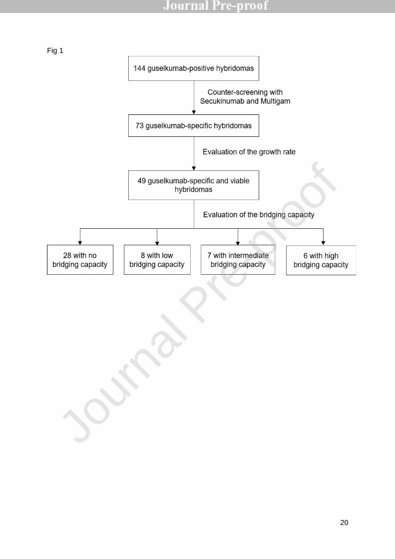
A total of 144 hybridomas producing monoclonal antibodies that bind to guselkumab
were generated. The cross-reactivity was evaluated by counter-screening for binding to
secukinumab (an anti-IL17A antibody) and Multigam(a human IgG mixture) resulting in a
total of 73 hybridomas binding exclusively to guselkumab (Figure 1). To facilitate further
antibody selection for assay development, the growth rate of the hybridoma and the ability of
the antibody to cross-link coated guselkumab with biotin-labeled guselkumab, i.e. the
bridging capacity, was evaluated. Of the 49 viable hybridomas, 28 had no bridging capacity,
8 had a low bridging capacity, 7 an intermediate bridging capacity and 6 a high bridging
capacity. Of each group, several hybridomas were selected in order to create a diverse panel
of 20 different guselkumab-specific antibodies that can be used for further assay
development.
3.1.2 Neutralizing capacity of guselkumab-specific MA-GUS
The neutralizing capacity of the 20 MA-GUS was determined using an IL-23-based
competition assay. Eight MA-GUS reduced the binding signal of guselkumab to IL-23 with ≥
65% when using at 2-fold excess of antibody over guselkumab and were considered
neutralizing. One antibody had no neutralizing capacity (<10%), while the 11 remaining
antibodies had an intermediate neutralizing capacity (ranging from 37 to 62%).
3.1.3 Epitope binning of guselkumab specific MA-GUS
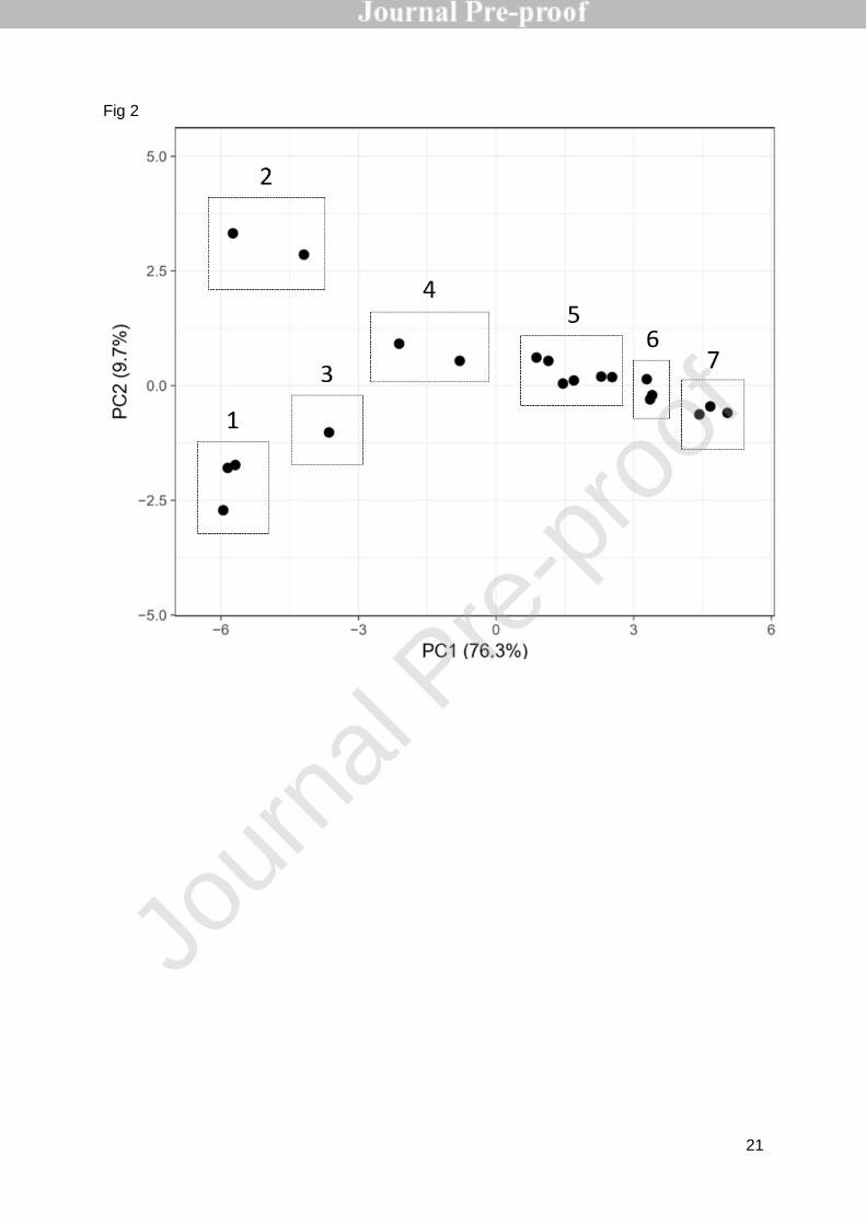
The results of the epitope binning experiment were visualized using principal
component analysis (PCA). The score plot shows the first two principal components (PC),
PC1 on the X-axis and PC2 on the Y-axis, explaining 76.3% and 9.7% of the total variance,
respectively (Fig. 2). The 20 MA-GUS were clustered in seven different groups, each group
containing antibodies that are directed towards a similar guselkumab binding site.
3.2 Development and validation of the guselkumab ELISA
3.2.1 Development of the guselkumab ELISA
The 20 selected MA-GUS were pairwise tested for their suitability as capture and
detection antibody in the development of an ELISA to quantify the concentration of
guselkumab. Based on the affinity, the absence of serum interference and the interdilution,
MA-GUS9F6 was selected as the capture antibody and MA-GUS12G12 as the detection
antibody. MA-GUS9F6 and MA-GUS12G12 had a neutralizing capacity of 72% and 58%,
respectively and bind to different epitopes of guselkumab (clusters 1 and 6). Using this
combination, a non-linear guselkumab calibration dose-response curve, that ranged from
0.08 to 5 ng/mL was obtained (Suppl. Fig. 1).
3.2.2 Specificity
The specificity of the guselkumab assay was determined by spiking secukinumab, a
human anti-IL-17A monoclonal antibody frequently used in the treatment of psoriasis, and
Jour
nal P
re-p
roof

10
guselkumab to drug-naive serum samples of psoriasis patients. In these samples,
guselkumab could accurately be measured (recovery of 95-111%), demonstrating the
specificity of the assay. Moreover, in five serum samples of secukinumab-treated
guselkumab-naive psoriasis patients (with secukinumab concentrations ranging from 13.1-
49.0 µg/mL) guselkumab was undetectable.
3.2.3 Selectivity
The selectivity of the guselkumab assay was determined by evaluating the impact of
the matrix (serum of psoriasis patients), the target (IL-23) and anti-guselkumab antibodies on
the measurement of guselkumab.
3.2.3.1 Impact of the matrix on guselkumab determination
Guselkumab was spiked to guselkumab-naive serum samples of psoriasis patients to
evaluate the impact of unrelated compounds in the matrix on guselkumab determination. The
matrix did not affect the quantification of guselkumab concentrations as the accuracy of 95%
of the spiked samples was within the acceptable range (recoveries ranging from 81-117%).
3.2.3.2 Impact of IL-23 on guselkumab determination
Varying concentrations of IL-23 were spiked to 1 µg/mL guselkumab to determine the
impact of IL-23 on guselkumab determination. The assay could quantify guselkumab
(recovery of 80%) in the presence of up to 1 µg/mL IL-23 (= 2.74:1 molar ratio of IL-23 versus
guselkumab, Fig. 3A). Once exceeding this IL-23 concentration, guselkumab could still be
detected but not accurately quantified (recovery below 80%). Nevertheless, even in the
presence of 2.5 µg/mL IL-23 (= 6.85:1 molar ratio of IL-23 versus guselkumab), the detection
of guselkumab was not completely blocked.
3.2.3.3 Impact of anti-guselkumab antibodies on guselkumab determination
The influence of a two-fold excess of five MA-GUS with varying neutralizing capacities
on the determination of 1 µg/mL guselkumab was evaluated. In the control samples with only
guselkumab or with the addition of a non-relevant anti-ixekizumab antibody (MA-IXE66F11),
the concentration of guselkumab was accurately detected (Fig. 3B). The addition of the non-
neutralizing antibody (MA-GUS14D11, neutralizing capacity = 5%) did not significantly impair
the quantification of guselkumab (recovery of 84%). In contrast, the addition of intermediate
neutralizing antibodies (MA-GUS6G1, neutralizing capacity = 40% and MA-GUS18A4
neutralizing capacity = 38%) revealed less than 25% recovery of guselkumab, while the
addition of neutralizing antibodies (MA-GUS12A9 and MA-GUS13A10, neutralizing capacity
more than 65%) completely hampered guselkumab detection.
3.2.4 Assay cut-off values for detection and quantification, accuracy and imprecision
The cut-off values for detection and quantification were determined based on the OD
of 15 guselkumab-naive serum samples of psoriasis patients. The mean OD was 0.061 with
a standard deviation of 0.012, resulting in a LLOD of 0.073 ng/mL and LLOQ of 0.137 ng/mL.
Taking into account the dilution factor of 1:100, the cut-off value for guselkumab detection
and quantification was 0.007 µg/mL and 0.014 µg/mL, respectively.
The assay complied with the requirements for intra-assay accuracy and precision with
recoveries ranging from 81% to 101% and CV of ≤12% and inter-assay accuracy and
Jour
nal P
re-p
roof
11
precision with recoveries ranging from 89% to 107% and CV of ≤13% for all quality control
samples tested. Furthermore, the total error did not exceed 17% and 25%, for the inter-assay
and intra-assay evaluation, respectively.
3.2.5 Minimum required dilution and dilutional linearity
The minimum required dilution was determined to be 1:100. For samples with a
guselkumab concentration between 0.5 µg/mL and 5 µg/mL, a dilution of 1:400 to 1:3200
was applied and concentrations >5 µg/mL were diluted 1:1600 to 1:12800. Quality control
samples of 2 and 5 µg/mL guselkumab could accurately (recovery of 89% and 94%,
respectively) and precisely (CV of 3% and 4%, respectively) be quantified showing that
concentrations exceeding the range of quantification can be reliably measured by the
guselkumab assay after proper dilution. Moreover, the precision between the measured
values in a dilution series of each quality control sample was acceptable (CV <30%).
3.2.6 Validation of the guselkumab ELISA in a cohort of moderate-to-severe psoriasis
patients
The ability of the newly developed assay to measure guselkumab concentrations in
patient’s serum was determined by the application of the guselkumab ELISA on serum
samples of 10 moderate-to-severe psoriasis patients under standard guselkumab treatment
(Figure 4). The guselkumab concentrations that were measured ranged from 0.46 to 10.36
µg/mL. Guselkumab could be detected during induction at weeks (w) 1, 2, 3 and 4 after the
start of guselkumab treatment (w1, 2, 3: intermediate time points and w4: trough, i.e. the time
point right before the next administration) and at weeks 12 and 20 (trough).
To evaluate accuracy of incurred samples, all patient samples were measured four
times. For each sample, at least 67% of the repeats lied within 30% of its mean. Moreover,
the precision between the measured values in a dilution series of all patient samples was
acceptable (CV <30%) demonstrating parallelism between the calibration standard curve and
serially diluted patient samples and excluding a possible matrix effect.
3.3 Development and validation of the anti-guselkumab antibody ELISAs
3.3.1 Development of the anti-guselkumab antibody assays
Two assays to measure anti-guselkumab antibodies were developed: an anti-
guselkumab antibody bridging assay and an anti-guselkumab antibody ACE assay.
The suitability of different MA-GUS as calibrator antibody in the anti-guselkumab
antibody bridging ELISA was evaluated. Based on the ability to cross-link plate-coated
guselkumab with biotinylated guselkumab and the obtained dose-response curve, MA-
GUS12A9 was selected as calibrator antibody for anti-guselkumab antibody quantification.
Using GUS12A9 as a calibrator, a non-linear dose-response curve was obtained in the range
of 0.16-10 ng/mL (Suppl. Fig. 2, panel A).
Additionally, an anti-guselkumab antibody ACE assay was developed that allows the
detection of anti-guselkumab antibodies in the presence of drug. The same calibrator as in
the bridging assay, GUS12A9, was used and yielded a non-linear dose-response curve
ranging from 0.98 to 1000 ng/mL (Suppl. Fig. 2, panel B).
Jour
nal P
re-p
roof

12
3.3.2 Specificity
The specificity of the anti-guselkumab antibody assays was determined by spiking
secukinumab, a human anti-IL-17A monoclonal antibody frequently used in the treatment of
psoriasis, and MA-GUS12A9 to drug-naive serum samples of psoriasis patients. In both the
bridging and ACE assay, anti-guselkumab antibodies could accurately be measured, as the
recovery of MA-GUS12A9 was 110-124% and 82-118%, respectively, demonstrating the
specificity of the assays.
3.3.3 Selectivity
MA-GUS12A9 was spiked to guselkumab-naive serum samples of psoriasis patients
to evaluate the impact of unrelated compounds in the matrix on anti-guselkumab antibody
determination. In the bridging assay, the matrix in a dilution of 1/20 did not affect the
quantification of anti-guselkumab antibodies as the accuracy of 90% of the spiked samples
was within the acceptable range with recoveries ranging from 90-120%. In the ACE assay,
the recovery of anti-guselkumab antibodies in spiked psoriasis serum was within 88-107% in
80% of the samples.
3.3.4 Assay cut-off values for detection and quantification, accuracy and imprecision
The cut-off values for detection and quantification of anti-guselkumab antibodies in
the bridging and ACE assay were determined based on the OD of 25 guselkumab-naive
serum samples of psoriasis patients. In the bridging assay, the mean OD was 0.06 with a
standard deviation of 0.01, resulting in a LLOD of 0.186 ng/mL and LLOQ of 0.348 ng/mL.
Taking into account the dilution factor of 1:20, the cut-off value for guselkumab detection and
quantification was 3.7 ng/mL and 7.0 ng/mL MA-GUS12A9 equivalents, respectively. In the
ACE assay, the mean OD was 0.075 with a standard deviation of 0.012, resulting in a LLOD
of 25.56 ng/mL and LLOQ of 50.38 ng/mL. Taking into account the dilution factor of 1:17.78,
the cut-off value for guselkumab detection and quantification were set at 454 ng/mL and 896
ng/mL MA-GUS12A9 equivalents, respectively.
The bridging assay complied with the requirements for intra-assay accuracy and
precision with recoveries ranging from 81% to 119% (and CV of ≤12%) and inter-assay
accuracy and precision with recoveries ranging from 101% to 103% (and CV of ≤17%) for all
quality control samples tested. A similar conclusion can be drawn for the ACE assay as the
intra-assay accuracy and precision was shown to be 83% to 118% (and CV of ≤19%) and the
inter-assay accuracy and precision was 91% to 113% (and CV of ≤14%) for all quality control
samples tested.
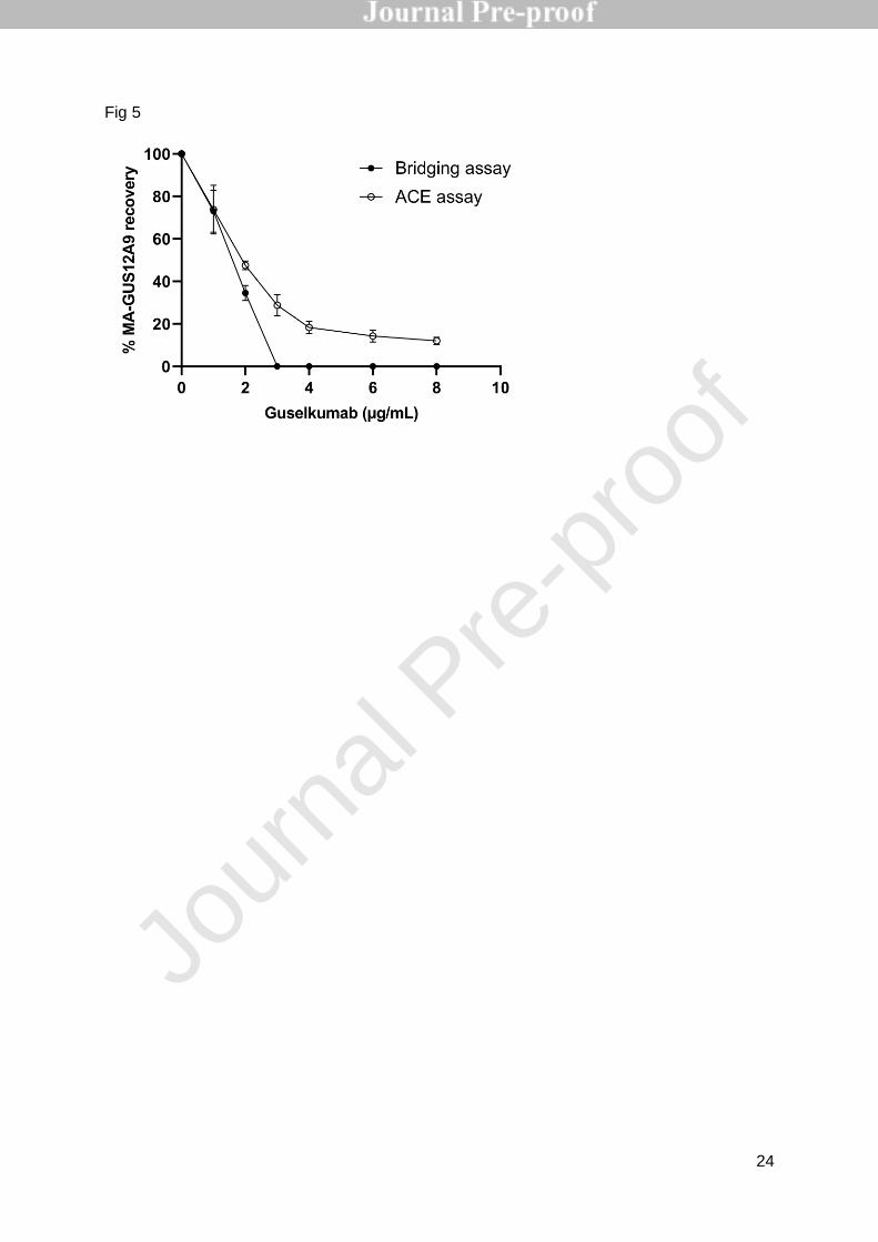
3.3.5 Evaluation of the drug tolerance
To evaluate the impact of guselkumab on the detection of anti-guselkumab
antibodies, i.e. the drug tolerance, increasing concentrations of guselkumab were spiked to 4
µg/mL MA-GUS12A9 resulting in GUS:MA-GUS ratios ranging from 0.25:1 to 2:1. The
addition of guselkumab reduced the detection of MA-GUS12A9 in a concentration-dependent
manner (Fig. 5). The presence of 1 µg/mL guselkumab (= ratio 0.25:1) already decreased the
recovery of MA-GUS12A9 to below 80% in both the bridging assay and the ACE assay. In
the anti-guselkumab antibody bridging assay, MA-GUS12A9 became undetectable in the
presence of 3 µg/mL guselkumab (= ratio 0.75:1), while an average recovery of 29% was
Jour
nal P
re-p
roof

13
observed in the ACE assay with the same guselkumab concentration. Interestingly, in
contrast to the bridging assay, MA-GUS12A9 remains detectable in the presence of up to 8
µg/mL guselkumab in the ACE assay, although in low concentrations.
Jour
nal P
re-p
roof
14
4. Discussion
TDM of biologicals is an emerging tool to optimize treatment as drug concentrations
have been linked to clinical outcomes in various chronic inflammatory diseases [9-11, 16-19].
Multiple studies indicate that monitoring drug concentrations together with a clinical
evaluation, will not only maximize treatment outcomes but will also improve the cost-
effectiveness of biological therapies [20, 21]. To date, TDM of guselkumab in patients with
moderate-to-severe psoriasis cannot be performed in daily clinical practice because no
commercial assays to measure guselkumab or anti-guselkumab antibody concentrations are
available. In this study, we aimed to develop immunoassays for the quantification of
guselkumab and anti-guselkumab antibody concentrations according to the guidelines of
EMA.
A diverse panel of 20 highly specific anti-guselkumab monoclonal antibodies was
generated and characterized for their neutralizing capacity and epitope binning profile. This
allowed to carefully select the best antibody combination for the development of an assay to
measure guselkumab. Using MA-GUS9F6 as the capture antibody and MA-GUS12G12 as
the detection antibody, an ELISA was developed which accurately and precisely quantified
guselkumab concentrations with a cut-off for quantification of 0.014 µg/mL. The mean (±SD)
steady-state trough serum guselkumab concentrations in two phase III studies were reported
to be 1.15±0.73µg/mL and 1.23±0.84 µg/mL [5]. As TDM is most often performed at trough,
our in-house developed guselkumab assay can quantify guselkumab in a clinically relevant
concentration range with a good sensitivity.
The guselkumab assay was shown to be selective as no matrix effect of serum of
psoriasis patients was observed. Moreover, the presence of the drug’s target, IL-23, did not
influence the quantification of guselkumab until a concentration of 0.25 µg/mL, indicating that
the presence of IL-23 at physiological concentrations (pg/mL) will not have an impact on the
quantification of guselkumab [22]. In contrast, the presence of neutralizing anti-guselkumab
monoclonal antibodies, but not non-neutralizing ones, severely hampered the detection of
guselkumab. This can be explained by the fact that the coating antibody MA-GUS9F6 is also
a neutralizing antibody and other neutralizing antibodies consequently compete for a similar
binding site. Hampering of drug detection by neutralizing anti-drug antibodies has also been
observed with other ELISAs that quantify biologicals [15, 23].
With the aim to perform a clinical validation, serum samples of 10 moderate-to-severe
psoriasis patients collected at several time points during standard guselkumab treatment
were applied on the guselkumab assay. The high sensitivity of the assay allowed to measure
guselkumab concentrations during the induction and maintenance phase, both at
intermediate time points (weeks 1, 2 and 3 after the start of treatment) and at trough (weeks
4, 12 and 20), indicating that this assay can be used to perform TDM in guselkumab-treated
psoriasis patients. Moreover, the guselkumab concentrations observed at week 20 were
comparable to the data reported in clinical trials [5]. Currently, clinical studies evaluating the
efficacy and safety of guselkumab in the treatment of inflammatory bowel disease and
psoriatic arthritis are ongoing [24-26]. If guselkumab is approved for these indications, the
developed assays will also be very useful to evaluate the value of monitoring guselkumab
and anti-guselkumab antibodies in these other chronic inflammatory diseases.
Jour
nal P
re-p
roof
15
Anti-drug antibodies can have a significant impact on treatment response. Multiple
studies have shown that anti-infliximab and anti-adalimumab antibodies are associated with
lower serum drug concentrations and loss of response in psoriasis patients [27-29]. The
clinical relevance of anti-guselkumab antibodies is not well established because of the lack of
appropriate assays. Therefore, two assays were developed to detect anti-guselkumab
antibodies, i.e. a bridging assay and an ACE assay. The specificity and selectivity of both
anti-guselkumab antibody assays was demonstrated.
Both anti-guselkumab antibody assays use the same calibrator, MA-GUS12A9, to
detect anti-guselkumab antibodies but differ in the threshold for anti-guselkumab antibody
detection and quantification and the tolerance to the presence of guselkumab. In the bridging
assay, the cut-off value for detection and quantification was determined to be 3.7 ng/mL and
7 ng/mL respectively. In contrast, the cut-off values of the ACE assay are 454 ng/mL and 896
ng/mL for detection and quantification, respectively. These are considerably higher
compared to the bridging assay, which can be explained by the fact that due to the different
acidification steps, the sample is automatically diluted to a certain extent. On the other hand,
the acid dissociation steps allow the detection of anti-drug antibodies that were previously in
complex with the drug. As a result, the ACE assay can still detect antibodies in the presence
of up to 8 µg/mL guselkumab, whereas the bridging assay cannot detect anti-guselkumab
antibodies from a guselkumab concentration of 3 µg/mL onwards. These assays will allow to
further elucidate the clinical relevance of anti-guselkumab-antibodies, either free or in
complex with the drug.
Together, the guselkumab and anti-guselkumab antibody assays will allow to
evaluate the presence of a concentration-response relationship and establish a therapeutic
window in patients receiving guselkumab treatment and may support the use of TDM in
guselkumab-treated patients.
5. Conclusion
In conclusion, 20 highly specific anti-guselkumab monoclonal antibodies were
generated, characterized and pairwise tested for the development of an ELISA for the
quantification of guselkumab concentrations in serum of patients with moderate-to-severe
psoriasis. Additionally, an anti-guselkumab antibody bridging and ACE assay were
developed for the detection of anti-guselkumab antibodies, either free or in complex with
guselkumab. All three assays were validated according to the EMA guidelines. These
validated immunoassays are essential to establish a concentration-response relationship and
will allow the future implementation of TDM in moderate-to-severe psoriasis patients
receiving guselkumab.
Jour
nal P
re-p
roof

16
Author statement
Nathalie Van den Berghe: conceptualization, investigation, formal analysis, Writing -
Original Draft, Writing - Review & Editing, visualization, project administration
Aurélie Truffot: conceptualization, investigation, formal analysis, Writing - Original
Draft, Writing - Review & Editing
Miet Peeters: investigation, formal analysis
Griet Compernolle: investigation, formal analysis
Els Brouwers: investigation, formal analysis
Rani Soenen: resources, Writing - Review & Editing
Lynda Grine: resources, Writing - Review & Editing
Ann Gils: conceptualization, supervision, Writing - Review & Editing
Maya Imbrechts: conceptualization, supervision, Writing - Review & Editing, Project
administration, validation
Declaration of interests
The authors declare that they have no known competing financial interests or personal
relationships that could have appeared to influence the work reported in this paper.
Acknowledgements
This work was in part supported by the TBM Grant T003218N of the Research Foundation
Flanders (FWO), Belgium. N. V. is a SB PhD fellow at FWO.
References
[1] G. Girolomoni, R. Strohal, L. Puig, H. Bachelez, J. Barker, W.H. Boehncke, J.C. Prinz, The role of IL-23 and the IL-23/TH 17 immune axis in the pathogenesis and treatment of psoriasis, J Eur Acad Dermatol Venereol 31 (2017) 1616-1626. [2] K.A. Papp, A. Blauvelt, A.B. Kimball, C. Han, B. Randazzo, Y. Wasfi, Y.K. Shen, S. Li, C.E.M. Griffiths, Patient-reported symptoms and signs of moderate-to-severe psoriasis treated with guselkumab or adalimumab: results from the randomized VOYAGE 1 trial, J Eur Acad Dermatol Venereol 32 (2018) 1515-1522. [3] C.E.M. Griffiths, K.A. Papp, A.B. Kimball, B. Randazzo, M. Song, S. Li, Y.K. Shen, A. Blauvelt, Long-Term Efficacy of Guselkumab for the Treatment of Moderate-to-Severe Psoriasis: Results from the Phase 3 VOYAGE 1 Trial Through Two Years, J Drugs Dermatol 17 (2018) 826-832. [4] K. Reich, A.W. Armstrong, P. Foley, M. Song, Y. Wasfi, B. Randazzo, S. Li, Y.K. Shen, K.B. Gordon, Efficacy and safety of guselkumab, an anti-interleukin-23 monoclonal antibody, compared with adalimumab for the treatment of patients with moderate to severe psoriasis with randomized withdrawal and retreatment: Results from the phase III, double-blind, placebo- and active comparator-controlled VOYAGE 2 trial, J Am Acad Dermatol 76 (2017) 418-431. [5] Tremfya (guselkumab, IL-23 inhibitor) [summary of product characteristics] Janssen Biotech, (2019). [6] C. Hermans, P. Herranz, S. Segaert, A. Gils, Current Practice of Therapeutic Drug Monitoring of Biopharmaceuticals in Psoriasis Patients, Ther Drug Monit 39 (2017) 356-359.
Jour
nal P
re-p
roof
17
[7] E. Dreesen, P. Bossuyt, D. Mulleman, A. Gils, D. Pascual-Salcedo, Practical recommendations for the use of therapeutic drug monitoring of biopharmaceuticals in inflammatory diseases, Clin Pharmacol 9 (2017) 101-111. [8] S.P. Menting, E. Coussens, M.F. Pouw, J.M. van den Reek, L. Temmerman, H. Boonen, E.M. de Jong, P.I. Spuls, J. Lambert, Developing a Therapeutic Range of Adalimumab Serum Concentrations in Management of Psoriasis: A Step Toward Personalized Treatment, JAMA Dermatol 151 (2015) 616-22. [9] N. Wilkinson, T. Tsakok, N. Dand, K. Bloem, M. Duckworth, D. Baudry, A. Pushpa-Rajah, C.E.M. Griffiths, N.J. Reynolds, J. Barker, R.B. Warren, A.D. Burden, T. Rispens, D. Stocken, C. Smith, Defining the Therapeutic Range for Adalimumab and Predicting Response in Psoriasis: A Multicenter Prospective Observational Cohort Study, J Invest Dermatol 139 (2019) 115-123. [10] M. Toro-Montecinos, F. Ballesca, C. Ferrandiz, A. Teniente-Serra, E. Martinez-Caceres, J.M. Carrascosa, Usefulness and correlation with clinical response of serum ustekinumab levels measured at 6 weeks versus 12 weeks, J Dermatolog Treat (2018) 1-5. [11] N. Van den Berghe, E. De Keyser, R. Soenen, L. Meuleman, S. Lanssens, A. Gils, J. Lambert, Clinical response correlates with 4-week postinjection ustekinumab concentrations in patients with moderate-to-severe psoriasis, Br J Dermatol 182 (2020) 390-397. [12] R. Soenen, E. Meulewaeter, L. Grine, N. Van den Berghe, E. Brouwers, R. Speeckaert, S. Lanssens, L. Temmerman, J. Lambert, A. Gils, Defining a Minimal Effective Serum Trough Concentration of Secukinumab in Psoriasis: A Step toward Personalized Therapy, J Invest Dermatol (2019). [13] European Medicines Agency (EMA). Guideline on bioanalytical method validation (version 21 July 2011).[Available from: https://www.ema.europa.eu/en/documents/scientific-guideline/guidelinebioanalytical-method-validation_en.pdf]. [14] G. Galfre, C. Milstein, Preparation of monoclonal antibodies: strategies and procedures, Methods Enzymol 73 (1981) 3-46. [15] T. Van Stappen, E. Brouwers, S. Tops, N. Geukens, S. Vermeire, P.J. Declerck, A. Gils, Generation of a Highly Specific Monoclonal Anti-Infliximab Antibody for Harmonization of TNF-Coated Infliximab Assays, Ther Drug Monit 37 (2015) 479-85. [16] E. Dreesen, B. Verstockt, S. Bian, M. de Bruyn, G. Compernolle, S. Tops, M. Noman, G. Van Assche, M. Ferrante, A. Gils, S. Vermeire, Evidence to Support Monitoring of Vedolizumab Trough Concentrations in Patients With Inflammatory Bowel Diseases, Clin Gastroenterol Hepatol 16 (2018) 1937-1946.e8. [17] S.P. Menting, J.M. van den Reek, E.M. Baerveldt, E.M. de Jong, E.P. Prens, L.L. Lecluse, G.J. Wolbink, D. Van der Kleij, P.I. Spuls, T. Rispens, The correlation of clinical efficacy, serum trough levels and antidrug antibodies in ustekinumab-treated patients with psoriasis in a clinical-practice setting, Br J Dermatol 173 (2015) 855-7. [18] I. Detrez, K. Van Steen, S. Segaert, A. Gils, The association between etanercept serum concentration and psoriasis severity is highly age-dependent, Clin Sci (Lond) 131 (2017) 1179-1189. [19] I. Detrez, E. Dreesen, T. Van Stappen, A. de Vries, E. Brouwers, G. Van Assche, S. Vermeire, M. Ferrante, A. Gils, Variability in Golimumab Exposure: A 'Real-Life' Observational Study in Active Ulcerative Colitis, J Crohns Colitis 10 (2016) 575-81. [20] L. Guidi, D. Pugliese, T.P. Tonucci, A. Berrino, B. Tolusso, M. Basile, L. Cantoro, P. Balestrieri, F. Civitelli, L. Bertani, M. Marzo, C. Felice, E. Gremese, F. Costa, F. Viola, M. Cicala, A. Kohn, A. Gasbarrini, G.L. Rapaccini, M. Ruggeri, A. Armuzzi, Therapeutic Drug Monitoring is More Cost-Effective than a Clinically Based Approach in the Management of Loss of Response to Infliximab in Inflammatory Bowel Disease: An Observational Multicentre Study, J Crohns Colitis 12 (2018) 1079-1088. [21] C. Steenholdt, J. Brynskov, O.O. Thomsen, L.K. Munck, J. Fallingborg, L.A. Christensen, G. Pedersen, J. Kjeldsen, B.A. Jacobsen, A.S. Oxholm, J. Kjellberg, K. Bendtzen, M.A. Ainsworth, Individualised therapy is more cost-effective than dose intensification in patients with Crohn's disease who lose response to anti-TNF treatment: a randomised, controlled trial, Gut 63 (2014) 919-27.
Jour
nal P
re-p
roof

18
[22] A. Kyriakou, A. Patsatsi, T.A. Vyzantiadis, D. Sotiriadis, Serum levels of TNF-alpha, IL-12/23p40, and IL-17 in plaque psoriasis and their correlation with disease severity, J Immunol Res 2014 (2014) 467541. [23] S. Bian, T.V. Stappen, F. Baert, G. Compernolle, E. Brouwers, S. Tops, A. Vries, T. Rispens, J. Lammertyn, S. Vermeire, A. Gils, Generation and characterization of a unique panel of anti-adalimumab specific antibodies and their application in therapeutic drug monitoring assays, J Pharm Biomed Anal 125 (2016) 62-7. [24] A. Deodhar, A.B. Gottlieb, W.H. Boehncke, B. Dong, Y. Wang, Y. Zhuang, W. Barchuk, X.L. Xu, E.C. Hsia, Efficacy and safety of guselkumab in patients with active psoriatic arthritis: a randomised, double-blind, placebo-controlled, phase 2 study, Lancet 39 (2018) 2213-2224. [25] ClinicalTrials.gov. National Library of Medicine (U.S.). A Study of the Efficacy and Safety of Guselkumab in Participants With Moderately to Severely Active Crohn's Disease (GALAXI) .Identifier NCT03466411 . Retrieved February, 2020 from: https://clinicaltrials.gov/ct2/show/NCT03466411. [26] ClinicalTrials.gov. National Library of Medicine (U.S.). A Study of Guselkumab in Participants With Moderately to Severely Active Ulcerative Colitis (QUASAR) Identifier NCT04033445 . Retrieved February, 2020 from: https://clinicaltrials.gov/ct2/show/NCT04033445. [27] L.L. Lecluse, R.J. Driessen, P.I. Spuls, E.M. de Jong, S.O. Stapel, M.B. van Doorn, J.D. Bos, G.J. Wolbink, Extent and clinical consequences of antibody formation against adalimumab in patients with plaque psoriasis, Arch Dermatol 146 (2010) 127-32. [28] H. Takahashi, H. Tsuji, A. Ishida-Yamamoto, H. Iizuka, Plasma trough levels of adalimumab and infliximab in terms of clinical efficacy during the treatment of psoriasis, J Dermatol 40 (2013) 39-42. [29] T. Bito, R. Nishikawa, M. Hatakeyama, A. Kikusawa, H. Kanki, H. Nagai, Y. Sarayama, T. Ikeda, H. Yoshizaki, H. Seto, A. Adachi, T. Horikawa, M. Oka, C. Nishigori, Influence of neutralizing antibodies to adalimumab and infliximab on the treatment of psoriasis, Br J Dermatol 170 (2014) 922-9.
Jour
nal P
re-p
roof

19
Figure captions
Figure 1: Flowchart of the selection of 20 guselkumab-specific MA-GUS
Figure 2: Principal component analysis and clustering of the 20 MA-GUS
Figure 3: Effect of IL-23 (panel A) and different antibodies (panel B) on the determination of
guselkumab (1 µg/mL) in the ELISA using MA-GUS9F6 as the capture antibody and MA-
GUS12G12 as the detection antibody (mean ± SD). Both graphs are representative of 2
independent experiments with 2 replicates each.
Figure 4: The average guselkumab serum concentration (mean ± SD) of 10 moderate-to-
severe psoriasis patients at different time points after the start of guselkumab treatment. The
patients received 100 mg guselkumab subcutaneously at weeks 0, 4, 12 and 20 according to
the standard dosing regimen. The graph shows the results of 2 independent experiments
with 2 replicates each.
Figure 5: The drug tolerance of the anti-guselkumab antibody bridging assay (black dots)
and anti-guselkumab antibody ACE assay (white dots) was evaluated by incubating
increasing concentrations of guselkumab with 4 µg/mL MA-GUS12A9. The percentage
recovery of MA-GUS12A9 in the presence of guselkumab was expressed relative to the
percentage recovery of MA-GUS12A9 in the absence of guselkumab, which was set at
100%. The graph shows the results of 2 independent experiments with 2 replicates each.
Jour
nal P
re-p
roof
20
Fig 1
Jour
nal P
re-p
roof
21
Fig 2
Jour
nal P
re-p
roof

22
Fig 3
Jour
nal P
re-p
roof
23
Fig 4
Jour
nal P
re-p
roof
24
Fig 5
Jour
nal P
re-p
roof
Related Documents


