Crystal structures of Ophiostoma piceae sterol esterase: Structural insights into activation mechanism and product release Javier Gutiérrez-Fernández a,1 , María Eugenia Vaquero b,1 , Alicia Prieto b , Jorge Barriuso b , María Jesús Martínez b,⇑ , Juan A. Hermoso a,⇑ a Departamento de Cristalografía y Biología Estructural, Instituto de Química-Física ‘‘Rocasolano’’, CSIC, Serrano 119, 28006 Madrid, Spain b Departamento de Biología Medioambiental, Centro de Investigaciones Biológicas, CSIC, Ramiro de Maeztu 9, 28040 Madrid, Spain article info Article history: Received 15 January 2014 Received in revised form 27 June 2014 Accepted 29 July 2014 Available online 7 August 2014 Keywords: Fungal lipase/esterase Activation mechanism abstract Sterol esterases are able to efficiently hydrolyze both sterol esters and triglycerides and to carry out syn- thesis reactions in the presence of organic solvents. Their high versatility makes them excellent candi- dates for biotechnological purposes. Sterol esterase from fungus Ophiostoma piceae (OPE) belongs to the family abH03.01 of the Candida rugosa lipase-like proteins. Crystal structures of OPE were solved in this study for the closed and open conformations. Enzyme activation involves a large displacement of the conserved lid, structural rearrangements of loop a16–a17, and formation of a dimer with a large opening. Three PEG molecules are placed in the active site, mimicking chains of the triglyceride substrate, demonstrating the position of the oxyanion hole and the three pockets that accommodate the sn-1, sn-2 and sn-3 fatty acids chains. One of them is an internal tunnel, connecting the active center with the outer surface of the enzyme 30 Å far from the catalytic Ser220. Based on our structural and biochemical results we propose a mechanism by which a great variety of different substrates can be hydrolyzed in OPE paving the way for the construction of new variants to improve the catalytic properties of these enzymes and their biotechnological applications. Ó 2014 Elsevier Inc. All rights reserved. 1. Introduction Triacylglycerol lipases (EC 3.1.1.3) are enzymes ubiquitous in nature, from microbes to plants and animals, catalyzing the hydro- lysis of triglycerides to diglycerides, monoglycerides, free fatty acids and glycerol. In addition, these enzymes can catalyze esteri- fication and transesterification reactions in the presence of organic solvents (Houde et al., 2004). Their interest is related to their appli- cation in a wide range of industrial processes: food, detergents, cosmetics, pharmaceutical, textile and paper industry (Reetz, 2002; Singh and Mukhopadhyay, 2012). On the other hand, sterol esterases (EC 3.1.1.13) are defined as enzymes that hydrolyze sterol esters releasing free sterols and fatty acids. These enzymes were described in mammal tissues and in some fungi and bacteria (Rahim and Sih, 1969; Ghosh et al., 1995). Initially they were proposed to be used to detect cho- lesterol in blood (Allain et al., 1974) but their importance increased during the last decade since, in addition to their ability to hydro- lyze sterol esters, they are able to hydrolyze triglycerides effi- ciently (Calero-Rueda et al., 2002b; Kontkanen et al., 2006; Maeda et al., 2008) and carry out synthesis reactions in the pres- ence of organic solvents (Morinaga et al., 2011; Barba et al., 2011). Due to their high versatility, these enzymes proved to have high biotechnological interest for pitch reduction during paper pulp manufacture (Calero-Rueda et al., 2002a) and they can also catalyze the synthesis of phytosterol esters used as food additives (Morinaga et al., 2011; Barba et al., 2011). Both lipases and sterol esterases are a/b-hydrolases showing a substrate-binding site formed by an extensive hydrophobic pocket (Grochulski et al., 1993; Mancheño et al., 2003). A traditional crite- rion accepted to define an enzyme as a lipase deals with the huge increase of their activity in the lipid–water interface, owing to a phenomenon known as interfacial activation (Verger, 1997), a pro- cess that can be also triggered by micelles (Hermoso et al., 1997, 1996). This property is linked usually to the existence of a http://dx.doi.org/10.1016/j.jsb.2014.07.007 1047-8477/Ó 2014 Elsevier Inc. All rights reserved. Abbreviations: pNPB, p-nitrophenylbutyrate; pNPL, p-nitrophenyl laureate; pNPP, p-nitrophenylpalmitate; CHAPS, 3-[(3-cholamidopropyl)dimethylammo- nio]-1-propanesulfonate; OPE, Ophiostoma piceae esterase; PEG, polyethylene glycol. ⇑ Corresponding authors. Fax: +34 91 536 04 32 (M.J. Martínez), +34 915642431 (J.A. Hermoso). E-mail addresses: [email protected] (M.J. Martínez), [email protected] (J.A. Hermoso). 1 Equally contributed to this work. Journal of Structural Biology 187 (2014) 215–222 Contents lists available at ScienceDirect Journal of Structural Biology journal homepage: www.elsevier.com/locate/yjsbi

Welcome message from author
This document is posted to help you gain knowledge. Please leave a comment to let me know what you think about it! Share it to your friends and learn new things together.
Transcript
Journal of Structural Biology 187 (2014) 215–222
Contents lists available at ScienceDirect
Journal of Structural Biology
journal homepage: www.elsevier .com/locate /y jsbi
Crystal structures of Ophiostoma piceae sterol esterase: Structuralinsights into activation mechanism and product release
http://dx.doi.org/10.1016/j.jsb.2014.07.0071047-8477/� 2014 Elsevier Inc. All rights reserved.
Abbreviations: pNPB, p-nitrophenylbutyrate; pNPL, p-nitrophenyl laureate;pNPP, p-nitrophenylpalmitate; CHAPS, 3-[(3-cholamidopropyl)dimethylammo-nio]-1-propanesulfonate; OPE, Ophiostoma piceae esterase; PEG, polyethyleneglycol.⇑ Corresponding authors. Fax: +34 91 536 04 32 (M.J. Martínez), +34 915642431
(J.A. Hermoso).E-mail addresses: [email protected] (M.J. Martínez), [email protected]
(J.A. Hermoso).1 Equally contributed to this work.
Javier Gutiérrez-Fernández a,1, María Eugenia Vaquero b,1, Alicia Prieto b, Jorge Barriuso b,María Jesús Martínez b,⇑, Juan A. Hermoso a,⇑a Departamento de Cristalografía y Biología Estructural, Instituto de Química-Física ‘‘Rocasolano’’, CSIC, Serrano 119, 28006 Madrid, Spainb Departamento de Biología Medioambiental, Centro de Investigaciones Biológicas, CSIC, Ramiro de Maeztu 9, 28040 Madrid, Spain
a r t i c l e i n f o
Article history:Received 15 January 2014Received in revised form 27 June 2014Accepted 29 July 2014Available online 7 August 2014
Keywords:Fungal lipase/esteraseActivation mechanism
a b s t r a c t
Sterol esterases are able to efficiently hydrolyze both sterol esters and triglycerides and to carry out syn-thesis reactions in the presence of organic solvents. Their high versatility makes them excellent candi-dates for biotechnological purposes. Sterol esterase from fungus Ophiostoma piceae (OPE) belongs tothe family abH03.01 of the Candida rugosa lipase-like proteins. Crystal structures of OPE were solvedin this study for the closed and open conformations. Enzyme activation involves a large displacementof the conserved lid, structural rearrangements of loop a16–a17, and formation of a dimer with a largeopening. Three PEG molecules are placed in the active site, mimicking chains of the triglyceride substrate,demonstrating the position of the oxyanion hole and the three pockets that accommodate the sn-1, sn-2and sn-3 fatty acids chains. One of them is an internal tunnel, connecting the active center with the outersurface of the enzyme 30 Å far from the catalytic Ser220. Based on our structural and biochemical resultswe propose a mechanism by which a great variety of different substrates can be hydrolyzed in OPE pavingthe way for the construction of new variants to improve the catalytic properties of these enzymes andtheir biotechnological applications.
� 2014 Elsevier Inc. All rights reserved.
1. Introduction fatty acids. These enzymes were described in mammal tissues
Triacylglycerol lipases (EC 3.1.1.3) are enzymes ubiquitous innature, from microbes to plants and animals, catalyzing the hydro-lysis of triglycerides to diglycerides, monoglycerides, free fattyacids and glycerol. In addition, these enzymes can catalyze esteri-fication and transesterification reactions in the presence of organicsolvents (Houde et al., 2004). Their interest is related to their appli-cation in a wide range of industrial processes: food, detergents,cosmetics, pharmaceutical, textile and paper industry (Reetz,2002; Singh and Mukhopadhyay, 2012).
On the other hand, sterol esterases (EC 3.1.1.13) are defined asenzymes that hydrolyze sterol esters releasing free sterols and
and in some fungi and bacteria (Rahim and Sih, 1969; Ghoshet al., 1995). Initially they were proposed to be used to detect cho-lesterol in blood (Allain et al., 1974) but their importance increasedduring the last decade since, in addition to their ability to hydro-lyze sterol esters, they are able to hydrolyze triglycerides effi-ciently (Calero-Rueda et al., 2002b; Kontkanen et al., 2006;Maeda et al., 2008) and carry out synthesis reactions in the pres-ence of organic solvents (Morinaga et al., 2011; Barba et al.,2011). Due to their high versatility, these enzymes proved to havehigh biotechnological interest for pitch reduction during paperpulp manufacture (Calero-Rueda et al., 2002a) and they can alsocatalyze the synthesis of phytosterol esters used as food additives(Morinaga et al., 2011; Barba et al., 2011).
Both lipases and sterol esterases are a/b-hydrolases showing asubstrate-binding site formed by an extensive hydrophobic pocket(Grochulski et al., 1993; Mancheño et al., 2003). A traditional crite-rion accepted to define an enzyme as a lipase deals with the hugeincrease of their activity in the lipid–water interface, owing to aphenomenon known as interfacial activation (Verger, 1997), a pro-cess that can be also triggered by micelles (Hermoso et al., 1997,1996). This property is linked usually to the existence of a

216 J. Gutiérrez-Fernández et al. / Journal of Structural Biology 187 (2014) 215–222
structural lid in these enzymes, allowing the access of the lipidicsubstrate to the catalytic site (Holmquist, 2000). Nevertheless,the existence of a lid seems not to be the single determinant forinterfacial activation (Mancheño et al., 2003). In the case of sterolesterases, no much information is available since most of themhave not been characterized.
The yeast Candida rugosa secretes a variety of closely relatedenzymes, referred as lipases or sterol esterases. In light of bio-chemical and structural studies, the best-characterized enzymesfrom this yeast (Lip1, Lip2 and Lip3) exhibit a high sequence iden-tity among them (77–88%), but they differ in their substrate spec-ificity on triglycerides and cholesterol esters. These differences inthe affinity towards these compounds are explained by smallchanges in the hydrophobicity of the substrate-binding site andthe lid region (Mancheño et al., 2003).
Sterol esterase from Ophiostoma piceae (OPE) (UniProt IdQ2TFW1) belongs to the family abH03.01 of the C. rugosa lipase likeproteins (The Lipase Engineering Database http://www.led.uni-stuttgart.de/) for which 326 members have been sequenced andthe three-dimensional structures of lipases from C. rugosa(Grochulski et al., 1993), the lipase from Geotrichum candidum(Schrag and Cygler, 1993) and the esterase from Aspergillus niger(Bourne et al., 2004) are the only members up to now reported.
Recently, we described a computational model of three-dimen-sional structure for OPE based on C. rugosa Lip1 and Lip3 structures(Calero-Rueda et al., 2009; Barriuso et al., 2013). OPE is more effi-cient than C. rugosa Lip3 against sterol esters (Barba et al., 2011).Our model indicated that differences in the tunnel region of theseenzymes could be related with their catalytic efficiency differences(Calero-Rueda et al., 2009).
Therefore, the development of structural studies is essential tounderstand the catalytic properties of these enzymes. The use ofnative O. piceae enzyme did not produce crystals suitable forX-ray diffraction (Calero-Rueda et al., 2002b). In this work wepresent the three-dimensional structure of this enzyme, in its openand closed conformations, by using the deglycosylated form of therecombinant protein expressed in Pichia pastoris, more solublethan the native one (Barba Cedillo et al., 2012). The activationmechanism and substrate stabilization is here described. This rep-resents a hotspot in this field, opening the way for the rationaldesign of new improved variants for industrial applications andallowing fine tuning enzyme activity.
2. Material and methods
2.1. OPE expression, purification and characterization
The enzyme was over-expressed in P. pastoris and purified in asingle hydrophobic chromatography step (Octyl-Sepharose car-tridge, GE Healthcare), as previously reported (Barba Cedilloet al., 2012), although in this case the protein was eluted after addi-tion of 18 mM CHAPS in 25 mM Tris–HCl pH 7.0. The purifiedenzyme was dialyzed against 25 mM Tris–HCl, pH 7 to reducethe detergent concentration below 0.5 lM and stored at �20 �Cuntil used.
Purified OPE was subjected to Size Exclusion Chromatography(SEC) using a Superose 12 GL 10/300 (GE Healthcare) with20 mM Tris–HCl pH 7 plus 0.15 M NaCl at 0.3 mL min�1 to separatethe multimolecular forms of the purified protein. Dextran blue(2000 kDa), Ferritin (460 kDa), Aldolase (170 kDa), BSA (73 kDa),Ovalbumin (48.2 kDa) and Chymotrypsinogen A (19.5 kDa), (cali-bration kits, Amersham Pharmacia), were used as standards to cal-ibrate the column. Before deglycosylation with Endoglycosidase H(Roche), OPE was dialyzed against sodium citrate buffer pH 5.5 andthen incubated at 37 �C for 24 h, following the manufacturer’sinstructions. Deglycosylation was checked by SDS–PAGE, staining
the protein bands with Coomassie R-250 or with silver reagent.The deglycosylated enzyme was dialyzed against 25 mM Tris–HCl, pH 7.0 for crystallization assays.
Enzyme activity against esters of p-nitrophenol was measuredby monitoring the hydrolysis of pNPB, pNPL and pNPP. A 20 mMstock solution of the substrates was prepared in HPLC grade ace-tone. The assay mixture contained 2 mM substrate in 20 mMTris–HCl, pH 7.0 buffer and 1% (v/v) Genapol X-100. The reactionswere monitored at 410 nm at room temperature in a ShimadzuUV-1800, in the case of pNPP and pNPL with magnetic stirring.One unit of activity (1 U) is defined as the amount of enzymereleasing 1 lmol of p-nitrophenol (e410 = 15,200 M�1cm�1) perminute under the defined conditions.
Protein concentration was determined by BCA bioassay(Thermo) using bovine serum albumin as standard.
Analytical ultracentrifugation was used to check the aggrega-tion behavior of recombinant proteins and the possible transitionstate between of both closed and open protein forms (monomerand dimer, respectively). Measurements were performed in a XL-Ianalytical ultracentrifuge (Beckman-Coulter Inc.) equipped withUV–VIS and interference detection optics. Samples, in 25 mMTris–HCl, pH 7.0, were centrifuged at 48,000 rpm and 20 �C usingan An50Ti eight hole rotor and double-sector Epon-charcoal cen-terpieces. Differential sedimentation coefficient distributions c(s)were calculated by least-squares boundary modeling of the exper-imental data using the program SEDFIT (version 12.44) (Schuck,2000). The experiments were carried out, by using both the puri-fied protein after hydrophobic chromatography (0.5 mg/mL), con-sisting in a mixture of aggregates states (principally monomerand dimer), and the monomer isolated after SEC (0.2 mg/mL).These proteins were analyzed in presence or absence of 5 mMdodecane sulfonyl chloride (Sigma–Aldrich), as enzyme inhibitor,and 5 mM of cholesteryl oleate, as enzyme substrate (after30 min of incubation at room temperature). The enzyme inhibitorwas prepared in isooctane, remaining in the samples at 0.5%, andthe substrate was dissolved in 0.02% Genapol X-100 and 150 mMNaCl (final concentration).
2.2. Construction of an OPE mutant
The mutant I544W was constructed by site-directed mutagene-sis. The mutation was introduced by polymerase chain reaction(PCR), using the expression plasmid pPIC9OPE as template (BarbaCedillo et al., 2012). Both a direct and a reverse primer weredesigned complementary to opposite strands of the same DNAregion. The following primers were used (with indication of thechanged triplets in bold): OPE I544W (Fw: 50-CAACAACTGGGG-CATCTTCC-30 and Rv: 50-GGAAGATGCCCCAGTTGTTG-30). PCRs(50 lL final volume) were carried out in a Mastercycler Pro S(Eppendorf) using 100 ng of template DNA, each dNTP at 250 lM,125 ng of direct and reverse primers, 5 units of expand long tem-plate enzyme mix (Roche), and the manufacturer’s buffer number3. Reaction conditions were as follows: (i) 95 �C for 1 min; (ii) 18cycles at 95 �C for 50 s, 55 �C for 50 s, and 68 �C for 10 min; and(iii) a final cycle at 68 �C for 10 min. After amplification, the PCRproducts were treated with DpnI restriction enzyme (Roche) todigest the parental strand. The mutated gene was completelysequenced using an ABI 3730 DNA analyzer (Applied Biosystems)to ensure that only the desired mutation was introduced.
2.3. Crystallization and data collection
Initial crystallization trials were performed by the sitting dropvapor-diffusion method at 18 �C. A wide range of crystallizationconditions were assayed by high-throughput techniques, using aNanoDrop robot with Innovadyne SD-2 microplates (Innovadyne
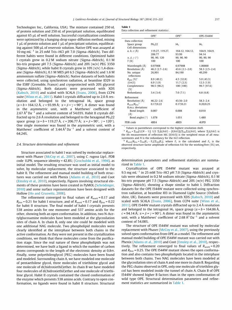
Table 1Data collection and refinement statistics*.
OPEc OPEo OPE-I544W
Data collectionSpace group P6322 I41 I41
Cell dimensionsa, b, c (Å) 119.27, 119.27,
206.77164.12, 164.12,93.99
164.9, 164.9,94.14
a, b, c (�) 90, 90, 120 90, 90, 90 90, 90, 90T (K) 100 100 100
Wavelength (Å) 0.97908 0.97908 1.00000Resolution (Å) 46.2 (2.7–2.6) 43.6 (2.1–2.0) 58.3 (2.5–2.4)No. uniquereflections
26,901 84,190 49,299
Rpim (%)a 8.9 (49.2) 4.5 (32.8) 5.0 (43.3)hI/r(I)i 6.9 (1.9) 11.7 (2.5) 12.5 (1.9)Completeness(%)
98.5 (98.2) 100 (100) 99.7 (97.6)
Redundancy 3.4 (3.4) 7.0 (7.1) 6.8 (6.8)
RefinementResolution (Å) 46.22–2.6 43.56–2.0 58.3–2.4Rwork/Rfree
b 0.17/0.22 0.17/0.21 0.20/0.25R.m.s. deviations
Bond length(Å)
0.007 0.008 0.010
Bond angles (�) 1.078 1.051 1.480
PDB code 4BE4 4BE9 4UPD
* Values in parentheses correspond to the highest resolution shell.a Rpim = Rhkl[1/(N � 1)] 1/2 Ri|Ii(hkl) � [I(hkl)]|/RhklRiIi(hkl), where RiIi(hkl) is
the ith measurement of reflection hkl, [I(hkl)] is the weighted mean of all mea-
J. Gutiérrez-Fernández et al. / Journal of Structural Biology 187 (2014) 215–222 217
Technologies Inc., California, USA). The mixture contained 250 nLof protein solution and 250 nL of precipitant solution, equilibratedagainst 65 lL of well solution. Successful crystallization conditionswere optimized by a hanging-drop vapor-diffusion method, mixing1 lL of protein solution and 1 lL of precipitant solution, equilibrat-ing against 500 lL of reservoir solution. Native OPE was assayed at10 mg mL�1 in 25 mM Tris–HCl pH 7.0 (Sigma–Aldrich). Two dif-ferent habits were found in different conditions. Optimized habitI crystals grew in 0.2 M sodium nitrate (Sigma–Aldrich), 0.1 Mbis-tris propane pH 7.5 (Sigma–Aldrich) and 20% (w/v) PEG 3350(Sigma–Aldrich), while habit II crystals grew in 10% (v/v) 1,4-diox-ane (Sigma–Aldrich), 0.1 M MES pH 6.5 (Sigma–Aldrich) and 1.6 Mammonium sulfate (Sigma–Aldrich). Native datasets of both habitswere collected, using synchrotron radiation, at beamline ID29 inthe ESRF (Grenoble, France) and cryoprotected with 20% glycerol(Sigma–Aldrich). Both datasets were processed with XDS(Kabsch, 2010) and scaled with SCALA (Evans, 2006), from CCP4suite (Winn et al., 2011). Habit I crystals diffracted up to 2.0 Å res-olution and belonged to the tetragonal I41 space group(a = b = 164.12 Å, c = 93.99 Å; a = b = c = 90�). A dimer was foundin the asymmetric unit, with a Matthews’ coefficient of2.67 Å3 Da�1 and a solvent content of 54.03%. Habit II crystals dif-fracted up to 2.6 Å resolution and belonged to the hexagonal P6322space group (a = b = 119.27 Å, c = 206.77 Å; a = b = 90�, c = 120�).One single monomer was found in the asymmetric unit, with aMatthews’ coefficient of 3.44 Å3 Da�1 and a solvent content of64.24%.
surements and N is the redundancy for the hkl reflection.b Rwork/Rfree = Rhkl|Fo � Fc|/Rhkl|Fo|, where Fc is the calculated and Fo is the
observed structure factor amplitude of reflection hkl for the working/free (5%) set,respectively.
2.4. Structure determination and refinement
Structure associated to habit I was solved by molecular replace-ment with Phaser (McCoy et al., 2007), using C. rugosa Lip1, PDBcode 1LPN, sequence identity = 42.8%; (Grochulski et al., 1994), asinitial model. The resulting structure was used as initial model tosolve, by molecular replacement, the structure associated to thehabit II. The refinement and manual model building of both struc-tures was carried out with Phenix (Adams et al., 2010) and Coot(Emsley et al., 2010), respectively. Figures involving structural ele-ments of these proteins have been created in PyMOL (Schrödinger,2010) and some surface representations have been designed withHollow (Ho and Gruswitz, 2008).
The refinement converged to final values of Rwork = 0.17 andRfree = 0.21 for habit I structure, and of Rwork = 0.17 and Rfree = 0.22for habit II structure. The final model of habit I crystals presents538 amino acids for one monomer and 537 amino acids for theother, showing both an open conformation. In addition, two N-Ace-tylglucosamine molecules have been modeled at the glycosilationsites of chain A. In chain B, only one site could be modeled withone additional NAG molecule. Two phospholipid molecules wereclearly identified at the interphase between both chains in theactive conformation. As they were not present in the crystallizationconditions, we think that these molecules come from the purifica-tion stage. Since the real nature of these phospholipids was notdetermined, we have built a ligand in which the number of carbonatoms corresponds to the length of the electronic density at 0.8r.Finally, some polyethilenglycol (PEG) molecules have been foundand modeled. Surrounding chain A, we have modeled one moleculeof pentaethilene glycol, three molecules of triethilene glycol andone molecule of di(hidroxiethil)ether. In chain B we have modeledfour molecules of di(hidroxiethil)ether and one molecule of triethi-lene glycol. Habit II crystals contained the closed conformation ofthe enzyme which presents 538 amino acids. Contrary to open con-formation, no ligands were found in habit II structure. Structural
determination parameters and refinement statistics are summa-rized in Table 1.
Crystallization of OPE I544W mutant was assayed at9.5 mg mL�1 in 25 mM Tris–HCl pH 7.0 (Sigma–Aldrich) and crys-tals were obtained in 0.2 M sodium nitrate (Sigma–Aldrich), 0.1 Mbis-tris propane pH 7.5 (Sigma–Aldrich) and 20% (w/v) PEG 3350(Sigma–Aldrich), showing a shape similar to habit I. Diffractiondatasets for the OPE I544W mutant were collected using synchro-tron radiation, at beamline I03 in Diamond Light Source (Oxford-shire, UK). Datasets were processed with XDS (Kabsch, 2010) andscaled with SCALA (Evans, 2006), from CCP4 suite (Winn et al.,2011). OPE I544W mutant crystals diffracted up to 2.4 Å resolutionand belonged to the tetragonal I41 space group (a = b = 164.86 Å,c = 94.14 Å; a = b = c = 90�). A dimer was found in the asymmetricunit, with a Matthews’ coefficient of 2.68 Å3 Da�1 and a solventcontent of 54.08%.
The structure of OPE I544W mutant was solved by molecularreplacement with Phaser (McCoy et al., 2007), using the previouslysolved open conformation from OPE as a model. The refinement andmanual model building of OPE I544W mutant was carried out withPhenix (Adams et al., 2010) and Coot (Emsley et al., 2010), respec-tively. The refinement converged to final values of Rwork = 0.20and Rfree = 0.25. The OPE I544W mutant shows the open conforma-tion and also contains two phospholipids located in the interphasebetween both chains. Two NAG molecules have been modeled atthe glycosilation sites of chain A and one more in chain B. Regardingthe PEG chains observed in OPE, only one molecule of triethilen gly-col has been modeled inside the tunnel of chain A. Chain B of OPEI544W showed higher B factors than in the open conformation ofwild type OPE. Structural determination parameters and refine-ment statistics are summarized in Table 1.

218 J. Gutiérrez-Fernández et al. / Journal of Structural Biology 187 (2014) 215–222
2.5. Accession numbers
Coordinates and structure factors have been deposited in theProtein Data Bank with accession numbers 4BE9 and 4BE4 forthe OPE open and closed conformations, respectively and 4UPDfor the OPE-I544W mutant.
3. Results and discussion
3.1. Overall structure
Two different three-dimensional structures are presented inthis report, namely, the structure of OPE in its closed conformation(OPEc) (Fig. 1a) obtained from habit II crystals and the open confor-mation of the enzyme (OPEo) (Fig. 1b) from habit I crystals. Crystalstructure of OPEc follows the protein scaffold of lipases fromC. rugosa based on an ab hydrolase fold (Ollis et al., 1992) with amajor 11-stranded mixed b-sheet, a small and nearly perpendicularN-terminal 3-stranded b-sheet and 16 helices (Fig. 1a). The majorb-sheet, which forms the core of the lipase, exhibits a pronouncedtwist, being the first (b0) and last (b10) strands almost perpendicu-lar to each other. In agreement with this, lipase 2 of C. rugosa (PDBcode 1GZ7, Mancheño et al., 2003) is the closest structural homo-logue of OPEc (sequence identity = 42.5%, rmsd = 0.81 Å for 538 Caatoms, Fig. S1 Supp. Mat.). OPEc presents a 37 amino acids lidcovering the active site. The lid consists of one a-helix (residues83–88) and two 310-helices (residues 90–94 and 96–98) flankedby two loops that end in a disulfide hinge (from Cys72 toCys108, Fig. 1c). OPE lid differs from that reported for C. rugosa
Fig.1. Overall structure of the closed and open conformations of OPE. (a) View of the closmagenta. Glycan molecules attached to the glycosylation sites are represented as blue sticthe open OPE structure with the ab hydrolase core colored in yellow and the lid coloredblue sticks. Catalytic Ser220 is shown in blue sticks. PEG chains are shown as black sticclosed and open states in OPE at the entry channel. Large displacements are observed fostates. (d) Dimeric form of OPE in its open conformation, showing the catalytic cavity anpatch of the other.
and G. candidum. While the lid in C. rugosa Lip1 and Lip2 containsone a-helix of 10 residues and one 310-helix of 3 residues, the lid inG. candidum is formed by three a-helices containing 12, 4 and 10residues, respectively.
A hydrophobic patch, formed by Ile407, Phe408, Phe458,Pro459 and Phe460, is located close to the lid (Fig. 1c). This Phe-Pro-Phe pattern appears only in OPE and not in the closest homo-logues. Both phenylalanines closely interact with some residues ofthe lid (Leu88 and Leu92).
The main structural difference between closed and open struc-tures is the displacement of the lid up to about 30 Å away from itsoriginal position (Fig. 1c and Fig. S2 Supp. Mat.). This movementinvolves a rearrangement of its secondary structure that upon acti-vation presents two a-helices (residues 84–93 and 96–101). A sim-ilar rearrangement is observed for the lipases/esterases from C.rugosa, but OPE presents the largest lid displacement amongrelated enzymes (�30 Å in the longest separation) (Fig. S3 Supp.Mat.). Analysis of both OPE forms (habit I and II) indicates thatthe lid regions in the closed conformation is not involved in crystalcontacts, while the lid in the open conformation is involved in olig-omeric arrangement. In agreement with that, observed B-factorsare higher for the lid in the closed conformation than in the open(Fig. S4 Supp. Mat.).
3.2. Oligomeric arrangement in OPE
A dimeric arrangement was observed for the open conformationOPEo as also reported for crystal structures of the open formsof Lip1 (Grochulski et al., 1993) and Lip3 (Ghosh et al., 1995).
ed OPE structure with the a/b hydrolase core colored in yellow and the lid colored inks. Catalytic Ser220 is shown in blue sticks. The a16–a17 loop is labeled. (b) View ofin magenta. Glycan molecules attached to the glycosylation sites are represented asks and the phospholipid as green sticks. (c) Detailed view of the changes betweenr Val476 (9 Å) and for Leu477 (4 Å) between the closed (blue) and open (magenta)d the non-bonded contacts between the open lid of one chain and the hydrophobic

J. Gutiérrez-Fernández et al. / Journal of Structural Biology 187 (2014) 215–222 219
However, in contrast with that observed for Lip1 and Lip3 whichmonomers tightly associate forming dimers, OPEo presents a dimerassociated by regions in which aliphatic residues from the lid ofone chain (Leu84, Leu85, Leu88, Leu92 and Ile95) interact withthe hydrophobic patch of the opposite chain (Fig. 1d) with fewcontacts between OPEo monomers and presenting a large cavityopened to the solvent (about 23 Å � 38 Å). Ultracentrifugationexperiments carried out with native O. piceae enzyme showedprotein multi-aggregates in aqueous solution while, in the sameconditions, monomeric and dimeric forms were observed in theprotein expressed in P. pastoris (Barba Cedillo et al., 2012).
Analytical ultracentrifugation studies (Table 2) showed achange in the percentages of monomer and dimer in the presenceof inhibitor or substrate enzyme. In both cases, the purified OPE ormonomeric-rich fraction, the predominant form was the monomer.In the presence of both, enzyme inhibitor or substrate, a significantamount of the monomer was converted into the dimer, suggestingthe transition from the closed to the open enzyme form in the pres-ence of both, inhibitor or substrate. The phenomenon of transitionbetween the two conformations has been previously described inC. rugosa lipases, where exposure of the monomeric form of theenzyme to triolein dramatically accelerated its transition to theopen form (Turner et al., 2001).
3.3. Activation mechanism in OPE
Besides opening of the lid, activation in OPE involves changes inthe conformation of the loop a16–a17 (residues Tyr474–Tyr480),
Fig.2. Phospholipid site and recognition in active OPE. (a) The phospholipid (green stickspatch (in yellow). Residues involved in substrate stabilization are labeled. Lid from the clobetween the phospholipid and the active OPE.
Table 2Percentage of monomer and dimer forms of the esterase, obtained after analyticalultracentrifugation, in protein samples in absence or presence of dodecane sulfonylchloride, as enzyme inhibitor, and cholesteryl oleate, as enzyme substrate. OPEcorresponds to the O. piceae esterase expressed in P. pastoris after hydrophobicinteraction columns. The monomeric fraction was obtained in an additional step byusing SEC to separate the different aggregate forms present in the purified protein.
% monomer % dimer
OPE 63.1 19.6OPE + inhibitor 33.4 40.1OPE + substrate 19.1 71.5
Monomer fraction 69.9 30.1Monomer fraction + inhibitor 18.6 75.9Monomer fraction + substrate 16.8 83.2
that stabilizes a phospholipid molecule located at the interfacebetween both monomers of the dimer into a groove (11 Å wide,8 Å deep and 15 Å long) connecting the outer part of the enzymeto the active site (Figs. 1c and 2). This phospholipid was notincluded in the crystallization conditions and very likely was incor-porated during the purification process (see Section 2). A twist inPro478 residue causes the displacement of the entire loop, withdisplacements of 9 Å in Val476 and 4 Å in Leu477 with regard toOPEc, increasing the interactions with the lipid found in this groove(Fig. 1c). H-bond interactions affecting residues at the extremes ofthe loop (Asn479, Tyr480 and Tyr474 in Fig. S3 Supp. Mat.) serve asanchoring point for the a16–a17 loop. Structural analysis also pro-vides indications about what could trigger lid movement. In OPEc,Leu92 is lying on the entry channel and interacting with Phe458and Phe464 from the hydrophobic patch. Upon activation, the posi-tion of Leu92 is now occupied by one of the aliphatic chains of thephospholipid (Fig. 2a).
Amino acid composition of loop a16–a17 is not conservedamong the abH03.01 family members and structural conformationof OPE loop a16–a17, in both closed and open states, is also unique(Fig. S3 Supp. Mat.).
3.4. The active site cleft and substrate binding
The catalytic machinery of OPE is formed by the triad Ser220,Glu352 and His465. As observed in other members of the fungalfamily, and unlike other lipases, the acidic residue of the triad isa Glu residue and not an Asp residue. The catalytic region is wellconserved between OPE and the other fungal lipases/esterases.The catalytic triad remains unchanged in the open and closeconformation of the enzyme. By comparison with homologousenzymes, the backbone NH groups of two Gly residues (Gly134–Gly135) build the oxyanion hole that stabilizes the oxyanionproduced in the tetrahedral intermediate generated during thehydrolysis. A water molecule is located close to the oxyanion hole.The oxyanion is the intermediate formed in the transient state ofthe hydrolysis reaction. Different molecules of PEG from thecrystallization condition (see Section 2) were identified occupyingthe hydrophobic active site cleft, very likely mimicking theposition of substrate (Fig. 3a).
The phospholipid molecule attached to the groove leading tothe OPE active site was found in both chains of OPEo and presentedan excellent electron density (Fig. 2 and Fig. S5 Supp. Mat.) This isthe first time in this family of lipases/esterases, that a potential
) is located in a groove formed by the a16–a17 loop (in blue) and the hydrophobicsed conformation is colored in pink. (b) Schematic representation of the interactions
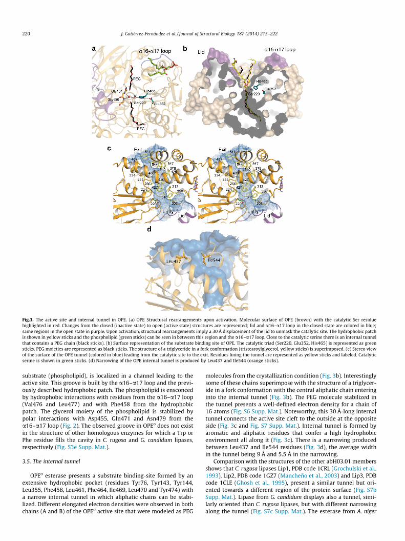
Fig.3. The active site and internal tunnel in OPE. (a) OPE Structural rearrangements upon activation. Molecular surface of OPE (brown) with the catalytic Ser residuehighlighted in red. Changes from the closed (inactive state) to open (active state) structures are represented; lid and a16–a17 loop in the closed state are colored in blue;same regions in the open state in purple. Upon activation, structural rearrangements imply a 30 Å displacement of the lid to unmask the catalytic site. The hydrophobic patchis shown in yellow sticks and the phospholipid (green sticks) can be seen in between this region and the a16–a17 loop. Close to the catalytic serine there is an internal tunnelthat contains a PEG chain (black sticks). (b) Surface representation of the substrate binding site of OPE. The catalytic triad (Ser220, Glu352, His465) is represented as greensticks. PEG moieties are represented as black sticks. The structure of a triglyceride in a fork conformation (tristearoylglycerol, yellow sticks) is superimposed. (c) Stereo viewof the surface of the OPE tunnel (colored in blue) leading from the catalytic site to the exit. Residues lining the tunnel are represented as yellow sticks and labeled. Catalyticserine is shown in green sticks. (d) Narrowing of the OPE internal tunnel is produced by Leu437 and Ile544 (orange sticks).
220 J. Gutiérrez-Fernández et al. / Journal of Structural Biology 187 (2014) 215–222
substrate (phospholipid), is localized in a channel leading to theactive site. This groove is built by the a16–a17 loop and the previ-ously described hydrophobic patch. The phospholipid is ensconcedby hydrophobic interactions with residues from the a16–a17 loop(Val476 and Leu477) and with Phe458 from the hydrophobicpatch. The glycerol moiety of the phospholipid is stabilized bypolar interactions with Asp455, Gln471 and Asn479 from thea16–a17 loop (Fig. 2). The observed groove in OPEo does not existin the structure of other homologous enzymes for which a Trp orPhe residue fills the cavity in C. rugosa and G. candidum lipases,respectively (Fig. S3e Supp. Mat.).
3.5. The internal tunnel
OPEo esterase presents a substrate binding-site formed by anextensive hydrophobic pocket (residues Tyr76, Tyr143, Tyr144,Leu355, Phe458, Leu461, Phe464, Ile469, Leu470 and Tyr474) witha narrow internal tunnel in which aliphatic chains can be stabi-lized. Different elongated electron densities were observed in bothchains (A and B) of the OPEo active site that were modeled as PEG
molecules from the crystallization condition (Fig. 3b). Interestinglysome of these chains superimpose with the structure of a triglycer-ide in a fork conformation with the central aliphatic chain enteringinto the internal tunnel (Fig. 3b). The PEG molecule stabilized inthe tunnel presents a well-defined electron density for a chain of16 atoms (Fig. S6 Supp. Mat.). Noteworthy, this 30 Å-long internaltunnel connects the active site cleft to the outside at the oppositeside (Fig. 3c and Fig. S7 Supp. Mat.). Internal tunnel is formed byaromatic and aliphatic residues that confer a high hydrophobicenvironment all along it (Fig. 3c). There is a narrowing producedbetween Leu437 and Ile544 residues (Fig. 3d), the average widthin the tunnel being 9 Å and 5.5 Å in the narrowing.
Comparison with the structures of the other abH03.01 membersshows that C. rugosa lipases Lip1, PDB code 1CRL (Grochulski et al.,1993), Lip2, PDB code 1GZ7 (Mancheño et al., 2003) and Lip3, PDBcode 1CLE (Ghosh et al., 1995), present a similar tunnel but ori-ented towards a different region of the protein surface (Fig. S7bSupp. Mat.). Lipase from G. candidum displays also a tunnel, simi-larly oriented than C. rugosa lipases, but with different narrowingalong the tunnel (Fig. S7c Supp. Mat.). The esterase from A. niger

Table 3Specific activity of the recombinant protein expressed in P. pastoris, as well as on themutant I544W constructed by site-directed mutagenesis, on 2 mM pNPB, pNPL andpNPP, in presence of 1% genapol.
Substrate Fatty acids Specific activity (U mg�1)
OPE native I544W
pNPB C4:0 103.17 ± 2.54 113.44 ± 3.52pNPL C12:0 162.72 ± 6.82 203.65 ± 12.93pNPP C16:0 65.78 ± 5.30 0.13 ± 0.013
J. Gutiérrez-Fernández et al. / Journal of Structural Biology 187 (2014) 215–222 221
completely lacks the tunnel (Fig. S7d Supp. Mat.). In this sense, it isworth to note how proteins sharing a similar overall structure(Fig. S3 Supp. Mat.) can display strong differences concerning itsinternal tunnel. As revealed by structural analysis, small changesin amino acid composition and/or rotamers can result in a com-pletely different structural arrangement of the tunnel, e.g., whilethe first half of the tunnel follows the same path in O. piceae, inC. rugosa and G. candidum lipases, a substitution of a serine byTyr377 in OPE blocks the path redirecting it about 150� apart(Fig. S7 Supp. Mat.).
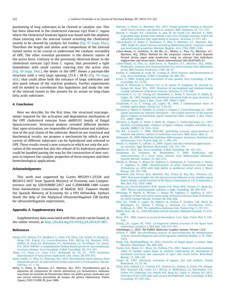
In an attempt to ascertain the potential role of the tunnel in theproduct release of the reaction, we constructed a site-directedmutant, replacing Ile544 by a Trp residue (OPE-I544W). The goalwith this mutant was to block the substrate exit by inserting abulky hydrophobic residue. The OPE-I544W mutant was purifiedand its structure solved at 2.4 Å resolution (Table 1). Both nativeand mutated proteins showed no differences in their secondarystructure (rmsd of 0.147 Å for 518 Ca atoms). As expected, substi-tution of Ile544 residue by bulky Trp residue completely blocks thepotential exit of the internal tunnel (Fig. 4).
Different activity studies were carried out comparing wild typeversus mutated enzyme (Fig. 4b). Our results (Table 3) show thatwhile enzyme activity against pNPB and pNPL was similar in themutated and wild type enzymes, the activity was completely lostwith pNPP (�0.2% of the wt enzyme). These results suggest thatthe length of the substrate affects the activity of the mutated pro-tein at the end of the tunnel; while short-medium substrates (4and 12 C atoms) remain mostly unaffected, hydrolytic activity withsubstrates having more than 16 C atoms is severely reduced versusthat of wt.
On the other hand, amino acid composition in the internal tun-nel of C. rugosa lipases has been associated to differences in sub-strate specificity between Lip1 and Lip3 (Ghosh et al., 1995).Structural comparison among the three C. rugosa isoenzymes(Lip1, Lip2 and Lip3) has been reported to show two distinctiveregions according to their amino acid composition: an aromatic-rich region at the beginning of the tunnel and an aliphatic-richregion at the bottom (Mancheño et al., 2003). Whereas this secondregion remained essentially identical in the three isoenzymes andthus the interactions with the alkyl chains of the substrates wouldbe very similar, the phenylalanine content of the first region differsin the three proteins (Mancheño et al., 2003), a fact that was asso-ciated with the substrate recognition properties of the isoenzymes,namely, to their lipase/esterase character. The differences ofC. rugosa lipases activity on cholesterol esters (Lip2 > Lip3 > Lip1)have been related to the higher Phe content in the hydrophobicregion of the tunnel (Lip1 > Lip3 > Lip2) (Mancheño et al., 2003).In the case of OPE, the Phe content in this region is similar to
Fig.4. Blocking tunnel exit by site directed mutagenesis. (a) View of the molecular surfacThe mutation blocks the potential exit of the tunnel (surface for the mutated position is
Lip2. It could explain its ability to hydrolyze sterol esters. However,its high efficiency, when compared to the commercial C. rugosacholesterol esterase to hydrolyze triglycerides with long chainfatty acids or different cholesterol esters (Calero-Rueda et al.,2009), could be related to its specific internal tunnel and its uniquedimeric structure. The presence of large opening in the activedimer could allow the entrance of different substrates that canbe stabilized in the 30 Å-long internal tunnel.
3.6. Proposed model of substrate entry and product release in OPE
Crystal structures of OPE provide clues about the fate of thesubstrate to be hydrolyzed by the enzyme. The closed conforma-tion of OPE is a monomer with the large lid masking the active site.Activation involves both, opening of the lid and the rearrangementof a16–a17 loop and the formation of a dimeric structure. As pro-ven by our experiments, isolation of the monomeric form results inan increase of the dimer upon activation of the enzyme. Once thelid is open, the hydrophobic surface behind it is exposed, allowingthe substrate to move to the catalytic cleft where the catalyticSer220 residue is located. The hydrocarbon chain of the substrateis inserted into the tunnel up to the point at which the ester bondreaches the Ser220. After reaction, the acidic moiety of the ester (afatty acid) could remain in the tunnel, while the sterol moiety isreleased. The fatty acid inside the tunnel has to be released in orderto recover the active state of the enzyme.
Our mutagenesis experiments indicate that a single modifica-tion blocking the exit of the tunnel 30 Å far from active site (byreplacing Ile544 by bulky Trp residue) did not affect significantlythe OPE activity on substrates with short-medium chain fattyacids. While an appealing hypothesis is that substrates could bereleased by the exit of the tunnel, our crystallographic and bio-chemical experiments indicate that the exit of the tunnel is notrequired for efficient hydrolysis in OPE against these substrates.The observed lost of activity of the OPE I544W mutant with pNPPcould be explained by the presence of bulky Trp residue at the endof the tunnel that decreases its effective length avoiding the right
e in the OPE I544W mutant (b) the molecular surface of the OPE mutant rotated 90�.colored in orange on the right panel).
222 J. Gutiérrez-Fernández et al. / Journal of Structural Biology 187 (2014) 215–222
positioning of long substrates to be cleaved at catalytic site. Thishas been observed in the cholesterol esterase Lip3 from C. rugosawhere the cholesteryl linoleate ligand was found with the aliphaticchain entering into the internal tunnel orienting the cholesterylmoiety to be cleaved by catalytic Ser residue (Fig. S7 Supp. Mat.).Therefore the length and amino acid composition of the internaltunnel seems to be crucial to understand the catalytic versatilityof OPE. The other essential parameter is the dimeric nature ofthe active form. Contrary to the previously observed dimer in thecholesterol esterase Lip3 from C. rugosa, that presented a tighthomodimer with small cavities entering into the active sites(Fig. S9 Supp. Mat.), the dimer in the OPEo shows a pacman-likestructure with a very large opening (23 Å � 38 Å) (Fig. S9 Supp.Mat.) that could allow both the entrance of large substrates andalso quick release of the reaction products. Further experimentswill be needed to corroborate this hypothesis and study the roleof the internal tunnel in this protein for its action on long-chainfatty acids substrates.
4. Conclusion
Here we describe, for the first time, the structural rearrange-ments required for the activation and degradation mechanism ofthe OPE cholesterol esterase from abH03.01 family of fungallipases/esterases. Structural analysis revealed different pocketsthat, upon activation, are responsible of dimerization and stabiliza-tion of the acyl chains of the substrate. Based on our structural andbiochemical results, we propose a mechanism by which a greatvariety of different substrates can be hydrolyzed and released inOPE. These results reveal a new scenario in which not only the acti-vation of the enzyme but also the release of its hydrolysis productscould be handled paving the way for the construction of new vari-ants to improve the catalytic properties of these enzymes and theirbiotechnological applications.
Acknowledgments
This work was supported by Grants BFU2011-25326 andBIO2012-3637 from Spanish Ministry of Economy and Competi-tiveness and by S2010/BMD-2457 and S-2009AMB-1480 Grantsfrom Autonomous Community of Madrid. M.E. Vaquero thanksthe Spanish Ministry of Economy for a FPU fellowship. Authorsthank the help of the Analytical Ultracentrifugation CIB facilityfor ultracentrifugation experiments.
Appendix A. Supplementary data
Supplementary data associated with this article can be found, inthe online version, at http://dx.doi.org/10.1016/j.jsb.2014.07.007.
References
Adams, P.D., Afonine, P.V., Bunkoczi, G., Chen, V.B., Davis, I.W., Echols, N., Headd, J.J.,Hung, L.W., Kapral, G.J., Grosse-Kunstleve, R.W., McCoy, A.J., Moriarty, N.W.,Oeffner, R., Read, R.J., Richardson, D.C., Richardson, J.S., Terwilliger, T.C., Zwart,P.H., 2010. PHENIX: a comprehensive Python-based system for macromolecularstructure solution. Acta Crystallogr. D Biol. Crystallogr. 66, 213–221.
Allain, C.C., Poon, L.S., Chan, C.S.G., Richmond, W., Fu, P.C., 1974. Enzymaticdetermination of total serum-cholesterol. Clin. Chem. 20, 470–475.
Barba Cedillo, V., Plou, F.J., Martinez, M.J., 2012. Recombinant sterol esterase fromOphiostoma piceae: an improved biocatalyst expressed in Pichia pastoris. Microb.Cell Fact. 11, 1–14.
Barba, V., Prieto, A., Martínez, A.T., Martínez, M.J., 2011. Procedimiento para laobtención de compuestos de interés alimenticio y/o farmacéutico mediantereacciones de acilación de fitoesteroles libres con ácidos grasos catalizadas poruna esterol esterasa procedente de hongos del género Ophiostoma. Patent(Spain), P201131098 20, June 2008.
Barriuso, J., Prieto, A., Martínez, M.J., 2013. Fungal genomes mining to discovernovel sterol esterases and lipases as catalysts. BMC Genomics 14, 712–719.
Bourne, Y., Hasper, A.A., Chahinian, H., Juin, M., de Graaff, L.H., Marchot, P., 2004.Aspergillus niger protein EstA defines a new class of fungal esterases within thealpha/beta hydrolase fold superfamily of proteins. Structure 12, 677–687.
Calero-Rueda, O., Barba, V., Rodriguez, E., Plou, F., Martínez, A.T., Martínez, M.J.,2009. Study of a sterol esterase secreted by Ophiostoma piceae: sequence, modeland biochemical properties. Biochim. Biophys. Acta 1794, 1099–1106.
Calero-Rueda, O., Gutiérrez, A., del Río, J.C., Muñoz, C., Plou, F.J., Martínez, A.T.,Martínez, M.J., 2002a. Method for the enzymatic control of pitch depositsformed during paper pulp production using an esterase that hydrolysestriglycerides and sterol esters. Patent (International) WO 02/075045 A1.
Calero-Rueda, O., Plou, F.J., Ballesteros, A., Martínez, A.T., Martínez, M.J., 2002b.Production, isolation and characterization of a sterol esterase from Ophiostomapiceae. BBA Proteins Proteomics 1599, 28–35.
Emsley, P., Lohkamp, B., Scott, W., Cowtan, K., 2010. Features and development ofCoot. Acta Crystallogr. D Biol. Crystallogr. 66, 486–501.
Evans, P., 2006. Scaling and assessment of data quality. Acta Crystallogr. D Biol.Crystallogr. 62, 72–82.
Ghosh, D., Wawrzak, Z., Pletnev, V.Z., Li, N., Kaiser, R., Pangborn, W., Jörnvall, H.,Erman, M., Duax, W.L., 1995. Structure of uncomplexed and linoleate-boundCandida cylindracea cholesterol esterase. Structure 3, 279–288.
Grochulski, P., Li, Y.G., Schrag, J.D., Bouthillier, F., Smith, P., Harrison, D., Rubin, B.,Cygler, M., 1993. Insights into interfacial activation from an open structure ofCandida rugosa lipase. J. Biol. Chem. 268, 12843–12847.
Grochulski, P., Li, Y., Schrag, J.D., Cygler, M., 1994. 2 conformational states ofCandida-rugosa lipase. Protein Sci. 3, 82–91.
Hermoso, J., Pignol, D., Kerfelec, B., Crenon, I., Chapus, C., FontecillaCamps, J.C., 1996.Lipase activation by nonionic detergents – the crystal structure of the porcinelipase–colipase–tetraethylene glycol monooctyl ether complex. J. Biol. Chem.271, 18007–18016.
Hermoso, J., Pignol, D., Penel, S., Roth, M., Chapus, C., FontecillaCamps, J.C., 1997.Neutron crystallographic evidence of lipase–colipase complex activation by amicelle. EMBO J. 16, 5531–5536.
Ho, B.K., Gruswitz, F., 2008. HOLLOW: generating accurate representations ofchannel and interior surfaces in molecular structures. BMC Struct. Biol., 8.
Holmquist, M., 2000. Alpha/beta-hydrolase fold enzymes: structures, functions andmechanisms. Curr. Protein Pept. Sci. 1, 209–235.
Houde, A., Kademi, A., Leblanc, D., 2004. Lipases and their industrial applications –an overview. Appl. Biochem. Biotechnol. 118, 155–170.
Kabsch, W., 2010. Xds. Acta Crystallogr. D Biol. Crystallogr. 66, 125–132.Kontkanen, H., Tenkanen, M., Reinikainen, T., 2006. Purification and
characterisation of a novel steryl esterase from Melanocarpus albomyces.Enzyme Microb. Technol. 39, 265–273.
Maeda, A., Mizuno, T., Bunya, M., Sugihara, S., Nakayama, D., Tsunasawa, S., Hirota,Y., Sugihara, A., 2008. Characterization of novel cholesterol esterase fromTrichoderma sp. AS59 with high ability to synthesize steryl esters. J. Biosci.Bioeng. 105, 341–349.
Mancheño, J.M., Pernas, M.A., Martínez, M.J., Ochoa, B., Rua, M.L., Hermoso, J.A.,2003. Structural insights into the lipase/esterase behavior in the Candida rugosalipases family: crystal structure of the lipase 2 isoenzyme at 1.97 A resolution. J.Mol. Biol. 332, 1059–1069.
McCoy, A.J., Grosse-Kunstleve, R.W., Adams, P.D., Winn, M.D., Storoni, L.C., Read, R.J.,2007. Phaser crystallographic software. J. Appl. Crystallogr. 40, 658–674.
Morinaga, N., Maeda, A., Mizuno, T., Bunya, M., Sugihara, S., Sugihara, A., 2011.Synthesis of fatty acid sterol esters using cholesterol esterase from Trichodermasp. AS59. Enzyme Microb. Technol. 48, 498–504.
Ollis, D.L., Cheah, E., Cygler, M., Dijkstra, B., Frolow, F., Franken, S.M., Harel, M.,Remington, S.J., Silman, I., Schrag, J., Sussman, J.L., Verschueren, K.H.G.,Goldman, A., 1992. The alpha/beta-hydrolase fold. Protein Eng. 5, 197–211.
Rahim, M.A., Sih, C.J., 1969. Microbial steroid esterases. Methods Enzymol. 15, 675–684.
Reetz, M.T., 2002. Lipases as practical biocatalysts. Curr. Opin. Chem. Biol. 6, 145–150.
Schrag, J.D., Cygler, M., 1993. 1.8-Angstrom refined structure of the lipase fromGeotrichum-candidum. J. Mol. Biol. 230, 575–591.
Schrödinger, L., 2010. The PyMOL Molecular Graphics System, Version 1.2r20.Schuck, P., 2000. Size-distribution analysis of macromolecules by sedimentation
velocity ultracentrifugation and Lamm equation modeling. Biophys. J. 78, 1606–1619.
Singh, A.K., Mukhopadhyay, M., 2012. Overview of fungal lipase: a review. Appl.Biochem. Biotechnol. 166, 486–520.
Turner, N.A., Needs, E.C., Khan, J.A., Vulfson, E.N., 2001. Analysis of conformationalstates of Candida rugosa lipase in solution: Implications for mechanism ofinterfacial activation and separation of open and closed forms. Biotechnol.Bioeng. 72, 108–118.
Verger, R., 1997. Interfacial activation of lipases: fact and artifacts. TrendsBiotechnol. 15, 32–38.
Winn, M.D., Ballard, C.C., Cowtan, K.D., Dodson, E.J., Emsley, P., Evans, P.R., Keegan,R.M., Krissinel, E.B., Leslie, A.G., McCoy, A., McNicholas, S.J., Murshudov, G.N.,Pannu, N.S., Potterton, E.A., Powell, H.R., Read, R.J., Vagin, A., Wilson, K.S., 2011.Overview of the CCP4 suite and current developments. Acta Crystallogr. D Biol.Crystallogr. 67, 235–242.
Related Documents