Editable text here Basic training Basic training ISUOG Basic Training Examining the Uterus, Cervix, Ovaries and Adnexae: Abnormal Findings Douglas Dumbrill, South Africa

Welcome message from author
This document is posted to help you gain knowledge. Please leave a comment to let me know what you think about it! Share it to your friends and learn new things together.
Transcript

Editable text here Basic training Basic training
ISUOG Basic Training
Examining the Uterus, Cervix, Ovaries and Adnexae:
Abnormal Findings Douglas Dumbrill, South Africa
Editable text here Basic training
Learning objective
At the end of the lecture you will be able to:
• compare the differences between typical normal and common
abnormal appearances presenting in gynecological ultrasound
examinations
Editable text here Basic training
Key questions • How do the ultrasound appearances of fibroids and adenomyosis differ?
• What are the typical ultrasound appearances of the most common endometrial and intracavitary pathologies?
• What are the typical ultrasound appearances of the most common pathologies in the adnexae?
• How do I describe my ultrasound findings using the standardized IOTA and IETA terminology?
• Which patients should I refer for specialist opinion?
Editable text here Basic training
The basis for ultrasound diagnosis in
gynecology • Gray scale ultrasound
• To use Doppler ultrasound, you must
– be familiar with Doppler physics
– understand the pitfalls of Doppler ultrasound
– recognize Doppler artefacts
• Doppler settings must be correct
– Pulse repetition frequency (PRF) 0.3- 0.6 KHz
Editable text here Basic training
Common myometrial pathology
• Myoma
• Adenomyosis
Editable text here Basic training
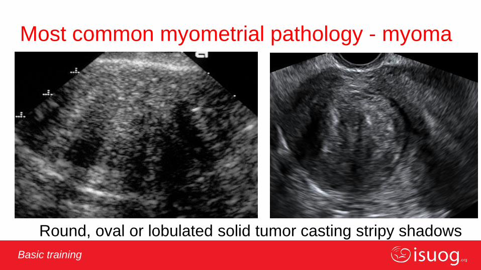
Most common myometrial pathology - myoma
Round, oval or lobulated solid tumor casting stripy shadows

Editable text here Basic training
Hyperechogenic uterine myoma
Editable text here Basic training
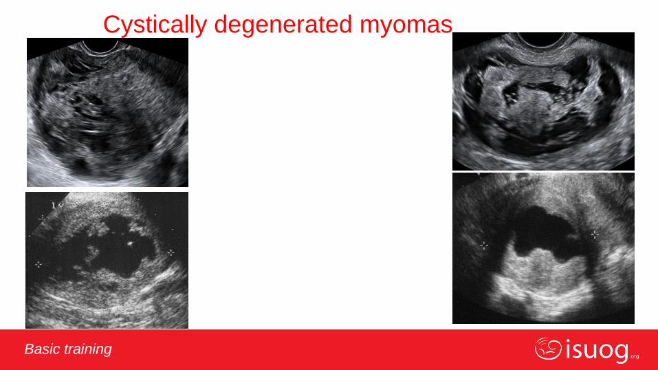
Cystically degenerated myomas
Editable text here Basic training
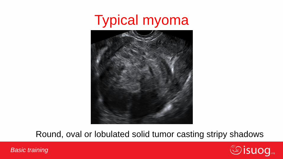
Typical myoma
Round, oval or lobulated solid tumor casting stripy shadows
Editable text here Basic training
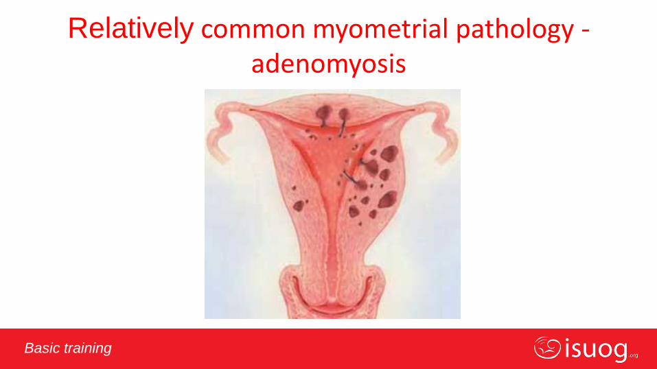
Relatively common myometrial pathology - adenomyosis
Editable text here Basic training
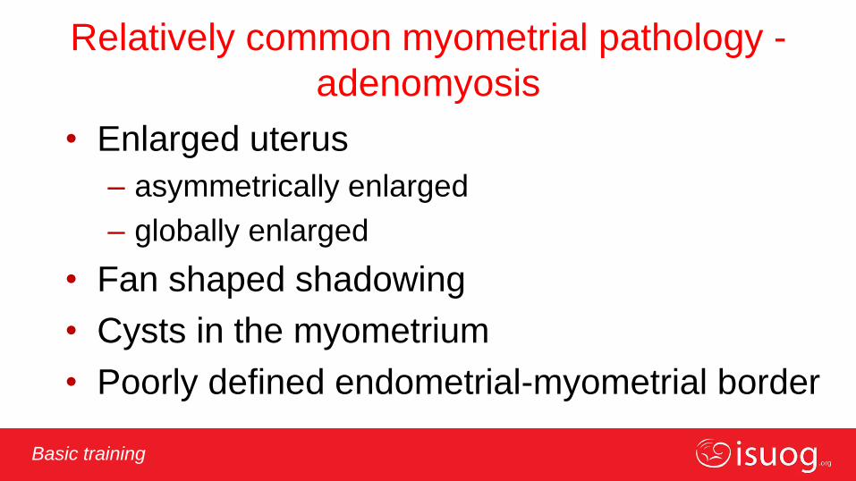
Relatively common myometrial pathology -
adenomyosis
• Enlarged uterus
– asymmetrically enlarged
– globally enlarged
• Fan shaped shadowing
• Cysts in the myometrium
• Poorly defined endometrial-myometrial border
Editable text here Basic training
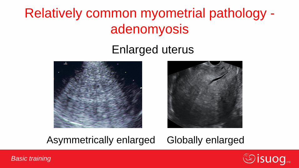
Relatively common myometrial pathology -
adenomyosis
Enlarged uterus
Asymmetrically enlarged Globally enlarged
Editable text here Basic training
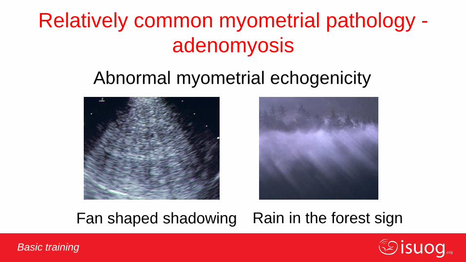
Relatively common myometrial pathology -
adenomyosis
Abnormal myometrial echogenicity
Fan shaped shadowing Fan shaped shadowing Rain in the forest sign

Editable text here Basic training
Relatively common myometrial pathology -
adenomyosis
Cysts in the myometrium Poorly defined endometrium
(subendometrial lines and buds)
Editable text here Basic training
Most common intracavitary pathology
• Polyp
• Submucuous myoma
• Hyperplasia
– without atypia
– with atypia
• Cancer
Editable text here Basic training
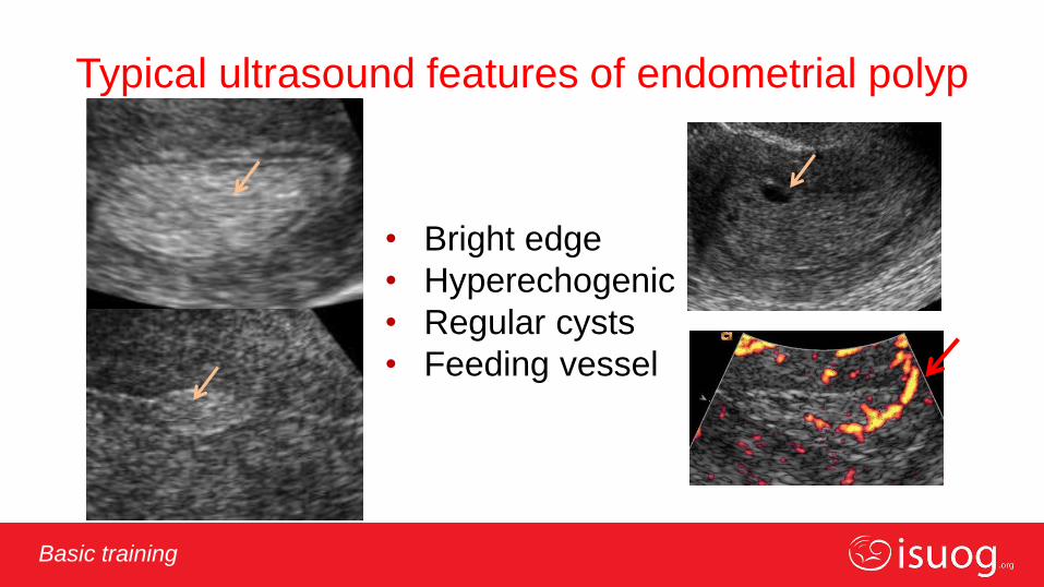
Typical ultrasound features of endometrial polyp
• Bright edge
• Hyperechogenic
• Regular cysts
• Feeding vessel
Editable text here Basic training
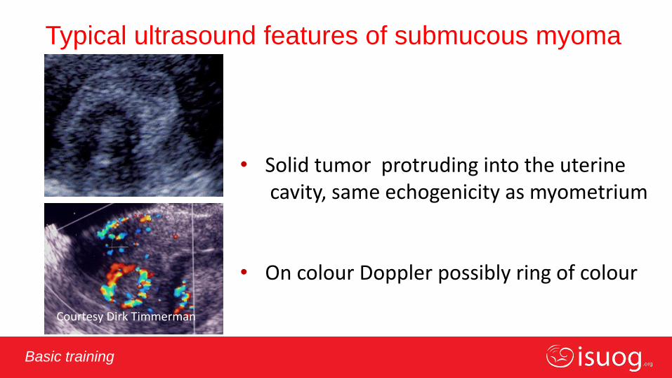
Typical ultrasound features of submucous myoma
• Solid tumor protruding into the uterine cavity, same echogenicity as myometrium • On colour Doppler possibly ring of colour
Courtesy Dirk Timmerman
Editable text here Basic training
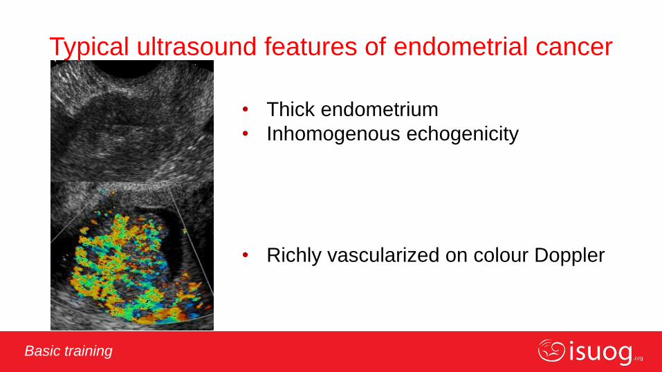
Typical ultrasound features of endometrial cancer
• Thick endometrium
• Inhomogenous echogenicity
• Richly vascularized on colour Doppler
Editable text here Basic training
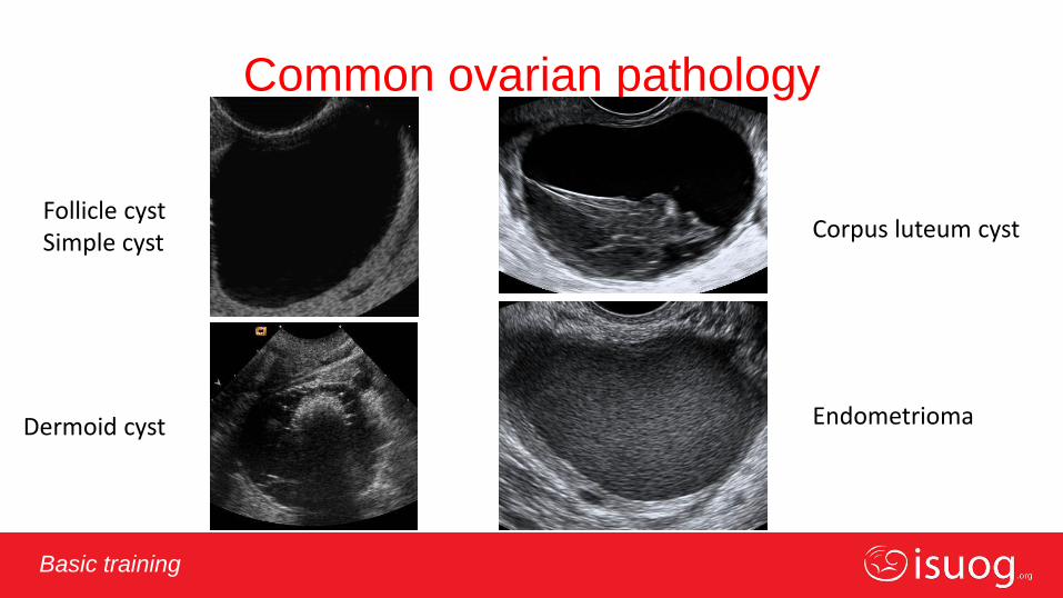
Common ovarian pathology
Follicle cyst Simple cyst
Corpus luteum cyst
Dermoid cyst Endometrioma

Editable text here Basic training
Dermoid cyst
White ball Shadowing
Shadowing Bright lines and dots
Editable text here Basic training
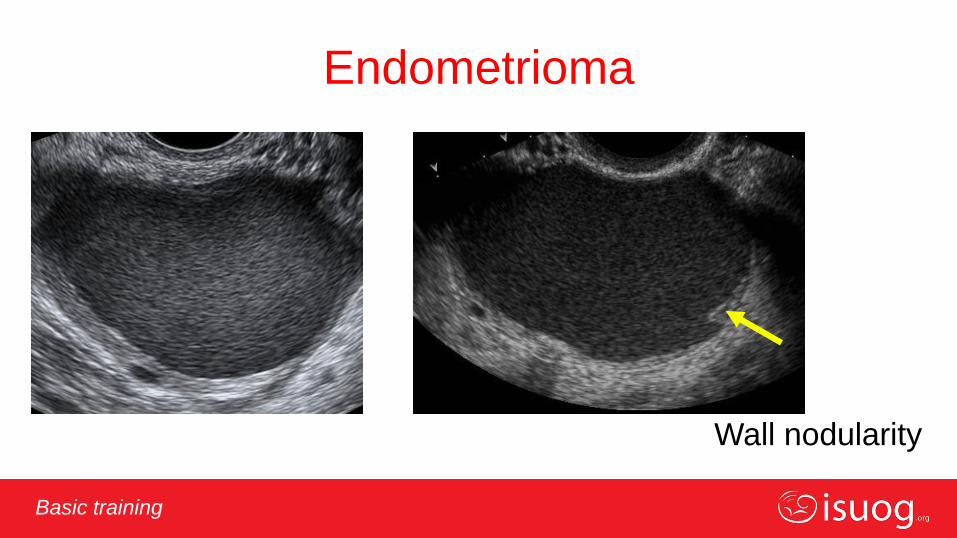
Endometrioma
Wall nodularity

Editable text here Basic training
Common ovarian pathology
Follicle cyst
Simple cyst Corpus luteum cyst
Dermoid cyst Endometrioma

Editable text here Basic training
Common extraovarian adnexal pathology
• Hydrosalpinx
• Paraovarian cysts
• Peritoneal inclusion cysts

Editable text here Basic training
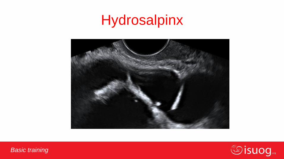
Hydro-pyo-hemato-salpinx
Sausage shape Cog wheel Beads on a string
Incomplete septa Incomplete septa
Editable text here Basic training
Hydrosalpinx
Editable text here Basic training
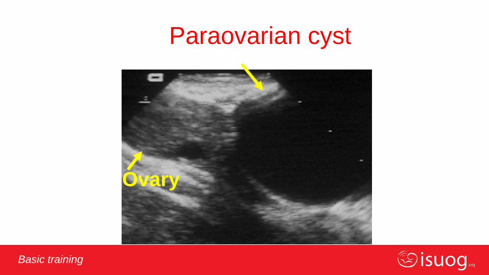
Paraovarian cyst
Ovary
Editable text here Basic training
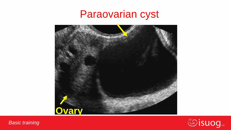
paraovarian
Paraovarian cyst
Ovary

Editable text here Basic training
Peritoneal pseudocyst
Ovary

Editable text here Basic training
Rules of thumb for discriminating between
benign and malignant adenxal masses
Malignant
irregularity
Benign
NO irregularity
Mucinous borderline
gastrointestinal type
Multilocular
with
many locules
Editable text here Basic training
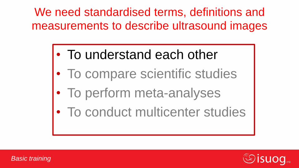
We need standardised terms, definitions and measurements to describe ultrasound images
• To understand each other
• To compare scientific studies
• To perform meta-analyses
• To conduct multicenter studies
Editable text here Basic training
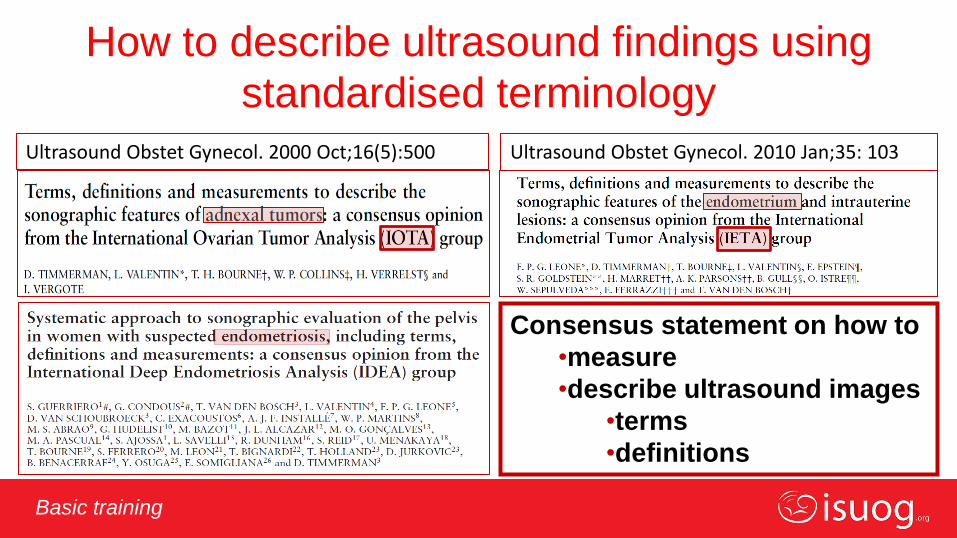
How to describe ultrasound findings using
standardised terminology
Consensus statement on how to
•measure
•describe ultrasound images
•terms
•definitions
Ultrasound Obstet Gynecol. 2000 Oct;16(5):500 Ultrasound Obstet Gynecol. 2010 Jan;35: 103
Editable text here Basic training
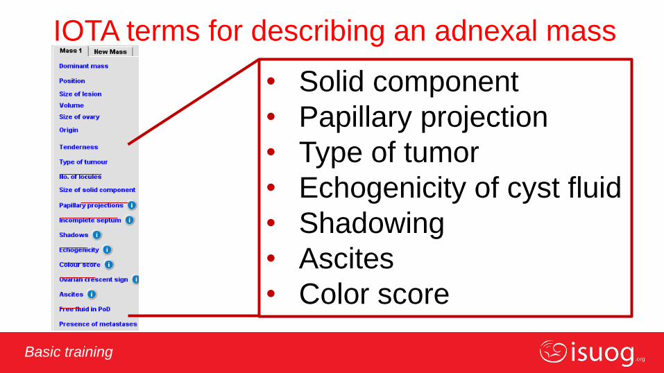
IOTA terms for describing an adnexal mass
• Solid component
• Papillary projection
• Type of tumor
• Echogenicity of cyst fluid
• Shadowing
• Ascites
• Color score

Editable text here Basic training
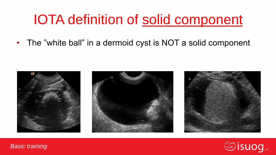
IOTA definition of solid component
• A structure that has
echogenicity
suggestive of tissue
– (myometrium, ovarian
stroma)
Editable text here Basic training
IOTA definition of solid component
• The ”white ball” in a dermoid cyst is NOT a solid component

Editable text here Basic training
IOTA definition of solid component
• Blood clot or other amorphous
material is NOT a solid
component

Editable text here Basic training
IOTA definition of solid component
• Blood clot, amorphous material or solid
tissue?
– push on the lesion

Editable text here Basic training
IOTA definition of solid component
• Blood clot, amorphous material
or solid tissue?
– colour Doppler
If in doubt – classify as solid tissue
Editable text here Basic training
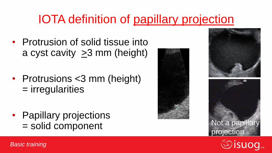
IOTA definition of papillary projection
• Protrusion of solid tissue into a cyst cavity >3 mm (height)
• Protrusions <3 mm (height) = irregularities
• Papillary projections = solid component
Not a papillary
projection

Editable text here Basic training
Five types of lesion/tumor (IOTA)
Unilocular
Solid
Unilocular solid
Multilocular solid
Multilocular
Unilocular
Solid
Unilocular solid
Multilocular solid
Multilocular
Editable text here Basic training
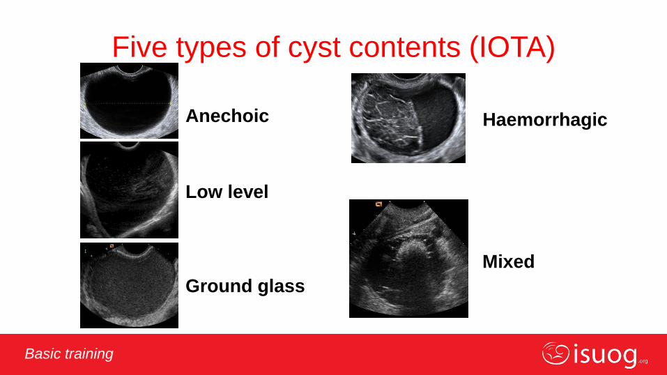
Five types of cyst contents (IOTA)
Anechoic
Low level
Ground glass
Mixed
Haemorrhagic
Editable text here Basic training
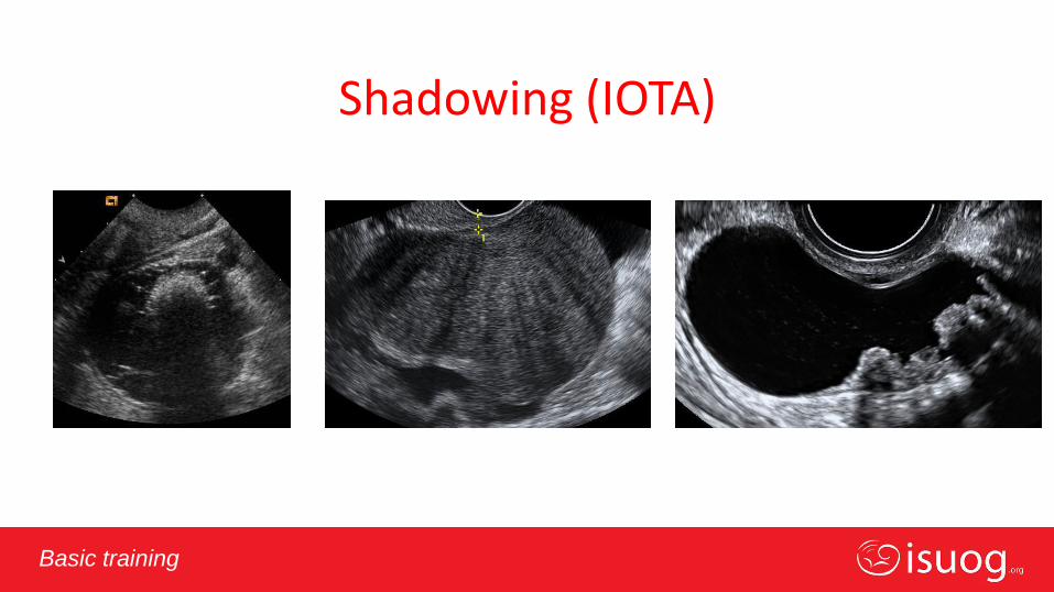
Shadowing (IOTA)

Editable text here Basic training
Ascites (IOTA)
Fluid outside the pouch of Douglas

Editable text here Basic training
The IOTA colour score
Adjust settings: maximize detection of flow without artifacts
(Pulse repetition frequency 0.3-0.6 KHz, 3-6 cm/s velocity scale)
Score 2
Score 3
Score 4
Score 1

Editable text here Basic training
Measurements
Editable text here Basic training
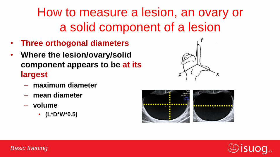
How to measure a lesion, an ovary or
a solid component of a lesion • Three orthogonal diameters
• Where the lesion/ovary/solid
component appears to be at its
largest
– maximum diameter
– mean diameter
– volume
• (L*D*W*0.5)
Editable text here Basic training
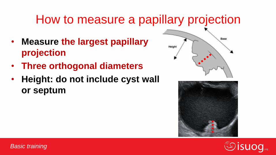
How to measure a papillary projection
• Measure the largest papillary
projection
• Three orthogonal diameters
• Height: do not include cyst wall
or septum
Editable text here Basic training
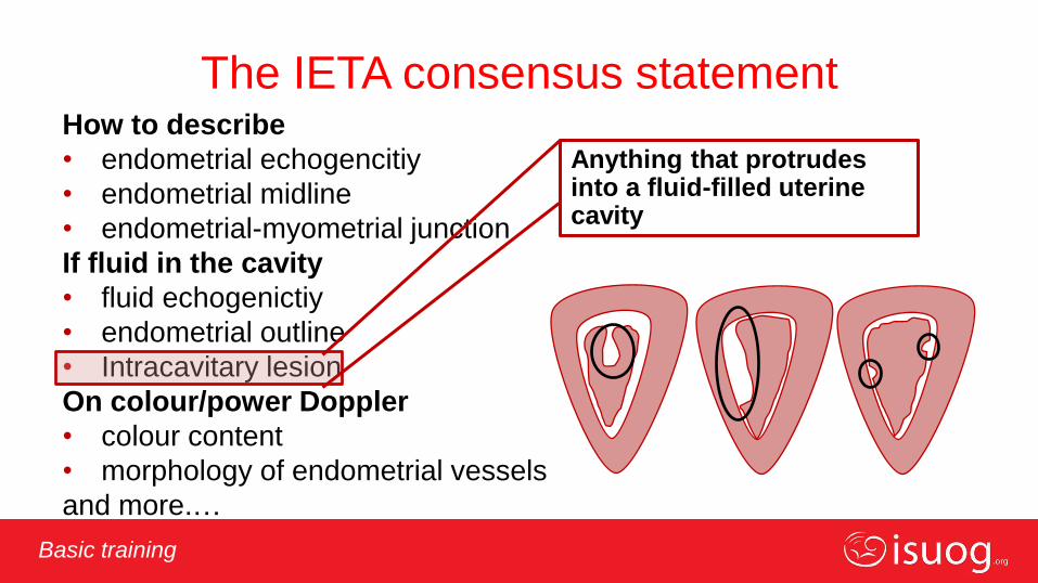
The IETA consensus statement How to describe
• endometrial echogencitiy
• endometrial midline
• endometrial-myometrial junction
If fluid in the cavity
• fluid echogenictiy
• endometrial outline
• Intracavitary lesion
On colour/power Doppler
• colour content
• morphology of endometrial vessels
and more.…
Anything that protrudes into a fluid-filled uterine cavity
Editable text here Basic training
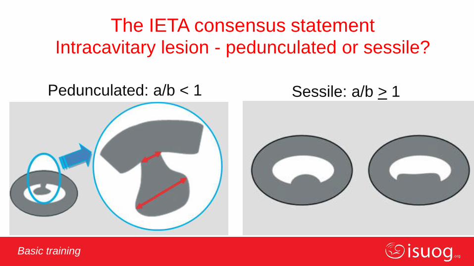
The IETA consensus statement Intracavitary lesion - pedunculated or sessile?
Pedunculated: a/b < 1 Sessile: a/b > 1

Editable text here Basic training
IETA consensus statment
Doppler ultrasound examination of the
endometrium
Quantification of the color content of the endometrial scan
Colour score 1
= no colour
Colour score 2
= minimal colour
Colour score 3
= moderate colour
Colour score 4
= abundant colour
Adjust settings: maximize detection of flow without artifacts
(Pulse repetition frequency 0.3-0.6 KHz, 3-6 cm/s velocity scale)

Editable text here Basic training
Which patients should I refer for
specialist opinion?
• Those in whom you are uncertain about the
diagnosis (especially if you suspect
malignancy)
Editable text here Basic training
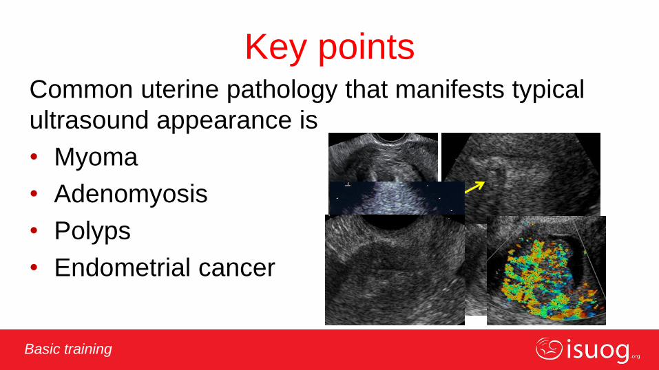
Key points Common uterine pathology that manifests typical
ultrasound appearance is
• Myoma
• Adenomyosis
• Polyps
• Endometrial cancer
Editable text here Basic training
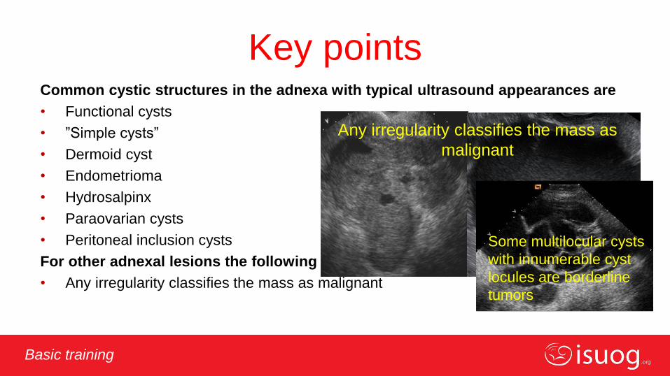
Key points Common cystic structures in the adnexa with typical ultrasound appearances are
• Functional cysts
• ”Simple cysts”
• Dermoid cyst
• Endometrioma
• Hydrosalpinx
• Paraovarian cysts
• Peritoneal inclusion cysts
For other adnexal lesions the following applies
• Any irregularity classifies the mass as malignant
Any irregularity classifies the mass as
malignant
Some multilocular cysts
with innumerable cyst
locules are borderline
tumors
Editable text here Basic training
Key points
We should use a standardised terminology when we describe ultrasound images of
• Adnexal lesions (IOTA)
• The endometrium /uterine cavity (IETA)
• The myometrium (MUSA)
• Deep infiltrating endometriosis (IDEA)

Editable text here Basic training
Key points
When in doubt:
refer for second opinion

Editable text here Basic training ISUOG’s basic training curriculum
THANK YOU
Related Documents