APPLIED AND ENVIRONMENTAL MICROBIOLOGY, July 2008, p. 4070–4078 Vol. 74, No. 13 0099-2240/08/$08.000 doi:10.1128/AEM.00428-08 Copyright © 2008, American Society for Microbiology. All Rights Reserved. Isolation and Characterization of Bacteriophages Infecting the Fish Pathogen Flavobacterium psychrophilum Anne Rønnest Stenholm, 1,2 Inger Dalsgaard, 2 and Mathias Middelboe 1 * Marine Biological Laboratory, University of Copenhagen, Strandpromenaden 5, 3000 Helsingør, Denmark, 1 and National Institute of Aquatic Resources, Fish Disease Laboratory, Technical University of Denmark, Stigbøjlen 4, 1870 Frederiksberg C, Denmark 2 Received 21 February 2008/Accepted 5 May 2008 Flavobacterium psychrophilum is a serious pathogen in trout aquaculture, responsible for the diseases rainbow trout fry syndrome (RTFS) and cold water disease (CWD). Bacteriophage control of F. psychrophilum may constitute a realistic approach in the treatment of these diseases; however, a detailed understanding of the phage-host interactions is needed to evaluate the potential of F. psychrophilum bacteriophages for that purpose. Twenty-two F. psychrophilum phages from Danish rainbow trout farms were isolated and characterized. The phage genome sizes differed considerably and fell into three major size classes (8.5 to 12 kb, 48 kb, and 90 kb). The phage host ranges comprised from 5 to 23 of the 28 tested F. psychrophilum strains, and 18 of the phage isolates showed unique host ranges. Each bacterial strain had a unique pattern of susceptibility to the 22 phages, and individual strains also showed large variations (up to 10 7 -fold differences) in susceptibility to specific phages. Phage burst size (7 to 162 phages infected cell 1 ) and latency period (4 to 6 h) also showed pronounced differences both between phages and, for a specific phage, between host strains. In general, the characterization documented the presence of diverse F. psychrophilum phage communities in Danish trout farms, with highly variable patterns of infectivity. The discovery and characterization of broad-host-range phages with strong lytic potential against numerous pathogenic F. psychrophilum host strains thus provided the foundation for future exploration of the potential of phages in the treatment of RTFS and CWD. Flavobacterium psychrophilum is responsible for the diseases rainbow trout fry syndrome (RTFS) and cold water disease, caus- ing considerable economic losses in salmonid aquaculture world- wide (27). Fish infected with F. psychrophilum have high mortality rates, and fry are especially affected, with mortalities of 50 to 60% (19). Treatment with antibiotics is required to limit economic losses; however, F. psychrophilum seems to be increasingly more resistant to the approved drugs (5). Today, florfenicol is the drug of choice, and no commercial vaccine is yet available. There is, therefore, a need for alternative treatments of F. psychrophilum infections in aquaculture, especially in infected fry. Phage therapy may be a realistic alternative approach for con- trolling pathogenic bacteria in aquaculture. The use of bacterio- phages to control bacterial pathogens has previously demon- strated promising potential for various types of bacteria in the food industry. Much of the recent research has focused on reduc- ing the impacts of food-borne human bacterial pathogens, such as Escherichia coli in domestic animals (e.g., references 28 and 34) and Salmonella (11), Listeria (18), and Campylobacter (3) species in fruit, diary products, and poultry (reviewed in reference 12). However, in addition to the current attempts to apply phages in the control of human pathogens, fish and shellfish pathogens have also been investigated as a target for phage therapy. A number of phages have been isolated for potential use in phage therapy against important fish and shellfish pathogens, such as Aeromonas salmonicida in brook trout (Oncorhynchus fontinalis) (15), Vibrio harveyi in shrimp (Penaeus monodon) (16, 31, 35), Pseudomonas plecoglossicida in ayu (Plecoglossus altivelis) (25, 29, 30), and Lactococcus garvieae in yellowtail (Seriola quinqueradiata) (26). All of these studies have dem- onstrated the potential of specific phages to significantly re- duce the impacts of their bacterial hosts, with a resulting pos- itive effect on fish survival. The studies emphasize the need for further investigations of the possibilities in using phages as an alternative to antibiotic treatment of other fish diseases in aquaculture. The application of phages to control a certain bacterial pathogen is complicated by the high degrees of phenotypic and genotypic diversity within populations of both phages and bac- teria (e.g., references 6 and 13). Consequently, individual strains of a pathogen may be more or less susceptible or even resistant to different cooccurring phages. For a successful phage control of pathogenic strains, it is therefore a prerequi- site that a detailed characterization of isolated phages be ob- tained with respect to host range, adsorption rate, lytic poten- tial, interaction with host, etc. Flavobacterium psychrophilum bacteriophages have to our knowledge not previously been described in the literature. The purpose of the present study was to isolate and characterize a number of lytic phages that infect F. psychrophilum and to inves- tigate their lytic properties toward their host bacterium under controlled conditions in the laboratory. The study thus provides the basis for an evaluation of the potential of phages to control the F. psychrophilum pathogen and consequently their potential application as a treatment of RTFS in aquaculture. MATERIALS AND METHODS Bacterial strains. The Flavobacterium psychrophilum strains used in this study are listed in Table 1. The strains included the type strain NCIMB 1947 T (serotype Fp T , nonvirulent for rainbow trout) as well as two well-characterized Danish * Corresponding author. Mailing address: Marine Biological Labora- tory, University of Copenhagen, Strandpromenaden 5, 3000 Helsingør, Denmark. Phone: 45 3532 1991. Fax: 45 3532 1951. E-mail: MMiddelboe @bio.ku.dk. Published ahead of print on 9 May 2008. 4070

Welcome message from author
This document is posted to help you gain knowledge. Please leave a comment to let me know what you think about it! Share it to your friends and learn new things together.
Transcript
APPLIED AND ENVIRONMENTAL MICROBIOLOGY, July 2008, p. 4070–4078 Vol. 74, No. 130099-2240/08/$08.00�0 doi:10.1128/AEM.00428-08Copyright © 2008, American Society for Microbiology. All Rights Reserved.
Isolation and Characterization of Bacteriophages Infecting the FishPathogen Flavobacterium psychrophilum�
Anne Rønnest Stenholm,1,2 Inger Dalsgaard,2 and Mathias Middelboe1*Marine Biological Laboratory, University of Copenhagen, Strandpromenaden 5, 3000 Helsingør, Denmark,1 and National Institute of
Aquatic Resources, Fish Disease Laboratory, Technical University of Denmark, Stigbøjlen 4, 1870 Frederiksberg C, Denmark2
Received 21 February 2008/Accepted 5 May 2008
Flavobacterium psychrophilum is a serious pathogen in trout aquaculture, responsible for the diseasesrainbow trout fry syndrome (RTFS) and cold water disease (CWD). Bacteriophage control of F. psychrophilummay constitute a realistic approach in the treatment of these diseases; however, a detailed understanding of thephage-host interactions is needed to evaluate the potential of F. psychrophilum bacteriophages for that purpose.Twenty-two F. psychrophilum phages from Danish rainbow trout farms were isolated and characterized. Thephage genome sizes differed considerably and fell into three major size classes (8.5 to 12 kb, 48 kb, and 90 kb).The phage host ranges comprised from 5 to 23 of the 28 tested F. psychrophilum strains, and 18 of the phageisolates showed unique host ranges. Each bacterial strain had a unique pattern of susceptibility to the 22phages, and individual strains also showed large variations (up to 107-fold differences) in susceptibility tospecific phages. Phage burst size (7 to 162 phages infected cell�1) and latency period (4 to 6 h) also showedpronounced differences both between phages and, for a specific phage, between host strains. In general, thecharacterization documented the presence of diverse F. psychrophilum phage communities in Danish troutfarms, with highly variable patterns of infectivity. The discovery and characterization of broad-host-rangephages with strong lytic potential against numerous pathogenic F. psychrophilum host strains thus provided thefoundation for future exploration of the potential of phages in the treatment of RTFS and CWD.
Flavobacterium psychrophilum is responsible for the diseasesrainbow trout fry syndrome (RTFS) and cold water disease, caus-ing considerable economic losses in salmonid aquaculture world-wide (27). Fish infected with F. psychrophilum have high mortalityrates, and fry are especially affected, with mortalities of 50 to 60%(19). Treatment with antibiotics is required to limit economiclosses; however, F. psychrophilum seems to be increasingly moreresistant to the approved drugs (5). Today, florfenicol is the drugof choice, and no commercial vaccine is yet available. There is,therefore, a need for alternative treatments of F. psychrophiluminfections in aquaculture, especially in infected fry.
Phage therapy may be a realistic alternative approach for con-trolling pathogenic bacteria in aquaculture. The use of bacterio-phages to control bacterial pathogens has previously demon-strated promising potential for various types of bacteria in thefood industry. Much of the recent research has focused on reduc-ing the impacts of food-borne human bacterial pathogens, such asEscherichia coli in domestic animals (e.g., references 28 and 34)and Salmonella (11), Listeria (18), and Campylobacter (3) speciesin fruit, diary products, and poultry (reviewed in reference 12).However, in addition to the current attempts to apply phages inthe control of human pathogens, fish and shellfish pathogens havealso been investigated as a target for phage therapy.
A number of phages have been isolated for potential use inphage therapy against important fish and shellfish pathogens,such as Aeromonas salmonicida in brook trout (Oncorhynchusfontinalis) (15), Vibrio harveyi in shrimp (Penaeus monodon)
(16, 31, 35), Pseudomonas plecoglossicida in ayu (Plecoglossusaltivelis) (25, 29, 30), and Lactococcus garvieae in yellowtail(Seriola quinqueradiata) (26). All of these studies have dem-onstrated the potential of specific phages to significantly re-duce the impacts of their bacterial hosts, with a resulting pos-itive effect on fish survival. The studies emphasize the need forfurther investigations of the possibilities in using phages as analternative to antibiotic treatment of other fish diseases inaquaculture.
The application of phages to control a certain bacterialpathogen is complicated by the high degrees of phenotypic andgenotypic diversity within populations of both phages and bac-teria (e.g., references 6 and 13). Consequently, individualstrains of a pathogen may be more or less susceptible or evenresistant to different cooccurring phages. For a successfulphage control of pathogenic strains, it is therefore a prerequi-site that a detailed characterization of isolated phages be ob-tained with respect to host range, adsorption rate, lytic poten-tial, interaction with host, etc.
Flavobacterium psychrophilum bacteriophages have to ourknowledge not previously been described in the literature. Thepurpose of the present study was to isolate and characterize anumber of lytic phages that infect F. psychrophilum and to inves-tigate their lytic properties toward their host bacterium undercontrolled conditions in the laboratory. The study thus providesthe basis for an evaluation of the potential of phages to controlthe F. psychrophilum pathogen and consequently their potentialapplication as a treatment of RTFS in aquaculture.
MATERIALS AND METHODS
Bacterial strains. The Flavobacterium psychrophilum strains used in this studyare listed in Table 1. The strains included the type strain NCIMB 1947T (serotypeFpT, nonvirulent for rainbow trout) as well as two well-characterized Danish
* Corresponding author. Mailing address: Marine Biological Labora-tory, University of Copenhagen, Strandpromenaden 5, 3000 Helsingør,Denmark. Phone: 45 3532 1991. Fax: 45 3532 1951. E-mail: [email protected].
� Published ahead of print on 9 May 2008.
4070
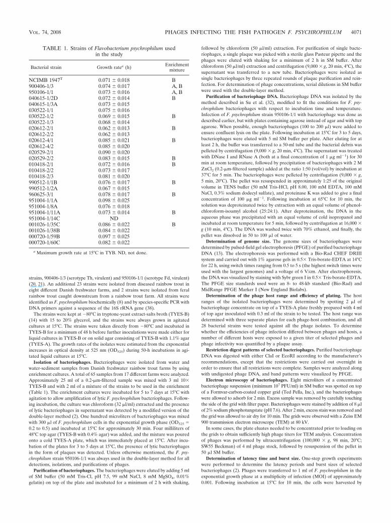
strains, 900406-1/3 (serotype Th, virulent) and 950106-1/1 (serotype Fd, virulent)(20, 21). An additional 23 strains were isolated from diseased rainbow trout ineight different Danish freshwater farms, and 2 strains were isolated from feralrainbow trout caught downstream from a rainbow trout farm. All strains wereidentified as F. psychrophilum biochemically (8) and by species-specific PCR withDNA primers against a sequence of the 16S rRNA gene (38).
The strains were kept at �80°C in tryptone-yeast extract-salts broth (TYES-B)(14) with 15 to 20% glycerol, and the strains were always grown in agitatedcultures at 15°C. The strains were taken directly from �80°C and incubated inTYES-B for a minimum of 48 h before further inoculations were made either forliquid cultures in TYES-B or on solid agar consisting of TYES-B with 1.1% agar(TYES-A). The growth rates of the isolates were estimated from the exponentialincreases in optical density at 525 nm (OD525) during 50-h incubations in agi-tated liquid cultures at 15°C.
Isolation of bacteriophages. Bacteriophages were isolated from water andwater-sediment samples from Danish freshwater rainbow trout farms by usingenrichment cultures. A total of 65 samples from 17 different farms were analyzed.Approximately 25 ml of a 0.2-�m-filtered sample was mixed with 3 ml 10�TYES-B and with 2 ml of a mixture of the strains to be used in the enrichment(Table 1). The enrichment cultures were incubated for 5 to 7 days at 15°C withagitation to allow amplification of lytic F. psychrophilum bacteriophages. Follow-ing incubation, the culture was chloroform (32 �l/ml) extracted and the presenceof lytic bacteriophages in supernatant was detected by a modified version of thedouble-layer method (2). One hundred microliters of bacteriophages was mixedwith 300 �l of F. psychrophilum cells in the exponential growth phase (OD525 �0.2 to 0.5) and incubated at 15°C for approximately 30 min. Four milliliters of48°C top agar (TYES-B with 0.4% agar) was added, and the mixture was pouredonto a cold TYES-A plate, which was immediately placed at 15°C. After incu-bation of the plates for 3 to 5 days at 15°C, the presence of lytic bacteriophagesin the form of plaques was detected. Unless otherwise mentioned, the F. psy-chrophilum strain 950106-1/1 was always used in the double-layer method for alldetections, isolations, and purifications of phages.
Purification of bacteriophages. The bacteriophages were eluted by adding 5 mlof SM buffer (50 mM Tris-Cl, pH 7.5, 99 mM NaCl, 8 mM MgSO4, 0.01%gelatin) on top of the plate and incubated for a minimum of 2 h with shaking,
followed by chloroform (50 �l/ml) extraction. For purification of single bacte-riophages, a single plaque was picked with a sterile glass Pasteur pipette and thephages were eluted with shaking for a minimum of 2 h in SM buffer. Afterchloroform (50 �l/ml) extraction and centrifugation (9,000 � g, 20 min, 4°C), thesupernatant was transferred to a new tube. Bacteriophages were isolated assingle bacteriophages by three repeated rounds of plaque purification and rein-fection. For determination of phage concentrations, serial dilutions in SM bufferwere used with the double-layer method.
Purification of bacteriophage DNA. Bacteriophage DNA was isolated by themethod described in Su et al. (32), modified to fit the conditions for F. psy-chrophilum bacteriophages with respect to incubation time and temperature.Infection of F. psychrophilum strain 950106-1/1 with bacteriophage was done asdescribed earlier, but with plates containing agarose instead of agar and with topagarose. When possible, enough bacteriophages (100 to 200 �l) were added toensure confluent lysis on the plate. Following incubation at 15°C for 3 to 5 days,bacteriophages were eluted with 5 ml SM buffer per plate. After eluting for atleast 2 h, the buffer was transferred to a 50-ml tube and the bacterial debris waspelleted by centrifugation (9,000 � g, 20 min, 4°C). The supernatant was treatedwith DNase I and RNase A (both at a final concentration of 1 �g ml�1) for 30min at room temperature, followed by precipitation of bacteriophages with 2 MZnCl2 (0.2-�m-filtered sample) added at the ratio 1:50 (vol/vol) by incubation at37°C for 5 min. The bacteriophages were pelleted by centrifugation (9,000 � g,5 min, 20°C). The pellet was resuspended in approximately 1:25 of the originalvolume in TENS buffer (50 mM Tris-HCl, pH 8.00, 100 mM EDTA, 100 mMNaCl, 0.3% sodium dodecyl sulfate), and proteinase K was added to give a finalconcentration of 100 �g ml�1. Following incubation at 65°C for 10 min, thesolution was deproteinated twice by extraction with an equal volume of phenol-chloroform-isoamyl alcohol (25:24:1). After deproteination, the DNA in theaqueous phase was precipitated with an equal volume of cold isopropanol andincubated at room temperature for 5 min, followed by centrifugation at 16,000 �g (10 min, 4°C). The DNA was washed twice with 70% ethanol, and finally, thepellet was dissolved in 50 to 100 �l of water.
Determination of genome size. The genome sizes of bacteriophages weredetermined by pulsed-field gel electrophoresis (PFGE) of purified bacteriophageDNA (13). The electrophoresis was performed with a Bio-Rad CHEF DRIIIsystem and carried out with 1% agarose gels in 0.5� Tris-borate-EDTA at 14°Cfor 22 h, using switch times ranging from 0.5 to 5 s (the highest switch times wereused with the largest genomes) and a voltage of 6 V/cm. After electrophoresis,the DNA was visualized by staining with Sybr green I in 0.5� Tris-borate-EDTA.The PFGE size standards used were an 8- to 48-kb standard (Bio-Rad) andMidRange PFGE Marker I (New England Biolabs).
Determination of the phage host range and efficiency of plating. The hostranges of the isolated bacteriophages were determined by spotting 2 �l ofbacteriophage concentrate on top of a TYES-A plate freshly prepared with 4 mlof top agar inoculated with 0.3 ml of the strain to be tested. The host range wasdetermined with three separate plates for each phage-host combination, and all28 bacterial strains were tested against all the phage isolates. To determinewhether the efficiencies of phage infection differed between phages and hosts, anumber of different hosts were exposed to a given titer of selected phages andphage infectivity was quantified by a plaque assay.
Restriction digest patterns of selected bacteriophages. Purified bacteriophageDNA was digested with either ClaI or EcoRI according to the manufacturer’srecommendations, except that the restrictions were carried out overnight inorder to ensure that all restrictions were complete. Samples were analyzed alongwith undigested phage DNA, and band patterns were visualized by PFGE.
Electron microscopy of bacteriophages. Eight microliters of a concentratedbacteriophage suspension (minimum 107 PFU/ml) in SM buffer was spotted on topof a Formvar-carbon-coated copper grid (Ted Pella, Inc.), and the bacteriophageswere allowed to adsorb for 2 min. Excess sample was removed by carefully touchingthe side of the grid with filter paper. Bacteriophages were stained by addition of 8 �lof 2% sodium phosphotungstate (pH 7.6). After 2 min, excess stain was removed andthe grid was allowed to air dry for 10 min. The grids were observed with a Zeiss EM900 transmission electron microscope (TEM) at 80 kV.
In some cases, the plate eluates needed to be concentrated prior to loading onthe grids to obtain sufficiently high phage titers for TEM analysis. Concentrationof phages was performed by ultracentrifugation (100,000 � g, 98 min, 20°C;SW55 Beckman) of 4 ml phage stock, followed by resuspension of the pellet in50 �l SM buffer.
Determination of latency time and burst size. One-step growth experimentswere performed to determine the latency periods and burst sizes of selectedbacteriophages (2). Phages were transferred to 1 ml of F. psychrophilum in theexponential growth phase at a multiplicity of infection (MOI) of approximately0.001. Following incubation at 15°C for 10 min, the cells were harvested by
TABLE 1. Strains of Flavobacterium psychrophilum usedin the study
Bacterial strain Growth ratea (h) Enrichmentmixture
NCIMB 1947T 0.071 � 0.018 B900406-1/3 0.074 � 0.017 A, B950106-1/1 0.073 � 0.016 A, B040615-1/2D 0.072 � 0.014 B040615-1/3A 0.073 � 0.015030522-1/1 0.075 � 0.016030522-1/2 0.069 � 0.015 B030522-1/3 0.068 � 0.014020612-2/1 0.062 � 0.013 B020612-2/2 0.062 � 0.013020612-4/1 0.085 � 0.021 B020612-4/2 0.085 � 0.020020529-2/1 0.090 � 0.020020529-2/2 0.083 � 0.015 B010418-2/1 0.072 � 0.016 B010418-2/2 0.073 � 0.017010418-2/3 0.081 � 0.020990512-1/1B 0.076 � 0.017 B990512-1/2A 0.067 � 0.015960625-3/1 0.078 � 0.017 B951004-1/1A 0.098 � 0.025951004-1/8A 0.076 � 0.018951004-1/11A 0.073 � 0.014 B951004-1/14C ND001026-1/35C 0.086 � 0.022 B001026-1/38B 0.084 � 0.022000720-1/59B 0.097 � 0.025000720-1/60C 0.082 � 0.022
a Maximum growth rate at 15°C in TYB. ND, not done.
VOL. 74, 2008 PHAGES INFECTING THE FISH PATHOGEN F. PSYCHROPHILUM 4071
centrifugation at 10,000 � g for 10 min at 15°C. Cells were then resuspended in1 ml of cold TYES-B and inoculated to 60 ml of cold TYES-B. From this pointon, samples were collected every 30 to 60 min for a minimum of 8 h. Theconcentration of bacteriophages was determined immediately by the double-layer method.
Phage adsorption rate. The rate of adsorption of FpV-9 to its host cells wasdetermined by adding the phage to a 30-ml culture of exponentially growing F.psychrophilum strain 950106-1/1 at an MOI of 0.0005 and incubating the infectedculture at 15°C with agitation (36). Samples (150 �l) were taken at 0, 5, 10, 15,20, 30, 40, 50, and 60 min, immediately diluted 1:10 in SM buffer with chloroform(50 �l ml�1), mixed thoroughly, and centrifuged. The supernatant was trans-ferred to a new tube, and the number of unadsorbed bacteriophages on F.psychrophilum strain 950106-1/1 was determined by the double-layer method.Phage adsorption rate was determined as the exponential rate of decrease inPFU over time during incubation.
RESULTS
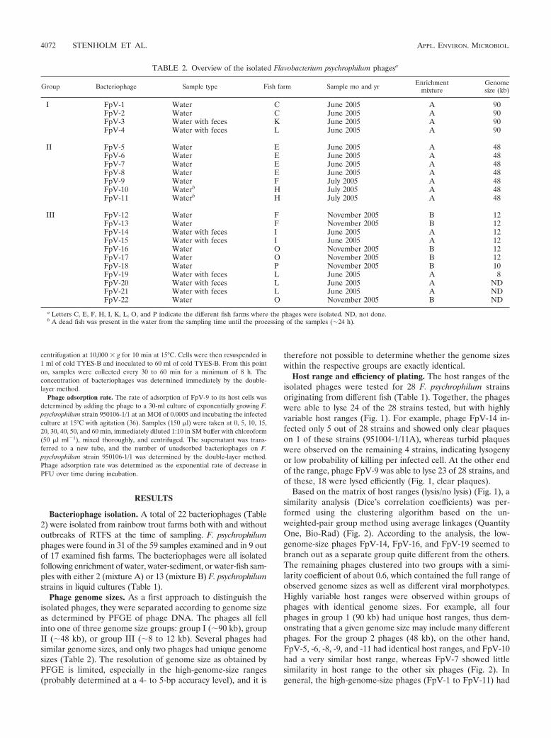
Bacteriophage isolation. A total of 22 bacteriophages (Table2) were isolated from rainbow trout farms both with and withoutoutbreaks of RTFS at the time of sampling. F. psychrophilumphages were found in 31 of the 59 samples examined and in 9 outof 17 examined fish farms. The bacteriophages were all isolatedfollowing enrichment of water, water-sediment, or water-fish sam-ples with either 2 (mixture A) or 13 (mixture B) F. psychrophilumstrains in liquid cultures (Table 1).
Phage genome sizes. As a first approach to distinguish theisolated phages, they were separated according to genome sizeas determined by PFGE of phage DNA. The phages all fellinto one of three genome size groups: group I (�90 kb), groupII (�48 kb), or group III (�8 to 12 kb). Several phages hadsimilar genome sizes, and only two phages had unique genomesizes (Table 2). The resolution of genome size as obtained byPFGE is limited, especially in the high-genome-size ranges(probably determined at a 4- to 5-bp accuracy level), and it is
therefore not possible to determine whether the genome sizeswithin the respective groups are exactly identical.
Host range and efficiency of plating. The host ranges of theisolated phages were tested for 28 F. psychrophilum strainsoriginating from different fish (Table 1). Together, the phageswere able to lyse 24 of the 28 strains tested, but with highlyvariable host ranges (Fig. 1). For example, phage FpV-14 in-fected only 5 out of 28 strains and showed only clear plaqueson 1 of these strains (951004-1/11A), whereas turbid plaqueswere observed on the remaining 4 strains, indicating lysogenyor low probability of killing per infected cell. At the other endof the range, phage FpV-9 was able to lyse 23 of 28 strains, andof these, 18 were lysed efficiently (Fig. 1, clear plaques).
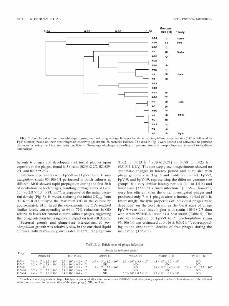
Based on the matrix of host ranges (lysis/no lysis) (Fig. 1), asimilarity analysis (Dice’s correlation coefficients) was per-formed using the clustering algorithm based on the un-weighted-pair group method using average linkages (QuantityOne, Bio-Rad) (Fig. 2). According to the analysis, the low-genome-size phages FpV-14, FpV-16, and FpV-19 seemed tobranch out as a separate group quite different from the others.The remaining phages clustered into two groups with a simi-larity coefficient of about 0.6, which contained the full range ofobserved genome sizes as well as different viral morphotypes.Highly variable host ranges were observed within groups ofphages with identical genome sizes. For example, all fourphages in group 1 (90 kb) had unique host ranges, thus dem-onstrating that a given genome size may include many differentphages. For the group 2 phages (48 kb), on the other hand,FpV-5, -6, -8, -9, and -11 had identical host ranges, and FpV-10had a very similar host range, whereas FpV-7 showed littlesimilarity in host range to the other six phages (Fig. 2). Ingeneral, the high-genome-size phages (FpV-1 to FpV-11) had
TABLE 2. Overview of the isolated Flavobacterium psychrophilum phagesa
Group Bacteriophage Sample type Fish farm Sample mo and yr Enrichmentmixture
Genomesize (kb)
I FpV-1 Water C June 2005 A 90FpV-2 Water C June 2005 A 90FpV-3 Water with feces K June 2005 A 90FpV-4 Water with feces L June 2005 A 90
II FpV-5 Water E June 2005 A 48FpV-6 Water E June 2005 A 48FpV-7 Water E June 2005 A 48FpV-8 Water E June 2005 A 48FpV-9 Water F July 2005 A 48FpV-10 Waterb H July 2005 A 48FpV-11 Waterb H July 2005 A 48
III FpV-12 Water F November 2005 B 12FpV-13 Water F November 2005 B 12FpV-14 Water with feces I June 2005 A 12FpV-15 Water with feces I June 2005 A 12FpV-16 Water O November 2005 B 12FpV-17 Water O November 2005 B 12FpV-18 Water P November 2005 B 10FpV-19 Water with feces L June 2005 A 8FpV-20 Water with feces L June 2005 A NDFpV-21 Water with feces L June 2005 A NDFpV-22 Water O November 2005 B ND
a Letters C, E, F, H, I, K, L, O, and P indicate the different fish farms where the phages were isolated. ND, not done.b A dead fish was present in the water from the sampling time until the processing of the samples (�24 h).
4072 STENHOLM ET AL. APPL. ENVIRON. MICROBIOL.
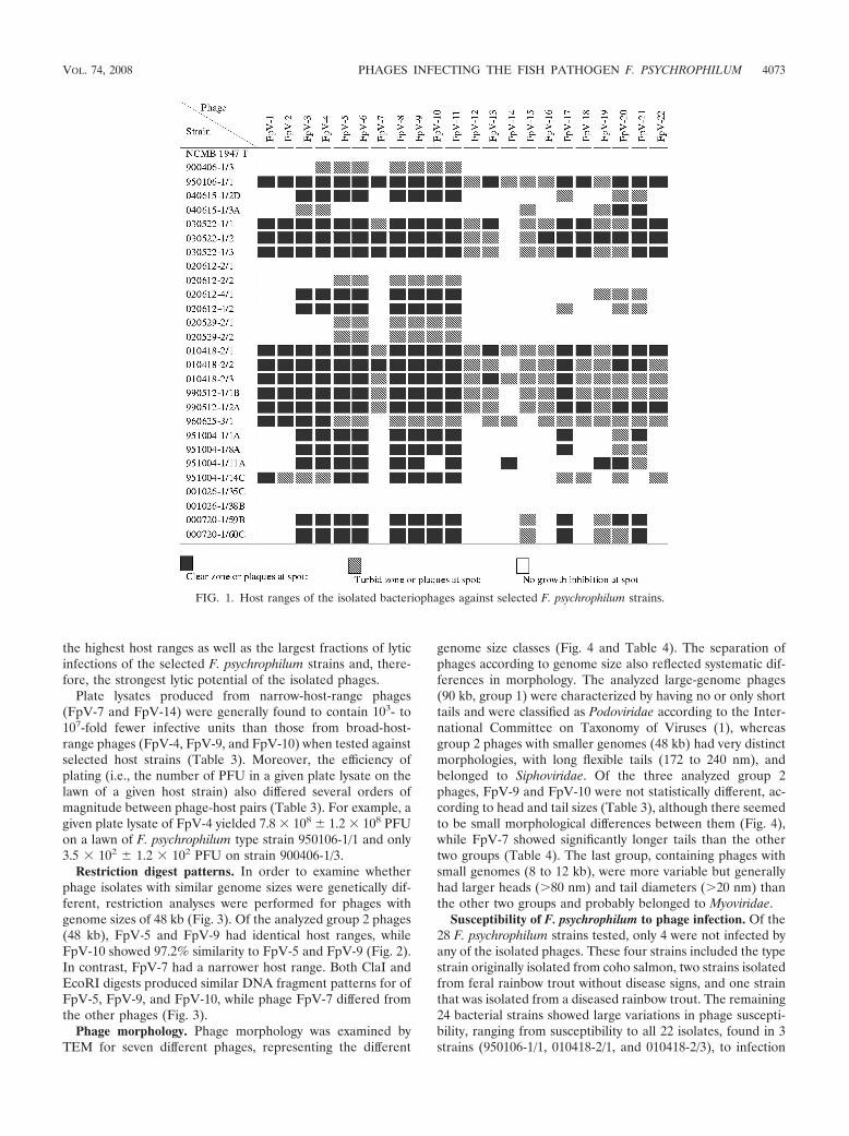
the highest host ranges as well as the largest fractions of lyticinfections of the selected F. psychrophilum strains and, there-fore, the strongest lytic potential of the isolated phages.
Plate lysates produced from narrow-host-range phages(FpV-7 and FpV-14) were generally found to contain 103- to107-fold fewer infective units than those from broad-host-range phages (FpV-4, FpV-9, and FpV-10) when tested againstselected host strains (Table 3). Moreover, the efficiency ofplating (i.e., the number of PFU in a given plate lysate on thelawn of a given host strain) also differed several orders ofmagnitude between phage-host pairs (Table 3). For example, agiven plate lysate of FpV-4 yielded 7.8 � 108 � 1.2 � 108 PFUon a lawn of F. psychrophilum type strain 950106-1/1 and only3.5 � 102 � 1.2 � 102 PFU on strain 900406-1/3.
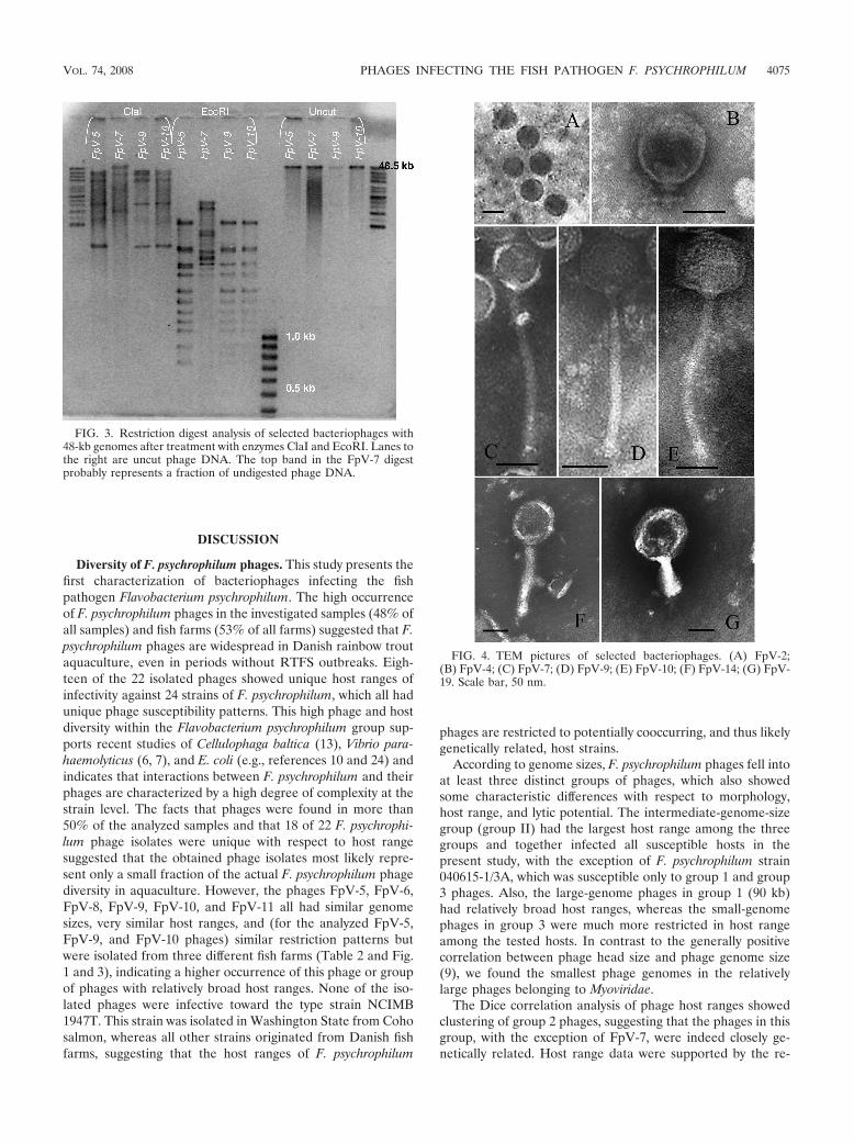
Restriction digest patterns. In order to examine whetherphage isolates with similar genome sizes were genetically dif-ferent, restriction analyses were performed for phages withgenome sizes of 48 kb (Fig. 3). Of the analyzed group 2 phages(48 kb), FpV-5 and FpV-9 had identical host ranges, whileFpV-10 showed 97.2% similarity to FpV-5 and FpV-9 (Fig. 2).In contrast, FpV-7 had a narrower host range. Both ClaI andEcoRI digests produced similar DNA fragment patterns for ofFpV-5, FpV-9, and FpV-10, while phage FpV-7 differed fromthe other phages (Fig. 3).
Phage morphology. Phage morphology was examined byTEM for seven different phages, representing the different
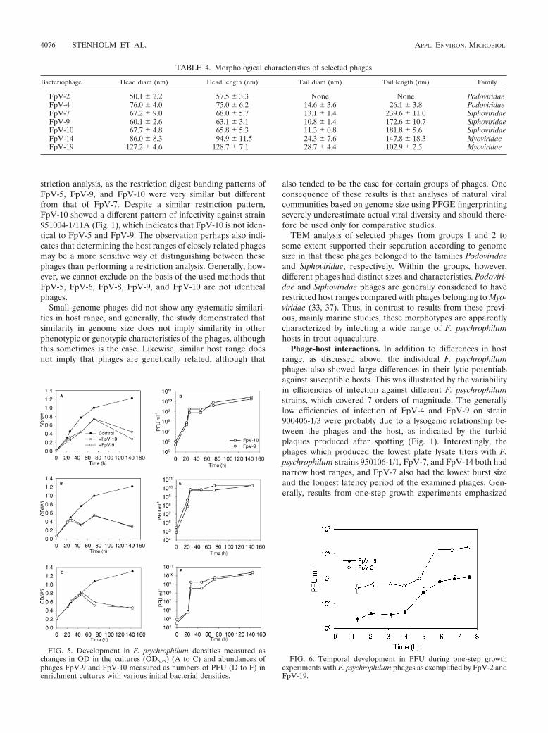
genome size classes (Fig. 4 and Table 4). The separation ofphages according to genome size also reflected systematic dif-ferences in morphology. The analyzed large-genome phages(90 kb, group 1) were characterized by having no or only shorttails and were classified as Podoviridae according to the Inter-national Committee on Taxonomy of Viruses (1), whereasgroup 2 phages with smaller genomes (48 kb) had very distinctmorphologies, with long flexible tails (172 to 240 nm), andbelonged to Siphoviridae. Of the three analyzed group 2phages, FpV-9 and FpV-10 were not statistically different, ac-cording to head and tail sizes (Table 3), although there seemedto be small morphological differences between them (Fig. 4),while FpV-7 showed significantly longer tails than the othertwo groups (Table 4). The last group, containing phages withsmall genomes (8 to 12 kb), were more variable but generallyhad larger heads (�80 nm) and tail diameters (�20 nm) thanthe other two groups and probably belonged to Myoviridae.
Susceptibility of F. psychrophilum to phage infection. Of the28 F. psychrophilum strains tested, only 4 were not infected byany of the isolated phages. These four strains included the typestrain originally isolated from coho salmon, two strains isolatedfrom feral rainbow trout without disease signs, and one strainthat was isolated from a diseased rainbow trout. The remaining24 bacterial strains showed large variations in phage suscepti-bility, ranging from susceptibility to all 22 isolates, found in 3strains (950106-1/1, 010418-2/1, and 010418-2/3), to infection
FIG. 1. Host ranges of the isolated bacteriophages against selected F. psychrophilum strains.
VOL. 74, 2008 PHAGES INFECTING THE FISH PATHOGEN F. PSYCHROPHILUM 4073
by only 6 phages and development of turbid plaques uponexposure to the phages, found in 3 strains (020612-2/2, 020529-2/1, and 020529-2/2).
Infection experiments with FpV-9 and FpV-10 and F. psy-chrophilum strain 950106-1/1 performed in batch cultures atdifferent MOI showed rapid propagation during the first 20 hof incubation for both phages, resulting in phage titers of 1.6 �1010 to 2.8 � 1010 PFU ml�1, irrespective of the initial bacte-rial density (Fig. 5). However, reducing the initial OD525 from0.216 to 0.013 delayed the maximum OD in the culture byapproximately 24 h. In all the experiments, the ODs reachedsimilar levels, corresponding to 64 to 77% reductions in ODrelative to levels for control cultures without phages, suggestingthat phage infection had a significant impact on host cell density.
Bacterial growth and phage-host interactions. F. psy-chrophilum growth was relatively slow in the enriched liquidcultures, with maximum growth rates at 15°C, ranging from
0.062 � 0.013 h�1 (020612-2/1) to 0.098 � 0.025 h�1
(951004-1/1A). The one-step growth experiments showed nosystematic changes in latency period and burst size withphage genome size (Fig. 6 and Table 5). In fact, FpV-2,FpV-9, and FpV-19, representing the different genome sizegroups, had very similar latency periods (4.0 to 4.5 h) andburst sizes (37 to 51 viruses infection�1). FpV-7, however,were less efficient than the other investigated phages andproduced only 7 � 1 phages after a latency period of 6 h.Interestingly, the lytic properties of individual phages weredependent on the host strain, as the burst sizes of phageFpV-9 were four times higher with strain 010418-2/3 thanwith strain 950106-1/1 used as a host strain (Table 5). Therate of adsorption of FpV-9 to F. psychrophilum strain950106-1/1 was estimated at 0.016 � 0.002 h�1, correspond-ing to the exponential decline of free phages during theincubation (Table 5).
FIG. 2. Tree based on the unweighted-pair group method using average linkages for the F. psychrophilum phage isolates (“#” is followed byFpV number) based on their host ranges of infectivity against the 28 bacterial isolates. The data in Fig. 1 were scored and converted to pairwisedistances by using the Dice similarity coefficient. Groupings of phages according to genome size and morphology are inserted to facilitatecomparison.
TABLE 3. Efficiencies of phage infection
PhageResult for indicated straina
950106-1/1 010418-2/3 900406-1/3 960625-3/1 951004-1/11a 95100-1/14c
FpV-4 7.8 � 108 � 1.2 � 108 2.7 � 109 � 4.2 � 108 3.5 � 102 � 1.2 � 102 1.5 � 109 � 3.3 � 108 1.5 � 108 � 5.3 � 107 NDFpV-7 3.2 � 104 � 7.8 � 103 4.1 � 104 � 0.5 � 103 ND ND ND NDFpV-9 �1012 6.9 � 109 � 3.5 � 108 1.5 � 105 � 3.3 � 104 ND 2.8 � 1010 � 4.3 � 109 2.8 � 1010 � 2.3 � 109
FpV-10 3.7 � 109 � 2.5 � 108 5.4 � 109 � 5.6 � 108 ND ND ND NDFpV-14 6.4 � 103 � 1.5 � 103 6.4 � 103 � 0.4 � 103 ND 6.5 � 103 � 0.7 � 103 5.7 � 103 � 2.9 � 103 ND
a Number of infecting units in phage plate lysates produced after infection of strain 950106-1/1 and subsequently exposed to selected host strains (i.e., the differentstrains were exposed to the same titer of the given phage). ND, not done.
4074 STENHOLM ET AL. APPL. ENVIRON. MICROBIOL.
DISCUSSION
Diversity of F. psychrophilum phages. This study presents thefirst characterization of bacteriophages infecting the fishpathogen Flavobacterium psychrophilum. The high occurrenceof F. psychrophilum phages in the investigated samples (48% ofall samples) and fish farms (53% of all farms) suggested that F.psychrophilum phages are widespread in Danish rainbow troutaquaculture, even in periods without RTFS outbreaks. Eigh-teen of the 22 isolated phages showed unique host ranges ofinfectivity against 24 strains of F. psychrophilum, which all hadunique phage susceptibility patterns. This high phage and hostdiversity within the Flavobacterium psychrophilum group sup-ports recent studies of Cellulophaga baltica (13), Vibrio para-haemolyticus (6, 7), and E. coli (e.g., references 10 and 24) andindicates that interactions between F. psychrophilum and theirphages are characterized by a high degree of complexity at thestrain level. The facts that phages were found in more than50% of the analyzed samples and that 18 of 22 F. psychrophi-lum phage isolates were unique with respect to host rangesuggested that the obtained phage isolates most likely repre-sent only a small fraction of the actual F. psychrophilum phagediversity in aquaculture. However, the phages FpV-5, FpV-6,FpV-8, FpV-9, FpV-10, and FpV-11 all had similar genomesizes, very similar host ranges, and (for the analyzed FpV-5,FpV-9, and FpV-10 phages) similar restriction patterns butwere isolated from three different fish farms (Table 2 and Fig.1 and 3), indicating a higher occurrence of this phage or groupof phages with relatively broad host ranges. None of the iso-lated phages were infective toward the type strain NCIMB1947T. This strain was isolated in Washington State from Cohosalmon, whereas all other strains originated from Danish fishfarms, suggesting that the host ranges of F. psychrophilum
phages are restricted to potentially cooccurring, and thus likelygenetically related, host strains.
According to genome sizes, F. psychrophilum phages fell intoat least three distinct groups of phages, which also showedsome characteristic differences with respect to morphology,host range, and lytic potential. The intermediate-genome-sizegroup (group II) had the largest host range among the threegroups and together infected all susceptible hosts in thepresent study, with the exception of F. psychrophilum strain040615-1/3A, which was susceptible only to group 1 and group3 phages. Also, the large-genome phages in group 1 (90 kb)had relatively broad host ranges, whereas the small-genomephages in group 3 were much more restricted in host rangeamong the tested hosts. In contrast to the generally positivecorrelation between phage head size and phage genome size(9), we found the smallest phage genomes in the relativelylarge phages belonging to Myoviridae.
The Dice correlation analysis of phage host ranges showedclustering of group 2 phages, suggesting that the phages in thisgroup, with the exception of FpV-7, were indeed closely ge-netically related. Host range data were supported by the re-
FIG. 3. Restriction digest analysis of selected bacteriophages with48-kb genomes after treatment with enzymes ClaI and EcoRI. Lanes tothe right are uncut phage DNA. The top band in the FpV-7 digestprobably represents a fraction of undigested phage DNA.
FIG. 4. TEM pictures of selected bacteriophages. (A) FpV-2;(B) FpV-4; (C) FpV-7; (D) FpV-9; (E) FpV-10; (F) FpV-14; (G) FpV-19. Scale bar, 50 nm.
VOL. 74, 2008 PHAGES INFECTING THE FISH PATHOGEN F. PSYCHROPHILUM 4075
striction analysis, as the restriction digest banding patterns ofFpV-5, FpV-9, and FpV-10 were very similar but differentfrom that of FpV-7. Despite a similar restriction pattern,FpV-10 showed a different pattern of infectivity against strain951004-1/11A (Fig. 1), which indicates that FpV-10 is not iden-tical to FpV-5 and FpV-9. The observation perhaps also indi-cates that determining the host ranges of closely related phagesmay be a more sensitive way of distinguishing between thesephages than performing a restriction analysis. Generally, how-ever, we cannot exclude on the basis of the used methods thatFpV-5, FpV-6, FpV-8, FpV-9, and FpV-10 are not identicalphages.
Small-genome phages did not show any systematic similari-ties in host range, and generally, the study demonstrated thatsimilarity in genome size does not imply similarity in otherphenotypic or genotypic characteristics of the phages, althoughthis sometimes is the case. Likewise, similar host range doesnot imply that phages are genetically related, although that
also tended to be the case for certain groups of phages. Oneconsequence of these results is that analyses of natural viralcommunities based on genome size using PFGE fingerprintingseverely underestimate actual viral diversity and should there-fore be used only for comparative studies.
TEM analysis of selected phages from groups 1 and 2 tosome extent supported their separation according to genomesize in that these phages belonged to the families Podoviridaeand Siphoviridae, respectively. Within the groups, however,different phages had distinct sizes and characteristics. Podoviri-dae and Siphoviridae phages are generally considered to haverestricted host ranges compared with phages belonging to Myo-viridae (33, 37). Thus, in contrast to results from these previ-ous, mainly marine studies, these morphotypes are apparentlycharacterized by infecting a wide range of F. psychrophilumhosts in trout aquaculture.
Phage-host interactions. In addition to differences in hostrange, as discussed above, the individual F. psychrophilumphages also showed large differences in their lytic potentialsagainst susceptible hosts. This was illustrated by the variabilityin efficiencies of infection against different F. psychrophilumstrains, which covered 7 orders of magnitude. The generallylow efficiencies of infection of FpV-4 and FpV-9 on strain900406-1/3 were probably due to a lysogenic relationship be-tween the phages and the host, as indicated by the turbidplaques produced after spotting (Fig. 1). Interestingly, thephages which produced the lowest plate lysate titers with F.psychrophilum strains 950106-1/1, FpV-7, and FpV-14 both hadnarrow host ranges, and FpV-7 also had the lowest burst sizeand the longest latency period of the examined phages. Gen-erally, results from one-step growth experiments emphasized
FIG. 5. Development in F. psychrophilum densities measured aschanges in OD in the cultures (OD525) (A to C) and abundances ofphages FpV-9 and FpV-10 measured as numbers of PFU (D to F) inenrichment cultures with various initial bacterial densities.
FIG. 6. Temporal development in PFU during one-step growthexperiments with F. psychrophilum phages as exemplified by FpV-2 andFpV-19.
TABLE 4. Morphological characteristics of selected phages
Bacteriophage Head diam (nm) Head length (nm) Tail diam (nm) Tail length (nm) Family
FpV-2 50.1 � 2.2 57.5 � 3.3 None None PodoviridaeFpV-4 76.0 � 4.0 75.0 � 6.2 14.6 � 3.6 26.1 � 3.8 PodoviridaeFpV-7 67.2 � 9.0 68.0 � 5.7 13.1 � 1.4 239.6 � 11.0 SiphoviridaeFpV-9 60.1 � 2.6 63.1 � 3.1 10.8 � 1.4 172.6 � 10.7 SiphoviridaeFpV-10 67.7 � 4.8 65.8 � 5.3 11.3 � 0.8 181.8 � 5.6 SiphoviridaeFpV-14 86.0 � 8.3 94.9 � 11.5 24.3 � 7.6 147.8 � 18.3 MyoviridaeFpV-19 127.2 � 4.6 128.7 � 7.1 28.7 � 4.4 102.9 � 2.5 Myoviridae
4076 STENHOLM ET AL. APPL. ENVIRON. MICROBIOL.
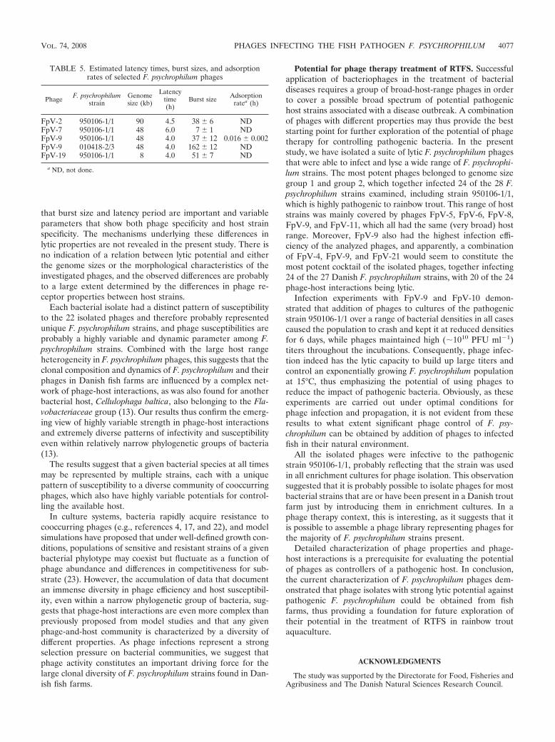
that burst size and latency period are important and variableparameters that show both phage specificity and host strainspecificity. The mechanisms underlying these differences inlytic properties are not revealed in the present study. There isno indication of a relation between lytic potential and eitherthe genome sizes or the morphological characteristics of theinvestigated phages, and the observed differences are probablyto a large extent determined by the differences in phage re-ceptor properties between host strains.
Each bacterial isolate had a distinct pattern of susceptibilityto the 22 isolated phages and therefore probably representedunique F. psychrophilum strains, and phage susceptibilities areprobably a highly variable and dynamic parameter among F.psychrophilum strains. Combined with the large host rangeheterogeneity in F. psychrophilum phages, this suggests that theclonal composition and dynamics of F. psychrophilum and theirphages in Danish fish farms are influenced by a complex net-work of phage-host interactions, as was also found for anotherbacterial host, Cellulophaga baltica, also belonging to the Fla-vobacteriaceae group (13). Our results thus confirm the emerg-ing view of highly variable strength in phage-host interactionsand extremely diverse patterns of infectivity and susceptibilityeven within relatively narrow phylogenetic groups of bacteria(13).
The results suggest that a given bacterial species at all timesmay be represented by multiple strains, each with a uniquepattern of susceptibility to a diverse community of cooccurringphages, which also have highly variable potentials for control-ling the available host.
In culture systems, bacteria rapidly acquire resistance tocooccurring phages (e.g., references 4, 17, and 22), and modelsimulations have proposed that under well-defined growth con-ditions, populations of sensitive and resistant strains of a givenbacterial phylotype may coexist but fluctuate as a function ofphage abundance and differences in competitiveness for sub-strate (23). However, the accumulation of data that documentan immense diversity in phage efficiency and host susceptibil-ity, even within a narrow phylogenetic group of bacteria, sug-gests that phage-host interactions are even more complex thanpreviously proposed from model studies and that any givenphage-and-host community is characterized by a diversity ofdifferent properties. As phage infections represent a strongselection pressure on bacterial communities, we suggest thatphage activity constitutes an important driving force for thelarge clonal diversity of F. psychrophilum strains found in Dan-ish fish farms.
Potential for phage therapy treatment of RTFS. Successfulapplication of bacteriophages in the treatment of bacterialdiseases requires a group of broad-host-range phages in orderto cover a possible broad spectrum of potential pathogenichost strains associated with a disease outbreak. A combinationof phages with different properties may thus provide the beststarting point for further exploration of the potential of phagetherapy for controlling pathogenic bacteria. In the presentstudy, we have isolated a suite of lytic F. psychrophilum phagesthat were able to infect and lyse a wide range of F. psychrophi-lum strains. The most potent phages belonged to genome sizegroup 1 and group 2, which together infected 24 of the 28 F.psychrophilum strains examined, including strain 950106-1/1,which is highly pathogenic to rainbow trout. This range of hoststrains was mainly covered by phages FpV-5, FpV-6, FpV-8,FpV-9, and FpV-11, which all had the same (very broad) hostrange. Moreover, FpV-9 also had the highest infection effi-ciency of the analyzed phages, and apparently, a combinationof FpV-4, FpV-9, and FpV-21 would seem to constitute themost potent cocktail of the isolated phages, together infecting24 of the 27 Danish F. psychrophilum strains, with 20 of the 24phage-host interactions being lytic.
Infection experiments with FpV-9 and FpV-10 demon-strated that addition of phages to cultures of the pathogenicstrain 950106-1/1 over a range of bacterial densities in all casescaused the population to crash and kept it at reduced densitiesfor 6 days, while phages maintained high (�1010 PFU ml�1)titers throughout the incubations. Consequently, phage infec-tion indeed has the lytic capacity to build up large titers andcontrol an exponentially growing F. psychrophilum populationat 15°C, thus emphasizing the potential of using phages toreduce the impact of pathogenic bacteria. Obviously, as theseexperiments are carried out under optimal conditions forphage infection and propagation, it is not evident from theseresults to what extent significant phage control of F. psy-chrophilum can be obtained by addition of phages to infectedfish in their natural environment.
All the isolated phages were infective to the pathogenicstrain 950106-1/1, probably reflecting that the strain was usedin all enrichment cultures for phage isolation. This observationsuggested that it is probably possible to isolate phages for mostbacterial strains that are or have been present in a Danish troutfarm just by introducing them in enrichment cultures. In aphage therapy context, this is interesting, as it suggests that itis possible to assemble a phage library representing phages forthe majority of F. psychrophilum strains present.
Detailed characterization of phage properties and phage-host interactions is a prerequisite for evaluating the potentialof phages as controllers of a pathogenic host. In conclusion,the current characterization of F. psychrophilum phages dem-onstrated that phage isolates with strong lytic potential againstpathogenic F. psychrophilum could be obtained from fishfarms, thus providing a foundation for future exploration oftheir potential in the treatment of RTFS in rainbow troutaquaculture.
ACKNOWLEDGMENTS
The study was supported by the Directorate for Food, Fisheries andAgribusiness and The Danish Natural Sciences Research Council.
TABLE 5. Estimated latency times, burst sizes, and adsorptionrates of selected F. psychrophilum phages
Phage F. psychrophilumstrain
Genomesize (kb)
Latencytime(h)
Burst size Adsorptionratea (h)
FpV-2 950106-1/1 90 4.5 38 � 6 NDFpV-7 950106-1/1 48 6.0 7 � 1 NDFpV-9 950106-1/1 48 4.0 37 � 12 0.016 � 0.002FpV-9 010418-2/3 48 4.0 162 � 12 NDFpV-19 950106-1/1 8 4.0 51 � 7 ND
a ND, not done.
VOL. 74, 2008 PHAGES INFECTING THE FISH PATHOGEN F. PSYCHROPHILUM 4077
We thank Jette Mundus Nikolajsen and Kirsten Kaas for excellenttechnical support.
REFERENCES
1. Ackermann, H.-W. 2005. Bacteriophage classification, p. 67–90. In E. Kutterand A. Sulakvelidze (ed.), Bacteriophages: biology and applications. CRCPress, Boca Raton, FL.
2. Adams, M. H. 1959. Bacteriophages. Interscience Publishers, New York,NY.
3. Atterbury, R. J., P. L. Connerton, C. E. R. Dodd, C. E. D. Rees, and I. F.Connerton. 2003. Application of host-specific bacteriophages to the surfaceof chicken leads to reduction in the recovery of Campylobacter jejuni. Appl.Environ. Microbiol. 69:6302–6306.
4. Bohannan, B. J. M., and R. E. Lenski. 2000. Linking genetic change tocommunity evolution: insights from studies of bacteria and bacteriophage.Ecol. Lett. 3:362–377.
5. Bruun, M. S., A. S. Schmidt, L. Madsen, and I. Dalsgaard. 2000. Antimi-crobial resistance patterns in Danish isolates of Flavobacterium psychrophi-lum. Aquaculture 187:201–212.
6. Comeau, A. M., A. M. Chan, and C. A. Suttle. 2006. Genetic richness ofvibriophages isolated in a coastal environment. Environ. Microbiol. 8:1164–1176.
7. Comeau, A. M., E. Buenaventura, and C. A. Suttle. 2005. A persistent,productive, and seasonally dynamic vibriophage population within Pacificoysters (Crassostrea gigas). Appl. Environ. Microbiol. 71:5324–5331.
8. Dalsgaard, I., and L. Madsen. 2000. Bacterial pathogens in rainbow troutOncorhyncus mykiss reared at Danish freshwater farms. J. Fish Dis. 23:199–209.
9. De Paepe, M., and F. Taddei. 2006. Viruses’ life history: towards a mecha-nistic basis for a trade-off between survival and reproduction among phages.PLoS Biol. 4:1248–1256.
10. Fischer, C. R., M. Yoichi, H. Unno, and Y. Tanji. 2004. The coexistence ofEscherichia coli serotype O157:H7 and its specific bacteriophage in contin-uous culture. FEMS Microbiol. Lett. 241:171–177.
11. Goode, D., V. M. Allen, and P. A. Barrow. 2003. Reduction of experimentalSalmonella and Campylobacter contamination of chicken skin by applicationof bacteriophages. Appl. Environ. Microbiol. 69:5032–5036.
12. Greer, G. G. 2005. Bacteriphage control of foodborne bacteria. J. Food Prot.68:1102–1111.
13. Holmfeldt, K., M. Middelboe, O. Nybroe, and L. Riemann. 2007. Largevariabilities in host strain susceptibility and phage host range govern inter-actions between lytic marine phages and their Flavobacterium hosts. Appl.Environ. Microbiol. 73:6730–6739.
14. Holt, R. A., J. S. Rohovec, and J. L. Fryer. 1993. Bacterial coldwaterdisease, p. 3–23 In V. Englis, R. J. Roberts, and N. R. Bromage (ed.),Bacterial diseases of fish. Blackwell Scientific Publications, Oxford,United Kingdom.
15. Imbeault, S., S. Parent, M. Lagace, C. F. Uhland, and J.-F. Blais. 2006.Using bacteriophages to prevent furunculosis caused by Aeromonas salmo-nicida in farmed brook trout. J. Aquat. Anim. Health 18:203–214.
16. Karunasagar, I., M. M. Shivu, S. K. Girisha, G. Krohne, and I. Karunasagar.2007. Biocontrol of pathogens in shrimp hatcheries using bacteriophages.Aquaculture 268:288–292.
17. Lenski, R. E. 1984. Coevolution of bacteria and phage: are there endlesscycles of bacterial defenses and phage counterdefenses? J. Theor. Biol.108:319–325.
18. Leverentz, B., W. S. Conway, W. Janisiewich, and M. J. Camp. 2004. Opti-mizing concentration and timing of a phage spray application to reduceListeria monocytogenes on honeydew melon tissue. J. Food Prot. 67:1682–1686.
19. Lorenzen, E., I. Dalsgaard, J. From, E. M. Hansen, V. Hørlyck, H. Korsholm, S.
Mellergaard, and N. J. Olesen. 1991. Preliminary investigations of fry mortalitysyndrom in rainbow trout. Bull. Eur. Assoc. Fish Pathol. 11:77–79.
20. Madsen, L., and I. Dalsgaard. 2000. Comparative studies of Danish Fla-vobacterium psychrophilum strains concerning ribotypes, plasmid profiles,serotypes and virulence. J. Fish Dis. 23:211–218.
21. Madsen, L., and I. Dalsgaard. 1999. Reproducible methods for experimentalinfection with Flavobacterium psychrophilum in rainbow trout Oncorhyncusmykiss. Dis. Aquat. Organ. 36:169–176.
22. Middelboe, M. 2000. Bacterial growth rate and marine virus-host dynamics.Microb. Ecol. 40:114–124.
23. Middelboe, M., Å. Hagstrom, N. Blackburn, B. Sinn, U. Fischer, N. H.Borch, J. Pinhassi, K. Simu, and M. G. Lorenz. 2001. Effects of bacterio-phages on the population dynamics of four strains of pelagic marine bacteria.Microb. Ecol. 42:395–406.
24. Mizoguchi, K., M. Morita, C. R. Fischer, M. Yoichi, Y. Tanji, and H. Unno.2003. Coevolution of bacteriophage PP01 and Escherichia coli O157:H7 incontinuous culture. Appl. Environ. Microbiol. 69:170–176.
25. Nakai, T., and K.-H. Park. 2002. Bacteriophage therapy of infectious dis-eases in aquaculture. Res. Microbiol. 153:13–18.
26. Nakai, T., R. Sugimoto, K.-H. Park, K. Mori, T. Nishioka, and K.Maruyama. 1999. Protective effects of bacteriophage on experimental Lac-tococcus garvieae infection in yellowtail. Dis. Aquat. Organ. 37:33–41.
27. Nematollahi, A., A. Decostere, F. Pasmans, and F. Haesebrouck. 2003. Fla-vobacterium psychrophilum infections in salmonid fish. J. Fish Dis. 26:563–574.
28. O’Flynn, G., R. P. Ross, G. F. Fitzgerald, and A. Coffey. 2004. Evaluation ofa cocktail of three bacteriophages for biocontrol of Escherichia coli O157:H7.Appl. Environ. Microbiol. 70:3417–3424.
29. Park, S. C., and T. Nakai. 2003. Bacteriophage control of Pseudomonasplecoglossicidae infection in ayu Plecoglossus altivelis. Dis. Aquat. Organ.53:33–39.
30. Park, S. C., I. Shimamura, M. Fukunaga, K.-I. Mori, and T. Nakai. 2000.Isolation of bacteriophages specific to a fish pathogen, Pseudomonas pleco-glossicidae, as a candidate for fish disease control. Appl. Environ. Microbiol.66:1416–1422.
31. Shivu, M. M., B. C. Rajeeva, S. K. Girisha, I. Karunasagar, G. Krohne, andI. Karunasagar. 2007. Molecular characterisation of Vibrio harveyi bacterio-phages isolated from aquaculture environments along the coast of India.Environ. Microbiol. 9:322–331.
32. Su, M.-T., T. V. Venkatesh, and R. Bodmer. 1998. Large- and small-scalepreparation of bacteriophage lysate and DNA. BioTechniques 25:44–46.
33. Suttle, C. A., and A. M. Chan. 1993. Marine cyanophages infecting oceanicand costal strains of Synechococcus: abundance, morphology, cross-infectiv-ity and growth characteristics. Mar. Ecol. Prog. Ser. 92:99–109.
34. Tanji, Y., T. Shimada, M. Yoichi, K. Miyanaga, K. Hori, and H. Unno. 2004.Toward rational control of Escherichia coli O157:H7 by a phage cocktail.Appl. Microbiol. Biotechnol. 64:270–274.
35. Vinod, M. G., M. M. Shivu, K. R. Umesha, B. C. Rajeeva, G. Krohne, I.Karunasagar, and I. Karunasagar. 2006. Isolation of Vibrio harveyi bacte-riophage with a potential for biocontrol of luminous vibriosis in hatcheryenvironments. Aquaculture 255:117–124.
36. Weiss, B. D., M. A. Capage, M. Kessel, and S. A. Benson. 1994. Isolation andcharacterization of a generalized transducing phage for Xanthomonascampestris pv. campestris. J. Bacteriol. 176:3354–3359.
37. Wichels, A., S. S. Biel, H. R. Gelderblom, T. Brinkhoff, G. Muyzer, and C.Schutt. 1998. Bacteriophage diversity in the North Sea. Appl. Environ. Mi-crobiol. 64:4128–4133.
38. Wiklund, T., L. Madsen, M. S. Bruun, and I. Dalsgaard. 2000. Detection ofFlavobacterium psychrophilum from fish tissue and water samples by PCRamplification. J. Appl. Microbiol. 87:299–307.
4078 STENHOLM ET AL. APPL. ENVIRON. MICROBIOL.
Related Documents