ISOLATION AND CHARACTERIZATION OF ANTmIOTICS FROM BACTERIA Siaw Wai Kian Bachelor of Science with Honours QR (Resource Biotechnology) 81 2009 S562 2009

Welcome message from author
This document is posted to help you gain knowledge. Please leave a comment to let me know what you think about it! Share it to your friends and learn new things together.
Transcript
ISOLATION AND CHARACTERIZATION OF ANTmIOTICS FROM BACTERIA
Siaw Wai Kian
Bachelor of Science with Honours QR (Resource Biotechnology) 81 2009 S562 2009
ISOLATION AND CHARACTERIZATION OF ANTffiIOTICS FROM.BACTERIA
SIA W W AI KlAN
A REPORT SUBMITTED IN PARTIAL FULFILMENT FOR THE DEGREE OF BACHELOR OF SCIENCE WITH HONOURS IN RESOURCE BIOTECHNOLOGY
DEPARTMENT OF MOLECULAR BIOLOGY FACULTY OF RESOURCE SCIENCE AND TECHNOLOGY
UNIVERSITI MALAYSIA SARAW AK
2009
ACKNOWLEDGEMENT
I want to thank the Department of Molecular Biology, Universiti Malaysia Sarawak
(Unimas) for giving me pennission to conunence this thesis in the first instance, to do the
necessary research work in the Virology Laboratory and to use departmental resources. I
really appreciate every moment working in the laboratory.
I would like to express my deep and sincere gratitude to my supervisor, Prof. Dr.
Ismail bin Ahmad. His wide knowledge and his logical way of thinking have been of great
value for me. His understanding, encouraging and personal guidance have provided a good
basis for the present thesis.
I warmly thank all the postgraduates and coursemates in Virology Laboratory, for
their valuable advice and friendly help. Their extensive discussions around my work and
interesting explorations in operations have been very helpful for this study. I really
appreciate every moment working with them.
lowe my loving thanks to my parents, my brothers and sister. Without their
encouragement and understanding it would have been impossible for me to fmish this work.
My special gratitude is due to friends for their loving support.
11
I'IIsat KhW...at Maldulll.t AIwIe VNIVERSm MALAYSIA SARAWAK
TABLE OF CONTENTS
ACKNOWLEDGEMENT
Page
11
TABLE OF CONTENTS
LIST OF TABLES Vill
III
LIST OF ABBREVIATIONS VI
LIST OF FIGURES IX
ABSTRACT X
ABSTRAK X
CHAPTER
1.0 INTRODUCTION
2.0 LITERATURE REVIEW
2.1 Sources and Production of Antibiotics 3
2.2 Soil as Sources ofAntibiotics 4
2.3 Soil Actinomycetes and Bacteria 5
2.4 Optimum Conditions for the Production ofAntibiotics 6
2.5 Antibiotics Sensitivity Screening 7
2.5.1 Spot Inoculation Method 7
2.5.2 Disk-diffusion Method 7
2.6 Identification ofActinomycetes and Bacteria 8
3.0 MATERIALS AND METHODS
3.1 Soil Sampling 9
3.2 Serial Dilutions of the Soil Samples 9
U\
3.3 Isolation of Soil Actinomycetes and Bacteria 9
3.4 Isolation of Pure Bacterial Culture 10
3.5 Cultivation ofTest Bacteria from Stock Cultures 10
3.6 Antibiotics Sensitivity Screening 10
3.6.1 Primary Screening: Spot Inoculation Method 11
3.6.2 Secondary Screening: Disk-diffusion Method 11
3.7 Sensitivity of Antibiotics Solubilized from Solid Culture 12
3.8 Sensitivity of the Concentrated Antibiotics Produced in Liquid Culture 12
3.9 Bacteria Identification 12
4.0 RESULTS
4.1 Isolation of Bacteria 14
4.2 Primary Screening Results: Spot Inoculation Method 14
4.3 Secondary Screening Results: Disk-diffusion Method 14
4.4 Comparison of Antibiotics Sensitivity between Primary and Secondary 18 Screening
4.5 Comparison between Sensitivity ofBI 4-1 Antibiotics Produced in 19 Different Medium
4.6 Comparison between Non-concentrated and Concentrated Supernatant of 21 B14-1 and BI 5-2
4.7 Bacteria Identification 21
5.0 DISCUSSION
5.1 Soil Sampling 24
5.2 Isolation of Soil Bacteria 25
5.2.1 Serial Dilution 25
5.2.2 Media and Optimum Conditions for Soil Bacterial Growth 25
IV
I
5.2.3 Storage of Bacterial Isolates 26
5.3 Primary Screening: Spot Inoculation Method 26
5.4 Secondary Screening: Disk-diffusion Method 28
5.5 Activity ofAntibiotics Produced by BI 4-1 Cultures 29
5.6 Activity ofConcentrated Antibiotics Produced by BI 4-1 and BI 5-2 29 Cultures
5.7 Bacteria Identification 30
6.0 CONCLUSION AND RECOMMENDATION 32
REFERENCES 34
APPENDICES
A Preparation of5 X Phosphate Buffered Saline (PBS)
B Preparation ofNutrient Agar
C Preparation of0.75 % Nutrient Agar (Soft Agar)
D Preparation ofNutrient Broth
E Preparation ofSimmons' Citrate Agar
F Preparation ofSIM Medium
G Recipe of Barritt's Reagent A and Reagent B
I
v
LIST OF ABBREVIATIONS
o
%
#L1
20th
BC
C
em
DNA
EA
EC
G
g
HPLC
L
MIC
ml
mm
MR
PBS
pH
rpm
Degree
Degree celsius
Percent
Micro liter
Twentieth
Bacillus cereus
Cytosine
Centimeter
Deoxyribose nucleic acid
Enterobacter aerogenes
Escherichia coli
Guanine
Gram
High perfonnance liquid chromatography
Hydrogen sulfide
Liter
Minimum inhibitory concentration
Milliliter
Millimeter
Methyl red
Phosphate buffered saline
a measurement of the acidity or alkalinity of solution [p stands for "potenz" (this means the potential to be) and H stands for Hydrogen]
Revolutions per minute
Staphylococcus aureus
VI
ST Salmonella typhi
SIM Sulfide, indole, motility
Unimas Universiti Malaysia Sarawak
VP Voges-Proskauer
w/v Weight over volume
Vll
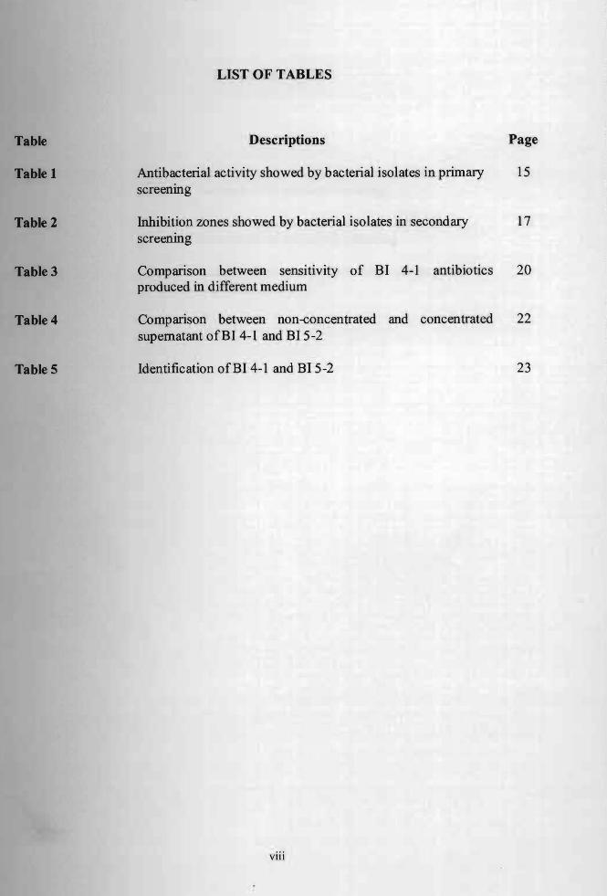
LIST OF TABLES
Table
Table 1
Table 2
Table 3
Table 4
Table 5
Descriptions Page
Antibacterial activity showed by bacterial isolates in primary screenmg
15
Inhibition zones showed by bacterial isolates in secondary screening
17
Comparison between senSItIVIty produced in different medium
of BI 4-1 antibiotics 20
Comparison between non-concentrated supernatant ofBI 4-1 and BI 5-2
and concentrated 22
Identification ofBI 4-1 and BI 5-2 23
VIII
Figure
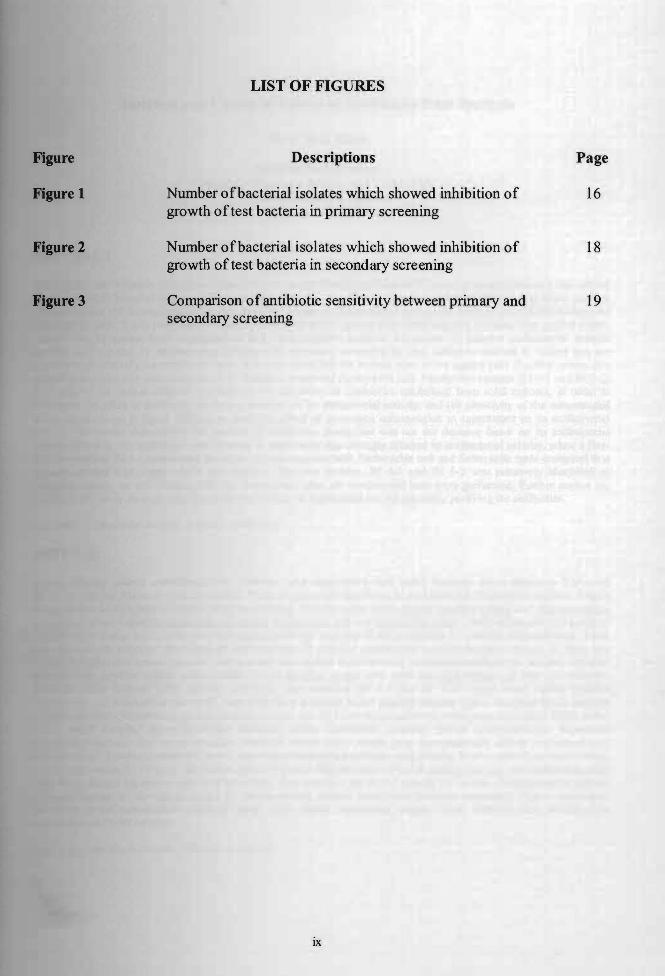
Figure 1
Figure 2
Figure 3
#
LIST OF FIGURES
Descriptions Page
Number ofbacterial isolates which showed inhibition of growth of test bacteria in primary screening
16
Number ofbacterial isolates which showed inhibition of growth of test bacteria in secondary screening
18
Comparison ofantibiotic sensitivity between primary and secondary screening
19
ix
Isolation and Characterization of Antibiotics from Bacteria
Siaw Wai Kian
Resource Biotechnology Faculty ofResource Science and Technology
Universiti Malaysia Sarawak
ABSTRACT
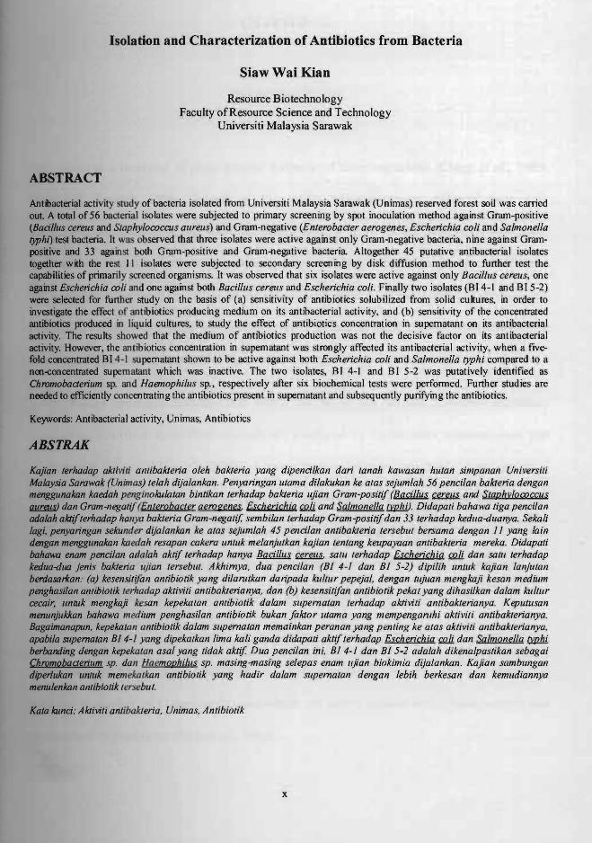
Antilacterial activity study of bacteria isolated from Universiti Malaysia Sarawak (Unimas) reserved forest soil was carried out. A total of 56 bacterial isolates were subjected to primary screening by spot inoculation method against Gram-positive (Bacillus cereus and Staphylococcus aureus) and Gram-negative (Enlerobacler aerogenes, Escherichia coli and Salmonella ryphl) test bacteria. It was observed that three isolates were active against only Gram-negative bacteria, nine against Grampositive and 33 against both Gram-positive and Gram-negative bacteria Altogether 45 putative antibacterial isolates together with the rest I I isolates were subjected to secondary screening by disk diffusion method to further test the capabilities of primarily screened organisms. It was observed that six isolates were active against only Bacilllls cereus, one against Escherichia coli and one against both Bacilllls cereus and Escherichia coli. Finally two isolates (BI 4-1 and BI 5-2) were selected for further study on the basis of (a) sensitivity of antibiotics solubilized from solid cuhures, in order to investigate the effect of antibiotics producing medium on its antibacterial activity, and (b) sensitivity of the concentrated antibiotics produced in liqu.id cultures, to study the effect of antibiotics concentration in supematant on its antibacterial activity. The results showed that the medium of antibiotics production was not the decisive factor on its antibacterial activity. However, the antibiotics concentration in supematant was strongly affected its antibacterial activity, when a fivefold concentrated BI 4-1 supernatant shown to be active against both Escherichia coli and Salmonella typhi compared to a noo-<:Oncentrated supernatant which was inactive. The two isolates, BI 4-1 and BI 5-2 was putatively identified as Chromobaaerium sp. and Haemophillls sp., respectively after six biochemical tests were performed. Further studies are needed to efficiently concentrating the antibiotics present in supernatant and subsequently purifying the antibiotics.
Keywords: Antibacterial activity, Unimas, Antibiotics
A BSTRAK
Kajian lerhadap aktivi# antibakteria oleh bakleria yang dipencilkan dari lanah kalVasan hllian simpanan Universili Malaysia Sarawak (Unimas) lelah dijalankan. Penyaringan ulama dilakukan ke alas sejumlah 56 pencilan bakteria dengan menggunakan kaedah penginokulalan binlikan lerhadap bakteria IIjian Gram-posilif (Bacillus ~ and SlaphylococClls flJIlIIIW dan Gram-negatif (,Enlerobacler aero genes. Escherichia illli and Salmonella !J!/2lJj). Didapali bahawa tiga pencilan adalah aktiflerhadap han)U bakleria Gram-negalif, sembilan lerhadap Gram-positif dan 33 lerhadap kedua-dllanya. Sekali lagi. pen)Uringan sekunder dijalankan ke alas sejllmlah 45 pencilan antibakleria lersebul bersama dengan II yang lain dengan menggunakan kaedah resapan cakera unlllk melanjulkan kajian lenlang keupayaan anlibakleria mereka. Didapali bahawa enam pencilan adalah aklif lerhadap hanya Bacillus ~ salll lerhadap Escherichia (;Q/i dan salll lerhadap kedua-duo Jenis bakteria ujian lerseblli. Akhimya, dua pencilan (BI 4-1 dan BI 5-2) dipilih IInluk kajian lanjulan berdasorlcan: (a) kesensilifan anlibwtik yang dilantlkan daripada kIIllur pepejal. dengan lujuan mengkaji kesan medium penghasilan antibiolik lerhadap aklivili anlibaklerianya, dan (b) kesensilifan antibiotik pekal yang dihasilkan dalam kIIllur cecair, IInlUk mengkaji kesan kepekalan anlibiolik dalam sllpemalan lerhadap aklivili anlibaklerianya. Kepull/san menunjukJcan bahawa medium penghasilan anlibiotik bukan faktor tltama yang mempengamhi aklivi/i antibaklerianya. Bagaimanapun. kepekamn anlibiolik dalam supemalan memainkan peranan yang penting ke alas aktivili anlibaklerianya, apabila supemaian BI 4- / yang dipekalkan lima kali ganda didapali aklif lerhadap Escherichia r;Q}1 dan Salmonella fY/2lJJ. berbanding dengan kepekatan asal yang lidak aktif Dua pencilan ini. BI 4-1 dan B15-2 adalah dikenalpastikan sebagai Chromobaaerjum sp. dan Haemophillis sp. masing-masing selepas enam IIjian biokimia dijalankan. Kajian sambungan diperlukan unluk menlekalkan antibiolik yang hadir dalam supemalan dengan lebih berkesan dan kemlldiannya menulenkan antibwtik lersebul.
Kala lame;: Alctivil; antibakleria, Unimas. Anlibiolik
x

CHAPTER 1.0
INTRODUCTION
Soil serves as a reservoir of great genetic diversity of microorganisms (Clegg et aI., 1998;
0vreas & Torsvik 1998; Torsvik et at., 1990). Microorganisms cultured from soil,
particularly actinomycetes and bacteria, have provided most of the antibiotics and many
other medicinal agents that have dramatically improved human health in the latter half of the
201ft century (Gillespie et aI., 2002). In order to combat with the newly emerging diseases and
antibiotic resistance problems worldwide, there is an urgent need for new antibiotics to be
discovered from natural bacteria living in soil.
Recent estimates indicate that nearly 50 % of the 20,000 bioactive secondary
metabolites described from 1900 onwards are produced by filamentous actinomycetes that
originated in the soil (Marinelli, 2009). A research also indicates two-thirds of the marketed
microbial drugs are produced by the genus Streptomyces, followed with the discovery of
actinomycin and streptomycin in 1940 and 1943, respectively. In tenns of total antibiotics
product coverage, other genera are trailing numerically. Micromonospora is next with less
than one-tenth as many as those produced by Streptomyces (Kieser et at., 2000).
Apart from actinomycetes, other soil bacteria also play an important role in
contributing worldwide antibiotics products. Chromobacterium, for example, is a genus that
produces a number of natural antibiotics which are active against both Gram-positive and
Gram-negative organisms (Imai et at., 1983).
In the study, ten soil samples were taken from different areas in the Universiti
Malaysia Sarawak (Unimas) reserved forest. Soil actinomycetes and bacteria were then grew
and isolated on artificial media. Those colonies which produced antibacterial substances
were able to inhibit other bacterial growth in the surroundings and formed clear zones known
as halos. After that, antibiotics producing isolates were selected and subcultured on fresh
solid media. After subculturing, these isolates were then subjected to primary screening and
subsequently secondary screening by spot inoculation and disk diffusion method,
respectively against Gram-positive (Bacillus cereus and Staphylococcus aureus) and Gram
negative (Enterobacter aerogenes, Escherichia coli and Salmonella typhi) test bacteria.
A further study was carried out to investigate the relationship between the antibiotics
producing medium and its antibacterial activity. Apart from that, the effect of the antibiotics
concentration on its antibacterial activity has also been studied, to further explain the
discrimination between the primary and secondary screening results.
The objectives of the study are:
(i) To isolate and identifY antibiotics producing actinomycetes and bacteria from soil,
(ii) To test the antibacterial activity of antibiotics produced by each isolates using spot
inoculation and disk-diffusion method against Gram-positive and Gram-negative test
bacteria, and
(iii) To carry out further studies to explain the discrimination between the primary and
secondary screening resu Its.
2
2.1
medicine.
CHAPTER 2.0
LITERATURE REVIEW
Sources and Production of Antibiotics
The tenn "antibiotic" literally means "against life". Antibiotics are a crucial line of defense
apinst bacterial infections because they attack the unique peptidoglycan cell wall or smaller
ribosomal unit ofthe bacteria (Dantas, 2008). Following of the first discovery of antibiotics,
penicillin from fungus Penicillium in 1928 by Scottish scientist Alexander Fleming, the
development and discovery of antibiotics have revolutionized the fight against bacterial
infection (Fleming, 1929). In the last half of the 20th Century, deaths from infectious diseases
sreatly decreased, partly due to the discovery of antibiotics (Gillespie et al., 2002). Until
now, antibiotics have become among the most frequently prescribed medications in modem
Antibiotics are primarily produced by microorganisms such as bacteria,
actinomycetes and fungus. Among them, actinomycetes are noteworthy as antibiotic
producers, making three quarters of all known products (Shantikumar et al., 2006). Apart
fiom that, some antibiotics are also found in animal and plant cells. For example, cecropin A,
I member of a family of antibiotic proteins produced by insects, may kill bacteria and avoid
resistance by entering bacterial cel1s and taking control of their genetic machinery (DeLucca
It al. , 1997).
3

Antibiotics are usually produced by an organism under extreme environments
because these extreme conditions require unique adaptation strategies leading to new natural
products from extreme organisms (Mahmoud, 2006). In order for an organism to adapt to the
extreme habitat, they need to produce certain products such as antibiotics that are essential
for their survival. Various extreme habitats that inhabit antibiotics producing microorganisms
including marine sediment (Kokare et aI., 2003), soil (Dastager et al., 2007), rhizosphere of
endemic plants (Anibou et al., 2008), extremely alkaline bauxite residue (Krishna et aI.,
2(08) and many others. Antibiotics are also produced by organisms to compete for a
particular niche and to sUlVive.
Apart from natural antibiotics obtained from organisms, synthetic and semi-synthetic
antibiotics are now being widely studied to produce novel antibiotics against various
bacterial infections and multidrug-resistance bacteria. For example, nitrofurans, a class of
antibacterial drugs in extensive use, interferes with gene expression in a highly specific
manner (Herrlich & Schweiger, 1976).
1.1 Soil as Source of Antibiotics
(l laas been established that the genetic diversity of soil bacteria is high (Janssen et aI., 2002),
and it serves as a potential reservoir of various novel antibiotics. DNA-DNA reassociation
measurements and other culture independent methods reveal that the total genetic diversity in
a soil sample of 100 g or less is likely between 4,000 and 13,000 species (Torsvik et al.,
199Oa, 1990b). Microorganisms cultured from soil have provided most of the antibiotics and
many other medicinal agents that have dramatically improved human health (Gillespie et al.,
2002).
4
lit .....atMtldamatAlwl VMWRSm MALAYSIA SA~ ..w..4f(
Different kinds of soil samples can be t~en from different areas for antibiotics ' .
,~
studies. For example, rhizophere soil (Wang et al., 2007},·.Antarctica soil (Chipeva et al.,
1996), fresh and salt water swamps, garden or greenhouse soil, forest soil (Sturgen & Casida,
1961), soil sample taken from the banks ofthe river (Ie Roes & Meyers, 2007), soil from take
lakes (Shantikumar et al., 2006) and geographical elevated regions (Agrawal, 2002).
However, researches have revealed that one of the richest sources of new antibiotics
may be the uncultured microorganisms of soil (Gillespie et ai., 2002). The number of
microorganisms typically cultured from soil represents 1 % or fewer of the total microbial
community (Torsvik et ai. , 1990a, 1990b). Hence there are many molecules, and perhaps
useful drugs, remain to be discovered from soil microorganisms.
2.3 Soil Actinomycetes and Bacteria
Actinomycetes are filamentous, branching bacteria with a fungal type of morphology. They
are part of the microbial flora of most natural substrates (EI-Nakeeb & Lechevalier, 1962).
Actinomycetes are an important source of new bioactive compounds such as antibiotics and
enzymes (Vining, 1992' Edwards, 1993; Demain, 1995; Xu et al., 2005) which have diverse
clinical effects and are active against many kinds of organisms (bacteria, fungi and
parasites). In fact more than 50 % of the known natural antibiotics produced are from
actinomycetes (Miyadoh, 1993). Genus Streptomyces, for example, have long been
appreciated for their abili ty to produce various kinds of medically important secondary
metabolites, such as antibiotics, anti-tumour agents, immunosuppressants and enzyme
iDhibitors (Choi et al., 2007). Besides, various species of Micromonospora are also main
JOUrCeS ofantibiotics (Kroppenstedt et al., 2005).
5

Apart from actinomycetes, other soil bacteria also produce useful antibiotics. Various
soil bacillus species are identified as important antibiotics producers. Bacillus subtilis, for
example, produces antibiotics such as aterrimin and bacitracin which are active against
Gram-positive bacteria (Stein, 2005), whereas Bacillus brevis are able to produce antibiotics
which are active against both Gram positive and Gram-negative bacteria (Haggag, 2008).
Other soil bacteria such as Paecilomyces varioti has been also reported to be active against
fimgi and yeasts (Yonehara et. al., 1959).
2A Optimum Conditions for the Production of Antibiotics
For optimum soil actinomycetes and bacteria growth and antibiotics production on nutrient
media, the appropriate incubation conditions need to be attained. According to Titus and
Pereira (2003), alkaline and neutral conditions are more favorable for the development of
actinomycetes. The optimum pH range for the activities of actinomycetes is in the range of
6.5 to 8.0 (Shin et al., 2000). They cannot survive in acidic environment. It is also suggested
that the ideal temperature for the growth of actinomycetes is in the range of25 to 30°C. And
this range oftemperature is suitable for other types of soil bacteria growth as well (McCarthy
lit al. 1994; Janssen et aI., 2002).
Considering that antibiotics are waste products of cellular metabolism and according
to this concept (Vanek & MikuIik, 1978), the ability to produce antibiotics will only occur in
the stationary phase of the bacterial growth, where the secondary metabolisms take place.
Hence, to ensure the production of antibiotics in soil actinomycetes and bacteria, the
incubation time must be long enough. It can be varies from 7 days (Kokare et aI., 2003) to 10
days as described by EI-Nakeeb & Lechevalier (1962) on nutrient agar.
6

'DlcR are
Antibiotics Sensitivity Screening
several methods for antibiotics sensitivity testing. The purpose is to test the
mtibacterial activity ofthe antibiotics produced by the soil actinomycetes and bacteria.
1 Spot Inoculation Method
ODe of the common methods is a modified in vitro assay called spot inoculation method
asa et al., 1971; Iwasa, 1978). The assay plate consists of double layers. On the lower
layer, four isolates to be tested are spot inoculated. These are allowed to grow for 7
days at 28°C and form colonies and to produce antibiotics (Omura, 1992). Overlaid is a
layer of 0.75 % soft agar that seeded with test bacteria. The petri dishes are incubated for a
Jbrther 24 hours. Antibacterial activity is estimated by the inhibition zone appearing around
the colonies (Shantikumar et al., 2006) .
.2 Disk-diffusion Method
Another simple technique to monitor antibiotics activity is Kirby-Bauer disk-diffusion
method (lrnai et al., 1983). This method is more suitable for routine testing where a large
DUJDber of isolates are tested for susceptibility to numerous antibiotics (Rodero et al., 2002) .
.An agar plate is uniformly inoculated with the test organism and a paper disk impregnated
a fixed concentration of an antibiotic is placed on the agar surface. Growth of the
Qf8anism and diffusion of the antibiotic commence simultaneously resulting in a circular
2IODe of inhibition in which the amount of antibiotic exceeds inhibitory concentrations. This
must be rigorously standardized since zone size is also dependent on inoculum size,
medium composition, temperature of incubation, excess moisture and thickness of the agar
7
(8ItoOllal et al., 2007). This method usually using 24 or 48 hours of incubation and the time
does not significantly affect the results, as described by Kostiala and Kostiala (1984).
deDdfieadon of Aetinomyeetes and Bacteria
are done by observing their
JlKtrpllOlogical characteristics. Then, a series of biochemical tests need to be carried out in
to complete the identification study. According to the classic Cowan and Steel's
Mol_Ill mr the Identification ofMedical Bacteria, 2nd edition, revised by Cowan (1974), the
Dowing tests can be carried to for the identification: Gram staining of young culture
Oram-positive or Gram-negative), shape detennination (coccus or rod shape, aggregated in
clusters, tetrads, chains or pairs), ability of aerobic and anaerobic growth, motility, catalase
_!COon, benzidine reaction, oxidase reaction, glucose fermentation to acid or to acid and
pa). Apart from that, hydrogen sulfide test, citrate test (Elston et aI., 1971), methyl red test
... Voges-Proskauer test are also common to use in bacteria confirmation (Yii, 2007).
8
.1
Ten
Sampl
CHAPTER 3.0
MATERIALS AND METHODS
SoU Sampling
il samples from different sites of Universiti Malaysia Sarawak (Unimas) reserved
were brought to the laboratory in aseptic condition on 22th of February, 2009
Agrawal, 2(02). The samples were taken with a spade (up to 5 cm depth) after removing
approximately 3 cm of the soil surface. The soil samples taken were near to the plants or
trees roots (Titus & Pereira, 2003). Samples were placed in sterile polyethylene bags, sealed
y and stored in the cold room until use (Anibou et at., 2008).
Serial Dilutions of the Soil Samples
of each soil were first mixed, suspended in sterile phosphate buffered saline (PBS)
Appendix A) (1 gin 10 ml) homogenized by using vortex and finally allowed it to settle for
oto 15 minutes according to Ouhdouch et at. (2001). All treated samples were serially
tIdIltecI up to 10.2 and 10-3 (Barnard, 1994). The serial dilutions were carried out complied
the aseptic technique in laminar flow hood.
Isolation of Soil Actinomycetes and Bacteria
diluted samples were spread (100 J.l.1) over the surface ofnutrient agar (Appendix B) with
••IImIIdin·g rod, accOIding to EI-Nakeeb & Lechevalier (1962). The plates were incubated at
°C for 7 days (Kokare el at., 2003). After incubation, the actinomycetes and bacterial
9
Ii
wth on nutrient agar were observed. Those colonies which were able to inhibit other
microbial growth on the media were isolated and inoculated on fresh nutrient agar. The
inhibition was identified by the halos produced around the antibiotics producing colonies
antikumar et al., 2006).
Isolation of Pure Bacterial Culture
of the antibacterial substances producing colony was selected and subcultured on fresh
medium using streak plate method to obtain pure isolates (Tortora et ai., 1998). The plates
ere again incubated at 28°C for 7 days. All the subculturing processes were conducted in
laminar flow hood using sterile inoculating loop. These stock cultures were then stored in the
refiigerator at 4°C for further testing (Shantikumar et ai., 2006).
Cultivation ofTest Bacteria from Stock Cultures
Two species ofGram-positive (Bacillus cereus and Staphylococcus aureus) and three species
r Gram-negative (Enterobacter aerogenes, Escherichia coli and Salmonella typhi) test
bcteria were cultured from the stock cultures on the fresh media prior to the antibiotics
"tivity screenings. The stock cultures of test bacteria were obtained from Virology
ratory, Department ofMolecular Biology, Unimas.
Antibiotics Sensitivity Screening
P.!iIIDII!y and secondary screenings were performed using spot inoculation and disk-diffusion
1DI1IIOI1I, respectively.
10
Primary Screening: Spot Inoculation Method
ofthe isolate was inoculated on fresh nutrient agar and incubated for 7 days at 28°C.
incubation, 6 ml of soft agar (0.75 % of nutrient agar) (Appendix C) seeded with 100
o test bacteria was overlaid on each sample in the petri dish (Fleming et al., 1975). The
atcd plates were incubated at 37°C for 24 hours and checked for the presence of
jalIibi1_ zones around the spots as a result ofantibacterial activity (Moreno, et al., 1999).
Secondary Screening: Disk-diffusion Method
isolate was cultured in 5 ml nutrient broth (Appendix D) in bijou bottle, and then
incubated for 7 days at 28°C. After incubation, 3 ml of each sample were divided into
1.5 ml eppendorftubes, and then subjected to centrifugation for 15 minutes at 7,000 rpm
temperature (Ames & Kustu, 1985). After centrifugation, the supernatant was
1.Ds1ii:m1d to a new tube for further usage and storage. Then, the supernatant was presumed
the existing of antibiotics produced by each isolates. As proposed by Bauer et al.
I._~.'_._" firstly a sterile cotton swab was placed in the bacterial suspension and the excess
removed by pressing and rotating the cotton against the inside of the tube above the
'....,001:·11-.,.1 The swab was streaked in at least three directions over the surface of the nutrient
obtain unifonn growth. A final sweep was made around the rim of the agar. After the
were dried for five minutes, the disk papers were gently and carefully put on the agar
fbrceps. Next, 10 J.d of upernatant were pipette onto the disk paper. Lastly, the plate
eel at 37°C for 24 hours. After 24 hours, the inhibition zones around the disk
obselVed and measured.
11

tivity of Antibiotics Solubilized from Solid Culture
·.......·..IDiate was cultured on nutrient agar and incubated for 7 days at 28°C. After incubation,
_._Ift· dish with colonies was added with adequate amount of phosphate buffered saline
•Then, the colonies were crushed in the buffer loaded plate, using an inoculating loop.
3 ml of the solvent were centrifuged at 7,000 rpm for 15 minutes in 2 separate
._Iorftubes. The clear supernatant was transferred to new tubes for further testing and
...~ Lastly, the disk diffusion method was carried out to study the antimicrobial
i__ties ofthe antibiotics.
Dlitivity of the Concentrated Antibiotics Produced in Liquid Culture
single colony of isolate was inoculated in 50 ml nutrient broth in conical flask and
riI.HIllcul~atc::d at 28°C for 7 days. After incubation, the sample was centrifuged at 7,000
fbr 15 minutes. The supernatant was filtered through the Whatman filter paper into a
By using disk-diffusion method, 10 J.l.1 of supernatant with original concentration
pipette onto a disk paper. The five-fold concentrated supernatant was achieved by added
ofSO J.I.I oforiginal supernatant gradually, 10 J.l.1 each time, and only added another 10
'. o" ...1f the previous 10 J.l.1 was fully evaporated on the disk paper inside the laminar flow
Baeteria Identification
hiochemical tests were performed to identifY the isolates: Hydrogen sulfide (H2S)
1JtJI.::tic)O (Darlan & Davis, 1974), Gram's staining (Bergey et aI., 1994), methyl red test
Kirner, 1941), motility (Luna et aI., 2005), Voges Proskauer (VP) test (Barry &
12
7) and citrate utilization (Wang et al., 1977). All the tests were done using
13
1
Related Documents