Isolation and characterisation of new putative probiotic bacteria from human colonic flora Irit Raz 1 , Natan Gollop 2 , Sylvie Polak-Charcon 3 and Betty Schwartz 1 * 1 Institute of Biochemistry, Food Science and Nutrition, Faculty of Agricultural, Food and Environmental Quality Sciences, The Hebrew University of Jerusalem PO Box 12, Rehovot 76100, Israel 2 Department of Food Science, The Volcani Center, Bet Dagan, Israel 3 Institute of Pathology, Sheba Medical Center, Ramat Gan, Tel-Aviv, Israel (Received 6 June 2006 – Revised 23 October 2006 – Accepted 24 October 2006) The present study describes a novel bacterial isolate exhibiting high ability to synthesise and secrete butyrate. The novel isolated bacterium was obtained from human faeces and grown in selective liquid intestinal microflora medium containing rumen fluid under microaerobic conditions. Its probiotic properties were demonstrated by the ability of the isolate to survive high acidity and medium containing bile acids and the ability to adhere to colon cancer cells (Caco-2) in vitro. Phylogenetic identity to Enterococcus durans was established using specific primers for 16S rRNA (99 % probability). PCR analyses with primers to the bacterial gene encoding butyrate kinase, present in the butyrogenic bacteria Clostri- dium, showed that this gene is present in E. durans. The in vivo immunoprotective and anti-inflammatory effects of E. durans were assessed in dextran sodium sulfate (DSS)-induced colitis in Balb/c mice. Administration of E. durans ameliorated histological, clinical and biochemical scores directly related to intestinal inflammation whereas the lactic acid bacterium Lactobacillus delbrueckii was ineffective in this regard. Colonic cDNA concentrations of IL-1b and TNF-a were significantly down regulated in DSS-treated E. durans-fed mice but not in control or DSS-treated L. delbrueckii- fed mice. Fluorescent in situ hybridisation analyses of colonic tissue from mice fed E. durans, using a butyrate kinase probe, demon- strated that E. durans significantly adheres to the colonic tissue. The novel isolated bacterium described in the present paper, upon further charac- terisation, can be developed into a useful probiotic aimed at the treatment of patients suffering from ulcerative colitis. Enterococcus durans: Butyrate: Butyrate kinase: Intestinal microflora: Probiotics: Short-chain fatty acids SCFA such as butyrate, propionate and acetate are the endpro- ducts of dietary carbohydrate breakdown by anaerobic bacteria in the large bowel. Butyrate is utilised by colonocytes for energetic purposes. Topical therapy with butyrate appears to be a promising development in the treatment of inflammatory bowel disease. The molecular mechanisms leading to the response to SCFA, and especially towards butyrate, are poorly understood. The mechanisms do not seem to be limited to the view that buty- rate only supplies the energy for colonocytes. Recent studies have demonstrated that butyrate exerts immunomodulatory effects, such as down regulation of T-cell responses, induction of Th1 cell anergy and modulation of antigen-presentation- associated molecules (Diakos et al. 2002). Additionally, a recent study has demonstrated that butyrate decreases pro- inflammatory cytokine expression via inhibition of NFkB acti- vation and inhibitory kBa degradation (Luhrs et al. 2002). Less consideration has been given to the possible use of buty- rate-producing bacteria as probiotics. A number of studies focusing on culturable human intestinal microflora (Frankel et al. 1994; Wilson et al. 2000; Duncan et al. 2002, 2003, 2004a,b; Pereira & Gibson, 2002; Vine et al. 2002; Louis et al. 2004) have demonstrated that the colon harbours signifi- cant populations of butyrate-producing bacteria, such as Clos- tridium, Eubacterium, Fusobacterium, etc. Recent studies have concentrated on combining molecular and cultural approaches to investigate the dominant butyrate-producing bacteria of the human colon and have identified a large number of butyrate-producing isolates based on sequencing and restriction fragment length polymorphism analyses of 16S rRNA genes. A probiotic is considered a live microbial feed supplement that has beneficial effects on the host by improving its intesti- nal microbial balance (O’Sullivan et al. 2005) or ‘a live microbial food ingredient that is beneficial to health’ (Otles et al. 2003). The criteria for a micro-organism to be defined as probiotic include that the strain be of human origin, be safe for human use, be stable in acid and bile acids, and adhere to the intestinal mucosa (Chen & Walker, 2005). Pro- biotic strains are often selected on an empirical basis from bacterial pools and include human and traditional fermented food isolates. Probiotic bacteria are shown to promote the * Corresponding author: Dr Betty Schwartz, fax þ972 8 936 3208, email [email protected] Abbreviations: cfu, colony-forming unit; DAI, disease activity index; DSS, dextran sodium sulfate; FISH, fluorescent in situ hybridisation; IMM, intestinal microflora medium; MPO, myeloperoxidase; MRS, De Man – Rogosa – Sharpe; SSC, saline sodium citrate.

Welcome message from author
This document is posted to help you gain knowledge. Please leave a comment to let me know what you think about it! Share it to your friends and learn new things together.
Transcript
Isolation and characterisation of new putative probiotic bacteria from human
colonic flora
Irit Raz1, Natan Gollop2, Sylvie Polak-Charcon3 and Betty Schwartz1*1Institute of Biochemistry, Food Science and Nutrition, Faculty of Agricultural, Food and Environmental Quality Sciences,
The Hebrew University of Jerusalem PO Box 12, Rehovot 76100, Israel2Department of Food Science, The Volcani Center, Bet Dagan, Israel3Institute of Pathology, Sheba Medical Center, Ramat Gan, Tel-Aviv, Israel
(Received 6 June 2006 – Revised 23 October 2006 – Accepted 24 October 2006)
The present study describes a novel bacterial isolate exhibiting high ability to synthesise and secrete butyrate. The novel isolated bacterium was
obtained from human faeces and grown in selective liquid intestinal microflora medium containing rumen fluid under microaerobic conditions. Its
probiotic properties were demonstrated by the ability of the isolate to survive high acidity and medium containing bile acids and the ability to
adhere to colon cancer cells (Caco-2) in vitro. Phylogenetic identity to Enterococcus durans was established using specific primers for 16S
rRNA (99% probability). PCR analyses with primers to the bacterial gene encoding butyrate kinase, present in the butyrogenic bacteria Clostri-
dium, showed that this gene is present in E. durans. The in vivo immunoprotective and anti-inflammatory effects of E. durans were assessed in
dextran sodium sulfate (DSS)-induced colitis in Balb/c mice. Administration of E. durans ameliorated histological, clinical and biochemical scores
directly related to intestinal inflammation whereas the lactic acid bacterium Lactobacillus delbrueckii was ineffective in this regard. Colonic cDNA
concentrations of IL-1b and TNF-a were significantly down regulated in DSS-treated E. durans-fed mice but not in control or DSS-treated L.
delbrueckii- fed mice. Fluorescent in situ hybridisation analyses of colonic tissue from mice fed E. durans, using a butyrate kinase probe, demon-
strated that E. durans significantly adheres to the colonic tissue. The novel isolated bacterium described in the present paper, upon further charac-
terisation, can be developed into a useful probiotic aimed at the treatment of patients suffering from ulcerative colitis.
Enterococcus durans: Butyrate: Butyrate kinase: Intestinal microflora: Probiotics: Short-chain fatty acids
SCFA such as butyrate, propionate and acetate are the endpro-ducts of dietary carbohydrate breakdown by anaerobic bacteriain the large bowel. Butyrate is utilised by colonocytes forenergetic purposes.
Topical therapy with butyrate appears to be a promisingdevelopment in the treatment of inflammatory bowel disease.The molecular mechanisms leading to the response to SCFA,and especially towards butyrate, are poorly understood. Themechanisms do not seem to be limited to the view that buty-rate only supplies the energy for colonocytes. Recent studieshave demonstrated that butyrate exerts immunomodulatoryeffects, such as down regulation of T-cell responses, inductionof Th1 cell anergy and modulation of antigen-presentation-associated molecules (Diakos et al. 2002). Additionally, arecent study has demonstrated that butyrate decreases pro-inflammatory cytokine expression via inhibition of NFkB acti-vation and inhibitory kBa degradation (Luhrs et al. 2002).Less consideration has been given to the possible use of buty-rate-producing bacteria as probiotics. A number of studiesfocusing on culturable human intestinal microflora (Frankelet al. 1994; Wilson et al. 2000; Duncan et al. 2002, 2003,
2004a,b; Pereira & Gibson, 2002; Vine et al. 2002; Louiset al. 2004) have demonstrated that the colon harbours signifi-cant populations of butyrate-producing bacteria, such as Clos-tridium, Eubacterium, Fusobacterium, etc. Recent studieshave concentrated on combining molecular and culturalapproaches to investigate the dominant butyrate-producingbacteria of the human colon and have identified a largenumber of butyrate-producing isolates based on sequencingand restriction fragment length polymorphism analyses of16S rRNA genes.
A probiotic is considered a live microbial feed supplementthat has beneficial effects on the host by improving its intesti-nal microbial balance (O’Sullivan et al. 2005) or ‘a livemicrobial food ingredient that is beneficial to health’ (Otleset al. 2003). The criteria for a micro-organism to be definedas probiotic include that the strain be of human origin, besafe for human use, be stable in acid and bile acids, andadhere to the intestinal mucosa (Chen & Walker, 2005). Pro-biotic strains are often selected on an empirical basis frombacterial pools and include human and traditional fermentedfood isolates. Probiotic bacteria are shown to promote the
*Corresponding author: Dr Betty Schwartz, fax þ972 8 936 3208, email [email protected]
Abbreviations: cfu, colony-forming unit; DAI, disease activity index; DSS, dextran sodium sulfate; FISH, fluorescent in situ hybridisation; IMM, intestinal microflora
medium; MPO, myeloperoxidase; MRS, De Man–Rogosa–Sharpe; SSC, saline sodium citrate.
host’s endogenous defence mechanisms by enhancing thehumoral immune responses and thus promoting the intestine’simmunological barrier (Saggioro, 2004). Moreover, probioticbacteria have been shown to stimulate non-specific host resist-ance to microbial pathogens (Resta-Lenert & Barrett, 2003),and thereby aid in immune elimination, and modulate thehost’s immune responses to potentially harmful antigenswith the potential to down regulate hypersensitivity reactions(Snelling, 2005). A previously reported study (Djouzi et al.1997) demonstrated an increase in SCFA in the caeca ofgerm-free rats inoculated with human intestinal microfloraand fed fermented milks without a concomitant change in lac-tate concentration. The authors concluded that the increase inSCFA was beneficial for the host since they were absorbedfrom the colon and provided an important source of energyfor tissues, especially of butyrate, which is the preferredenergy source for colonocytes.The aim of the present study was to isolate and characterise
in-depth a putatively functional butyrate-producing facultativeanaerobe, a bacterium belonging to the species Enterococcusdurans. We tested the ability of this novel bacterial isolateto suppress inflammation and concomitantly ameliorate thesymptoms of dextran sodium sulfate (DSS)-induced colitis inmice.
Materials and methods
Faecal sample preparation
The bacterium described in the present paper was isolatedfrom the fresh faeces of a healthy human vegetarian. The vol-unteer had not consumed any antibiotics or other drug knownto affect faecal flora for at least 6 months before taking thefaecal sample. Sterile peptone solution (9ml; 0·1%, w/v) inwater was added to 1 g faeces and then mixed with astomacher. A dilute faecal sample was incubated in liquidintestinal microflora medium (IMM) containing 15ml rumenfluid, 0·2 g peptone, 0·2 g bacto soytone, 0·1 g yeast extract,1 £ 1024 g resazurin, 0·4 g Na2CO3, 0·05 g cysteine hydro-chloride, 0·5 g carboxymethylcellulose, 5ml of a mineral sol-ution containing (per litre): 0·004 g KH2PO4, 0·09 g NaCl,0·009 g MgSO4.7H2O, 0·009 g CaCl2.2H2O, 0·014 g(NH4)2SO4, 15 £ 1026 g MnSO4.4H2O, 18 £ 1027 gCoCl2.6H2O, 0·5ml of a vitamin solution containing perlitre: 0·1mg B12, 20mg thiamine hydrochloride, 20mg Ca-D-pantothenate, 20mg pyridoxine hydrochloride, 20mg nico-tinamide, 20mg riboflavin, 1mg p-aminobenzoic acid,0·25mg biotin and 0·25mg folic acid, essentially as previouslydescribed by Barcenilla et al. (2000). The dilute faecal samplewas incubated in IMM broth under microanaerobic conditions(using anaerobic sachets AN-35 (Oxoid Unipath Ltd, Basing-stoke, Hants, UK) in anaerobic chambers) at 378C, and atdifferent pH: 5·0, 5·5, 6·0, 6·5, 7·0, 7·5, 8·0 and 9·6.
Quantitative detection of butyrate
Detection and quantification of butyrate concentration wasperformed following injection of a sample of growth mediuminto a gas chromatograph with a flame ionisation detectorand capillary column (HP-FFAP 30m £ 0·53mm £ 1·0mm).Running conditions were as described in the capillary column
instructions (Agilent Technologies, Palo Alto, CA, USA).Briefly, the samples were injected at 2408C, He was run at15ml/min and the flame ionisation detector was brought to2408C. The initial temperature was 408C for 2min. and thenrose gradually at 308C/min or 358C/min, until the desired tem-perature was reached.
Selection of high-butyrate-producing bacterial isolates
The faecal sample bacteria were inoculated on IMM agar for48 h and fifty colonies were then randomly chosen. The colon-ies were incubated in IMM broth and those which secreted thehighest quantities of butyrate were selected for furthercharacterisation.
Identification of the faecal isolate producing butyrate
Gram staining was performed as described in the Gram stainkit instructions (Difco Laboratories, Detroit, MI, USA).
The selected bacterial isolate was grown in 10ml De Man–Rogosa–Sharpe (MRS) medium (Difco Laboratories) for onenight. Putative bacterial plasmids were isolated using theQiagen Plasmid Mini purification kit (Hilden, Germany)according to the instruction manual. Isolated and purified plas-mids were monitored on an agarose-analytical gel.
Genomic DNA was isolated from the bacterial isolate by thegenomic DNA purification kit (Promega, Madison, WI, USA).The nucleotide sequence of the reverse primer used in thePCR was 1392r universal 50-ACGGGCGGTGTGTAC-30 andthe nucleotide sequence of the forward sequence was 11f uni-versal 50-GTTTGATCCTGGCTCAG-30 (Sigma Chemicals, StLouis, MO, USA). PCR amplification was performed by aPCR thermal cycler under the following conditions: denatura-tion at 958C for 1min, followed by twenty-nine cycles ofdenaturation at 948C for 30 s, annealing at 448C for 40 s, andextension at 728C for 1min and 40 s. After completion, anadditional extension step was performed at 728C for 4min,and the samples were then chilled to 48C. The final mixturevolume was 50ml, and contained deoxynucleotide triphos-phates (2mmol/l), 4mM-MgCl2, bovine serum albumin(250mg/ml), 30ml PCR water, forward primer (50 pmol/ml),reverse primer (50 pmol/ml), 1·5ml red Taq DNA polymeraseand its 5ml buffer (Sigma) and purified bacterial genomicDNA (30 ng/ml). The primers were tested in PCR amplifica-tion with E. coli genomic DNA. Analyses were performedby Blast on the website of the National Center of Biotechnol-ogy Information (www.ncbi.nlm.nih.gov/blast/Blast.cgi).
Butyrate kinase sequence analysis
The nucleotide sequences of the PCR primers were chosenfrom the genomic DNA sequence of the butyrate-producingbacterium Clostridium acetobutylicum taken from the websiteof the National Center of Biotechnology Information. Thenucleotide sequence of the forward primer used in PCR was50-TGCGTCAAATCTTGGTGGAA-30, which is located at1385–1404; that of the reverse sequence was 50-AAGTACC-TCCACCCAGGTGT-30, which is located at 1615–1635(MWG-Biotech AG, Reinach, Switzerland). PCR amplifica-tion was performed by a gradient-PCR thermal cycler underthe following conditions: denaturation at 958C for 5min,
followed by thirty-five cycles of denaturation at 958C for 30 s,annealing at 408C to 608C (with 28C jumps) for 1min, andextension at 728C for 1min. After completion, an additionalextension step was performed at 728C for 5min, and thesamples were then chilled to 48C. The final mixture volumewas 50ml, and contained deoxynucleotide triphosphates(10mmol/l), MgSO4 (25mmol/l), 31ml PCR water, forwardprimer (10mmol/l), reverse primer (10mmol/l), 1·5ml TflTaq DNA polymerase and its 10ml buffer (RT PCR kit; Pro-mega) and purified bacterial genomic DNA (25 ng/ml).
All PCR products were visualised in ethidium bromide-stained agarose gels, purified by gel extraction kit (Qiagen;Westburg, Leusden, The Netherlands) and nucleotide se-quences were determined by automated DNA sequencer(ABI 3100) at the DNA analysis laboratory, Faculty ofScience of the Hebrew University of Jerusalem (Israel).
Gelatin zymography
We examined the bacterial isolate’s ability to synthesise someof the harmful factors previously described to be produced bypathogenic Enterococcus strains, such as gelatinases. To thisend zymographic analyses using gelatin gels were performedon concentrated medium samples from bacterial culturesessentially as described by Kawalec et al. (2005) with modifi-cations. Samples were treated with Laemmli SDS-PAGEsample buffer for 30min at 378C, subjected to electrophoreticseparation on a gelatin-impregnated (1mg/ml; Difco) SDS-8% polyacrylamide gel under non-reducing conditions, fol-lowed by 30min of shaking in 2·5% Triton X-100 (BDH,Poole, Dorset, UK). The gels were then incubated for 16 hat 378C in tri(hydroxymethyl)-aminomethane (50mmol/l),NaCl (0·2mol/l), CaCl2 (5mmol/l) at pH 7·6. At the end ofthe incubation, the gels were stained with 0·5% (w/v) Coo-massie Blue G 250 (Bio-Rad, Richmond, CA, USA) in metha-nol–acetic acid–water (30:10:60, by vol.).
Effects of low pH and bile acids on growth rate of selectedbacterial isolate
Selected bacterial colonies were grown to mid log phase (up to0·4–0·5 optical density units at 600 nm) and then incubated for1, 2, 3 and 4 h at 378C in MRS medium, adjusted to pH 2 or 3with HCl (5mol/l). The number of surviving bacteria wasdetermined by a plate-count procedure on MRS agar at pH6·0. The sensitivity of the selected bacterial colonies to bilewas tested by incubating the bacteria in MRS medium contain-ing 0·05% (w/v) ox-gall (Oxoid Unipath Ltd) for 2, 4 and 6 h.MRS medium at pH 6·0 was used as a control. The number ofsurviving bacteria was again determined by the plate-countprocedure as above. The number of colonies was countedafter each incubation time. All the experiments were per-formed in quadruplicate.
E. durans was grown in IMM as described previously. MRSmedium was used to maintain the Enterococcus isolate. Lacto-bacillus delbrueckii subsp. lactis ATCC 4797 was grown inMRS broth at 378C without shaking. Bacterial isolateswere stored in appropriate media containing 50% (v/v)glycerol at 2808C.
Assays in tissue cultures
A Caco-2 cell line, originally isolated from a human colonadenocarcinoma, was obtained from ATCC (number: HTB-38). Cells were routinely grown at 378C in a 5% CO2–95%air atmosphere in Dulbecco’s modified Eagle’s medium with1% (v/v) L-glutamine, 10% (v/v) fetal calf serum and 0·2%(v/v) penicillin (Biological Industries, Kibbutz Beit Haemek,Israel). Cells were seeded at a concentration of 105 cells/ml.The culture medium was replaced every 2 d.
In vitro adhesion assay. Caco-2 monolayers at late stagesof post-confluence (after 14 d of culture) were prepared onplastic coverslips, which were placed in eight-well tissue cul-ture plates. The selected E. durans was compared withadditional established probiotic bacteria, L. rhamnosus GGand L. casei Shirota, which were included as high- and low-adhesive strains (Tuomola & Salminen, 1998), respectively.Overnight bacterial cultures (50ml) in 5ml of MRS mediumand 200ml Dulbecco’s modified Eagle’s medium withoutadditions were added to each well of the tissue culture plateand incubated. After 0·5, 1, 1·5 and 2 h of incubation, themonolayers were washed twice with sterile PBS, fixed with95% (v/v) methanol, stained with Giemsa stain solution(1:20) (Sigma), washed until no colour was observed in thewashing solution, dried and examined microscopically at1000 £ magnification under oil immersion. For each mono-layer on a coverslip well, the number of cells with adherentbacteria was evaluated compared with the number of cellswithout adherent bacteria in ten random microscopic areas.This comparison represents the adherence percentage. Theadhesion capacity was determined by counting Giemsa-stainedCaco-2 and bacterial cells under a microscope at 1000 £
magnification.
Effect of Enterococcus durans on dextran sodium sulfate-induced colitis
The experiments were conducted in full compliance with thestrict guidelines of the Hebrew University policy on animalcare and use. Colitis was induced in male 10- to 12-week-old Balb/c mice (Harlan Laboratories Ltd, Rehovot, Israel),which were randomly divided into three different groups ofsix mice each. The DSS group received 2·5% (w/v) DSS(Sigma) in the drinking water and was fed only Purina chow(Harlan Laboratories Ltd, Rehovot, Israel), whereas the con-trol group received tap water and Purina chow. There weretwo experimental groups: one received 5% (w/v) DSS in thedrinking water and fed Purina chow containing 1 £ 108
colony-forming units (cfu) L. delbrueckii/g; the other alsoreceived 5% DSS in the drinking water as above and fedPurina chow containing 1 £ 108 cfu E. durans/g.
We choose as control the food-grade lactic acid bacteriumL. delbrueckii subsp. lactis ATCC 4797, since its measuredability to produce butyrate as assessed by gas chromatographmethodology resulted in negligible concentrations as com-pared with E. durans (data not shown).
We evaluated animal body weight, stool consistency and thepresence of blood in the stools daily. After 8 d of DSS treat-ment and feeding with the different diets, mice were killedand tissue samples were harvested from the colon. Thetissue samples were removed and divided. One portion was
snap-frozen in liquid N2 for evaluation of myeloperoxidase(MPO) activity. Another was fixed in buffered formalin(4%, v/v) at 48C and embedded in paraffin, and then sections(5mm thick) stained with haematoxylin and eosin were sub-jected to histological evaluation or fluorescent in situ hybrid-isation (FISH) analysis to assess the presence of E. duransadhered to the colonic mucosa.The extent of clinical activity inDSS-induced colitiswas deter-
mined and scored according to typical clinical signs of intestinalinflammation (disease activity index; DAI). Scores were definedas follows: loss in body weight, 0 ¼ no loss; 1 ¼ 5 to 10%;2 ¼ 11 to 15%; 3 ¼ 16 to 20%; 4 ¼ over 20%; haemoccult,0 ¼ no blood; 2–3 ¼ positive; 4 ¼ gross blood.Histological findings were also graded according to stage of
intestinal inflammation, using a previously described gradingscale (Schultz et al. 2004) with the following criteria: no inflam-mation ¼ 0; slightly disturbed mucosal architecture ¼ 1; moredisturbed mucosal architecture ¼ 2; marked mucosal hyper-plasia ¼ 3; severe hyperplasia and mucosal ulcerations ¼ 4.
Determination of myeloperoxidase activity
Colonic tissue samples (about 100–120mg) were homogen-ised in 50mM-potassium phosphate buffer (pH 6·0, Sigma)and centrifuged at 41 400 g for 10min; pellets were suspendedin 50 mM-potassium phosphate buffer containing 0·5% (w/v)hexadecyltrimethylammonium bromide (Sigma). After threefreeze-and-thaw cycles, with sonication between cycles, thesamples were centrifuged at 41 400 g for 10min. Samples(0·3ml) were added to 2·3ml of reaction mixture containing50mM-potassium phosphate buffer, o-dianisidine (Sigma),and H2O2 solution (20mmol/l). One unit of enzyme activitywas defined as the amount of the MPO present that caused achange in absorbance measured at 460 nm for 3min. MPOactivity was expressed as U/g tissue.
Expression analyses of colonic interleukin-1b and tumournecrosis factor-a
Total RNA was isolated from colonic tissue samples homogen-ised in Tri Reagent solution (MRC, Cincinnati, OH, USA).RT-PCR assay was performed using the Promega kit assay.The selected primers were as follows: IL-1b (sense) 50-ATGAA-GCTTTGTACAAGGAGAACCA-30; (antisense) 50-TTAGGA-AGACACAGATTCCATGGT-30; TNF-a (sense) 50-ATCAGT-TCTATGGCCCAGACCT-30; (antisense) 50-TCACAGAGCA-ATGACTCCAAAGTA-30. Glyceraldehyde-3-phosphate de-hydrogenase primers were used for RNA control loading:(sense) 50-ACCACAGTCCATGCCATCAC-30; (antisense) 50-TCCACCACCCTGTTGCTGTA-30. The PCR protocol was5min at 948C, then twenty-eight cycles (1min, 948C, 1min,688C, 1min, 728C) for the test genes and twenty cycles at similarconditions for glyceraldehyde-3-phosphate dehydrogenase, and afinal 10min at 728C for all samples.
Localisation of butyrate-producing bacteria by fluorescent insitu hybridisation analyses
The FISH method was used to determine whether the bacterialisolate adhered to the large intestinal tissue thereby affectingthe composition of the mouse intestinal microflora.
Fluorescent in situ hybridisation analysis
The 250 bp PCR product from the butyrate kinase gene waslabelled with digoxigenin-11-20-deoxy-uridine-50-triphosphate(DIG-11-UTP) under the PCR conditions described earlierbut with the addition of 0·5ml (1 nmol/ml) DIG-11-UTP(Roche Diagnostics GmbH, Mannheim, Germany).
The slides containing large-intestinal sections were incu-bated at 568C overnight. Sections were then deparaffinisedin xylene for 10min while shaking at 608C. This processwas repeated three times and then the slides were washedtwice with 100% ethanol for 5min each time. The slideswere then incubated in 2 £ saline sodium citrate (SSC)buffer at room temperature for 2min. This process wasrepeated three times and then the slides were dehydratedfor 1min in 70%, then 85% and finally 100% (v/v) coldethanol (aqueous solution) at 2208C. The slides were thenincubated for 1 h in tri(hydroxymethyl)-aminomethane(100mmol/l), EDTA (50mmol/l) buffer containing lysozyme(0·1mg/ml), at 378C, to dissolve the bacterial wall. Then theslides were dehydrated again in cold graded cold ethanolsolutions as before.
The slides were warmed in a slide plate for 5min at 378Cfollowing 5min incubation, at 758C, in 60ml denaturationbuffer containing 49ml 70% (v/v) formamide, 7ml20 £ SSC and 14ml sterile water. The slides were washedin graded cold ethanol solutions as before.
Hybridisation buffer was prepared to contain 250ml 70%formamide, 100ml 10 £ SSC phosphate buffer, 100ml 50%(w/v) dextran sulfate and 20ml of salmon sperm DNA(10mg/ml). The hybridisation buffer was warmed at 708Cfor 10min and 9ml warm hybridisation buffer and 1ml ofprobe (25 ng/ml) were applied to the treated slides andthen covered with cover slips and sealed with Fixogumrubber cement. The slides were then incubated at 378C inan incubator overnight.
The rubber cement was removed and the slides werewashed in 20 £ SSC at 728C and twice for 1min in PBScontaining Tween 20 (50ml Tween 20 in 200ml PBS).The slides were incubated for 10min at room temperaturewith Reagent A from a Spot-light CISH translocation detec-tion kit (Zymed Laboratories Inc., San Francisco, CA,USA). Anti-digoxigenin-rhodamine (50ml; from a stock sol-ution containing anti-digoxigenin-rhodamine (200ml/ml)with 10% (v/v) 1:250 horse serum) was added to theslides and incubated for 40min at 378C. The slides werethen washed twice in PBS–Tween 20 for 2min each timeand the slides were dried. Finally, slides were counterstainedwith 40,6-diamidino-2-phenylindole (1mg/ml) in anti-fadesolution and examined with an Olympus U-RFL-T micro-scope (Olympus Europe GmbH, Hamburg, Germany) suit-able for FISH analyses.
Statistical analysis
Every experiment was conducted under a definite experimen-tal design and specific statistical models were used to discerndifferences among treatments. Values are expressed as meanvalues with their standard errors. Data analyses were per-formed by t test or by the Tukey–Kramer test. Differenceswere considered significant at P,0·05.
Results
Butyrate detection and quantification
A diluted fresh faecal sample was grown in IMM broth pre-pared at a wide range of different pH (5·0, 5·5, 6·0, 6·5, 7·0,7·5, 8·0, 9·6) and incubated for 3 weeks. Butyrate concen-tration was determined weekly by GC. The control consistedof IMM solution without faeces prepared at pH 9·6. DifferentpH were used to mimic the intraluminal pH of the differentcompartments of the gastrointestinal tract. The highly acidpH in the stomach is changed to about pH 6 in the duodenumand increases to about pH 7·4 in the terminal ileum and thenthe pH drops to 5·7 in the caecum, but again graduallyincreases, reaching pH 6·7 in the rectum. Therefore we con-centrated on pH from 5·0 to 9·6. The highest amount of buty-rate was recorded in samples incubated at pH 5·0, 5·5 and 6·0following 2 weeks of incubation, relative to the control;however, the optimal pH for butyrate production was 6·0(Table 1). We elected the optimal and not the maximal buty-rate secretion, i.e. at pH 6·0 a steady butyrate secretion isachieved already at 1, 2, 3 and 4 h. The faecal culture produ-cing the optimal amount of butyrate was spread on an IMMagar plate and colonies were isolated.
Bacterial characterisation of high-butyrate producing selectedisolate
Gram staining of the bacterial isolates demonstrated that it is aGram-positive coccus. The PCR product obtained followingamplification with the 16S rRNA sequence was 1000 bp insize. The 1000 bp sequence exhibited phylogenetic identityto E. durans category (GenBank accession no. DSM20;99% probability).
Since the selected bacterium produced butyrate, it wastested for the expression of genes encoding one of the keyenzymes participating in the butyrate biosynthesis pathway,butyrate kinase. Using the primers described earlier (p. 726),a fragment of 250 bp was amplified and sequenced (data notshown). The sequence was found to have100% similarity (atthe amplified sequence) to the gene encoding the bacterial
enzyme butyrate kinase expressed in the genome of otheradditional butyrogenic bacteria such as Clostridium (Duncanet al. 2002). The selected bacterial isolate was devoid of gela-tinase activity (Franz et al. 1999), as obtained by gelatin-zymography assay (data not shown).
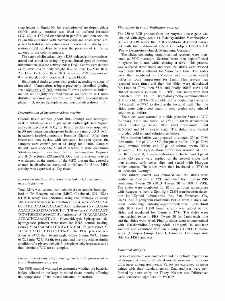
As indicated in Fig. 1 (A), the selected bacterial isolate wasable to grow at pH 2 and 3 for 4 h. Although the number ofsurviving bacteria following 1 h of incubation at pH 2 and 3was significantly lower than in the control at pH 6, a signifi-cant number still survived and kept growing after 4 h incu-bation under the acidic conditions. It fulfilled additionalprobiotic criteria such as resistance to bile acids (Fig. 1(B)), and survived 4 h in 0·5% ox-gall (bile acid mixture).
Percentage adhesion of E. durans to Caco-2 cells was com-pared with the well-known probiotic strains L. rhamnosus GGand L. casei Shirota, which are considered to be high- andlow-adhesive strains, respectively (Tuomola & Salminen,1998). The percentage of Caco-2 cells in each field withadhering bacteria and demonstrated that the number ofadhered E. durans cells was comparable with the highlyadhesive strain L. rhamnosus GG but significantly higher(P,0·005 as calculated by the Tukey–Kramer test) than thelow-adhesive strain L. casei Shirota (data not shown).
The selected bacterial isolate was devoid of gelatinaseactivity according to the gelatin-zymography assay (data notshown).
Effect of Enterococcus durans on dextran sodium sulfate-induced colitis
Acute colitis was induced in mice via administration of DSSin the drinking water. At the end of the experiment, significantdifferences were observed among the different groups in termsof body-weight gain and extent of rectal bleeding, measure-ments that were all scored as DAI. Daily administration of1 £ 108 cfu E. durans/g chow to DSS-treated mice induceda significant lowering effect in DAI, i.e. E. durans preventedweight loss in the DSS-treated mice and concomitantly theadministered bacteria prevented colonic bleeding. In contrast,the food-grade bacteria L. delbrueckii subsp. lactis ATCC
Table 1. Determination of optimal intestinal microflora medium (IMM) conditions for faecal butyrateproduction†
(Mean values with their standard errors)
Butyrate concentration (mmol/l)
Time (weeks) . . . 0 1 2 3
Medium conditions Mean SEM Mean SEM Mean SEM Mean SEM
Control‡ 1·9 0·2 2·1 0·3 2·0 0·5 1·3 0·7pH 5·0 1·8 0·4 4·7* 0·8b 6·8** 1·2 5·3** 1·1pH 5·5 2·1 0·2 5·1* 0·9 4·8* 0·4 5·6** 0·8pH 6·0 2·3 0·3 5·1* 0·5 5·8** 0·4c 5·6** 0·6pH 6·5 1·9 0·2 2·8 0·2 2·8 0·1 3·5* 0·3pH 7·0 1·7 0·4 3·0 0·6 2·5 0·3 3·4* 0·5pH 7·5 1·7 0·2 2·7 0·4 2·7 0·5 4·2* 0·4pH 8·0 2·0 0·1 2·9 0·2 2·7 0·2 3·8* 0·4pH 9·6 2·2 0·4 3·4 0·5 3·8* 0·7 3·8* 0·6
Mean value was significantly different from that of the control condition: * P,0·05, ** P,0·01 (Tukey–Kramer test).†Four different independent experiments were conducted.‡Control culture contained IMM at pH 9 without faecal bacterial samples.
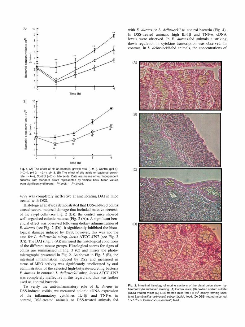
4797 was completely ineffective at ameliorating DAI in micetreated with DSS.Histological analyses demonstrated that DSS-induced colitis
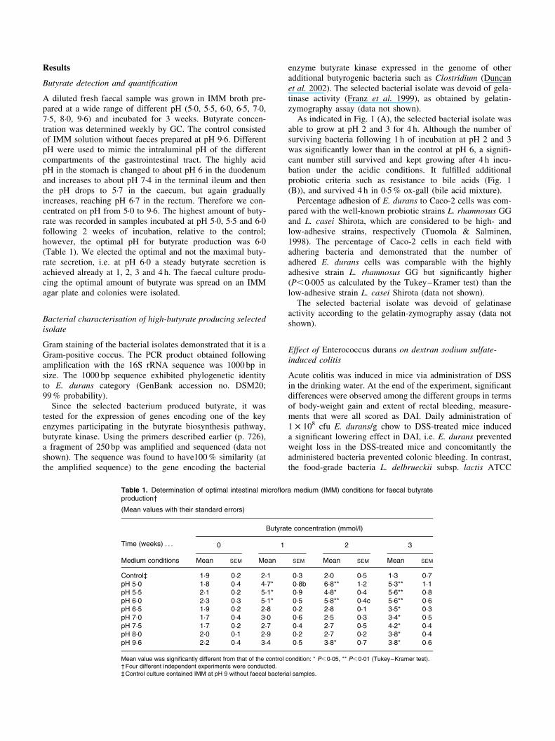
caused severe mucosal damage that included massive necrosisof the crypt cells (see Fig. 2 (B)); the control mice showedwell-organised colonic mucosa (Fig. 2 (A)). A significant ben-eficial effect was observed following dietary administration ofE. durans (see Fig. 2 (D)); it significantly inhibited the histo-logical damage induced by DSS; however, this was not thecase for L. delbrueckii subsp. lactis ATCC 4797 (see Fig. 2(C)). The DAI (Fig. 3 (A)) mirrored the histological conditionsof the different mouse groups. Histological scores for signs ofcolitis are summarised in Fig. 3 (C) and mirror the photo-micrographs presented in Fig. 2. As shown in Fig. 3 (B), theintestinal inflammation induced by DSS and measured interms of MPO activity was significantly ameliorated by oraladministration of the selected high-butyrate-secreting bacteriaE. durans. In contrast, L. delbrueckii subsp. lactis ATCC 4797was completely ineffective in this regard and thus was furtherused as control bacteria.To verify the anti-inflammatory role of E. durans in
DSS-induced colitis, we measured colonic cDNA expressionof the inflammatory cytokines IL-1b and TNF-a incontrol, DSS-treated animals or DSS-treated animals fed
with E. durans or L. delbrueckii as control bacteria (Fig. 4).In DSS-treated animals, high IL-1b and TNF-a cDNAlevels were observed. In E. durans-fed animals a strikingdown regulation in cytokine transcription was observed. Incontrast, in L. delbrueckii-fed animals, the concentrations of
10
9
8
7
6
5
4
3
2
1
0 0 1 2 3 4
Time (h)
0 1 2 3 4
Time (h)
Bac
teri
al c
on
cen
trat
ion
× 1
010
(cfu
/ml)
10
9
8
7
6
5
4
3
2
1
0
Bac
teri
al c
on
cen
trat
ion
× 1
010
(cfu
/ml)
****
**
**
(A)
(B)
Fig. 1. (A) The effect of pH on bacterial growth rate. (–V–), Control (pH 6);
(–A–), pH 2; (–D–), pH 3. (B) The effect of bile acids on bacterial growth
rate. (–V–), Control (–A–), bile acids. Data are means of four independent
cultures, with standard errors represented by vertical bars. Mean values
were significantly different: * P,0·05, ** P,0·001.
Fig. 2. Intestinal histology of murine sections of the distal colon shown by
haematoxylin and eosin staining. (A) Control mice; (B) dextran sodium sulfate
(DSS)-treated mice; (C) DSS-treated mice fed 1 £ 108 colony-forming units
(cfu) Lactobacillus delbrueckii subsp. lactis/g feed; (D) DSS-treated mice fed
1 £ 108 cfu Enterococcus durans/g feed.
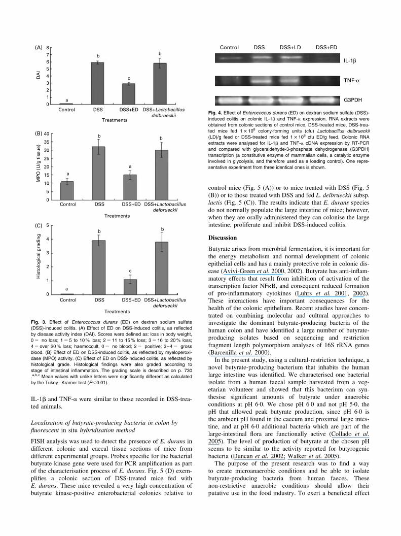
IL-1b and TNF-a were similar to those recorded in DSS-trea-ted animals.
Localisation of butyrate-producing bacteria in colon byfluorescent in situ hybridisation method
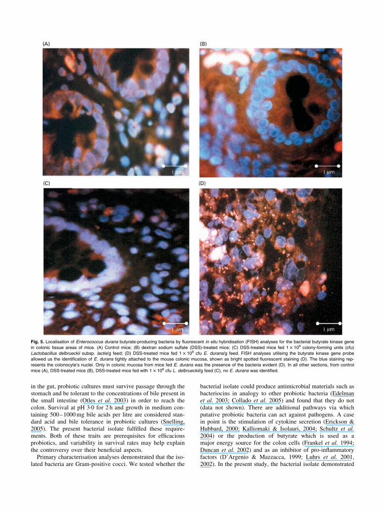
FISH analysis was used to detect the presence of E. durans indifferent colonic and caecal tissue sections of mice fromdifferent experimental groups. Probes specific for the bacterialbutyrate kinase gene were used for PCR amplification as partof the characterisation process of E. durans. Fig. 5 (D) exem-plifies a colonic section of DSS-treated mice fed withE. durans. These mice revealed a very high concentration ofbutyrate kinase-positive enterobacterial colonies relative to
control mice (Fig. 5 (A)) or to mice treated with DSS (Fig. 5(B)) or to those treated with DSS and fed L. delbrueckii subsp.lactis (Fig. 5 (C)). The results indicate that E. durans speciesdo not normally populate the large intestine of mice; however,when they are orally administered they can colonise the largeintestine, proliferate and inhibit DSS-induced colitis.
Discussion
Butyrate arises from microbial fermentation, it is important forthe energy metabolism and normal development of colonicepithelial cells and has a mainly protective role in colonic dis-ease (Avivi-Green et al. 2000, 2002). Butyrate has anti-inflam-matory effects that result from inhibition of activation of thetranscription factor NFkB, and consequent reduced formationof pro-inflammatory cytokines (Luhrs et al. 2001, 2002).These interactions have important consequences for thehealth of the colonic epithelium. Recent studies have concen-trated on combining molecular and cultural approaches toinvestigate the dominant butyrate-producing bacteria of thehuman colon and have identified a large number of butyrate-producing isolates based on sequencing and restrictionfragment length polymorphism analyses of 16S rRNA genes(Barcenilla et al. 2000).
In the present study, using a cultural-restriction technique, anovel butyrate-producing bacterium that inhabits the humanlarge intestine was identified. We characterised one bacterialisolate from a human faecal sample harvested from a veg-etarian volunteer and showed that this bacterium can syn-thesise significant amounts of butyrate under anaerobicconditions at pH 6·0. We chose pH 6·0 and not pH 5·0, thepH that allowed peak butyrate production, since pH 6·0 isthe ambient pH found in the caecum and proximal large intes-tine, and at pH 6·0 additional bacteria which are part of thelarge-intestinal flora are functionally active (Collado et al.2005). The level of production of butyrate at the chosen pHseems to be similar to the activity reported for butyrogenicbacteria (Duncan et al. 2002; Walker et al. 2005).
The purpose of the present research was to find a wayto create microanaerobic conditions and be able to isolatebutyrate-producing bacteria from human faeces. Thesenon-restrictive anaerobic conditions should allow theirputative use in the food industry. To exert a beneficial effect
8(A)
(B)
(C)
7654D
AI
3210
40
35
30
25
20
15
10
5
0
5
4
3
2
1
0
Control
Control
MP
O (
U/g
tis
sue)
DSS
DSS
DSS+ED
DSS+ED
DSS+Lactobacillusdelbrueckii
DSS+Lactobacillusdelbrueckii
Treatments
Treatments
Control DSS DSS+ED DSS+Lactobacillusdelbrueckii
Treatments
a
a
a
a
b
b
b b
b
b
c
c
His
tolo
gic
al g
rad
ing
Fig. 3. Effect of Enterococcus durans (ED) on dextran sodium sulfate
(DSS)-induced colitis. (A) Effect of ED on DSS-induced colitis, as reflected
by disease activity index (DAI). Scores were defined as: loss in body weight,
0 ¼ no loss; 1 ¼ 5 to 10% loss; 2 ¼ 11 to 15% loss; 3 ¼ 16 to 20% loss;
4 ¼ over 20% loss; haemoccult, 0 ¼ no blood; 2 ¼ positive; 3–4 ¼ gross
blood. (B) Effect of ED on DSS-induced colitis, as reflected by myeloperoxi-
dase (MPO) activity. (C) Effect of ED on DSS-induced colitis, as reflected by
histological grade. Histological findings were also graded according to
stage of intestinal inflammation. The grading scale is described on p. 730
.a,b,c Mean values with unlike letters were significantly different as calculated
by the Tukey–Kramer test (P,0·01).
Control DSS DSS+LD DSS+ED
IL-1β
TNF-α
G3PDH
Fig. 4. Effect of Enterococcus durans (ED) on dextran sodium sulfate (DSS)-
induced colitis on colonic IL-1b and TNF-a expression. RNA extracts were
obtained from colonic sections of control mice, DSS-treated mice, DSS-trea-
ted mice fed 1 £ 108 colony-forming units (cfu) Lactobacillus delbrueckii
(LD)/g feed or DSS-treated mice fed 1 £ 108 cfu ED/g feed. Colonic RNA
extracts were analysed for IL-1b and TNF-a cDNA expression by RT-PCR
and compared with glyceraldehyde-3-phosphate dehydrogenase (G3PDH)
transcription (a constitutive enzyme of mammalian cells, a catalytic enzyme
involved in glycolysis, and therefore used as a loading control). One repre-
sentative experiment from three identical ones is shown.
in the gut, probiotic cultures must survive passage through thestomach and be tolerant to the concentrations of bile present inthe small intestine (Otles et al. 2003) in order to reach thecolon. Survival at pH 3·0 for 2 h and growth in medium con-taining 500–1000mg bile acids per litre are considered stan-dard acid and bile tolerance in probiotic cultures (Snelling,2005). The present bacterial isolate fulfilled these require-ments. Both of these traits are prerequisites for efficaciousprobiotics, and variability in survival rates may help explainthe controversy over their beneficial aspects.Primary characterisation analyses demonstrated that the iso-
lated bacteria are Gram-positive cocci. We tested whether the
bacterial isolate could produce antimicrobial materials such asbacteriocins in analogy to other probiotic bacteria (Edelmanet al. 2003; Collado et al. 2005) and found that they do not(data not shown). There are additional pathways via whichputative probiotic bacteria can act against pathogens. A casein point is the stimulation of cytokine secretion (Erickson &Hubbard, 2000; Kalliomaki & Isolauri, 2004; Schultz et al.2004) or the production of butyrate which is used as amajor energy source for the colon cells (Frankel et al. 1994;Duncan et al. 2002) and as an inhibitor of pro-inflammatoryfactors (D’Argenio & Mazzacca, 1999; Luhrs et al. 2001,2002). In the present study, the bacterial isolate demonstrated
Fig. 5. Localisation of Enterococcus durans butyrate-producing bacteria by fluorescent in situ hybridisation (FISH) analyses for the bacterial butyrate kinase gene
in colonic tissue areas of mice. (A) Control mice; (B) dextran sodium sulfate (DSS)-treated mice; (C) DSS-treated mice fed 1 £ 108 colony-forming units (cfu)
Lactobacillus delbrueckii subsp. lactis/g feed; (D) DSS-treated mice fed 1 £ 108 cfu E. durans/g feed. FISH analyses utilising the butyrate kinase gene probe
allowed us the identification of E. durans tightly attached to the mouse colonic mucosa, shown as bright spotted fluorescent staining (D). The blue staining rep-
resents the colonocyte’s nuclei. Only in colonic mucosa from mice fed E. durans was the presence of the bacteria evident (D). In all other sections, from control
mice (A), DSS-treated mice (B), DSS-treated mice fed with 1 £ 108 cfu L. delbrueckii/g feed (C), no E. durans was identified.
adherence to Caco-2 cells in vitro. In the colon, the bacterianeed to adhere to the epithelial cells and form active colonies.The colonised bacteria can influence the host via their meta-bolic activity and can compete with pathogenic bacteria(Jacobsen et al. 1999).
We genetically characterised the bacterial isolate by PCRamplification of the 16S rRNA. The sequence obtained following16S rRNA amplification was 99% identical to an E. durans iso-late in the GeneBank. Enterococcus bacteria such as E. durans,E. faecalis, E. faecium, E. hirae, E. casseliflavus and E. galli-narum are known to be significant for the gastrointestinal ecologyaswell as for that of some foods (Klein, 2003).Enterococcus bac-teria exist in soil, water and in human and animal intestines (105–107 cfu/g faeces). E. faecalis and E. faecium are the most domi-nant strains found in the human intestine. Enterococcus strainsare already known as probiotic bacteria and, to a certain extent,are used to treat gastroenteritis in human patients and animals(Franz et al. 1999). In this regard, Pereira & Gibson (2002)have recently identified E. duransDSM20633 as a potential pro-biotic isolated from faecal material.
Sequencing the 250 bp PCRproduct obtained following ampli-fication with specific primers to bacterial butyrate kinase indi-cated that in the isolated butyrate-producing E. durans this geneexists in its genome, as judged from 100% identity of the250 bp amplified to the respective sequence in the gene presentin the anaerobic butyrate-producing strain C. acetobutylicum.Two key enzymes have been found to be involved in thelast stage of the butyrate-synthesising process inbacteria; butyratekinase and butyryl CoA:acetate CoA transferase (Duncan et al.2002, 2003, 2004a,b; Louis et al. 2004). The butyryl CoA:ace-tate-CoA transferase gene has been found inmost butyrate-produ-cing bacteria from the human intestine, whereas butyrate kinase isless common (Duncan et al. 2002, 2004a,b; Louis et al. 2004).Nonetheless, both pathways can be expressed at once. The signifi-cance of these metabolic differences has yet to be established.
An additional important issue to be addressed relates to bac-terial safety for human use. We analysed whether the isolatedE. durans has a plasmid and found none. This suggests that ifthe bacteria become resistant to any antibiotics, transfer of thistrait to other bacteria would be impossible. We found that thebacterial isolate cannot survive in the presence of ampicillin,tetracycline and chloramphenicol. We also examined the abil-ity of the bacterial isolate to synthesise gelatinase, an activityassociated with harmful factors such as adhesins, invasins andhaemolysin (Franz et al. 1999; Kawalec et al. 2005). Weshowed that E. durans isolated in the present study does notbear gelatinase activity. Cumulatively these qualities furthersupport the view that the bacterial isolate can be safe forhuman use.
An additional important finding of the present study relatesto the ability of the bacterial isolate to prevent DSS-inducedcolitis. We tested the effect of E. durans on a murine modelof ulcerative colitis simultaneously induced by DSS. Severalanimal models of experimental ulcerative colitis have beendescribed and, of these, colitis induced in mice by oral admin-istration of DSS has been widely used because of its manysimilarities to human ulcerative colitis (Cooper et al. 1993).E. durans was able to prevent disease induction almost com-pletely as reflected by DAI, MPO activity and histologicalgrade, while L. delbrueckii subsp. lactis ATCC 4797 bacteriawas completely ineffective in this regard.
Using a preventive setup in the present study, we demon-strated that following daily feeding of DSS-treated mice with1 £ 108 E. durans per g feed, there is significant inhibition inthe development of chronic experimental colitis. The mechan-ism by which E. durans exerts its protection in DSS-inducedcolitis may reside in the bacteria’s effect on immune-systemregulation, or on the ability of the E. durans isolate to producehigh concentrations of butyrate. Butyrate’s role in preventingand/or ameliorating conditions such as ulcerative colitis hasbeen studied (Hallert et al. 2003; Vernia et al. 2003; Galvezet al. 2005). It has been proposed that an inadequate supply ofenergy to colonocytes, 70% of which is normally obtainedfrom butyrate, can be a causative factor in colitis. DSS, whichinhibits butyrate oxidation without affecting glucose metab-olism, induces colitis when given orally to mice (Ahmad et al.2000), and it has been proposed that sulfide toxicity resultslargely from inhibition of the butyrate energy supply to colono-cytes. It is surmised that DSS changes in epithelial cell metab-olism are secondary to mucosal inflammation. E. durans cansupply sufficient butyrate concentrations to counteract theeffect of DSS or inhibit intestinal mucosal inflammation, orboth concomitantly. Present work in our laboratory is addressingeach pathway and its significance. We conclude that E. duransprevents the induction of ulcerative colitis by DSS, significantlyimproving the clinical conditions. This could have importantbiological consequences for human health.
We used FISH analysis in order to localise the fate ofE. durans in the mouse intestinal tissue. We used a 250 bpprobe obtained by PCR amplification of the bacterial butyratekinase gene to localise the E. durans in the mice colonictissue. A high concentration of fluorescent dots on the colonictissue surfaces of mice fed E. durans were obtained, whereasno such fluorescent dots were detected in control mice or inthose fed with L. delbrueckii. This suggests that E. duransdoes not normally populate the mouse caecal and large-intes-tinal tissue surface; however, when these bacteria colonise therodent intestine they are not only harmless to the mice, butalso prevent DSS-induced colitis.
In conclusion, we described in the present paper the charac-terisation of a bacterial isolate exhibiting 99% homology toE. durans and expressing typical probiotic features. Oraladministration of the bacterial isolate prevented and/or ame-liorated experimental colitis, suggesting that this isolate pro-vides a unique oral anti-inflammatory tool that deservesfurther studies specifically addressing the pathogenesis andtreatment of human inflammatory bowel disease.Additionalin vivo and in vitro studies are needed, to determine the mech-anism by which this bacterial isolate exerts its preventiveeffect on ulcerative colitis. Moreover, its influence on the indi-genous microflora and overall metabolic activity of the gutshould be further assessed in vitro in mixed-culture andmixed-substrate environments before the design of any clinicalintervention trials. We believe that the putative probiotic bac-teria described in the present paper could potentially make asignificant contribution to the food and medicinal industries.
Acknowledgements
This study was supported in part by Danone Research Center,Israel.
References
Ahmad MS, Krishnan S, Ramakrishna BS, Mathan M, Pulimood AB
& Murthy SN (2000) Butyrate and glucose metabolism by colono-
cytes in experimental colitis in mice. Gut 46, 493–499.Avivi-Green C, Madar Z & Schwartz B (2000) Pectin-enriched diet
affects distribution and expression of apoptosis-cascade proteins
in colonic crypts of dimethylhydrazine-treated rats. Int J Mol
Med 6, 689–698.Avivi-Green C, Polak-Charcon S, Madar Z & Schwartz B (2002)
Different molecular events account for butyrate-induced apoptosis
in two human colon cancer cell lines. J Nutr 132, 1812–1818.Barcenilla A, Pryde SE, Martin JC, Duncan SH, Stewart CS, Hender-
son C & Flint HJ (2000) Phylogenetic relationships of butyrate-
producing bacteria from the human gut. Appl Environ Microbiol
66, 1654–1661.Chen CC & Walker WA (2005) Probiotics and prebiotics: role in
clinical disease states. Adv Pediatr 52, 77–113.Collado MC, Hernandez M & Sanz Y (2005) Production of bacterio-
cin-like inhibitory compounds by human fecal Bifidobacterium
strains. J Food Prot 68, 1034–1040.Cooper HS, Murthy SN, Shah RS & Sedergran DJ (1993) Clinico-
pathologic study of dextran sulfate sodium experimental murine
colitis. Lab Invest 69, 238–249.D’Argenio G & Mazzacca G (1999) Short-chain fatty acid in the
human colon. Relation to inflammatory bowel diseases and colon
cancer. Adv Exp Med Biol 472, 149–158.Diakos C, Prieschl EE, Saemann M, Novotny V, Bohmig G, Csonga R,
Baumruker T & Zlabinger GJ (2002) Novel mode of interference
with nuclear factor of activated T-cells regulation in T-cells by the
bacterial metabolite n-butyrate. J Biol Chem 277, 24243–24251.Djouzi Z, Andrieux C, Degivry MC, Bouley C & Szylit O (1997) The
association of yogurt starters with Lactobacillus casei DN 114.001
in fermented milk alters the composition and metabolism of intes-
tinal microflora in germ-free rats and in human flora-associated
rats. J Nutr 127, 2260–2266.Duncan SH, Barcenilla A, Stewart CS, Pryde SE & Flint HJ (2002)
Acetate utilization and butyryl coenzyme A (CoA):acetate-CoA
transferase in butyrate-producing bacteria from the human large
intestine. Appl Environ Microbiol 68, 5186–5190.Duncan SH, Holtrop G, Lobley GE, Calder AG, Stewart CS & Flint
HJ (2004a) Contribution of acetate to butyrate formation by human
faecal bacteria. Br J Nutr 91, 915–923.Duncan SH, Louis P & Flint HJ (2004b) Lactate-utilizing bacteria,
isolated from human faeces, that produce butyrate as a major fer-
mentation product. Appl Environ Microbiol 70, 5810–5817.Duncan SH, Scott KP, Ramsay AG, Harmsen HJ, Welling GW, Stew-
art CS & Flint HJ (2003) Effects of alternative dietary substrates on
competition between human colonic bacteria in an anaerobic fer-
mentor system. Appl Environ Microbiol 69, 1136–1142.Edelman MJ, Bauer K, Khanwani S, Tait N, Trepel J, Karp J, Nemie-
boka N, Chung EJ & Van Echo D (2003) Clinical and pharmaco-
logic study of tributyrin: an oral butyrate prodrug. Cancer
Chemother Pharmacol 51, 439–444.Erickson KL & Hubbard NE (2000) Probiotic immunomodulation in
health and disease. J Nutr 130, 403S–409S.Frankel WL, Zhang W, Singh A, Klurfeld DM, Don S, Sakata T,
Modlin I & Rombeau JL (1994) Mediation of the trophic effects
of short-chain fatty acids on the rat jejunum and colon. Gastroen-
terology 106, 375–380.Franz CM, Holzapfel WH & Stiles ME (1999) Enterococci at the
crossroads of food safety? Int J Food Microbiol 47, 1–24.Galvez J, Rodriguez-CabezasME&ZarzueloA (2005) Effects of dietary
fiber on inflammatory bowel disease.Mol Nutr Food Res 49, 601–608.Hallert C, Bjorck I, Nyman M, Pousette A, Granno C & Svensson H
(2003) Increasing fecal butyrate in ulcerative colitis patients by
diet: controlled pilot study. Inflamm Bowel Dis 9, 116–121.
Jacobsen CN, Rosenfeldt Nielsen V, Hayford AE, Moller PL,
Michaelsen KF, Paerregaard A, Sandstrom B, Tvede M &
Jakobsen M (1999) Screening of probiotic activities of forty-
seven strains of Lactobacillus spp. by in vitro techniques and
evaluation of the colonization ability of five selected strains in
humans. Appl Environ Microbiol 65, 4949–4956.Kalliomaki MA & Isolauri E (2004) Probiotics and down-regulation
of the allergic response. Immunol Allergy Clin North Am 24,739–752, viii.
Kawalec M, Potempa J, Moon JL, Travis J & Murray BE (2005) Mol-
ecular diversity of a putative virulence factor: purification and
characterization of isoforms of an extracellular serine glutamyl
endopeptidase of Enterococcus faecalis with different enzymatic
activities. J Bacteriol 187, 266–275.Klein G (2003) Taxonomy, ecology and antibiotic resistance of enter-
ococci from food and the gastro-intestinal tract. Int J Food Micro-
biol 88, 123–131.Louis P, Duncan SH, McCrae SI, Millar J, Jackson MS & Flint HJ
(2004) Restricted distribution of the butyrate kinase pathway
among butyrate-producing bacteria from the human colon. J Bac-
teriol 186, 2099–2106.Luhrs H, Gerke T, Boxberger F, Backhaus K, Melcher R, Scheppach
W & Menzel T (2001) Butyrate inhibits interleukin-1-mediated
nuclear factor-kappa B activation in human epithelial cells. Dig
Dis Sci 46, 1968–1973.Luhrs H, Gerke T, Muller JG, Melcher R, Schauber J, Boxberge F,
Scheppach W & Menzel T (2002) Butyrate inhibits NF-kappaB
activation in lamina propria macrophages of patients with ulcera-
tive colitis. Scand J Gastroenterol 37, 458–466.O’Sullivan GC, Kelly P, O’Halloran S, Collins C, Collins JK, Dunne
C & Shanahan F (2005) Probiotics: an emerging therapy. Curr
Pharm Des 11, 3–10.Otles S, Cagindi O & Akcicek E (2003) Probiotics and health. Asian
Pac J Cancer Prev 4, 369–372.Pereira DI & Gibson GR (2002) Cholesterol assimilation by lactic
acid bacteria and bifidobacteria isolated from the human gut.
Appl Environ Microbiol 68, 4689–4693.Resta-Lenert S & Barrett KE (2003) Live probiotics protect intestinal
epithelial cells from the effects of infection with enteroinvasive
Escherichia coli (EIEC). Gut 52, 988–997.Saggioro A (2004) Probiotics in the treatment of irritable bowel syn-
drome. J Clin Gastroenterol 38, S104–S106.Schultz M, Strauch UG, Linde HJ, Watzl S, Obermeier F, Gottl C,
Dunger N, Grunwald N, Scholmerich J & Rath HC (2004) Preven-
tive effects of Escherichia coli strain Nissle 1917 on acute and
chronic intestinal inflammation in two different murine models of
colitis. Clin Diagn Lab Immunol 11, 372–378.Snelling AM (2005) Effects of probiotics on the gastrointestinal tract.
Curr Opin Infect Dis 18, 420–426.Tuomola EM & Salminen SJ (1998) Adhesion of some probiotic and
dairy Lactobacillus strains to Caco-2 cell cultures. Int J Food
Microbiol 41, 45–51.Vernia P, Annese V, Bresci G, et al. (2003) Topical butyrate
improves efficacy of 5-ASA in refractory distal ulcerative
colitis: results of a multicentre trial. Eur J Clin Invest 33, 244–248.Vine DF, Charman SA, Gibson PR, Sinclair AJ & Porter CJ (2002)
Effect of dietary fatty acids on the intestinal permeability of
marker drug compounds in excised rat jejunum. J Pharm Pharma-
col 54, 809–819.Walker AW, Duncan SH, McWilliam Leitch EC, ChildMW&Flint HJ
(2005) pH and peptide supply can radically alter bacterial
populations and short-chain fatty acid ratios within microbial com-
munities from the human colon. Appl Environ Microbiol 71,3692–3700.
Wilson M, Hall V, Brazier J & Lewis MA (2000) Evaluation of a
phenotypic scheme for identification of the ‘butyrate-producing’
Peptostreptococcus species. J Med Microbiol 49, 747–751.
Related Documents


![WallFlex Colonic Stent - Boston Scientific- US · WallFlex ™ Colonic Stent Visualization Expertise in combining stent materials has resulted ... (BTS). “The WallFlex™ [Colonic]](https://static.cupdf.com/doc/110x72/5ae601bc7f8b9a8b2b8ca931/wallflex-colonic-stent-boston-scientific-us-colonic-stent-visualization-expertise.jpg)