Iron oxide nanoparticle-containing microbubble composites as contrast agents for MR and ultrasound dual-modality imaging Zhe Liu a , Twan Lammers a , Josef Ehling a, b , Stanley Fokong a , Jörg Bornemann c , Fabian Kiessling a , Jessica Gätjens a, * a Department of Experimental Molecular Imaging (ExMI), Helmholtz Institute for Biomedical Engineering, Medical Faculty, RWTH Aachen University, Aachen 52074, Germany b Institute of Pathology, University Hospital Aachen (UKA), Aachen 52074, Germany c Electron Microscopic Facility, University Hospital Aachen (UKA), Aachen 52074, Germany article info Article history: Received 24 February 2011 Accepted 5 May 2011 Available online 31 May 2011 Keywords: Nanoparticle MRI (magnetic resonance imaging) Contrast agent Polymerization Molecular imaging abstract Magnetic resonance (MR) and ultrasound (US) imaging are widely used diagnostic modalities for various experimental and clinical applications. In this study, iron oxide nanoparticle-embedded polymeric microbubbles were designed as multi-modal contrast agents for hybrid MReUS imaging. These magnetic nano-in-micro imaging probes were prepared via a one-pot emulsion polymerization to form poly(butyl cyanoacrylate) microbubbles, along with the oil-in-water (O/W) encapsulation of iron oxide nano- particles in the bubble shell. The nano-in-micro embedding strategy was validated using NMR and electron microscopy. These hybrid imaging agents exhibited strong contrast in US and an increased transversal relaxation rate in MR. Moreover, a significant increase in longitudinal and transversal relaxivities was observed after US-induced bubble destruction, which demonstrated triggerable MR imaging properties. Proof-of-principle in vivo experiments confirmed that these nanoparticle-embedded microbubble composites are suitable contrast agents for both MR and US imaging. In summary, these magnetic nano-in-micro hybrid materials are highly interesting systems for bimodal MReUS imaging, and their enhanced relaxivities upon US-induced destruction recommend them as potential vehicles for MR-guided US-mediated drug and gene delivery. Ó 2011 Elsevier Ltd. All rights reserved. 1. Introduction In the field of disease-specific imaging, magnetic resonance (MR) and ultrasound (US) imaging are widely used modalities for various experimental and clinical applications [1,2]. Magnetic nanoparticles or metal complexes have been developed as power- ful contrast agents for MR imaging, which is non-invasive, and capable of providing morphological and functional information with a high spatial resolution and with excellent soft-tissue contrast. In comparison, ultrasound is highly cost-effective, and suitable for real-time imaging. Stabilized gas microbubbles are routinely used as US contrast agents, and they can be functionalized with antibodies or peptides to specifically bind receptors over- expressed on vascular endothelial cells, thereby generating sensi- tive intravascular molecular imaging probes. Moreover, they can also be loaded with drugs and genes, and release their payloads either upon magnetic actuation or upon exposure to destructive ultrasound pulses [3e7]. These features of MR and US contrast agents not only make them favorable tools for precisely visualizing biological and physiological processes at molecular and cellular level, but also suggest that they might render synergistic efficacy if one single imaging probe can be developed for both modalities. Recently, nanoparticle-containing composites have attracted unprecedented attention for functional and molecular imaging investigations [8e10]. It has been reported that some solid nano- particles, such as silica, polystyrene and superparamagnetic iron oxide (SPIO) nanoparticles, are able to boost the acoustic impedance, increase backscattered signals, and consequently contribute to contrast enhancement for US [11e 13]. On the other hand, gas-filled microbubbles proved to be detectable by MR since the shell oscil- lations of bubbles result in a proportionate contribution to magnetic susceptibility [14,15]. Therefore, constructing a hybrid contrast agent would be highly useful for both imaging modalities, and could facilitate MR-guided US-mediated transfection and gene delivery, MR-guided high-intensity focused ultrasound treatment, hyper- thermia therapy, and even broader biomedical applications [16e21]. As appealing carrier materials for nanoparticles, polymer- based hard-shell microbubbles show obvious advantages over * Corresponding author. Tel.: þ49 241 8080116; fax: þ49 241 8082006. E-mail address: [email protected] (J. Gätjens). Contents lists available at ScienceDirect Biomaterials journal homepage: www.elsevier.com/locate/biomaterials 0142-9612/$ e see front matter Ó 2011 Elsevier Ltd. All rights reserved. doi:10.1016/j.biomaterials.2011.05.019 Biomaterials 32 (2011) 6155e6163

Welcome message from author
This document is posted to help you gain knowledge. Please leave a comment to let me know what you think about it! Share it to your friends and learn new things together.
Transcript
lable at ScienceDirect
Biomaterials 32 (2011) 6155e6163
Contents lists avai
Biomaterials
journal homepage: www.elsevier .com/locate/biomater ia ls
Iron oxide nanoparticle-containing microbubble composites as contrast agentsfor MR and ultrasound dual-modality imaging
Zhe Liu a, Twan Lammers a, Josef Ehling a,b, Stanley Fokong a, Jörg Bornemann c, Fabian Kiessling a,Jessica Gätjens a,*
aDepartment of Experimental Molecular Imaging (ExMI), Helmholtz Institute for Biomedical Engineering, Medical Faculty, RWTH Aachen University, Aachen 52074, Germanyb Institute of Pathology, University Hospital Aachen (UKA), Aachen 52074, Germanyc Electron Microscopic Facility, University Hospital Aachen (UKA), Aachen 52074, Germany
a r t i c l e i n f o
Article history:Received 24 February 2011Accepted 5 May 2011Available online 31 May 2011
Keywords:NanoparticleMRI (magnetic resonance imaging)Contrast agentPolymerizationMolecular imaging
* Corresponding author. Tel.: þ49 241 8080116; faxE-mail address: [email protected] (J. Gätjens
0142-9612/$ e see front matter � 2011 Elsevier Ltd.doi:10.1016/j.biomaterials.2011.05.019
a b s t r a c t
Magnetic resonance (MR) and ultrasound (US) imaging are widely used diagnostic modalities for variousexperimental and clinical applications. In this study, iron oxide nanoparticle-embedded polymericmicrobubbles were designed as multi-modal contrast agents for hybrid MReUS imaging. These magneticnano-in-micro imaging probes were prepared via a one-pot emulsion polymerization to form poly(butylcyanoacrylate) microbubbles, along with the oil-in-water (O/W) encapsulation of iron oxide nano-particles in the bubble shell. The nano-in-micro embedding strategy was validated using NMR andelectron microscopy. These hybrid imaging agents exhibited strong contrast in US and an increasedtransversal relaxation rate in MR. Moreover, a significant increase in longitudinal and transversalrelaxivities was observed after US-induced bubble destruction, which demonstrated triggerable MRimaging properties. Proof-of-principle in vivo experiments confirmed that these nanoparticle-embeddedmicrobubble composites are suitable contrast agents for both MR and US imaging. In summary, thesemagnetic nano-in-micro hybrid materials are highly interesting systems for bimodal MReUS imaging,and their enhanced relaxivities upon US-induced destruction recommend them as potential vehicles forMR-guided US-mediated drug and gene delivery.
� 2011 Elsevier Ltd. All rights reserved.
1. Introduction
In the field of disease-specific imaging, magnetic resonance(MR) and ultrasound (US) imaging are widely used modalities forvarious experimental and clinical applications [1,2]. Magneticnanoparticles or metal complexes have been developed as power-ful contrast agents for MR imaging, which is non-invasive, andcapable of providing morphological and functional informationwith a high spatial resolution and with excellent soft-tissuecontrast. In comparison, ultrasound is highly cost-effective, andsuitable for real-time imaging. Stabilized gas microbubbles areroutinely used as US contrast agents, and they can be functionalizedwith antibodies or peptides to specifically bind receptors over-expressed on vascular endothelial cells, thereby generating sensi-tive intravascular molecular imaging probes. Moreover, they canalso be loaded with drugs and genes, and release their payloadseither upon magnetic actuation or upon exposure to destructive
: þ49 241 8082006.).
All rights reserved.
ultrasound pulses [3e7]. These features of MR and US contrastagents not only make them favorable tools for precisely visualizingbiological and physiological processes at molecular and cellularlevel, but also suggest that they might render synergistic efficacy ifone single imaging probe can be developed for both modalities.
Recently, nanoparticle-containing composites have attractedunprecedented attention for functional and molecular imaginginvestigations [8e10]. It has been reported that some solid nano-particles, such as silica, polystyrene and superparamagnetic ironoxide (SPIO)nanoparticles, are able toboost theacoustic impedance,increase backscattered signals, and consequently contribute tocontrast enhancement for US [11e13]. On the other hand, gas-filledmicrobubbles proved to be detectable by MR since the shell oscil-lations of bubbles result in a proportionate contribution tomagneticsusceptibility [14,15]. Therefore, constructing a hybrid contrastagentwould be highly useful for both imagingmodalities, and couldfacilitate MR-guided US-mediated transfection and gene delivery,MR-guided high-intensity focused ultrasound treatment, hyper-thermia therapy, and evenbroader biomedical applications [16e21].
As appealing carrier materials for nanoparticles, polymer-based hard-shell microbubbles show obvious advantages over
Z. Liu et al. / Biomaterials 32 (2011) 6155e61636156
phospholipid-based soft-shell microbubbles [22,23]. In particular,poly(butyl cyanoacrylate) (PBCA) microbubbles are potentialcandidates, due to the following characteristics: (i) the shellmaterial of PBCA is biodegradable and biocompatible, whichensures safety for in vivo applications and clinical translation; (ii)PBCA-based microbubbles have a higher stability than soft-shellbubbles, so that enhanced acoustic properties and prolongedcirculation times will be ensured by minimizing gas-coreshrinkage. Some reports show that PBCA nanoparticles can beproduced as drug delivery vectors via cationic polymerization ofBCA monomers by low-speed magnetic stirring in suitablesurfactant emulsions, but little research has been dedicated to thedesign of monodisperse PBCA gas-filled bubbles as a potentialbifunctional platform for image-guided biomedical purposes,along with the incorporation of paramagnetic nanoparticlesand/or therapeutic agents [24e27]. Although multistep proce-dures and layer-by-layer (LBL) processes have been reported forthe construction of hybridmultifunctional agents, it is challengingbut of great interest to develop a simple and straightforward one-pot protocol to generate such nano-in-micro formulations[28e31].
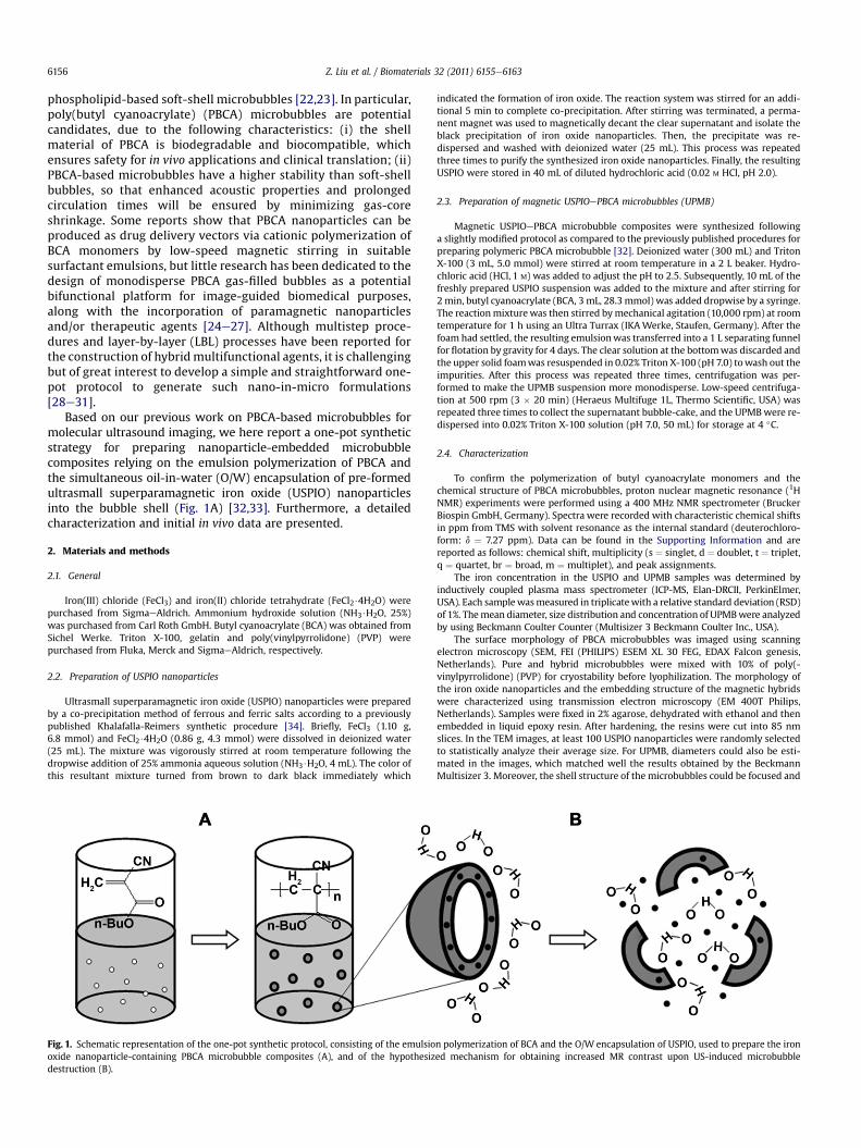
Based on our previous work on PBCA-based microbubbles formolecular ultrasound imaging, we here report a one-pot syntheticstrategy for preparing nanoparticle-embedded microbubblecomposites relying on the emulsion polymerization of PBCA andthe simultaneous oil-in-water (O/W) encapsulation of pre-formedultrasmall superparamagnetic iron oxide (USPIO) nanoparticlesinto the bubble shell (Fig. 1A) [32,33]. Furthermore, a detailedcharacterization and initial in vivo data are presented.
2. Materials and methods
2.1. General
Iron(III) chloride (FeCl3) and iron(II) chloride tetrahydrate (FeCl2$4H2O) werepurchased from SigmaeAldrich. Ammonium hydroxide solution (NH3$H2O, 25%)was purchased from Carl Roth GmbH. Butyl cyanoacrylate (BCA) was obtained fromSichel Werke. Triton X-100, gelatin and poly(vinylpyrrolidone) (PVP) werepurchased from Fluka, Merck and SigmaeAldrich, respectively.
2.2. Preparation of USPIO nanoparticles
Ultrasmall superparamagnetic iron oxide (USPIO) nanoparticles were preparedby a co-precipitation method of ferrous and ferric salts according to a previouslypublished Khalafalla-Reimers synthetic procedure [34]. Briefly, FeCl3 (1.10 g,6.8 mmol) and FeCl2$4H2O (0.86 g, 4.3 mmol) were dissolved in deionized water(25 mL). The mixture was vigorously stirred at room temperature following thedropwise addition of 25% ammonia aqueous solution (NH3$H2O, 4 mL). The color ofthis resultant mixture turned from brown to dark black immediately which
Fig. 1. Schematic representation of the one-pot synthetic protocol, consisting of the emulsiooxide nanoparticle-containing PBCA microbubble composites (A), and of the hypothesizdestruction (B).
indicated the formation of iron oxide. The reaction system was stirred for an addi-tional 5 min to complete co-precipitation. After stirring was terminated, a perma-nent magnet was used to magnetically decant the clear supernatant and isolate theblack precipitation of iron oxide nanoparticles. Then, the precipitate was re-dispersed and washed with deionized water (25 mL). This process was repeatedthree times to purify the synthesized iron oxide nanoparticles. Finally, the resultingUSPIO were stored in 40 mL of diluted hydrochloric acid (0.02 M HCl, pH 2.0).
2.3. Preparation of magnetic USPIOePBCA microbubbles (UPMB)
Magnetic USPIOePBCA microbubble composites were synthesized followinga slightly modified protocol as compared to the previously published procedures forpreparing polymeric PBCA microbubble [32]. Deionized water (300 mL) and TritonX-100 (3 mL, 5.0 mmol) were stirred at room temperature in a 2 L beaker. Hydro-chloric acid (HCl, 1 M) was added to adjust the pH to 2.5. Subsequently, 10 mL of thefreshly prepared USPIO suspension was added to the mixture and after stirring for2 min, butyl cyanoacrylate (BCA, 3 mL, 28.3 mmol) was added dropwise by a syringe.The reactionmixturewas then stirred bymechanical agitation (10,000 rpm) at roomtemperature for 1 h using an Ultra Turrax (IKAWerke, Staufen, Germany). After thefoam had settled, the resulting emulsionwas transferred into a 1 L separating funnelfor flotation by gravity for 4 days. The clear solution at the bottomwas discarded andthe upper solid foamwas resuspended in 0.02% Triton X-100 (pH 7.0) towash out theimpurities. After this process was repeated three times, centrifugation was per-formed to make the UPMB suspension more monodisperse. Low-speed centrifuga-tion at 500 rpm (3 � 20 min) (Heraeus Multifuge 1L, Thermo Scientific, USA) wasrepeated three times to collect the supernatant bubble-cake, and the UPMBwere re-dispersed into 0.02% Triton X-100 solution (pH 7.0, 50 mL) for storage at 4 �C.
2.4. Characterization
To confirm the polymerization of butyl cyanoacrylate monomers and thechemical structure of PBCA microbubbles, proton nuclear magnetic resonance (1HNMR) experiments were performed using a 400 MHz NMR spectrometer (BruckerBiospin GmbH, Germany). Spectra were recorded with characteristic chemical shiftsin ppm from TMS with solvent resonance as the internal standard (deuterochloro-form: d ¼ 7.27 ppm). Data can be found in the Supporting Information and arereported as follows: chemical shift, multiplicity (s ¼ singlet, d ¼ doublet, t ¼ triplet,q ¼ quartet, br ¼ broad, m ¼ multiplet), and peak assignments.
The iron concentration in the USPIO and UPMB samples was determined byinductively coupled plasma mass spectrometer (ICP-MS, Elan-DRCII, PerkinElmer,USA). Each samplewasmeasured in triplicatewith a relative standard deviation (RSD)of 1%. Themean diameter, size distribution and concentration of UPMBwere analyzedby using Beckmann Coulter Counter (Multisizer 3 Beckmann Coulter Inc., USA).
The surface morphology of PBCA microbubbles was imaged using scanningelectron microscopy (SEM, FEI (PHILIPS) ESEM XL 30 FEG, EDAX Falcon genesis,Netherlands). Pure and hybrid microbubbles were mixed with 10% of poly(-vinylpyrrolidone) (PVP) for cryostability before lyophilization. The morphology ofthe iron oxide nanoparticles and the embedding structure of the magnetic hybridswere characterized using transmission electron microscopy (EM 400T Philips,Netherlands). Samples were fixed in 2% agarose, dehydrated with ethanol and thenembedded in liquid epoxy resin. After hardening, the resins were cut into 85 nmslices. In the TEM images, at least 100 USPIO nanoparticles were randomly selectedto statistically analyze their average size. For UPMB, diameters could also be esti-mated in the images, which matched well the results obtained by the BeckmannMultisizer 3. Moreover, the shell structure of the microbubbles could be focused and
n polymerization of BCA and the O/W encapsulation of USPIO, used to prepare the ironed mechanism for obtaining increased MR contrast upon US-induced microbubble
Z. Liu et al. / Biomaterials 32 (2011) 6155e6163 6157
magnified, to display the USPIO nanoparticles embedded. Optical microscopy (AxioImager M2, Carl Zeiss Microimaging GmbH, Jena, Germany) was also used to visu-alize the shape of the pure and USPIO-containing microbubbles.
2.5. US and MR phantom imaging
The ultrasound phantomwas prepared by dispersingmicrobubbles in 2.5% (w/v)aqueous gelatin solution (concentration ¼ 40 microbubbles/mL). An ultrasoundscanner (Vevo2100 Visualsonics Inc., Canada) was used to obtain the ultrasoundphantom images. The phantom was fixed on an imaging panel with a transducer(25 MHz center frequency) oriented vertically to the gelatin phantom. Ultrasoundimaging was performed by first running a predefined continuous imaging sequencefor the first 4.5 s, and was followed by a destructive pulse (high mechanical index) tomeasure the background signal. Phantoms of both non-magnetic and magneticPBCA microbubbles were investigated by randomly selecting 5 different positions(15 mm � 15 mm) as regions of interest (ROI) before and after microbubbledestruction, to acquire the trend of signal intensity (SI). Contrast intensities (CI) ofeach of the UPMB batches were calculated by deducting background SI (post-destruction) from total SI (pre-destruction).
The MR phantom studies were carried out using a 3.0T clinical whole-body MRimaging system (Achieva 3.0T Philips, Netherlands), in combinationwith a knee coil(SENSE Flex M Philips, Netherlands). USPIO nanoparticles were diluted in deionizedwater and a series of fractions with gradient iron content were placed into a custom-made micro-well plate (300 mL for each well). Gas bubbles were introduced inEppendorf tubes (300 mL for each sample), and these tubes were placed in a plasticrack and submerged into deionized water to eliminate unwanted external suscep-tibility effects arising from airewater interfaces. The samples were gently andcontinuously rotated by hand for a uniform suspension prior to phantom imaging.Longitudinal relaxation times (T1) were measured using a multi-slice multi spinecho sequence with a 10� excitation pulse followed by a train of equally spacedrefocusing pulses (repetition time (TR)¼ 6.8 ms, echo time (TE)¼ 3.3 ms, number ofexcitations (NEX) ¼ 1, slice thickness ¼ 5 mm, field of view (FOV) ¼ 170 mm� 148.75 mm, matrix size ¼ 152 � 130). Relaxivity r1 was calculated by linear fittingof the inverse relaxation times as a function of iron concentrations or bubbleconcentration. Transversal relaxation times (T2) were measured using a multi-slicemulti spin echo sequence with a 90� excitation pulse followed by a train ofequally spaced refocusing pulses (repetition time (TR) ¼ 1500 ms, echo time(TE)¼ 162ms, number of excitations (NEX)¼ 1, slice thickness¼ 3mm, field of view(FOV)¼ 130mm� 162.5 mm, matrix size¼ 64 � 81). Relaxivity r2 was calculated bylinear fitting of the inverse relaxation times as a function of iron concentrations orbubble concentration.
To destroy the magnetic microbubbles and release the embedded USPIOnanoparticles, an ultrasonication bath was used. UPMB samples were placed inEppendorf tubes with Parafilm sealing. These tubes were put on a plastic rack whichsubmerged into the water bath. After exposure to ultrasonic waves for 30 min,samples were taken out for MR imaging.
2.6. In vivo USeMR imaging with mice
The human epithelial ovarian carcinoma cell line MLS was kindly provided byProf. Michal Neeman (Weizmann Institute of Science, Rehovot, Israel) and culturedin MEM Alpha þ 10% FCS in a cell culture incubator (C150 CO2-Incubator, Binder,Germany). Female CD1-nu/nu mice were purchased from Charles River Laboratory(Wilmington, USA). Approximately 2.5 � 106 MLS cells were inoculated subcuta-neously into the right flank of themice, and tumors were grown for 10e14 days priorto analysis. All experiments were approved by the government review committee onanimal care. Animals were anesthetized with 2% isofluorane in oxygen-enriched airwith a facemask during the whole imaging process of US and MRI.
In vivo ultrasound imaging was performed by using a preclinical ultrasoundscanner (Vevo2100 Visualsonics Inc., Canada). Fifty mL of magnetic hybrid micro-bubbles (batch d, [MB]¼ 0.9� 109microbubbles/mL) were injected i.v. via a tail-veincatheter. Tumors were scanned three-dimensionally prior to injection to confirma clear background signal. After the magnetic microbubbles were injected, a secondscan was carried out (in contrast mode; for 1 min) to record microbubble inflow.Acoustic signal intensityetime curves were acquired within a region of interest(ROI) of the maximum tumor area (28 mm2) to display the kinetics of the UPMB.
For in vivo MRI, magnetic hybrid microbubbles were administered through thetail vein of each mouse at a dose of 500 mg/kg iron. Using a 3.0 T clinical whole-bodyMRI scanner (Achieva 3.0T Philips, Netherlands) equipped with a mouse coil,T*2-weighted echo planar imaging (EPI) dynamic contrast-enhanced (DCE) MR wasperformed before and after UPMB administration, to monitor the contrast. Twentyseconds after the EPI DCE MR sequence was activated, 100 mL of magnetic micro-bubbles (batch d) were injected. The sequence parameters were repetition time(TR) ¼ 71 ms, echo time (TE) ¼ 11 ms, number of excitations (NEX) ¼ 1, slicethickness ¼ 2 mm, field of view (FOV) ¼ 30 mm � 8 mm, matrix size ¼ 64 � 60,number of dynamic measurements 600, and total scan time ¼ 533 s. To evaluate thecontrast, signal intensities (SI) were measured before and after injection of themagnetic microbubbles. SI over time curves were generated in a defined region ofinterest (ROI) covering the tumor using the imaging analysis software Dynalab
(MEVIS Research, Bremen, Germany). The T2-weighted MR images of the tumorbefore UPMB injection were obtained with a TSE sequence (TR ¼ 2391 ms,TE ¼ 100 ms, NEX ¼ 3, slice thickness ¼ 1 mm, FOV ¼ 25 mm � 22 mm, matrixsize ¼ 124 � 120).
2.7. Statistical analysis
Differences between the experimental groups were analyzed using a standardStudent’s t-test. A p value of <0.05 was considered to be statistically significant.
3. Results and discussion
3.1. Synthesis and characterization of iron oxide nanoparticle-containing microbubble composites
MagneticUSPIOePBCAmicrobubbles (UPMBs)were synthesizedvia a one-pot protocol consisting of the O/Wencapsulation of USPIOas embedded magnetic nanoparticles, and the emulsion polymeri-zation of PBCA as a carrier material for the USPIO (Fig. 1A) [35,36].BCA monomers formed tiny droplets, which were suspended in anaqueous solution of Triton X-100. Upon mixing, iron oxide nano-particles could be effectively entrapped inside the BCA droplets,which were stabilized by Triton X-100 surfactant molecules. Thisprocedure largely enhanced the efficacy of encapsulation during theprocess of monomer nucleation, polymer growth and final PBCAformation. Via high-speed agitation (10,000 rpm), air was incorpo-rated into the core of the bubble composites. Triton X-100 as neutralsurfactant stabilized the polymer structure with its hydrophobicchains, which ensured good stability of the PBCAmicrobubbles andincreased storage time (up to severalmonths). After polymerization,the resulting mixture was subjected to flotation, in order to differ-entially discardmicrobubble fragments, excessivenanoparticles andother impurities, as a result of distinct buoyancy by gravity. Then,low-speed centrifugation followed, to efficiently discriminatebubble populationswith different sizes, due to theirflotation time ina centrifugal field. Via this simple and straightforward syntheticprotocol, nanoparticle-embedded microbubble hybrids withnarrow size distribution could be isolated from polydisperse pop-ulations [37]. As compared to the multistep and layer-by-layerdeposition protocols reported in the literature, this syntheticstrategy is milder, easier and much more rapid. By replacing reac-tions conditions and/or components, and by using different gases,various types of nanoparticles or drugs could in principle beencapsulated in such materials, in order to obtain other diagnostic,therapeutic and theranostic nano-in-micro composites [38].
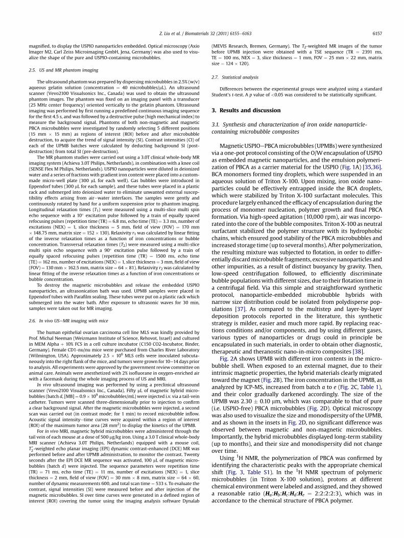
Fig. 2A shows UPMB with different iron contents in the micro-bubble shell. When exposed to an external magnet, due to theirintrinsic magnetic properties, the hybrid materials clearly migratedtoward themagnet (Fig. 2B). The iron concentration in the UPMB, asanalyzed by ICP-MS, increased from batch a to e (Fig. 2C, Table 1),and their color gradually darkened accordingly. The size of theUPMB was 2.30 � 0.10 mm, which was comparable to that of pure(i.e. USPIO-free) PBCA microbubbles (Fig. 2D). Optical microscopywas also used to visualize the size andmonodispersity of the UPMB,and as shown in the insets in Fig. 2D, no significant difference wasobserved between magnetic and non-magnetic microbubbles.Importantly, the hybrid microbubbles displayed long-term stability(up to months), and their size and monodispersity did not changeover time.
Using 1H NMR, the polymerization of PBCA was confirmed byidentifying the characteristic peaks with the appropriate chemicalshift (Fig. 3, Table S1). In the 1H NMR spectrum of polymericmicrobubbles (in Triton X-100 solution), protons at differentchemical environment were labeled and assigned, and they showeda reasonable ratio (Ha:Hb:Hc:Hd:He ¼ 2:2:2:2:3), which was inaccordance to the chemical structure of PBCA polymer.
Fig. 2. (A) Pure (a) and USPIO-containing (bee) PBCA microbubbles were prepared via a one-pot synthetic protocol. (B) Exposure of UPMB to an external magnetic field confirmedtheir magnetic properties. (C) Iron content of magnetic and non-magnetic microbubbles, as determined by ICP-MS. (D) Size distribution of pure (a) and USPIO-containing (bee)PBCA microbubbles. Inset: optical microscopy analysis, exemplifying narrow size distribution. Scale bar: 5 mm.
Z. Liu et al. / Biomaterials 32 (2011) 6155e61636158
To determine the successful encapsulation of USPIO nano-particles into the microbubble shell and to investigate the detailedhybrid microstructure, electron microscopy was performed. Thesurface morphology of iron oxide nanoparticles and of the pure andhybrid microbubbles was visualized using scanning electronmicroscopy (SEM) (Fig. 4; left panel). Iron oxide nanoparticles wereestimated to be w10 nm in diameter, and both types of micro-bubbles showed a relatively rough surface morphology due to thepolymeric structure. It should be noted that no obvious difference
in size or shape were observed upon the encapsulation of USPIOnanoparticles into the microbubble shell. Furthermore, trans-mission electron microscopy (TEM) was performed, to providehigh-resolution images for visualizing the embedding structure(Fig. 4; right panel). In batch e ([Fe] ¼ 113.43 mg/L), the nano-particles were arranged in a chain-like structure around themicrobubble core, while in batch b ([Fe] ¼ 29.67 mg/L), only fewUSPIO nanoparticles were evenly distributed within the micro-bubble shell. In contrast to UPMB, pure PBCAmicrobubbles without
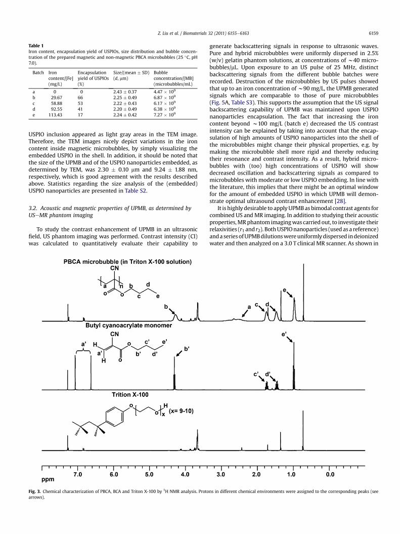
Table 1Iron content, encapsulation yield of USPIOs, size distribution and bubble concen-tration of the prepared magnetic and non-magnetic PBCA microbubbles (25 �C, pH7.0).
Batch Ironcontent/[Fe](mg/L)
Encapsulationyield of USPIOs(%)
Size/(mean � SD)(d, mm)
Bubbleconcentration/[MB](microbubbles/mL)
a 0 0 2.43 � 0.37 4.47 � 109
b 29.67 66 2.25 � 0.49 6.87 � 109
c 58.88 53 2.22 � 0.43 6.17 � 109
d 92.55 41 2.20 � 0.49 6.38 � 109
e 113.43 17 2.24 � 0.42 7.27 � 109
Z. Liu et al. / Biomaterials 32 (2011) 6155e6163 6159
USPIO inclusion appeared as light gray areas in the TEM image.Therefore, the TEM images nicely depict variations in the ironcontent inside magnetic microbubbles, by simply visualizing theembedded USPIO in the shell. In addition, it should be noted thatthe size of the UPMB and of the USPIO nanoparticles embedded, asdetermined by TEM, was 2.30 � 0.10 mm and 9.24 � 1.88 nm,respectively, which is good agreement with the results describedabove. Statistics regarding the size analysis of the (embedded)USPIO nanoparticles are presented in Table S2.
3.2. Acoustic and magnetic properties of UPMB, as determined byUSeMR phantom imaging
To study the contrast enhancement of UPMB in an ultrasonicfield, US phantom imaging was performed. Contrast intensity (CI)was calculated to quantitatively evaluate their capability to
Fig. 3. Chemical characterization of PBCA, BCA and Triton X-100 by 1H NMR analysis. Protoarrows).
generate backscattering signals in response to ultrasonic waves.Pure and hybrid microbubbles were uniformly dispersed in 2.5%(w/v) gelatin phantom solutions, at concentrations of w40 micro-bubbles/mL. Upon exposure to an US pulse of 25 MHz, distinctbackscattering signals from the different bubble batches wererecorded. Destruction of the microbubbles by US pulses showedthat up to an iron concentration ofw90 mg/L, the UPMB generatedsignals which are comparable to those of pure microbubbles(Fig. 5A, Table S3). This supports the assumption that the US signalbackscattering capability of UPMB was maintained upon USPIOnanoparticles encapsulation. The fact that increasing the ironcontent beyond w100 mg/L (batch e) decreased the US contrastintensity can be explained by taking into account that the encap-sulation of high amounts of USPIO nanoparticles into the shell ofthe microbubbles might change their physical properties, e.g. bymaking the microbubble shell more rigid and thereby reducingtheir resonance and contrast intensity. As a result, hybrid micro-bubbles with (too) high concentrations of USPIO will showdecreased oscillation and backscattering signals as compared tomicrobubbles with moderate or low USPIO embedding. In line withthe literature, this implies that there might be an optimal windowfor the amount of embedded USPIO in which UPMB will demon-strate optimal ultrasound contrast enhancement [28].
It is highly desirable to applyUPMBas bimodal contrast agents forcombined US andMR imaging. In addition to studying their acousticproperties,MRphantom imagingwas carried out, to investigate theirrelaxivities (r1 and r2). BothUSPIOnanoparticles (used as a reference)anda seriesofUPMBdilutionswereuniformlydispersed indeionizedwater and then analyzed on a 3.0 T clinical MR scanner. As shown in
ns in different chemical environments were assigned to the corresponding peaks (see
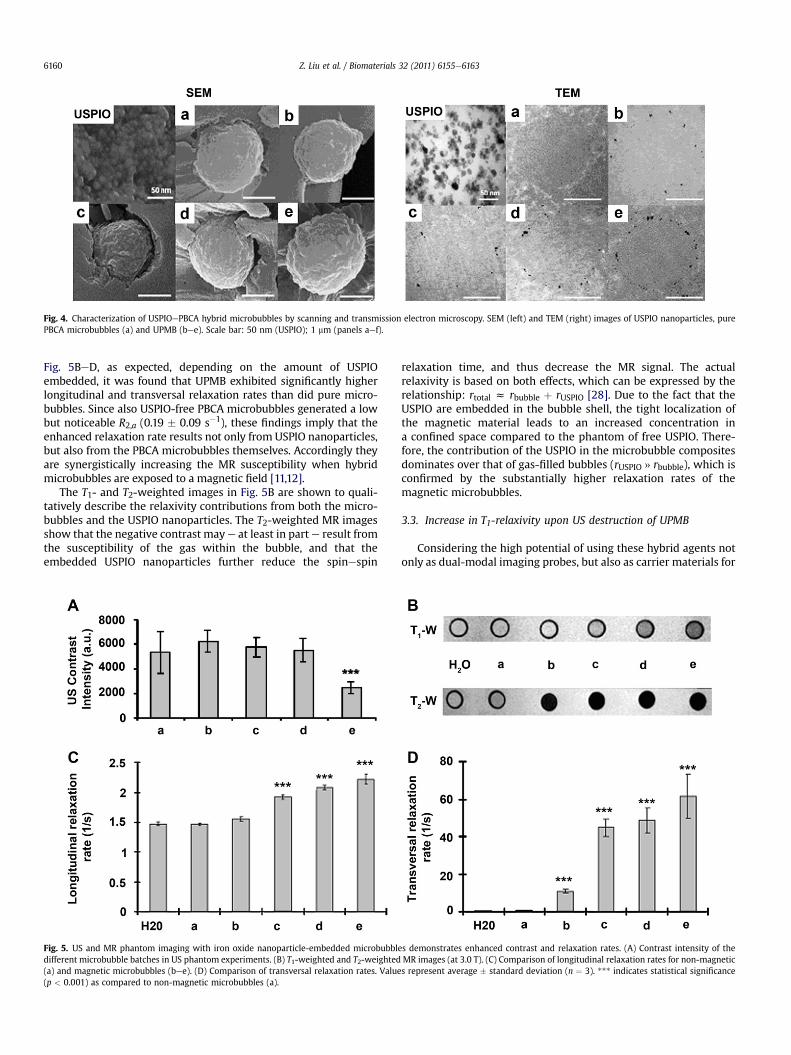
Fig. 4. Characterization of USPIOePBCA hybrid microbubbles by scanning and transmission electron microscopy. SEM (left) and TEM (right) images of USPIO nanoparticles, purePBCA microbubbles (a) and UPMB (bee). Scale bar: 50 nm (USPIO); 1 mm (panels aef).
Z. Liu et al. / Biomaterials 32 (2011) 6155e61636160
Fig. 5BeD, as expected, depending on the amount of USPIOembedded, it was found that UPMB exhibited significantly higherlongitudinal and transversal relaxation rates than did pure micro-bubbles. Since also USPIO-free PBCA microbubbles generated a lowbut noticeable R2,a (0.19 � 0.09 s�1), these findings imply that theenhanced relaxation rate results not only from USPIO nanoparticles,but also from the PBCA microbubbles themselves. Accordingly theyare synergistically increasing the MR susceptibility when hybridmicrobubbles are exposed to a magnetic field [11,12].
The T1- and T2-weighted images in Fig. 5B are shown to quali-tatively describe the relaxivity contributions from both the micro-bubbles and the USPIO nanoparticles. The T2-weighted MR imagesshow that the negative contrast maye at least in part e result fromthe susceptibility of the gas within the bubble, and that theembedded USPIO nanoparticles further reduce the spinespin
Fig. 5. US and MR phantom imaging with iron oxide nanoparticle-embedded microbubbldifferent microbubble batches in US phantom experiments. (B) T1-weighted and T2-weighted(a) and magnetic microbubbles (bee). (D) Comparison of transversal relaxation rates. Value(p < 0.001) as compared to non-magnetic microbubbles (a).
relaxation time, and thus decrease the MR signal. The actualrelaxivity is based on both effects, which can be expressed by therelationship: rtotal z rbubble þ rUSPIO [28]. Due to the fact that theUSPIO are embedded in the bubble shell, the tight localization ofthe magnetic material leads to an increased concentration ina confined space compared to the phantom of free USPIO. There-fore, the contribution of the USPIO in the microbubble compositesdominates over that of gas-filled bubbles (rUSPIO » rbubble), which isconfirmed by the substantially higher relaxation rates of themagnetic microbubbles.
3.3. Increase in T1-relaxivity upon US destruction of UPMB
Considering the high potential of using these hybrid agents notonly as dual-modal imaging probes, but also as carrier materials for
es demonstrates enhanced contrast and relaxation rates. (A) Contrast intensity of theMR images (at 3.0 T). (C) Comparison of longitudinal relaxation rates for non-magnetics represent average � standard deviation (n ¼ 3). *** indicates statistical significance
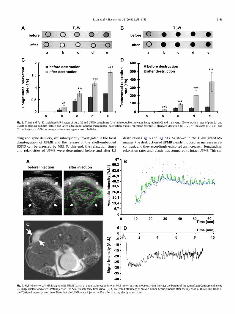
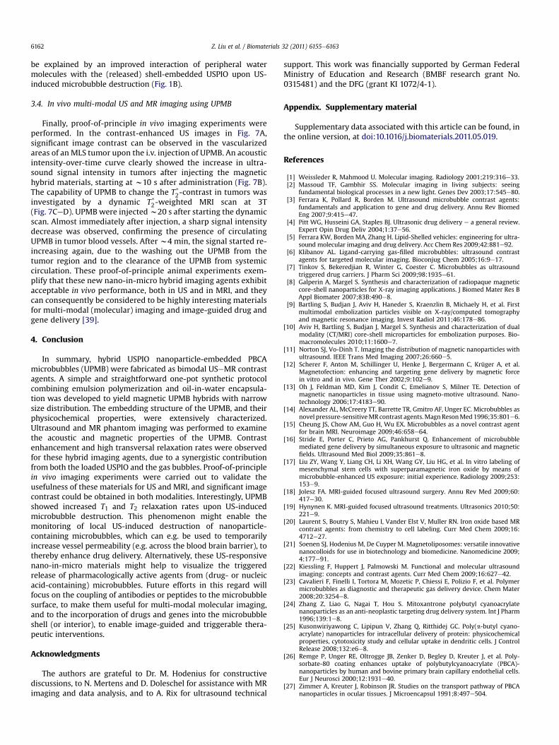
Fig. 6. T1 (A) and T2 (B) -weighted MR images of pure (a) and USPIO-containing (bee) microbubbles in water. Longitudinal (C) and transversal (D) relaxation rates of pure (a) andUSPIO-containing bubbles before and after ultrasound-induced microbubble destruction Values represent average � standard deviation (n ¼ 7). ** indicates p < 0.01 and*** indicates p < 0.001 as compared to non-magnetic microbubbles.
Z. Liu et al. / Biomaterials 32 (2011) 6155e6163 6161
drug and gene delivery, we subsequently investigated if the localdisintegration of UPMB and the release of the shell-embeddedUSPIO can be assessed by MRI. To this end, the relaxation timesand relaxivities of UPMB were determined before and after US
Fig. 7. Hybrid in vivo USeMR imaging with UPMB (batch d) upon i.v. injection into an MLS tUS images before and after UPMB injection. (B) Acoustic intensity-time curve. (C) T2-weightthe T*
2-signal intensity over time. Note that the UPMB were injected w20 s after starting th
destruction (Fig. 6 and Fig. S1). As shown in the T1-weighted MRimages, the destruction of UPMB clearly induced an increase in T1-contrast, and they accordingly exhibited an increase in longitudinalrelaxation rates and relaxivities compared to intact UPMB. This can
umor-bearing mouse (arrows indicate the border of the tumor). (A) Contrast-enhanceded MR image of an MLS tumor-bearing mouse after the injection of UPMB. (D) Trend ofe dynamic scan.
Z. Liu et al. / Biomaterials 32 (2011) 6155e61636162
be explained by an improved interaction of peripheral watermolecules with the (released) shell-embedded USPIO upon US-induced microbubble destruction (Fig. 1B).
3.4. In vivo multi-modal US and MR imaging using UPMB
Finally, proof-of-principle in vivo imaging experiments wereperformed. In the contrast-enhanced US images in Fig. 7A,significant image contrast can be observed in the vascularizedareas of an MLS tumor upon the i.v. injection of UPMB. An acousticintensity-over-time curve clearly showed the increase in ultra-sound signal intensity in tumors after injecting the magnetichybrid materials, starting at w10 s after administration (Fig. 7B).The capability of UPMB to change the T*2-contrast in tumors wasinvestigated by a dynamic T*
2-weighted MRI scan at 3T(Fig. 7CeD). UPMB were injectedw20 s after starting the dynamicscan. Almost immediately after injection, a sharp signal intensitydecrease was observed, confirming the presence of circulatingUPMB in tumor blood vessels. After w4 min, the signal started re-increasing again, due to the washing out the UPMB from thetumor region and to the clearance of the UPMB from systemiccirculation. These proof-of-principle animal experiments exem-plify that these new nano-in-micro hybrid imaging agents exhibitacceptable in vivo performance, both in US and in MRI, and theycan consequently be considered to be highly interesting materialsfor multi-modal (molecular) imaging and image-guided drug andgene delivery [39].
4. Conclusion
In summary, hybrid USPIO nanoparticle-embedded PBCAmicrobubbles (UPMB) were fabricated as bimodal USeMR contrastagents. A simple and straightforward one-pot synthetic protocolcombining emulsion polymerization and oil-in-water encapsula-tion was developed to yield magnetic UPMB hybrids with narrowsize distribution. The embedding structure of the UPMB, and theirphysicochemical properties, were extensively characterized.Ultrasound and MR phantom imaging was performed to examinethe acoustic and magnetic properties of the UPMB. Contrastenhancement and high transversal relaxation rates were observedfor these hybrid imaging agents, due to a synergistic contributionfrom both the loaded USPIO and the gas bubbles. Proof-of-principlein vivo imaging experiments were carried out to validate theusefulness of these materials for US and MRI, and significant imagecontrast could be obtained in both modalities. Interestingly, UPMBshowed increased T1 and T2 relaxation rates upon US-inducedmicrobubble destruction. This phenomenon might enable themonitoring of local US-induced destruction of nanoparticle-containing microbubbles, which can e.g. be used to temporarilyincrease vessel permeability (e.g. across the blood brain barrier), tothereby enhance drug delivery. Alternatively, these US-responsivenano-in-micro materials might help to visualize the triggeredrelease of pharmacologically active agents from (drug- or nucleicacid-containing) microbubbles. Future efforts in this regard willfocus on the coupling of antibodies or peptides to the microbubblesurface, to make them useful for multi-modal molecular imaging,and to the incorporation of drugs and genes into the microbubbleshell (or interior), to enable image-guided and triggerable thera-peutic interventions.
Acknowledgments
The authors are grateful to Dr. M. Hodenius for constructivediscussions, to N. Mertens and D. Doleschel for assistance with MRimaging and data analysis, and to A. Rix for ultrasound technical
support. This work was financially supported by German FederalMinistry of Education and Research (BMBF research grant No.0315481) and the DFG (grant KI 1072/4-1).
Appendix. Supplementary material
Supplementary data associated with this article can be found, inthe online version, at doi:10.1016/j.biomaterials.2011.05.019.
References
[1] Weissleder R, Mahmood U. Molecular imaging. Radiology 2001;219:316e33.[2] Massoud TF, Gambhir SS. Molecular imaging in living subjects: seeing
fundamental biological processes in a new light. Genes Dev 2003;17:545e80.[3] Ferrara K, Pollard R, Borden M. Ultrasound microbubble contrast agents:
fundamentals and application to gene and drug delivery. Annu Rev BiomedEng 2007;9:415e47.
[4] Pitt WG, Husseini GA, Staples BJ. Ultrasonic drug delivery e a general review.Expert Opin Drug Deliv 2004;1:37e56.
[5] Ferrara KW, Borden MA, Zhang H. Lipid-Shelled vehicles: engineering for ultra-sound molecular imaging and drug delivery. Acc Chem Res 2009;42:881e92.
[6] Klibanov AL. Ligand-carrying gas-filled microbubbles: ultrasound contrastagents for targeted molecular imaging. Bioconjug Chem 2005;16:9e17.
[7] Tinkov S, Bekeredjian R, Winter G, Coester C. Microbubbles as ultrasoundtriggered drug carriers. J Pharm Sci 2009;98:1935e61.
[8] Galperin A, Margel S. Synthesis and characterization of radiopaque magneticcore-shell nanoparticles for X-ray imaging applications. J Biomed Mater Res BAppl Biomater 2007;83B:490e8.
[9] Bartling S, Budjan J, Aviv H, Haneder S, Kraenzlin B, Michaely H, et al. Firstmultimodal embolization particles visible on X-ray/computed tomographyand magnetic resonance imaging. Invest Radiol 2011;46:178e86.
[10] Aviv H, Bartling S, Budjan J, Margel S. Synthesis and characterization of dualmodality (CT/MRI) core-shell microparticles for embolization purposes. Bio-macromolecules 2010;11:1600e7.
[11] Norton SJ, Vo-Dinh T. Imaging the distribution of magnetic nanoparticles withultrasound. IEEE Trans Med Imaging 2007;26:660e5.
[12] Scherer F, Anton M, Schillinger U, Henke J, Bergermann C, Krüger A, et al.Magnetofection: enhancing and targeting gene delivery by magnetic forcein vitro and in vivo. Gene Ther 2002;9:102e9.
[13] Oh J, Feldman MD, Kim J, Condit C, Emelianov S, Milner TE. Detection ofmagnetic nanoparticles in tissue using magneto-motive ultrasound. Nano-technology 2006;17:4183e90.
[14] Alexander AL, McCreery TT, Barrette TR, Gmitro AF, Unger EC. Microbubbles asnovel pressure-sensitiveMR contrast agents.MagnResonMed1996;35:801e6.
[15] Cheung JS, Chow AM, Guo H, Wu EX. Microbubbles as a novel contrast agentfor brain MRI. Neuroimage 2009;46:658e64.
[16] Stride E, Porter C, Prieto AG, Pankhurst Q. Enhancement of microbubblemediated gene delivery by simultaneous exposure to ultrasonic and magneticfields. Ultrasound Med Biol 2009;35:861e8.
[17] Liu ZY, Wang Y, Liang CH, Li XH, Wang GY, Liu HG, et al. In vitro labeling ofmesenchymal stem cells with superparamagnetic iron oxide by means ofmicrobubble-enhanced US exposure: initial experience. Radiology 2009;253:153e9.
[18] Jolesz FA. MRI-guided focused ultrasound surgery. Annu Rev Med 2009;60:417e30.
[19] Hynynen K. MRI-guided focused ultrasound treatments. Ultrasonics 2010;50:221e9.
[20] Laurent S, Boutry S, Mahieu I, Vander Elst V, Muller RN. Iron oxide based MRcontrast agents: from chemistry to cell labeling. Curr Med Chem 2009;16:4712e27.
[21] Soenen SJ, Hodenius M, De Cuyper M. Magnetoliposomes: versatile innovativenanocolloids for use in biotechnology and biomedicine. Nanomedicine 2009;4:177e91.
[22] Kiessling F, Huppert J, Palmowski M. Functional and molecular ultrasoundimaging: concepts and contrast agents. Curr Med Chem 2009;16:627e42.
[23] Cavalieri F, Finelli I, Tortora M, Mozetic P, Chiessi E, Polizio F, et al. Polymermicrobubbles as diagnostic and therapeutic gas delivery device. Chem Mater2008;20:3254e8.
[24] Zhang Z, Liao G, Nagai T, Hou S. Mitoxantrone polybutyl cyanoacrylatenanoparticles as an anti-neoplastic targeting drug delivery system. Int J Pharm1996;139:1e8.
[25] Kusonwiriyawong C, Lipipun V, Zhang Q, Ritthidej GC. Poly(a-butyl cyano-acrylate) nanoparticles for intracellular delivery of protein: physicochemicalproperties, cytotoxicity study and cellular uptake in dendritic cells. J ControlRelease 2008;132:e6e8.
[26] Remge P, Unger RE, Oltrogge JB, Zenker D, Begley D, Kreuter J, et al. Poly-sorbate-80 coating enhances uptake of polybutylcyanoacrylate (PBCA)-nanoparticles by human and bovine primary brain capillary endothelial cells.Eur J Neurosci 2000;12:1931e40.
[27] Zimmer A, Kreuter J, Robinson JR. Studies on the transport pathway of PBCAnanoparticles in ocular tissues. J Microencapsul 1991;8:497e504.
Z. Liu et al. / Biomaterials 32 (2011) 6155e6163 6163
[28] Yang F, Li Y, Chen Z, Zhang Y, Wu J, Gu N. Superparamagnetic iron oxidenanoparticle-embedded encapsulated microbubbles as dual contrast agents ofmagnetic resonance and ultrasound imaging. Biomaterials 2009;30:3882e90.
[29] Ke H, Xing ZW, Zhao B, Wang JR, Liu JB, Guo CX, et al. Quantum-dot-modifiedmicrobubbleswithBi-mode imagingcapabilities.Nanotechnology2009;20:1e8.
[30] Cavalieri F, Ashokkumar M, Grieser F, Caruso F. Ultrasonic synthesis of stable,functional lysozyme microbubbles. Langmuir 2008;24:10078e83.
[31] Park JI, Jagadeesan D, Williams R, Oakden W, Chung S, Stanisz GJ, et al.Microbubbles loaded with nanoparticles: a route to multiple imagingmodalities. ACS Nano 2010;4:6579e86.
[32] Palmowski M, Huppert J, Ladewig G, Hauff P, Reinhardt M, Mueller MM, et al.Molecular profiling of angiogenesis with targeted ultrasound imaging: earlyassessment of antiangiogenic therapy effects. Mol Cancer Ther 2008;7:101e9.
[33] Palmowski M, Peschke P, Huppert J, Hauff P, Reinhardt M, Maurer M, et al.Molecular ultrasound imaging of early vascular response in prostate tumorsirradiated with carbon ions. Neoplasia 2009;11:856e63.
[34] Khalafalla SE, Reimers GW. Preparation of dilution-stable aqueous magneticfluids. IEEE Trans Magn 1980;16:178e83.
[35] Behan N, Birkinshaw C, Clarke N. Poly n-butyl cyanoacrylate nanoparticles:a mechanistic study of polymerisation and particle formation. Biomaterials2001;22:1335e44.
[36] Olbrich C, Hauff P, Scholle F, Schmidt W, Bakowsky U, Briel A, et al. The in vitrostability of air-filled polybutylcyanoacrylate microparticles. Biomaterials2006;27:3549e59.
[37] Feshitan JA, Chen CC, Kwan JJ, Borden MA. Microbubble size isolation bydifferential centrifugation. J Colloid Interface Sci 2009;329:316e24.
[38] Lammers T, Kiessling F, Hennink WE, Storm G. Nanotheranostics and image-guided drug delivery: current concepts and future directions. Mol Pharm2010;7:1899e912.
[39] Deckers R, Rome C, Moonen CTW. The role of ultrasound and magneticresonance in local drug delivery. J Magn Reson Imaging 2008;27:400e9.
Related Documents