NOT FOR DISTRIBUTION Revised JBC/2012/366732 Iron inhibits AID activity and CSR Page 1 of 19 Iron Inhibits Activation-induced Cytidine Deaminase Enzymatic Activity and Modulates Immunoglobulin Class Switch DNA Recombination Guideng Li, Egest J Pone, Daniel C Tran, Pina J Patel, Lisa Dao, Zhenming Xu * , Paolo Casali From the Institute for immunology and Department of Medicine, School of Medicine, University of California, Irvine, CA 92697-4120, USA Background: AID is critical for effective immune responses, as it initiates antibody class switching and somatic hypermutation by deaminating deoxycytidines in the immunoglobulin locus switch region and V(D)J DNA. Results: Fe 2+ inhibited AID-mediated deoxycytidine deamination and class-switch DNA recombination in B cells. Conclusion: Iron inhibits the enzymatic function of AID. Significance: Iron can modulate antibody responses and may dampen AID-dependent autoimmunity and neoplastic transformation. Immunoglobulin (Ig) class switch DNA recombination (CSR) and somatic hypermutation (SHM) are critical for the maturation of the antibody response. Activation-induced cytidine deaminase (AID) initiates CSR and SHM by deaminating deoxycytidines (dCs) in switch (S) and V(D)J region DNA, respectively, to generate deoxyuracils (dUs). Processing of dUs by uracil DNA glycosylase (UNG) yields abasic sites, which are excised by apurinic/apyrimidinic endonucleases, eventually generating double strand DNA breaks, the obligatory intermediates of CSR. Here, we found that the bivalent iron ion (Fe 2+ , ferrous) suppressed CSR, leading to decreased number of switched B cells, decreased post-recombination Iμ -C H transcripts and reduced titers of secreted class- switched IgG1, IgG3 and IgA antibodies, without alterations in critical CSR factors, such as AID, 14-3-3γ or PTIP, or in general germline I H -S-C H transcription. Fe 2+ did not affect B cell proliferation or plasmacytoid differentiation. Rather, it inhibited AID-mediated dC deamination in a dose-dependent fashion. The inhibition of intrinsic AID enzymatic activity by Fe 2+ was specific, as shown by lack of inhibition of AID-mediated dC deamination by other bivalent metal ions, such as Zn 2+ , Mn 2+ , Mg 2+ or Ni 2+ , and inability of Fe 2+ to inhibit UNG- mediated dU excision. Overall, our findings have outlined a novel role of iron in modulating a B cell differentiation process (CSR) that is critical to the generation of effective antibody responses to microbial pathogens and tumoral cells. They also suggest a possible role of iron in dampening AID-dependent autoimmunity and neoplastic transformation. * To whom correspondence should be addressed. Tel: 949-824-0193; Fax: 949-824-2305; E-mail: [email protected]. Keywords: activation-induced cytidine deaminase (AID); antibody diversification; class switch DNA recombination (CSR); immunoglobulin; iron; plasmacytoid differentiation; uracil DNA glycosylase (UNG). Immunoglobulin (Ig) class switch DNA recombination (CSR) and somatic hypermutation (SHM) are central to the generation of neutralizing antibodies to pathogens, such as bacteria, viruses and tumoral cells as well anti-self antigen autoantibodies in autoimmunity and IgE antibodies in allergy and anaphylaxis (1). SHM inserts mainly point-mutations in Ig V(D)J region DNA at a high rate (2,3), thereby providing the structural substrate for positive selection of high- affinity antibody mutants by antigen (4). CSR substitutes the Ig heavy chain (IgH) constant region – such as Cμ, which is expressed in all naïve B cells – with a downstream Cγ, Cα or Cε region, thereby giving rise to IgG, IgA or IgE antibodies. Class-switched antibodies display different biological effector functions but maintain the same structure and specificity of the antigen- binding site, as compared to IgM antibodies produced by unswitched B cells (5). Both CSR and SHM require activation- induced cytidine deaminase (AID), a member of the AID/APOBEC cytidine deaminase family (6- 8). CSR also entails IgH locus germline I H -S-C H transcription, which initiates at specific I H promoters (Iμ, Iγ, Iα or Iε) and elongates through the upstream (donor) and downstream (acceptor) S and C H regions, giving rise to germline Iμ-Cμ, Iγ- Cγ, Iα-Cα or Iε-Cε transcripts. The “core” of each of S regions contains a tandem array of 5′- AGCT-3′ repeats, which are specifically targeted by 14-3-3 adaptor proteins. These adaptors, in turn, recruit AID to S region DNA (9). There, AID http://www.jbc.org/cgi/doi/10.1074/jbc.M112.366732 The latest version is at JBC Papers in Press. Published on May 3, 2012 as Manuscript M112.366732 Copyright 2012 by The American Society for Biochemistry and Molecular Biology, Inc. at Univ of California - Irvine, on May 3, 2012 www.jbc.org Downloaded from

Welcome message from author
This document is posted to help you gain knowledge. Please leave a comment to let me know what you think about it! Share it to your friends and learn new things together.
Transcript
NOT FOR DIS
TRIBUTIO
N
Revised JBC/2012/366732 Iron inhibits AID activity and CSR
Page 1 of 19
Iron Inhibits Activation-induced Cytidine Deaminase Enzymatic Activity and Modulates Immunoglobulin Class Switch DNA Recombination Guideng Li, Egest J Pone, Daniel C Tran, Pina J Patel, Lisa Dao, Zhenming Xu*, Paolo Casali From the Institute for immunology and Department of Medicine, School of Medicine, University of California, Irvine, CA 92697-4120, USA Background: AID is critical for effective immune responses, as it initiates antibody class switching and somatic hypermutation by deaminating deoxycytidines in the immunoglobulin locus switch region and V(D)J DNA. Results: Fe2+ inhibited AID-mediated deoxycytidine deamination and class-switch DNA recombination in B cells. Conclusion: Iron inhibits the enzymatic function of AID. Significance: Iron can modulate antibody responses and may dampen AID-dependent autoimmunity and neoplastic transformation.
Immunoglobulin (Ig) class switch DNA recombination (CSR) and somatic hypermutation (SHM) are critical for the maturation of the antibody response. Activation-induced cytidine deaminase (AID) initiates CSR and SHM by deaminating deoxycytidines (dCs) in switch (S) and V(D)J region DNA, respectively, to generate deoxyuracils (dUs). Processing of dUs by uracil DNA glycosylase (UNG) yields abasic sites, which are excised by apurinic/apyrimidinic endonucleases, eventually generating double strand DNA breaks, the obligatory intermediates of CSR. Here, we found that the bivalent iron ion (Fe2+, ferrous) suppressed CSR, leading to decreased number of switched B cells, decreased post-recombination Iµ-CH transcripts and reduced titers of secreted class-switched IgG1, IgG3 and IgA antibodies, without alterations in critical CSR factors, such as AID, 14-3-3γ or PTIP, or in general germline IH-S-CH transcription. Fe2+ did not affect B cell proliferation or plasmacytoid differentiation. Rather, it inhibited AID-mediated dC deamination in a dose-dependent fashion. The inhibition of intrinsic AID enzymatic activity by Fe2+ was specific, as shown by lack of inhibition of AID-mediated dC deamination by other bivalent metal ions, such as Zn2+, Mn2+, Mg2+ or Ni2+, and inability of Fe2+ to inhibit UNG-mediated dU excision. Overall, our findings have outlined a novel role of iron in modulating a B cell differentiation process (CSR) that is critical to the generation of effective antibody responses to microbial pathogens and tumoral cells. They also suggest a possible role of iron in dampening AID-dependent autoimmunity and neoplastic transformation. * To whom correspondence should be addressed. Tel: 949-824-0193; Fax: 949-824-2305; E-mail: [email protected].
Keywords: activation-induced cytidine deaminase (AID); antibody diversification; class switch DNA recombination (CSR); immunoglobulin; iron; plasmacytoid differentiation; uracil DNA glycosylase (UNG).
Immunoglobulin (Ig) class switch DNA recombination (CSR) and somatic hypermutation (SHM) are central to the generation of neutralizing antibodies to pathogens, such as bacteria, viruses and tumoral cells as well anti-self antigen autoantibodies in autoimmunity and IgE antibodies in allergy and anaphylaxis (1). SHM inserts mainly point-mutations in Ig V(D)J region DNA at a high rate (2,3), thereby providing the structural substrate for positive selection of high-affinity antibody mutants by antigen (4). CSR substitutes the Ig heavy chain (IgH) constant region – such as Cµ, which is expressed in all naïve B cells – with a downstream Cγ, Cα or Cε region, thereby giving rise to IgG, IgA or IgE antibodies. Class-switched antibodies display different biological effector functions but maintain the same structure and specificity of the antigen-binding site, as compared to IgM antibodies produced by unswitched B cells (5).
Both CSR and SHM require activation-induced cytidine deaminase (AID), a member of the AID/APOBEC cytidine deaminase family (6-8). CSR also entails IgH locus germline IH-S-CH transcription, which initiates at specific IH promoters (Iµ, Iγ, Iα or Iε) and elongates through the upstream (donor) and downstream (acceptor) S and CH regions, giving rise to germline Iµ-Cµ, Iγ-Cγ, Iα-Cα or Iε-Cε transcripts. The “core” of each of S regions contains a tandem array of 5′-AGCT-3′ repeats, which are specifically targeted by 14-3-3 adaptor proteins. These adaptors, in turn, recruit AID to S region DNA (9). There, AID
http://www.jbc.org/cgi/doi/10.1074/jbc.M112.366732The latest version is at JBC Papers in Press. Published on May 3, 2012 as Manuscript M112.366732
Copyright 2012 by The American Society for Biochemistry and Molecular Biology, Inc.
at Univ of C
alifornia - Irvine, on May 3, 2012
ww
w.jbc.org
Dow
nloaded from
NOT FOR DIS
TRIBUTIO
N
Revised JBC/2012/366732 Iron inhibits AID activity and CSR
Page 2 of 19
will deaminate dCs, particularly those within 5′-AGCT-3′ repeats in the core of S regions, thereby generating a high density of dUs (10). These can then be excised by uracil DNA glycosylase (UNG), as recruited to S region DNA by the REV1 DNA polymerase (11), to yield abasic sites, which are excised by apurinic/apyrimidinic endonucleases (APEs), leading to single-strand DNA breaks (SSBs). High-density SSBs in both DNA strands readily form double-strand DNA breaks (DSBs), the obligatory CSR intermediates (12,13). Looping-out of the intervening DNA between the donor and acceptor S regions and re-ligation of DSB free ends result in S-S junctions and recombination. Post-recombination IgH sequences are transcribed, giving rise to post-recombination transcripts, such as Iµ-Cγ, Iµ-Cα or Iµ-Cε (5).
CSR levels are determined by overall expression level, targeting and activity of AID (14). AID expression is strictly regulated in a B cell lineage-specific and differentiation stage-specific manner. It is induced by primary CSR-inducing stimuli – for example, CD40 or dual engagement of Toll-like receptors (TLRs) and B cell receptor (BCR) – through transcription factors such as HOXC4 (15), E2A (16) and NF-κB (16-18), and enhanced by cytokines, such as IL-4 and TGFβ (17). AID levels are also controlled at the post-transcriptional stage (e.g., by microRNAs) and post-translational stage (e.g., by proteasome-mediated degradation) (14). Further, to mediate CSR, AID needs to be targeted to S region DNA by 14-3-3 adaptors through direct protein-protein interaction (9). AID C-terminal truncation mutants cannot bind 14-3-3 and are defective in mediating CSR. Finally, AID dC deamination activity is enhanced by 14-3-3 and regulated by replication protein A (RPA) and RNA exosomes (19,20). The important role of 14-3-3, RNA and RNA exosome components in CSR strongly suggest that the regulation of AID activity constitutes an important step in regulation of CSR.
Iron is a crucial metal element. It mediates many metabolic pathways and is required for proliferation of cells, including B and T lymphocytes (21). B lymphocyte proliferation is inhibited by iron chelators, such as desferoxamine and salicylaldehyde isonicotinoyl hydrazone, or depletion of ferritin, a ferrous ion (Fe2+) transporter (21,22). Despite the importance of iron in B cell proliferation, iron overload is associated with impaired immune defense to viruses and bacteria, including Mycobacterium
tuberculosis, Candida albicans and Pasteurella septica, in humans and experimental animals (23-26), possibly due to impaired antibody responses. Accordingly, patients with hemochromatosis display iron overload, as caused by excess absorption of iron, and a reduced level of class-switched antibodies (27). Finally, heme, an iron-containing molecule essential for diverse organisms, has been implicated in inhibiting CSR (28), suggesting a role of iron in modulating this important B cell differentiation process.
Here, we have tested the hypothesis that iron suppresses CSR, thereby inhibiting the generation of class-switched B cells and antibodies. We have used a well-defined B cell culture system to analyze the role of Fe2+ in CSR and B cell plasmacytoid differentiation, which underpin the high-rate production of class-switched antibodies. We have further measured core CSR parameters, such as levels of germline IH-S-CH transcription, AID and other CSR factors. Finally, we have used in vitro dC DNA deamination assays involving purified recombinant AID to analyze Fe2+-mediated inhibition of CSR at the molecular level.
EXPERIMENTAL PROCEDURES B cells—Preparation and purification of
mouse spleen and lymph node B cells were as described (18). B cells were cultured in RPMI-1640 (Invitrogen) supplemented with penicillin-streptomycin and amphotericin B (1% v/v), FBS (10% v/v, Hyclone) and 50 µM of β-mercaptoethanol (RPMI-FBS). To induce CSR, B cells were stimulated with LPS (5 µg/ml, from E. coli, serotype 055:B5 and deproteinized by chloroform extraction, Sigma-Aldrich) alone for CSR to IgG3 or with IL-4 (3 ng/ml, R&D Systems) for CSR to IgG1 or with IL-4, TGFβ (3 ng/ml, R&D Systems) plus rat anti–mouse Igδ chain monoclonal antibody (clone 11-26)-conjugated to dextran (anti–δ mAb/dex, 30 ng/ml, Fina Biosolutions – Igδ is expressed in all naïve B cells) for CSR to IgA. FeCl2 and FeSO4 (ACS grade; Sigma-Aldrich) dissolved in PBS were freshly prepared and added at a final concentration of 80 µM or 20 µM and 40 µM as indicated.
Surface Ig analysis—After 4 d of stimulation, B cells were harvested for flow cytometry analysis of surface expression of Ig (Igµ, and/or sIgγ3, sIgγ1 and sIgα for CSR to IgG3, IgG1 and IgA, respectively), B220 (clone RA3-6B2, eBioscience) and/or CD138 (after 5 d of stimulation), as we
at Univ of C
alifornia - Irvine, on May 3, 2012
ww
w.jbc.org
Dow
nloaded from
NOT FOR DIS
TRIBUTIO
N
Revised JBC/2012/366732 Iron inhibits AID activity and CSR
Page 3 of 19
described (18). Briefly, B cells were centrifuged at 400 g for 5 m, and then stained with fluorochrome-conjugated mAbs in Hank’s Buffered Salt Solution (HBSS) containing BSA (1%, w/v) for 15 m. After washing, cells were resuspended in HBSS-BSA buffer and analyzed using a FACSCalibur® (BD Biosciences). Data were analyzed by using the FlowJo® software (Tree Star). Dead (7–AAD+) cells were excluded from analysis.
B cell proliferation and viability analysis—CFSE-labeled B cells were stimulated for 4 d and harvested for flow cytometry analysis of CFSE intensity (which halves in two daughter cells when a cell divides) and surface expression of Ig, as described above. To analyze B cell proliferation, individual cell divisions were first determined by the cell proliferation platform of FlowJo®, and CSR to IgG3, IgG1 or IgA as a function of division number was analyzed by the ratio of IgG3+, IgG1+ or IgA+ B cells, respectively, in each division over total B cells in that division. For B cell viability analysis, cells were stained with 7–AAD, which enters apoptotic and necrotic cells, but not intact cells, to intercalate into DNA, and analyzed by flow cytometry.
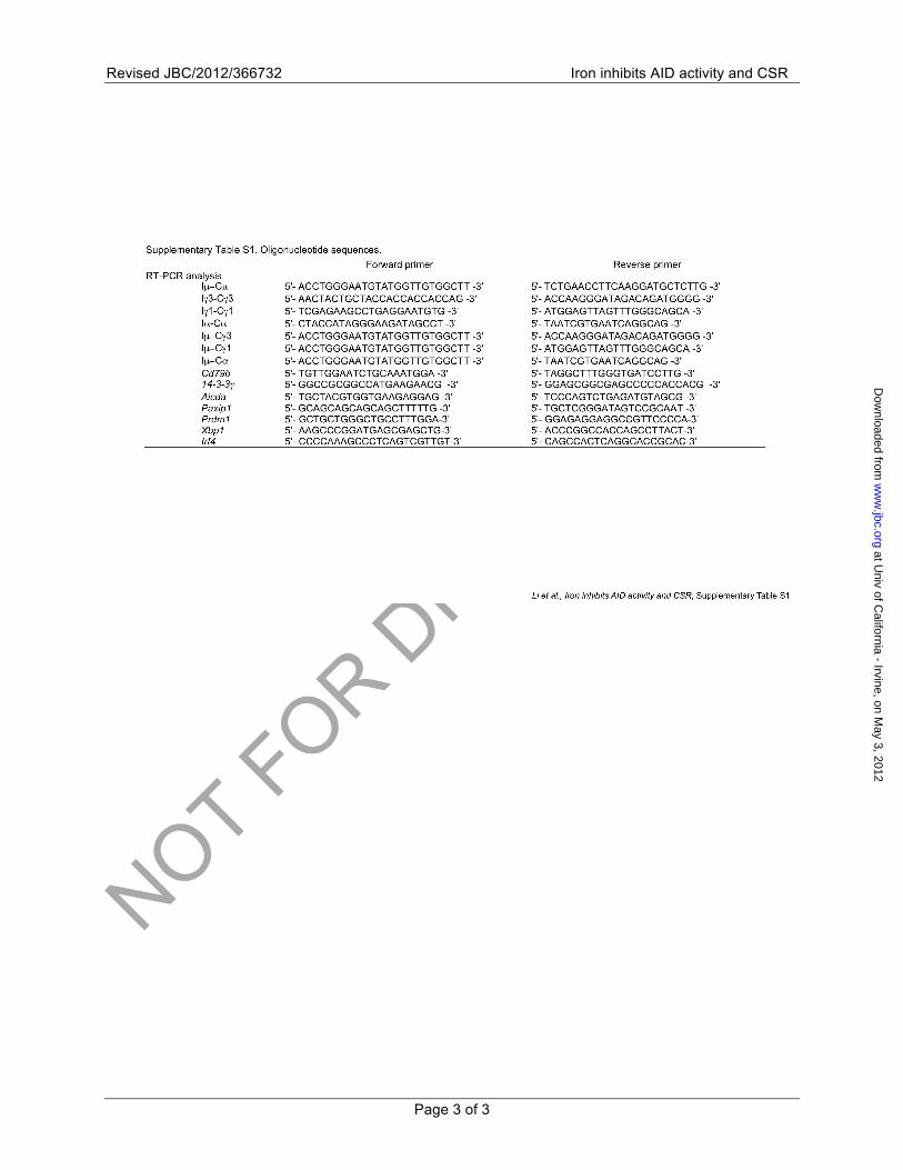
RNA isolation and transcripts analysis by quantitative real-time PCR (qRT-PCR)—Total RNA was extracted from 5 x 10⁶ B cells using RNeasy Mini Kit (Qiagen) according to the manufacturer's instruction. First strand cDNA were synthesized from 2 µg of total RNA using the SuperScript III System with Oligo dT primer (Invitrogen) and measured by qRT-PCR using appropriate primers (Supplementary Table S1) and SYBR Green (Dynamo HS kit; New England Biolabs). PCR was performed in the MyiQ Single-color Real-Time PCR Detection System (Bio-Rad Laboratories) according to the following protocol: 95°C for 5 m, 40 cycles of 95°C for 10 s, 60°C for 30 s, 72°C for 30 s. Melting curve analysis was performed at 72°-95°C. The ΔΔCt method was used to analyze levels of transcripts and data were normalized to the level of Cd79b, which encodes the BCR Igβ chain, as constitutively expressed in B cells.
Secreted Ig analysis—To analyze titers of IgG3, IgG1 or IgA secreted from cultured B cells, culture supernatants from B cells stimulated for 4 d were diluted 4-, 10- and 4-fold, respectively, with PBS plus 0.05% (v/v) Tween-20 (PBST).
Two-fold serially diluted samples and the standard for each Ig isotype (IgG3, IgG1 or IgA) were incubated in a 96-well plate pre-blocked with PBST and BSA and coated with a pre-adsorbed goat Ab to mouse IgG to capture IgG3 and IgG1 or goat Ab to mouse Ig to capture IgA (all goat Abs were from Southern Biotech). Captured Igs were reacted with biotin-labeled rat mAb to IgG3, IgG1 or IgA (BD Biosciences), followed by incubation with horseradish peroxidase (HRP)-labeled streptavidin (Sigma-Aldrich) and development with o-phenylenediamine. OD492 nm was measured with a Luminescence Detector 400 (Beckman Coulter). Ig concentrations were analyzed using the Excel® software (Microsoft).
AID dC DNA deamination assay—Purified recombinant GST-AID (1 ng) and UNG (0.2 U, in excess to convert all AID-generated dUs to abasic sites; New England Biolabs) were incubated with the HPLC-purified single-strand oligonucleotide 5′-AGCTAAAAAGCTAAAAAGCTAAAA-3′ labeled with Alexa Fluor 647 at the 5′ end (Alexa-[5′-AGCT-3′]3-24nt, 1 pmol) in a 20–µl reaction mixture containing Tris-HCl (25 mM, pH 7.5), NaCl (50 mM), DTT (0.5 mM), CHAPS (0.01% v/v) and glutathione (1 mM) at 37°C for indicated time. RNase A (Qiagen) and FeCl2, ZnCl2, MnCl2, MgCl2 or NiCl2 (ACS grade; all from Sigma-Aldrich) were freshly prepared and added, within 30 m of preparation, at final concentrations as indicated. After the reaction, heat-labile abasic sites were cleaved by heating at 95°C for 10 m in the presence of NaOH (200 mM). The intact substrate Alexa-[5′-AGCT-3′]3-24nt and cleavage products were fractionated by denaturing 15% PAGE containing 8 M urea and visualized using a TyphoonTM 9410 scanner (GE Healthcare). The intensity of substrate and cleavage product bands was quantified using the ImageQuantTM TL software (GE Healthcare). AID deamination activity was defined as the percentage of DNA substrate showing deamination (sum of the three cleavage products over the sum of the substrate and cleavage products).
UNG dU excision assay—The Alexa Fluo 647-labeled dU-containing oligonucleotide 5′-AGU(/C)TAAAAAGU(/C)TAAAAAGU(/C)T AAAA-3′ (dUs are underlined) were used as the substrate for UNG. This oligonucleotide (termed Alexa-[5′-AGU/(C)T-3′]3-24nt) was generated by incubating 1 pmol of Alexa-[5′-AGCT-3′]3-24nt with GST-AID (1 ng) in a 20–µl reaction mixture
at Univ of C
alifornia - Irvine, on May 3, 2012
ww
w.jbc.org
Dow
nloaded from

NOT FOR DIS
TRIBUTIO
N
Revised JBC/2012/366732 Iron inhibits AID activity and CSR
Page 4 of 19
(25 mM Tris-HCl, pH 7.5, 50 mM NaCl, 0.5 mM DTT, 0.01% (v/v) CHAPS, 1 mM glutathione and 10 ng/ml RNase A) at 37°C for 30 m. Incubation of Alexa-[5′-AGU/(C)T-3′]3-24nt with UNG was carried out in reaction buffer (25 mM Tris-HCl, pH 7.5, 50 mM NaCl, 0.5 mM DTT, 0.01% (v/v) CHAPS, 1 mM glutathione and 10 ng/ml RNase A) in the absence or presence of 20 µM of FeCl2, ZnCl2, MnCl2, MgCl2 or NiCl2 at 37°C for 20 m. After the reaction, heat-labile abasic sites were cleaved to completion by heating at 95°C for 10 m in the presence of NaOH (200 mM). The intact substrates Alexa-[5′-AGU/(C)T-3′]3-24nt and cleavage products were fractionated by denaturing 15% PAGE containing 8 M urea and visualized using a TyphoonTM 9410 scanner. The intensity of the substrate and cleavage product bands was quantified using the ImageQuantTM TL software. The UNG dU excision activity was defined as the percentage of substrates showing cleavage (sum of the three cleavage products over the sum of the substrate and cleavage products).
Mice—C57BL/6 mice were maintained in a pathogen-free vivarium at the University of California, Irvine. The mice used in all experiments were 8-12 weeks old and without any apparent infection or disease. All animal protocols were approved by the Institutional Animal Care and Use Committee (IACUC) of the University of California, Irvine, CA.
Statistical analysis—Statistical analysis was performed using the Excel® software to determine P values by paired student t-test. P values less than 0.05 were considered significant.
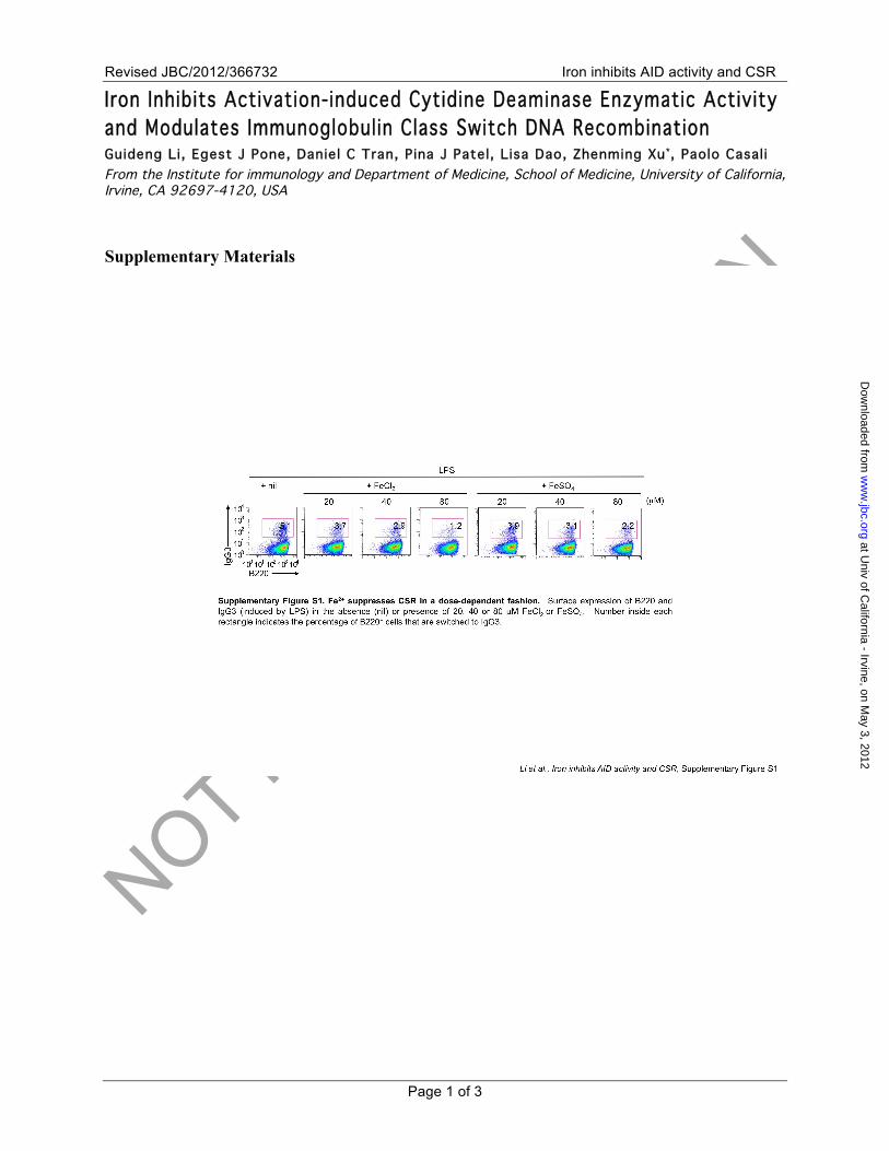
RESULTS Fe2+ suppresses CSR to multiple Ig isotypes—Decreased levels of class-switched antibodies and impaired immune responses in human and mice with iron overload prompted us to hypothesize that CSR is inhibited by iron. To test this hypothesis, we analyzed CSR in B cells stimulated with LPS (for induction of CSR to IgG3, which mediates the antibody response to many pyogenic bacteria), LPS plus IL-4 (to IgG1, which is the most abundant IgG subclass and provides neutralizing antibodies to blood-borne viral and bacterial pathogens) or LPS, IL-4, TGFβ plus anti–δ mAb/dex (to IgA, which mediates the front-line immune defense on respiratory and digestive mucosae) in the presence of Fe2+ (FeCl2 or FeSO4). The proportion of surface IgG3+ B cells was
significantly decreased in the presence of FeCl2 or FeSO4, as compared to B cells cultured in the absence of Fe2+ (Fig. 1). Fe2+ inhibition of generation of IgG3+ B cells was dose-dependent, starting at 20 µM of Fe2+, further at 40 µM and by as much as 77% and 57% at 80 µM of FeCl2 and FeSO4, respectively (Supplementary Figure S1) – iron concentrations range from 10 µM to 30 µM in the serum of healthy adults and from 30 µM to over 40 µM in the serum of patients with hemochromatosis (29). Likewise, in the presence of Fe2+, the proportion of surface IgG1+ and IgA+ B cells was reduced by as much as 50% (FeCl2) and 84% (FeSO4), respectively. FeCl2 or FeSO4 did not alter B cell surface expression of Igµ (Fig. 2A), showing that Fe2+ did not interfere with expression of membrane Igs; rather, it specifically inhibited the generation of class-switched (IgG3+, IgG1+ and IgA+) B cells. Further, titers of IgG3, IgG1 and IgA antibodies secreted from stimulated B cells were decreased in the presence of FeCl2 (by 91%, 68% and 66%, respectively) or FeSO4 (by 75%, 54% and 79%, respectively) (Fig. 1B). Finally, Fe2+-mediated impairment in the generation of surface and secreted class-switched Igs was associated with decreased levels of post-recombination Iµ-Cγ3 (53% and 83% lower by FeCl2 and FeSO4, respectively), Iµ-Cγ1 (40% and 24% lower) or Iµ-Cα (46% and 90% lower) transcripts, which are direct indexes of DNA recombination involving Sµ and Sγ3, Sγ1 or Sα (Fig. 1C).
Thus, Fe2+ suppresses CSR to multiple Ig isotypes, thereby inhibiting the generation of class-switched antibodies.
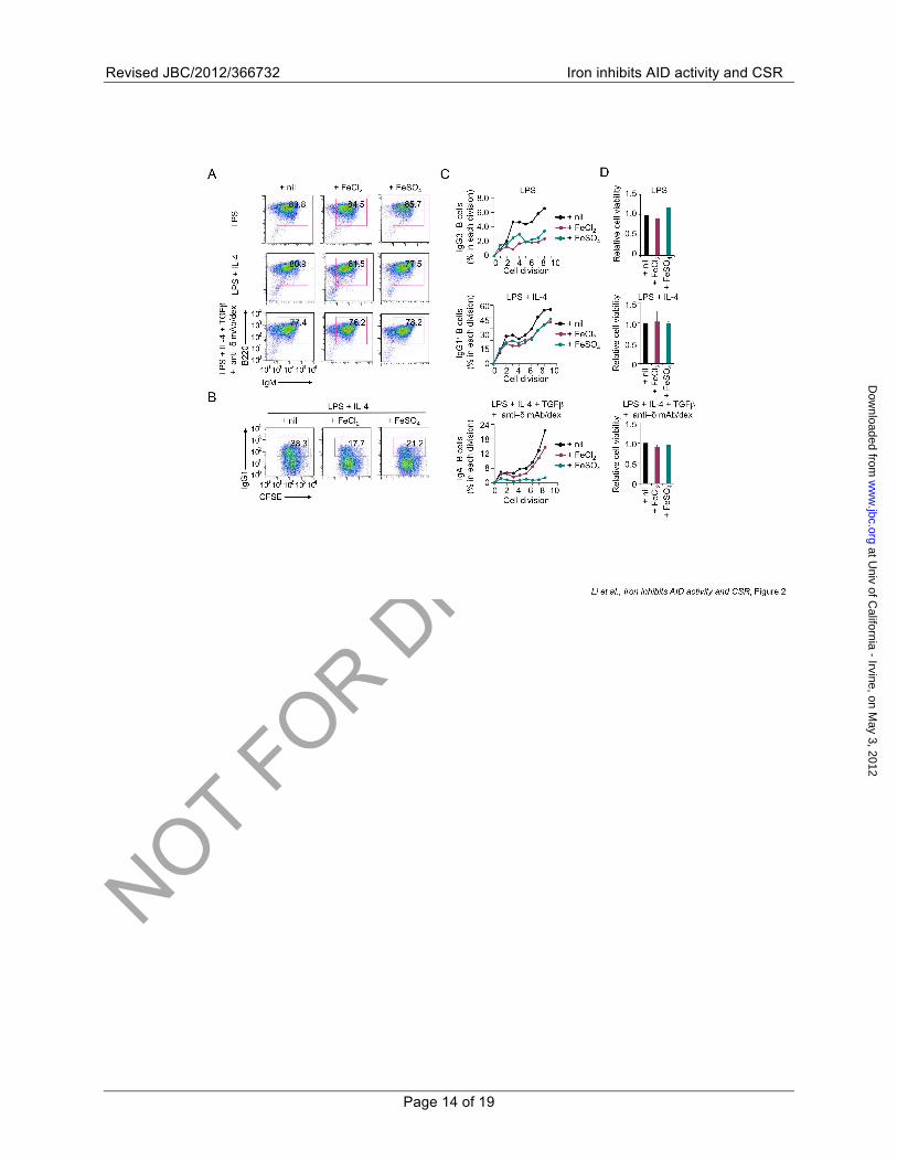
Fe2+ does not impair B cell proliferation or viability—B cell proliferation is required for efficient CSR. We next analyzed whether B cell proliferation or viability was affected by Fe2+ under the same stimulation conditions as those used for CSR induction. B cells stimulated with LPS (for CSR to IgG3), LPS plus IL-4 (to IgG1) or LPS, IL-4, TGFβ plus anti–δmAb/dex (to IgA) for 4 d and cultured with FeCl2 or FeSO4 completed virtually the same number of cell divisions as their counterparts cultured without the Fe2+ treatment (Figs. 2B, 2C). Further, the proportion of class-switched (IgG3+, IgG1+ and IgA+) B cells within each cell division was decreased in the presence of FeCl2 or FeSO4, showing that Fe2+-mediated CSR inhibition was independent of B cell proliferation. Finally, B
at Univ of C
alifornia - Irvine, on May 3, 2012
ww
w.jbc.org
Dow
nloaded from
NOT FOR DIS
TRIBUTIO
N
Revised JBC/2012/366732 Iron inhibits AID activity and CSR
Page 5 of 19
cells stimulated with LPS, LPS plus IL-4 or LPS, IL-4, TGFβ plus anti–δmAb/dex displayed comparable viabilities in the absence or presence of FeCl2 or FeSO4 (Fig. 2D).
Thus, Fe2+ inhibits CSR without altering B cell proliferation or viability.
Fe2+ does not affect plasmacytoid differentiation—During the antibody response, unswitched (Igµ+) and switched (IgG+, IgA+ and IgE+) B cells differentiate into short-lived plasmablasts and long-lived plasma cells (plasmacytes), respectively, thereby generating secreted IgM Abs or class-switched (IgG, IgA and IgE) Abs. Both plasmablasts and plasma cells express high levels of surface CD138 (syndecan-1), the hallmark of plasmacytoid differentiation (30). Accordingly, B cells stimulated in vitro by LPS, LPS plus IL-4 or LPS, IL-4, TGFβ plus anti–δ mAb/dex expressed surface CD138. Generation of CD138+ cells was not affected or even slightly enhanced in the presence of FeSO4 (Fig. 3A).
Plasmacytoid differentiation requires B-lymphocyte-induced maturation protein 1 (BLIMP1), which activates expression of secreted IgH protein chains and, by downregulating PAX5, de-represses expression of X-box binding protein 1 (XBP1) (31-33). XBP1 is a member of the CREB/ATF transcription factor family and activates transcription of genes encoding proteins that function in secretory pathways. Transcription of both the Prdm1 (encoding BLIMP1) and Xbp1 gene is also regulated by the IRF4 transcription factor, which plays a role in both CSR and plasmacytoid differentiation. In stimulated B cells, expression of Irf4, Prdm1 or Xbp1 was not suppressed by FeCl2 or FeSO4 (Fig. 3B). In fact, expression of Prdm1 and Xbp1 was upregulated by FeSO4 in B cells stimulated with LPS plus IL-4 or LPS, IL-4, TGFβ plus anti–δ mAb/dex, largely in agreement with the role of Fe2+ in the generation of CD138+ plasmacytoid cells and plasmacytes.
Thus, Fe2+ does not suppress plasmacytoid differentiation, indicating that the significant decrease in titers of secreted class-switched antibodies in the presence of Fe2+ is due to Fe2+–mediated CSR inhibition.
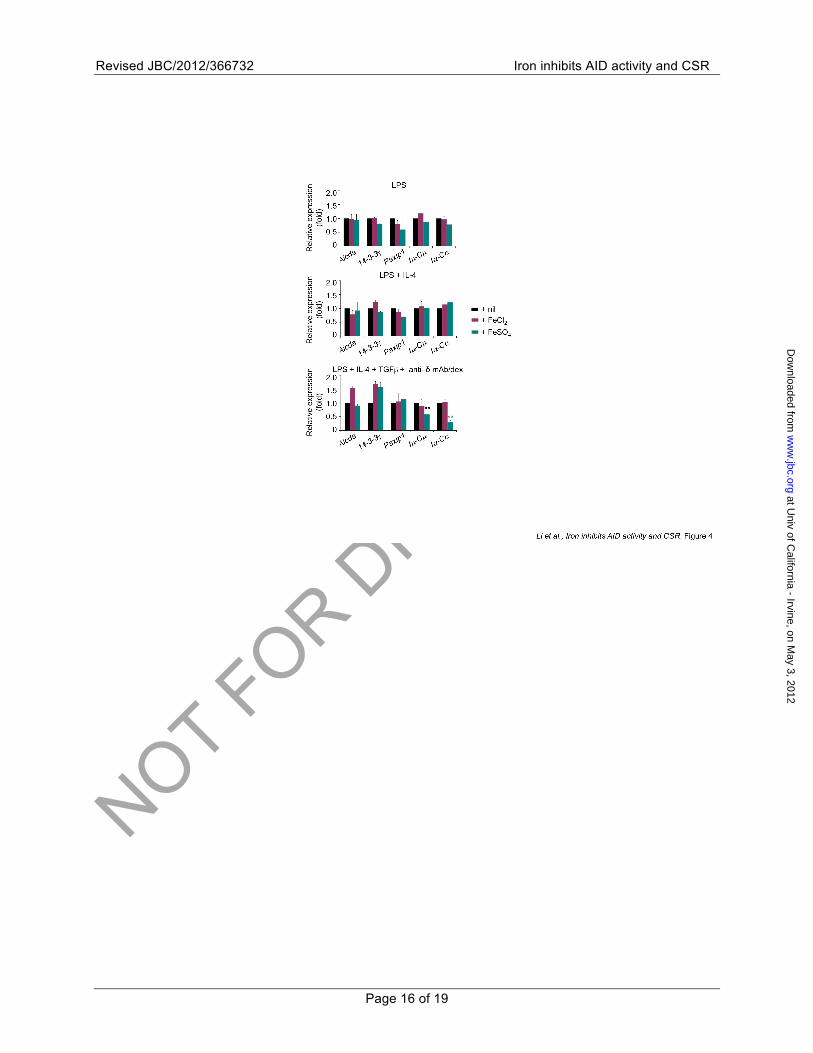
Fe2+does not alter induction of AID, 14-3-3γ or PTIP—To address the molecular mechanisms underlying Fe2+–mediated CSR inhibition, we analyzed the effect of FeCl2 and FeSO4 on AID, 14-3-3γ and PTIP. In B cells simulated by LPS, LPS plus IL-4 or LPS, IL-4, TGFβ plus anti–δ
mAb/dex, FeCl2 or FeSO4 did not affect the induction of AID (encoded by the Aicda gene), which is essential for CSR, 14-3-3γ, which is rapidly induced (Mai, Xu & Casali, unpublished data) and recruit AID to the S regions that will undergo recombination, or PTIP (encoded by the Paxip1 gene), which mediates multiple histone modifications and germline transcription in acceptor S regions (34) (Fig. 4). Likewise, FeCl2 or FeSO4 did not affect the induction of (spliced) germline Iµ-Cµ, Iγ3-Cγ3, Iγ1-Cγ1 and/or Iα-Cα transcripts, with the exception of some reduction in Iµ-Cµ and Iα-Cα transcript levels in B cells stimulated with LPS, IL-4, TGFβ plus anti–δ mAb/dex in the presence of FeSO4.
Thus, Fe2+ impairs CSR without affecting the induction of AID, 14-3-3, PTIP or, in general, IH–S–CH transcription. This suggests that Fe2+ does not affect chromatin accessibility of the S regions that will undergo recombination or the targeting of the CSR machinery by 14-3-3 adaptors.
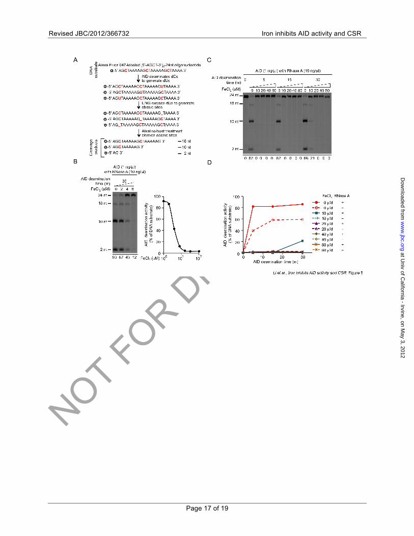
Fe2+ inhibits AID-mediated dC DNA deamination—The normal expression (and likely targeting) of AID in B cells stimulated by LPS, LPS plus IL-4 or LPS, IL-4, TGFβ plus anti–δ mAb/dex in the presence of FeCl2 or FeSO4
prompted us to hypothesize that Fe2+ suppresses CSR by inhibiting AID enzymatic activity. Our hypothesis was further strengthened by the suggestion that the AID catalytic residues are coordinated to Zn2+ (35) and findings that selected cytidine/cytosine deaminases can be bound by Zn2+ or Fe2+ (36). To test our hypothesis, we performed in vitro AID dC deamination assays, in which dCs within the three 5′-AGCT-3′ motifs of a 24-nt single-strand oligonucleotide Alexa-[5′-AGCT-3′]3-24nt was converted by AID to dUs. These were, in turn, excised by UNG to yield labile abasic sites, which were then cleaved by alkaline and heat treatment to generate 18-nt, 10-nt and 2-nt cleavage products (Fig. 5A). FeCl2 inhibited AID-mediated deamination of dCs within Alexa-[5′-AGCT-3′]3-24nt in a dose-dependent fashion and within a narrow range, i.e., from 2 µM (7% inhibition of the AID enzymatic activity) to 8 µM (87% inhibition) (Fig. 5B).
RNase A has been shown to enhance AID-mediated dC DNA deamination in vitro (37-39), possibly by degrading an RNA molecule that tightly binds to recombinant AID and inhibits its enzymatic activity. To ascertain that Fe2+
does not merely interfere with the activity of RNase A used
at Univ of C
alifornia - Irvine, on May 3, 2012
ww
w.jbc.org
Dow
nloaded from
NOT FOR DIS
TRIBUTIO
N
Revised JBC/2012/366732 Iron inhibits AID activity and CSR
Page 6 of 19
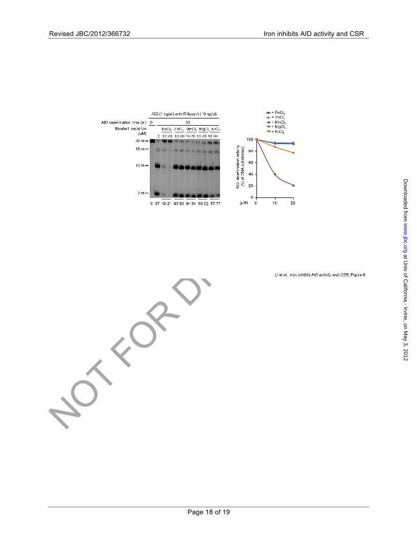
in our assays, we performed AID deamination assays without RNase A. In the absence of RNase A, AID-mediated dC deamination plateaued at 60% deamination activity within 15 m, as compared to 80% deamination activity within 5 m in the presence of RNase A. The (RNase A-independent) intrinsic AID dC deamination activity was efficiently inhibited by FeCl2 (Figs. 5C, 5D). In contrast to Fe2+, bivalent metal ions Zn2+, Mn2+, Mg2+ and Ni2 exhibited little, if any, inhibitory effects on AID-mediated deamination (Fig. 6).
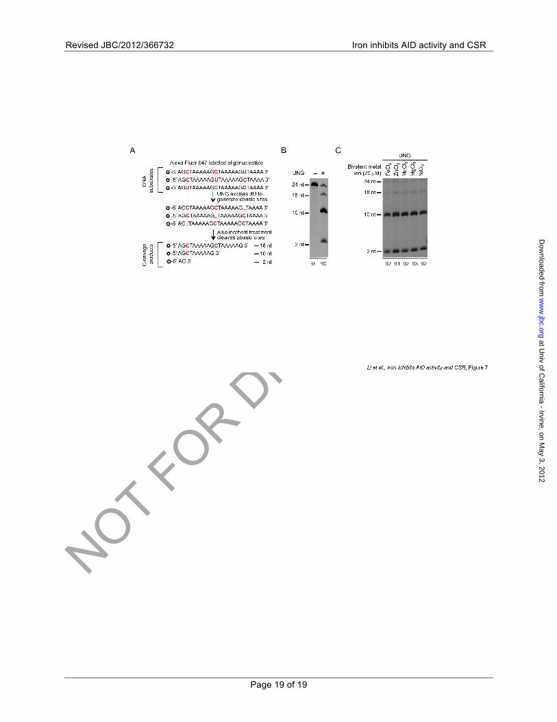
To analyze whether Fe2+ also affects UNG dU excision activity, we generated dU-containing oligonucleotides that could give rise to abasic sites for cleavage by alkaline and heat treatment only in after UNG-mediated dU excision (Figs. 7A, 7B). This was not affected by Fe2+, Zn2+, Mn2+, Mg2+ or Ni2+, as shown by over 90% of the substrates showing cleavage (Fig. 7C).
Thus, Fe2+, but not other bivalent metal ions (Zn2+, Mn2+, Mg2+ or Ni2+), effectively inhibits the intrinsic AID dC DNA deamination activity in a dose-dependent fashion. Fe2+ does not affect UNG-mediated dU excision.
DISCUSSION
Here we have shown that Fe2+ efficiently inhibited the generation of class-switched B cells and antibodies by interfering with the intrinsic CSR mechanisms without altering B cell proliferation or viability. Fe2+ inhibition of CSR, such as from IgM to IgG3 (by FeCl2) or IgA (by FeSO4), was profound and concomitant with normal plasmacytoid differentiation of B cells. It was associated with normal expression levels of important CSR factors, including AID, 14-3-3 and PTIP, and mainly reflected Fe2+ inhibition of AID enzymatic activity (dC DNA deamination). Such inhibition was specific, as emphasized by the failure of other bivalent metal ions (Zn2+, Mn2+, Mg2+ or Ni2+) to inhibit AID enzymatic activity and the inability of Fe2+ to inhibit UNG dU excision activity (whether Fe2+ inhibits enzymatic or scaffold functions of other CSR factors remains to be determined). By inhibiting AID enzymatic activity, Fe2+ not only inhibits CSR, but would also interfere with AID-mediated SHM and generation of high-affinity antibody mutants, which are positively selected by antigen in germinal centers of peripheral lymphoid organs (e.g., lymph nodes and spleen).
Fe2+ inhibition of enzymatic function of AID in CSR is further emphasized by our findings that
Fe2+ did not generally affect germline IH-S-CH transcription. The only exception was the reduction of germline Iµ-Sµ-Cµ (by 30%) and Iα-Sα-Cα (by 70%) transcription in B cells stimulated with LPS, IL-4, TGFβ plus anti–δ mAb/dex by FeSO4. Such inhibition likely resulted from FeSO4-mediated interference with TGFβ-induced activation of transcription factors that are critical for germline Iα-Sα-Cα transcription, such as RUNX and SMAD. Reduction in germline Iµ-Sµ-Cµ and Iα-Sα-Cα transcription compounded the Fe2+ inhibition of AID-mediated dC DNA deamination, leading to the profound decrease (90%, as assessed by post-recombination Iµ-Cα transcripts) of CSR to IgA. Lack of inhibition of germline Iα-Sα-Cα transcription by FeCl2 suggests that FeCl2 does not modulate TGFβ signaling and extends previous findings showing the differential ability of FeSO4 and FeCl2 in modulating selected biological processes, such as xylanase enzymatic activity and production of homopolymer ε-poly-L-lysine in Kitasatospora kifunense (40,41).
Our findings that the generation of class-switched and, likely, high-affinity antibodies can be suppressed by iron (21,22) strongly suggest that cytoplasmic/nuclear iron levels need be temporarily downregulated in antigen-activated B cells, perhaps as signaled by CSR-inducing stimuli, such as CD154:CD40 engagement and/or BCR crosslinking, which – as we have shown – is critical for TLR-dependent and T cell-independent CSR in vivo and in vitro (18). Our findings suggest that in antigen-primed B cells, such as those engaged in the germinal center reaction, a lower level of iron than that steadily maintained in naïve B cells would enable efficient class switching while maintaining proper B cell proliferation. Downregulation of iron levels in germinal center B cells could result from reduced iron uptake dependent on transferring receptor 2 (TFR2), as suggested by downregulation of TFR2 expression in those B cells (42). B cells existing the germinal center reaction (after undergoing CSR and SHM) would replenish their iron pool in the transition to memory B cells or plasma cells, and might to do so by “siphoning” iron from macrophages, a major iron reservoir in peripheral lymphoid organs, via long-range “nanotubes” (43) that function as conduits for molecular exchange between cells (44).
at Univ of C
alifornia - Irvine, on May 3, 2012
ww
w.jbc.org
Dow
nloaded from
NOT FOR DIS
TRIBUTIO
N
Revised JBC/2012/366732 Iron inhibits AID activity and CSR
Page 7 of 19
Due to the high mutagenicity of AID, expression and targeting of this cytidine deaminase are tightly regulated (45). Our findings that Fe2+ suppresses AID enzymatic activity outline another level of control (by endogenous metabolites, i.e., iron and factors that regulate iron transport and uptake) of overall AID activity, in addition to the regulation of AID expression. Genetic defects, infectious agents or tissue injuries can perturb the B cell-intrinsic homeostatic mechanisms that regulate intracellular iron level and lead to iron overload. Consistent with the principle outlined here by our in vitro findings, high iron levels are associated in different contexts with impaired antibody responses (25,46). Most notably, patients with hemochromatosis, a genetic disease characterized by iron overload (ranging from 30 µM to over 40 µM), have reduced level of class-switched IgG and IgA antibodies, which can be readily explained by our findings that CSR was substantially decreased by Fe2+ at 40 and 80 µM, and lowered defenses with increased susceptibility to viral and, more frequently, bacterial infections (26,27). Once established, infections could in turn interfere with antibody class switching and the immune response by further upregulating B cell iron levels. For instance, by inducing macrophage expression of ferritin, HIV-1 accessory protein Nef would lead to upregulation of B cell intracellular iron levels, leading to decreased AID and inhibition of class-switching of HIV-1-specific antibodies (22,44,47,48) – Nef can also directly inhibit CSR (49).
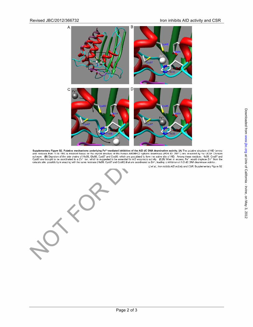
Our data showed that the AID enzymatic activity was highly sensitive to Fe2+ concentrations in vitro (2 – 8 µM) and likely in switching B cells, which, as we contend, would temporarily downregulate intracellular iron levels to enable efficient CSR. Notably, the narrow range of concentrations at which Fe2+ inhibited AID enzymatic functions fell within the putative range of intracellular free iron levels (0.5 – 10 µM) in many cell types, likely including B cells (50,51). Fe2+ would inhibit AID enzymatic activity by displacing Zn2+ in this enzyme catalytic site by virtue of the similar chemical coordination properties of these two metal ions (Supplementary Figure S2). Accordingly, the endogenous cytosine deaminase purified from E. coli contains mostly Fe2+ but also Zn2+, whereas the cytosine deaminase isolated from yeast contains mostly Zn2+ but also Fe2+. The subtle coordination differences of Fe2+ and Zn2+ would,
however, result in significantly different catalytic rates (36,52). Fe2+ can inhibit the activity of Zn2+-containing yeast cytosine deaminase and, conversely, Zn2+ can inhibit the activity of Fe2+-containing E. coli cytosine deaminase (52,53). How Fe2+ would displace Zn2+, which generally coordinates catalytic residues in metalloenzymes – likely including AID – with a high affinity (54), from AID remains to be elucidated. An alternative possibility is that Fe2+ binds to residues different from those of Zn2+, thereby inducing allosteric changes that lead to impairment of AID enzymatic activity. Oxidation of Fe2+ to Fe3+ within the E. coli cytosine deaminase over the course of hours results in inactivation of that enzyme (53). It is, however, unlikely that AID-mediated dC DNA deamination and CSR were inhibited by Fe3+ oxidized from Fe2+, as B cells maintain a reducing environment in the cytoplasm and the nucleus. In addition, all Fe2+ solutions used in our assays were freshly prepared and used within 30 m in the presence of glutathione. Finally, catalytic residues in UNG are not coordinated by metal ions (55).
Fe2+-mediated inhibition of AID and CSR can be exploited by the body to inhibit unwanted class-switching of autoantibodies associated with chronic inflammation, as suggested by increased iron levels in peripheral lymphoid organs in patients with chronic inflammatory diseases (56). Patients with systemic lupus erythematosus and lupus-prone MRL/Faslpr/lpr mice, however, would overcome the iron inhibition of AID by expressing abnormally high levels of AID, resulting from estrogen upregulation of HoxC4 induction (57). In these autoimmune patients and mice, increased AID expression leads to high levels of class-switched and high-affinity pathogenic autoantibodies, mainly to double-strand DNA, increased organ and tissue damage and increased mortality (58-61). These could be reversed in MRL/Faslpr/lpr mice by dietary iron (62), suggesting that the effects of high levels of AID can be overridden by iron supplement, thereby pointing to a potential iron-based therapeutic intervention in lupus.
High AID levels, perhaps compounded by potential systemic decrease of iron levels associated with loss of red blood cells in lupus, would lead to significantly increased insertion/deletions in VDJ region DNA and chromosomal translocations involving the IgH and c-Myc loci (59,61,63). Thus, iron might also protect from AID-mediated genome-wide damage,
at Univ of C
alifornia - Irvine, on May 3, 2012
ww
w.jbc.org
Dow
nloaded from
NOT FOR DIS
TRIBUTIO
N
Revised JBC/2012/366732 Iron inhibits AID activity and CSR
Page 8 of 19
mutations, generation of DSBs and chromosomal translocations (64), particularly in proto-oncogenes, such as c-Myc, and tumor suppressor genes, such as p53 (65,66), thereby inhibiting tumorigenesis. In conclusion, our findings provide an important mechanistic underpinning to the association between iron overload and defective antibody responses. They also suggest a therapeutic application of iron in autoimmunity and neoplasia.
Acknowledgements⎯ We thank Amanda Lin, Nancy Dang and J’aime Moehlman for excellent technical assistance. We thank Drs. Fergus Byrne and Robert Kurzeja (Amgen Inc.) for providing purified recombinant AID. This work was supported by NIH grants AI 045011, AI 060573 and AI 079705 to P.C.
Author contribution⎯ G.L., E.J.P., D.C.T., P.J.P. and L.D. performed experiments; G.L., E.J.P. and Z.X. designed the experiment, analyzed the data and wrote the manuscript; P.C. provided financial support, supervised the project and wrote the manuscript; Z.X. supervised the project.
REFERENCES 1. Murphy, K. M. (2011) The humoral immune
response. in Janeway's Immunobiology (Murphy, K. M. ed.), 8th Ed., Garland Science, New York, NY.
2. Odegard, V. H., and Schatz, D. G. (2006) Targeting of somatic hypermutation. Nat. Rev. Immunol. 6, 573-583
3. Casali, P. (2009) Somatic recombination and hypermutation in the immune system. in Lewin's Genes X (Krebs, J. E., Goldstein, E. S., and Kilpatrick, S. T. eds.), 10th Ed., Jones & Bartlett, Sudbury, MA.
4. Goodnow, C. C., Vinuesa, C. G., Randall, K. L., Mackay, F., and Brink, R. (2010) Control systems and decision making for antibody production. Nat. Immunol. 11, 681-688
5. Stavnezer, J., Guikema, J. E., and Schrader, C. E. (2008) Mechanism and regulation of class switch recombination. Annu. Rev. Immunol. 26, 261-292
6. Muramatsu, M., Sankaranand, V. S., Anant, S., Sugai, M., Kinoshita, K., Davidson, N. O., and Honjo, T. (1999) Specific expression of activation-induced cytidine deaminase (AID), a novel member of the RNA-editing deaminase family in germinal center B cells. J. Biol. Chem. 274, 18470-18476
7. Petersen-Mahrt, S. K., Harris, R. S., and Neuberger, M. S. (2002) AID mutates E. coli suggesting a DNA deamination mechanism for antibody diversification. Nature 418, 99-103
8. Kohli, R. M., Maul, R. W., Guminski, A. F., McClure, R. L., Gajula, K. S., Saribasak, H., McMahon, M. A., Siliciano, R. F., Gearhart, P. J., and Stivers, J. T. (2010) Local sequence targeting in the AID/APOBEC family differentially impacts retroviral restriction and antibody diversification. J. Biol. Chem. 285, 40956-40964
9. Xu, Z., Fulop, Z., Wu, G., Pone, E. J., Zhang, J., Mai, T., Thomas, L. M., Al-Qahtani, A., White, C. A., Park, S. R., Steinacker, P., Li, Z., Yates, J., 3rd, Herron, B., Otto, M., Zan, H., Fu, H., and Casali, P. (2010) 14-3-3 adaptor proteins recruit AID to 5'-AGCT-3'-rich switch regions for class switch recombination. Nat. Struct. Mol. Biol. 17, 1124-1135
10. Maul, R. W., Saribasak, H., Martomo, S. A., McClure, R. L., Yang, W., Vaisman, A., Gramlich, H. S., Schatz, D. G., Woodgate, R., Wilson, D. M., 3rd, and Gearhart, P. J. (2011) Uracil residues dependent on the deaminase AID in immunoglobulin gene variable and switch regions. Nat. Immunol. 12, 70-76
11. Zan, H., White, C. A., Thomas, L. M., Zhang, J., Li, G., Yu, E. S., Xu, Z., Mai, T., and Casali, P. (2012) The translesion DNA synthesis polymerase Rev1 recruits Ung to switch regions and enhances dU excision for immunoglobulin class switch DNA recombination. Cell Reports, Under revision
12. Schrader, C. E., Linehan, E. K., Mochegova, S. N., Woodland, R. T., and Stavnezer, J. (2005) Inducible DNA breaks in Ig S regions are dependent on AID and UNG. J. Exp. Med. 202, 561-568
13. Zan, H., and Casali, P. (2008) AID- and Ung-dependent generation of staggered double-strand DNA breaks in immunoglobulin class switch DNA recombination: a post-cleavage role for AID. Mol. Immunol. 46, 45-61
14. Delker, R. K., Fugmann, S. D., and Papavasiliou, F. N. (2009) A coming-of-age story: activation-induced cytidine deaminase turns 10. Nat. Immunol. 10, 1147-1153
15. Park, S. R., Zan, H., Pal, Z., Zhang, J., Al-Qahtani, A., Pone, E. J., Xu, Z., Mai, T., and Casali, P. (2009) HoxC4 binds to the promoter of the cytidine deaminase AID gene to induce AID expression, class-switch DNA recombination and somatic hypermutation. Nat. Immunol. 10, 540-550
at Univ of C
alifornia - Irvine, on May 3, 2012
ww
w.jbc.org
Dow
nloaded from
NOT FOR DIS
TRIBUTIO
N
Revised JBC/2012/366732 Iron inhibits AID activity and CSR
Page 9 of 19
16. Sayegh, C. E., Quong, M. W., Agata, Y., and Murre, C. (2003) E-proteins directly regulate expression of activation-induced deaminase in mature B cells. Nat. Immunol. 4, 586-593
17. Tran, T. H., Nakata, M., Suzuki, K., Begum, N. A., Shinkura, R., Fagarasan, S., Honjo, T., and Nagaoka, H. (2010) B cell-specific and stimulation-responsive enhancers derepress Aicda by overcoming the effects of silencers. Nat. Immunol. 11, 148-154
18. Pone, E. J., Zhang, J., Mai, T., White, C. A., Li, G., Sakakura, J. K., Patel, P. J., Al-Qahtani, A., Zan, H., Xu, Z., and Casali, P. (2012) BCR-signalling synergizes with TLR-signalling for induction of AID and immunoglobulin class-switching through the non-canonical NF-kappaB pathway. Nat. Commun. 3, 767. DOI 710.1038/ncomms1769
19. Chaudhuri, J., Basu, U., Zarrin, A., Yan, C., Franco, S., Perlot, T., Vuong, B., Wang, J., Phan, R. T., Datta, A., Manis, J., and Alt, F. W. (2007) Evolution of the immunoglobulin heavy chain class switch recombination mechanism. Adv. Immunol. 94, 157-214
20. Basu, U., Meng, F. L., Keim, C., Grinstein, V., Pefanis, E., Eccleston, J., Zhang, T., Myers, D., Wasserman, C. R., Wesemann, D. R., Januszyk, K., Gregory, R. I., Deng, H., Lima, C. D., and Alt, F. W. (2011) The RNA exosome targets the AID cytidine deaminase to both strands of transcribed duplex DNA substrates. Cell 144, 353-363
21. Golenser, J., Domb, A., Mordechai-Daniel, T., Leshem, B., Luty, A., and Kremsner, P. (2006) Iron chelators: correlation between effects on Plasmodium spp. and immune functions. J. Parasitol. 92, 170-177
22. Swingler, S., Zhou, J., Swingler, C., Dauphin, A., Greenough, T., Jolicoeur, P., and Stevenson, M. (2008) Evidence for a pathogenic determinant in HIV-1 Nef involved in B cell dysfunction in HIV/AIDS. Cell Host Microbe 4, 63-76
23. Kochan, I., Wagner, S. K., and Wasynczuk, J. (1984) Effect of iron on antibacterial immunity in vaccinated mice. Infect. Immun. 43, 543-548
24. Gordeuk, V. R., McLaren, C. E., MacPhail, A. P., Deichsel, G., and Bothwell, T. H. (1996) Associations of iron overload in Africa with hepatocellular carcinoma and tuberculosis: Strachan's 1929 thesis revisited. Blood 87, 3470-3476
25. Mencacci, A., Cenci, E., Boelaert, J. R., Bucci, P., Mosci, P., Fe d'Ostiani, C., Bistoni, F., and Romani, L. (1997) Iron overload alters innate
and T helper cell responses to Candida albicans in mice. J. Infect. Dis. 175, 1467-1476
26. Khan, F. A., Fisher, M. A., and Khakoo, R. A. (2007) Association of hemochromatosis with infectious diseases: expanding spectrum. Int. J. Infect. Dis. 11, 482-487
27. Barton, J. C., Bertoli, L. F., and Acton, R. T. (2003) Common variable immunodeficiency and IgG subclass deficiency in central Alabama hemochromatosis probands homozygous for HFE C282Y. Blood Cells Mol. Dis. 31, 102-111
28. Watanabe-Matsui, M., Muto, A., Matsui, T., Itoh-Nakadai, A., Nakajima, O., Murayama, K., Yamamoto, M., Ikeda-Saito, M., and Igarashi, K. (2011) Heme regulates B-cell differentiation, antibody class switch, and heme oxygenase-1 expression in B cells as a ligand of Bach2. Blood 117, 5438-5448
29. Van Vlierberghe, H., Langlois, M., Delanghe, J., Horsmans, Y., Michielsen, P., Henrion, J., Cartuyvels, R., Billiet, J., De Vos, M., and Leroux-Roels, G. (2001) Haptoglobin phenotype 2-2 overrepresentation in Cys282Tyr hemochromatotic patients. J. Hepatol. 35, 707-711
30. Fairfax, K. A., Kallies, A., Nutt, S. L., and Tarlinton, D. M. (2008) Plasma cell development: from B-cell subsets to long-term survival niches. Semin. Immunol. 20, 49-58
31. Klein, U., Casola, S., Cattoretti, G., Shen, Q., Lia, M., Mo, T., Ludwig, T., Rajewsky, K., and Dalla-Favera, R. (2006) Transcription factor IRF4 controls plasma cell differentiation and class-switch recombination. Nat. Immunol. 7, 773-782
32. Shaffer, A. L., Lin, K. I., Kuo, T. C., Yu, X., Hurt, E. M., Rosenwald, A., Giltnane, J. M., Yang, L., Zhao, H., Calame, K., and Staudt, L. M. (2002) Blimp-1 orchestrates plasma cell differentiation by extinguishing the mature B cell gene expression program. Immunity 17, 51-62
33. Reimold, A. M., Iwakoshi, N. N., Manis, J., Vallabhajosyula, P., Szomolanyi-Tsuda, E., Gravallese, E. M., Friend, D., Grusby, M. J., Alt, F., and Glimcher, L. H. (2001) Plasma cell differentiation requires the transcription factor XBP-1. Nature 412, 300-307
34. Daniel, J. A., Santos, M. A., Wang, Z., Zang, C., Schwab, K. R., Jankovic, M., Filsuf, D., Chen, H. T., Gazumyan, A., Yamane, A., Cho, Y. W., Sun, H. W., Ge, K., Peng, W., Nussenzweig, M. C., Casellas, R., Dressler, G. R., Zhao, K., and Nussenzweig, A. (2010)
at Univ of C
alifornia - Irvine, on May 3, 2012
ww
w.jbc.org
Dow
nloaded from
NOT FOR DIS
TRIBUTIO
N
Revised JBC/2012/366732 Iron inhibits AID activity and CSR
Page 10 of 19
PTIP promotes chromatin changes critical for immunoglobulin class switch recombination. Science 329, 917-923
35. Prochnow, C., Bransteitter, R., Klein, M. G., Goodman, M. F., and Chen, X. S. (2007) The APOBEC-2 crystal structure and functional implications for the deaminase AID. Nature 445, 447-451
36. Ireton, G. C., McDermott, G., Black, M. E., and Stoddard, B. L. (2002) The structure of Escherichia coli cytosine deaminase. J. Mol. Biol. 315, 687-697
37. Pham, P., Bransteitter, R., Petruska, J., and Goodman, M. F. (2003) Processive AID-catalysed cytosine deamination on single-stranded DNA simulates somatic hypermutation. Nature 424, 103-107
38. Bransteitter, R., Pham, P., Scharff, M. D., and Goodman, M. F. (2003) Activation-induced cytidine deaminase deaminates deoxycytidine on single-stranded DNA but requires the action of RNase. Proc. Natl. Acad. Sci. U S A. 100, 4102-4107
39. Yu, K., Huang, F. T., and Lieber, M. R. (2004) DNA substrate length and surrounding sequence affect the activation-induced deaminase activity at cytidine. J. Biol. Chem. 279, 6496-6500
40. Kabayashi, K., and Nishikawa, M. (2007) Promotion of epsilon-poly-lysine production by iron in Kitasatospora kifunense. World. J. Microbiol. Biotechnol. 23, 1033-1036
41. Annamalai, N., Thavasi, R., Jayalakshmi, S., and Balasubramanian, T. (2009) Thermostable and alkaline tolerant xylanase production by Bacillus subtilis isolated from marine environment. Ind. J. Biotechnol. 8, 291-297
42. Luckey, C. J., Bhattacharya, D., Goldrath, A. W., Weissman, I. L., Benoist, C., and Mathis, D. (2006) Memory T and memory B cells share a transcriptional program of self-renewal with long-term hematopoietic stem cells. Proc. Natl. Acad. Sci. U.S.A. 103, 3304-3309
43. Kadiu, I., and Gendelman, H. E. (2011) Macrophage bridging conduit trafficking of HIV-1 through the endoplasmic reticulum and Golgi network. J. Proteome Res. 10, 3225-3238
44. Xu, W., Santini, P. A., Sullivan, J. S., He, B., Shan, M., Ball, S. C., Dyer, W. B., Ketas, T. J., Chadburn, A., Cohen-Gould, L., Knowles, D. M., Chiu, A., Sanders, R. W., Chen, K., and Cerutti, A. (2009) HIV-1 evades virus-specific IgG2 and IgA responses by targeting systemic and intestinal B cells via long-range
intercellular conduits. Nat. Immunol. 10, 1008-1017
45. Xu, Z., Pone, E. J., Al-Qahtani, A., Park, S. R., Zan, H., and Casali, P. (2007) Regulation of aicda expression and AID activity: relevance to somatic hypermutation and class switch DNA recombination. Crit. Rev. Immunol. 27, 367-397
46. Bullen, J. J., Wilson, A. B., Cushnie, G. H., and Rogers, H. J. (1968) The abolition of the protective effect of Pasteurella septica antiserum by iron compounds. Immunology 14, 889-898
47. Kohyama, M., Ise, W., Edelson, B. T., Wilker, P. R., Hildner, K., Mejia, C., Frazier, W. A., Murphy, T. L., and Murphy, K. M. (2009) Role for Spi-C in the development of red pulp macrophages and splenic iron homeostasis. Nature 457, 318-321
48. Rudnicka, D., and Schwartz, O. (2009) Intrusive HIV-1-infected cells. Nat. Immunol. 10, 933-934
49. Qiao, X., He, B., Chiu, A., Knowles, D. M., Chadburn, A., and Cerutti, A. (2006) Human immunodeficiency virus 1 Nef suppresses CD40-dependent immunoglobulin class switching in bystander B cells. Nat. Immunol. 7, 302-310
50. Konijn, A. M., Glickstein, H., Vaisman, B., Meyron-Holtz, E. G., Slotki, I. N., and Cabantchik, Z. I. (1999) The cellular labile iron pool and intracellular ferritin in K562 cells. Blood 94, 2128-2134
51. Petrat, F., de Groot, H., and Rauen, U. (2001) Subcellular distribution of chelatable iron: a laser scanning microscopic study in isolated hepatocytes and liver endothelial cells. Biochem J. 356, 61-69
52. Ireton, G. C., Black, M. E., and Stoddard, B. L. (2003) The 1.14 A crystal structure of yeast cytosine deaminase: evolution of nucleotide salvage enzymes and implications for genetic chemotherapy. Structure 11, 961-972
53. Porter, D. J., and Austin, E. A. (1993) Cytosine deaminase. The roles of divalent metal ions in catalysis. J. Biol. Chem. 268, 24005-24011
54. Christianson, D. W., and Cox, J. D. (1999) Catalysis by metal-activated hydroxide in zinc and manganese metalloenzymes. Annu. Rev. Biochem. 68, 33-57
55. Mol, C. D., Arvai, A. S., Slupphaug, G., Kavli, B., Alseth, I., Krokan, H. E., and Tainer, J. A. (1995) Crystal structure and mutational analysis of human uracil-DNA glycosylase:
at Univ of C
alifornia - Irvine, on May 3, 2012
ww
w.jbc.org
Dow
nloaded from
NOT FOR DIS
TRIBUTIO
N
Revised JBC/2012/366732 Iron inhibits AID activity and CSR
Page 11 of 19
structural basis for specificity and catalysis. Cell 80, 869-878
56. Theurl, I., Aigner, E., Theurl, M., Nairz, M., Seifert, M., Schroll, A., Sonnweber, T., Eberwein, L., Witcher, D. R., Murphy, A. T., Wroblewski, V. J., Wurz, E., Datz, C., and Weiss, G. (2009) Regulation of iron homeostasis in anemia of chronic disease and iron deficiency anemia: diagnostic and therapeutic implications. Blood 113, 5277-5286
57. Mai, T., Zan, H., Zhang, J., Hawkins, J. S., Xu, Z., and Casali, P. (2010) Estrogen receptors bind to and activate the HOXC4/HoxC4 promoter to potentiate HoxC4-mediated activation-induced cytosine deaminase induction, immunoglobulin class switch DNA recombination, and somatic hypermutation. J. Biol. Chem. 285, 37797-37810
58. Jiang, C., Foley, J., Clayton, N., Kissling, G., Jokinen, M., Herbert, R., and Diaz, M. (2007) Abrogation of lupus nephritis in activation-induced deaminase-deficient MRL/lpr mice. J. Immunol. 178, 7422-7431
59. Zan, H., Zhang, J., Ardeshna, S., Xu, Z., Park, S. R., and Casali, P. (2009) Lupus-prone MRL/faslpr/lpr mice display increased AID expression and extensive DNA lesions, comprising deletions and insertions, in the immunoglobulin locus: concurrent upregulation of somatic hypermutation and class switch DNA recombination. Autoimmunity 42, 89-103
60. Hsu, H. C., Yang, P., Wu, Q., Wang, J. H., Job, G., Guentert, T., Li, J., Stockard, C. R., Le, T. V., Chaplin, D. D., Grizzle, W. E., and Mountz, J. D. (2011) Inhibition of the catalytic function of activation-induced cytidine deaminase promotes apoptosis of germinal center B cells in BXD2 mice. Arthritis Rheum. 63, 2038-2048
61. White, C. A., Seth Hawkins, J., Pone, E. J., Yu, E. S., Al-Qahtani, A., Mai, T., Zan, H., and Casali, P. (2012) AID dysregulation in lupus-prone MRL/Fas(lpr/lpr) mice increases class switch DNA recombination and promotes interchromosomal c-Myc/IgH loci translocations: modulation by HoxC4. Autoimmunity 44, 585-598
62. Leiter, L. M., Reuhl, K. R., Racis, S. P., Jr., and Sherman, A. R. (1995) Iron status alters murine systemic lupus erythematosus. J. Nutr. 125, 474-484
63. Liu, M., Duke, J. L., Richter, D. J., Vinuesa, C. G., Goodnow, C. C., Kleinstein, S. H., and Schatz, D. G. (2008) Two levels of protection
for the B cell genome during somatic hypermutation. Nature 451, 841-845
64. Xu, Z., Zan, H., Pone, E. J., Mai, T., and Casali, P. (2012) Immunoglobulin class switching: induction, targeting and beyond. Nat. Rev. Immunol. , In press
65. Matsumoto, Y., Marusawa, H., Kinoshita, K., Endo, Y., Kou, T., Morisawa, T., Azuma, T., Okazaki, I. M., Honjo, T., and Chiba, T. (2007) Helicobacter pylori infection triggers aberrant expression of activation-induced cytidine deaminase in gastric epithelium. Nat. Med. 13, 470-476
66. Pasqualucci, L., Bhagat, G., Jankovic, M., Compagno, M., Smith, P., Muramatsu, M., Honjo, T., Morse, H. C., 3rd, Nussenzweig, M. C., and Dalla-Favera, R. (2008) AID is required for germinal center-derived lymphomagenesis. Nat. Genet. 40, 108-112
ABBREVIATIONS The abbreviations used are: AID, activation-induced cytidine deaminase; BLIMP1, B-lymphocyte-induced maturation protein 1; CSR, class switch DNA recombination; dC, deoxycytidine; DSBs, double-strand DNA breaks; dU, deoxyuracil; Ig, immunoglobulin; Paxip1, PAX interacting (with transcription- activation domain) protein 1; S, switch; SHM, somatic hypermutation; UNG, uracil DNA glycosylase; XBP1, X-box binding protein 1.
FIGURE LEGENDS FIGURE 1. Fe2+ suppresses CSR. (A) Surface expression of B220 and IgG3 (induced by LPS), IgG1 (induced by LPS plus IL-4) or IgA (induced by LPS, IL-4, TGFβ plus anti–δ mAb/dex) in the absence (nil, black) or presence of 80 µM of FeCl2 (plum) or FeSO4 (teal). The number inside each rectangle indicates the percentage of B220+ cells that are switched to the indicated Ig isotypes. Ratios of the proportion of switched B cells in B cells stimulated in the presence of Fe2+ to that of B cells stimulated in the absence of Fe2+ are depicted in histograms (mean and s.e.m. of data from three independent experiments). (B) Titers of IgG3, IgG1 or IgA in supernatants of B cells stimulated with LPS, LPS plus IL-4 or LPS, IL-4, TGFβ plus anti–δ mAb/dex in the absence (nil, black) or presence of 80 µM of FeCl2 (plum) or FeSO4 (teal) (mean and s.d. of data from three independent experiments). (C) Levels of post-recombination Iµ-Cγ3, Iµ-Cγ1 or Iµ-Cα transcripts in B cells stimulated with LPS, LPS plus IL-4 or LPS, IL-4, TGFβ plus anti–δ mAb/dex in the absence (nil, black) or presence of 80 µM of FeCl2 (plum) or FeSO4 (teal). Data were normalized to
at Univ of C
alifornia - Irvine, on May 3, 2012
ww
w.jbc.org
Dow
nloaded from
NOT FOR DIS
TRIBUTIO
N
Revised JBC/2012/366732 Iron inhibits AID activity and CSR
Page 12 of 19
the expression of Cd79b and are expressed as ratios of values in B cells stimulated in the presence of Fe2+ to those in B cells stimulated in the absence of Fe2+ (mean and s.e.m. of data from three independent experiments). **, P < 0.05 (P values: t-test).
FIGURE 2. Fe2+ does not alter B cell surface Igµ expression, proliferation or viability. (A) Surface expression of B220 and Igµ in B cells stimulated with of LPS, LPS plus IL-4 or LPS, IL-4, TGFβ plus anti–δ mAb/dex in the absence (nil) or presence of 80 µM of FeCl2 or FeSO4. The number inside each rectangle indicates the percentage of cells that are Igµ+. Data are representative of three independent experiments. (B) Proportion of surface IgG1+ B cells after stimulation of CFSE-labeled B cells with LPS plus IL-4 in the absence (nil) or presence of 80 µM of FeCl2 or FeSO4. Data are representative of three independent experiments. (C) Proportion of IgG3+, IgG1+ or IgA+ B cells in each cell division after CFSE-labeled B cells were stimulated with LPS, LPS plus IL-4 or LPS plus IL-4 or LPS, IL-4, TGFβ plus anti–δ mAb/dex in the absence (nil, black) or presence of 80 µM of FeCl2 (plum) or FeSO4 (teal). Data are representative of three independent experiments. (D) The proportion of live (7-AAD–) B cells after stimulation with LPS, LPS plus IL-4 or LPS, IL-4, TGFβ plus anti–δ mAb/dex in the absence (nil, black) or presence of 80 µM of FeCl2 (plum) or FeSO4 (teal). Data are expressed as ratios of values in B cells stimulated in the presence of Fe2+ to those in B cells stimulated in the absence of Fe2+ (mean and s.e.m. of data from three independent experiments).
FIGURE 3. Fe2+ does not inhibit plasmacytoid differentiation. (A) Proportion of CD138+
(Syndecan-1+) plasmacytoid cells/plasmacytes after stimulation of B cells with LPS, LPS plus IL-4 or LPS, IL-4, TGFβ plus anti–δ mAb/dex in the absence (nil) or presence of 80 µM of FeCl2 or FeSO4. B220 surface expression was downregulated in B cells stimulated with LPS or LPS plus IL-4. Data are representative of three independent experiments. (B) Levels of Irf4, Prdm1 and Xbp1 transcripts in B cells stimulated with LPS, LPS plus IL-4 or LPS, IL-4, TGFβ plus anti–δ mAb/dex in the absence (nil, black) or presence of 80 µM of FeCl2 (plum) or FeSO4 (teal). Data were normalized to the level of Cd79b and are expressed as ratios of values in B cells cultured with LPS, LPS plus IL-4 or LPS, IL-4, TGFβ plus anti–δ mAb/dex presence of Fe2+ to those in B cells cultured in the absence of Fe2+
(mean and s.e.m. of data from three independent experiments).
FIGURE 4. Fe2+ does not alter induction of Aicda, 14-3-3γ or Paxip1, or germline IH-S-CH transcription in general. Levels of Aicda, 14-3-3γ and Paxip1 transcripts in B cells stimulated with LPS, LPS plus IL-4 or LPS, IL-4, TGFβ plus anti–δ mAb/dex in the absence (nil, black) or presence of 80 µM of FeCl2 (plum) or FeSO4 (teal) as well as germline Iµ-Cµ and Iγ3-Cγ3, Iγ1-Cγ1 or Iα-Cα transcripts in those B cells. Data were normalized to the level of Cd79b and are expressed as ratios of values in B cells stimulated in the presence of Fe2+ to those in B cells stimulated in the absence of Fe2+ (mean and s.e.m. of data from three independent experiments). **, P < 0.05 (P values: t-test).
FIGURE 5. Fe2+ inhibits AID-mediated dC DNA deamination. (A) Schematics of the AID deamination assay. (B) Dose-dependent inhibition of AID mediated dC DNA deamination by FeCl2. AID dC DNA deamination activity in the presence of different concentrations of FeCl2 is indicated below each lane in left panel and depicted by an inhibition curve in the right panel. (C) Inhibition of AID dC DNA deamination over different periods of the enzymatic reaction by different concentrations FeCl2, as indicated. AID dC DNA deamination activity is indicated below each lane. (D) Inhibition of AID dC DNA deamination over different periods of the enzymatic reaction by different concentrations FeCl2, as indicated, in the absence or presence of RNase A. AID dC DNA deamination activity is depicted by kinetic curves in the right panel. Data are representative of three independent experiments.
FIGURE 6. Bivalent metal ion Zn2+, Mn2+, Mg2+ or Ni2+ does not affect AID-mediated dC DNA deamination. AID dC DNA deamination activity in the presence of absence or 10 µM or 20 µM of FeCl2, ZnCl2, MnCl2, MgCl2 or NiCl2 is indicated below each lane in left panel and depicted by inhibition curves in the right panel. Data are representative of three independent experiments.
FIGURE 7. Fe2+ does not alter the UNG dU excision activity. (A) Schematics of the UNG-mediated dU excision assay. (B) Omission of UNG yielded no cleavage products. (C) UNG-mediated dU excision in the absence or presence of FeCl2, ZnCl2, MnCl2, MgCl2 or NiCl2. Numbers below each lane indicate the percentage of the substrate showing dU excision. Data are representative of three independent experiments.
at Univ of C
alifornia - Irvine, on May 3, 2012
ww
w.jbc.org
Dow
nloaded from

NOT FOR DIS
TRIBUTIO
N
Revised JBC/2012/366732 Iron inhibits AID activity and CSR
Page 13 of 19
at Univ of C
alifornia - Irvine, on May 3, 2012
ww
w.jbc.org
Dow
nloaded from
NOT FOR DIS
TRIBUTIO
N
Revised JBC/2012/366732 Iron inhibits AID activity and CSR
Page 14 of 19
at Univ of C
alifornia - Irvine, on May 3, 2012
ww
w.jbc.org
Dow
nloaded from

NOT FOR DIS
TRIBUTIO
N
Revised JBC/2012/366732 Iron inhibits AID activity and CSR
Page 15 of 19
at Univ of C
alifornia - Irvine, on May 3, 2012
ww
w.jbc.org
Dow
nloaded from
NOT FOR DIS
TRIBUTIO
N
Revised JBC/2012/366732 Iron inhibits AID activity and CSR
Page 16 of 19
at Univ of C
alifornia - Irvine, on May 3, 2012
ww
w.jbc.org
Dow
nloaded from
NOT FOR DIS
TRIBUTIO
N
Revised JBC/2012/366732 Iron inhibits AID activity and CSR
Page 17 of 19
at Univ of C
alifornia - Irvine, on May 3, 2012
ww
w.jbc.org
Dow
nloaded from
NOT FOR DIS
TRIBUTIO
N
Revised JBC/2012/366732 Iron inhibits AID activity and CSR
Page 18 of 19
at Univ of C
alifornia - Irvine, on May 3, 2012
ww
w.jbc.org
Dow
nloaded from
NOT FOR DIS
TRIBUTIO
N
Revised JBC/2012/366732 Iron inhibits AID activity and CSR
Page 19 of 19
at Univ of C
alifornia - Irvine, on May 3, 2012
ww
w.jbc.org
Dow
nloaded from
NOT FOR DIS
TRIBUTIO
N
Revised JBC/2012/366732 Iron inhibits AID activity and CSR
Page 1 of 3
Iron Inhibits Activation-induced Cytidine Deaminase Enzymatic Activity and Modulates Immunoglobulin Class Switch DNA Recombination Guideng Li, Egest J Pone, Daniel C Tran, Pina J Patel, Lisa Dao, Zhenming Xu*, Paolo Casali From the Institute for immunology and Department of Medicine, School of Medicine, University of California, Irvine, CA 92697-4120, USA
Supplementary Materials
at Univ of C
alifornia - Irvine, on May 3, 2012
ww
w.jbc.org
Dow
nloaded from
NOT FOR DIS
TRIBUTIO
N
Revised JBC/2012/366732 Iron inhibits AID activity and CSR
Page 2 of 3
at Univ of C
alifornia - Irvine, on May 3, 2012
ww
w.jbc.org
Dow
nloaded from
NOT FOR DIS
TRIBUTIO
N
Revised JBC/2012/366732 Iron inhibits AID activity and CSR
Page 3 of 3
at Univ of C
alifornia - Irvine, on May 3, 2012
ww
w.jbc.org
Dow
nloaded from
Related Documents