Activation-Induced Cytidine Deaminase Targets DNA at Sites of RNA Polymerase II Stalling by Interaction with Spt5 Rushad Pavri, 1 Anna Gazumyan, 1,2 Mila Jankovic, 1 Michela Di Virgilio, 1 Isaac Klein, 1 Camilo Ansarah-Sobrinho, 3 Wolfgang Resch, 3 Arito Yamane, 3 Bernardo Reina San-Martin, 1,4 Vasco Barreto, 1,5 Thomas J. Nieland, 6 David E. Root, 6 Rafael Casellas, 3, * and Michel C. Nussenzweig 1,2, * 1 Laboratory of Molecular Immunology 2 Howard Hughes Medical Institute The Rockefeller University, New York, New York 10065, USA 3 Genomics and Immunity, The National Institute of Arthritis and Musculoskeletal and Skin Diseases (NIAMS), and Center for Cancer Research, National Cancer Institute (NCI), National Institutes of Health, Bethesda, MD 20892, USA 4 Institut de Ge ´ ne ´ tique et de Biologie Mole ´ culaire et Cellulaire (IGBMC), INSERM U964 / CNRS UMR7104 / Universite ´ de Strasbourg, 67404, Illkirch, France 5 Laboratory of Epigenetics and Soma, Instituto Gulbenkian de Cie ˆ ncia, P-2780-156 Oeiras Portugal 6 RNAi Platform, The Broad Institute of MIT and Harvard, Cambridge, MA 02142, USA *Correspondence: [email protected] (M.C.N.), [email protected] (R.C.) DOI 10.1016/j.cell.2010.09.017 SUMMARY Activation-induced cytidine deaminase (AID) initiates antibody gene diversification by creating U:G mis- matches. However, AID is not specific for antibody genes; Off-target lesions can activate oncogenes or cause chromosome translocations. Despite its importance in these transactions little is known about how AID finds its targets. We performed an shRNA screen to identify factors required for class switch recombination (CSR) of antibody loci. We found that Spt5, a factor associated with stalled RNA polymerase II (Pol II) and single stranded DNA (ssDNA), is required for CSR. Spt5 interacts with AID, it facilitates association between AID and Pol II, and AID recruitment to its Ig and non-Ig targets. ChIP-seq experiments reveal that Spt5 colocalizes with AID and stalled Pol II. Further, Spt5 accumula- tion at sites of Pol II stalling is predictive of AID- induced mutation. We propose that AID is targeted to sites of Pol II stalling in part via its association with Spt5. INTRODUCTION AID is a cytidine deaminase that initiates immunoglobulin somatic hypermutation (SHM) and class switch recombination (CSR) (Muramatsu et al., 2000, 1999; Revy et al., 2000). It does so by deaminating cytidine residues in ssDNA (Bransteitter et al., 2003; Chaudhuri et al., 2003; Dickerson et al., 2003; Pham et al., 2003; Ramiro et al., 2003; Sohail et al., 2003). The resulting U:G mismatches can be processed by several different DNA repair pathways to produce mutations or DNA double- strand breaks (Di Noia and Neuberger, 2007; Peled et al., 2008). In addition to diversifying the antibody repertoire by SHM and CSR, AID also contributes to malignant transformation by initi- ating chromosome translocations (Ramiro et al., 2006; Ramiro et al., 2004; Robbiani et al., 2008; Nussenzweig and Nussenz- weig, 2010) and by producing mutations in non-Ig genes such as Bcl-6 (Pasqualucci et al., 1998, 2001; Shen et al., 1998). Although the comparative frequency of mutation at non-Ig genes is low, AID mutates 25% of the genes transcribed in germinal center B cells, where it is normally expressed (Liu et al., 2008). Furthermore, even low levels of mutation are sufficient to produce substrates for translocation (Robbiani et al., 2008; Rob- biani et al., 2009). Consistent with the breadth of genes found mutated by AID in germinal center B cells, AID overexpression in transgenic mice leads to extensive translocation of non-Ig genes and cancer (Robbiani et al., 2009). In addition, AID dereg- ulation has been associated with H. pylori infection and gastric cancer (Matsumoto et al., 2007), and with translocation in pros- tate malignancy (Lin et al., 2009). Finally, AID is also of interest because it has been implicated as a cytosine demethylase involved in reprogramming pluripotent cells (Bhutani et al., 2010; Morgan et al., 2004; Popp et al., 2010; Rai et al., 2008). Although the precise mechanism which targets AID to Ig genes is unknown, AID-induced mutations are associated with tran- scription and are most prevalent in a 2 kb region beginning downstream of the promoter (Di Noia and Neuberger, 2007; Peled et al., 2008; Stavnezer et al., 2008; Storb et al., 2007). Transcription is also required for CSR, suggesting that RNA polymerase II (Pol II) might facilitate AID access to target DNA (Di Noia and Neuberger, 2007; Peled et al., 2008; Stavnezer- Nordgren and Sirlin, 1986; Stavnezer et al., 2008; Storb et al., 2007; Yancopoulos et al., 1986). This idea was confirmed by the observation that transcriptional regulatory elements are essential to both hypermutation and CSR (reviewed in (Di Noia 122 Cell 143, 122–133, October 1, 2010 ª2010 Elsevier Inc.

Welcome message from author
This document is posted to help you gain knowledge. Please leave a comment to let me know what you think about it! Share it to your friends and learn new things together.
Transcript
Activation-Induced Cytidine DeaminaseTargets DNA at Sites of RNA Polymerase IIStalling by Interaction with Spt5Rushad Pavri,1 Anna Gazumyan,1,2 Mila Jankovic,1 Michela Di Virgilio,1 Isaac Klein,1 Camilo Ansarah-Sobrinho,3
Wolfgang Resch,3 Arito Yamane,3 Bernardo Reina San-Martin,1,4 Vasco Barreto,1,5 Thomas J. Nieland,6 David E. Root,6
Rafael Casellas,3,* and Michel C. Nussenzweig1,2,*1Laboratory of Molecular Immunology2Howard Hughes Medical InstituteThe Rockefeller University, New York, New York 10065, USA3Genomics and Immunity, TheNational Institute of Arthritis andMusculoskeletal andSkin Diseases (NIAMS), andCenter for Cancer Research,
National Cancer Institute (NCI), National Institutes of Health, Bethesda, MD 20892, USA4Institut de Genetique et de Biologie Moleculaire et Cellulaire (IGBMC), INSERM U964 / CNRS UMR7104 / Universite de Strasbourg, 67404,
Illkirch, France5Laboratory of Epigenetics and Soma, Instituto Gulbenkian de Ciencia, P-2780-156 Oeiras Portugal6RNAi Platform, The Broad Institute of MIT and Harvard, Cambridge, MA 02142, USA*Correspondence: [email protected] (M.C.N.), [email protected] (R.C.)
DOI 10.1016/j.cell.2010.09.017
SUMMARY
Activation-induced cytidine deaminase (AID) initiatesantibody gene diversification by creating U:G mis-matches. However, AID is not specific for antibodygenes; Off-target lesions can activate oncogenes orcause chromosome translocations. Despite itsimportance in these transactions little is knownabout how AID finds its targets. We performed anshRNA screen to identify factors required for classswitch recombination (CSR) of antibody loci. Wefound that Spt5, a factor associated with stalledRNA polymerase II (Pol II) and single stranded DNA(ssDNA), is required for CSR. Spt5 interacts withAID, it facilitates association between AID and PolII, and AID recruitment to its Ig and non-Ig targets.ChIP-seq experiments reveal that Spt5 colocalizeswith AID and stalled Pol II. Further, Spt5 accumula-tion at sites of Pol II stalling is predictive of AID-induced mutation. We propose that AID is targetedto sites of Pol II stalling in part via its associationwith Spt5.
INTRODUCTION
AID is a cytidine deaminase that initiates immunoglobulin
somatic hypermutation (SHM) and class switch recombination
(CSR) (Muramatsu et al., 2000, 1999; Revy et al., 2000). It does
so by deaminating cytidine residues in ssDNA (Bransteitter
et al., 2003; Chaudhuri et al., 2003; Dickerson et al., 2003;
Pham et al., 2003; Ramiro et al., 2003; Sohail et al., 2003). The
resulting U:G mismatches can be processed by several different
122 Cell 143, 122–133, October 1, 2010 ª2010 Elsevier Inc.
DNA repair pathways to produce mutations or DNA double-
strand breaks (Di Noia and Neuberger, 2007; Peled et al., 2008).
In addition to diversifying the antibody repertoire by SHM and
CSR, AID also contributes to malignant transformation by initi-
ating chromosome translocations (Ramiro et al., 2006; Ramiro
et al., 2004; Robbiani et al., 2008; Nussenzweig and Nussenz-
weig, 2010) and by producing mutations in non-Ig genes such
as Bcl-6 (Pasqualucci et al., 1998, 2001; Shen et al., 1998).
Although the comparative frequency of mutation at non-Ig genes
is low, AID mutates 25% of the genes transcribed in germinal
center B cells, where it is normally expressed (Liu et al., 2008).
Furthermore, even low levels of mutation are sufficient to
produce substrates for translocation (Robbiani et al., 2008; Rob-
biani et al., 2009). Consistent with the breadth of genes found
mutated by AID in germinal center B cells, AID overexpression
in transgenic mice leads to extensive translocation of non-Ig
genes and cancer (Robbiani et al., 2009). In addition, AID dereg-
ulation has been associated with H. pylori infection and gastric
cancer (Matsumoto et al., 2007), and with translocation in pros-
tate malignancy (Lin et al., 2009). Finally, AID is also of interest
because it has been implicated as a cytosine demethylase
involved in reprogramming pluripotent cells (Bhutani et al.,
2010; Morgan et al., 2004; Popp et al., 2010; Rai et al., 2008).
Although the precisemechanismwhich targets AID to Ig genes
is unknown, AID-induced mutations are associated with tran-
scription and are most prevalent in a 2 kb region beginning
downstream of the promoter (Di Noia and Neuberger, 2007;
Peled et al., 2008; Stavnezer et al., 2008; Storb et al., 2007).
Transcription is also required for CSR, suggesting that RNA
polymerase II (Pol II) might facilitate AID access to target DNA
(Di Noia and Neuberger, 2007; Peled et al., 2008; Stavnezer-
Nordgren and Sirlin, 1986; Stavnezer et al., 2008; Storb et al.,
2007; Yancopoulos et al., 1986). This idea was confirmed by
the observation that transcriptional regulatory elements are
essential to both hypermutation and CSR (reviewed in (Di Noia
and Neuberger, 2007; Peled et al., 2008; Stavnezer et al., 2008;
Storb et al., 2007)). Consistent with these findings, AID is associ-
ated with Pol II (Nambu et al., 2003). In E. coli and in in vitro
assays, transcription liberates ssDNA, the substrate for AID
(Bransteitter et al., 2003; Chaudhuri et al., 2003; Dickerson
et al., 2003; Pham et al., 2003; Ramiro et al., 2003; Sohail
et al., 2003). In more complex systems, transcription is also
required for AID to access chromatinized substrates (Shen
et al., 2009); however, the role of transcription in SHM and
CSR is not completely understood.
AID is a relatively small enzyme composed of 198 amino acids
(Muramatsu et al., 1999). It preferentially deaminates cytosine
residues embedded in WRCY consensus sequences (where
W=adenosine/thymine, R = purine, and Y = pyrimidine) (Rogozin
and Kolchanov, 1992). This preference is dictated in part by the
composition of the active site (Wang et al., 2010). However,
WRCY motifs are present throughout the genome and cannot
fully account for AID target choice. While several AID cofactors
have been reported, including replication protein A (RPA),
protein kinase-Ar1a, and CTNNBL1, none of these are known
to impart specificity to AID (Basu et al., 2005; Chaudhuri et al.,
2004; Conticello et al., 2008; McBride et al., 2006; Pasqualucci
et al., 2006).
Here we report that Spt5, a factor normally associated with
stalled or paused Pol II, is required for CSR. Spt5 is required
for AID recruitment to switch regions, for switch region mutation,
and for AID association with Pol II. Furthermore, genes that
accumulate Spt5 also accumulate AID and suffer AID-dependent
mutations.
RESULTS
shRNA Screen for CSR in CH12 CellsTo identify factors required for CSR, we developed a lentiviral-
based shRNA screening strategy using the murine B cell line,
CH12. This cell line expresses AID and undergoes CSR to IgA
in response to stimulation with interleukin 4 (IL-4), CD40 ligation
and transforming growth factor b (TGFb) (Nakamura et al., 1996).
AID is limiting for CSR in these cells because its knockdown by
specific shRNA results in reduction of CSR in a manner consis-
tent with the decrease in AID protein levels (Figures 1A and 1B).
In addition, shRNA-induced knockdown of other known regula-
tors of the reaction result in the expected decrease in CSR
(Figure 1C). Therefore, the level of CSR in CH12 cells is limited
by the amount of AID and its cofactors suggesting that CSR
can be used as an assay for additional factors that might be
required for AID function in these cells. To screen for such
factors, we developed an shRNA screen for CSR in CH12 cells.
We assembled an shRNA lentiviral library containing 8797
hairpins representing 1745 genes selected primarily on the basis
of their expression in CH12 cells (Table S1A available online) and
germinal center B cells (Klein et al., 2003; Moffat et al., 2006;
Root et al., 2006) (Table S1B). Factors directly involved in tran-
scription, or in cotranscriptional and posttranscriptional events,
such as mRNA processing, turnover and export, and DNA repair
factors, kinases and phosphatases were preferentially retained
(reviewed in (Di Noia and Neuberger, 2007; Peled et al., 2008;
Stavnezer et al., 2008; Storb et al., 2007)). Finally, we added
several DNA repair factors and transcription-associated factors
that were not selected based on their expression, but that might
be required based on the literature (Table S1B).
The recombinant lentiviruses were prepared and screened in
a 96-well format in triplicate (Moffat et al., 2006; Root et al.,
2006), and CSR and viability for each sample was evaluated by
flow cytometry (Figure 1D). Each plate contained three negative
control shRNAs (shLacZ, shGFP and shRFP) and a positive
control AID shRNA (Figure 1E, shAID). Positive hits were defined
as viable shRNA-expressing clones that exhibited at least 50%
reduction in CSR compared to the controls (Figures 1E). Positive
hits were rearrayed and rescreened in triplicate. The screen
uncovered 181 hits of which 28 were previously shown to be
involved directly or indirectly in CSR (Figure 1F and Table S2).
We tested the candidate hairpins for knockdown of the target
mRNA and their effects on AID mRNA, and m-and a�germline
transcripts (GLTs). We focused on those genes that did not alter
AID mRNA or m-and a�GLTs and assayed for association of the
corresponding protein with AID by coimmunoprecipitation.
Spt5 Is Required for CSR in CH12 and Primary B CellsSuppressor of Ty 5 homolog (Spt5), a transcription elongation
factor associated with paused Pol II, was selected for further
analysis (reviewed in Gilmour, 2009; Lis, 2007; Peterlin and Price,
2006). Two unique shRNAs targeting Spt5 decreased CSR
(Figure 2A), and the decrease was specific as determined by
complementation with an Spt5 cDNA lacking the sequence
targeted by shSpt5-1 (Spt5D), but not by a cDNA with intact
target sites (Figure S1). Spt5 knockdown also decreased switch
region hypermutation (Figure 2B), but did not alter the steady
state levels of AID, or m- or a�germline mRNA (Figure 2C), or
cell division as measured by CFSE dye dilution (Figure S2A).
Finally, CH12 cells expressing these shRNAs showed decreased
Spt5 protein, whereas AID protein levels were unaltered
(Figure 2D).
Similarly, primary B cells treated with LPS and IL-4 and in-
fected with retroviruses directing the synthesis of shRNAs
specific for Spt5 showed decreased Spt5 protein (Figure S2B)
and a concomitant decrease in CSR to IgG1 (Figure 2E). We
conclude that Spt5 is required for CSR in primary B cells.
Spt5 Associates with AID in Fibroblastsand Primary B CellsSince both Spt5 (Wada et al., 1998) and AID (Nambu et al., 2003)
associate with Pol II, we asked if Spt5 is also associated with
AID. Endogenous Spt5 was coprecipitated from 293T cells
transfected with Flag-tagged AID (F-AID) using anti-Flag anti-
bodies (Figure 3A). Conversely, F-AID was coprecipitated by
anti-Spt5 antibodies from the same cells under identical condi-
tions (Figure 3B). In contrast, APOBEC-2, a closely related
deaminase, did not coprecipitate with Spt5 in either direction
(Figures 3A and 3B). Finally, endogenous Spt5was also coimmu-
noprecipitated with F-AID from activated B cells isolated from
F-AID knockin mice (AIDF/F mice) that express physiological
levels of AID, and undergo near-normal levels of CSR (Figure 3C
and Figure S3). DNA or RNA was not required for the Spt5-AID
interaction since the extracts were treated with Benzonase,
a nuclease that digests all nucleic acids. We conclude that
Cell 143, 122–133, October 1, 2010 ª2010 Elsevier Inc. 123
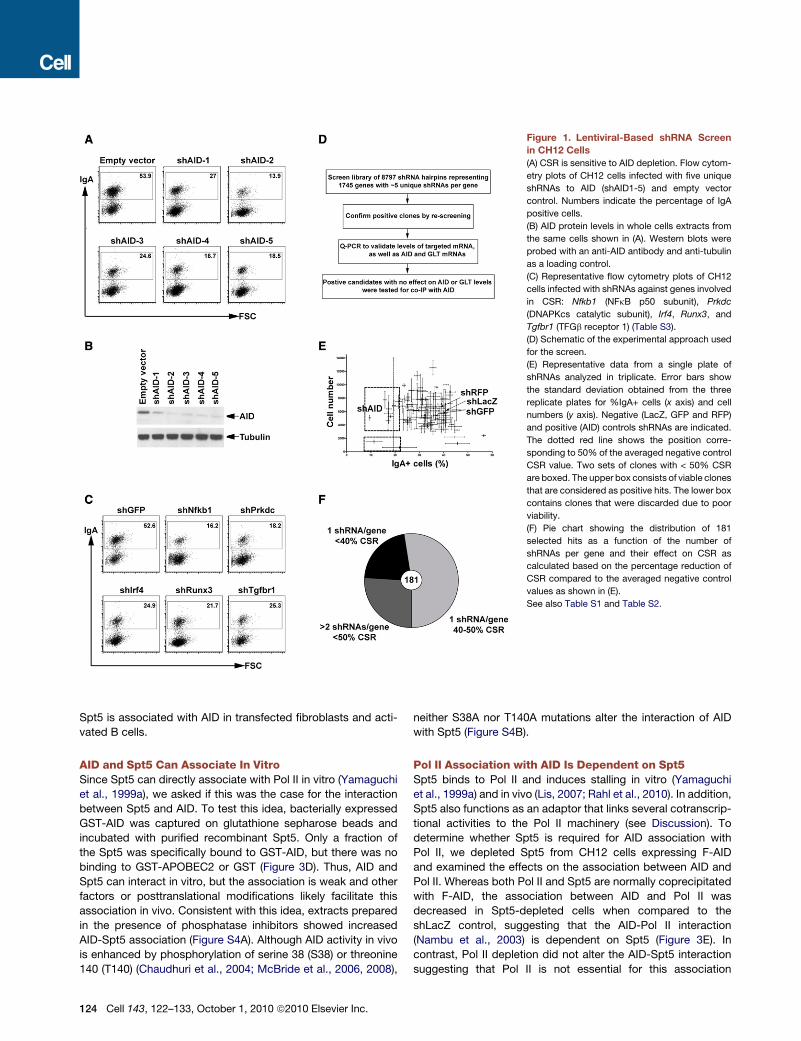
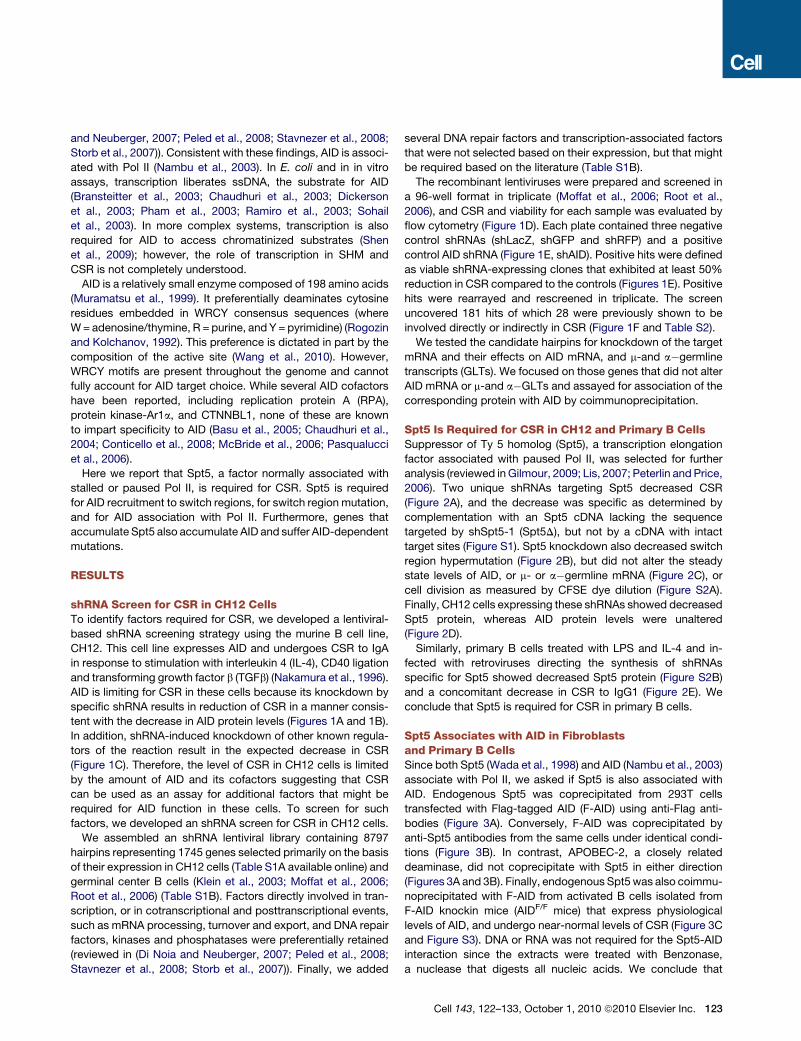
Figure 1. Lentiviral-Based shRNA Screen
in CH12 Cells
(A) CSR is sensitive to AID depletion. Flow cytom-
etry plots of CH12 cells infected with five unique
shRNAs to AID (shAID1-5) and empty vector
control. Numbers indicate the percentage of IgA
positive cells.
(B) AID protein levels in whole cells extracts from
the same cells shown in (A). Western blots were
probed with an anti-AID antibody and anti-tubulin
as a loading control.
(C) Representative flow cytometry plots of CH12
cells infected with shRNAs against genes involved
in CSR: Nfkb1 (NFkB p50 subunit), Prkdc
(DNAPKcs catalytic subunit), Irf4, Runx3, and
Tgfbr1 (TFGb receptor 1) (Table S3).
(D) Schematic of the experimental approach used
for the screen.
(E) Representative data from a single plate of
shRNAs analyzed in triplicate. Error bars show
the standard deviation obtained from the three
replicate plates for %IgA+ cells (x axis) and cell
numbers (y axis). Negative (LacZ, GFP and RFP)
and positive (AID) controls shRNAs are indicated.
The dotted red line shows the position corre-
sponding to 50% of the averaged negative control
CSR value. Two sets of clones with < 50% CSR
are boxed. The upper box consists of viable clones
that are considered as positive hits. The lower box
contains clones that were discarded due to poor
viability.
(F) Pie chart showing the distribution of 181
selected hits as a function of the number of
shRNAs per gene and their effect on CSR as
calculated based on the percentage reduction of
CSR compared to the averaged negative control
values as shown in (E).
See also Table S1 and Table S2.
Spt5 is associated with AID in transfected fibroblasts and acti-
vated B cells.
AID and Spt5 Can Associate In VitroSince Spt5 can directly associate with Pol II in vitro (Yamaguchi
et al., 1999a), we asked if this was the case for the interaction
between Spt5 and AID. To test this idea, bacterially expressed
GST-AID was captured on glutathione sepharose beads and
incubated with purified recombinant Spt5. Only a fraction of
the Spt5 was specifically bound to GST-AID, but there was no
binding to GST-APOBEC2 or GST (Figure 3D). Thus, AID and
Spt5 can interact in vitro, but the association is weak and other
factors or posttranslational modifications likely facilitate this
association in vivo. Consistent with this idea, extracts prepared
in the presence of phosphatase inhibitors showed increased
AID-Spt5 association (Figure S4A). Although AID activity in vivo
is enhanced by phosphorylation of serine 38 (S38) or threonine
140 (T140) (Chaudhuri et al., 2004; McBride et al., 2006, 2008),
124 Cell 143, 122–133, October 1, 2010 ª2010 Elsevier Inc.
neither S38A nor T140A mutations alter the interaction of AID
with Spt5 (Figure S4B).
Pol II Association with AID Is Dependent on Spt5Spt5 binds to Pol II and induces stalling in vitro (Yamaguchi
et al., 1999a) and in vivo (Lis, 2007; Rahl et al., 2010). In addition,
Spt5 also functions as an adaptor that links several cotranscrip-
tional activities to the Pol II machinery (see Discussion). To
determine whether Spt5 is required for AID association with
Pol II, we depleted Spt5 from CH12 cells expressing F-AID
and examined the effects on the association between AID and
Pol II. Whereas both Pol II and Spt5 are normally coprecipitated
with F-AID, the association between AID and Pol II was
decreased in Spt5-depleted cells when compared to the
shLacZ control, suggesting that the AID-Pol II interaction
(Nambu et al., 2003) is dependent on Spt5 (Figure 3E). In
contrast, Pol II depletion did not alter the AID-Spt5 interaction
suggesting that Pol II is not essential for this association
A B C
FSC
IgA48.4 16.7
21.9 12.2
shLacZ shSpt5-1
shSpt5-2 shAID
% Ig
A
0
20
30
40
10
50
D
shLacZ
shSpt5
-1
p = 0.02
0
1
2
3
4
5
Mu
tatio
n
freq
uen
cy (x10
-4)
Sμμ
shLacZ 26/73776
shSpt5-1 13/88418
p value 0.02
AID
Spt5
0
1
2
3
0.0
0.5
1.0
1.5
0.0
0.5
1.0
1.5
2.0
2.5
0.0
0.5
1.0
1.5
Igμ GLTs
Igα GLTs
1
1
0.94 0.38 0.32
0.2 0.96 0.9
E
14.1 10 23
2211.515.3
shSpt5-1 shSpt5-2 Vector
FSC
IgG1
% Ig
G1
shSpt5-1 shSpt5-2 Vector
p=0.002
p<0.0001
shLacZ
shAID
shSpt5
-1
shSpt5
-2
shLacZ
shAID
shSpt5
-1
shSpt5
-2
shLacZ
shAID
shSpt5
-1
shSpt5
-2
Spt5
AID
β-Actin
Spt5
AID
Figure 2. Spt5 Is Required for CSR and
Switch Region Mutation in CH12 Cells
(A) Upper panel shows representative flow cytom-
etry plots of CH12 infected with two unique
shRNAs to Spt5 (shSpt5-1 and shSpt5-2) and
controls (shLacZ and shAID) and stimulated to
undergo CSR. Numbers indicate percentage of
IgA positive cells. The graph in the lower panel
summarizes the data from four to six independent
experiments.
(B) Decreased switch region mutation after Spt5
knockdown in CH12 cells. Upper panel represents
the mutation frequency and corresponding p value
from control (shLacZ) and shSpt5-1 infected cells
stimulated to undergo CSR for 48 hr. The table in
the lower panel summarizes the mutation analysis
(represented as unique mutations/nucleotides
sequenced).
(C) Graphs show Q-PCR analysis for Spt5, AID, Iga
and Igm germline (GLT) mRNA levels in activated
CH12 cells infected as in (A) with the indicated
shRNAs. The data summarizes three independent
experiments with standard deviation indicated as
error bars. In all cases, shLacZ was assigned an
arbitrary value of 1.0.
(D) Western blot analysis of Spt5 and AID protein
levels in WCEs from activated CH12 cells infected
with the indicated shRNAs. Threefold serial dilu-
tions of WCEs were loaded. b-Actin was used as
a loading control. Numbers below the blots repre-
sent normalized band intensities for Spt5 and AID
with the shLacZ lanes assigned an arbitrary value
of 1.
(E) Spt5 is required for CSR in primary B cells.
Representative flow cytometry plots of B cells
stimulated with LPS + IL-4 and infected with retro-
viruses expressing shSpt5-1, shSpt5-2 or LMP
vector alone. Efficiency of switching was deter-
mined by gating on GFP-positive cells. Numbers
indicate percentage of IgG1 positive cells. The
graph in the lower panel summarizes the data
from three independent experiments.
See also Figure S1 and Figure S2.
(Figure S4C). We conclude that Spt5 serves as an adaptor that
recruits AID to Pol II.
AID Recruitment to Ig Switch RegionsIs Dependent on Spt5To determine whether AID recruitment to the Ig switch region is
dependent on Spt5, we performed quantitative PCR-based ChIP
analysis with two different anti-AID antibodies (Chaudhuri et al.,
2004; McBride et al., 2006). Spt5 depletion resulted in significant
reduction of AID occupancy in the switch region (Figure 3F,
p = 1.4 3 10�6). We conclude that Spt5 is required for AID
recruitment to the Ig switch region in B cells undergoing CSR.
Spt5 Is Associated with Stalled Pol II in B CellsAID mutates Ig genes and up to 25% of the expressed genes in
germinal center B cells (Liu et al., 2008; Pasqualucci et al., 1998,
2001; Shen et al., 1998). To determine whether Spt5 localization
in the genome of activated B cells coincides with AID-dependent
mutation, we performed genome-wide chromatin immunopre-
cipitation and sequencing (ChIP-seq) with antibodies against
Spt5 and Pol II. Spt5 was found throughout the genome of
activated B cells undergoing CSR (Figure 4 and Table S3A). As
in other cell types that have been assayed for Spt5 localization,
this protein was also concentrated at promoter regions coinci-
dent with Pol II peaks in activated B cells (Gilmour, 2009; Lis,
2007; Peterlin and Price, 2006; Rahl et al., 2010) (Figures 4A–4C).
Spt5 is a stalling factor in vitro (Wada et al., 1998; Yamaguchi
et al., 1999b) and associated with stalled Pol II in various cell
types in vivo (Rahl et al., 2010; Zeitlinger et al., 2007). The amount
ofPol II stalling canbequantitatedbycalculating a stallingor trav-
eling index (Is), which is a ratio of the Pol II density at promoter
regions compared to the gene body (Zeitlinger et al., 2007; see
Experimental Procedures). Genes with Is > 3 are considered
stalledwhereas thosewith Is<1are consideredelongating genes
(Zeitlinger et al., 2007). Stalling iswidespread in theBcell genome
(5594 genes, 61%, Table S3A), and in addition, the Pol II andSpt5
stalling indices were significantly correlated (Spearman’s corre-
lation coefficient, r = 0.8), consistent with previous observations
in other cell types (Nechaev et al., 2010; Rahl et al., 2010)
(Figure 4D, and A.Y. and R.C., unpublished data, accession
Cell 143, 122–133, October 1, 2010 ª2010 Elsevier Inc. 125

Input
shLa
cZ
Anti-Flag IPsh
Spt5
shLacZ shSpt5
Spt5
Pol II
E1 E2 E1 E2
IP
F-AID
Spt5
F-Apo2F-AID
Spt5
InputIP
anti-Blnk
pMX
IP anti-Spt5
A B C
E
Inpu
t
Elutions
GST
-AID
GST
-Apo
2
GST
GST
GST-Apo2GST-AID
Spt5
D
IgG anti-F
lag
Inpu
t
shLac
Z
shSpt5-
1
1.0
0.5
0
p = 1.4 x 10⁻⁶
Nor
mal
ized
A
ID C
hIP
F
F-AID
F-A
po2
F-A
ID
pMX
F-A
po2
F-A
ID
pMX
F-A
po2
F-A
ID
F-Apo2F-AID
Spt5
pMX
F-A
po2
F-A
IDpM
XF-
Apo
2F-
AID
InputIP
anti-Flag
Figure 3. Spt5 Interacts with AID in Fibroblasts and Primary B Cells
(A) Anti-Flag immunoprecipitates from whole cell extracts (WCEs) from 293T cells transfected with Flag-tagged AID (F-AID), or Flag-tagged Apobec2 (F-Apo2) or
pMX vector probed with anti-Flag or anti-Spt5 antibodies as indicated.
(B) Anti-Spt5 immunoprecipitates from WCEs from 293T cells transfected as in (A). Blots were probed as in (A). Anti-Blnk was used as an isotype control.
(C) Anti-Flag immunoprecipitates from WCEs from cultured splenic AIDF/F B cells. Blots were probed as in (A). E1 and E2 represent first and second elutions
with Flag peptide respectively.
(D) Bacterially expressed GST-AID, GST-APOBEC2 (GST-Apo2) or GST alone were bound to glutathione sepharose beads and incubated with purified recombi-
nant Spt5-Spt4 heterodimer (DSIF). Boundmaterial was eluted and analyzed by SDS-PAGE and blotted using antibodies against Spt5 andGST. The input lane for
DSIF represents 1% of the amount used in the reaction.
(E) Anti-Flag immunoprecipitates from WCEs of CH12 cells transfected with F-AID and depleted of Spt5 by shSpt5-1. shLacZ is used as a control. Blots were
probed as in (A) and with anti-Pol II.
(F) ChIP analysis for AID occupancy in Sm regions of CH12 cells infected with shSpt5-1 or shLacZ control. Data represents a total of 7 experiments using two
different anti-AID antibodies (Chaudhuri et al., 2004; McBride et al., 2006). For each experiment, shLacZ was assigned an arbitrary value of 1. The p value is
indicated.
See also Figure S3 and Figure S4.
number GSE24178). Most strikingly, AID occupancy in activated
B cells is also tightly correlatedwith Spt5 (see below andA.Y. and
R.C., unpublished data, accession number GSE24178).
TodeterminehowSpt5accumulation relates tomRNAlevels,we
compared the density of Spt5 sequence reads toB cellmRNA-seq
levels (both measured as reads per kbp per million sequences
(RPKM) (Figure 4E, and (Kuchen et al., 2010)). Although there
was some correlation between Spt5 and mRNA levels (Figure 4E,
r= 0.55), therewas a 1- to 2-log variation inmRNA levels for genes
accumulating similar levels of Spt5. Thus, in B cells, as in other
cells (Nechaev et al., 2010; Rahl et al., 2010), Spt5 (or Pol II)
accumulation is not necessarily equivalent to cellularmRNA levels.
Spt5 Genomic Occupancy Is Predictiveof AID-Dependent MutationUpon genome-wide analysis of Spt5 occupancy in the promoter
proximal region (�1–2 kb relative to the transcriptional start site
126 Cell 143, 122–133, October 1, 2010 ª2010 Elsevier Inc.
[TSS]), we found that Im bore the greatest tag count (Figure 5A
and Table S3B). The IgVH region could not be mapped because
each B cell has a unique rearrangement; however, a strong Spt5
signal was found from the IgH enhancer region through the
switch region (Figure 5A). Mir142, a robust AID target (Robbiani
et al., 2009), is also embedded in a region of high Spt5 accumu-
lation (Figure 5B and Table S3C). In contrast, Taci,Whsc1,H2Ea,
A20, Anxa4, andWdfy3, all of which are expressed in activated B
cells (Kuchen et al., 2010), but are not mutated (Liu et al., 2008;
Robbiani et al., 2009), do not accumulate Spt5 (Figure 5C and
Tables S3A and S3B).
To determine whether Spt5 accumulation is predictive of
mutations, we sequenced 10 genes that ranked within the
top 5% of genes analyzed for Spt5 tag density (Spt5hi),
measured as the density of sequence tags or reads per million
base-pairs (TPM), in the promoter-proximal region (Table S3B,
Figure 5B and Figure 6, and Kuchen et al., 2010). As controls,
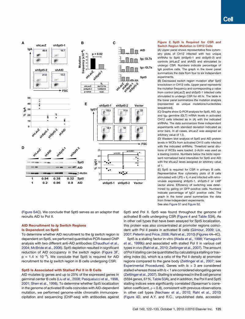
Figure 4. ChIP-Seq Analysis of Spt5 Genomic Occupancy
(A) Venn diagram showing overlap between genes recruiting Spt5 and Pol II using ChIP-Seq data from LPS+IL4 activated B cells (Table S4). There is a significant
association between the presence of Spt5 and Pol II at genes (Pearson’s Chi-square test; p < 0.0005).
(B) Correlation between Spt5 and Pol II density per gene. For each gene that recruited above-background amounts of Pol II and Spt5, the number of sequence
tags aligning between �1 Kb upstream of the transcriptional start site to its transcriptional termination site were normalized per gene length (in Kb), per million
aligned reads (reads per Kb per million, RPKM) and shown as a hexagonal binning plot. Spearman’s correlation coefficient (r) is indicated.
(C) Spt5 profile at all Spt5+ genes from�2 Kb to +5 Kb of the TSS. Data was normalized as reads per million per nucleotide. Dots represent densities at individual
nucleotides and the line a 10 nucleotide moving average.
(C) Correlation between the stalling index calculated based on Pol II or Spt5 occupancy (see Experimental Procedures). Spearman’s correlation coefficient (r) is
indicated.
(E) Comparative analysis of transcript levels (determined by mRNA-Seq, [Kuchen et al., 2010]) and Spt5 recruitment at all Spt5+ genes. Spearman’s correlation
coefficient (r) is indicated.
See also Table S3.
we sequenced 8 highly expressed genes (Kuchen et al., 2010)
that had a �4- to 6-fold lower Spt5 tag density (Spt5lo) in the
same region (Figure 5C and Figure 6 and Table S3B). For
each selected gene, a region starting around the TSS, corre-
sponding to the peak of Spt5, and extending �500–600 bp
downstream was sequenced (Figure 5, Figure 6, and Figure S5).
Because the rate of mutation at non-Ig genes is normally very
low unless repair is impaired (Liu et al., 2008; Pasqualucci
et al., 1998, 2001; Shen et al., 1998), we used B cells derived
from transgenic mice overexpressing AID from the Igk
promoter (IgkAID) (Robbiani et al., 2009). These mice display
elevated levels of AID protein with concomitant increases in
CSR and somatic mutation; nevertheless, they retain AID tar-
geting specificity (Robbiani et al., 2009). All 10 Spt5hi genes
(Table S3B) were mutated with frequencies from 4.6 3 10�4
for miR142 to 0.8 3 10�4 for H3f3b (Figure 6A and Fig-
ure S5). In contrast, none of the eight Spt5lo genes (Table S5)
were mutated above background levels (Figure 6A and
Figure S5).
To determine whether genes occupied by Spt5 correspond to
sites of AID recruitment, we compared Spt5 and AID ChIP-seq
data (Figure 6B and A.Y. and R.C., unpublished data, accession
number GSE24178). Strikingly, the tag density for AID per gene
(measured as readsper kilobase permillion [RPKM]) was uniformly
and directly proportional to the tag density of Spt5 (r = 0.75,
Figure 6B and A.Y. and R.C., unpublished data, accession number
GSE24178). To determine if AID recruitment to non-Ig genes was
dependent on Spt5, we performed ChIP for AID localization at the
Gas5 gene, a stalled gene (Table S3A) which accumulates AID-
mediatedmutation (Figure 6A). As shown in Figure 6C, AID recruit-
ment to Gas5 is impaired upon Spt5 depletion. We conclude that
Spt5 and AID accumulation coincide genome-wide and that high
density Spt5 occupancy is predictive of AID-mediated mutation.
DISCUSSION
Genetic and biochemical evidence indicate that AID initiates
SHM, CSR and chromosome translocation by deaminating
Cell 143, 122–133, October 1, 2010 ª2010 Elsevier Inc. 127

Figure 5. ChIP-seq Profiles of Spt5 on
Selected Genes
(A, B, and C) Pol II and Spt5 reads per million
plotted in 100 bpwindows across (A) the Igm locus,
(B) Spt5hi, and (C) Spt5lo genes. The axes scales
are identical for all histograms. Tag mappability
(shown below) was calculated based on the
percentage of 36 nt sequences that uniquely
aligned to the genomic site with a 10 bp window
resolution. Only windows with a significant enrich-
ment compared to a random background model
are shown. The location of the TSS for each
gene is indicated. The histograms cover the length
of the gene. Whsc1 and Tnfaip3 were previously
sequenced (Robbiani et al., 2009). All profiles
were generated using the UCSC genome browser.
See also Table S3.
cytidine residues in ssDNA that are exposed during transcription
(Chaudhuri and Alt, 2004; Di Noia and Neuberger, 2007; Nus-
senzweig and Nussenzweig, 2010; Peled et al., 2008; Stavnezer
et al., 2008). AID initiated processes are therefore limited by
regulators of transcription initiation such as PTIP, which facili-
tates Pol II access to specific switch regions by regulating their
H3K4 methylation (Daniel et al., 2010). However, active tran-
scription is not sufficient to allow AID access to DNA, and cannot
explain why AID-mediated lesions are found primarily in the
promoter proximal region of only some transcribed genes. Since
Pol II stalling is a feature of promoter-proximal regions, the
observation that Spt5, a stalling factor, associates with AID
and is required for AID localization to target genes, provides
a molecular explanation for the pattern of mutation.
Inducible transcription of genes carrying paused Pol II is an
important mechanism for regulating gene expression (Gilmour,
2009; Lis, 2007; Peterlin and Price, 2006; Bai et al., 2010; Core
128 Cell 143, 122–133, October 1, 2010 ª2010 Elsevier Inc.
et al., 2008; Guenther et al., 2007; Lefeb-
vre et al., 2002; Muse et al., 2007; Zeitlin-
ger et al., 2007; Bentley and Groudine,
1986; Krumm et al., 1992;Raschke et al.,
1999;Kao et al., 1987). Pausing is typi-
cally found downstream of promoters
and is associated with permanganate
sensitivity, which is indicative of the pres-
ence of ssDNA (Giardina et al., 1992).
Spt5 Is Required for Pol II StallingIn Vitro and In VivoSpt5 was originally identified as an elon-
gation factor in a yeast suppressor
screen (Swanson et al., 1991). It was
subsequently purified biochemically as
a heterodimeric complex with Spt4
called 5,6-dichloro-1-b-d-ribofuranosyl-
benzimidazole (DRB) sensitivity inducing
factor (DSIF) (Wada et al., 1998; Yamagu-
chi et al., 1999b). DSIF, in association
with negative elongation factor (NELF),
binds to Pol II and induces pausing
in vitro (Wada et al., 1998; Yamaguchi et al., 1999a). Genome-
wide ChIP studies have established a strong correlation between
Spt5 and Pol II stalling in vivo (Rahl et al., 2010). These and
related studies showed that the presence of Pol II in promoter
regions does not necessarily correlate with transcription (Bai
et al., 2010; Gilmour, 2009; Lefebvre et al., 2002; Lis, 2007;
Nechaev et al., 2010; Peterlin and Price, 2006; Rahl et al.,
2010). Consistent with these studies, we find only a partial corre-
lation between Spt5 or Pol II occupancy and mRNA levels in
activated B cells (Figure 4E), and importantly, that shRNA knock-
down of Spt5 did not decrease AID mRNA, or Igm or Iga sterile
transcripts (Figures 2C and 2D).
Current models suggest that the stalled Pol II complex is reac-
tivated by inductive signals that recruit the P-TEFb kinase, which
phosphorylates Pol II and Spt5, thereby releasing NELF from the
complex and activating transcription (Kim and Sharp, 2001;
Marshall et al., 1996; Marshall and Price, 1995; Wada et al.,
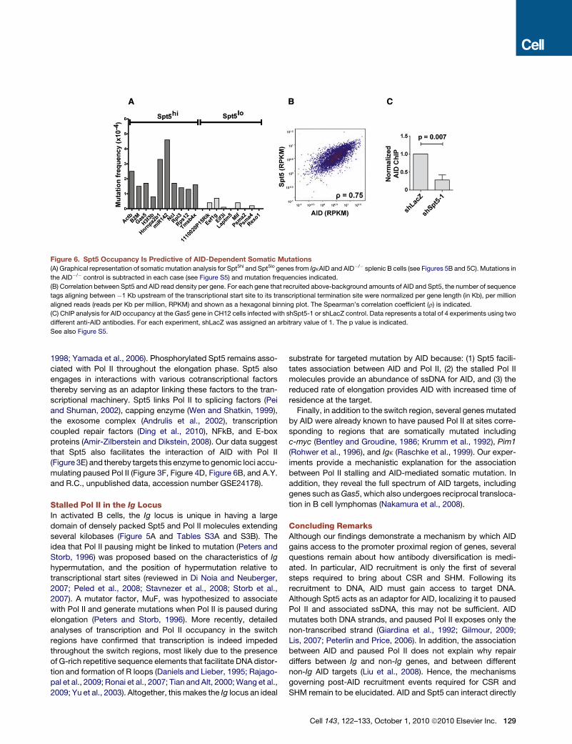
Figure 6. Spt5 Occupancy Is Predictive of AID-Dependent Somatic Mutations
(A) Graphical representation of somatic mutation analysis for Spt5hi and Spt5lo genes from IgkAID and AID�/� splenic B cells (see Figures 5B and 5C). Mutations in
the AID�/� control is subtracted in each case (see Figure S5) and mutation frequencies indicated.
(B) Correlation between Spt5 and AID read density per gene. For each gene that recruited above-background amounts of AID and Spt5, the number of sequence
tags aligning between �1 Kb upstream of the transcriptional start site to its transcriptional termination site were normalized per gene length (in Kb), per million
aligned reads (reads per Kb per million, RPKM) and shown as a hexagonal binning plot. The Spearman’s correlation coefficient (r) is indicated.
(C) ChIP analysis for AID occupancy at the Gas5 gene in CH12 cells infected with shSpt5-1 or shLacZ control. Data represents a total of 4 experiments using two
different anti-AID antibodies. For each experiment, shLacZ was assigned an arbitrary value of 1. The p value is indicated.
See also Figure S5.
1998; Yamada et al., 2006). Phosphorylated Spt5 remains asso-
ciated with Pol II throughout the elongation phase. Spt5 also
engages in interactions with various cotranscriptional factors
thereby serving as an adaptor linking these factors to the tran-
scriptional machinery. Spt5 links Pol II to splicing factors (Pei
and Shuman, 2002), capping enzyme (Wen and Shatkin, 1999),
the exosome complex (Andrulis et al., 2002), transcription
coupled repair factors (Ding et al., 2010), NFkB, and E-box
proteins (Amir-Zilberstein and Dikstein, 2008). Our data suggest
that Spt5 also facilitates the interaction of AID with Pol II
(Figure 3E) and thereby targets this enzyme to genomic loci accu-
mulating paused Pol II (Figure 3F, Figure 4D, Figure 6B, and A.Y.
and R.C., unpublished data, accession number GSE24178).
Stalled Pol II in the Ig LocusIn activated B cells, the Ig locus is unique in having a large
domain of densely packed Spt5 and Pol II molecules extending
several kilobases (Figure 5A and Tables S3A and S3B). The
idea that Pol II pausing might be linked to mutation (Peters and
Storb, 1996) was proposed based on the characteristics of Ig
hypermutation, and the position of hypermutation relative to
transcriptional start sites (reviewed in Di Noia and Neuberger,
2007; Peled et al., 2008; Stavnezer et al., 2008; Storb et al.,
2007). A mutator factor, MuF, was hypothesized to associate
with Pol II and generate mutations when Pol II is paused during
elongation (Peters and Storb, 1996). More recently, detailed
analyses of transcription and Pol II occupancy in the switch
regions have confirmed that transcription is indeed impeded
throughout the switch regions, most likely due to the presence
of G-rich repetitive sequence elements that facilitate DNA distor-
tion and formation of R loops (Daniels and Lieber, 1995; Rajago-
pal et al., 2009; Ronai et al., 2007; Tian and Alt, 2000;Wang et al.,
2009; Yu et al., 2003). Altogether, this makes the Ig locus an ideal
substrate for targeted mutation by AID because: (1) Spt5 facili-
tates association between AID and Pol II, (2) the stalled Pol II
molecules provide an abundance of ssDNA for AID, and (3) the
reduced rate of elongation provides AID with increased time of
residence at the target.
Finally, in addition to the switch region, several genes mutated
by AID were already known to have paused Pol II at sites corre-
sponding to regions that are somatically mutated including
c-myc (Bentley and Groudine, 1986; Krumm et al., 1992), Pim1
(Rohwer et al., 1996), and Igk (Raschke et al., 1999). Our exper-
iments provide a mechanistic explanation for the association
between Pol II stalling and AID-mediated somatic mutation. In
addition, they reveal the full spectrum of AID targets, including
genes such asGas5, which also undergoes reciprocal transloca-
tion in B cell lymphomas (Nakamura et al., 2008).
Concluding RemarksAlthough our findings demonstrate a mechanism by which AID
gains access to the promoter proximal region of genes, several
questions remain about how antibody diversification is medi-
ated. In particular, AID recruitment is only the first of several
steps required to bring about CSR and SHM. Following its
recruitment to DNA, AID must gain access to target DNA.
Although Spt5 acts as an adaptor for AID, localizing it to paused
Pol II and associated ssDNA, this may not be sufficient. AID
mutates both DNA strands, and paused Pol II exposes only the
non-transcribed strand (Giardina et al., 1992; Gilmour, 2009;
Lis, 2007; Peterlin and Price, 2006). In addition, the association
between AID and paused Pol II does not explain why repair
differs between Ig and non-Ig genes, and between different
non-Ig AID targets (Liu et al., 2008). Hence, the mechanisms
governing post-AID recruitment events required for CSR and
SHM remain to be elucidated. AID and Spt5 can interact directly
Cell 143, 122–133, October 1, 2010 ª2010 Elsevier Inc. 129

in vitro but the interaction is weak suggesting that additional
factors or posttranslational modifications may be required.
Nevertheless, our data suggests that Spt5 links Pol II and AID,
thereby providing a mechanistic explanation for the well-estab-
lished correlation between AID and transcription. The associa-
tion between Spt5 and AID also explains intrinsic features of
hypermutation and CSR, including the enrichment of mutation
in the promoter-proximal regions, which correspond to sites of
Pol II stalling (Nechaev et al., 2010; Rahl et al., 2010; Zeitlinger
et al., 2007).
In conclusion, we propose that AID utilizes the phenomenon of
Pol II stalling, which is widespread in the B cell genome, and is
particularly prominent on Ig loci, to gain access to its target
genes across the genome.
EXPERIMENTAL PROCEDURES
Library Preparation
The lentiviral shRNA library (Table S1B) was prepared, titered, arrayed and
validated as described (Moffat et al., 2006; Root et al., 2006; http://www.
broadinstitute.org/rnai/trc/lib).
Library Screening
The lentiviral library was screened in a 96-well plate format in triplicate, starting
from the infection stage through to flow cytometry analysis (schematically
represented in Figure 1C). Each plate contained negative control viruses
targeting LacZ, GFP and RFP and a positive control shRNA targeting AID. Cells
were infected, selected, and stimulated to undergo CSR followed by FACS
analysis (details in Supplemental Information).
shRNA Knockdown in Primary B Cells
The hairpin sequences for shSpt5-1 and shSpt5-2 (Table S1B) were cloned
into the LMP retroviral vector (Open Biosystems) and transfected into
BOSC23 cells to produce retrovirus (Robbiani et al., 2008). Primary B cells
stimulated with LPS and IL-4 were cultured as described (Robbiani et al.,
2008). After 24 hr in culture, B cells were infected with shRNA-expressing
retroviral supernatants as described (Robbiani et al., 2008) and cultured for
an additional 3 days with LPS and IL-4, followed by FACS analysis for IgG1
and Spt5 protein analysis by western blotting.
Immunoprecipitation
For Flag-IPs, 2 mg of WCE (prepared as described in Supplemental Informa-
tion) was incubated with 20 ml Flag Agarose resin (Sigma) for 2 hr at 4�C in
IP buffer (identical to WCE preparation buffers above adjusted to 150 mM
NaCl for fibroblasts assays, and 200 mM NaCl for B cells and CH12 assays).
This was followed by three washes in IP buffer and elution with 0.2 mg/ml
Flag peptide (Sigma) for 1 hr at 4�C. Eluates were subjected to SDS-PAGE
and western blot analysis. For anti-Spt5 IPs, 2 mgs of WCE were incubated
with 3 mg of anti-Spt5 (Santa Cruz Biotechnology) for 2 hr at 4�C followed by
capture of the immune complexes with 20 ml Protein A agarose (Roche) for
1 hr at 4�C. Beads were washed three times with IP buffer and bound material
was extracted by boiling in 100 ml of Laemmli sample buffer. Eluted material
was analyzed by SDS-PAGE and western blot. Antibodies used for probing
western blots were as follows: Flag (Sigma), Spt5 (H300) (SantaCruz Biotech-
nology), Pol II (4H8) (Abcam) and Phospho-Ser PKC Substrate (Cell Signaling).
AID-Spt5 Interaction In Vitro
GST fusion proteins were expressed in E. coli and immobilized on Glutathione
Sepharose 4 Fast Flow beads (GE Healthcare). Beads were incubated with
500 ng of purified DSIF (Spt5-Spt4) complex (a generous gift from Dr. Sohail
Malik, The Rockefeller University) in 200 ml final volume of binding buffer
(20 mM Tris [pH 7.5], 150 mMNaCl, 0.1%NP-40, 1 mM EDTA, Protease Inhib-
itor cocktail (Roche), 0.5 mM PMSF, 1 mMDTT, 0.5 mg/ml BSA) for 2 hr at 4�Cwith gentle rotation. After four washeswith binding buffer, bound proteins were
130 Cell 143, 122–133, October 1, 2010 ª2010 Elsevier Inc.
eluted by boiling in NuPAGE LDS loading buffer (Invitrogen). Samples were
then subjected to SDS-PAGE followed by western blot analysis.
Chromatin Immunoprecipitation and Sequencing
ChIP-seq was performed exactly as described (Kuchen et al., 2010). In brief,
cells were fixed with 1% paraformaldehyde at 37�C for 10 min followed by
sonication. Chromatin fragments were then immunoprecipitated with anti-
bodies specific for Spt5 (Santa Cruz Biotechnology [H300] and BD Biosci-
ences [anti-DSIF]), RNA Pol II (Abcam, [4H8]) or Ser5-phosphorylated RNA
Pol II (Abcam, [phospho-S5]). Immunoprecipitates were processed following
Illumina’s protocol and sequenced on a Genome Analyzer. During analysis,
short sequence tags were trimmed to 32 nts and aligned to themouse genome
(NCBI37/mm9) using Bowtie. Uniquely aligned reads were analyzed by SICER
(Zang et al., 2009) using an expectation value E of 0.05 in a random back-
ground model. The requirement for unique alignment was not applied for
IgSm or IgSg1 because of their high repetitive nature and low mappability
(Figure 5A). Reads on significant islands as defined by SICER were normalized
to the total number of reads on islands. Downstream analysis was carried out
in R and Python.
Quantitative AID ChIP
CH12 cells were infected with shRNAs to Spt5 as above and subjected to ChIP
analysis using two different anti-AID antibodies (Chaudhuri et al., 2004;
McBride et al., 2006). Assays were performed as described (Vuong et al.,
2009). The ChIP’d material was analyzed by Q-PCR and raw values were
normalized to the input signals for each sample (Vuong et al., 2009). Reactions
were performed in triplicate. Forward and reverse primers used for Sm ampli-
fication were 50 TAGTAAGCGAGGCTCTAAAAAGCAT 30 and 50 AGAACAGT
CCAGTGTAGGCAGTAGA 3‘ respectively. Forward and reverse primers
used for Gas5 amplification were 5‘ TATGGCTTCGGGCCTTGGA 3‘ and 5‘
CCTCCTAAAGTTTCCAGCTTGTGC 3‘ respectively.
Calculation of the Stalling Index
The stalling index was calculated based on Pol II ChIP-seq reads as described
(Rahl et al., 2010; Zeitlinger et al., 2007). Briefly, the Pol II and Spt5 stalling
indices are calculated in the same way and represent the ratio of read density
at the promoter to the average gene body density. The promoter was defined
as a 1 kb region extending from�0.5 kb to +0.5 kb relative to the TSS, and the
gene body was defined as the region from +1kb downstream of the TSS up to
the transcription termination site (TTS) (Rahl et al., 2010; Zeitlinger et al., 2007).
Additional experimental procedures can be found in the Supplemental
Information.
ACCESSION NUMBERS
The ChIP-seq data for Spt5, Pol II and AID are deposited in GEO under acces-
sion number GSE24178.
SUPPLEMENTAL INFORMATION
Supplemental Information includes Extended Experimental Procedures, five
figures, and four tables and can be found with this article online at doi:10.
1016/j.cell.2010.09.017.
ACKNOWLEDGMENTS
We thank members of the Nussenzweig lab for helpful discussions, Klara Ve-
linzon for FACS sorting, and Tom Eisenreich and David Bosque for animal
management. We thank Drs Jayanta Chaudhuri and Urszula Nowak for ChIP
protocols and anti-AID antibody, Dr. Sohail Malik for generously providing
purified recombinant DSIF and Dr. Alan Derr for assistance with informatics.
M.D.V. is a fellow of the American-Italian Cancer Foundation. R.P was a recip-
ient of The Irvington Institute Postdoctoral Fellowship of the Cancer Research
Institute. The work was supported by NIH grant (AI037526) toM.C.N. M.C.N. is
a Howard Hughes Medical Institute Investigator.
Received: May 22, 2010
Revised: August 2, 2010
Accepted: September 13, 2010
Published: September 30, 2010
REFERENCES
Amir-Zilberstein, L., and Dikstein, R. (2008). Interplay between E-box and
NF-kappaB in regulation of A20 gene by DRB sensitivity-inducing factor
(DSIF). J. Biol. Chem. 283, 1317–1323.
Andrulis, E.D., Werner, J., Nazarian, A., Erdjument-Bromage, H., Tempst, P.,
and Lis, J.T. (2002). The RNA processing exosome is linked to elongating
RNA polymerase II in Drosophila. Nature 420, 837–841.
Bai, X., Kim, J., Yang, Z., Jurynec, M., Akie, T., Lee, J., LeBlanc, J., Sessa, A.,
Jiang, H., DiBiase, A., et al. (2010). TIF1g Controls Erythroid Cell Fate by
Regulating Transcription Elongation. Cell 142, 133–143.
Basu, U., Chaudhuri, J., Alpert, C., Dutt, S., Ranganath, S., Li, G., Schrum, J.P.,
Manis, J.P., and Alt, F.W. (2005). The AID antibody diversification enzyme is
regulated by protein kinase A phosphorylation. Nature 438, 508–511.
Bentley, D.L., and Groudine, M. (1986). A block to elongation is largely respon-
sible for decreased transcription of c-myc in differentiated HL60 cells. Nature
321, 702–706.
Bhutani, N., Brady, J.J., Damian, M., Sacco, A., Corbel, S.Y., and Blau, H.M.
(2010). Reprogramming towards pluripotency requires AID-dependent DNA
demethylation. Nature 463, 1042–1047.
Bransteitter, R., Pham, P., Scharff, M.D., and Goodman, M.F. (2003).
Activation-induced cytidine deaminase deaminates deoxycytidine on single-
stranded DNA but requires the action of RNase. Proc. Natl. Acad. Sci. USA
100, 4102–4107.
Chaudhuri, J., and Alt, F.W. (2004). Class-switch recombination: interplay of
transcription, DNA deamination and DNA repair. Nat. Rev. Immunol. 4,
541–552.
Chaudhuri, J., Khuong, C., and Alt, F.W. (2004). Replication protein A interacts
with AID to promote deamination of somatic hypermutation targets. Nature
430, 992–998.
Chaudhuri, J., Tian, M., Khuong, C., Chua, K., Pinaud, E., and Alt, F.W. (2003).
Transcription-targeted DNA deamination by the AID antibody diversification
enzyme. Nature 422, 726–730.
Conticello, S.G., Ganesh, K., Xue, K., Lu, M., Rada, C., and Neuberger, M.S.
(2008). Interaction between antibody-diversification enzyme AID and spliceo-
some-associated factor CTNNBL1. Mol. Cell 31, 474–484.
Core, L.J., Waterfall, J.J., and Lis, J.T. (2008). Nascent RNA sequencing
reveals widespread pausing and divergent initiation at human promoters.
Science 322, 1845–1848.
Daniel, J.A., Santos, M.A., Wang, Z., Zang, C., Schwab, K.R., Jankovic, M.,
Filsuf, D., Chen, H., Gazumyan, A., Yamane, A., et al. (2010). PTIP Promotes
Chromatin Changes Critical for Immunoglobulin Class Switch Recombination.
Science 329, 917–923.
Daniels, G.A., and Lieber, M.R. (1995). RNA:DNA complex formation upon
transcription of immunoglobulin switch regions: implications for the mecha-
nism and regulation of class switch recombination. Nucleic Acids Res. 23,
5006–5011.
Di Noia, J.M., and Neuberger, M.S. (2007). Molecular mechanisms of antibody
somatic hypermutation. Annu. Rev. Biochem. 76, 1–22.
Dickerson, S.K., Market, E., Besmer, E., and Papavasiliou, F.N. (2003). AID
mediates hypermutation by deaminating single stranded DNA. J. Exp. Med.
197, 1291–1296.
Ding, B., LeJeune, D., and Li, S. (2010). The C-terminal repeat domain of Spt5
plays an important role in suppression of Rad26-independent transcription
coupled repair. J. Biol. Chem. 285, 5317–5326.
Giardina, C., Perez-Riba, M., and Lis, J.T. (1992). Promoter melting and TFIID
complexes on Drosophila genes in vivo. Genes Dev. 6, 2190–2200.
Gilmour, D.S. (2009). Promoter proximal pausing on genes in metazoans.
Chromosoma 118, 1–10.
Guenther, M.G., Levine, S.S., Boyer, L.A., Jaenisch, R., and Young, R.A.
(2007). A chromatin landmark and transcription initiation at most promoters
in human cells. Cell 130, 77–88.
Kao, S.Y., Calman, A.F., Luciw, P.A., and Peterlin, B.M. (1987). Anti-termina-
tion of transcription within the long terminal repeat of HIV-1 by tat gene
product. Nature 330, 489–493.
Kim, J.B., and Sharp, P.A. (2001). Positive transcription elongation factor B
phosphorylates hSPT5 and RNA polymerase II carboxyl-terminal domain inde-
pendently of cyclin-dependent kinase-activating kinase. J. Biol. Chem. 276,
12317–12323.
Klein, U., Tu, Y., Stolovitzky, G.A., Keller, J.L., Haddad, J., Jr., Miljkovic, V.,
Cattoretti, G., Califano, A., and Dalla-Favera, R. (2003). Transcriptional
analysis of the B cell germinal center reaction. Proc. Natl. Acad. Sci. USA
100, 2639–2644.
Krumm, A., Meulia, T., Brunvand, M., and Groudine, M. (1992). The block to
transcriptional elongation within the human c-myc gene is determined in the
promoter-proximal region. Genes Dev. 6, 2201–2213.
Kuchen, S., Resch, W., Yamane, A., Kuo, N., Li, Z., Chakraborty, T., Wei, L.,
Laurrence, A., Yasuda, T., Peng, S., et al. (2010). Regulation of MicroRNA
Expression and Abundance during Lymphopoesis. Immunity 32, 828–839.
Lefebvre, B., Brand, C., Lefebvre, B., and Ozato, K. (2002). Chromosomal
Integration of Retinoic Acid Response Elements Prevents Cooperative Tran-
scriptional Activation by Retinoic Acid Receptor and Retinoid X Receptor.
Mol. Cell. Biol. 22, 1446–1459.
Lin, C., Yang, L., Tanasa, B., Hutt, K., Ju, B.G., Ohgi, K., Zhang, J., Rose, D.W.,
Fu, X.D., Glass, C.K., et al. (2009). Nuclear receptor-induced chromosomal
proximity and DNA breaks underlie specific translocations in cancer. Cell
139, 1069–1083.
Lis, J.T. (2007). Imaging Drosophila gene activation and polymerase pausing
in vivo. Nature 450, 198–202.
Liu, M., Duke, J.L., Richter, D.J., Vinuesa, C.G., Goodnow, C.C., Kleinstein,
S.H., and Schatz, D.G. (2008). Two levels of protection for the B cell genome
during somatic hypermutation. Nature 451, 841–845.
Marshall, N.F., Peng, J., Xie, Z., and Price, D.H. (1996). Control of RNA poly-
merase II elongation potential by a novel carboxyl-terminal domain kinase.
J. Biol. Chem. 271, 27176–27183.
Marshall, N.F., and Price, D.H. (1995). Purification of P-TEFb, a transcription
factor required for the transition into productive elongation. J. Biol. Chem.
270, 12335–12338.
Matsumoto, Y., Marusawa, H., Kinoshita, K., Endo, Y., Kou, T., Morisawa, T.,
Azuma, T., Okazaki, I.M., Honjo, T., and Chiba, T. (2007). Helicobacter pylori
infection triggers aberrant expression of activation-induced cytidine deami-
nase in gastric epithelium. Nat. Med. 13, 470–476.
McBride, K.M., Gazumyan, A., Woo, E.M., Barreto, V.M., Robbiani, D.F., Chait,
B.T., and Nussenzweig, M.C. (2006). Regulation of hypermutation by activa-
tion-induced cytidine deaminase phosphorylation. Proc. Natl. Acad. Sci.
USA 103, 8798–8803.
McBride, K., Gazumyan, A., Woo, E., Schwickert, T., Chait, B., and
Nussenzweig, M. (2008). Regulation of class switch recombination and
somatic mutation by AID phosphorylation. J. Exp. Med. 205, 2585–2594.
Moffat, J., Grueneberg, D.A., Yang, X., Kim, S.Y., Kloepfer, A.M., Hinkle, G.,
Piqani, B., Eisenhaure, T.M., Luo, B., Grenier, J.K., et al. (2006). A lentiviral
RNAi library for human and mouse genes applied to an arrayed viral high-
content screen. Cell 124, 1283–1298.
Morgan, H.D., Dean, W., Coker, H.A., Reik, W., and Petersen-Mahrt, S.K.
(2004). Activation-induced cytidine deaminase deaminates 5-methylcytosine
in DNA and is expressed in pluripotent tissues: implications for epigenetic
reprogramming. J. Biol. Chem. 279, 52353–52360.
Muramatsu, M., Kinoshita, K., Fagarasan, S., Yamada, S., Shinkai, Y., and
Honjo, T. (2000). Class switch recombination and hypermutation require
Cell 143, 122–133, October 1, 2010 ª2010 Elsevier Inc. 131
activation-induced cytidine deaminase (AID), a potential RNA editing enzyme.
Cell 102, 553–563.
Muramatsu, M., Sankaranand, V.S., Anant, S., Sugai, M., Kinoshita, K.,
Davidson, N.O., and Honjo, T. (1999). Specific expression of activation-
induced cytidine deaminase (AID), a novel member of the RNA-editing deam-
inase family in germinal center B cells. J. Biol. Chem. 274, 18470–18476.
Muse, G.W., Gilchrist, D.A., Nechaev, S., Shah, R., Parker, J.S., Grissom, S.F.,
Zeitlinger, J., and Adelman, K. (2007). RNA polymerase is poised for activation
across the genome. Nat. Genet. 39, 1507–1511.
Nakamura, M., Kondo, S., Sugai, M., Nazarea, M., Imamura, S., and Honjo, T.
(1996). High frequency class switching of an IgM+ B lymphoma clone CH12F3
to IgA+ cells. Int. Immunol. 8, 193–201.
Nakamura, Y., Takahashi, N., Kakegawa, E., Yoshida, K., Ito, Y., Kayano, H.,
Niitsu, N., Jinnai, I., and Bessho, M. (2008). The GAS5 (growth arrest-specific
transcript 5) gene fuses to BCL6 as a result of t(1;3)(q25;q27) in a patient with
B-cell lymphoma. Cancer Genet. Cytogenet. 182, 144–149.
Nambu, Y., Sugai, M., Gonda, H., Lee, C.G., Katakai, T., Agata, Y., Yokota, Y.,
and Shimizu, A. (2003). Transcription-coupled events associating with immu-
noglobulin switch region chromatin. Science 302, 2137–2140.
Nechaev, S., Fargo, D., Santos, G., Liu, L., Gao, Y., and Adelman, K. (2010).
Global Analysis of Short RNAs Reveals Widespread Promoter-Proximal
Stalling and Arrest of Pol II in Drosophila. Science 327, 335–338.
Nussenzweig, A., and Nussenzweig, M.C. (2010). Origin of chromosomal
translocations in lymphoid cancer. Cell 141, 27–38.
Pasqualucci, L., Kitaura, Y., Gu, H., and Dalla-Favera, R. (2006). PKA-medi-
ated phosphorylation regulates the function of activation-induced deaminase
(AID) in B cells. Proc. Natl. Acad. Sci. USA 103, 395–400.
Pasqualucci, L., Migliazza, A., Fracchiolla, N., William, C., Neri, A., Baldini, L.,
Chaganti, R.S., Klein, U., Kuppers, R., Rajewsky, K., et al. (1998). BCL-6 muta-
tions in normal germinal center B cells: evidence of somatic hypermutation
acting outside Ig loci. Proc. Natl. Acad. Sci. USA 95, 11816–11821.
Pasqualucci, L., Neumeister, P., Goossens, T., Nanjangud, G., Chaganti, R.S.,
Kuppers, R., and Dalla-Favera, R. (2001). Hypermutation of multiple proto-
oncogenes in B-cell diffuse large-cell lymphomas. Nature 412, 341–346.
Pei, Y., and Shuman, S. (2002). Interactions between fission yeast mRNA
capping enzymes and elongation factor Spt5. J. Biol. Chem. 277, 19639–
19648.
Peled, J.U., Kuang, F.L., Iglesias-Ussel, M.D., Roa, S., Kalis, S.L., Goodman,
M.F., and Scharff, M.D. (2008). The Biochemistry of Somatic Hypermutation.
Annu. Rev. Immunol. 26, 481–511.
Peterlin, B.M., and Price, D.H. (2006). Controlling the elongation phase of tran-
scription with P-TEFb. Mol. Cell 23, 297–305.
Peters, A., and Storb, U. (1996). Somatic hypermutation of immunoglobulin
genes is linked to transcription initiation. Immunity 4, 57–65.
Pham, P., Bransteitter, R., Petruska, J., and Goodman, M.F. (2003).
Processive AID-catalysed cytosine deamination on single-stranded DNA
simulates somatic hypermutation. Nature 424, 103–107.
Popp, C., Dean, W., Feng, S., Cokus, S.J., Andrews, S., Pellegrini, M.,
Jacobsen, S.E., and Reik, W. (2010). Genome-wide erasure of DNA methyla-
tion in mouse primordial germ cells is affected by AID deficiency. Nature
463, 1101–1105.
Rahl, P.B., Lin, C.Y., Seila, A.C., Flynn, R.A., McCuine, S., Burge, C.B., Sharp,
P.A., and Young, R.A. (2010). c-Myc Regulates Transcriptional Pause Release.
Cell 141, 432–445.
Rai, K., Huggins, I.J., James, S.R., Karpf, A.R., Jones, D.A., and Cairns, B.R.
(2008). DNA demethylation in zebrafish involves the coupling of a deaminase,
a glycosylase, and gadd45. Cell 135, 1201–1212.
Rajagopal, D., Maul, R.W., Ghosh, A., Chakraborty, T., Khamlichi, A.A., Sen,
R., and Gearhart, P.J. (2009). Immunoglobulin switch mu sequence causes
RNA polymerase II accumulation and reduces dA hypermutation. J. Exp.
Med. 206, 1237–1244.
132 Cell 143, 122–133, October 1, 2010 ª2010 Elsevier Inc.
Ramiro, A.R., Jankovic, M., Callen, E., Difilippantonio, S., Chen, H.T., McBride,
K.M., Eisenreich, T.R., Chen, J., Dickins, R.A., Lowe, S.W., et al. (2006). Role of
genomic instability and p53 in AID-induced c-myc-Igh translocations. Nature
440, 105–109.
Ramiro, A.R., Jankovic, M., Eisenreich, T., Difilippantonio, S., Chen-Kiang, S.,
Muramatsu, M., Honjo, T., Nussenzweig, A., and Nussenzweig, M.C. (2004).
AID is required for c-myc/IgH chromosome translocations in vivo. Cell 118,
431–438.
Ramiro, A.R., Stavropoulos, P., Jankovic, M., and Nussenzweig, M.C. (2003).
Transcription enhances AID-mediated cytidine deamination by exposing
single-stranded DNA on the nontemplate strand. Nat. Immunol. 4, 452–456.
Raschke, E.E., Albert, T., and Eick, D. (1999). Transcriptional regulation of the
Ig kappa gene by promoter-proximal pausing of RNA polymerase II. J. Immu-
nol. 163, 4375–4382.
Revy, P., Muto, T., Levy, Y., Geissmann, F., Plebani, A., Sanal, O., Catalan, N.,
Forveille, M., Dufourcq-Labelouse, R., Gennery, A., et al. (2000). Activation-
induced cytidine deaminase (AID) deficiency causes the autosomal recessive
form of the Hyper-IgM syndrome (HIGM2). Cell 102, 565–575.
Robbiani, D.F., Bothmer, A., Callen, E., Reina-San-Martin, B., Dorsett, Y.,
Difilippantonio, S., Bolland, D.J., Chen, H.T., Corcoran, A.E., Nussenzweig,
A., et al. (2008). AID is required for the chromosomal breaks in c-myc that
lead to c-myc/IgH translocations. Cell 135, 1028–1038.
Robbiani, D.F., Bunting, S., Feldhahn, N., Bothmer, A., Camps, J., Deroubaix,
S., McBride, K.M., Klein, I.A., Stone, G., Eisenreich, T.R., et al. (2009). AID
produces DNA double-strand breaks in non-Ig genes and mature B cell
lymphomas with reciprocal chromosome translocations. Mol. Cell 36,
631–641.
Rogozin, I.B., and Kolchanov, N.A. (1992). Somatic hypermutagenesis in
immunoglobulin genes. II. Influence of neighbouring base sequences onmuta-
genesis. Biochim. Biophys. Acta 1171, 11–18.
Rohwer, F., Todd, S., andMcGuire, K.L. (1996). The effect of IL-2 treatment on
transcriptional attenuation in proto-oncogenes pim-1 and c-myb in human
thymic blast cells. J. Immunol. 157, 643–649.
Ronai, D., Iglesias-Ussel, M.D., Fan, M., Li, Z., Martin, A., and Scharff, M.D.
(2007). Detection of chromatin-associated single-stranded DNA in regions
targeted for somatic hypermutation. J. Exp. Med. 204, 181–190.
Root, D.E., Hacohen, N., Hahn, W.C., Lander, E.S., and Sabatini, D.M. (2006).
Genome-scale loss-of-function screening with a lentiviral RNAi library. Nat.
Methods 3, 715–719.
Shen, H.M., Peters, A., Baron, B., Zhu, X., and Storb, U. (1998). Mutation of
BCL-6 gene in normal B cells by the process of somatic hypermutation of Ig
genes. Science 280, 1750–1752.
Shen, H.M., Poirier, M.G., Allen, M.J., North, J., Lal, R., Widom, J., and Storb,
U. (2009). The activation-induced cytidine deaminase (AID) efficiently targets
DNA in nucleosomes but only during transcription. J. Exp. Med. 206,
1057–1071.
Sohail, A., Klapacz, J., Samaranayake, M., Ullah, A., and Bhagwat, A.S. (2003).
Human activation-induced cytidine deaminase causes transcription-depen-
dent, strand-biased C to U deaminations. Nucleic Acids Res. 31, 2990–2994.
Stavnezer-Nordgren, J., and Sirlin, S. (1986). Specificity of immunoglobulin
heavy chain switch correlates with activity of germline heavy chain genes prior
to switching. EMBO J. 5, 95–102.
Stavnezer, J., Guikema, J.E., and Schrader, C.E. (2008). Mechanism and
regulation of class switch recombination. Annu. Rev. Immunol. 26, 261–292.
Storb, U., Shen, H.M., Longerich, S., Ratnam, S., Tanaka, A., Bozek, G., and
Pylawka, S. (2007). Targeting of AID to immunoglobulin genes. Adv. Exp.
Med. Biol. 596, 83–91.
Swanson, M.S., Malone, E.A., and Winston, F. (1991). SPT5, an essential gene
important for normal transcription in Saccharomyces cerevisiae, encodes an
acidic nuclear protein with a carboxy-terminal repeat. Mol. Cell. Biol. 11, 4286.
Tian, M., and Alt, F.W. (2000). Transcription-induced cleavage of immunoglob-
ulin switch regions by nucleotide excision repair nucleases in vitro. J. Biol.
Chem. 275, 24163–24172.
Vuong, B., Lee, M., Kabir, S., Irimia, C., Macchiarulo, S., McKnight, G., and
Chaudhuri, J. (2009). Specific recruitment of protein kinase A to the immuno-
globulin locus regulates class-switch recombination. Nat. Immunol. 10,
420–426.
Wada, T., Takagi, T., Yamaguchi, Y., Ferdous, A., Imai, T., Hirose, S.,
Sugimoto, S., Yano, K., Hartzog, G.A., Winston, F., et al. (1998). DSIF, a novel
transcription elongation factor that regulates RNA polymerase II processivity,
is composed of human Spt4 and Spt5 homologs. Genes Dev. 12, 343–356.
Wang, L., Wuerffel, R., Feldman, S., Khamlichi, A.A., and Kenter, A.L. (2009). S
region sequence, RNA polymerase II, and histone modifications create
chromatin accessibility during class switch recombination. J. Exp. Med. 206,
1817–1830.
Wang, M., Rada, C., and Neuberger, M.S. (2010). Altering the spectrum of
immunoglobulin V gene somatic hypermutation by modifying the active site
of AID. J Exp Med 207, 141–153.
Wen, Y., and Shatkin, A.J. (1999). Transcription elongation factor hSPT5
stimulates mRNA capping. Genes Dev. 13, 1774–1779.
Yamada, T., Yamaguchi, Y., Inukai, N., Okamoto, S., Mura, T., and Handa, H.
(2006). P-TEFb-mediated phosphorylation of hSpt5 C-terminal repeats is
critical for processive transcription elongation. Mol. Cell 21, 227–237.
Yamaguchi, Y., Takagi, T., Wada, T., Yano, K., Furuya, A., Sugimoto, S.,
Hasegawa, J., and Handa, H. (1999a). NELF, a multisubunit complex contain-
ing RD, cooperates with DSIF to repress RNA polymerase II elongation. Cell
97, 41–51.
Yamaguchi, Y., Wada, T., Watanabe, D., Takagi, T., Hasegawa, J., and Handa,
H. (1999b). Structure and function of the human transcription elongation factor
DSIF. J. Biol. Chem. 274, 8085–8092.
Yancopoulos, G.D., DePinho, R.A., Zimmerman, K.A., Lutzker, S.G.,
Rosenberg, N., and Alt, F.W. (1986). Secondary genomic rearrangement
events in pre-B cells: VHDJH replacement by a LINE-1 sequence and directed
class switching. EMBO J. 5, 3259–3266.
Yu, K., Chedin, F., Hsieh, C.L.,Wilson, T.E., and Lieber, M.R. (2003). R-loops at
immunoglobulin class switch regions in the chromosomes of stimulated B
cells. Nat. Immunol. 4, 442–451.
Zang, C., Schones, D.E., Zeng, C., Cui, K., Zhao, K., and Peng, W. (2009).
A clustering approach for identification of enriched domains from histone
modification ChIP-Seq data. Bioinformatics 25, 1952–1958.
Zeitlinger, J., Stark, A., Kellis, M., Hong, J.W., Nechaev, S., Adelman, K.,
Levine, M., and Young, R.A. (2007). RNA polymerase stalling at developmental
control genes in the Drosophila melanogaster embryo. Nat. Genet. 39,
1512–1516.
Cell 143, 122–133, October 1, 2010 ª2010 Elsevier Inc. 133
Related Documents