Clin. Cardiol. 23, 507-5 15 (2000) Intravascular Ultrasound Predictors of Major Adverse Cardiac Events in Patients with Unstable Angina MARIA” GYONGYOSI, M.D., P€f.D., PAUL YANG, M.D., ALI HASSAN, M.D., HANS DOMANOVITS, M.D.,* ANTON LAGGNER, M.D.,* FRANZ WEIDLNGER,M.D., DrETMAR GLOCAR, M.D.. FESC Division of Cardiology, Second Department of Internal Medicine, and *Department of Emergency Medicine, University of Vienna, Vienna. Austria Summary Background: Intravawular ultrasound (IVUS) predictors of native culprit lesion morphology for occurrence of major ad- verse cardiac events (MACE) have not been reported. More- over, the published data on IVUS predictors of restenosis in- clude patients with stable and unstable angina, although the development and progression of atherosclerosis related to un- stable coronary syndrome is different from that of stable angina. Hypothesis: This study investigated whether IVUS-de- rived qualitative and quantitative parameters of native (prean- gioplastic) plaque morphologic features can predict major ad- verse cardiac events in patients with unstable angina. Methods: Clinical (age, gender, coronary risk factors), qual- itative and quantitativeangiographic (lesion localization, mor- phology, pre- and postangioplastic minimal lumen diameter, reference diameter, and percent diameter stenosis), and IVUS vanables (soft/fibrocalcific plaque, calcification, presence of thrombus or plaque disruption, different types of arterial re- modeling, pre- or postangioplastic minimal lumen, external elastic membrane and plaque cross-sectional area, and plaque burden of the target lesion and reference segments) were ma- lyzed by regression analyses using the Cox model, assuming proportional hazards. Results: Of 6 0 consecutively enrolled patients, 2 1 suffered from MACE, while 39 remained event-free during the follow- up period. Multivariate regression analyses revealed that the presence of adaptive remodeling [p = 0.01 77, risk ratio (RR) = 3.108, with 95% confidence interval (CI) = 1.371-8.2891 and the preangioplastic lumen cross-sectional area (p = 0.01 30, RR = 0.869, with 95% C1= 0.6674.9 13)are independent pre- dictors of MACE during follow-up, as is postangioplastic an- Address for reprints: Mariann Gyongyosi, M.D., Ph.D. Division of Cardiology 2nd Department of Internal Medicine Wahringer Gurtel 18-20 A- 1090 Vienna. Austria Received: July 6, I999 Accepted with revision: October 1, 1999 giographic minimal lumen diameter (p = 0.0330, RR = 0.7 15 with95%CI=O.6784.812). Conclusions: Adaptive remodeling and preangioplastic lumen cross-sectional area determined by IVUS and postan- gioplastic minimal lumen diameter calculated by quantitative angiography are significant independent predictors of time- dependent MACE in patients with unstable angina. Key words: coronary artery disease, unstable coronary syn- drome, coronary angiography, intravascular ultrasound, re- modeling, angina pectoris Introduction In spite of several advantages of catheter-based interven- tional therapy of coronary artery disease, restenosis remains the crucial problem. Several studies have been undertaken dur- ing recent years to define qualitative and quantitative culprit le- sion characteristics as predictors of restenosis after coronary angioplasty,and. have related these data to clinical, laborato- angi~graphic,~~ and intravascular ultrasound (IVUS) data.*-I4A variety of clinical and angiographic variables, such as coronary risk factors, angiographic morphology of the tar- get lesion, postprocedural minimal lumen diameter, stenosis length, the presence of either calcification or major dissection after coronary intervention determined by coronary angiogra- phy, biochemical changes after angioplasty, and the type of conservative and invasive therapy have been correlated with restenosis and major adverse cardiac events (MACE) at fol- Iow-uP.~~. Is, l6 These numerous variations of the factors in- fluencing the outcome of the patients after angioplasty reveal the difficulty of estimating the exact risk of MACE, although the outcome of the patients also depends on the native mor- phology of the culprit lesion. Few angiographic data have been reported on the predictive value of the native culprit lesion morphology (eccentricity, stenosis location at a bend point) and lesion severity before an- gioplasty with regard to subsequent restenosis,I2. ] ’ . and few IVUS-derived morphologic predictors have been published (absence of plaque fracture, presence of major dissection, and greater plaque b ~ r d e n ) . ~ Up to the date of submission of this manuscript, only one abstract had been published about the higher degree of restenosis severity being related to preexist- ing compensatory vessel er11argernent.l~ Moreover, NUS pre-

Welcome message from author
This document is posted to help you gain knowledge. Please leave a comment to let me know what you think about it! Share it to your friends and learn new things together.
Transcript
Clin. Cardiol. 23, 507-5 15 (2000)
Intravascular Ultrasound Predictors of Major Adverse Cardiac Events in Patients with Unstable Angina
MARIA” GYONGYOSI, M.D., P€f.D., PAUL YANG, M.D., ALI HASSAN, M.D., HANS DOMANOVITS, M.D.,* ANTON LAGGNER, M.D.,* FRANZ WEIDLNGER, M.D., DrETMAR GLOCAR, M.D.. FESC
Division of Cardiology, Second Department of Internal Medicine, and *Department of Emergency Medicine, University of Vienna, Vienna. Austria
Summary
Background: Intravawular ultrasound (IVUS) predictors of native culprit lesion morphology for occurrence of major ad- verse cardiac events (MACE) have not been reported. More- over, the published data on IVUS predictors of restenosis in- clude patients with stable and unstable angina, although the development and progression of atherosclerosis related to un- stable coronary syndrome is different from that of stable angina.
Hypothesis: This study investigated whether IVUS-de- rived qualitative and quantitative parameters of native (prean- gioplastic) plaque morphologic features can predict major ad- verse cardiac events in patients with unstable angina.
Methods: Clinical (age, gender, coronary risk factors), qual- itative and quantitative angiographic (lesion localization, mor- phology, pre- and postangioplastic minimal lumen diameter, reference diameter, and percent diameter stenosis), and IVUS vanables (soft/fibrocalcific plaque, calcification, presence of thrombus or plaque disruption, different types of arterial re- modeling, pre- or postangioplastic minimal lumen, external elastic membrane and plaque cross-sectional area, and plaque burden of the target lesion and reference segments) were ma- lyzed by regression analyses using the Cox model, assuming proportional hazards.
Results: Of 60 consecutively enrolled patients, 2 1 suffered from MACE, while 39 remained event-free during the follow- up period. Multivariate regression analyses revealed that the presence of adaptive remodeling [p = 0.01 77, risk ratio (RR) = 3.108, with 95% confidence interval (CI) = 1.371-8.2891 and the preangioplastic lumen cross-sectional area (p = 0.01 30, RR = 0.869, with 95% C1= 0.6674.9 13) are independent pre- dictors of MACE during follow-up, as is postangioplastic an-
Address for reprints:
Mariann Gyongyosi, M.D., Ph.D. Division of Cardiology 2nd Department of Internal Medicine Wahringer Gurtel 18-20 A- 1090 Vienna. Austria
Received: July 6, I999 Accepted with revision: October 1, 1999
giographic minimal lumen diameter (p = 0.0330, RR = 0.7 15 with95%CI=O.6784.812).
Conclusions: Adaptive remodeling and preangioplastic lumen cross-sectional area determined by IVUS and postan- gioplastic minimal lumen diameter calculated by quantitative angiography are significant independent predictors of time- dependent MACE in patients with unstable angina.
Key words: coronary artery disease, unstable coronary syn- drome, coronary angiography, intravascular ultrasound, re- modeling, angina pectoris
Introduction
In spite of several advantages of catheter-based interven- tional therapy of coronary artery disease, restenosis remains the crucial problem. Several studies have been undertaken dur- ing recent years to define qualitative and quantitative culprit le- sion characteristics as predictors of restenosis after coronary angioplasty, and. have related these data to clinical, laborato-
a n g i ~ g r a p h i c , ~ ~ and intravascular ultrasound (IVUS) data.*-I4 A variety of clinical and angiographic variables, such as coronary risk factors, angiographic morphology of the tar- get lesion, postprocedural minimal lumen diameter, stenosis length, the presence of either calcification or major dissection after coronary intervention determined by coronary angiogra- phy, biochemical changes after angioplasty, and the type of conservative and invasive therapy have been correlated with restenosis and major adverse cardiac events (MACE) at fol- I o w - u P . ~ ~ . Is, l 6 These numerous variations of the factors in- fluencing the outcome of the patients after angioplasty reveal the difficulty of estimating the exact risk of MACE, although the outcome of the patients also depends on the native mor- phology of the culprit lesion.
Few angiographic data have been reported on the predictive value of the native culprit lesion morphology (eccentricity, stenosis location at a bend point) and lesion severity before an- gioplasty with regard to subsequent restenosis,I2. ]’. and few IVUS-derived morphologic predictors have been published (absence of plaque fracture, presence of major dissection, and greater plaque b ~ r d e n ) . ~ Up to the date of submission of this manuscript, only one abstract had been published about the higher degree of restenosis severity being related to preexist- ing compensatory vessel er11argernent.l~ Moreover, NUS pre-
508 Clin. Cardiol. Vol. 23, July 2000
dictors of native culprit lesion morphology for occurrence of MACE have not been reported. The published data on IVUS predictors of restenosis include patients with stable and unsta- ble angina. Emerging evidence suggests that the development and progression of the atherosclerotic coronary artery lesion related to unstable coronary syndrome is different from that of stable angina.
Therefore, the purpose of the present study was to investi- gate whether NUS-derived parameters of native (before coro- nary intervention) plaque morphologic features, luminal di- mensions, together with demographic variables, coronary risk factors, and angiographic variables can predict major adverse cardiac events in patients with unstable angina pectoris.
Methods
Patients
Between September 1995 and March 1997,95 consecutive patients with unstable angina (70 men and 25 women, mean age 62 * 12 years) and no history of previous ischemic heart disease were included in a prospective IVUS study. Unstable angina was defined as a new onset of severe or accelerated angina(< 2 months of duration, Braunwald Class 1 B), angina at rest (Braunwald Class 2B), or angina within 2 weeks of acute myocardial infarction (Braunwald Class 3C).*O All pa- tients were admitted to the Department of Emergency Med- icine, University of Vienna, and were treated with the same antianginal therapy, including intravenous nitroglycerin and heparin. The medical history, including the coronary risk fac- tors diabetes (medication-dependent only), hypertension (medication-dependent only), hypercholesterolemia (medi- cation-dependent or serum cholesterol > 240 mg/dl), and smoking, was recorded for each patient.
All patients underwent coronary angiography. Culprit le- sions were localized according to the angiographic vessel morphology and the echocardiographic (ECG) pattern. In pa- tients with single-vessel disease, the diseased artery was con- sidered to be the ischemia-related artery. In patients with mul- tivessel disease, the localization of the ECG changes during anginal episodes was used to identify the culprit lesion. The angiographic evidence of thrombus, the most severe stenosis or complex lesion morphology, and the regional wall motion abnormalities in contrast ventriculography were additional factors used for identification of the culprit lesion. If, for any reason, no clear culprit lesion could be reliably identified (nor- mal or nearly normal coronary angiographic and/or IVUS findings or multivessel disease), the patient was excluded from the study.
Of 95 patients initially enrolled in the study, a culprit lesion could not be determined in 17 patients because of the presence of multivessel disease in 9 patients and a normal coronary an- giogram in 8 patients. In another 12 patients, IVUS could not be performed because of severe main stem stenosis (4 pa- tients) or total vessel occlusion (8 patients). Furthermore, in six patients quantitative IVUS measurements were not available because of extensive calcification (two patients) or the pres-
ence of a side branch at the site of the culprit lesion (four pa- tients), making it impossible to delineate the vessel wall boundaries. Thus, qualitative and quantitative data on the cul- prit lesion and the proximal and distal reference segments could be determined in 60 of the 95 patients (41 men and 19 women, mean age 61 f 8 years).
Balloon angioplasty of the target lesion was perfonned in 30 patients, stent implantation in 25, and aortocoronary by- pass grafting in 4 patients. All patients, even if asymptomatic, were requested to return for follow-up angiography, which was performed at 6 months or earlier in the presence of recur- rent symptoms. All 60 patients were followed 1 to 30 months (mean 8.2 * 6.2 months) after the study inclusion. Medication at hospital discharge generally included platelet-aggregation inhibitor and beta blocker, with or without nitrate. Control coronary angiography was performed whenever indicated by the recurrence of symptoms or a positive exercise stress test. Angiographic restenosis was defined as > 50% diameter nar- rowing at the site of angioplasty. Major adverse cardiac events were defined as death, nonfatal acute myocardial infarction, target lesion revascularization (TLR) by coronary artery by- pass graft (CABG) or percutaneous transluminal coronary an- gioplasty (PTCA) for angiographically proven restenosis of the treated lesion.
The study protocol was approved by the local Ethical Committee, and written informed consent was obtained from all patients. All investigations were in accordance with the Declaration of Helsinki.
Coronary Angiography and IVUS Procedures
After completion of diagnostic coronary angiography and identification of the culprit lesion, IVUS imaging was per- formed. Before each IVUS procedure, intracoronary (0.14.2 mg) nitroglycerin was administered to prevent vasospasm. Intravascular ultrasound images were obtained with 2.9 or 3.2 F mechanical (CVIS, Sunnyvale, Calif., USA) or 3.0 F elec- tronic (EndoSonics, Rancho Cordova, Calif., USA) imaging catheters. The imaging catheter was placed distally to the cul- prit lesion and was subsequently withdrawn manually. Correct assessment of the IVUS catheter position and the site of the culprit lesion were achieved by fluoroscopic control and/or an- giographic documentation of the tip of the catheter. All IVUS images were obtained at 30 frames/s and recorded on VHS videotapes for subsequent off-line analysis. Selected images from the videotape were digitized (MediaGrabber, Rasterops) and stored in computer-based patient data files.
Qualitative and Quantitative Angiography
Cineangiograms were analyzed in two different sessions by two experienced observers blinded to the ultrasound results, using a computer-assisted quantitative coronary arteriograph- ic edge-detection algorithm (Medis, The Netherlands). Cali- bration was performed on catheters filled with contrast medi- um. Minimal lumen diameter (MLD) and reference diameters,
M. Gyongyosi P t al.: Predictors of MACE in unslable angina 509
lengths of stenoses, and percent diameter stenoses were mea- sured at end-diastolic frames to minimize the variation caused by the cardiac motion and to maximize the contrast filling of the coronary vessel. Lesion length was measured as the dis- tance (in mm) from the proximal to the distal shoulder of the lesion. Acute lumen gain was calculated as the difference be- tween post- and preangioplastic MLD. Lesion localization [left anterior descending (LAD)- or non-LAD lesion] and le- sion morphology (Type A, B, or C lesion) were determined for all patients.
Qualitative and Quantitative IVUS
Intravascular ultrasound images were analyzed in off-line mode with a computer-assisted IVUS analysis system (Tape Measure“’; Indec System Inc., Sunnyvale, Calif., USA) by two experienced observers. The culprit lesions and the proximal and distal reference segments adjacent to the culprit lesion sites were analyzed. The proximal and distal reference seg- ments were selected on the basis ofthe segment morphology determined by IVUS (normal or less diseased vessel segment with <SO% area stenoses and without active plaques proxi- mally or distally adjacent to the culprit lesion). Consensus be- tween the two independent observers with regard to the quali- tative features ofthe culprit lesion and reference segments was reached in all cases.
The qualitative IVUS analysis included assessment of plaque composition (soft or fibrocalcific plaque, presence of thrombus, plaque disruption, and calcification) and eccentric- ity. Plaque tissue less dense than the reference adventitia was classified as soft. Tissue producing echoes that were as bright as or brighter than the reference adventitia was classified as fi- brocalcific. Bright echoes with acoustic shadowing were re- garded as calcification. Calcified or mixed (soft and fibrocal- cific) plaques were considered to be fibrocalcific plaque. Plaque was considered eccentric if the ratio of plaque thick- ness on opposite sides of the lumen was < 0.5, or if there was an arc of disease-free vessel wall. Plaque disruptions or throni- bi were identified visually. Plaque disruption was defined as an abrupt, focal, superficial break in the linear continuity of the plaque surface that extended in only a radial direction. Vascu- lar thrombi were considered present upon the typical appear- ance of speckled echoes softer than the dense atheroma echo signal within a soft plaque.
The site of the minimum lumen cross-sectional area (CSA) was identified by carefully scrolling the tape forward and back: if there were multiple image slices with the same mini- mum lumen CSA, then the image slice with the largest plaque burden was selected. For each lesion site, the lumen CSA and external elastic membrane (EEM) CSA (defined as the area encompassed by the adventitia) were measured at the point of maximal lumen narrowing and in adjacent proximally and dis- tally located segments (reference segments). For each lesion, the lumen CSA [area within the lumen-intima border (mm’)] and the EEM CSA were manually delineated. The plaque CSA was defined as the intima + media area, calculated as EEM CSA - lumen CSA (mm’). The plaque burden was ex-
pressed as a percentage and calculated as (plaque CSAEEM CSA) X 100 (%).
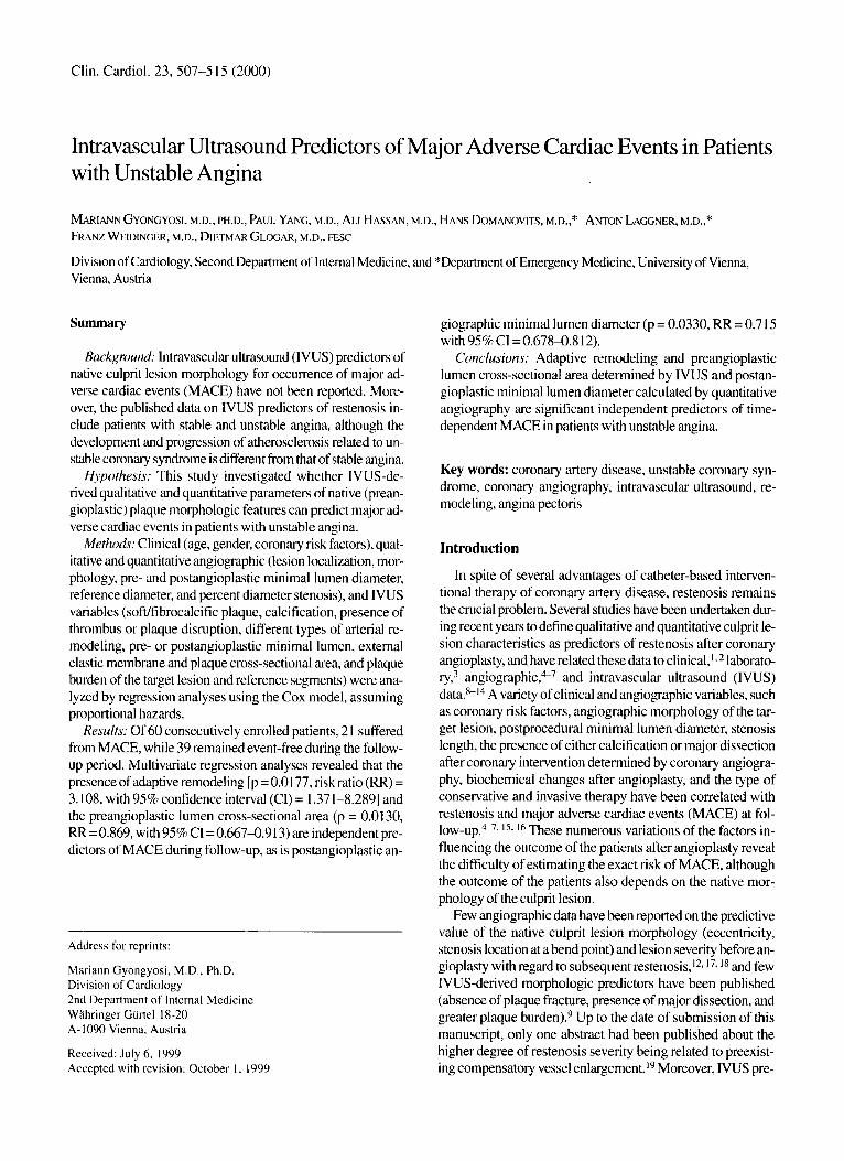
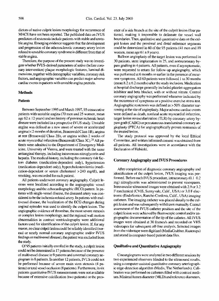
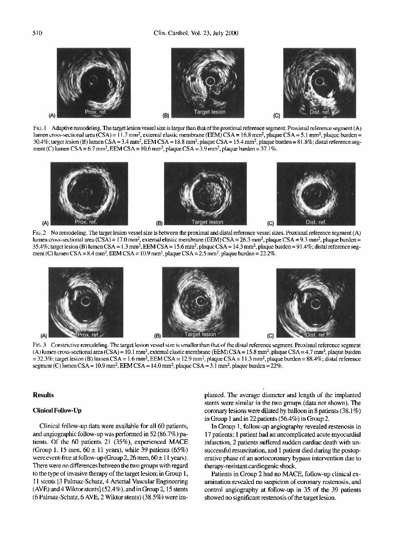
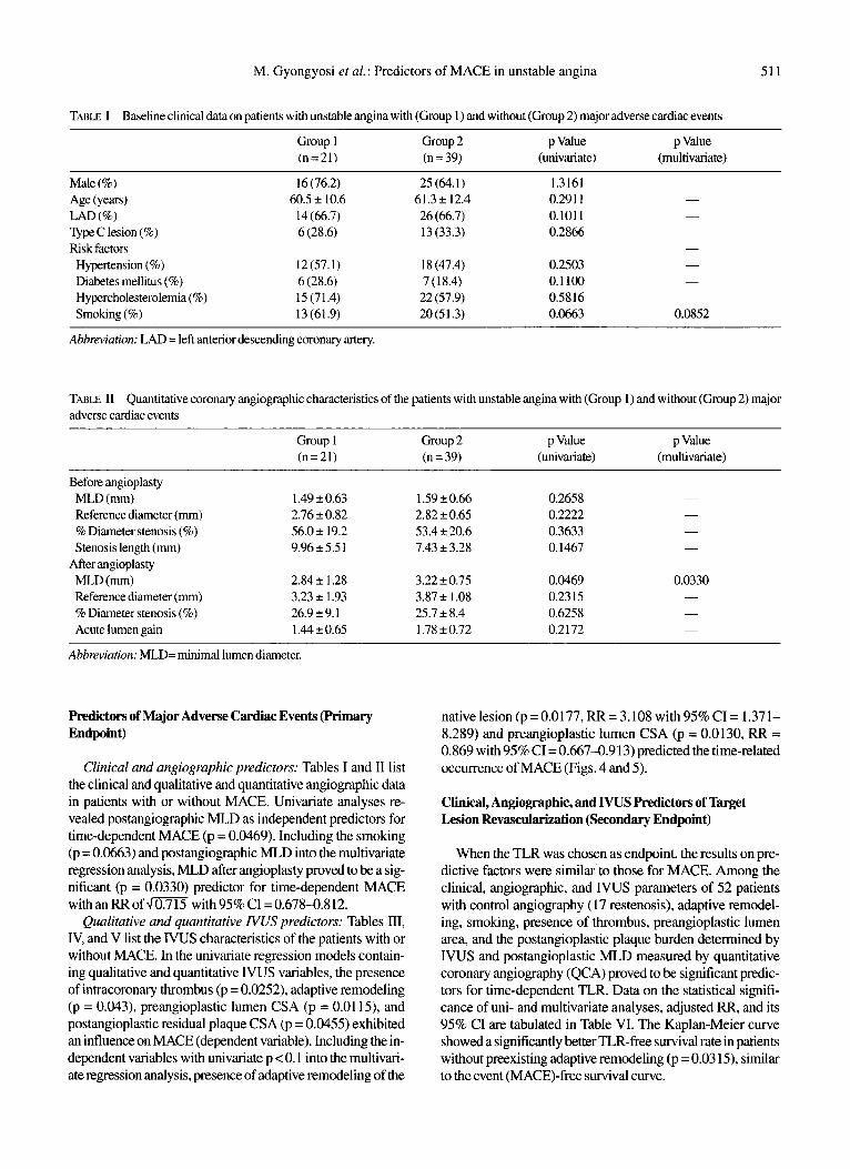
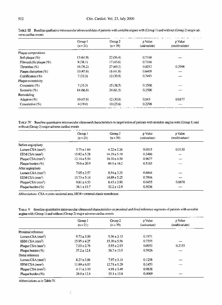
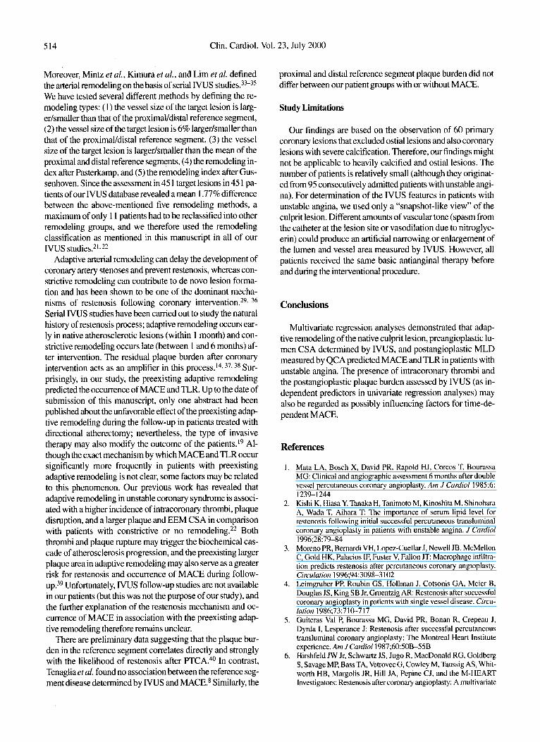
Compensatory vessel enlargement (adaptive remodeling) was assumed when the EEM CSA at the lesion site was larger than the proximal reference EEM CSA. Coronary shrinkage (constrictive remodeling) was considered present when the EEM CSA at the lesion site was smaller than that at the distal reference site EEM CSA. The definitions of different remod- eling types are the same as those previously published from our and from other (Figs. 1,2,3).
Statistics
The statistical analyses included descriptive statistics, uni- and multivariate regression analyses, and analysis assuming the Cox proportional hazards model. Descriptive statistics were used to determine the mean k standard deviation (SD) for continuous variables, frequencies, and percentages of cate- goric variables, regression analysis to assess the interobserver variability, and one-way analysis of variance (ANOVA) with repeated measurement for assessment of the reproducibility and intraobserver variability for all IVUS measurements. The exact method for the detennination of intra- and interobserver variability of IVUS measurements used by our laboratory has been described in detail elsewhere.’’ Brietly, the coefficient of correlation of the interobserver variability was r = 0.956 (p < 0.001). The coefficient of variation of the repeated measure- ments of the lumen and EEM diameter was 3% while that of the lumen and EEM CSA was I .7%. The methodologic error of the measurement of the lumen and EEM diameter was therefore 0.19 mm, and that of the lumen and EEM CSA was 0.38 mm’. Since the twofold coefficient of variation (0.76 mm2) was 5.52% of the mean target lesion vessel size of45 1 patients in our IVUS database, a difference of at least 6% be- tween the target lesion and reference segment vessel size was considered to be significant.
The Cox proportional hazards model was run using univari- ate and multivariate regression approaches. The MACE (pri- mary endpoint of the study) and TLR (secondary endpoint) and their relation to time were chosen as dependent variables. First, clinical variables (age, gender, and coronary risk fac- tors), qualitative IVUS parameters (soft or fibrocalcific plaque, the presence of thrombus, plaque disruption, calcification, plaque eccentricity, adaptive and constrictive remodeling), and quantitative IVUS variables (proximal and distal reference pre- and target lesion pre- and postangioplastic lumen CSA, plaque and EEM CSA, and the plaque burden), qualitative an- giographic variables (LAD and type C lesion) and quantitative angiographic parameters [stenosis length, pre- and postangio- plastic MLD, reference diameter and % diameter stenosis (%DS)] were analyzed by univariate regression analyses. In the second step, all variables showing a p value < 0.1 in the uni- variate regression models were entered in stepwise fashion into the multivariate regression model. The adjusted risk ratio (RR) and its 95% contidence interval (CI) for significant inde- pendent variables in multivariate analysis were calculated. The corresponding event-free survival function (Kaplan-Meier curve) was estimated and supplemented with log-rank test.
510 Clin. Cardiol. Vol. 23, July 2000
FIG. 1 Adaptive remodeling. The target lesion vessel size is larger than that of the proximal reference segment. Proximal reference segment (A) lumen cross-sectional area (CSA) = 11.7 mm2, external elastic membrane (EEM) CSA = 16.8 mm2, plaque CSA = 5.1 m2, plaque burden = 30.4%; target lesion (B) lumen CSA = 3.4 mm2, EEM CSA = 18.8 mm2, plaqueCSA = 15.4 mm2, plaque burden = 81.8%; distal reference seg- ment (C) lumen CSA = 6.7 mm2, EEM CSA = 10.6 nun2, plaque CSA = 3.9 mm2, plaque burden = 37.1%.
FIG. 2 No remodeling. The target lesion vessel size is between the proximal and distal reference vessel sizes. Proximal reference segment (A) lumen cross-sectional area (CSA) = 17.0 mm2, external elastic membrane (EEM) CSA = 26.3 mm2, plaque CSA = 9.3 mm2, plaque burden = 35.4%; target lesion (B) lumen CSA = 1.3 m2, EEM CSA = 15.6 mm2, plaqueCSA = 14.3 m2, plaque burden = 91.4%; distal reference seg- ment (C) lumen CSA = 8.4 mm2, EEM CSA = 10.9 mm2, plaque CSA = 2.5 nun2, plaque burden = 22.2%.
FIG. 3 Constrictive remodeling. The target lesion vessel size is smaller than that of the distal reference segment. Proximal reference segment (A) lumen cross-sectional area (CSA) = 10.1 mm2, external elastic membrane (EEM) CSA = 15.8 mm2, plaque CSA = 4.7 mm2, plaque burden = 32.3%; target lesion (B) lumen CSA = 1.6 mm2, EEM CSA = 12.9 mm2, plaque CSA = 11.3 m*, plaque burden = 88.4%; distal reference segment (C) lumen CSA = 10.9 nun2, EEM CSA = 14.0mm2, plaque CSA = 3.1 mm2, plaque burden = 22%.
Results
Clinical Follow-Up
planted. The average diameter and length of the implanted stents were similar in the two groups (data not shown). The coronary lesions were dilated by balloon in 8 patients (38.1 %) in Group 1 and in 22 patients (56.4%) in Group 2.
In Group 1, follow-up angiography revealed restenosis in 17 patients; 1 patient had an uncomplicated acute myocardial infarction, 2 patients suffered sudden cardiac death with un- successful resuscitation, and 1 patient died during the postop- erative phase of an aortocoronary bypass intervention due to therapy-resistant cardiogenic shock.
Patients in Group 2 had no MACE, follow-up clinical ex- amination revealed no suspicion of coronary restenosis, and control angiography at follow-up in 35 of the 39 patients showed no significant restenosis of the target lesion.
Clinical follow-up data were available for all 60 patients, and angiographic follow-up was performed in 52 (86.7%) pa- tients. Of the 60 patients 21 (35%), experienced MACE (Group 1, 15 men, 60 f 1 1 years), while 39 patients (65%) were event-free at follow-up (Group 2,26 men, 60 * 1 1 years). There were no differences between the two groups with regard to the type of invasive therapy of the target lesion; in Group 1, 1 1 stents [3 Palmaz-Schatz, 4 Arterial Vascular Engineering (AVE) and 4 Wiktor stents] (52.4%), and in Group 2,15 stents (6 Palmaz-Schatz, 6 AVE, 2 Wiktor stents) (38.5%) were im-
M. Gyongyosi et al.: Predictors of MACE in unstable angina 51 1
TABLE I Baseline clinical data on patients with unstable angina with (Group 1) and without (Group 2) major adverse cardiac events
Group 1 Group 2 p Value p Value (n=21) (n = 39) (univariate) (multivariate)
Male (%) Age (years) LAD (%) Type C lesion (%) Risk factors Hypertension (%) Diabetes mellitus (%) Hypercholesterolemia (%) Smoking (%)
16 (76.2) 60.5 f 10.6 14(66.7) 6 (28.6)
12 (57.1) 6 (28.6) 15 (71.4) 13(61.9)
25 (64.1) 61.3f 12.4 26 (66.7) 13 (33.3)
18 (47.4) 7 (18.4) 22 (57.9) 20(51.3)
1.3161 0.291 1 -
0.101 1 - 0.2866
0.2503 - 0.1 100 -
0.5816 0.0663 0.0852
-
Abbreviation: LAD = left anterior descending coronary artery.
TABLE I1 Quantitative coronary angiographic characteristics of the patients with unstable angina with (Group 1) and without (Group 2) major adverse cardiac events
Group 1 Group 2 p Value p Value (n=21) (n = 39) (univariate) (multivariate)
Before angioplasty MLD (mm) Reference diameter (mm) % Diameter stenosis (%) Stenosis length (mm)
MLD (mm) Reference diameter (mm) % Diameter stenosis (%) Acute lumen gain
After angioplasty
1.49 f 0.63 2.76 f 0.82 56.0f 19.2 9.96f5.51
2.84 f 1.28 3.23 f 1.93 26.9 f 9.1 1.44 f 0.65
1.59 f 0.66 2.82 f 0.65 53.4 f 20.6 7.43 f 3.28
3.22 f 0.75 3.87 f 1.08 25.7 f 8.4 1.78 f 0.72
0.2658 0.2222 0.3633 0.1467
0.0469 0.2315 0.6258 0.2172
Abbreviation: MLD= minimal lumen diameter,
predictors of Major Adverse Cardiac Events (Primary Endpoint)
Clinical and angiographic predictors: Tables I and I1 list the clinical and qualitative and quantitative angiographic data in patients with or without MACE. Univariate analyses re- vealed postangiographic MLD as independent predictors for time-dependent MACE (p = 0.0469). Including the smoking (p = 0.0663) and postangiographic MLD into the multivariate regression analysis, MLD after angioplasty proved to be a sig- nificant (p = 0.0330) predictor for time-dependent MACE w i t h a n R R o f W with95%CI=0.678-0.812.
Qualitative and quantitative NUS predictors: Tables III, IV, and V list the NUS characteristics of the patients with or without MACE. In the univariate regression models contain- ing qualitative and quantitative IVUS variables, the presence of intracoronary thrombus (p = 0.0252), adaptive remodeling (p = 0.043), preangioplastic lumen CSA (p = 0.01 15), and postangioplastic residual plaque CSA (p = 0.0455) exhibited an influence on MACE (dependent variable). Including the in- dependent variables with univariate p < 0.1 into the multivari- ate regression analysis, presence of adaptive remodeling of the
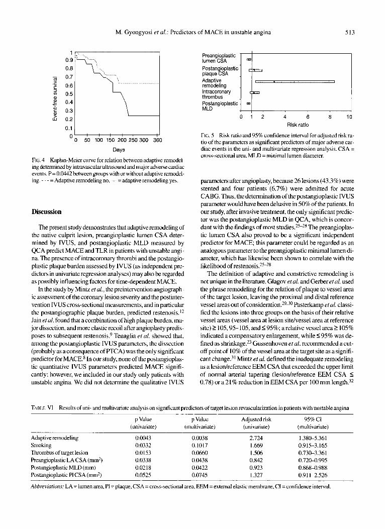
nativelesion(p=0.0177,RR=3.108 with95%CI= 1.371- 8.289) and preangioplastic lumen CSA (p = 0.0130, RR = 0.869 with 95% CI = 0.6674.913) predicted the time-related occurrence of MACE (Figs. 4 and 5).
Clinical, Angiographic, and IVUS Predictors of Target Lesion Revascularbation (Secondary Endpoint)
When the TLR was chosen as endpoint, the results on pre- dictive factors were similar to those for MACE. Among the clinical, angiographic, and IVUS parameters of 52 patients with control angiography (17 restenosis), adaptive remodel- ing, smoking, presence of thrombus, preangioplastic lumen area, and the postangioplastic plaque burden determined by IVUS and postangioplastic MLD measured by quantitative coronary angiogaphy (QCA) proved to be significant predic- tors for time-dependent TLR. Data on the statistical signifi- cance of uni- and multivariate analyses, adjusted RR, and its 95% CI are tabulated in Table VI. The Kaplan-Meier curve showed a signlficantly better TLR-free survival rate in patients without preexisting adaptive remodeling (p = 0.0315), similar to the event (MACE)-free survival curve.
512 Clin. Cardiol. Vol. 23, July 2000
TABLE III Baseline qualitative intravascular ultrasound data of patients with unstable angina with (Group 1) and without (Group 2) major ad- verse cardiac events
Group 1 Group 2 p Value p Value (n=21) (n = 39) (univariate) (multivariate)
Plaque compositions Soft plaque (%) Fibrocalcific plaque (%) Thrombus (%) Plaque disruption (%) Calcification (%)
Plaque eccentricity Concentric (%) Eccentric (%)
Adaptive (%) Constrictive (%)
Remodeling
I3 (61.9) 8 (38.1) 16 (76.2) 10 (47.6) 7 (33.3)
22 (56.4) 17 (43.6) 27 (69.2) 16 (41 .O) 12 (30.8)
0.7 168 0.7 168 0.0252 0.6409 0.7493
0.2998 - -
7 (33.3) 14 (66.6)
15 (38.5) 24(61.5)
0.1500 0.1500
0.0 177 -
lO(47.6) 4 (19.0)
12 (30.8) 10 (25.6)
0.043 0.2298
TABLE N Baseline quantitative intravascular ultrasound characteristics on target lesion of patients with unstable angina with (Group 1 ) and without (Group 2) major adverse cardiac events
Group 1 Group 2 p Value p Value (n=21) (n = 39) (univariate) (multivariate)
Before angioplasty Lumen CSA (mm2) 3.75 f 1.64 4.22 f 2.26 EEM CSA (mm2) 15.82 f 5.28 14.19f5.19 Plaque CSA (mm2) 12.14 2 5.54 10.10f4.50 Plaque burden (%) 70.6 f 20.9 69.1 2 16.2
Lumen CSA (mm2) 7.95 f 3.57 8.54 f 3.35 EEM CSA (mm2) 157325.14 14.89 f 5.25 Plaque CSA (mm2) 9.8 1 f 4.53 6.43 2 2.90 Plaque burden (%) 38.1 f 13.7 32.2 f 12.9
After angioplasty
Abbreviations: CSA =cross-sectional area, EEM =external elastic membrane.
0.01 15 0.3486 0.9677 0.5 165
0.0130 - - -
0.8844 0.7816 0.0455 0.5036
-
0.0678 -
TABLE V Baseline quantitative intravascular ultrasound characteristics on proximal and distal reference segments of patients with unstable angina with (Group 1) and without (Group 2) major adverse cardiac events
Group 1 Group 2 p Value p Value (n = 21) (n = 39) (univariate) (multivariate)
Proximal reference Lumen CSA (nun2) EEM CSA (mm2) Plaque CSA (nun2) Plaque burden (%)
Lumen CSA (mm2) EEM CSA (m2) Plaque CSA (nun2) Plaque burden (%)
Distal reference
9.73 f 3.00 15.95 f4.25 7.03 f 2.76 37.2 f 12.8
9.36f3.13 15.3025.56 5.93 f 2.93 36.7 2 13.5
0.1971 0.7355 -
0.0953 0.2155 0.5926 -
8.27 f 3.88 1 1.89 f 6.07 4.1 123.10 28.02 12.8
7.97 f 3.1 1 12.71 f5.29 4.91 f 3.49 35.1 f 13.8
0.1258 0.1453 0.9838 0.4069
Abbreviations as in Table IV.
1 0.9 0.8
0.7
0.6 0.5 0.4 0.3
0.2
0.1 I
Preangio lastic ~
lumen &A
M. Gyongyosi et al.;
1
'b 50 I00 150 200 250300 3iO
Days
FIG. 4 Kaplan-Meier curve for relation between adaptive remodel- ing determined by intravascular ultrasound and major adverse cardiac events. P= 0.0442between groups with or without adaptive remodel- ing. - - - = Adaptive remodeling no, - = adaptive remodeling yes.
Discussion
The present study demonstrates that adaptive remodeling of the native culprit lesion, preangioplastic lumen CSA deter- mined by IVUS, and postangioplastic MLD measured by QCA predict MACE and TLR in patients with unstable angi- na. The presence of intracoronary thrombi and the postangio- plastic plaque burden assessed by IVUS (as independent pre- dictors in univariate regression analyses) may also be regarded as possibly influencing factors for time-dependent MACE.
In the study by Mintz rt al., the preintervention angiograph- ic assessment of the coronary lesion severity and the postinter- vention IVUS cross-sectional measurements, and in particular the postangiographic plaque burden, predicted restenosis. I 2
Jain eta/. found that acombination of high plaque burden, ma- jor dissection, and more elastic recoil after angioplasty predis- poses to subsequent resteno~is.~ Tenaglia et al. showed that, among the postangioplastic IVUS parameters, the dissection (probably as a consequence of F'TCA) was the only significant predictor for MACE.8 In our study, none of the postangioplas- tic quantitative IVUS parameters predicted MACE signifi- cantly; however, we included in our study only patients with unstable angina. We did not determine the qualitative IVUS
Predictors of MACE in unstable angina 513
Postangioplastic plaque CSA I Adaptive remodeling lntracoronary thrombus Postangioplastic MLD
0 1 2 4 6 8 10 Risk ratio
FIG. 5 Risk ratio and 95% confidence interval for adjusted risk ra- tio of the parameters as significant predictors of major adverse car- diac events in the uni- and multivariate regression analysis. CSA = cross-sectional area, MLD = minimal lumen diameter.
parameters after angioplasty, because 26 lesions (43.3%) were stented and four patients (6.7%) were admitted for acute CABG. Thus, the determination of the postangioplastic IVUS parameter would have been delusive in 50% of the patients. In our study, after invasive treatment, the only significant predic- tor was the postangioplastic MLD in QCA, which is concor- dant with the findings of most studies.2s-28 The preangioplaq- tic lumen CSA also proved to be a significant independent predictor for MACE; this parameter could be regarded as an analogous parameter to the preangioplastic minimal lumen di- ameter, which has likewise been shown to correlate with the likelihood of restenosis.2s-28
The definition of adaptive and constrictive remodeling is not unique in the literature. Glagov eta/. and Gerber eta/. used the phrase remodeling for the relation of plaque to vessel area of the target lesion, leaving the proximal and distal reference vessel areas out of ~ons idera t ion .~~.~" Pasterkamp eta/. classi- fied the lesions into three groups on the basis of their relative vessel areas (vessel area at lesion sitehessel area at reference site) 1 105,95-105, and 5 95%; a relative vessel area I 105% indicated a compensatory enlargement, while 595% was de- fined as ~hrinkage.~' Gussenhoven et al. recommended a cut- off point of 10% of the vessel area at the target site as a signifi- cant change.31 Mintz et al. defined the inadequate remodeling as a lesiodreference EEM CSA that exceeded the upper limit of normal arterial tapering (lesiodreference EEM CSA 5 0.78) or a 21 % reduction in EEM CSA per 100 mm length.'*
TABLE V1 Results of uni- and multivariate analysis on significant predictors oftarget lesion revascularization in patients with unstable angina
p Value p Value Adjusted risk 9.5% CI (univariate) (multivariate) (univariate) (multivariate)
Adaptive remodeling 0.0043 0.0038 2.724 1.380-5.361
Thrombus of target lesion 0.0 153 0.0660 1 SO6 0.73C3.361 Preangioplastic LA CSA (mm') 0.0338 0.0438 0.842 0.720-0.995 Postangioplastic MLD (mm) 0.02 18 0.0422 0.923 0.868-0.988 Postangioplastic PI CSA (mm2) 0.0525 0.0745 1.327 0.91 1-2.526
Abbreviations: LA = lumen area, PI = plaque, CSA =cross-sectional area, EEM = external elastic membrane, CI =confidence interval.
Smoking 0.0332 0.1017 1.669 0.9 15-3.165
514 Clin. Cardiol. Vol. 23, July 2000
Moreover, Mintz et al., Kimura et al., and Lim et al. defined the arterial remodeling on the basis of serial IVUS ~ t u d i e s . ~ * ~ ~ We have tested several different methods by defining the re- modeling types: (1) the vessel size of the target lesion is larg- erhmaller than that of the proximalldistal reference segment, ( 2 ) the vessel size of the target lesion is 6% largerhmaller than that of the proximaVdista1 reference segment, ( 3 ) the vessel size of the target lesion is larger/sroaller than the mean of the proximal and distal reference segments, (4) the remodeling in- dex after Pasterkamp, and (5) the remodeling index after Gus- senhoven. Since the assessment in 45 1 target lesions in 45 1 pa- tients of our I W S database revealed a mean l .77% difference between the above-mentioned five remodeling methods, a maximum of only 1 1 patients had to be reclassified into other remodeling groups, and we therefore used the remodeling classification as mentioned in this manuscript in all of our IVUS
Adaptive arterial remodeling can delay the development of coronary artery stenoses and prevent restenosis, whereas con- strictive remodeling can contribute to de novo lesion forma- tion and has been shown to be one of the dominant mecha- nisms of restenosis following coronary interventi~n.~~. 36
Serial NUS studies have been carried out to study the natural history of restenosis process; adaptive remodeling occurs ear- ly in native atherosclerotic lesions (within l month) and con- strictive remodeling occurs late (between 1 and 6 months) af- ter intervention. The residual plaque burden after coronary intervention acts as an amplifier in this process.I4 37. 38 Sur- prisingly, in our study, the preexisting adaptive remodeling predicted the occurrence of MACE and TLR. Up to the date of submission of this manuscript, only one abstract had been published about the unfavorable effect of the preexisting adap- tive remodeling during the follow-up in patients treated with directional atherectomy; nevertheless, the type of invasive therapy may also modify the outcome of the patients.I9 Al- though the exact mechanism by which MACE and TLR occur significantly more frequently in patients with preexisting adaptive remodeling is not clear, some factors may be related to this phenomenon. Our previous work has revealed that adaptive remodeling in unstable coronary syndrome is associ- ated with a higher incidence of intracoronary thrombi, plaque disruption, and a larger plaque and EEM CSA in comparison with patients with constrictive or no remodeling2* Both thrombi and plaque rupture may trigger the biochemical cas- cade of atherosclerosis progression, and the preexisting larger plaque area in adaptive remodeling may also serve as a greater risk for restenosis and occurrence of MACE during follow- up.39 Unfortunately, I W S follow-up studies are not available in our patients (but this was not the purpose of our study), and the further explanation of the restenosis mechanism and oc- currence of MACE in association with the preexisting adap- tive remodeling therefore remains unclear.
There are preliminary data suggesting that the plaque bur- den in the reference segment correlates directly and strongly with the likelihood of restenosis after FTCA.40 In contrast, Tenaglia et al. found no association between the reference seg- ment disease determined by IVUS and MACE.8 Similarly, the
proximal and distal reference segment plaque burden did not differ between our patient groups with or without MACE.
Study Limitations
Our findings are based on the observation of 60 primary coronary lesions that excluded ostial lesions and also coronary lesions with severe calchcation. Therefore, our findings might not be applicable to heavily calcified and ostial lesions. The number of patients is relatively small (although they originat- ed from 95 consecutively admitted patients with unstable angi- na). For determination of the IVUS features in patients with unstable angina, we used only a “snapshot-like view” of the culprit lesion. Different amounts of vascular tone (spasm from the catheter at the lesion site or vasodilation due to nitroglyc- erin) could produce an artificial narrowing or enlargement of the lumen and vessel area measured by IVUS. However, all patients received the same basic antianginal therapy before and during the interventional procedure.
Conclusions
Multivariate regression analyses demonstrated that adap- tive remodeling of the native culprit lesion, preangioplastic lu- men CSA determined by IVUS, and postangioplastic MLD measured by QCA predicted MACE and TLR in patients with unstable angina. The presence of intracoronary thrombi and the postangioplastic plaque burden assessed by IVUS (as in- dependent predictors in univariate regression analyses) may also be regarded as possibly influencing factors for time-de- pendent MACE.
References
1.
2.
3.
4.
5.
6.
Mata LA, Bosch X, David PR, Rapold HJ, Corcos T, Bourassa MG: Clinical and angiographic assessment 6 months after double vessel percutaneous coronary angioplasty. Am J Curdiol 1985:6:
Kishi K, Hiasa Y, Tanaka H, Tanimoto M, Kinoshita M. Shinohara A, Wada T, Aihara T The importance of serum lipid level for restenosis following initial successful percutaneous transluminal coronary angioplasty in patients with unstable angina. J Cardiol
Moreno PR, Bemardi VH, Lopez-Cuellar J, Newell JB, McMellon C, Gold HK, Palacios IF, Fuster V, Fallon JT: Macrophage infiltra- tion predicts restenosis after percutaneous coronary angioplasty. Circulation 1996;94:3098-3 102 Leimgruber PP, Roubin GS, Hollman J, Cotsonis GA, Meier B, Douglas JS, King SB Jr. Gruentzig AR: Restenosisafter successful coronary angioplasty in patients with single vessel disease. Circu- lation 1986;73:7 10-7 17 Guiteras Val P, Bourassa MG, David PR, Bonan R, Crepeau J, Dyrda I, Lespemnce J: Restenosis after successful percutaneous transluminal coronary angioplasty: The Montreal Heart Institute experience. Am J Cardiol1987;60:50B-55B Hirshfeld JW Jr, Schwartz JS. Jug0 R, MacDonald RG, Goldberg S, Savage Mp, Bass TA, Vetrovec G , Cowley M, Taussig AS, Whit- worth HB, Margolis JR, Hill JA, Pepine CJ, and the M-HEART Investigators: Restenosis after coronary angioplasty: A multivariate
1239-1 244
1996;28:79-84
M. Gyongyosi et al.: Predictors of MACE in unstable angina 515
7.
8.
9.
10.
I I .
12.
13.
14.
15.
16.
17.
18.
19.
20.
21.
22.
23.
24.
statistical model to relate lesion and procedurc variables to stenosis. The M-HEARTInvestigators. J A W CollCtrrdiol 1991: l8:617-656 Kuntz RE, Safian RD. Carrozza JP, Fishinan RE Mansour M. Baini DS: The importance of acute luminal diameter i n determining restenosis after coronary atherectomy or stenting. Circulrrtiorr
Tenaglia AN, Buller CE, Kisslo KB, Phillips HR. Stack RS. David- son CJ: Intracoronary ultrasound predictors of adverse outcomes after coronary artery interventions. J Anz Coll Crrrtfiol 1992:20: 1385-1390 Jain SP, Jain A, Collins TJ, Raniee SR, White CJ: Predictors of
A morphometric and quantitative evaluation by in- travascular ultrasound. Am Heart J 1994; 128:664-673 Post MJ, de Smet BJ, van der Helm Y. Borst C, Kuntz RE: Arterial remodeling after balloon angioplasty or stcnting in an atheroscle- rotic experimental model. Circdution I997:96:99&1003 Dussaillant GR. Mintz GS. Pichard AD, Kent KM, Satlcr LF, Popma JJ, Wong SC, Leon MB: Small stent size and intinial hyper- plasia contribute to restenosis: A volumetric intravascular ultra- sound analysis. JAin Coll Curdiol I995:26:720-724 Mintz GS. Popma JJ, Pichard AD. Kent KM. Salter LE Chuang YC, Griflin J , Leon MB: lntravascular ultrasound predictors of restenosis after percutaneous transcatheter coronary revasculariza- tion. J Am Coll Cardiol I996:27: 1678- I687 Colombo A, Ferraro M, ltoh A, Martini G. Blengino S, Finci L: Results of coronary stenting for restenosis. J An] Coll Curdiol 1996;28:830-836 Mintz GS, Kent KM, Pichard AD, Popma JJ. Satler LF, Leon MB: lntravascular ultrasound insights into mechanisms of stenosis for- mation and restenosis. Ctrrdiol Clin 1997; 15: 17-29 Williams MJ. Morison IM, Parker JH, Stewart RA: Progression of the culprit lesion in unstable coronary artery disease with warkin and aspirin versus aspirin alone: Preliminary study. Am J Curdiol I997;30:364369 Mittal S, Weiss DL, Hirshfeld JW Jr, Kolansky DM, Herrmann HC: Comparison of outcome after stenting for de novo versus restenotic nmowings in native coronary arteries. Am J Cardiol 1997;80:711-715 Tousoulis D, Kaski JC, Davies G. Pereira W, el Tamimi H. McFadden E, Maseri A: Preangioplasty complicated coronary stenosis morphology as a predictor of’ restenosis. Am Heart J 1992; 123: 15-20 Ellis SG. Roubin GS, King SB 111, Douglas JS Jr, Cox WR: Importance of stenosis morphology in the estimation of restenosis risk after elective percutaneous transluniinal coronary angioplasty. Am J Ccirdiol 1989;63:30-34 Wexberg P. Hassan A, Yang P. Palisek-Kiss K, Clogar D: Pre-exist- ing compensatory vessel enlargement is associated with higher de- gree ofrestenosis severity (abstr). Z Kardiol 1998;87:398 Braunwald E: Unstable angina: A classification. Circirla/iori 1989; 8O:4l M I 4 Gyongyosi M, Yang P, Hassan A, Weidinger F, Domanovits H, Laggner A, Glogar D: Coronary risk factors influence plaque mor- phology in patients with unstable angina. Corm Art Dis 1999; 10:211-219 Gyongyosi M, Yang P, Hassan A, Weidinger F, Domanovits H, Laggner A, Glogar D: Arterial remodeling of native human coro- nary arteries in patients with unstable angina pectoris: A prospec- tive intravascular ultrasound study. Hecrrt 1999; 82:68-74 Pasterkamp G, Peters RJG, Kok WEM, van Leuwen TG, Borst C for the PICTURE investigators. Arterial remodeling after balloon angioplasty of the coronaiy artery: An intravascular ultrasound study. Am Heart J 1997; 134:68(M84 Nishioka T, Luo H, Eigler NL, Berglund H, Kim CJ, Siege1 RJ: Contribution of inadequate compensatory enlargement to develop-
1992236: 1827-1 835
25.
26.
27.
28.
29.
30.
31.
32.
33.
34.
35.
36.
37.
38.
39.
40.
ment of human coronary artery stenosis: An in vivo intravascular ultrasound study. JAni Coll Cbrdinl 1996;27: 1571-1576 Fleisch M. Meier B: Present status ofcoronary stenting: Hopes and hypes. Indicrn Hew? J l998;5O(suppl I): 14-1 7 Ellis SG, Savage M, Fischrnan D. Baim DS, Leon M, Goldberg S, Hirshfeld JW, Cleman MW, Teirstein PS, Walker C: Restenosis af- ter placement of Palmaz-Schatz stents in native coronary arteries. Initial results of a multicenter experience. Circulation I992;86: 1836-1844 Bauters C, Hubert E, Rat A, Bougrimi K, Van Belle E, McFadden EP, Amouyel P, Lablanche JM, Bertrand M: Predictors of resteno- sis after coronary stent implantation. Am J Cardiol 1998:3 1: 1291-1 298 Bauters C, Lablanche JM, Van Belle E, Niculescu R, Meurice T. Mc Fadden EP, Bertrand ME: Effects of coronary stenting on restenosis and occlusion after angioplasty of the culprit vessel in pa- tients with recent myocardial infarction. Circulrrtion 1997;96: 28542858 Glagov S, Weisenberg E, Zarins CK, Stankunavicius R, Kolettis GJ: Compensatory enlargement of human atherosclerotic coronary arteries. N Engl JMed l987;3 16: 1371-1375 GerberTC, Erbel R, GBrge G, Ge J, Rupprecht HJ, MeyerJ: Extent of atherosclerosis and remodeling of the left main coronary artery determined by intravascular ultrasound. Am J Curdiol 1994;73: 6 6 M 7 1 Gussenhoven EJ, Geselschap JH, van Lankeren W, Posthuma DJ, van der Lugt A: Remodeling of the atherosclerotic coronary arter- ies assessed with intravascular ultrasound in vitro. Am J Curdiol
Mintz GS, Kent KM, Pichard AD, Satler LF, Popma JJ, Leon MB: Contribution of inadequate arterial remodeling to the development of focal coronary artery stenoses. An intravascular ultrasound study. Circulation I997;95: 179 1- I798 Mintz GS, Popma JJ, Pichard AD, Kent KM, Satler LF, Wong SC, Hong MK, Kovach JA, Leon MB: Arterial remodeling after coro- nary angioplasty. A serial intravascular ultrasound study. Circulu- tion 1996;94:3543 Kimura T, Kabiragi S, Tamura T, Yokoi H, Nakagawa Y, Yokoi H, Hamasaki N, Nosaka H, Nobuyoshi M, Mintz GS, Popma JJ, Leon MB: Remodeling of human coronary arteries undergoing coronary angioplasty or atherectomy. Circulation 1997;96:457-483 Lim lT, Liang DH, Botas J, Schroeder JS, Oesterle SN, Yeung AC: Role of compensatory enlargement and shrinkage in transplant coronary artery disease. Serial intravascular ultrasound study. Circularion 1997;95:855-859 Glagov S: Intimal hyperplasia, vascular remodeling and the restenosis problem. Circulation 1994;89:2888-289 1 McPherson DD, Sirna SJ, Hiratzka LF, Thorpe L, Armstrong ML, Marcus ML, Kerber RE: Coronary arterial remodeling studies by high-frequency epicardial echocardiography: An early compen- satory mechanism in patients with obstructive coronary atheroscle- rosis. JAni Coll Cardiol 1991; 17:79-86 Kakuta T, Currier JW, Haudenschild CC, Ryan TJ, Faxon DP: Differences in compensatory vessel enlargement, not intimal for- mation, account for restenosis after angioplasty in the hypercholes- terolemic rabbit model. Circulation 1994;89:2809-28 15 Libby P, Schwartz D, Brogi E, Tanaka H, Clinton SK: A cascade model for restenosis: A special case of atherosclerosis progression.
Nissen SE, De Franco AC, Raymond RE: Angiographically un- recognised disease at “normal” reference sites: A risk factor for sub-optimal results after coronary interventions (abstr). Circulation 1993;8 1:1-412
1997;79:699-702
Circulatiort l992;86(~~ppl III):47-52
Related Documents