495 Intradural Lumbar Disk Herniation Virgil B. Graves,' H. Lee Finney, and James Mailander Intradural lumbar disk herniation is an uncommon occur- rence in the natural course and clinical spectrum of lumbar disk disease. The reported cases suggest an incidence of 0.13%. This case report presents some of the commonly occurring clinical and radiographic findings in intradural lumbar disk herniation, including those of CT. It also demonstrates the importance of metrizamide-enhanced CT in the evaluation of intradural lesions. Case Report A 60-year-old man had a long history of low back pain. Two months before admission, he developed increasing lumbar back pain and intermittent radicular pain. At the time of admission, he had acute radicular pain and exhibited acute and progressive neurologic changes, including lower extremity weakness, more marked on the right than the left, and some bowel and bladder dysfunction. The patient had absent lower extremity reflexes. An unenhanced CT scan (Fig. 1 A) showed a density posterior to the disk that appeared to be distinctly different from the thecal sac. CT at the level of the L3-L4 disk space showed a protruding disk not only posteriorly but also anteriorly and laterally (Fig. 1 B). The epidural fat space was obliterated. Metrizamide myelography, per- formed for further evaluation of the severe and progressive neurologic changes, demonstrated a complete block at the level of the L3-L4 disk interspace (Fig. 2). A cerebrospinal fluid (CSF) protein level at the time of myelography from below the block was greater than 1800 mg/dl. The CSF fluid was clear. A delayed CT scan (Fig. 3) after metrizamide myelography demonstrated a complete block at the L3- L4 disk interspace level secondary to an irregular lobulated intradural mass. Exploratory laminectomy at the L3 level revealed the dura to be tense, slightly discolored, and firm. A firm intradural mass could be felt with transdural palpation. On opening the dura a lobular white mass was seen to be enmeshed among the nerve roots of the cauda equina. The nerve roots of the cauda equina were displaced dorsally by the mass. A 2.5 cm lobular white mass was removed. This mass originated from a defect in the ventral dura and arachnoid at the level of the L3-L4 interspace. The defect in the ventral dura and arachnoid measured about 3-4 mm . The ventral dura appeared to be adherent to the posterior longitudinal ligament at this level. This appeared to be secondary to epidural scarring. Intradural and extradural exploration failed to reveal any additional mass or fragments. The disk space was curetted and the defect in the ventral dura closed. Subsequent histologic examination of the Received August 23, 1984; accepted after revision January 16, 1985. intradural mass identified it as cartilage with focal degenerative changes consistent with herniated disk material. The patient's recovery was uneventful. He had complete relief of the radicular pain and gradual resolution of the neurologic deficit s. On discharge, he demonstrated increased strength in his lower extremities. Discussion Forty-nine cases of intradural disk herniation have been reported (1). Thirty-six involved the lumbar disk interspaces [2]. The earliest reported case was by Dandy [3] in 1942. The incidence has been variously reported by numerous authors, including Dandy [3], 1:300 (0.33 %); Peyser and Harari [4], 2:753 (0.27%); Lyons and Wise [5], 1:2500 (0.04%); Slater et al. [6], 2:1 000 (0.2%); Carcavilla et al. [7], 2:740 (0 .27%); Paine et al. [8],2:1078 (0.2%); and our own experience of 1 :1 816 (0.06%). The overall combined incidence from these reported studies is 11 :81 87 (0.13%). Intradural lumbar disk herniation has occurred so infre- quently that generalizations regarding its clinical and radio- logic presentation and appearance are difficult to make; how- ever, several commonly occurring characteristics have been observed in this and the other reported cases. Most reported intradural lumbar disk herniations have histories of chronic low back pain of some duration, followed by an acute, often severe episode of back pain or radicular pain and progressive neurologic deficits suggestive of multiple root involvement. The neurologic deficits have been reported to be slightly more severe than usually associated with extradural disk hernia- tions. Several cases have been associated with previous lumbar surgical procedures [4, 6, 9, 10]. CSF protein has nearly always been elevated, even if a complete block has not been present [10]. Plain radiographs mayor may not demonstrate intradiskal narrowing. Myelograms have usually revealed a complete block, and it is difficult to differentiate intradural disk herniation from other intradural mass lesions of the lumbar area, includ- ing neurofibroma, meningioma, lipoma, epidermoid and arach- noid cyst, metastases, and even large extradural disk hernia- tions [10] . Unenhanced CT failed to reveal significant abnormalities 1 All authors: Department of Radiology, Columbus Hospital, Great Fall s, MT 59405. Address reprint requests to V. B. Graves. AJNR 7:495-497, May/June 1986 0195-6108/86/0703-0495 © American Society of Neuroradiology

Welcome message from author
This document is posted to help you gain knowledge. Please leave a comment to let me know what you think about it! Share it to your friends and learn new things together.
Transcript
495
Intradural Lumbar Disk Herniation Virgil B. Graves,' H. Lee Finney, and James Mailander
Intradural lumbar disk herniation is an uncommon occurrence in the natural course and clinical spectrum of lumbar disk disease. The reported cases suggest an incidence of 0.13%. This case report presents some of the commonly occurring clinical and radiographic findings in intradural lumbar disk herniation, including those of CT. It also demonstrates the importance of metrizamide-enhanced CT in the evaluation of intradural lesions.
Case Report
A 60-year-old man had a long history of low back pain. Two months before admission, he developed increasing lumbar back pain and intermittent radicular pain . At the time of admission, he had acute radicular pain and exhibited acute and progressive neurologic changes, including lower extremity weakness, more marked on the right than the left , and some bowel and bladder dysfunction. The patient had absent lower extremity reflexes.
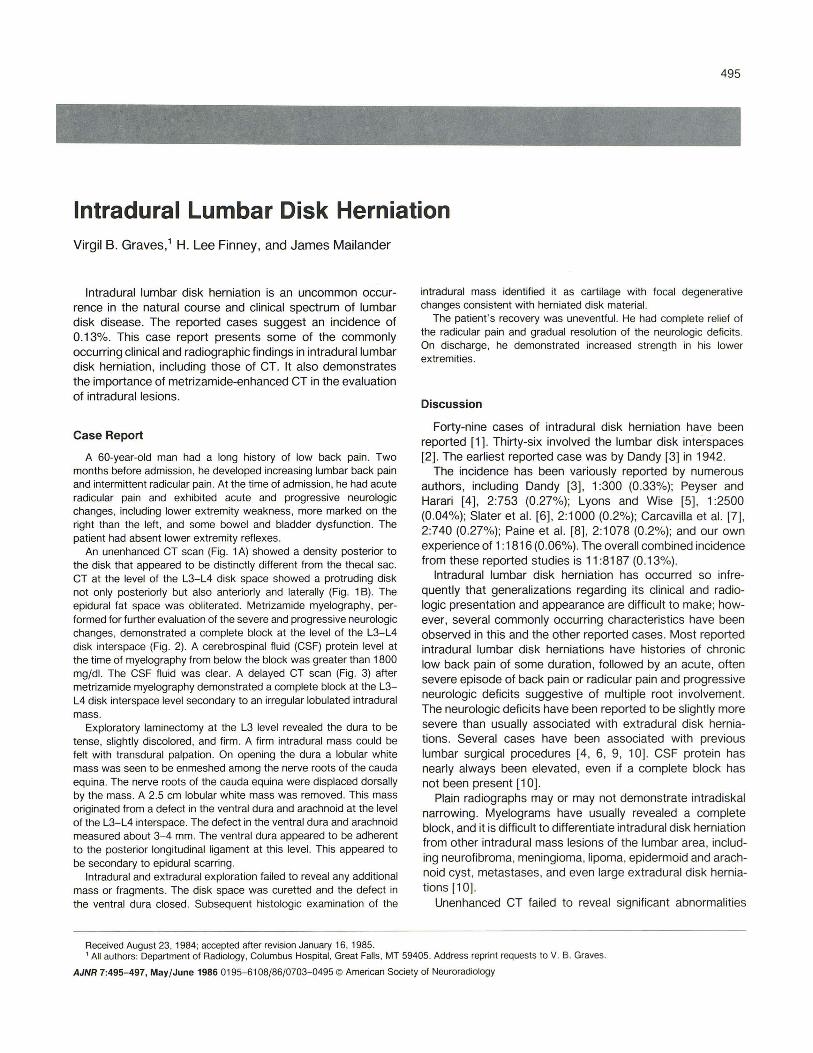
An unenhanced CT scan (Fig. 1 A) showed a density posterior to the disk that appeared to be distinctly different from the thecal sac. CT at the level of the L3-L4 disk space showed a protruding disk not only posteriorly but also anteriorly and laterally (Fig. 1 B). The epidural fat space was obliterated. Metrizamide myelography, performed for further evaluation of the severe and progressive neurologic changes, demonstrated a complete block at the level of the L3-L4 disk interspace (Fig. 2). A cerebrospinal fluid (CSF) protein level at the time of myelography from below the block was greater than 1800 mg/dl. The CSF fluid was clear. A delayed CT scan (Fig. 3) after metrizamide myelography demonstrated a complete block at the L3-L4 disk interspace level secondary to an irregular lobulated intradural mass.
Exploratory laminectomy at the L3 level revealed the dura to be tense , slightly discolored, and firm. A firm intradural mass could be felt with transdural palpation. On opening the dura a lobular white mass was seen to be enmeshed among the nerve roots of the cauda equina. The nerve roots of the cauda equina were displaced dorsally by the mass. A 2.5 cm lobular white mass was removed . This mass originated from a defect in the ventral dura and arachnoid at the level of the L3-L4 interspace. The defect in the ventral dura and arachnoid measured about 3-4 mm. The ventral dura appeared to be adherent to the posterior longitudinal ligament at this level. This appeared to be secondary to epidural scarring.
Intradural and extradural exploration failed to reveal any additional mass or fragments. The disk space was curetted and the defect in the ventral dura closed. Subsequent histologic examination of the
Received August 23, 1984; accepted after revision January 16, 1985.
intradural mass identified it as cartilage with focal degenerative changes consistent with herniated disk material.
The patient's recovery was uneventful. He had complete relief of the radicular pain and gradual resolution of the neurologic deficits. On discharge, he demonstrated increased strength in his lower extremities.
Discussion
Forty-nine cases of intradural disk herniation have been reported (1). Thirty-six involved the lumbar disk interspaces [2] . The earliest reported case was by Dandy [3] in 1942.
The incidence has been variously reported by numerous authors, including Dandy [3], 1 :300 (0.33%); Peyser and Harari [4] , 2:753 (0.27%); Lyons and Wise [5], 1 :2500 (0.04%); Slater et al. [6], 2:1 000 (0.2%); Carcavilla et al. [7], 2:740 (0.27%); Paine et al. [8],2:1078 (0.2%); and our own experience of 1 :1 816 (0.06%). The overall combined incidence from these reported studies is 11 :81 87 (0.13%).
Intradural lumbar disk herniation has occurred so infrequently that generalizations regarding its clinical and radiologic presentation and appearance are difficult to make; however, several commonly occurring characteristics have been observed in this and the other reported cases. Most reported intradural lumbar disk herniations have histories of chronic low back pain of some duration, followed by an acute, often severe episode of back pain or radicular pain and progressive neurologic deficits suggestive of multiple root involvement. The neurologic deficits have been reported to be slightly more severe than usually associated with extradural disk herniations. Several cases have been associated with previous lumbar surgical procedures [4, 6, 9, 10]. CSF protein has nearly always been elevated , even if a complete block has not been present [10].
Plain radiographs mayor may not demonstrate intradiskal narrowing. Myelograms have usually revealed a complete block, and it is difficult to differentiate intradural disk herniation from other intradural mass lesions of the lumbar area, including neurofibroma, meningioma, lipoma, epidermoid and arachnoid cyst, metastases, and even large extradural disk herniations [10] .
Unenhanced CT failed to reveal significant abnormalities
1 All authors: Department of Radiology, Columbus Hospital, Great Falls, MT 59405. Address reprint requests to V. B. Graves.
AJNR 7:495-497, May/June 1986 0195-6108/86/0703-0495 © American Society of Neuroradiology

496 GRAVES ET AL. AJNR:7. May/June 1986
A B
A B
sufficient to explain our patient's neurologic findings . The density posterior to the disk at the level of Figure 1 A may represent the fibrous adhesion between the ventral dura and the posterior longitudinal ligament later observed at surgery.
Fig. 1.-Preenhancement CT scans. A. Level of intradural disk herniation. Density posterior to disk may represent fibrous adhesion later found at surgery between ventral dura and posterior longitudinalligament. B. L3-L4 level. Generalized protrusion of L3-L4 disk with obliteration of epidural fat space.
Fig. 2.-Lumbar metrizamide myelogram. A. Posteroanterior view. Complete block. B. Lateral view with head down 45°. Myelographic appearance is suggestive of extradural compression of thecal sac.
No extradural disk material was found at surgery. The contrast-enhanced (metrizamide) CT scan demonstrated the lobular intradural mass. This allowed for a confident preoperative diagnosis of an intradural mass, and it also helped to define
AJNR:7, May/June 1986 INTRADURAL LUMBAR DISK HERNIATION 497
A B c Fig. 3.-Metrizamide-enhanced CT scans. A and B, Same level as Fig. 1A. Lobulated left-sided intradural mass. B, L3-L4 disk level (ct . Fig . 1 B). No contrast
material at this level.
its extent. This was useful in planning the surgery preoperatively.
Hodge et al. [10J suggested that metrizamide myelography and metrizamide CT can be used to suggest the diagnosis of lumbar intradural disk herniation preoperatively by demonstrating an irregular or lobular intradural mass. With the exception of metastatic disease, the more common causes of intradural masses in the lower lumbar region have characteristically smooth margins. Smith [2] recommended analysis of the CSF macrophages for evidence of fibrocartilage as a way to make the preoperative diagnosis of intradural disk herniation.
The mechanism of intradural disk herniation is not known with certainty. It is probable that a primary or preexisting abnormality must be present to allow intradural disk herniation to occur.
Blikra [11 J demonstrated with autopsy material that there is generally only a loose connection between the ventral surface of the dura and the posterior longitudinal ligament in the lumbar region. But in some cases, there was a very firm , dense connection between the dura, posterior ligament, and anulus. Other reported cases have shown dense, firm adhesions of the ventral dura and posterior ligament both in patients without [2, 5, 11, 12J and with [4, 6, 9, 10] histories of previous surgery.
On the basis of reported cases, the existence of a firm attachment of the ventral dura and posterior longitudinal ligament probably plays a significant role in the occurrence of intradural disk herniation. This probably occurs as a result of several mechanisms, including long-standing degenerative or
inflammatory changes of the disk, minor trauma, surgery, and developmental anatomic variations.
In general, the prognosis of these patients has been good, with postoperative recovery essentially the same as with patients having extradural disk herniations and similar neurologic presentation.
REFERENCES
1. Barbera J, Gonzalez-Darder J, Garcia-Vazquez F. Intraradicular herniated lumbar disc. J Neurosurg 1984;60:858-860
2. Smith RV. Intradural disc rupture. J Neurosurg 1981 ;55 :117-120
3. Dandy WE. Serious complications of ruptured intervertebral disks. JAMA 1942;119:474-477
4. Peyser E, Harari A. Intradural rupture of lumbar intervertebral disk. Surg Neuro/1977;8 :95- 98
5. Lyons AE, Wise BL. Subarachnoid rupture of intervertebral disc fragments. J Neurosurg 1961 ;18 :242-244
6. Slater RA, Pineda A, Porter RW. Intradural herniation of lumbar intervertebral discs. Arch Surg 1965;90 :266- 269
7. Carcavilla LI , Perun JG, Firass A-KR. Hernie discale lombaire intra-durale. Neurochirurgie 1982 ;28 :421- 424
8. Paine GP, Barra N, Balde PG, Pasetti S. Rottura intradurale del disco intervertebrale. Acta Biomed 1978;49 : 189-1 96
9. Coppola AR. Pseudoneurilemona of the spinal canal. South Med J 1979 ;72 :1625-1626
10. Hodge CJ, Binet EF, Kieffer SA. Intradural herniation of lumbar intervertebral discs. Spine 1978;3 :346-350
11 . Blikra G. Intradural herniated lumbar disc. J Neurosurg 1969;31 :676-679
12. Weuzel V, Molot MJ , Murray JK. Intradural herniation of a lumbar disc. J Can Assoc Radio/1981 ;31 :64-65
Related Documents


![Percutaneous Endoscopic Lumbar Spine Surgery for …1.1. Lumbar Disc Herniation (LDH) Lumbar disc herniation [1][2] [3] (Figure 1(a)) is a medical condition affecting the spine in](https://static.cupdf.com/doc/110x72/5f01e5a17e708231d4019244/percutaneous-endoscopic-lumbar-spine-surgery-for-11-lumbar-disc-herniation-ldh.jpg)