Interferon-induces expression of cyclin-dependent kinase-inhibitors p21 WAF1 and p27 Kip1 that prevent activation of cyclin-dependent kinase by CDK-activating kinase (CAK) Mahitosh Mandal*, Debdutta Bandyopadhyay*, Thea M Goepfert and Rakesh Kumar Cell Growth Regulation Laboratory, Department of Clinical Investigation, The University of Texas MD Anderson Cancer Center; Houston, Texas 77030, USA To understand the mechanism of interferon (IFN)- mediated suppression of cell cycle progression, we have earlier shown that IFN-a enhances the expression of underphosphorylated retinoblastoma protein by inhibiting the cyclin-dependent kinase-2 (CDK-2) activity (Kumar and Atlas, Proc. Natl. Acad. Sci. 89, 6599 – 6603, 1992; Zhang and Kumar, Biochem. Biophysi. Res. Comm., 200, 522 – 528, 1994). In the studies presented here, we investigated the mechanism of inhibition of CDKs in IFN-treated cells by delineating the potential role(s) of CDK-inhibitors (CKIs) and CDK-activating kinase (CAK). We report that IFN-a inhibits the H-1 kinase activity associated with CDK-4 or CDK-2 due to induction of expression of CDK-inhibitor p21 WAF1 (but not p27 Kip1 ) as its immunodepletion from IFN-treated extracts restored the CDK-associated H-1 kinase activity. In addition, we also show that IFN-g induces expression of CDK-inhibitors p21 WAF1 and p27 Kip1 and inhibited the H-1 kinase activity associated with CDK-2 or CDK-4. The observed IFN-g-mediated inhibition of CDK-2 and CDK-4 kinase activity was due to enhanced interactions with p21 WAF1 and p27 Kip1 , respectively. We also demonstrated that IFN-induced CKIs prevent CAK from activating the CDK-2 as immunodepletion of induced CKIs from the inhibitory extracts resulted in the restoration of CAK-mediated activation of CDK-2. Keywords: interferons; cell cycle; CDK; CDK-inhibi- tors; CDK-activating kinase Introduction Regulation of cell proliferation is a complex process involving the regulated expression and/or modification of discrete gene products. There is a growing list of evidence to support the involvement of paracrine growth inhibitors and intracellular growth suppressors in the regulation of cell growth. Representatives of these classes of molecules are secretory proteins like interferons (IFN), and cell cycle inhibitors like p21 WAF1 and p27 Kip1 that inhibit the cyclin-dependent kinases (CDKs). An obvious question is whether the growth inhibition by IFN is mediated through the pathway(s) that control the activation of CDKs. Interferons (IFNs) are a family of hormone-like secretory proteins which interact with neighboring cells and bring about many phenotypical changes in these cells. Like most cytokines, IFNs must bind to specific high anity cellular receptors to exert biological eects. These eects include regulation of expression of specific genes, antiviral properties, and inhibition of cell growth and proliferation (Sokawa, 1977; Pestka et al., 1987; Kumar and Atlas, 1992; Kumar et al., 1994; Yamada et al., 1994; Tiefenbrum et al., 1996). The inhibition of cell proliferation by IFNs is an active process, and generally involves arrest of cells in the G0/ G1-phase of the cell cycle. IFN-a and IFN-b have also been considered to have a natural negative regulatory role in the autocrine regulation of cell proliferation, as several growth factors induce the formation of IFN in the cells they stimulate (Zullo et al., 1985; Moore et al., 1984), and this possibly could prevent uncontrolled cell proliferation. The mechanisms underlying growth inhibitory eects by IFNs are not well established. The regulation of cell cycle is controlled by families of essential proteins which control transition between dierent stages of the cell cycle. These proteins either facilitate cell cycle progression or serve to break the progression. The cell cycle progression through the specific phases of the cell cycle is regulated by the sequential formation, activation and inactivation of a family of serine/threonine kinases, the CDKs (reviewed in Reed, 1992; Sherr, 1993; Hunter and Pine, 1994). The CDKs are a family of enzymes that are inactive as monomers, and require association with cyclins for activation. The activities of CDK-4 and CDK-2 appear to be most critical to the restriction point transitions, particularly, passage through the G1-phase and entry into the S-phase (Hunter and Pine, 1994). CDK- complexes also associate with proliferating cell nuclear antigen (PCNA) to form a catalytically active kinase complex that phosphorylates retinoblastoma protein (pRB) (abrogate growth-inhibitory function of pRB). This allows release of transcription factor E2F, which activates the expression of target genes required in DNA synthesis (Reed, 1991; Dulic et al., 1992; Xiong et al., 1993). The activity of CDKs is negatively controlled by CDK-inhibitors (CKIs) such as p21 WAF1 and p27 Kip1 . Both p21 WAF1 and p27 Kip1 have been shown to inhibit the activity of CDK-2- and CDK4-cyclin complex’s in vitro (Xiong et al., 1993; Harper et al., 1993; Kato et al., 1994; Hall et al., 1995). The p21 WAF1 has been shown to also bind to PCNA and block DNA replication (Waga et al., 1994). It is believed that CKIs bind to cyclin/CDK complexes and set stoichiometric inhibitory thresholds of CDKs kinase activity, which prevents premature or inappropriate progression of the cell cycle by impeding their ability to phosphorylate target substrates such as pRB family proteins. Correspondence: R Kumar *MM and DB contributed equally to this study Received 7 May 1997; revised 29 August 1997; accepted 29 August 1997 Oncogene (1998) 16, 217–225 1998 Stockton Press All rights reserved 0950 – 9232/98 $12.00

Welcome message from author
This document is posted to help you gain knowledge. Please leave a comment to let me know what you think about it! Share it to your friends and learn new things together.
Transcript
Interferon-induces expression of cyclin-dependent kinase-inhibitorsp21WAF1 and p27Kip1 that prevent activation of cyclin-dependent kinase byCDK-activating kinase (CAK)
Mahitosh Mandal*, Debdutta Bandyopadhyay*, Thea M Goepfert and Rakesh Kumar
Cell Growth Regulation Laboratory, Department of Clinical Investigation, The University of Texas MD Anderson Cancer Center;Houston, Texas 77030, USA
To understand the mechanism of interferon (IFN)-mediated suppression of cell cycle progression, we haveearlier shown that IFN-a enhances the expression ofunderphosphorylated retinoblastoma protein by inhibitingthe cyclin-dependent kinase-2 (CDK-2) activity (Kumarand Atlas, Proc. Natl. Acad. Sci. 89, 6599 ± 6603, 1992;Zhang and Kumar, Biochem. Biophysi. Res. Comm.,200, 522 ± 528, 1994). In the studies presented here, weinvestigated the mechanism of inhibition of CDKs inIFN-treated cells by delineating the potential role(s) ofCDK-inhibitors (CKIs) and CDK-activating kinase(CAK). We report that IFN-a inhibits the H-1 kinaseactivity associated with CDK-4 or CDK-2 due toinduction of expression of CDK-inhibitor p21WAF1 (butnot p27Kip1) as its immunodepletion from IFN-treatedextracts restored the CDK-associated H-1 kinaseactivity. In addition, we also show that IFN-g inducesexpression of CDK-inhibitors p21WAF1 and p27Kip1 andinhibited the H-1 kinase activity associated with CDK-2or CDK-4. The observed IFN-g-mediated inhibition ofCDK-2 and CDK-4 kinase activity was due to enhancedinteractions with p21WAF1 and p27Kip1, respectively. Wealso demonstrated that IFN-induced CKIs prevent CAKfrom activating the CDK-2 as immunodepletion ofinduced CKIs from the inhibitory extracts resulted inthe restoration of CAK-mediated activation of CDK-2.
Keywords: interferons; cell cycle; CDK; CDK-inhibi-tors; CDK-activating kinase
Introduction
Regulation of cell proliferation is a complex processinvolving the regulated expression and/or modi®cationof discrete gene products. There is a growing list ofevidence to support the involvement of paracrinegrowth inhibitors and intracellular growth suppressorsin the regulation of cell growth. Representatives ofthese classes of molecules are secretory proteins likeinterferons (IFN), and cell cycle inhibitors like p21WAF1
and p27Kip1 that inhibit the cyclin-dependent kinases(CDKs). An obvious question is whether the growthinhibition by IFN is mediated through the pathway(s)that control the activation of CDKs.Interferons (IFNs) are a family of hormone-like
secretory proteins which interact with neighboring cells
and bring about many phenotypical changes in thesecells. Like most cytokines, IFNs must bind to speci®chigh a�nity cellular receptors to exert biologicale�ects. These e�ects include regulation of expressionof speci®c genes, antiviral properties, and inhibition ofcell growth and proliferation (Sokawa, 1977; Pestka etal., 1987; Kumar and Atlas, 1992; Kumar et al., 1994;Yamada et al., 1994; Tiefenbrum et al., 1996). Theinhibition of cell proliferation by IFNs is an activeprocess, and generally involves arrest of cells in the G0/G1-phase of the cell cycle. IFN-a and IFN-b have alsobeen considered to have a natural negative regulatoryrole in the autocrine regulation of cell proliferation, asseveral growth factors induce the formation of IFN inthe cells they stimulate (Zullo et al., 1985; Moore et al.,1984), and this possibly could prevent uncontrolled cellproliferation. The mechanisms underlying growthinhibitory e�ects by IFNs are not well established.The regulation of cell cycle is controlled by families
of essential proteins which control transition betweendi�erent stages of the cell cycle. These proteins eitherfacilitate cell cycle progression or serve to break theprogression. The cell cycle progression through thespeci®c phases of the cell cycle is regulated by thesequential formation, activation and inactivation of afamily of serine/threonine kinases, the CDKs (reviewedin Reed, 1992; Sherr, 1993; Hunter and Pine, 1994).The CDKs are a family of enzymes that are inactive asmonomers, and require association with cyclins foractivation. The activities of CDK-4 and CDK-2 appearto be most critical to the restriction point transitions,particularly, passage through the G1-phase and entryinto the S-phase (Hunter and Pine, 1994). CDK-complexes also associate with proliferating cell nuclearantigen (PCNA) to form a catalytically active kinasecomplex that phosphorylates retinoblastoma protein(pRB) (abrogate growth-inhibitory function of pRB).This allows release of transcription factor E2F, whichactivates the expression of target genes required inDNA synthesis (Reed, 1991; Dulic et al., 1992; Xionget al., 1993). The activity of CDKs is negativelycontrolled by CDK-inhibitors (CKIs) such as p21WAF1
and p27Kip1. Both p21WAF1 and p27Kip1 have been shownto inhibit the activity of CDK-2- and CDK4-cyclincomplex's in vitro (Xiong et al., 1993; Harper et al.,1993; Kato et al., 1994; Hall et al., 1995). The p21WAF1
has been shown to also bind to PCNA and block DNAreplication (Waga et al., 1994). It is believed that CKIsbind to cyclin/CDK complexes and set stoichiometricinhibitory thresholds of CDKs kinase activity, whichprevents premature or inappropriate progression of thecell cycle by impeding their ability to phosphorylatetarget substrates such as pRB family proteins.
Correspondence: R Kumar*MM and DB contributed equally to this studyReceived 7 May 1997; revised 29 August 1997; accepted 29 August1997
Oncogene (1998) 16, 217±225 1998 Stockton Press All rights reserved 0950 ± 9232/98 $12.00
In addition to CKIs, the activity of CDKs isregulated by cycles of phosphorylation and depho-sphorylation (reviewed in Clarke, 1995; Morgan, 1995).One of the key phosphorylation event controllingCDKs activity is the activation of phosphorylationon a residue in various CDKs, corresponding tothreonine (Thr) 161 in human prototypic CDK-1.Phosphorylation of this residue is positively requiredfor function of both human and yeast CDK-1 andrecent reports have shown that an analogousphosphorylation step is essential for activities ofCDK-2 and CDK-4 (Poon et al., 1993; Matsuoka etal., 1994). A kinase responsible for phosphorylating theThr161 of CDK-1 and the corresponding residue inother CDKs (T172 in CDK-4, T160 in CDK-2) has beenidenti®ed in various organisms and designated CAK,for CDK-activating kinase (also known as CDK-7).The catalytic subunit of CDK-7 requires binding of aregulating cyclin H subunit to become active (Fisherand Morgan, 1994).To understand the mechanism of inhibitory e�ect of
IFN in the G0/G1-phase of the cell cycle, we andothers have earlier shown that IFN-a enhances theexpression of underphosphorylated RB protein (pRB)by inhibiting the pRB phosphorylation in humanBurkitt's lymphoma Daudi cells (Burke et al., 1992;Kumar and Atlas, 1992; Resnitzky et al., 1992). Wehave also demonstrated that IFN-g inhibits the growthof human epidermoid A-431 cells in the G0/G1-phaseof the cell cycle (Kumar and Mendelsohn, 1989).Recently, Harvat and Jetten (1996) have shown thatIFN-g, like IFN-a, also enhances the levels of theunderphosphorylated pRB form. The inhibition ofpRB phosphorylation in IFN-a-treated Daudi cellshas been shown to be related to the inhibition of H-1kinase activity associated with cyclin D1- and E-associated CDK complexes which contain pRB andE2F (Zhang and Kumar, 1994), and suppression ofDNA-binding activity of E2F (Melamed et al., 1993).Using prolonged (48 ± 72 h) IFN-a-treatment of Daudicells, Yamada et al. have shown the downregulation ofcyclin H (Yamada et al., 1995). While this study wasin-progress, recent reports have shown a correlationbetween the induction of p21WAF1 and inhibition ofCDK-2 activity in IFN-a-treated cells (Sangfelt et al.,1997; Hobeika et al., 1997). Taken together, inspite ofour increased understanding of the e�ect of IFN on thecell cycle during the last few years, the precisemechanism of inhibition of CDKs (CDK-2 andCDK-4) including the potential roles of CKI (p21WAF1
and p27Kip1) and CAK in the growth inhibition byIFNs, remains still unclear.In the present study, we have investigated the
possible roles of CKIs and CAK in the regulation ofCDKs in IFN-treated cells. Here, we report that IFN-ainhibits the H-1 kinase activity associated with CDK2or CDK-4 without any change in its expression. Theinhibitory e�ects of IFN-a on CDKs activities were in-part due to induction of expression of p21WAF1 (but notp27Kip1) as its immunodepletion restored CDK-asso-ciated H-1 kinase activity. In addition, we have nowshown that IFN-g induces expression of both p21WAF1
and p27Kip1, and inhibited the kinase activity associatedwith CDKs. It was observed that IFN-g inhibitedCDK-2 and CDK-4 kinase activity due to enhancedinteraction with p21WAF1 and p27Kip1, respectively. We
also show that immunodepletion of induced CKIs fromthe inhibitory extracts resulted in the restoration ofCAK-mediated activation of CDK-2. This indicatesthat IFN-induced CKIs prevent the CAK fromactivating the CDK-2 in IFN-treated cells.
Results
IFN-a inhibits RB protein phosphorylation and CDK-associated H-1 kinase activity
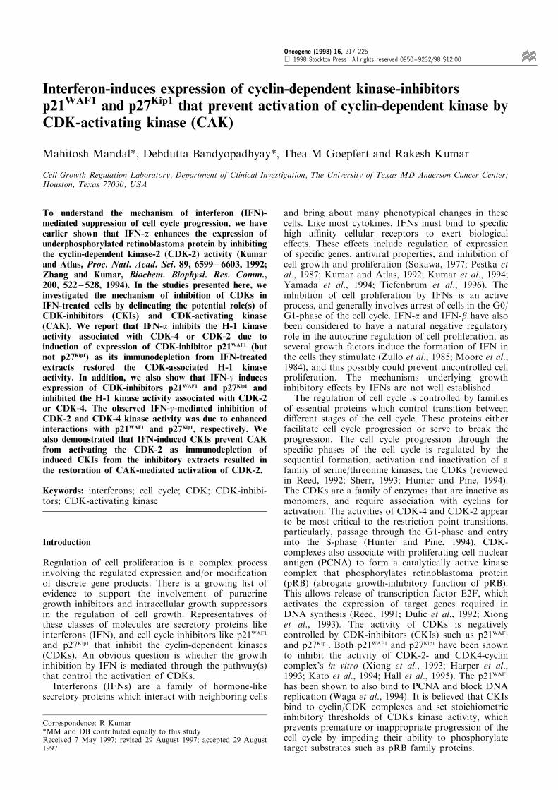
To understand the mechanism of IFN-mediatedgrowth-inhibition of cells in the G0/G1-phase of thecell-cycle, we and others have earlier shown that IFN-a-treatment of Daudi cells leads to enhanced expres-sion of under-phosphorylated form of pRB (Kumarand Atlas, 1992; Resnitzky et al., 1992). To illustratethis e�ect, Figure 1 shows the kinetics of inhibition ofpRB phosphorylation (Figure 1a) and inhibition ofCDK-2 associated H-1 kinase activity without anye�ect on CDK-2 expression (Figure 1b). Results inFigure 1a show that in exponentially growing cells, wecan resolve three di�erent electrophoretically migratingforms and IFN-a predominantly inhibits the expressionof slowest migrating form (Figure 1a, lane 1, bandnumber 1) and enhances the expression of intermediatemigrating band (band number 2) as early as 6 h post-treatment. However, at 24 h post-treatment, there was
0 3 6 12 24
IFN-α (h)
RBP
RB
1 2
3
0 3 6 12 24
— CDK2
— H-1
IFN-α (h)
a
b
Figure 1 IFN-a inhibits pRB phosphorylation and CDK-2kinase activity. (a and b) Daudi cells were treated with IFN-a(500 U/ml). Cell extracts (30 mg protein) were blotted with anti-RB mAb or anti-CDK-2 mAb. Extracts (50 mg protein) were alsoimmunoprecipitated (IP) with anti-CDK-2 Ab, and assayed forH-1 kinase activity
Regulation of CDKs in IFN-treated cellsM Mandal et al
218
also enhancement of expression of slowest migratingpRB band (Figure 1a, 24 h, band number 3). In brief,these results suggested that IFN-a may have di�erentiale�ect on the expression of di�erent forms of RBprotein, presumably by targeting di�erent CDKscomplexes at di�erent stages of cell cycle.
Induction of expression of CDK-inhibitor p21WAF1 andinhibition of CDK-associated H-1 kinase activity in IFN-a treated cells
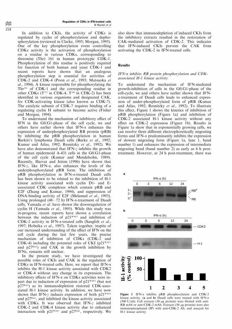
Understand the possible basis of IFN-a mediatedinhibition of CDK kinase activity in Daudi cells, weinvestigated the possibility of regulation of CKIsinhibitors-p21WAF1 and p27Kip1 by IFNs. Figure 2ashows that IFN-a induces the expression of p21WAF1
(lower band, marked by *) starting at 6 h post-treatment. The levels of induced p21WAF1 proteinpersisted up to 48 h after IFN-a-treatment of Daudicells. There was no e�ect of IFN-a on p27Kip1
expression in Daudi cells (data not shown). Sincep21WAF1 is known to inhibit the H-1 kinase activityassociated with CDK-2 or CDK-4 (Clarke, 1995), we
examined the e�ect of IFN-a on the inhibition of bothCDK-2 and CDK-4 kinase activity, and investigatedwhether depletion of induced p21WAF1 would have anye�ect on CDK-2- and CDK-4-associated kinaseactivity using H-1 as a substrate. Results in Figure 2also show that IFN-a di�erentially inhibited the H-1kinase activity associated with CDKs as CDK-2- andCDK-4-associated H-1 kinase activities were inhibitedby 3 h and 6 h (Figure 2b, lanes 1 ± 5), respectively. Inaddition, data in Figure 2b (lanes 6 ± 10) also illustratethat the depletion of p21WAF1 before immunoprecipita-tion (IP) with CDKs could partially restore theinhibition of CDKs kinase activity. The observedIFN-a-mediated early inhibition (6 ± 24 h treatment)of CDK-4-associated H-1 kinase activity was not dueto the inhibitory e�ect of CDK-4 expression, as IFN-a-treatment did not inhibit the expression of CDK-4protein upto 24 h treatment (Figure 2c). Results inFigure 2d show that the induced p21WAF1 also interactswith CDK-4, in addition to the reported interactionwith CDK-2 (Poon et al., 1993; Matsuoka et al., 1994).Since the kinetics of restoration (due to depletion ofp21WAF1) of H-1 activity associated with CDK-2 or
0 3 6 12 24
IFN-α (h)
1 2 3 4 5
— HSP-70
— p21WAF1
0 3 6 12 24
1 2 3 4 5
0 3 6 12 24
– +p21 depletion
IFN-α (h)
IP: CDK2 H-1 kinase
IP: CDK4 H-1 kinase
6 7 8 9 10
0 12 24 48 72
CDK4 —
1 2 3 4 5
IFN-α (h)C α
— CDK4
— p21WAF1
1 2
c d
b
a
Figure 2 IFN-a induces the expression of p21WAF1 in Daudi cells. (a) Induction of p21WAF1. Daudi cells were treated with IFN-a,and cell extracts (30 mg protein) were immunoblotted with a anti-p21WAF1 mAb (a). The position of p21WAF1 is shown by *, lowerband. As an internal control, the upper part of the same blot was probed with an unrelated anti-heat shock protein (HSP)-70 mAb.(b) E�ect of immunodepletion of p21WAF1 on H-1 activity associated with CDK-2 or CDK-4. Extracts (50 mg) from a were IP withanti-CDK-2 or anti-CDK-4 mAb. Samples in lanes 6 ± 10 were ®rst IP with a anti-p21WAF1 mAb before IP with anti-CDK mAbs.Precipitated CDKs were subjected to H-1 kinase assay. (c) E�ect of IFN-a on the expression of CDK-4. Daudi cells were treatedwith IFN-a for di�erent times, and extracts (30 mg) were blotted with a anti-CDK-4 mAb. (d) Enhanced interaction betweenp21WAF1 and CDK-4. Daudi cells treated with IFN-a (24 h) were immunoprecipitated with CDK-4 and blotted with an anti-p21WAF1 mAb (lower panel) or anti-CDK-4 mAb (upper panel)
Regulation of CDKs in IFN-treated cellsM Mandal et al
219
CDK-4 was in-parallel with the kinetics of p21WAF1
induction, these results suggested that the observedinhibition of CDK-2 or CDK-4 kinase activity in IFN-a-treated Daudi cells may be due to conditionalinduction of CDK-inhibitor p21WAF1 by IFN-a.
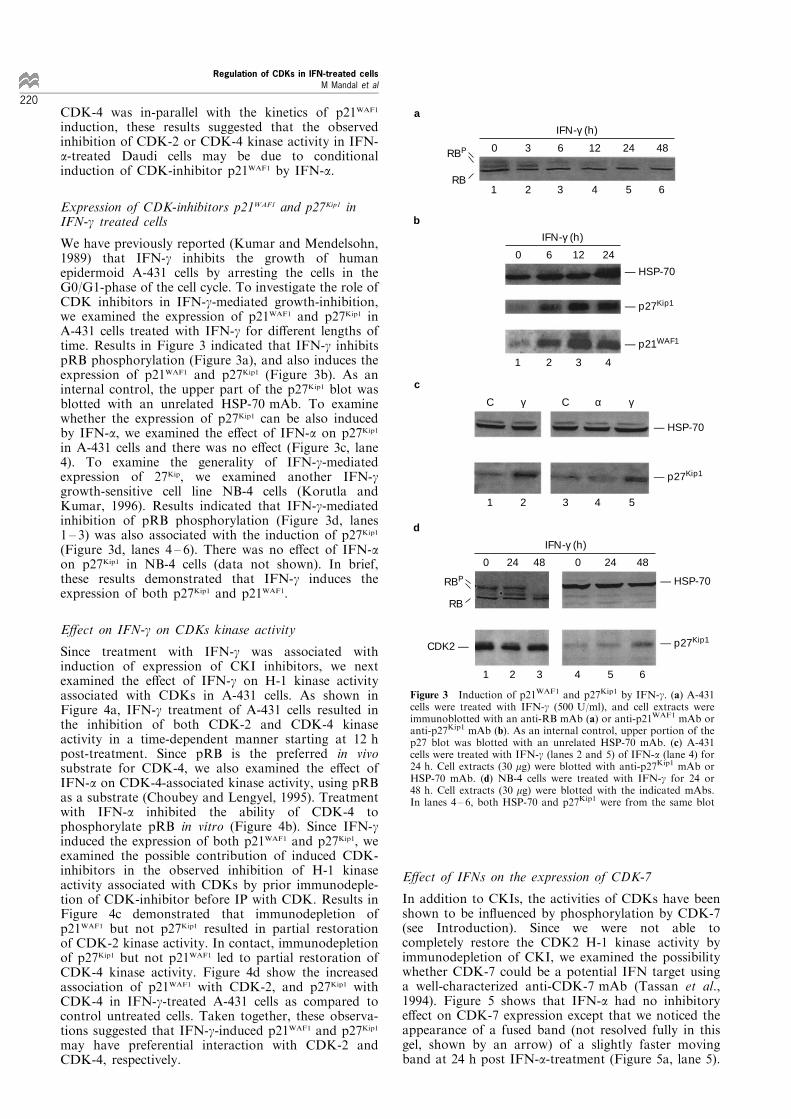
Expression of CDK-inhibitors p21WAF1 and p27Kip1 inIFN-g treated cells
We have previously reported (Kumar and Mendelsohn,1989) that IFN-g inhibits the growth of humanepidermoid A-431 cells by arresting the cells in theG0/G1-phase of the cell cycle. To investigate the role ofCDK inhibitors in IFN-g-mediated growth-inhibition,we examined the expression of p21WAF1 and p27Kip1 inA-431 cells treated with IFN-g for di�erent lengths oftime. Results in Figure 3 indicated that IFN-g inhibitspRB phosphorylation (Figure 3a), and also induces theexpression of p21WAF1 and p27Kip1 (Figure 3b). As aninternal control, the upper part of the p27Kip1 blot wasblotted with an unrelated HSP-70 mAb. To examinewhether the expression of p27Kip1 can be also inducedby IFN-a, we examined the e�ect of IFN-a on p27Kip1
in A-431 cells and there was no e�ect (Figure 3c, lane4). To examine the generality of IFN-g-mediatedexpression of 27Kip, we examined another IFN-ggrowth-sensitive cell line NB-4 cells (Korutla andKumar, 1996). Results indicated that IFN-g-mediatedinhibition of pRB phosphorylation (Figure 3d, lanes1 ± 3) was also associated with the induction of p27Kip1
(Figure 3d, lanes 4 ± 6). There was no e�ect of IFN-aon p27Kip1 in NB-4 cells (data not shown). In brief,these results demonstrated that IFN-g induces theexpression of both p27Kip1 and p21WAF1.
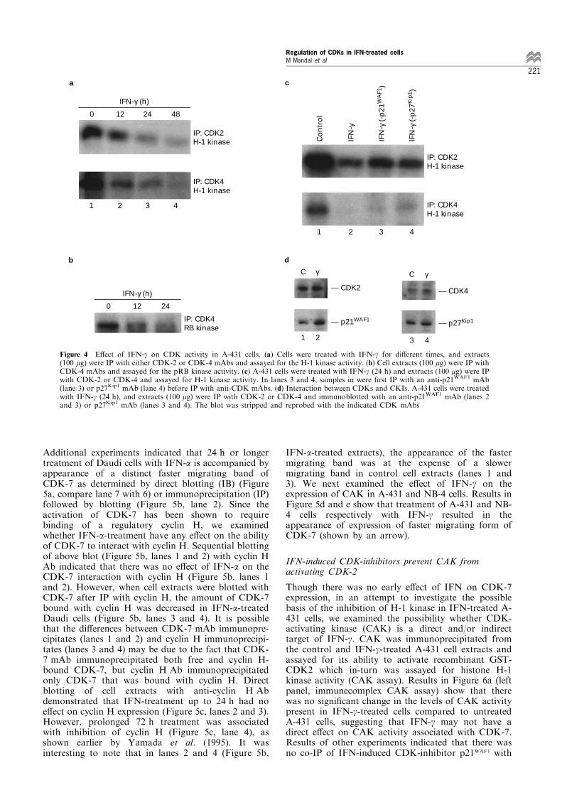
E�ect on IFN-g on CDKs kinase activity
Since treatment with IFN-g was associated withinduction of expression of CKI inhibitors, we nextexamined the e�ect of IFN-g on H-1 kinase activityassociated with CDKs in A-431 cells. As shown inFigure 4a, IFN-g treatment of A-431 cells resulted inthe inhibition of both CDK-2 and CDK-4 kinaseactivity in a time-dependent manner starting at 12 hpost-treatment. Since pRB is the preferred in vivosubstrate for CDK-4, we also examined the e�ect ofIFN-a on CDK-4-associated kinase activity, using pRBas a substrate (Choubey and Lengyel, 1995). Treatmentwith IFN-a inhibited the ability of CDK-4 tophosphorylate pRB in vitro (Figure 4b). Since IFN-ginduced the expression of both p21WAF1 and p27Kip1, weexamined the possible contribution of induced CDK-inhibitors in the observed inhibition of H-1 kinaseactivity associated with CDKs by prior immunodeple-tion of CDK-inhibitor before IP with CDK. Results inFigure 4c demonstrated that immunodepletion ofp21WAF1 but not p27Kip1 resulted in partial restorationof CDK-2 kinase activity. In contact, immunodepletionof p27Kip1 but not p21WAF1 led to partial restoration ofCDK-4 kinase activity. Figure 4d show the increasedassociation of p21WAF1 with CDK-2, and p27Kip1 withCDK-4 in IFN-g-treated A-431 cells as compared tocontrol untreated cells. Taken together, these observa-tions suggested that IFN-g-induced p21WAF1 and p27Kip1
may have preferential interaction with CDK-2 andCDK-4, respectively.
E�ect of IFNs on the expression of CDK-7
In addition to CKIs, the activities of CDKs have beenshown to be in¯uenced by phosphorylation by CDK-7(see Introduction). Since we were not able tocompletely restore the CDK2 H-1 kinase activity byimmunodepletion of CKI, we examined the possibilitywhether CDK-7 could be a potential IFN target usinga well-characterized anti-CDK-7 mAb (Tassan et al.,1994). Figure 5 shows that IFN-a had no inhibitorye�ect on CDK-7 expression except that we noticed theappearance of a fused band (not resolved fully in thisgel, shown by an arrow) of a slightly faster movingband at 24 h post IFN-a-treatment (Figure 5a, lane 5).
0 3 6 12 24 48
1 2 3 4 5 6
IFN-γ (h)
RBP
RB
0 6 12 24
IFN-γ (h)
— HSP-70
— p27Kip1
— p21WAF1
1 2 3 4
C γ C α γ
— HSP-70
— p27Kip1
1 2 3 4 5
0 24 48 0 24 48
IFN-γ (h)
— HSP-70
— p27Kip1
1 2 3 4 5 6
RBP
RB
CDK2 —
d
c
b
a
Figure 3 Induction of p21WAF1 and p27Kip1 by IFN-g. (a) A-431cells were treated with IFN-g (500 U/ml), and cell extracts wereimmunoblotted with an anti-RB mAb (a) or anti-p21WAF1 mAb oranti-p27Kip1 mAb (b). As an internal control, upper portion of thep27 blot was blotted with an unrelated HSP-70 mAb. (c) A-431cells were treated with IFN-g (lanes 2 and 5) of IFN-a (lane 4) for24 h. Cell extracts (30 mg) were blotted with anti-p27Kip1 mAb orHSP-70 mAb. (d) NB-4 cells were treated with IFN-g for 24 or48 h. Cell extracts (30 mg) were blotted with the indicated mAbs.In lanes 4 ± 6, both HSP-70 and p27Kip1 were from the same blot
Regulation of CDKs in IFN-treated cellsM Mandal et al
220
Additional experiments indicated that 24 h or longertreatment of Daudi cells with IFN-a is accompanied byappearance of a distinct faster migrating band ofCDK-7 as determined by direct blotting (IB) (Figure5a, compare lane 7 with 6) or immunoprecipitation (IP)followed by blotting (Figure 5b, lane 2). Since theactivation of CDK-7 has been shown to requirebinding of a regulatory cyclin H, we examinedwhether IFN-a-treatment have any e�ect on the abilityof CDK-7 to interact with cyclin H. Sequential blottingof above blot (Figure 5b, lanes 1 and 2) with cyclin HAb indicated that there was no e�ect of IFN-a on theCDK-7 interaction with cyclin H (Figure 5b, lanes 1and 2). However, when cell extracts were blotted withCDK-7 after IP with cyclin H, the amount of CDK-7bound with cyclin H was decreased in IFN-a-treatedDaudi cells (Figure 5b, lanes 3 and 4). It is possiblethat the di�erences between CDK-7 mAb immunopre-cipitates (lanes 1 and 2) and cyclin H immunoprecipi-tates (lanes 3 and 4) may be due to the fact that CDK-7 mAb immunoprecipitated both free and cyclin H-bound CDK-7, but cyclin H Ab immunoprecipitatedonly CDK-7 that was bound with cyclin H. Directblotting of cell extracts with anti-cyclin H Abdemonstrated that IFN-treatment up to 24 h had noe�ect on cyclin H expression (Figure 5c, lanes 2 and 3).However, prolonged 72 h treatment was associatedwith inhibition of cyclin H (Figure 5c, lane 4), asshown earlier by Yamada et al. (1995). It wasinteresting to note that in lanes 2 and 4 (Figure 5b,
IFN-a-treated extracts), the appearance of the fastermigrating band was at the expense of a slowermigrating band in control cell extracts (lanes 1 and3). We next examined the e�ect of IFN-g on theexpression of CAK in A-431 and NB-4 cells. Results inFigure 5d and e show that treatment of A-431 and NB-4 cells respectively with IFN-g resulted in theappearance of expression of faster migrating form ofCDK-7 (shown by an arrow).
IFN-induced CDK-inhibitors prevent CAK fromactivating CDK-2
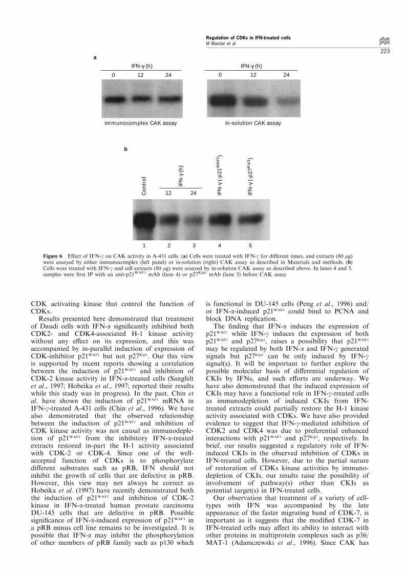
Though there was no early e�ect of IFN on CDK-7expression, in an attempt to investigate the possiblebasis of the inhibition of H-1 kinase in IFN-treated A-431 cells, we examined the possibility whether CDK-activating kinase (CAK) is a direct and/or indirecttarget of IFN-g. CAK was immunoprecipitated fromthe control and IFN-g-treated A-431 cell extracts andassayed for its ability to activate recombinant GST-CDK2 which in-turn was assayed for histone H-1kinase activity (CAK assay). Results in Figure 6a (leftpanel, immunecomplex CAK assay) show that therewas no signi®cant change in the levels of CAK activitypresent in IFN-g-treated cells compared to untreatedA-431 cells, suggesting that IFN-g may not have adirect e�ect on CAK activity associated with CDK-7.Results of other experiments indicated that there wasno co-IP of IFN-induced CDK-inhibitor p21WAF1 with
0 12 24 48
IFN-γ (h)
IP: CDK2 H-1 kinase
IP: CDK4 H-1 kinase
1 2 3 4
0 12 24
IFN-γ (h)
IP: CDK4 RB kinase
C γ C γ
— CDK2
— p21WAF1
1 2
— CDK4
— p27Kip1
3 4
1 2 3 4
Co
ntr
ol
IFN
-γ
IFN
-γ (
-p21
WA
F1)
IFN
-γ (
-p27
Kip
1 )
IP: CDK2 H-1 kinase
IP: CDK4 H-1 kinase
a
b
c
d
Figure 4 E�ect of IFN-g on CDK activity in A-431 cells. (a) Cells were treated with IFN-g for di�erent times, and extracts(100 mg) were IP with either CDK-2 or CDK-4 mAbs and assayed for the H-1 kinase activity. (b) Cell extracts (100 mg) were IP withCDK-4 mAbs and assayed for the pRB kinase activity. (c) A-431 cells were treated with IFN-g (24 h) and extracts (100 mg) were IPwith CDK-2 or CDK-4 and assayed for H-1 kinase activity. In lanes 3 and 4, samples in were ®rst IP with an anti-p21WAF1 mAb(lane 3) or p27Kip1 mAb (lane 4) before IP with anti-CDK mAbs. (d) Interaction between CDKs and CKIs. A-431 cells were treatedwith IFN-g (24 h), and extracts (100 mg) were IP with CDK-2 or CDK-4 and immunoblotted with an anti-p21WAF1 mAb (lanes 2and 3) or p27Kip1 mAb (lanes 3 and 4). The blot was stripped and reprobed with the indicated CDK mAbs
Regulation of CDKs in IFN-treated cellsM Mandal et al
221
CDK-7 (data not shown). In addition to theimmunecomplex CAK assay, we also assayed CAKactivity by in-solution method that allows othercomponents of cell lysate to in¯uence the activationof GST-CDK2 by CAK. In contrast to the results fromthe immunecomplex CAK assay, results in Figure 6a(right panel) indicated that IFN-g treatment wasassociated with the inhibition CAK activity. Ascontrol, GST-beads were treated in an identicalmanner and there was no e�ect (data not shown).Since IFN-g induces expression of CDK-inhibitors, wereasoned that the observed inhibition of in-solutionCAK activity in IFN-treated A-431 cell extracts maybe due to induced expression of CDK-inhibitors whichmay prevent CAK from phosphorylating GST-CDK2.Data in Figure 6b show that immunedepletion ofp21WAF1 (lane 4) or p27Kip1 (lane 5) from the IFN-g-treated (24 h) inhibitory cell extract (lane 3) resulted in
partial restoration of CAK activity, suggesting thatIFN-induced CDK-inhibitors prevents the activation ofGST-CDK2 by CAK.
Discussion
IFNs are potent regulators of cellular proliferation.The growth inhibitory action of IFN is an activeprocess, and involves suppression of the cell cycleprogression. Both IFN-a and IFN-g have been shownto arrest human cells in the G0/G1 phase of the cellcycle. Earlier we and others have shown that IFNinhibits pRB protein phosphorylation by inhibitingCDK-2 kinase activity. We have undertaken thepresent investigation to further delineate the basis ofIFN-mediated inhibition of CDK activity by examiningthe role(s) of pathways such as CDK-inhibitors and
Regulation of CDKs in IFN-treated cellsM Mandal et al
222
CDK activating kinase that control the function ofCDKs.Results presented here demonstrated that treatment
of Daudi cells with IFN-a signi®cantly inhibited bothCDK2- and CDK4-associated H-1 kinase activitywithout any e�ect on its expression, and this wasaccompanied by in-parallel induction of expression ofCDK-inhibitor p21WAF1 but not p27Kip1. Our this viewis supported by recent reports showing a correlationbetween the induction of p21WAF1 and inhibition ofCDK-2 kinase activity in IFN-a-treated cells (Sangfeltet al., 1997; Hobeika et al., 1997; reported their resultswhile this study was in progress). In the past, Chin etal. have shown the induction of p21WAF1 mRNA inIFN-g-treated A-431 cells (Chin et al., 1996). We havealso demonstrated that the observed relationshipbetween the induction of p21WAF1 and inhibition ofCDK kinase activity was not causal as immunodeple-tion of p21WAF1 from the inhibitory IFN-a-treatedextracts restored in-part the H-1 activity associatedwith CDK-2 or CDK-4. Since one of the well-accepted function of CDKs is to phosphorylatedi�erent substrates such as pRB, IFN should notinhibit the growth of cells that are defective in pRB.However, this view may not always be correct asHobeika et al. (1997) have recently demonstrated boththe induction of p21WAF1 and inhibition of CDK-2kinase in IFN-a-treated human prostate carcinomaDU-145 cells that are defective in pRB. Possiblesigni®cance of IFN-a-induced expression of p21WAF1 ina pRB minus cell line remains to be investigated. It ispossible that IFN-a may inhibit the phosphorylationof other members of pRB family such as p130 which
is functional in DU-145 cells (Peng et al., 1996) and/or IFN-a-induced p21WAF1 could bind to PCNA andblock DNA replication.The ®nding that IFN-a induces the expression of
p21WAF1 while IFN-g induces the expression of bothp21WAF1 and p27Kip1, raises a possibility that p21WAF1
may be regulated by both IFN-a and IFN-g generatedsignals but p27Kip1 can be only induced by IFN-gsignal(s). It will be important to further explore thepossible molecular basis of di�erential regulation ofCKIs by IFNs, and such e�orts are underway. Wehave also demonstrated that the induced expression ofCKIs may have a functional role in IFN-g-treated cellsas immunodepletion of induced CKIs from IFN-treated extracts could partially restore the H-1 kinaseactivity associated with CDKs. We have also providedevidence to suggest that IFN-g-mediated inhibition ofCDK2 and CDK4 was due to preferential enhancedinteractions with p21WAF1 and p27Kip1, respectively. Inbrief, our results suggested a regulatory role of IFN-induced CKIs in the observed inhibition of CDKs inIFN-treated cells. However, due to the partial natureof restoration of CDKs kinase activities by immuno-depletion of CKIs, our results raise the possibility ofinvolvement of pathway(s) other than CKIs aspotential target(s) in IFN-treated cells.Our observation that treatment of a variety of cell-
types with IFN was accompanied by the lateappearance of the faster migrating band of CDK-7, isimportant as it suggests that the modi®ed CDK-7 inIFN-treated cells may a�ect its ability to interact withother proteins in multiprotein complexes such as p36/MAT-1 (Adamczewski et al., 1996). Since CAK has
0 12 24
IFN-γ (h)
Immunocomplex CAK assay
0 12 24
IFN-γ (h)
In-solution CAK assay
1 2 3 4 5
Co
ntr
ol
IFN
-γ (
-p21
WA
F1)
IFN
-γ (
-p27
Kip
1 )
12 24
IFN
-γ (h
)
b
a
Figure 6 E�ect of IFN-g on CAK activity in A-431 cells. (a) Cells were treated with IFN-g for di�erent times, and extracts (80 mg)were assayed by either immunocomplex (left panel) or in-solution (right) CAK assay as described in Materials and methods. (b)Cells were treated with IFN-g and cell extracts (80 mg) were assayed by in-solution CAK assay as described above. In lanes 4 and 5,samples were ®rst IP with an anti-p21WAF1 mAb (lane 4) or p27Kip1 mAb (lane 5) before CAK assay
Regulation of CDKs in IFN-treated cellsM Mandal et al
223
been shown to be responsible for the C-terminaldomain (CTD) kinase activity associated with amultiprotein transcription factor complex TFIIH(Feaver et al., 1994), it is possible that IFN-mediatedappearance of the faster migrating CAK form mayin¯uence the CTD kinase activity due to possiblemodulation of interactions between components ofCAK (CDK-7, cyclin H and p36/MAT1) of TFIIH(p89 and p62). (Devault et al., 1995). Furtherinvestigations are required to examine these possibi-lities.Another notable ®nding of this study was the
indirect inhibitory e�ect of IFNs on CAK activity.Although recent studies have demonstrated theinduction of p21WAF1 by IFN-a but the mechanismthrough which CDK-inhibitors inhibit CDK in IFN-treated cells remains poorly understood. Since activa-tion of CDKs requires an essential phosphorylationstep by CAK, we therefore examined the hypothesisthat IFN-induced CDK-inhibitors may interfere withthe activation of CDK by CAK. In agreement with thisview, our results have shown that the presence of aCAK inhibitor(s) in IFN-treated extracts, and immu-nodepletion of p21WAF1 or p27Kip1 from the inhibitoryIFN-treated extracts resulted in part restoration ofCAK-mediated activation of CDK-2. Taken together,these results suggested that IFN-induced CDK-inhibitors prevent CAK from activating the CDKholoenzyme.
Materials and methods
Cell culture
Human Burkitt's lymphoma Daudi cells (Kumar andAtlas, 1992) and acute promyelocytic leukemia NB-4 cells(Korutla and Kumar, 1996) were grown in RPMI-1640medium containing 10% fetal bovine medium. Humanepidermoid carcinoma A-431 cells (Kumar and Mendel-sohn, 1989) were maintained in Dulbecco's modi®edeagle's medium (DMEM) supplemented with 10% fetalbovine serum. Human recombinant IFN-a 2a (Speci®cactivity, 56107 IU/mg protein) and recombinant IFN-g(Speci®c activity 46106 IU/mg protein) was obtained fromthe Ho�man La Roche Inc. and Genentech Inc.,respectively.
Cell extracts and immunoblotting
All experiments were performed with cells in logarithmicphase by controlling the plating density. Cells' extractswere prepared as described (Kumar and Atlas, 1992). Celllysates containing equal amount of total protein (15 ±30 mg) were resolved on a 7% (for RB protein) or 10% (forother proteins) SDS ± PAGE, followed by immunoblottingusing alkaline phosphatase or 125I-protein A or ECLmethod (Mandal et al., 1996). As an internal control, thesame blot was reprobed with an unrelated antibody. Low-molecular-mass colored markers (Amersham Corp.) wereused as molecular weight standards. The followingantibodies were used purchased from the Neomarkers,Inc: anti-RB mAb (clone IF8/Rb-1), anti-p21 mAb (cloneDCS 60.2), anti-p27 mAb (clone DCS 72.F6), anti-CDK-7mAb (clone MO 1-1), and anti-HSP70 mAb (clone BMR22). Anti-CDK2 mAb (clone 55) and anti-CDK4 mAb(clone 97) were from the Transduction Laboratories.Antibody against cyclin H (SC-609) was purchased from
the Santa Cruz, Inc. Quantitation of speci®c protein bandswas performed by using protein databases scanner(Molecular Dynamics).
Histone H-1 kinase assay
Speci®c CDK was immunoprecipitated from 100 mg totalprotein by using a speci®c Ab as described (Zhang andKumar, 1994). Assay of CDK kinase is based on thetransfer of 32P from labeled ATP by CDK kinase todephosphorylated H-1 histone. The kinase reaction mixturecontained 25 mM glycerophosphate pH 7.3, 10 mM EGTA,10 mM MgCl2, 1 mM DTT, 25 mM HEPES pH 7.3, 5 mg ofH-1 histone, 100 pmole ATP and 5 mCi [32P]-ATP in avolume of 25 ml. Reaction product was resolved onto a gel,transferred on a nitrocellulose and subjected to autoradio-graphy. Subsequently the blot was immunoblotted with aspeci®c CDK Ab to evaluate the immunoprecipitatione�ciency.
Expression and puri®cation of recombinant proteins
GST-CDK2 and Histidine-tagged Protein A-cyclin A (PA-cyclin A) were generous gifts from Randy Poon anddescribed previously (Poon et al., 1993). GST-CDK2 wasexpressed in E. coli, cultures were grown up to log phaseand expression of recombinant GST-CDK2 was inducedwith isopropyl-b-D-thiogalactopyranoside (0.1 mM) for6 h. Cells were lysed, sonicated and centrifuged at 10000 g for 30 min as described (Poon et al., 1993). GST-CDK2 was puri®ed from the supernatant using glu-tathione-Sepharose 4B protein puri®cation kit from thePharmacia Fine Chemicals as per manufacturer's instruc-tions. Histidine-tagged Protein A-cyclin A (PA-cyclin A)was also expressed in E. coli and puri®ed by NTA-Ni2+
agarose a�nity chromatography following a kit from theQiagen.
CAK Assay
Immumne-complex CAK assay Cell lysates are IP withanti-CDK-7 mAb, immunoprecipitated complexes (sourceof CDK-7) were washed with 36lysis bu�er and 36 withCAK kinase bu�er, resuspended in 50 ml of CAK bu�er(Poon et al., 1993), and 0.2 mg of bacterially producedcyclin A and GST-CDK2 in CAK bu�er containing 1 mM
ATP. The mixture was incubated at 228C for 1 h (CAK-mediated activation step). Activated cyclin-CDK com-plexes in the supernatant were recovered onto glu-tathione-Sepharose beads, washed with kinase bu�er, andassayed for H-1 kinase activity.
Soluble CAK assay Cell extracts (100 mg protein, sourceof CDK-7) were mixed with 0.2 mg of bacterially producedcyclin A and GST-CDK-2 in CAK bu�er containing 1 mM
ATP. The mixture was incubated at 228C for 1 h (CAK-mediated activation step). Activated cyclin-CDK com-plexes were recovered onto glutathione-Sepharose beads,washed with kinase bu�er, and assay for H-1 kinaseactivity.
AcknowledgementsWe thank Randy Poon for a generous gift of recombinantPA-cyclin A and GST-CDK2 plasmids, and helpfulsuggestions to establish the CAK assay in our laboratory.We also thank Divakar Choubey for providing us with thepuri®ed recombinant pRB. This work was supported inpart by National Institutes of Health grant CA56564.
Regulation of CDKs in IFN-treated cellsM Mandal et al
224
References
Adamczewski JP, Rossignol M, Tassan JP, Nigg EA,Moncollin V and Egly JM. (1996). EMBO J., 15, 1877 ±1884.
Burke LC, Bybee A and Thomas NS. (1992). Oncogene, 7,783 ± 788.
Chin YE, Kitagawa M, Su W-S, You Z-H, Iwamoto Y andFu XY. (1996) Science, 272, 719 ± 722.
Clarke PR. (1995). Current Biol., 5, 40 ± 42.Choubey D and Lengyel P. (1995). J. Biol. Chem., 270,6134 ± 6140.
Devault A, Martinez AC, Fesquet D, Labbe JC et al. (1995).EMBO J., 15, 5027 ± 5036.
Dulic V, Lees E and Reed SI. (1992). Science, 257, 1958 ±1961.
Feaver WJ, Svejstrup JQ, Henry NL and Kornberg RD.(1994). Cell, 79, 1103 ± 1109.
Fisher RP and Morgan DO. (1994). Cell, 78, 713 ± 724.Hall M, Bates S and Peters G. (1995). Oncogene, 11, 1581 ±1588.
Harper JW, Adami GR, Wei N, Keyomarsi K and ElledgeSJ. (1993). Cell, 75, 805 ± 816.
Hobeika AC, Subramaniam PS and Johnson HM. (1997).Oncogene, 14, 1165 ± 1170.
Harvet BL and Jetten AM. (1996). Cell Growth & Di�., 7,289 ± 300.
Hunter T and Pines J. (1994). Cell, 79, 573 ± 582.Kato JY, Matsuoka M, Polyak K, Massague J, Sherr CJ.(1994). Cell, 79, 487 ± 496.
Korutla L and Kumar R. (1996). Anticancer Res., 16, 2789 ±2796.
Kumar R and Mendelsohn J. (1989). Cancer Res., 49, 5180 ±5184.
Kumar R and Atlas I. (1992). Proc. Natl. Acad. Sci. USA, 89,6599 ± 6603.
Kumar R, Korutla L and Zhang K. (1994). J. Biol. Chem.,269, 25437 ± 25441.
Mandal M, Maggirwar SB, Sharma N, Kaufmann SH, SunSC and Kumar R. (1996). J. Biol. Chem., 271, 30354 ±30359.
Matsuoka M, Kato J, Fisher RP, Morgan DO and Sherr CJ.(1994). Mol. Cell. Biol., 14, 7265 ± 7275.
Melamed DN, Tiefenbrun A, Yarden A and Kimchi A.(1993). Mol. Cell. Biol., 13, 5255 ± 5265.
Moore RN, Larsen HS, Horohov DW and Rouse BT. (1984).Science, 223, 178 ± 180.
Morgan DO. (1995). Nature, 374, 131 ± 134.Pestka S, Langer JA, Zoon KC and Samuel CE. (1987). Ann.
Rev. Biochem., 56, 727 ± 777.Peng D, Fan X, Lu Y, Deblasio T, Scher H and MendelsohnJ. (1996). Can. Res., 56, 3666 ± 3669.
Poon RY, Yamashita K, Adamczewski JP, Hunt T andShuttleworth J. (1993). EMBO J., 12, 3123 ± 3132.
Reed SI. (1991). Trends Genet., 7, 95 ± 99.Reed SI. (1992). Ann. Rev. Cell. Biol., 8, 529 ± 561.Resnitzky D, Tiefenbrun N, Berissi H and Kimchi A. (1992).
Proc. Natl. Acad. Sci. USA, 89, 402 ± 406.Sangfelt O, Erickson S, Einhorn S and Grander D. (1997).
Oncogene, 14, 415 ± 423.Seroz T, Hwang JR, Moncollin V and Egly JM. (1995). Curr.
Opin. Genet. Dev., 5, 217 ± 221.Sherr CJ. (1993). Cell, 73, 1059 ± 1065.Sokawa Y, Watanabe Y, Watanabe Y and Yawada Y.(1977). Nature, 268, 236 ± 238.
Tassan JP, Schultz SJ, Bartek J and Nigg EA. (1994). J. CellBiol., 127, 467 ± 478.
Tiefenbrun N, Malamed D, Levy N, Resnitzky D, Ho�mannI, Reed SI and Kimchi A. (1996). Mol. Cell. Biol., 16,3934 ± 3944.
Waga S, Hannon GJ, Beach D and Stillman B. (1994).Nature, 369, 574 ± 578.
Xiong Y, Hannon GJ, Zhang H, Casso D, Kobayashi R andBeach D. (1993). Nature, 366, 701 ± 704.
Yamada H, Ochi K, Nakada S, Memot T and Horiguchi YJ.(1994). Mol. Cellular Biochem., 136, 117 ± 123.
Yamada H, Ochi K, Nakada S, Takahara S, Nemoto T,Sekikawwa T and Horiguchi YJ. (1995). Mol. CellularBiochem., 152, 149 ± 158.
Zhang K and Kumar R. (1994). Biochem. Biophysi. Res.Comm., 200, 522 ± 528.
Zullo J, Cochran BH, Huang A and Stiles CD. (1985). Cell,43, 793 ± 800.
Regulation of CDKs in IFN-treated cellsM Mandal et al
225
Related Documents


![Elevated Cyclins and Cyclin-dependent Kinase Activity in ...[CANCER RESEARCH 58, 2042-2049, May I, 1998] Elevated Cyclins and Cyclin-dependent Kinase Activity in the Rhabdomyosarcoma](https://static.cupdf.com/doc/110x72/5e4e63ca3358114ff2317f00/elevated-cyclins-and-cyclin-dependent-kinase-activity-in-cancer-research-58.jpg)