Chapter 18 Interceptive Orthodontics — Current Evidence Maen H. Zreaqat Additional information is available at the end of the chapter http://dx.doi.org/10.5772/59256 1. Introduction From evidence found in human skulls, crooked teeth have been around since the time of Neanderthal man (about 50,000 BC), but it was not until about 3000 years ago that we had the first written record of attempts to correct crowded or protruding teeth. Long before braces, long before the word orthodontics” was coined, it was known that teeth moved in response to pressure. Primitive (and surprisingly well-designed) orthodontic appliances have been found with Greek and Etruscan artifacts. Archaeologists have discovered Egyptian mummies with crude metal bands wrapped around individual teeth. It is speculated that catgut was used to close the gaps [1]. The earliest description of irregularities of the teeth was given about 400 BC by Hippocrates (460-377 BC). The first treatment of an irregular tooth was recorded by Celsus (25 BC-50 AD), a Roman writer, who said, “If a second tooth should happen to grow in children before the first has fallen out, that which ought to be shed is to be drawn out and the new one daily pushed toward its place by means of the finger until it arrives at its just proportion.” A clear mechanical treatment was advocated by Pliny the Elder (23-79 AD), who suggested filing elongated teeth to bring them into proper alignment. This method remained in practice until the 1800s [2]. Dentistry entered a period of marked decline during middle ages (5 th to 15 th centuries), as did all sciences. After the 16th century, considerable progress was made. Matthaeus Gottfried Purmann (1692) was the first to report taking wax impressions. In 1756, Phillip Pfaff used plaster of Paris impressions. Malocclusions were called “irregularities” of the teeth, and their correction was termed “regulating.” It remained for the Enlightenment to reawaken the spirit of scientific thought necessary to advance dentistry and other disciplines. Beginning in the 18th century, Pierre Fauchard (1678-1761) was leading efforts in the field of dentistry. He has been called the “Father of Orthodontia.” He was the first to remove dentistry from the bonds of empiricism and put it on a scientific foundation. In 1728, he published the first general work on dentistry, a 2-volume opus entitled "The Surgeon Dentist: A Treatise on the Teeth". © 2015 The Author(s). Licensee InTech. This chapter is distributed under the terms of the Creative Commons Attribution License (http://creativecommons.org/licenses/by/3.0), which permits unrestricted use, distribution, and reproduction in any medium, provided the original work is properly cited.

Welcome message from author
This document is posted to help you gain knowledge. Please leave a comment to let me know what you think about it! Share it to your friends and learn new things together.
Transcript

Chapter 18
Interceptive Orthodontics — Current Evidence
Maen H. Zreaqat
Additional information is available at the end of the chapter
http://dx.doi.org/10.5772/59256
1. Introduction
From evidence found in human skulls, crooked teeth have been around since the time ofNeanderthal man (about 50,000 BC), but it was not until about 3000 years ago that we had thefirst written record of attempts to correct crowded or protruding teeth. Long before braces,long before the word orthodontics” was coined, it was known that teeth moved in responseto pressure. Primitive (and surprisingly well-designed) orthodontic appliances have beenfound with Greek and Etruscan artifacts. Archaeologists have discovered Egyptian mummieswith crude metal bands wrapped around individual teeth. It is speculated that catgut was usedto close the gaps [1]. The earliest description of irregularities of the teeth was given about 400BC by Hippocrates (460-377 BC). The first treatment of an irregular tooth was recorded byCelsus (25 BC-50 AD), a Roman writer, who said, “If a second tooth should happen to growin children before the first has fallen out, that which ought to be shed is to be drawn out andthe new one daily pushed toward its place by means of the finger until it arrives at its justproportion.” A clear mechanical treatment was advocated by Pliny the Elder (23-79 AD), whosuggested filing elongated teeth to bring them into proper alignment. This method remainedin practice until the 1800s [2].
Dentistry entered a period of marked decline during middle ages (5th to 15th centuries), as didall sciences. After the 16th century, considerable progress was made. Matthaeus GottfriedPurmann (1692) was the first to report taking wax impressions. In 1756, Phillip Pfaff usedplaster of Paris impressions. Malocclusions were called “irregularities” of the teeth, and theircorrection was termed “regulating.” It remained for the Enlightenment to reawaken the spiritof scientific thought necessary to advance dentistry and other disciplines. Beginning in the18th century, Pierre Fauchard (1678-1761) was leading efforts in the field of dentistry. He hasbeen called the “Father of Orthodontia.” He was the first to remove dentistry from the bondsof empiricism and put it on a scientific foundation. In 1728, he published the first general workon dentistry, a 2-volume opus entitled "The Surgeon Dentist: A Treatise on the Teeth".
© 2015 The Author(s). Licensee InTech. This chapter is distributed under the terms of the Creative CommonsAttribution License (http://creativecommons.org/licenses/by/3.0), which permits unrestricted use, distribution,and reproduction in any medium, provided the original work is properly cited.
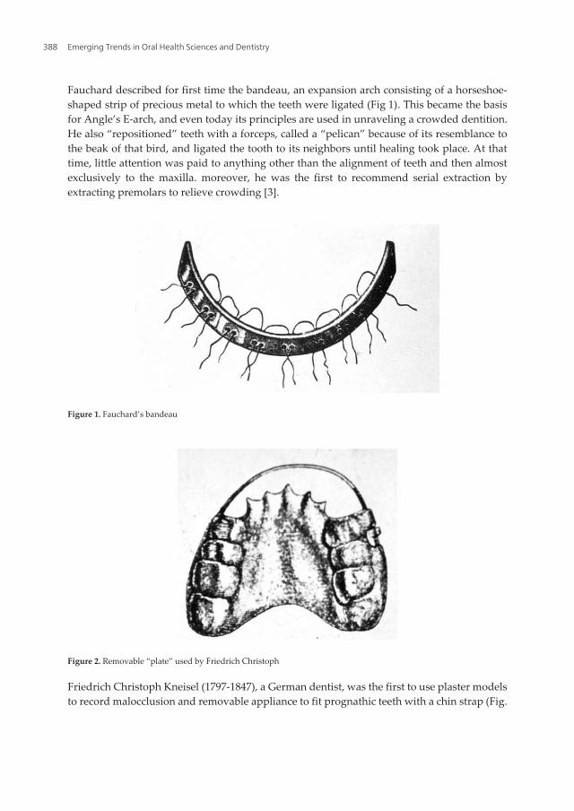
Fauchard described for first time the bandeau, an expansion arch consisting of a horseshoe-shaped strip of precious metal to which the teeth were ligated (Fig 1). This became the basisfor Angle’s E-arch, and even today its principles are used in unraveling a crowded dentition.He also “repositioned” teeth with a forceps, called a “pelican” because of its resemblance tothe beak of that bird, and ligated the tooth to its neighbors until healing took place. At thattime, little attention was paid to anything other than the alignment of teeth and then almostexclusively to the maxilla. moreover, he was the first to recommend serial extraction byextracting premolars to relieve crowding [3].
Figure 1. Fauchard’s bandeau
Figure 2. Removable “plate” used by Friedrich Christoph
Friedrich Christoph Kneisel (1797-1847), a German dentist, was the first to use plaster modelsto record malocclusion and removable appliance to fit prognathic teeth with a chin strap (Fig.
Emerging Trends in Oral Health Sciences and Dentistry388
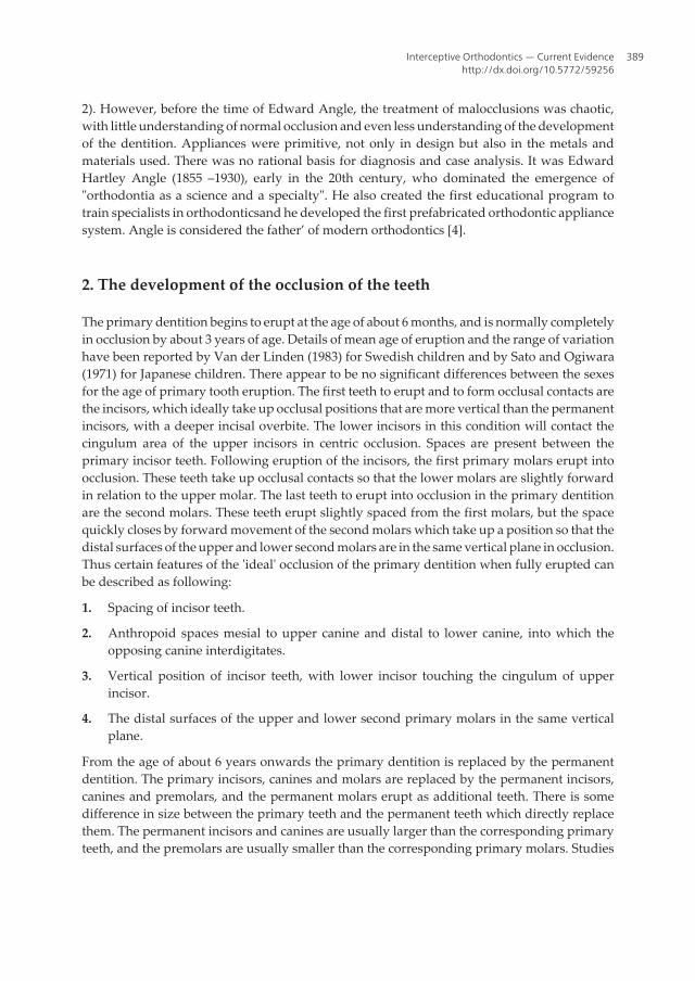
2). However, before the time of Edward Angle, the treatment of malocclusions was chaotic,with little understanding of normal occlusion and even less understanding of the developmentof the dentition. Appliances were primitive, not only in design but also in the metals andmaterials used. There was no rational basis for diagnosis and case analysis. It was EdwardHartley Angle (1855 –1930), early in the 20th century, who dominated the emergence of"orthodontia as a science and a specialty". He also created the first educational program totrain specialists in orthodonticsand he developed the first prefabricated orthodontic appliancesystem. Angle is considered the father’ of modern orthodontics [4].
2. The development of the occlusion of the teeth
The primary dentition begins to erupt at the age of about 6 months, and is normally completelyin occlusion by about 3 years of age. Details of mean age of eruption and the range of variationhave been reported by Van der Linden (1983) for Swedish children and by Sato and Ogiwara(1971) for Japanese children. There appear to be no significant differences between the sexesfor the age of primary tooth eruption. The first teeth to erupt and to form occlusal contacts arethe incisors, which ideally take up occlusal positions that are more vertical than the permanentincisors, with a deeper incisal overbite. The lower incisors in this condition will contact thecingulum area of the upper incisors in centric occlusion. Spaces are present between theprimary incisor teeth. Following eruption of the incisors, the first primary molars erupt intoocclusion. These teeth take up occlusal contacts so that the lower molars are slightly forwardin relation to the upper molar. The last teeth to erupt into occlusion in the primary dentitionare the second molars. These teeth erupt slightly spaced from the first molars, but the spacequickly closes by forward movement of the second molars which take up a position so that thedistal surfaces of the upper and lower second molars are in the same vertical plane in occlusion.Thus certain features of the 'ideal' occlusion of the primary dentition when fully erupted canbe described as following:
1. Spacing of incisor teeth.
2. Anthropoid spaces mesial to upper canine and distal to lower canine, into which theopposing canine interdigitates.
3. Vertical position of incisor teeth, with lower incisor touching the cingulum of upperincisor.
4. The distal surfaces of the upper and lower second primary molars in the same verticalplane.
From the age of about 6 years onwards the primary dentition is replaced by the permanentdentition. The primary incisors, canines and molars are replaced by the permanent incisors,canines and premolars, and the permanent molars erupt as additional teeth. There is somedifference in size between the primary teeth and the permanent teeth which directly replacethem. The permanent incisors and canines are usually larger than the corresponding primaryteeth, and the premolars are usually smaller than the corresponding primary molars. Studies
Interceptive Orthodontics — Current Evidencehttp://dx.doi.org/10.5772/59256
389

reported by Van der Linden (1983), have shown that the overall difference in size between thetwo dentitions is not large, amounting on average to about 3 mm in the upper teeth and lessthan 1 mm in the lower teeth. There is, however, not a strong correlation between the sizes ofthe primary dentition and the permanent teeth. The relationship of the jaws to each other willhave a large influence on the relationship of the dental arches. The relationship of the jaws toeach other can also vary in all three planes of space, and variation in any plane can affect theocclusion of the teeth. The antero-posterior positional relationship of the basal parts of theupper and lower jaws to each other, with the teeth in occlusion, is known as the skeletalrelationship. This is sometimes called the dental base relationship, or the skeletal pattern. Aclassification of the skeletal relationship is in common use, namely:
1. Skeletal Class 1—in which the jaws are in their ideal antero-posterior relationship inocclusion.
2. Skeletal Class 2—in which the lower jaw in occlusion is positioned further back in relationto the upper jaw than in skeletal Class 1.
3. Skeletal Class 3—in which the lower jaw in occlusion is positioned further forward thanin skeletal Class 1.
In addition, the teeth erupt into an environment of functional activity governed by the musclesof mastication, of the tongue and of the face. The muscles of the tongue, lips and cheeks are ofparticular importance in guiding the teeth into their final position, and variation in muscleform and function can affect the position and occlusion of the teeth. Moreover, some dentaland local factors can affect the development of occlusion. These include: alterations in size ofthe dentition in relation to jaw size, crossbite, aberrant developmental position of individualteeth, presence of supernumerary teeth, developmental hypodontia, labial frenum, thumb orfinger sucking. Early interference and modification of these basic etiological features can helpto avoid malocclusion or reduce the need for treatment in some cases. Consequently intercep‐tive orthodontic treatment has been set as an important aspect of orthodontic care [5].
3. Interceptive orthodontics: Definition of the concept
The concept and the necessity of interceptive orthodontic treatment, so called early, have beencontroversial. Some define it as removable or fixed appliance intervention in the deciduous,early mixed, or midmixed dentition. Others place it in the late mixed dentition stage ofdevelopment (before emergence of the second premolars and the permanent maxillarycanines). The American Association of Orthodontists’ Council of Orthodontic Educationdefines interceptive orthodontics as “that phase of the science and art of orthodontics em‐ployed to recognize and eliminate potential irregularities and malpositions in the developingdentofacial complex.” [6]. While some profession's leaders advocate that early treatment isalways desirable because tissue tolerance and their power of adjustment are at or near theirmaximum, others warn that there is no assurance that the results of early treatment will besustained, and that several-phased treatment will always lengthen overall treatment time.
Emerging Trends in Oral Health Sciences and Dentistry390
Early treatment not only may do some damage or prolong therapy, it may exhaust the child’sspirit of cooperation and compliance [7]. Joseph Fox (1776-1816, English), in his "NaturalHistory of the Human Teeth" (London, 1803), recommended that treatment be started “before13 or 14 years of age, and as much earlier as possible.” Angle advocated the institution oforthodontic treatment “as near the beginning of the variation from the normal in the processof the development of the dental apparatus as possible”. Although Nance advocated that“active treatment in the mixed dentition period is desirable only in Class III cases, crossbites,and Class II cases wherein facial appearance is markedly affected,” he freed orthodontists fromtheir hesitancy to treat patients before the development of the adult dentition [8].
4. Orthodontic interceptive measures during primary and mixed dentition
4.1. Space maintainers
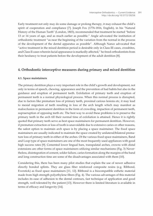
The primary dentition plays a very important role in the child’s growth and development, notonly in terms of speech, chewing, appearance and the prevention of bad habits but also in theguidance and eruption of permanent teeth. Exfoliation of primary teeth and eruption ofpermanent teeth is a normal physiological process. When this normal process is disrupted,due to factors like premature loss of primary teeth, proximal carious lesions etc, it may leadto mesial migration of teeth resulting in loss of the arch length which may manifest asmalocclusion in permanent dentition in the form of crowding, impaction of permanent teeth,supraeruption of opposing teeth etc. The best way to avoid these problems is to preserve theprimary teeth in the arch till their normal time of exfoliation is attained. Hence it is rightlyquoted that primary teeth serve as best space maintainers for permanent dentition. However,if premature extraction or loss of tooth is unavoidable due to extensive caries or other reasons,the safest option to maintain arch space is by placing a space maintainer. The fixed spacemaintainers are usually indicated to maintain the space created by unilateral/bilateral prema‐ture loss of primary teeth in either of the arches. Of the various fixed space maintainers, Bandand Loop type of space maintainers are one of the most frequently used appliances with goodhigh success rates [9]. Cemented lower lingual bars, transpalatal arches, crowns with distalextensions are other forms of space maintainers utilizing similar mechanisms (Fig. 3) Never‐theless, disintegration of cement, solder failure, caries formation along the margins of the bandand long construction time are some of the disadvantages associated with them [10].
Considering this, there has been many pilot studies that explain the use of newer adhesivedirectly bonded splints. They are glass fiber reinforced composite resins (e.g. Ribbond,Everstick) as fixed space maintainers [11, 12]. Ribbond is a biocompatible esthetic materialmade from high strength polyethylene fibers (Fig. 4). The various advantages of this materialincludes its ease of adhesion to the dental contours, fast technique of application and goodstrength, well tolerated by the patient [13]. However there is limited literature is available interms of efficacy and longevity [14].
Interceptive Orthodontics — Current Evidencehttp://dx.doi.org/10.5772/59256
391
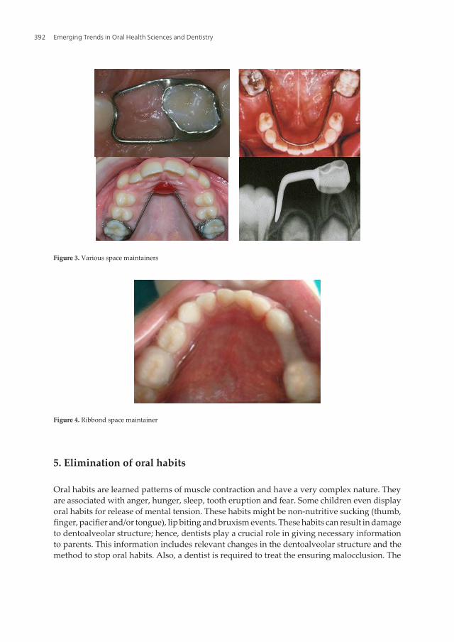
Figure 4. Ribbond space maintainer
5. Elimination of oral habits
Oral habits are learned patterns of muscle contraction and have a very complex nature. Theyare associated with anger, hunger, sleep, tooth eruption and fear. Some children even displayoral habits for release of mental tension. These habits might be non-nutritive sucking (thumb,finger, pacifier and/or tongue), lip biting and bruxism events. These habits can result in damageto dentoalveolar structure; hence, dentists play a crucial role in giving necessary informationto parents. This information includes relevant changes in the dentoalveolar structure and themethod to stop oral habits. Also, a dentist is required to treat the ensuring malocclusion. The
Figure 3. Various space maintainers
Emerging Trends in Oral Health Sciences and Dentistry392
prevalence of oral habits in high school girls and primary school students has been reportedto be 87.9 and 30%, respectively [15].
Oral habits could be divided into 2 main groups:
1. Acquired oral habits: Include those behaviors which are learned and could be stoppedeasily and when the child grows up, he or she can give up that behavior and start anotherone.
2. Compulsive oral habits: Consist of those behaviors which are fixed in child and whenemotional pressures are intolerable for the child, he or she can feel safety with this habit,and preventing the child from these habits make him or her anxious and worried.
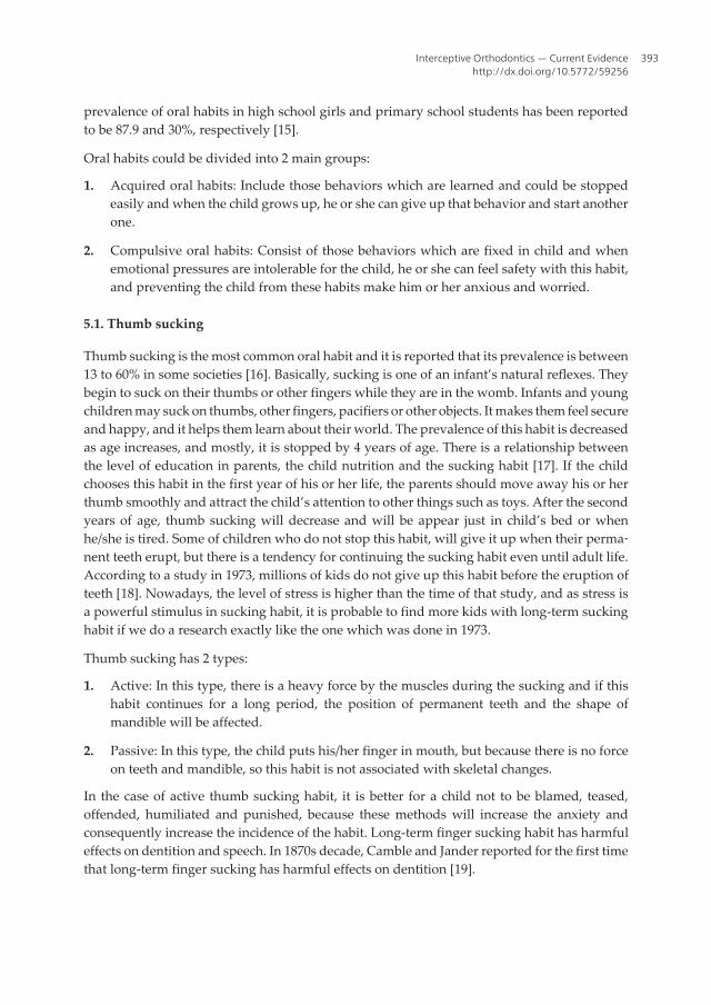
5.1. Thumb sucking
Thumb sucking is the most common oral habit and it is reported that its prevalence is between13 to 60% in some societies [16]. Basically, sucking is one of an infant’s natural reflexes. Theybegin to suck on their thumbs or other fingers while they are in the womb. Infants and youngchildren may suck on thumbs, other fingers, pacifiers or other objects. It makes them feel secureand happy, and it helps them learn about their world. The prevalence of this habit is decreasedas age increases, and mostly, it is stopped by 4 years of age. There is a relationship betweenthe level of education in parents, the child nutrition and the sucking habit [17]. If the childchooses this habit in the first year of his or her life, the parents should move away his or herthumb smoothly and attract the child’s attention to other things such as toys. After the secondyears of age, thumb sucking will decrease and will be appear just in child’s bed or whenhe/she is tired. Some of children who do not stop this habit, will give it up when their perma‐nent teeth erupt, but there is a tendency for continuing the sucking habit even until adult life.According to a study in 1973, millions of kids do not give up this habit before the eruption ofteeth [18]. Nowadays, the level of stress is higher than the time of that study, and as stress isa powerful stimulus in sucking habit, it is probable to find more kids with long-term suckinghabit if we do a research exactly like the one which was done in 1973.
Thumb sucking has 2 types:
1. Active: In this type, there is a heavy force by the muscles during the sucking and if thishabit continues for a long period, the position of permanent teeth and the shape ofmandible will be affected.
2. Passive: In this type, the child puts his/her finger in mouth, but because there is no forceon teeth and mandible, so this habit is not associated with skeletal changes.
In the case of active thumb sucking habit, it is better for a child not to be blamed, teased,offended, humiliated and punished, because these methods will increase the anxiety andconsequently increase the incidence of the habit. Long-term finger sucking habit has harmfuleffects on dentition and speech. In 1870s decade, Camble and Jander reported for the first timethat long-term finger sucking has harmful effects on dentition [19].
Interceptive Orthodontics — Current Evidencehttp://dx.doi.org/10.5772/59256
393

The side effects of finger sucking are: Anterior open bite, increased overjet, lingual inclinationlower incisor and labial inclination upper incisor, posterior cross bite, compensatory tonguethrust, deep palate, speech defect, and finger defects (Eczema of the finger due to alternatedryness and moisture that occurs and even angulations of the finger). The severity of changesin dentition due to finger sucking is related to the duration and times of doing the habit. Also,the position of finger in mouth, dental arches relation and child’s health affect the severity ofchanges [20].
Dental changes due to finger sucking do not need any treatment if the habit stopped beforethe 5 years of age and as soon as giving up the habit, dental changes will be corrected spon‐taneously [21]. At the time of permanent anterior teeth eruption and if the child is motivatedto stop the sucking habit, it is time to start the treatment as follows:
1. Direct interview with child if he/she is mature enough to understand
2. Encouragement: This can give the child more pride and self-confidence
3. Reward system
4. Reminder therapy
5. Orthodontic appliance: The final stage in treatment is the use of orthodontic appliancewhether fixed or removable, which can play the role of reminder and can reduce thewilling of finger sucking. For long-term habits or unwilling patient, the fixed intra oralappliance is the most effective inhibitor. In the case of using fixed or removable appliance,we should alarm the parents about potential problems in speaking or eating during thefirst 24 to 48 h, which are usual and self correcting. After active phase of treatment, theappliance should remain in place for more 3 to 6 month to minimize the relapse potential[22, 23].
5.2. Use of pacifier
The use of pacifier is common in most countries and it will not cause permanent changes indentition if it is stopped at the age of 2 or 3 years. After that, the use of pacifier has harmfuleffects on dentition development, and if it is used more than 5 years old, these effects wouldbe more severe [24]. The children who use pacifier are not willing to suck their fingers. pacifierhas the following negative effects:
1. Anterior open bite
2. Shallow palate
3. Increased width of lower arch
4. Posterior cross bite.
5. Median otitis
It is suggested that pacifier should be replaced in children who have the habit of finger sucking,because the harmful effects of sucking pacifier are less than finger. In comparison between
Emerging Trends in Oral Health Sciences and Dentistry394
different pacifiers, despite the claims, it has been shown that there is no significant advantagefor physiologic pacifiers over conventional ones [25].
5.3. Nail biting or onychophagia
Nail biting is a common and untreated medical problem among children. This habit starts after3 to 4 years of age and is in its peak in 10 years of age. Its rate increases in adolescency, whileit declines later. This problem is not gender dependent in children less than 10 years of age,but its incidence in boys is more than girls among adolescents [26]. This problem is a reactionin response to psychological disorders and some children will shift their habits from thumbsucking to nail biting. Complications caused by nail biting include malocclusion of the anteriorteeth, teeth root resorption, bacterial infection and alveolar destruction. Moreover, about oneforth of patients with temporo-mandibular joint pain and dysfunction have been shown tosuffer from nail biting habit [27]. It is seen in clinic that boys with nail biting have a kind ofpsychological disorder especially attention deficient hyperactivity disorder (ADHD) morethan girls. This habit in higher ages will be replaced with some habits such as lip chewing,gum chewing or smoking (Finn, 1998). Children with nail biting should be evaluated foremotional problems. In addition, putting nail polish or distasteful liquids on nails may be atherapeutic choice.
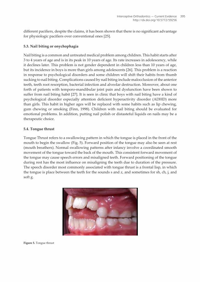
5.4. Tongue thrust
Tongue Thrust refers to a swallowing pattern in which the tongue is placed in the front of themouth to begin the swallow (Fig. 5). Forward position of the tongue may also be seen at rest(mouth breathers). Normal swallowing patterns after infancy involve a coordinated smoothmovement of the tongue toward the back of the mouth. This consistent forward movement ofthe tongue may cause speech errors and misaligned teeth. Forward positioning of the tongueduring rest has the most influence on misaligning the teeth due to duration of the pressure.The speech disorder most commonly associated with tongue thrust is a frontal lisp, in whichthe tongue is place between the teeth for the sounds s and z, and sometimes for sh, ch, j, andsoft g.
Figure 5. Tongue thrust
Interceptive Orthodontics — Current Evidencehttp://dx.doi.org/10.5772/59256
395

The line of treatment for these habits includes removal of the etiology, retraining exercises,and use of mechanical restraining appliances. Tongue bead appliances are commonly used asretraining exercise devices. In severe tongue thrusting cases and in cases with anterior openbite, a bead appliance alone may not be effective in restricting the habit. Tongue crib appliances(Fig. 6) are extremely effective in breaking the tongue thrust habit [28]. They create a mechan‐ical barrier and prevent the tongue from thrusting between the incisors. In most of the caseswith severe thumb/digit sucking habit, an anterior open bite develops. This will result in thedevelopment of a secondary tongue thrust habit. Hence, in cases with severe prolonged thumbor digit sucking, an appliance which can eliminate both of these habits. The Hybrid HabitCorrecting Appliance (HHCA) can be used to effectively restrain and correct tongue thrustingas well as thumb sucking habit (Fig.7). HHCA incorporates a tongue bead, a palatal crib anda U-loop which is attached to the molar bands on either sides. The tongue bead consists of aspinnable acrylic bead of 3mm diameter. The appliance is designed to position the acrylic beadover the posterior one-third of the incisive papilla. The bead acts as a tongue retrainer. Thepatient is asked to constantly pull the bead towards the posterior region of the mouth. Thepalatal crib and the U-loop are made of 0.9mm stainless steel wire. Three to four spurs are benton either sides of the bead, starting from the canine region on one side, running anteriorly asa smooth curve (in conventional crib appliances, the cribs run obliquely from one canine tothe other side canine) and lying 1mm lingual to the cervical margin of the maxillary anteriorteeth. In the region of the incisive papilla, the acrylic bead is incorporated in such a way thatit lies over the posterior one-third of the incisive papilla. The tip of the crib should be almostin line with the incisor tip of the maxillary central incisor or 2 mm longer without interferingwith the lower incisors when in occlusion. In cases with anterior open bite, the crib should belonger and can be up to 3/4th of the interincisal distance between the upper and lower centralincisors. This is to avoid the tongue from thrusting over the tip of the crib. The palatal crib actsas a barrier against the thrusting tongue and works as a mechanical restrainer. The U-loop isincorporated in the second premolar region and it helps to reposition the appliance posteriorlyduring the retraction phase, when it is used along with fixed orthodontic appliances.
Figure 6. Tongue crib appliances
Emerging Trends in Oral Health Sciences and Dentistry396

Figure 7. Hybrid Habit Correcting Appliance (HHCA).
5.5. Bruxism
The actions of masticatory system are divided into 2 groups. Functional actions such asmastication, speaking and swallowing, and parafunctional actions such as teeth impacting(clenching) and bruxism.
Functional activities are controllable and occurred daily. Parafunctional actions may beconscious or unconscious and are normally without sound. However, bruxism in nights isunconscious and mostly it is with sound production. Sleep bruxism occurs during stages firstand second of non rapid eye movement (REM) sleep and REM sleep. These people do not haveany complaint about bruxism, and it would not affect their quality of sleep. But in the old andpeople with sleep apnea, bruxism can reduce the quality of sleep [29]. Sleep bruxism has 2types: Primary or idiopathic and secondary or iatrogenic. The first type is without any medicalreason and the secondary type is whether with use of drug or without the use of drug. Riskfactors are as follows: Genetics: 20 to 50% of patients with sleep bruxism have positive familyhistory [30]; age: The prevalence of this habit decrease with age; cigarette smoking: Theprevalence of sleep bruxism in smokers is 1.9 times more than non-smokers; use of alcohol andcaffeine [31]; tension and stresses. Clinical findings of sleep bruxism include; report of grindingor impacting sounds of teeth; erosion of the teeth occlusal surfaces and breakdown of repairs;hypertrophy of masticatory muscles; hypersensitivity of teeth to cold air, and joint sounds.The treatment includes no special recommended regimen, but increasing awareness of thepatient, intra oral appliances, behavioral treatment and drugs like diazepam and clonazepamhave been reported to be effective [32,33].
6. Anterior cross bite correction
Anterior crossbite is defined as a malocclusion resulting from the lingual positioning of themaxillary anterior teeth in relationship to the mandibular anterior teeth. An anterior crossbite
Interceptive Orthodontics — Current Evidencehttp://dx.doi.org/10.5772/59256
397
is present when one or more of the upper incisors are in linguo-occlusion (reverse over jet).This may involve just a single tooth or could include all four upper incisors. Anterior dentalcrossbite has a reported incidence of 4-5% and usually becomes evident during the early mixed-dentition phase [34]. Anterior crossbite correction in early mixed dentition is highly recom‐mended as this kind of malocclusion does not diminish with age. Uncorrected anteriorcrossbite may lead to abnormal wear of the lower incisors, dental compensation of mandibularincisors leading to thinning of labial alveolar plate and/or gingival recession. However earlytreatment does not always eliminates orthodontic treatment need in permanent occlusion. Theaim of early treatment of this type of malocclusion is to correct anterior crossbite, as otherwiseoften can lead to very serious Class III mallocclusion which would be possible to treat onlywith combined orthodontic and orthognatic method.
A variety of factors has been reported to cause anterior dental crossbite, including a palataleruption path of the maxillary anterior incisors; trauma to the primary incisor resulting inlingual displacement of the permanent tooth germ; supernumerary anterior teeth; an over-retained necrotic or pulpless deciduous tooth or root; odontomas; crowding in the incisorregion; inadequate arch length; and a habit of biting the upper lip. Various treatment methodshave been proposed to correct anterior dental crossbite, such as tongue blades, reversedstainless steel crowns, fixed acrylic planes, bonded resin-composite slopes, and removableacrylic appliances with finger springs.
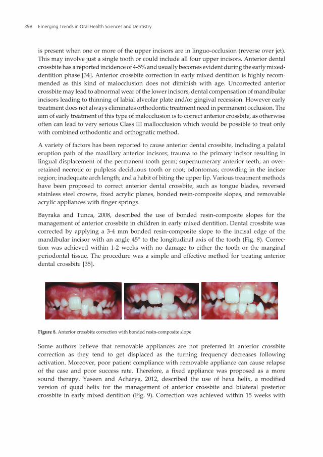
Bayraka and Tunca, 2008, described the use of bonded resin-composite slopes for themanagement of anterior crossbite in children in early mixed dentition. Dental crossbite wascorrected by applying a 3-4 mm bonded resin-composite slope to the incisal edge of themandibular incisor with an angle 45° to the longitudinal axis of the tooth (Fig. 8). Correc‐tion was achieved within 1-2 weeks with no damage to either the tooth or the marginalperiodontal tissue. The procedure was a simple and effective method for treating anteriordental crossbite [35].
Fig. 8 Anterior crossbite correction with bonded resin-composite slope Some authors believe that removable appliances are not preferred in anterior crossbite correction as they tend to get displaced as the turning frequency decreases following activation. Moreover, poor patient compliance with removable appliance can cause relapse of the case and poor success rate. Therefore, a fixed appliance was proposed as a more sound therapy. Yaseen and Acharya, 2012, described the use of hexa helix, a modified version of quad helix for the management of anterior crossbite and bilateral posterior crossbite in early mixed dentition (Fig. 9). Correction was achieved within 15 weeks with no damage to the tooth or the marginal periodontal tissue. The procedure is a simple and effective method for treating anterior and bilateral posterior crossbite simultaneously. it provides advantages such as minimal discomfort, reduces need for patient cooperation, and better control of tooth movements [37].
Fig. 9 Hexa helix appliance. In a recent study, Wiedel and Bondemark, 2014, evaluated and compared the stability of correction of anterior crossbite in the mixed dentition by fixed or removable appliance therapy. The study comprised 64 consecutive patients who met the following inclusion criteria: early to late mixed dentition, anterior crossbite affecting one or more incisors, no inherent skeletal Class III discrepancy, moderate space deficiency, a nonextraction treatment plan, and no previous orthodontic treatment. The study was designed as a randomized controlled trial with two parallel arms. The patients were randomized for treatment with a removable appliance with protruding springs or with a fixed appliance with multi-brackets. The outcome measures were success rates for crossbite correction, overjet, overbite, and arch length. Measurements were made on study casts before treatment (T0), at the end of the retention period (T1), and 2 years after retention (T2). Results showed that at T1 the anterior crossbite had been corrected in all patients in the fixed appliance group and all except one in the removable appliance group. At T2, almost all treatment results remained stable and equal in both groups. From T0 to T1, minor differences were observed between the fixed and removable appliance
Figure 8. Anterior crossbite correction with bonded resin-composite slope
Some authors believe that removable appliances are not preferred in anterior crossbitecorrection as they tend to get displaced as the turning frequency decreases followingactivation. Moreover, poor patient compliance with removable appliance can cause relapseof the case and poor success rate. Therefore, a fixed appliance was proposed as a moresound therapy. Yaseen and Acharya, 2012, described the use of hexa helix, a modifiedversion of quad helix for the management of anterior crossbite and bilateral posteriorcrossbite in early mixed dentition (Fig. 9). Correction was achieved within 15 weeks with
Emerging Trends in Oral Health Sciences and Dentistry398
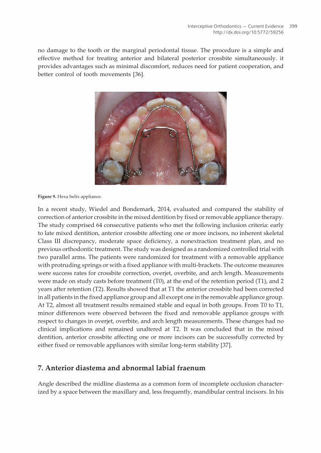
no damage to the tooth or the marginal periodontal tissue. The procedure is a simple andeffective method for treating anterior and bilateral posterior crossbite simultaneously. itprovides advantages such as minimal discomfort, reduces need for patient cooperation, andbetter control of tooth movements [36].
Figure 9. Hexa helix appliance.
In a recent study, Wiedel and Bondemark, 2014, evaluated and compared the stability ofcorrection of anterior crossbite in the mixed dentition by fixed or removable appliance therapy.The study comprised 64 consecutive patients who met the following inclusion criteria: earlyto late mixed dentition, anterior crossbite affecting one or more incisors, no inherent skeletalClass III discrepancy, moderate space deficiency, a nonextraction treatment plan, and noprevious orthodontic treatment. The study was designed as a randomized controlled trial withtwo parallel arms. The patients were randomized for treatment with a removable appliancewith protruding springs or with a fixed appliance with multi-brackets. The outcome measureswere success rates for crossbite correction, overjet, overbite, and arch length. Measurementswere made on study casts before treatment (T0), at the end of the retention period (T1), and 2years after retention (T2). Results showed that at T1 the anterior crossbite had been correctedin all patients in the fixed appliance group and all except one in the removable appliance group.At T2, almost all treatment results remained stable and equal in both groups. From T0 to T1,minor differences were observed between the fixed and removable appliance groups withrespect to changes in overjet, overbite, and arch length measurements. These changes had noclinical implications and remained unaltered at T2. It was concluded that in the mixeddentition, anterior crossbite affecting one or more incisors can be successfully corrected byeither fixed or removable appliances with similar long-term stability [37].
7. Anterior diastema and abnormal labial fraenum
Angle described the midline diastema as a common form of incomplete occlusion character‐ized by a space between the maxillary and, less frequently, mandibular central incisors. In his
Interceptive Orthodontics — Current Evidencehttp://dx.doi.org/10.5772/59256
399

classical article, Andrews stated that interdental diastemas should not exist and all contactsshould be tight so that the patient has ‘straight and attractive teeth as well as a correct overalldental occlusion’.
groups with respect to changes in overjet, overbite, and arch length measurements. These changes had no clinical implications and remained unaltered at T2. It was concluded that in the mixed dentition, anterior crossbite affecting one or more incisors can be successfully corrected by either fixed or removable appliances with similar long-term stability [38]. Anterior diastema and abnormal labial fraenum Angle described the midline diastema as a common form of incomplete occlusion characterized by a space between the maxillary and, less frequently, mandibular central incisors. In his classical article, Andrews stated that interdental diastemas should not exist and all contacts should be tight so that the patient has ‘straight and attractive teeth as well as a correct overall dental occlusion’.
Fig. 10 midline diastema
Sanin et al., developed a method that could predict whether the space would close spontaneously in the developing dentition. This method is based on millimeter measurements in the early mixed dentition and is claimed to have an accuracy of 88%. As the size of the diastema increases the possibility of space closure without treatment reduces. Sanin’s prediction is as follows: � For a 1 mm space in the early mixed dentition the possibility of spontaneous space closure is 99%. � For a 1.5 mm space the possibility is 85%. � For a 1.85 mm diastema it is 50%. � For a 2.7 mm space the possibility of closure without treatment is only 1%. The measurement should be made after the eruption of the lateral incisors. Hence it is advisable to intervene early if the midline diastema is more than 1.85 mm after the eruption of the permanent lateral incisors [39]. To treat the midline diastema effectively, an accurate diagnosis of the etiology and an intervention relevant to the specific etiology is necessary. Timing of the treatment is important to achieve satisfactory results. Most of the researchers do not recommend tooth movement until the eruption of the permanent canines, but in certain cases, where very large diastemas exist, treatment can be initiated early [40]. Nainar and Gnanasundaram noted in their study of midline diastemas on 9774 Southern Indian individuals, that there was a relatively increased frequency of familial occurrence and hence proposed the presence of a genetic factor in the expression of midline diastema. Treatment methods include orthodontic correction with a fixed or removable appliance and prosthetic correction with composites and crowns. If the diastema is large, it is advisable to close the space using orthodontic appliances. In most cases, simple removable appliances incorporating finger springs or a split labial bow can give good results [41]. A hypertrophic labial frenum may be considered as a major etiological factor for midline diastema. In a thick and fleshy labial frenum, the fibro-elastic band crosses the alveolus and inserts into the incisive papilli, preventing the approximation of the maxillary central incisors. The blanching test is a simple diagnostic test to predict whether a normal tight contact between the central incisors. Most of the researchers, like Angle, Sicher, and Edwards, [42-44] are of the opinion that superior labial frenum causes midline diastema. Some researchers, like Popovich et al, believe that there is an inverse relationship between high frenal attachment and midline diastema. According to them, labial frenum persists owing to the existing diastema and, as the dentition applies little or no pressure on the tissues, here is little or no atrophy of the frenum [45]. However, most of the researchers agree that removal of the high bulbous labial frenum is important for the stability after the closure of the midline diastema. Excessive anterior overbite is another major contributing factor for midline diastema. As a result of trauma to the maxillary anterior teeth from the mandibular incisors, the maxillary incisors procline. This results in an increase of the upper arch circumference, leading to a diastema. Practitioners should not
Figure 10. Midline diastema
Sanin et al., developed a method that could predict whether the space would close spontane‐ously in the developing dentition. This method is based on millimeter measurements in theearly mixed dentition and is claimed to have an accuracy of 88%. As the size of the diastemaincreases the possibility of space closure without treatment reduces. Sanin’s prediction is asfollows:
• For a 1 mm space in the early mixed dentition the possibility of spontaneous space closureis 99%.
• For a 1.5 mm space the possibility is 85%.
• For a 1.85 mm diastema it is 50%.
• For a 2.7 mm space the possibility of closure without treatment is only 1%.
The measurement should be made after the eruption of the lateral incisors. Hence it is advisableto intervene early if the midline diastema is more than 1.85 mm after the eruption of thepermanent lateral incisors [38].
To treat the midline diastema effectively, an accurate diagnosis of the etiology and an inter‐vention relevant to the specific etiology is necessary. Timing of the treatment is important toachieve satisfactory results. Most of the researchers do not recommend tooth movement untilthe eruption of the permanent canines, but in certain cases, where very large diastemas exist,treatment can be initiated early [39].
Nainar and Gnanasundaram noted in their study of midline diastemas on 9774 SouthernIndian individuals, that there was a relatively increased frequency of familial occurrence andhence proposed the presence of a genetic factor in the expression of midline diastema.Treatment methods include orthodontic correction with a fixed or removable appliance andprosthetic correction with composites and crowns. If the diastema is large, it is advisable to
Emerging Trends in Oral Health Sciences and Dentistry400
close the space using orthodontic appliances. In most cases, simple removable appliancesincorporating finger springs or a split labial bow can give good results [40].
A hypertrophic labial frenum may be considered as a major etiological factor for midlinediastema. In a thick and fleshy labial frenum, the fibro-elastic band crosses the alveolus andinserts into the incisive papilli, preventing the approximation of the maxillary central incisors.The blanching test is a simple diagnostic test to predict whether a normal tight contact betweenthe central incisors. Most of the researchers, like Angle, Sicher, and Edwards, [41-43] are of theopinion that superior labial frenum causes midline diastema. Some researchers, like Popovichet al, believe that there is an inverse relationship between high frenal attachment and midlinediastema. According to them, labial frenum persists owing to the existing diastema and, as thedentition applies little or no pressure on the tissues, here is little or no atrophy of the frenum[44]. However, most of the researchers agree that removal of the high bulbous labial frenumis important for the stability after the closure of the midline diastema.
Excessive anterior overbite is another major contributing factor for midline diastema. As aresult of trauma to the maxillary anterior teeth from the mandibular incisors, the maxillaryincisors procline. This results in an increase of the upper arch circumference, leading to adiastema. Practitioners should not fail to identify deep bite as an aetiology for the diastema.Any attempt to close the midline spacing without correcting the deep bite and anteriortraumatic bite will lead to a speedy relapse of the condition.
Oral habits such as tongue thrusting and finger sucking can be other etiological factors for theappearance of the midline diastema. According to Proffit and Fields, tongue position at restmay have a greater impact on tooth position than tongue pressure, as the tongue only brieflycontacts the lingual surface of the anterior teeth during thrusting [45]. The tongue pushes theanterior teeth to a forward position, increasing the circumference which results in spacing. Anabnormal habit of the tongue can be detected by the tip of the tongue popping out through theanterior spacing when the patient is asked to swallow. In cases of anterior open bite, the tonguemay be seen thrusting between incisal edges of the maxillary and mandibular incisors. Patientswith tongue thrust often produce a snap sound on swallowing and also have hyperactivity ofthe orbicularis oris muscle.Deleterious habits have to be corrected by using habit-breakingappliances and by psychological approaches. The use of fixed tongue cribs are found to beeffective in breaking the tongue-thrusting habit.
Peg-shaped laterals Supernumerary teeth/mesiodens, missing teeth, pathologic migrationof teeth Tooth size, arch size discrepancy, angulation of teeth, odontomas occurring in themaxillary midline, developmental cysts in the orofacial midline, and flaccid lips are otherproposed etiological factors leading to midline diastema. Relapse is a major concern in thecorrection of midline diastema. However, exact diagnosis and removal of the etiology isthe key to obtaining a stable result. Long-term use of retainers or even permanent bond‐ed lingual retainers are advocated, especially in cases with large diastema. Large pre-treatment diastema presence of at least one family member with a similar conditionincreases the risk of relapse [46,47].
Interceptive Orthodontics — Current Evidencehttp://dx.doi.org/10.5772/59256
401

8. Serial extraction
The term serial extraction describes an orthodontic treatment procedure that involves theorderly removal of selected deciduous and permanent teeth in a predetermined sequence(Dewel, 1969). Serial extraction can be defined as the correctly timed, planned removal ofcertain deciduous and permanent teeth in mixed dentition cases with dento-alveolar dispro‐portion in order to: Alleviate crowding of incisor teeth and to allow unerupted teeth to guidethemselves into improved positions (canines in particular), and to lessen (or eliminate) theperiod of active appliance therapy. Thus, it is one of the positive interceptive orthodonticprocedure generally applied in most discrepancy cases where supporting bone is less than thetotal tooth material [48].
Serial extraction has been of interest to dentist for many years. Throughout the history ofdentistry it has been recognized that the removal of one or more irregular teeth would improvethe appearance of the reminder. Nance presented clinics on his technique of progressiveextraction in 1940 and has been called as the father of serial extraction philosophy in the UnitedStates. Kjellgren in 1940 termed this extraction procedure as planned or progressive extractionprocedure of teeth. Hotz, 1970, named the same procedure on “Guidance of eruption”.According to him the term guidance of eruption is comprehensive and encompasses allmeasures available for influencing tooth eruption [49]. Widespread adoption of serial extrac‐tion as a corrective treatment procedure continues to be a source of concern to all pedodontistswho are aware of its limitations as well as of its possibilities. The principle reason is that itsapplication involves growth prediction. Every serial extraction diagnosis is based on thepromise that future growth will be inadequate to accommodate all of the teeth in a normalalignment.
If primary teeth are extracted prematurely, this will influence the eruption rate and positionof the permanent successors. In general, the eruption will be delayed if the primary toothoverlying the permanent tooth is extracted 1 ½ years or more from the time the primary toothwould normally exfoliate. Conversely, the eruption rate can be accelerated if the primary toothoverlying the permanent tooth is extracted less than a year before the primary tooth wouldnormally exfoliate. Biologic variation in eruption rates will affect these time tables, as willperiapical inflammation of the primary tooth. Another useful principle is that crowded teethadjacent to an extraction site tend to align themselves [50].
Normal dental, skeletal and profile development – influences the rationale for serial extraction.The work of Moorrees et al on arch dimensions and serial extractions indicates that there isminimal increase in mandibular intercanine width between 8 and 18 years, occurring usuallyaround the time the permanent mandibular canines erupt. The maxillary intercanine widthincreases slightly more and over a longer time. The dental arch perimeter from the distal ofthe mandibular primary second molar to its antimere is less in the permanent dentition thanin the primary. Also the principles of leeway space, interrelationship of overjet, overbite, axialinclinations, and mesial shift, and arch-length analysis must be considered in determiningwhether to institute a serial extraction procedure. The skeletal and profile factors that influenceserial extractions are the another-posterior, vertical, and transverse relationships as well as the
Emerging Trends in Oral Health Sciences and Dentistry402
developmental pattern. Specifically the relation of the maxilla to the mandible and of the bothto the cranial base must be determined to identify protrusions, retrusions, hyperdivergences,hypodivergences, crossbites, and asymmetries. Also rotational, vertical, and transversegrowth patterns need to be integrated into the decision-making process [51].
The idea of serial extraction started when Pedodontist sees a child 5 or 6 years of age with allthe deciduous teeth present in a slightly crowded state or with no spaces between them, hecan predict, with a fair degree of certainly, that there will not be enough space in the jaws toaccommodate all the permanent teeth in their proper alignment. As Nance (1940), Dewel(1954), and others have pointed out, after the eruption of the first permanent molars at 6 yearsof age, there is probably no increase in the distance from the mesial aspect of the first molaron one side around the arch to the mesial aspect of the first molar on the opposite side. If thereis any change, it may be an actual reduction of the molar-to-molar arch length, as the "leeway"space is lost through the mesial migration of the first permanent molars during the tooth-exchange process and correction of the flush terminal plane relationship. At that time, a list ofpossible clinical clues for serial extraction were proposed: Premature loss of deciduous teeth,arch-length deficiency and tooth size discrepancies, lingual eruption of lateral incisors,unilateral deciduous canine loss and shift to the same side, mesial eruption of caninesoverlateral incisors, mesial drift of buccal segments, abnormal eruption direction and eruptionsequence, flaring of incisors, ectopic eruption of mandibular first deciduous molar, abnormalresorption of II deciduous molar, ankylosis, labial stripping, and gingival recession, usuallyof lower incisor. However, a number of contraindications for serial extractions were addressed:Congenital absence of teeth providing space, mild to moderate crowding, deep or open bites,severe Class II, III of dental/skeletal origin, cleft lip and palate, spaced dentition, anodontia /oligodontia, Midline diastemia, dilacerations extensive caries, disportion between arc lengthand tooth material.
9. Considerations in serial extraction
1. Extracting primary canines will produce maximum amounts of self improvement incrowding with greatest inter-ception of lingual cross bite.
2. Extracting primary first molars produces earliest eruption of first premolars but reducesspeed and amount of improvement in permanent central and lateral incisors crowdingand position due to retention of C that it has limited application.
3. Extracting primary canines and first molars is a compromise between rapid improvementin and desired early eruption of permanent central and lateral incisors due to simultane‐ous eruption of first premolars with this extraction sequence.
There is no single technique for Serial Extraction. It is a long-range guidance program and itmay be necessary to reevaluate and change tentative decisions several times. Usually the childis 7-8 years of age when he/she brought to the pedodontist. At this time the maxillary andmandibular central incisors are usually erupted, but there is inadequate space in anterior
Interceptive Orthodontics — Current Evidencehttp://dx.doi.org/10.5772/59256
403

segments to allow normal eruption and positioning of lateral incisors. In some cases, mandib‐ular lateral incisors have already erupted but they are usually lingually positioned and rotated.The same is with the maxillary lateral incisors.
9.1. Dewel’s method
There are 3 stages in Serial Extraction Therapy:
First: Removal of deciduous canines : to permit eruption and optimal alignment of lateralincisors. There is some amount of improvement in position of central incisors also.
Second: Removal of first deciduous molars: to accelerate eruption of first premolars ahead ofcanine if possible.
Third: Removal of erupting first premolars: Before the first premolars are extracted, all thediagnostic criteria must again be evaluated. The status of developing third molars must beevaluated, because if the third molars are congenitally missing then extraction of 1st premolarswould be unnecessary because there would be enough space. So in short, Dewel’s method is:
9.2. Tweed’s method
According to Tweed, if diagnosis shows the discrepancy exists between teeth and basal bonestructures and if patient is between 7 ½ to 8 ½ years, Serial Extraction program is should becarried out. Sequence is:
First: At approximately 8 years all deciduous molars are extracted. It is preferable to maintainin deciduous canines to retard eruption of permanent canines.
Second: extract of first premolar and deciduous canines should he done 4-6 months prior toeruption of permanent canines when they erupt they migrate posteriorly into good position.Any irregularities in mandibular incisors if not too severe, get corrected themselves and theyare also tipped lingually due to normal muscular forces.
9.3. Moyers method
Proposed when crowding seen in central incisor region. Fairly eruption of lateral incisors.
Stage I (Extraction of all deciduous lateral incisors). It helps in alignment of central incisors.
Stage II (Extraction of all deciduous canines after 7-8 months). It helps in alignment of lateralincisors and provides space for lateral incisors.
Emerging Trends in Oral Health Sciences and Dentistry404
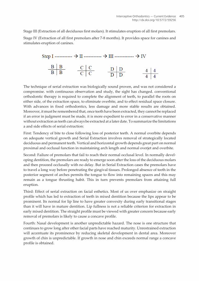
Stage III (Extraction of all deciduous first molars). It stimulates eruption of all first premolars.
Stage IV (Extraction of all first premolars after 7-8 months). It provides space for canines andstimulates eruption of canines.
The technique of serial extraction was biologically sound proven, and was not considered acompromise. with continuous observation and study, the sight has changed. conventionalorthodontic therapy is required to complete the alignment of teeth, to parallel the roots oneither side, of the extraction space, to eliminate overbite, and to effect residual space closure.With advances in fixed orthodontics, less damage and more stable results are obtained.Moreover, it must be remembered that, once teeth have been extracted, they cannot be replacedif an error in judgment must be made, it is more expedient to error in a conservative mannerwithout extraction as teeth can always be extracted at a later date. To summarize the limitationsa and side effects of serial extraction:
First: Tendency of bite to close following loss of posterior teeth. A normal overbite dependson adequate vertical growth and Serial Extraction involves removal of strategically locateddeciduous and permanent teeth. Vertical and horizontal growth depends great part on normalproximal and occlusal function in maintaining arch length and normal overjet and overbite.
Second: Failure of premolars that fail to reach their normal occlusal level. In normally devel‐oping dentition, the premolars are ready to emerge soon after the loss of the deciduous molarsand then proceed occlusally with no delay. But in Serial Extraction cases the premolars haveto travel a long way before penetrating the gingival tissues. Prolonged absence of teeth in theposterior segment of arches permits the tongue to flow into remaining spaces and this mayremain as a tongue thrusting habit. This in turn prevents premolars from attaining fulleruption.
Third: Effect of serial extraction on facial esthetics. Most of us over emphasize on straightprofile which has led to extraction of teeth in mixed dentition because the lips appear to beprominent. Its normal for lip line to have greater convexity during early transitional stagesthan it will have in mature dentition. Lip fullness is not a reliable criterion for extraction inearly mixed dentition. The straight profile must be viewed with greater concern because earlyremoval of premolars is likely to cause a concave profile.
Fourth: Nasal development is another unpredictable hazard. The nose is one structure thatcontinues to grow long after other facial parts have reached maturity. Unrestrained extractionwill accentuate its prominence by reducing skeletal development in dental area. Moreovergrowth of chin is unpredictable. If growth in nose and chin exceeds normal range a concaveprofile is obtained.
Interceptive Orthodontics — Current Evidencehttp://dx.doi.org/10.5772/59256
405

In conclusion, one team of clinicians and practioners demonstrate that undertaking a serialextraction protocol can afforded an improvement of the patient's self-esteem, resulting in apositive social impact due to esthetic enhancement. Furthermore, the low cost of this protocolpermits the use of this therapy in underprivileged communities provided the diagnosis iscertain and the post extraction movement of teeth is controlled by mechanical means. The otherteam suggest that serial extraction is counter-productive. The early extraction of primarycuspids will invariably result in crowding of the permanent cuspids region. In reality, theyadopt the idea that the problem is maintained and the crowding shifts to involve the permanentcuspids. They remind us with the most basic canon of the health profession which is "first dono harm" [52].
10. Interceptive functional therapy
There is little doubt that functional appliances produce tooth movement and in many casescan correct occlusal discrepancies. The controversy over their use relates mainly to their modeof action, and in particular to two aspects. The first is the question of modification of growthof the basal parts of the jaws. Many authorities believe that basal jaw growth can be altered byfunctional means. The temporo-mandibular joint area has been thought to be a reactive growthsite, i.e. any prolonged change in the position of the mandible during the growth period, suchas is induced by wearing the appliance, results in bone apposition on the mandibular andtemporal surfaces of the enlarged joint cavity. Baume, (1969) quotes histological evidence tosupport this concept, and ample clinical evidence has been produced in attempts to show thatthe use of functional appliances can alter the skeletal relationship of the jaws [53]. On the otherhand, this clinical evidence does not always take into account the effects of normal growth. Asfunctional appliances are normally used during the mixed dentition stage a considerableamount of normal growth must occur which could alter jaw size and relationships. Severalinvestigators have failed to find evidence of altered growth with functional appliances, butinstead have found the main effects to be tipping of the incisors and an opening rotation of themandible [54,55].
The first practitioner to use functional jaw orthopedics to treat a malocclusion was Pierre Robin(1902).
His appliance influenced muscular activity by changing the spatial relationship of the jaws.Robin’s monoloc was actually a modification of Kingsley’s maxillary plate. It extended allalong the lingual surfaces of the mandibular teeth, but it had sharp lingual imprints of thecrown surfaces of both maxillary and mandibular teeth. It incorporated an expansion screwin the palate to expand the dental arches. In 1909, Viggo Andresen, a Danish dentist, usedlingual horseshoe flange that guides the mandible forward to eliminate Class II malocclusioncases. The original Andresen activator was a tooth-borne, loosely fitting passive applianceconsisting of a block of plastic covering the palate and the teeth of both arches, designed toadvance the mandible several millimeters for Class II correction and open the bite 3 to 4 mm.The original design had facets incorporated into the body of the appliance to direct erupting
Emerging Trends in Oral Health Sciences and Dentistry406

posterior teeth mesially or distally, so, despite the simple design, dental relationships in all 3planes of space could be changed.
The Herbst appliance (Dentaurum, Newtown, Pa) is suitable for slightly older children whosecooperation might not be dependable, because it is a fixed appliance worn 24 hours a day. TheHerbst was introduced in 1905 by Emil Herbst, but his findings were not published until 1935.Little more was published on the appliance until the late 1970s, when Hans Pancherz, recog‐nizing its possibilities for mandibular growth stimulation, revived interest. The typical Herbstconsists of a telescoping mechanism connected to the maxillary first molars at one end and acantilevered arm attached to the mandibular first molars at the other end; it forces the mandibleforward (Fig. 11).
Figure 11. Herbst Appliance
In 1950, Wilhelm Balters (1893-1973), in an effort to treat Class II malocclusions characterizedby deficient mandibles, began to modify Andresen’s activator. He gave it the name bionator.It is indicated for patients with favorable facial growth patterns and is designed to produceforward positioning of the mandible. As with the function regulator, the bionator is availablein 3 designs. Consisting of 2 halves connected by a Coffin spring, it is less restrictive of speechthan Andresen’s appliance. However, the treatment also highly depends on patient compli‐ance, especially with regard to exercising
The Clark twin-block (Clark 1988) consists of separate upper and lower removable appliances,each with a 45° posterior bite plane designed to induce a mandibular posture of the desiredamount and direction (Fig. 12) One or both sections may incorporate a mid-line screw to effectarch expansion(Fig. 13), and there is provision for the addition of extraoral traction.
Many clinical studies have been done on skeletal and dentoalveolar changes associated withfunctional appliances therapy in Class II malocclusions, but the scientific data are still contro‐versial. Concerning skeletal effects induced by the functional appliances some authors
Interceptive Orthodontics — Current Evidencehttp://dx.doi.org/10.5772/59256
407

demonstrate significant influences on mandibular growth [56], the others claim that it may beinduced only small skeletal changes by this type of treatment [57]. The latter group of re‐searchers found that the main changes occurred with functional appliance therapy weredentoalveolar distalization of the buccal and retroclination of the frontal upper teeth, alongwith mesial movement of the lower buccal segments and proclination of the lower labialsegments[58]. Such diversity of results on skeletal changes might be related first of all todifficulties in applying treatment at the maximum growth spurt time. Another reason for theinconsistence in assessment of treatment results might be the use of not reliable reference linesand/or structures for cephalometric analysis before and after treatment. This makes difficultto assess real contribution of skeletal and dental components to occlusal changes [59]. A newparadigm for successful treatment presents a philosophical challenge to combine the benefitsof orthodontic and orthopedic techniques to extend our horizons in the treatment of maloc‐clusion that requires dental and skeletal correction.
The prefabricated myofunctional appliances are a series of prefabricated appliances producedby myoresearch company, Queensland, Australia. These appliances were also called "Train‐ersTM" which include T4KTM and T4FTM appliances (Fig.14,15). The idea of prefabricated
Figure 12. Twin-block appliance
Figure 13. Expansion screw within Twin block appliance
Emerging Trends in Oral Health Sciences and Dentistry408
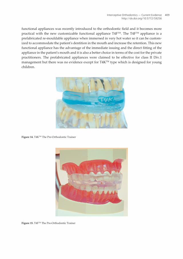
functional appliances was recently introduced to the orthodontic field and it becomes morepractical with the new customizable functional appliance T4FTM. The T4FTM appliance is aprefabricated re-mouldable appliance when immersed in very hot water so it can be custom‐ized to accommodate the patient's dentition in the mouth and increase the retention. This newfunctional appliance has the advantage of the immediate issuing and the direct fitting of theappliance in the patient's mouth and it is also a better choice in terms of the cost for the privatepractitioners. The prefabricated appliances were claimed to be effective for class II Div.1management but there was no evidence except for T4KTM type which is designed for youngchildren.
Figure 14. T4KTM The Pre-Orthodontic Trainer
Figure 15. T4FTM The Pre-Orthodontic Trainer
Interceptive Orthodontics — Current Evidencehttp://dx.doi.org/10.5772/59256
409

Uysal et al., 2012, evaluated the effects of Pre-Orthodontic Trainer (T4FTM) appliance on theanterior temporal, mental, orbicularis oris, and masseter muscles through electromyography(EMG) evaluations in subjects with Class II division 1 malocclusion and incompetent lips.Twenty patients (mean age: 9.8 ± 2.2 years) with a Class II division 1 malocclusion were treatedwith T4FTM (Myofunctional Research Co., Queensland, Australia). A group of 15 subjects (meanage: 9.2 ± 0.9 years) with untreated Class II division 1 malocclusions was used as a control.EMG recordings of treatment group were taken at the beginning and at the end of the T4FTM
therapy (mean treatment period: 7.43 ± 1.06 months). Follow-up records of the control groupwere taken after 8 months of the first records. Recordings were taken during different oralfunctions: clenching, sucking, and swallowing. Statistical analyses were undertaken withWilcoxon and Mann-Whitney U-tests. During the T4FTM treatment, activity of anteriortemporal, mental, and masseter muscles was decreased and orbicularis oris activity wasincreased during clenching and these differences were found statistically significant whencompared to control. Orbicularis oris activity during sucking was increased in the treatmentgroup (P < 0.05). In the control group, significant changes were determined for anteriortemporal (P < 0.05) and masseter (P < 0.01) muscle at clenching and orbicularis oris muscle atswallowing during observation period (P< 0.05). Findings indicated that treatment withT4FTM appliance showed a positive influence on the masticatory and perioral musculature [60].
Usumez et al., 2004, evaluated the dentoskeletal treatment effects induced by a preorthodontictrainer appliance (T4KTM) treatment on Class II, division 1 cases. Twenty patients (10 girls and10 boys, mean age 9.6+/-1.3 years) with a Class II, division 1 malocclusion were treated withT4KTM (Myofunctional Research Co., Queensland, Australia). The patients were instructed touse the trainer every day for one hour and overnight while they slept. A control group of 20patients (mean age 10.2+/-0.8 years) with untreated Class II, division 1 malocclusions was usedto eliminate possible growth effects. Lateral cephalograms were taken at the start and end oftreatment. Final cephalograms were taken 13.1+/-1.8 months after trainer application, com‐pared with a mean of 11.2+/-2.4 months later for the control group. The mean and standarddeviations for cephalometric measurements were analyzed by paired-samples t-test andindependent-samples t-tests. At the end of the study period, the trainer group subjects showedsignificant changes including anterior rotation and sagittal growth of the mandible, increasedSNB and facial height, reduced ANB, increased lower incisor proclination, retroclination ofupper incisors, and overjet reduction. However, only total facial height increase, lower incisorproclination, and overjet reduction were significantly higher when compared with the changesobserved in the control group. This study was the first that demonstrated that the preortho‐dontic trainer application induces basically dentoalveolar changes that result in significantreduction of overjet and can be used with appropriate patient selection [61].
In a very recent study, Dr. Hanoun and his colleagues evaluated the effectiveness of the pre‐fabricated myofunctional appliance T4FTM (compared to Twin Block appliance) in the treat‐ment of Class II Div.1 malocclusion. The study was a prospective randomized clinical trial.All subjects were growing patients aged 11 -14 years old with Class II Div.1 malocclusionbased on Class II skeletal relationship with overjet of 7 mm or more. Those subjects who hadanterior open bite or previous orthodontic therapy or craniofacial anomalies or history of fa‐
Emerging Trends in Oral Health Sciences and Dentistry410

cial trauma were all excluded from the trial. The overjet was reduced more favourably in theTwin Block group than in the T4FTM group with a mean difference between the two groupsof 2.14 mm (p <0.01). Moreover, there was a significant difference between both groups interms of horizontal skeletal linear dimensions of the mandible with more favourable in‐crease in the Twin Block group (p < 0.05).
11. Conclusion
Interceptive orthodontics is employed to recognize and eliminate potential irregularities andmalposition in the developing dentofacial complex. These procedures are directed to lessen orto eliminate the severity of developing malocclusion. The early assessment of the child,followed by regular review, and treatment at the appropriate time if necessary, will do muchto reduce malocclusion to the basic non-preventable level. The key to prevention of this kindis awareness. This part examines the key areas relating to interceptive orthodontics with theavailable evidence to support the clinical management of common problems presenting in themixed dentition.
Acknowledgements
I offer my sincerest gratitude to my wife, Mrs. Huda Zurigat who has supported me throughoutmy project with a lot of patience and enthusiasm. Her assistance in typing and organizingwords and figures in this article was indispensible.
Author details
Maen H. Zreaqat*
Address all correspondence to: [email protected]
Al-Ogaly Medical Group, Saudi Arabia
References
[1] Weinberger BW. Historical résumé of the evolution and growth of orthodontia. J AmDent Assoc 1934;21:201-221.
[2] Asbell MB. A brief history of orthodontics. Am J Orthod Dentofacial Orthop1990;98:176-83.
Interceptive Orthodontics — Current Evidencehttp://dx.doi.org/10.5772/59256
411

[3] Casto FM. A historical sketch of orthodontia. Dent Cosmos 1934;76:111-35.
[4] Brodie AG. Orthodontic history and what it teaches. Angle Orthod 1934;4:85-97.
[5] Irurita J, Alemán I, López-Lázaro S, Viciano J, Botella MC. Chronology of the devel‐opment of the deciduous dentition in Mediterranean population. Forensic Sci Int.2014;240:95-103.
[6] Borrie F, Bonetti D, Bearn D. What influences the implementation of interceptive or‐thodontics in primary care? Br Dent J. 2014;216(12):687-91.
[7] Kerosuo H1, Väkiparta M, Nyström M, Heikinheimo K. The seven-year outcome ofan early orthodontic treatment strategy. J Dent Res. 2008;87(6):584-8.
[8] Norman W. Orthodontics in 3 millennia. Chapter 12: Two controversies: Early treat‐ment and occlusion Am J Orthod Dentofacial Orthop. 2006;130(6):799-804.
[9] Baroni C; Franchini A; Rimondini L: Survival of different types of space maintainers.Pediatr Dent. 1994;16:360-61.
[10] Kirzioglu Z, Ozay MS Z, Ozay MS. Success of reinforced fibre material space main‐tainers. J Dent Child. 2004;71;2:158-62.
[11] Kargul B, Caglar E, Kabalay U. Glass fibre reinforced composite resin as fixed spacemaintainer in children 12 month clinical follow up. J Dent Child. 2005; 72(3):109-12
[12] Kargul B, Çaglar E, Kabalay U. Glass fiber-reinforced composite resin space main‐tainer: case reports. J Dent Child. 2003;71:258-61.
[13] Goldberg AJ, Frelich MA. Tooth splinting and stabilisation Dent Clin North Am.1999; 43 (1) :127-33.
[14] Setia V, Pandit IK, Srivastava N, Gugnani N, Sekhon HK. Space maintainers in den‐tistry: past to present. J Clin Diagn Res. 2013;7(10):2402-5.
[15] Kharbanda O. P, Sidhu S. S, Sundaram K, Shukla D. K, “Oral habits in school goingchildren of Delhi: a prevalence study,” Journal of the Indian Society of Pedodonticsand Preventive Dentistry. 2003; 21 (3): 120–124.
[16] Larson EF. The prevalence and etiology of prolonged dumy and finger sucking habit.Am. J. Orthod., 1985; 87(5):172-174.
[17] Farsi NM, Salama FS.. Sucking habits in Saudi children: prevalence, contributing fac‐tors and effects on the primary dentition. Pediatr. Dent. 1997; 19(1):28-33.
[18] N. L. Villa, “Changes in the dentition secondary to palatal crib therapy in digit-suck‐ers: a preliminary study,” Pediatric Dentistry, vol. 19, no. 5, pp. 323–326, 1997.
[19] B. S. Haskell and J. R. Mink, “An aid to stop thumb sucking: the “Bluegrass” appli‐ance,” Pediatric Dentistry. 1991; 13 (2): 83– 85.
Emerging Trends in Oral Health Sciences and Dentistry412

[20] Van Norman RA (1997). Digit-sucking: A review of the literature, clinical observa‐tions and treatment recommendations. Int. J. Orofacial Myol., 23: 14-34
[21] Warren JJ, Bishara SE (2002). Duration of nutritive and nonnutritive sucking behav‐ior and their effects on the dental arches in the primary dentition. Am. J. Orthod.,121: 347-356.
[22] Moimaz SA, Garbin AJ, Lima AM, Lolli LF, Saliba O, Garbin CA. Longitudinal studyof habits leading to malocclusion development in childhood. BMC Oral Health. 2014;(4):14-96.
[23] Maia-Nader M, Silva de Araujo Figueiredo C, Pinheiro de Figueiredo F, Moura daSilva AA, Thomaz EB, Saraiva MC, Barbieri MA, Bettiol H. Factors associated withprolonged non-nutritive sucking habits in two cohorts of Brazilian children. BMCPublic Health. 2014;14:14-17.
[24] Fleming PJ, Blaive PS Pacifier use an sudden infant death syndrome. Arch. Dis. Chil‐dren. 1999; 81(2): 112-116.
[25] Lopes TS, Moura Lde F, Lima MC. Breastfeeding and sucking habits in children en‐rolled in a mother-child health program. BMC Res Notes. 2014;14(7): 23-38.
[26] Tanaka OM, Vitral RW, Tanaka GY, Guerrero AP, Camargo ES. (. Nailbiting, or ony‐chophagia: A special habit. Am. J. Orthod. Dentofacial Orthop. 2008;134(2): 305-308.
[27] Saheeb D. Prevalence of oral and parafunctional habits in Nigerian patients sufferingtemporomandibular joint pain and dysfunftion. J. Med. Biomed. Res., (2005; 4(1):59-64
[28] Feu D1, Menezes LM, Quintão AP, Quintão CC. A customized method for palatalcrib fabrication. J Clin Orthod. 2013 Jul;47(7):406-12.
[29] Kato T, Thein NMR, Montplaisir JY, Lavigne GJ. Bruxism and orofacial movementsduring sleep. Dent. Clin. North Am. 2001;45(4):651-676.
[30] Hublin C, Kaprio J, Partinen M, Koskenvuo M. Sleep bruxism based on self – reportin a nationwide twin cohort. Sleep Res. 1998;7(1): 61-68
[31] Dahan JS, Lelong BA, Celant S, Leysen V. Oral perception in tounge thrust and otheroral habits. Am. J. Orthod. Dentofacial Orthop., 2000;118:385-91.
[32] Pierce CJ, Gale EN. A comparison of different treatments for nocturnal bruxism. J.Dent. Res., 1988;67: 597-601.
[33] Ferreira NM, Dos Santos JF, Dos Santos MB, Marchini L. Sleep bruxism associatedwith obstructive sleep apnea syndrome in children. Cranio. 2014 Sep [Epub ahead ofprint].
[34] Major PW, Glover K. Treatment of anterior cross-bites in the early mixed dentition.Journal of the Canadian Dental Association. 1992;58(7):574–578.
Interceptive Orthodontics — Current Evidencehttp://dx.doi.org/10.5772/59256
413
[35] Bayrak S1, Tunc ES. Treatment of anterior dental crossbite using bonded resin-com‐posite slopes: case reports. Eur J Dent. 2008;2(4):303-6.
[36] Yaseen SM, Acharya R. Hexa Helix:Modified Quad Helix Appliance to Correct Ante‐rior and Posterior Crossbites in Mixed Dentition. Case Reports in Dentistry. 2012: 1-5.
[37] Wiedel AP1, Bondemark L. Stability of anterior crossbite correction: A randomizedcontrolled trial with a 2-year follow-up. Angle Orthod. 2014 Jul 8. [Epub ahead ofprint]
[38] Sanin C, Sekiguchi T, Savara BS. A clinical method for the prediction of closure of thecentral diastema. ASDC J Dent Child 1969; 36: 415−418.
[39] Baum AT. The midline diastema. J Oral Med 1966; 21: 30–39.
[40] Nainar SM, Gnanasundaram N. Incidence and etiology of midlinediastema in a pop‐ulation in southIndia (Madras). Angle Orthod 1989; 59: 277−282.
[41] Angle EH. Treatment of Malocclusion of the Teeth 7th edn. Philadelphia: SS WhiteDental Manufacturing Company, 1907: p167.
[42] Sicher H. Oral Anatomy 2nd edn. St Louis: CV Mosby Co, 1952: pp272−273.
[43] Edwards JG. The diastema, the frenum, the frenectomy: a clinical study. Am J Orthod1977; 71:489−508.
[44] Popovich F, Thompson GW, Main PA. Persisting maxillary diastema: indications fortreatment. Am JOrthod 1979; 75(4): 399−404.
[45] Proffit WR, Fields HW. Contemporary Orthodontics 2nd edn. St Louis: Mosby Year‐book, 1993: p467
[46] Shashua D, Artun J. Relapse afterorthodontic correction of maxillarymedian diaste‐ma: a follow-up evaluation of consecutive cases. Angle Orthod 1999; 69: 257−263.
[47] Nagalakshmi S, Sathish R, Priya K, Dhayanithi D. Changes in quality of life duringorthodontic correction of midline diastema. J Pharm Bioallied Sci. 2014 (Suppl 1):162-4
[48] Almeida RR, Almeida MR, Oltramari-Navarro PV, Conti AC, Navarro Rde L, SouzaKR. Serial extraction: 20 years of follow-up. J Appl Oral Sci. 2012; 20(4):486-92.
[49] Hotz P.R. Guidance of eruption versus serial extraction. Am. J. Orthod, 1970; 58(1):1-20.
[50] Nagalakshmi S, Sathish R, Priya K, Dhayanithi D. Changes in quality of life duringorthodontic correction of midline diastema. J Pharm Bioallied Sci. 2014; 6 (Suppl 1):.[Epub ahead of print]
[51] Lee KP. The fallacy of serial extractions. Aust Orthod J. 2013;29(2):217-21.
Emerging Trends in Oral Health Sciences and Dentistry414

[52] Almeida RR, Almeida MR, Oltramari-Navarro PV, Conti AC, Navarro Rde L, SouzaKR. Serial extraction: 20 years of follow-up. J Appl Oral Sci. 2012 Jul-Aug;20(4):486-92.
[53] Baume, L. J. Cephalo-facial growth patterns and the functional adaptation of the tem‐poro-mandibular joint structures. Eur Orthod Soc Trans, 1969: 79—98.
[54] Calvert, F. J. () An assessment of Andresen therapy on Class II Division I malocclu‐sion. Br J Orthodk 1982; 9: 149-153.
[55] Hamilton, S. D., Sinclair, P. M. & Hamilton, R. H. A cephalometric, tomographic anddental cast evaluation of Frankei therapy. Am J Orthod. 1987; 92: 427-434.
[56] Pancherz H. A cephalometric analysis of skeletal and dental changes contributing toClass II correction in activator treatment. Am J Orthod 1984;85:125-134
[57] Mills C, McCulloch K. Treatment effects of the Twin-block appliance: a cephalomet‐ric study. Am J Orthod 1998;114:15-24.
[58] Windmiller EC. Acrylic splint Herbst appliance: cephalometric evaluation. Am J Or‐thod Dentofac Orthop 1993;104:73-84.
[59] Baccetti T. et al. Treatment timing for Twin-block therapy. Am J Orthod dentofacialOrhop 2000;118:159-70.
[60] Usumez S, Uysal T, Sari Z, Basciftci FA, Karaman AI, Guray E. The effects of earlypreorthodontic trainer treatment on Class II, division 1 patients. Angle Orthod. 2004Oct;74(5):605-9.
[61] Uysal T1, Yagci A, Kara S, Okkesim S. Influence of pre-orthodontic trainer treatmenton the perioral and masticatory muscles in patients with Class II division 1 malocclu‐sion. Eur J Orthod. 2012 34(1):96-101
[62] Hanoun AB, Khamis MF Mokhtar N. The effectiveness of the prefabricated myofunc‐tional appliance T4FTM in comparison with Twin Block (TB) appliance in the treat‐ment of Class II Div.1 malocclusion. A randomized clinical trial. Master thesis.School of Dental Sciences, University Science Malaysia, 2014.
Interceptive Orthodontics — Current Evidencehttp://dx.doi.org/10.5772/59256
415

Related Documents