Interactions of ADF/cofilin, Arp2/3 complex, capping protein and profilin in remodeling of branched actin filament networks Laurent Blanchoin, Thomas D. Pollard and R. Dyche Mullins* Background: Cellular movements are powered by the assembly and disassembly of actin filaments. Actin dynamics are controlled by Arp2/3 complex, the Wiskott–Aldrich syndrome protein (WASp) and the related Scar protein, capping protein, profilin, and the actin-depolymerizing factor (ADF, also known as cofilin). Recently, using an assay that both reveals the kinetics of overall reactions and allows visualization of actin filaments, we showed how these proteins co-operate in the assembly of branched actin filament networks. Here, we investigated how they work together to disassemble the networks. Results: Actin filament branches formed by polymerization of ATP–actin in the presence of activated Arp2/3 complex were found to be metastable, dissociating from the mother filament with a half time of 500 seconds. The ADF/cofilin protein actophorin reduced the half time for both dissociation of γ-phosphate from ADP–P i –actin filaments and debranching to 30 seconds. Branches were stabilized by phalloidin, which inhibits phosphate dissociation from ADP–P i –filaments, and by BeF 3 , which forms a stable complex with ADP and actin. Arp2/3 complex capped pointed ends of ATP–actin filaments with higher affinity (K d ~40 nM) than those of ADP–actin filaments (K d ~1 µM), explaining why phosphate dissociation from ADP–P i –filaments liberates branches. Capping protein prevented annealing of short filaments after debranching and, with profilin, allowed filaments to depolymerize at the pointed ends. Conclusions: The low affinity of Arp2/3 complex for the pointed ends of ADP–actin makes actin filament branches transient. By accelerating phosphate dissociation, ADF/cofilin promotes debranching. Barbed-end capping proteins and profilin allow dissociated branches to depolymerize from their free pointed ends. Background The assembly of actin filament networks at the leading edge of motile cells pushes forward the plasma membrane [1,2]. Arp2/3 complex nucleates actin filament branches and links the pointed end of the new daughter filament to the side of the mother filament, a mechanism that has been called dendritic nucleation [3,4]. Highly purified Arp2/3 complex nucleates poorly [3,5] but can be activated by ActA, the surface protein of the bacterial pathogen Listeria monocytogenes [6], or by the carboxyl termini of members of the Wiskott–Aldrich syndrome protein (WASp)/Scar family [7,8]. WASp/Scar proteins also bind a number of proteins thought to provide signals for actin polymerization [5]. A new assay using light microscopy to visualize branched actin filaments generated by the activated Arp2/3 complex reveals that these branches are metastable [4]. Here, we established that phosphate release from aged actin fila- ments favors dissociation of Arp2/3 complex from the pointed ends of filaments by showing that proteins of the actin depolymerizing factor (ADF, also known as cofilin) family promote both dissociation of γ-phosphate from ADP–P i -bound actin (ADP–Pi–actin) filaments and debranching. We also established that Arp2/3 complex has a lower affinity for the pointed ends of ADP-bound actin (ADP–actin) filaments than those of ATP-bound actin (ATP–actin) filaments. The products of debranching were long unbranched filaments, as short dissociated branches annealed rapidly unless they were capped at one end. We found that, if a barbed-end capping protein was present to block annealing and profilin was present to block addi- tion of dissociated actin subunits at free pointed ends, ADF/cofilin could depolymerize the free branches. We also found that ADF/cofilin binds Arp2/3 complex, another possible mechanism to promote disassembly. Results Using single turnover experiments [9,10], we followed the time course of the dissociation of subunits from the ends of actin filaments. Filaments were assembled from actin monomers with a nucleotide (εATP) that has a higher flu- orescence when bound to actin monomers or filaments than when free. When labeled subunits dissociate from Address: Structural Biology Laboratory, The Salk Institute for Biological Studies, 10010 N. Torrey Pines Road, La Jolla, California 92037, USA. *Present address: Department of Cellular and Molecular Pharmacology, University of California, San Francisco, California 94143, USA. Correspondence: Thomas D. Pollard E-mail: [email protected] Received: 1 July 2000 Revised: 7 August 2000 Accepted: 25 August 2000 Published: 2 October 2000 Current Biology 2000, 10:1273–1282 0960-9822/00/$ – see front matter © 2000 Elsevier Science Ltd. All rights reserved. Research Paper 1273

Welcome message from author
This document is posted to help you gain knowledge. Please leave a comment to let me know what you think about it! Share it to your friends and learn new things together.
Transcript
Interactions of ADF/cofilin, Arp2/3 complex, capping protein andprofilin in remodeling of branched actin filament networksLaurent Blanchoin, Thomas D. Pollard and R. Dyche Mullins*
Background: Cellular movements are powered by the assembly anddisassembly of actin filaments. Actin dynamics are controlled by Arp2/3complex, the Wiskott–Aldrich syndrome protein (WASp) and the related Scarprotein, capping protein, profilin, and the actin-depolymerizing factor (ADF, alsoknown as cofilin). Recently, using an assay that both reveals the kinetics ofoverall reactions and allows visualization of actin filaments, we showed howthese proteins co-operate in the assembly of branched actin filament networks.Here, we investigated how they work together to disassemble the networks.
Results: Actin filament branches formed by polymerization of ATP–actin in thepresence of activated Arp2/3 complex were found to be metastable, dissociatingfrom the mother filament with a half time of 500 seconds. The ADF/cofilin proteinactophorin reduced the half time for both dissociation of γ-phosphate fromADP–Pi–actin filaments and debranching to 30 seconds. Branches werestabilized by phalloidin, which inhibits phosphate dissociation fromADP–Pi–filaments, and by BeF3, which forms a stable complex with ADP andactin. Arp2/3 complex capped pointed ends of ATP–actin filaments with higheraffinity (Kd ~40 nM) than those of ADP–actin filaments (Kd ~1 µM), explainingwhy phosphate dissociation from ADP–Pi–filaments liberates branches. Cappingprotein prevented annealing of short filaments after debranching and, withprofilin, allowed filaments to depolymerize at the pointed ends.
Conclusions: The low affinity of Arp2/3 complex for the pointed ends ofADP–actin makes actin filament branches transient. By accelerating phosphatedissociation, ADF/cofilin promotes debranching. Barbed-end capping proteins andprofilin allow dissociated branches to depolymerize from their free pointed ends.
BackgroundThe assembly of actin filament networks at the leadingedge of motile cells pushes forward the plasma membrane[1,2]. Arp2/3 complex nucleates actin filament branchesand links the pointed end of the new daughter filament tothe side of the mother filament, a mechanism that hasbeen called dendritic nucleation [3,4]. Highly purifiedArp2/3 complex nucleates poorly [3,5] but can be activatedby ActA, the surface protein of the bacterial pathogen Listeria monocytogenes [6], or by the carboxyl termini ofmembers of the Wiskott–Aldrich syndrome protein(WASp)/Scar family [7,8]. WASp/Scar proteins also bind anumber of proteins thought to provide signals for actinpolymerization [5].
A new assay using light microscopy to visualize branchedactin filaments generated by the activated Arp2/3 complexreveals that these branches are metastable [4]. Here, weestablished that phosphate release from aged actin fila-ments favors dissociation of Arp2/3 complex from thepointed ends of filaments by showing that proteins of theactin depolymerizing factor (ADF, also known as cofilin)
family promote both dissociation of γ-phosphate fromADP–Pi-bound actin (ADP–Pi–actin) filaments anddebranching. We also established that Arp2/3 complex hasa lower affinity for the pointed ends of ADP-bound actin(ADP–actin) filaments than those of ATP-bound actin(ATP–actin) filaments. The products of debranching werelong unbranched filaments, as short dissociated branchesannealed rapidly unless they were capped at one end. Wefound that, if a barbed-end capping protein was presentto block annealing and profilin was present to block addi-tion of dissociated actin subunits at free pointed ends,ADF/cofilin could depolymerize the free branches. Wealso found that ADF/cofilin binds Arp2/3 complex,another possible mechanism to promote disassembly.
ResultsUsing single turnover experiments [9,10], we followed thetime course of the dissociation of subunits from the endsof actin filaments. Filaments were assembled from actinmonomers with a nucleotide (εATP) that has a higher flu-orescence when bound to actin monomers or filamentsthan when free. When labeled subunits dissociate from
Address: Structural Biology Laboratory, The SalkInstitute for Biological Studies, 10010 N. TorreyPines Road, La Jolla, California 92037, USA.
*Present address: Department of Cellular andMolecular Pharmacology, University of California,San Francisco, California 94143, USA.
Correspondence: Thomas D. PollardE-mail: [email protected]
Received: 1 July 2000Revised: 7 August 2000Accepted: 25 August 2000
Published: 2 October 2000
Current Biology 2000, 10:1273–1282
0960-9822/00/$ – see front matter © 2000 Elsevier Science Ltd. All rights reserved.
Research Paper 1273
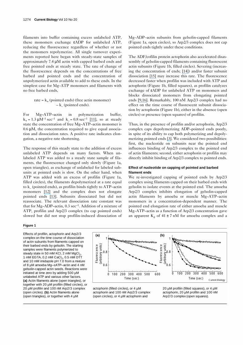
filaments into buffer containing excess unlabeled ATP,these monomers exchange εADP for unlabeled ATP,reducing the fluorescence regardless of whether or notthe monomers repolymerize. All single turnover experi-ments reported here began with steady-state samples ofapproximately 7.4 µM actin with capped barbed ends andfree pointed ends at steady state. The rate of change ofthe fluorescence depends on the concentrations of freebarbed and pointed ends and the concentration ofunpolymerized actin available to add to these ends. In thesimplest case for Mg–ATP monomers and filaments withno free barbed ends,
rate = k+ (pointed ends) (free actin monomer)– k– (pointed ends).
For Mg–ATP–actin in polymerization buffer,k+ = 1.3 µM–1 sec–1 and k– = 0.8 sec–1 [11], so at steadystate the concentration of free Mg–ATP–actin monomer is0.6 µM, the concentration required to give equal associa-tion and dissociation rates. A positive rate indicates elon-gation, a negative rate shortening.
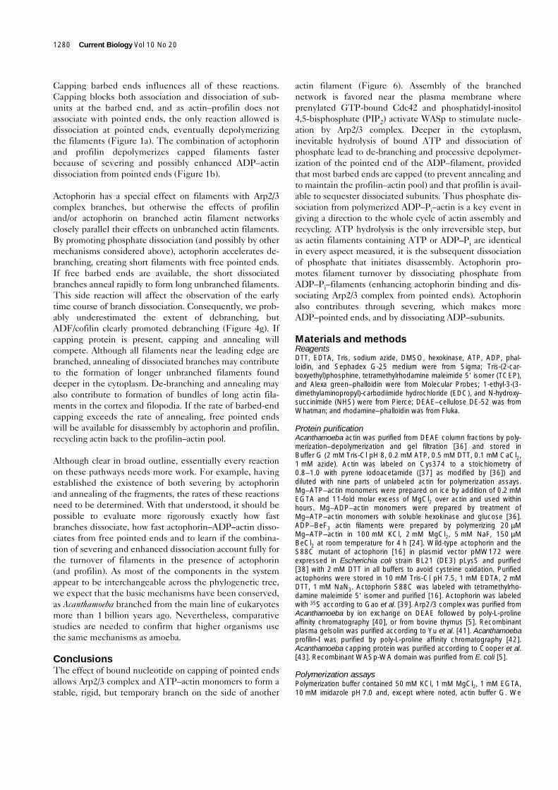
The response of this steady state to the addition of excessunlabeled ATP depends on many factors. When un-labeled ATP was added to a steady state sample of fila-ments, the fluorescence changed only slowly (Figure 1a,open triangles), as exchange of unlabeled for labeled sub-units at pointed ends is slow. On the other hand, whenATP was added with an excess of profilin (Figure 1a,filled circles), the filaments depolymerized at a rate equalto k– (pointed ends), as profilin binds tightly to ATP–actinmonomers [12] and the complex does not elongatepointed ends [13]. Subunits dissociated but did notreassociate. The relevant dissociation rate constant wasthat for Mg–ADP–actin, 0.3 sec–1. Addition of a mixture ofATP, profilin and Arp2/3 complex (to cap pointed ends)slowed but did not stop profilin-induced dissociation of
Mg–ADP–actin subunits from gelsolin-capped filaments(Figure 1a, open circles), so Arp2/3 complex does not cappointed ends tightly under these conditions.
The ADF/cofilin protein actophorin also accelerated disas-sembly of gelsolin-capped filaments containing fluorescentactin subunits (Figure 1b, filled circles). Severing (increas-ing the concentration of ends; [14]) and/or faster subunitdissociation [15] may increase this rate. The fluorescencedecreased faster when profilin was included with ATP andactophorin (Figure 1b, filled squares), as profilin catalyzesexchange of εADP for unlabeled ATP on monomers andblocks dissociated monomers from elongating pointedends [9,16]. Remarkably, 100 nM Arp2/3 complex had noeffect on the time course of fluorescent subunit dissocia-tion by actophorin (Figure 1b), either in the absence (opencircles) or presence (open squares) of profilin.
Thus, in the presence of profilin and/or actophorin, Arp2/3complex caps depolymerizing ADP–pointed ends poorly,in spite of its ability to cap both polymerizing and depoly-merizing pointed ends [3]. We considered two possibilities:first, the nucleotide on subunits near the pointed endinfluences binding of Arp2/3 complex to the pointed endof actin filaments; second, either actophorin or profilin maydirectly inhibit binding of Arp2/3 complex to pointed ends.
Effect of nucleotide on capping of pointed and barbedfilament endsWe re-investigated capping of pointed ends by Arp2/3complex using filaments capped on their barbed ends withgelsolin to isolate events at the pointed end. The amoebaArp2/3 complex inhibits elongation of gelsolin-cappedactin filaments by amoeba or muscle Mg–ATP–actinmonomers in a concentration-dependent manner. Thepointed end elongation rate of either amoeba and muscleMg–ATP–actin as a function of Arp2/3 concentration gavean apparent Kd of 41 ± 7 nM for amoeba complex and a
1274 Current Biology Vol 10 No 20
Figure 1
Effects of profilin, actophorin and Arp2/3complex on the time course of dissociationof actin subunits from filaments capped ontheir barbed ends by gelsolin. The startingsamples were filaments polymerized tosteady state in 50 mM KCl, 2 mM MgCl2,1 mM EGTA, 0.2 mM CaCl2, 0.5 mM DTTand 10 mM imidazole pH 7.0 from a mixtureof 8 µM amoeba Mg–εATP–actin and 4 nMgelsolin-capped actin seeds. Reactions wereinitiated at time zero by adding 500 µMunlabeled ATP and various other factors.(a) Actin filaments alone (open triangles), ortogether with 20 µM profilin (filled circles), or20 µM profilin and 100 nM Arp2/3 complex(open circles). (b) Actin filaments alone(open triangles), or together with 4 µM
actophorin (filled circles), or 4 µMactophorin and 100 nM Arp2/3 complex(open circles), or 4 µM actophorin and
20 µM profilin (filled squares), or 4 µMactophorin, 20 µM profilin and 100 nMArp2/3 complex (open squares).
Current Biology
100 200 300 400 500 600
Fluo
resc
ence
(arb
itrar
y un
its)
Fluo
resc
ence
(arb
itrar
y un
its)
Time (sec) Time (sec)
0.6
0.8
1
0 100 200 300 400 500 600
(a) (b)
0.6
0.8
1
0
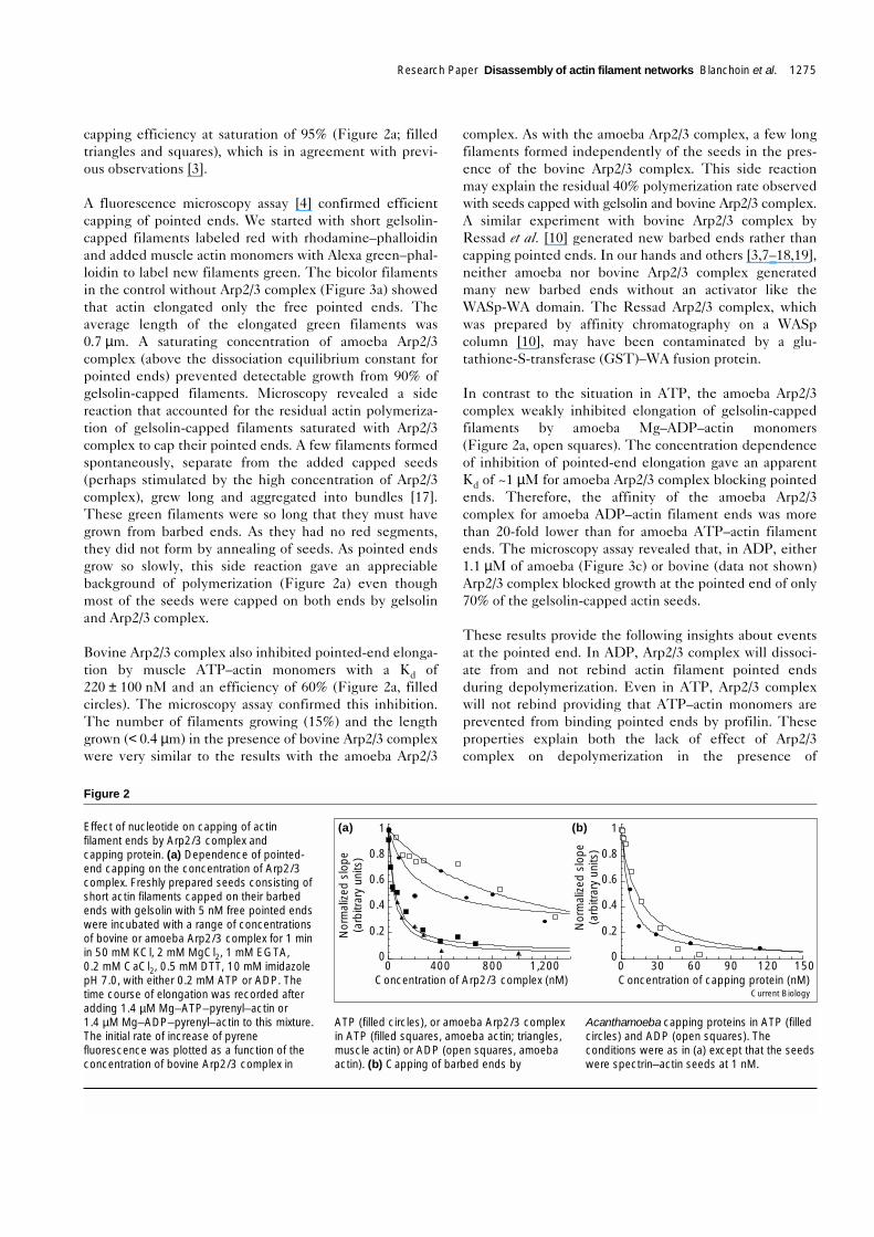
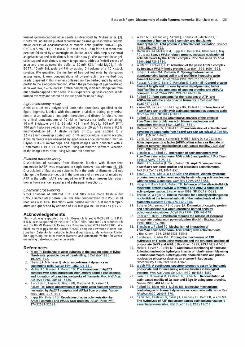
capping efficiency at saturation of 95% (Figure 2a; filledtriangles and squares), which is in agreement with previ-ous observations [3].
A fluorescence microscopy assay [4] confirmed efficientcapping of pointed ends. We started with short gelsolin-capped filaments labeled red with rhodamine–phalloidinand added muscle actin monomers with Alexa green–phal-loidin to label new filaments green. The bicolor filamentsin the control without Arp2/3 complex (Figure 3a) showedthat actin elongated only the free pointed ends. Theaverage length of the elongated green filaments was0.7 µm. A saturating concentration of amoeba Arp2/3complex (above the dissociation equilibrium constant forpointed ends) prevented detectable growth from 90% ofgelsolin-capped filaments. Microscopy revealed a sidereaction that accounted for the residual actin polymeriza-tion of gelsolin-capped filaments saturated with Arp2/3complex to cap their pointed ends. A few filaments formedspontaneously, separate from the added capped seeds(perhaps stimulated by the high concentration of Arp2/3complex), grew long and aggregated into bundles [17].These green filaments were so long that they must havegrown from barbed ends. As they had no red segments,they did not form by annealing of seeds. As pointed endsgrow so slowly, this side reaction gave an appreciablebackground of polymerization (Figure 2a) even thoughmost of the seeds were capped on both ends by gelsolinand Arp2/3 complex.
Bovine Arp2/3 complex also inhibited pointed-end elonga-tion by muscle ATP–actin monomers with a Kd of220 ± 100 nM and an efficiency of 60% (Figure 2a, filledcircles). The microscopy assay confirmed this inhibition.The number of filaments growing (15%) and the lengthgrown (< 0.4 µm) in the presence of bovine Arp2/3 complexwere very similar to the results with the amoeba Arp2/3
complex. As with the amoeba Arp2/3 complex, a few longfilaments formed independently of the seeds in the pres-ence of the bovine Arp2/3 complex. This side reactionmay explain the residual 40% polymerization rate observedwith seeds capped with gelsolin and bovine Arp2/3 complex.A similar experiment with bovine Arp2/3 complex byRessad et al. [10] generated new barbed ends rather thancapping pointed ends. In our hands and others [3,7–18,19],neither amoeba nor bovine Arp2/3 complex generatedmany new barbed ends without an activator like theWASp-WA domain. The Ressad Arp2/3 complex, whichwas prepared by affinity chromatography on a WASpcolumn [10], may have been contaminated by a glu-tathione-S-transferase (GST)–WA fusion protein.
In contrast to the situation in ATP, the amoeba Arp2/3complex weakly inhibited elongation of gelsolin-cappedfilaments by amoeba Mg–ADP–actin monomers(Figure 2a, open squares). The concentration dependenceof inhibition of pointed-end elongation gave an apparentKd of ~1 µM for amoeba Arp2/3 complex blocking pointedends. Therefore, the affinity of the amoeba Arp2/3complex for amoeba ADP–actin filament ends was morethan 20-fold lower than for amoeba ATP–actin filamentends. The microscopy assay revealed that, in ADP, either1.1 µM of amoeba (Figure 3c) or bovine (data not shown)Arp2/3 complex blocked growth at the pointed end of only70% of the gelsolin-capped actin seeds.
These results provide the following insights about eventsat the pointed end. In ADP, Arp2/3 complex will dissoci-ate from and not rebind actin filament pointed endsduring depolymerization. Even in ATP, Arp2/3 complexwill not rebind providing that ATP–actin monomers areprevented from binding pointed ends by profilin. Theseproperties explain both the lack of effect of Arp2/3complex on depolymerization in the presence of
Research Paper Disassembly of actin filament networks Blanchoin et al. 1275
Figure 2
Effect of nucleotide on capping of actinfilament ends by Arp2/3 complex andcapping protein. (a) Dependence of pointed-end capping on the concentration of Arp2/3complex. Freshly prepared seeds consisting ofshort actin filaments capped on their barbedends with gelsolin with 5 nM free pointed endswere incubated with a range of concentrationsof bovine or amoeba Arp2/3 complex for 1 minin 50 mM KCl, 2 mM MgCl2, 1 mM EGTA,0.2 mM CaCl2, 0.5 mM DTT, 10 mM imidazolepH 7.0, with either 0.2 mM ATP or ADP. Thetime course of elongation was recorded afteradding 1.4 µM Mg–ATP–pyrenyl–actin or1.4 µM Mg–ADP–pyrenyl–actin to this mixture.The initial rate of increase of pyrenefluorescence was plotted as a function of theconcentration of bovine Arp2/3 complex in
ATP (filled circles), or amoeba Arp2/3 complexin ATP (filled squares, amoeba actin; triangles,muscle actin) or ADP (open squares, amoebaactin). (b) Capping of barbed ends by
Acanthamoeba capping proteins in ATP (filledcircles) and ADP (open squares). Theconditions were as in (a) except that the seedswere spectrin–actin seeds at 1 nM.
Current Biology
Nor
mal
ized
slo
pe(a
rbitr
ary
units
)
Concentration of Arp2/3 complex (nM)
Nor
mal
ized
slo
pe(a
rbitr
ary
units
)
Concentration of capping protein (nM)
0
0.2
0.4
0.6
0.8
1
0
0.2
0.4
0.6
0.8
1
0 30 60 90 120 1500 400 800 1,200
(a) (b)
actophorin (Figure 1b, open circles) and the partial inhibi-tion of pointed-end depolymerization by Arp2/3 complexin the presence of profilin (Figure 1a, open circles). Theyalso highlight the importance of ATP hydrolysis and therate-limiting release of phosphate from ADP–Pi–actin sub-units for polymer stability in the presence of regulatoryproteins. The situation at the barbed end was dramaticallydifferent. First, although capping protein bound better inATP than ADP (Figure 2b), this 2-fold difference wassmall compared with the 40-fold difference in affinity ofArp2/3 complex for ATP– and ADP–pointed ends. Thisemphasizes the sensitivity of Arp2/3 complex to the
nucleotide on subunits at pointed ends. Second, barbedends were more likely to have ATP than pointed ends inthe presence of profilin because of exchange of ATP–sub-units and exchange of ATP from the medium with theterminal subunits at the barbed end [20].
Another series of microscopy experiments showed that thebovine Arp2/3 complex blocked all pointed ends of thedaughter filaments at the branch point but that barbed-endcapping by capping protein was intermittent (Figure 3d).Experiments started with branched filaments labeled redwith rhodamine–phalloidin. In the presence of 100 nMcapping protein, muscle ATP–actin monomers elongatedpointed ends rapidly and barbed ends slowly (labeledgreen with Alexa green–phalloidin). Pointed ends growingfaster than barbed ends is opposite to what happens withuncapped filaments. We never observed new filamentgrowth at the pointed end of filaments at branch points.Thus, pointed ends residing at branches are capped moreeffectively by Arp2/3 complex than unbranched pointedends. This observation also shows that branches rarely,if ever, form by Arp2/3 complex joining the pointed endof a preformed filament (even a newly polymerizedATP–pointed end) to the side of another filament.
The fact that some barbed ends had short green extensions(Figure 3d) means that capping protein dissociates inter-mittently from barbed ends to allow for elongation orannealing. As growth on these barbed ends was short,recapping must be efficient or else barbed-end growthwould have greatly exceeded pointed-end growth. Fromthe concentration of capping protein and the associationrate constant [21], we estimated that the half time forcapping protein to rebind was about 2 seconds, which is inagreement with the limited growth of barbed ends. Slowgrowth in the presence of saturating capping protein sug-gests that capping protein may dissociate from barbed endsfaster than proposed previously (t1/2 = 30 minutes, [21]).
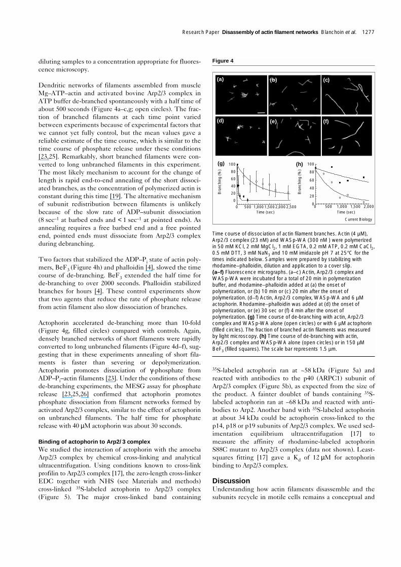
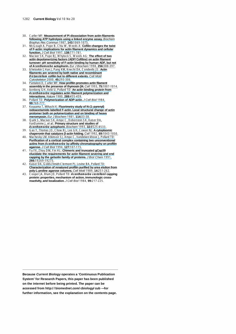
Dissociation of branches during aging anddepolymerization of dendritic networksAs Arp2/3 complex bound ADP–pointed ends much moreweakly than ATP–pointed ends, we examined how threefactors that affect phosphate dissociation — phalloidin,BeF3 and actophorin — affect the stability of actin fila-ment branches formed by Arp2/3 complex on the side ofactin filaments (Figure 4). Phalloidin inhibits phosphaterelease [22] whereas actophorin promotes phosphaterelease [23]. BeF3 substitutes for γ-phosphate, forming amuch more stable complex with ADP and actin than phos-phate [24]. We assembled branched networks from con-centrations of pure actin and activated Arp2/3 complexthat yielded branching density and lengths suitable forlight microscopy. To follow the time course of anyrearrangements in these samples, we terminated the reac-tions at intervals by adding rhodamine–phalloidin and
1276 Current Biology Vol 10 No 20
Figure 3
Visualization by fluorescence microscopy of the products generated byelongation of red-labeled (a–c) gelsolin-capped actin filament seeds or(d) branched filaments nucleated by Arp2/3 complex. The reactioncontained 50 mM KCl, 1 mM MgCl2, 1 mM EGTA, 0.5 mM DTT,0.1 mM CaCl2, 0.2 mM ATP or ADP, 3 mM NaN3 and 10 mM imidazolepH 7. Phalloidin conjugated to Alexa green (Alexa green–phalloidin)was present during the elongation to stabilize the new filaments.Samples were prepared by dilution and application to a cover slip.(a) Gelsolin-capped seeds (5 nM) stabilized with rhodamine–phalloidinwere incubated with 1 µM Mg–ATP–actin for 15 min in the presenceof Alexa green–phalloidin and with an additional 1 µM Mg–ATP–actinfor 15 min. (b) Gelsolin-capped red seeds were pre-incubated for1 min with 1080 nM amoeba Arp2/3 complex, then with a mixture of1 µM Mg–ATP–actin monomers and 1 µM Alexa green–phalloidin15 min, followed by an additional 1 µM Mg–ATP–actin for 15 min.(c) Gelsolin-capped red seeds were pre-incubated with 1080 nMamoeba Arp2/3 complex, then with 1 µM Mg–ADP–actin monomersand Alexa green–phalloidin for 15 min, followed by an additional 1 µMMg–ADP–actin for 15 min. (d) Branched actin filaments nucleated byArp2/3 complex, labeled with rhodamine–phalloidin (4.8 nM of barbedends) and capped with 100 nM capping protein, were incubated for20 min with 4 µM Mg–ATP–actin monomers in the presence of Alexagreen–phalloidin. The scale bars represent 0.6 µm.
Current Biology
(a) (b)
(c) (d)
diluting samples to a concentration appropriate for fluores-cence microscopy.
Dendritic networks of filaments assembled from muscleMg–ATP–actin and activated bovine Arp2/3 complex inATP buffer de-branched spontaneously with a half time ofabout 500 seconds (Figure 4a–c,g; open circles). The frac-tion of branched filaments at each time point variedbetween experiments because of experimental factors thatwe cannot yet fully control, but the mean values gave areliable estimate of the time course, which is similar to thetime course of phosphate release under these conditions[23,25]. Remarkably, short branched filaments were con-verted to long unbranched filaments in this experiment.The most likely mechanism to account for the change oflength is rapid end-to-end annealing of the short dissoci-ated branches, as the concentration of polymerized actin isconstant during this time [19]. The alternative mechanismof subunit redistribution between filaments is unlikelybecause of the slow rate of ADP–subunit dissociation(8 sec–1 at barbed ends and < 1 sec–1 at pointed ends). Asannealing requires a free barbed end and a free pointedend, pointed ends must dissociate from Arp2/3 complexduring debranching.
Two factors that stabilized the ADP–Pi state of actin poly-mers, BeF3 (Figure 4h) and phalloidin [4], slowed the timecourse of de-branching. BeF3 extended the half time forde-branching to over 2000 seconds. Phalloidin stabilizedbranches for hours [4]. These control experiments showthat two agents that reduce the rate of phosphate releasefrom actin filament also slow dissociation of branches.
Actophorin accelerated de-branching more than 10-fold(Figure 4g, filled circles) compared with controls. Again,densely branched networks of short filaments were rapidlyconverted to long unbranched filaments (Figure 4d–f), sug-gesting that in these experiments annealing of short fila-ments is faster than severing or depolymerization.Actophorin promotes dissociation of γ-phosphate fromADP–Pi–actin filaments [23]. Under the conditions of thesede-branching experiments, the MESG assay for phosphaterelease [23,25,26] confirmed that actophorin promotesphosphate dissociation from filament networks formed byactivated Arp2/3 complex, similar to the effect of actophorinon unbranched filaments. The half time for phosphaterelease with 40 µM actophorin was about 30 seconds.
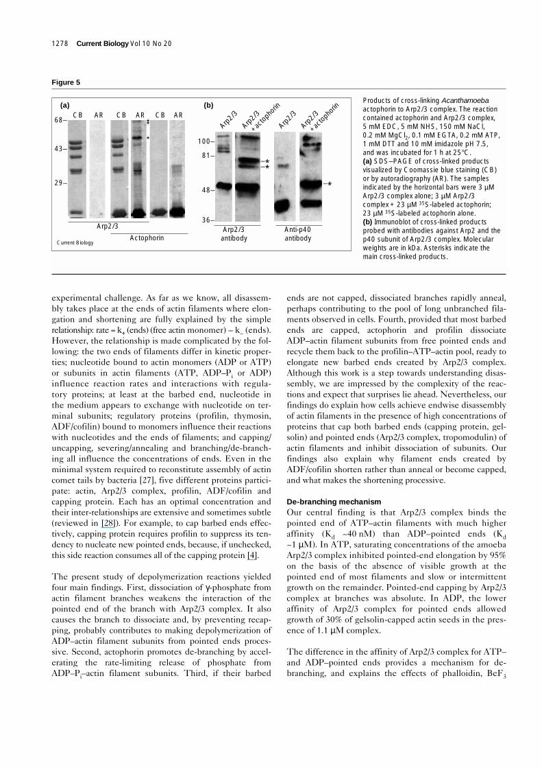
Binding of actophorin to Arp2/3 complexWe studied the interaction of actophorin with the amoebaArp2/3 complex by chemical cross-linking and analyticalultracentrifugation. Using conditions known to cross-linkprofilin to Arp2/3 complex [17], the zero-length cross-linkerEDC together with NHS (see Materials and methods)cross-linked 35S-labeled actophorin to Arp2/3 complex(Figure 5). The major cross-linked band containing
35S-labeled actophorin ran at ~58 kDa (Figure 5a) andreacted with antibodies to the p40 (ARPC1) subunit ofArp2/3 complex (Figure 5b), as expected from the size ofthe product. A fainter doublet of bands containing 35S-labeled actophorin ran at ~68 kDa and reacted with anti-bodies to Arp2. Another band with 35S-labeled actophorinat about 34 kDa could be actophorin cross-linked to thep14, p18 or p19 subunits of Arp2/3 complex. We used sed-imentation equilibrium ultracentrifugation [17] tomeasure the affinity of rhodamine-labeled actophorinS88C mutant to Arp2/3 complex (data not shown). Least-squares fitting [17] gave a Kd of 12 µM for actophorinbinding to Arp2/3 complex.
DiscussionUnderstanding how actin filaments disassemble and thesubunits recycle in motile cells remains a conceptual and
Research Paper Disassembly of actin filament networks Blanchoin et al. 1277
Figure 4
Time course of dissociation of actin filament branches. Actin (4 µM),Arp2/3 complex (23 nM) and WASp-WA (300 nM ) were polymerizedin 50 mM KCl, 2 mM MgCl2, 1 mM EGTA, 0.2 mM ATP, 0.2 mM CaCl2,0.5 mM DTT, 3 mM NaN3 and 10 mM imidazole pH 7 at 25°C for thetimes indicated below. Samples were prepared by stabilizing withrhodamine–phalloidin, dilution and application to a cover slip.(a–f) Fluorescence micrographs. (a–c) Actin, Arp2/3 complex andWASp-WA were incubated for a total of 20 min in polymerizationbuffer, and rhodamine–phalloidin added at (a) the onset ofpolymerization, or (b) 10 min or (c) 20 min after the onset ofpolymerization. (d–f) Actin, Arp2/3 complex, WASp-WA and 6 µMactophorin. Rhodamine–phalloidin was added at (d) the onset ofpolymerization, or (e) 30 sec or (f) 4 min after the onset ofpolymerization. (g) Time course of de-branching with actin, Arp2/3complex and WASp-WA alone (open circles) or with 6 µM actophorin(filled circles). The fraction of branched actin filaments was measuredby light microscopy. (h) Time course of de-branching with actin,Arp2/3 complex and WASp-WA alone (open circles) or in 150 µMBeF3 (filled squares). The scale bar represents 1.5 µm.
Current Biology
0 500 1,0001,5002,0002,500
Bra
nchi
ng (%
)
Bra
nchi
ng (%
)
Time (sec) Time (sec)
(g)
0 500 1,000 1,500 2,000
(h)
0
20
40
60
80
100
0
20
40
60
80
100
(a) (b)
(d) (e)
(c)
(f)
experimental challenge. As far as we know, all disassem-bly takes place at the ends of actin filaments where elon-gation and shortening are fully explained by the simplerelationship: rate = k+ (ends) (free actin monomer) – k– (ends).However, the relationship is made complicated by the fol-lowing: the two ends of filaments differ in kinetic proper-ties; nucleotide bound to actin monomers (ADP or ATP)or subunits in actin filaments (ATP, ADP–Pi or ADP)influence reaction rates and interactions with regula-tory proteins; at least at the barbed end, nucleotide inthe medium appears to exchange with nucleotide on ter-minal subunits; regulatory proteins (profilin, thymosin,ADF/cofilin) bound to monomers influence their reactionswith nucleotides and the ends of filaments; and capping/uncapping, severing/annealing and branching/de-branch-ing all influence the concentrations of ends. Even in theminimal system required to reconstitute assembly of actincomet tails by bacteria [27], five different proteins partici-pate: actin, Arp2/3 complex, profilin, ADF/cofilin andcapping protein. Each has an optimal concentration andtheir inter-relationships are extensive and sometimes subtle(reviewed in [28]). For example, to cap barbed ends effec-tively, capping protein requires profilin to suppress its ten-dency to nucleate new pointed ends, because, if unchecked,this side reaction consumes all of the capping protein [4].
The present study of depolymerization reactions yieldedfour main findings. First, dissociation of γ-phosphate fromactin filament branches weakens the interaction of thepointed end of the branch with Arp2/3 complex. It alsocauses the branch to dissociate and, by preventing recap-ping, probably contributes to making depolymerization ofADP–actin filament subunits from pointed ends proces-sive. Second, actophorin promotes de-branching by accel-erating the rate-limiting release of phosphate fromADP–Pi–actin filament subunits. Third, if their barbed
ends are not capped, dissociated branches rapidly anneal,perhaps contributing to the pool of long unbranched fila-ments observed in cells. Fourth, provided that most barbedends are capped, actophorin and profilin dissociateADP–actin filament subunits from free pointed ends andrecycle them back to the profilin–ATP–actin pool, ready toelongate new barbed ends created by Arp2/3 complex.Although this work is a step towards understanding disas-sembly, we are impressed by the complexity of the reac-tions and expect that surprises lie ahead. Nevertheless, ourfindings do explain how cells achieve endwise disassemblyof actin filaments in the presence of high concentrations ofproteins that cap both barbed ends (capping protein, gel-solin) and pointed ends (Arp2/3 complex, tropomodulin) ofactin filaments and inhibit dissociation of subunits. Ourfindings also explain why filament ends created byADF/cofilin shorten rather than anneal or become capped,and what makes the shortening processive.
De-branching mechanism Our central finding is that Arp2/3 complex binds thepointed end of ATP–actin filaments with much higheraffinity (Kd ~40 nM) than ADP–pointed ends (Kd~1 µM). In ATP, saturating concentrations of the amoebaArp2/3 complex inhibited pointed-end elongation by 95%on the basis of the absence of visible growth at thepointed end of most filaments and slow or intermittentgrowth on the remainder. Pointed-end capping by Arp2/3complex at branches was absolute. In ADP, the loweraffinity of Arp2/3 complex for pointed ends allowedgrowth of 30% of gelsolin-capped actin seeds in the pres-ence of 1.1 µM complex.
The difference in the affinity of Arp2/3 complex for ATP–and ADP–pointed ends provides a mechanism for de-branching, and explains the effects of phalloidin, BeF3
1278 Current Biology Vol 10 No 20
Figure 5
Products of cross-linking Acanthamoebaactophorin to Arp2/3 complex. The reactioncontained actophorin and Arp2/3 complex,5 mM EDC, 5 mM NHS, 150 mM NaCl,0.2 mM MgCl2, 0.1 mM EGTA, 0.2 mM ATP,1 mM DTT and 10 mM imidazole pH 7.5,and was incubated for 1 h at 25°C.(a) SDS–PAGE of cross-linked productsvisualized by Coomassie blue staining (CB)or by autoradiography (AR). The samplesindicated by the horizontal bars were 3 µMArp2/3 complex alone; 3 µM Arp2/3complex + 23 µM 35S-labeled actophorin;23 µM 35S-labeled actophorin alone.(b) Immunoblot of cross-linked productsprobed with antibodies against Arp2 and thep40 subunit of Arp2/3 complex. Molecularweights are in kDa. Asterisks indicate themain cross-linked products.
Current Biology
(a)CB AR CB AR
**
*
–*–*–*
CB AR
Arp2/3
Actophorin
(b)
Arp2/3
Arp2/3
+ac
topho
rin
Arp2/3
Arp2/3
+ac
topho
rin
81–
100–
48–
36–Arp2/3
antibodyAnti-p40antibody
68–
43–
29–
and actophorin on the rate of de-branching and whypointed-end depolymerization is processive. All of thedata are consistent with phosphate dissociation from thepointed end of the branch being the key event in chang-ing the affinity and with the de-branching reaction beingdissociation of the pointed end from Arp2/3 complex,which may or may not remain associated with the motherfilament. Nevertheless, more detailed mechanistic studiesare desirable in the future. In principle, the nucleotidestate of the mother filament and of Arp2/3 complex mightalso influence the stability of the branch point. However,the frequency of branching from ADP–filaments isapproximately the same as from newly polymerized fila-ments (ATP–filaments and ADP–Pi–filaments) [4], suggest-ing that the lateral interaction with the mother filament isless sensitive to the nucleotide in the filament.
As irreversible ATP hydrolysis [29] followed by slow dis-sociation of the γ-phosphate [30] are inevitable followingpolymerization of ATP–actin, de-branching may also beinevitable, although external factors may influence therate of these reactions. The first example is the ability ofADF/cofilin to accelerate de-branching. The ability ofactophorin to promote phosphate dissociation fromADP–Pi–actin filaments is sufficient to explain its effecton de-branching, but actophorin may influence de-branching and disassembly in other ways. Binding ofactophorin to Arp2/3 complex, although weak, may reducethe affinity of Arp2/3 complex for pointed ends. In addi-tion, the conformational change induced when ADF/cofil-ins bind actin filaments [31] may dissociate Arp2/3complex from filament ends or sides. This conformationalchange may also influence binding of capping protein tobarbed ends. On the other hand, WASp/Scar proteins maystabilize branches by interacting with both Arp2/3complex and actin. All of these possibilities deserve moredetailed investigation.
Response of filaments and branched networks toactophorin and profilin Much previous work considered how individual actin-binding proteins interact with actin monomers and fila-ments, but it is now clear that understanding actin filamentturnover in cells requires insights about ensembles of regu-latory proteins. This task is built on knowledge of the indi-vidual reactions, but light microscopy has revealed that it ismuch more complex than one might have anticipated. Wehave found that the responses of actin filaments andbranched actin filament networks to actophorin and profilininvolve not only effects on subunit dissociation at filamentends [15] and severing [14,23,32,33], but also effects ofADF/cofilin on the rate of phosphate dissociation, de-branching, capping and annealing. Capping is particularlyimportant. Barbed-end capping is required for profilin tosequester ATP–actin monomers [34], but it also preventsannealing of filaments [35] created by severing or de-branching, allowing these free pointed ends to disassemble.Thus barbed-end capping is essential for effective disas-sembly of pointed ends. Addition of actophorin to uncappedfilaments results in severing, but the mean length of the fil-aments reaches a plateau with time [23,32]. We postulatedthat this steady state results from a balance between therates of severing and annealing. The light micrographs inthis paper confirmed annealing, but its rate and extent needfurther quantitative studies. Given the impressive annealingobserved by light microscopy, we expect that previous solu-tion studies underestimated the extent of severing. Lightmicroscopy with immobilized filaments [14,33] may havegiven a more accurate account of severing. Addition of pro-filin to uncapped filaments has no effect on actin monomeror polymer concentrations, but most of the actin monomerbinds profilin. Addition of actophorin and profilin touncapped filaments promotes subunit flux from filaments toADP–actin–actophorin to ATP–actin–profilin and back tofree barbed ends [15,16,23].
Research Paper Disassembly of actin filament networks Blanchoin et al. 1279
Figure 6
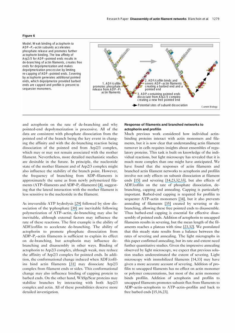
Model. Weak binding of actophorin toADP–Pi–actin subunits acceleratesphosphate release and promotes furtheractophorin binding. The low affinity ofArp2/3 for ADP–pointed ends results inde-branching of actin filaments, creates freeends for depolymerization and makesdepolymerization processive by limitingre-capping of ADP–pointed ends. Severingby actophorin generates additional pointedends, which depolymerize provided barbedends are capped and profilin is present tosequester monomers.
1. ADF/cofilinpromotes phosphaterelease from ADP–Pi
actin filaments
2. ADF/cofilin binds andsevers ADP–actin filamentscreating a barbed end and apointed end
3. ADP-containing pointed endsdissociate from Arp2/3 complexcreating a new free pointed end
Potential sites of subunit dissociation Current Biology
Capping barbed ends influences all of these reactions.Capping blocks both association and dissociation of sub-units at the barbed end, and as actin–profilin does notassociate with pointed ends, the only reaction allowed isdissociation at pointed ends, eventually depolymerizingthe filaments (Figure 1a). The combination of actophorinand profilin depolymerizes capped filaments fasterbecause of severing and possibly enhanced ADP–actindissociation from pointed ends (Figure 1b).
Actophorin has a special effect on filaments with Arp2/3complex branches, but otherwise the effects of profilinand/or actophorin on branched actin filament networksclosely parallel their effects on unbranched actin filaments.By promoting phosphate dissociation (and possibly by othermechanisms considered above), actophorin accelerates de-branching, creating short filaments with free pointed ends.If free barbed ends are available, the short dissociatedbranches anneal rapidly to form long unbranched filaments.This side reaction will affect the observation of the earlytime course of branch dissociation. Consequently, we prob-ably underestimated the extent of debranching, butADF/cofilin clearly promoted debranching (Figure 4g). Ifcapping protein is present, capping and annealing willcompete. Although all filaments near the leading edge arebranched, annealing of dissociated branches may contributeto the formation of longer unbranched filaments founddeeper in the cytoplasm. De-branching and annealing mayalso contribute to formation of bundles of long actin fila-ments in the cortex and filopodia. If the rate of barbed-endcapping exceeds the rate of annealing, free pointed endswill be available for disassembly by actophorin and profilin,recycling actin back to the profilin–actin pool.
Although clear in broad outline, essentially every reactionon these pathways needs more work. For example, havingestablished the existence of both severing by actophorinand annealing of the fragments, the rates of these reactionsneed to be determined. With that understood, it should bepossible to evaluate more rigorously exactly how fastbranches dissociate, how fast actophorin–ADP–actin disso-ciates from free pointed ends and to learn if the combina-tion of severing and enhanced dissociation account fully forthe turnover of filaments in the presence of actophorin(and profilin). As most of the components in the systemappear to be interchangeable across the phylogenetic tree,we expect that the basic mechanisms have been conserved,as Acanthamoeba branched from the main line of eukaryotesmore than 1 billion years ago. Nevertheless, comparativestudies are needed to confirm that higher organisms usethe same mechanisms as amoeba.
ConclusionsThe effect of bound nucleotide on capping of pointed endsallows Arp2/3 complex and ATP–actin monomers to form astable, rigid, but temporary branch on the side of another
actin filament (Figure 6). Assembly of the branchednetwork is favored near the plasma membrane whereprenylated GTP-bound Cdc42 and phosphatidyl-inositol4,5-bisphosphate (PIP2) activate WASp to stimulate nucle-ation by Arp2/3 complex. Deeper in the cytoplasm,inevitable hydrolysis of bound ATP and dissociation ofphosphate lead to de-branching and processive depolymer-ization of the pointed end of the ADP–filament, providedthat most barbed ends are capped (to prevent annealing andto maintain the profilin–actin pool) and that profilin is avail-able to sequester dissociated subunits. Thus phosphate dis-sociation from polymerized ADP–Pi–actin is a key event ingiving a direction to the whole cycle of actin assembly andrecycling. ATP hydrolysis is the only irreversible step, butas actin filaments containing ATP or ADP–Pi are identicalin every aspect measured, it is the subsequent dissociationof phosphate that initiates disassembly. Actophorin pro-motes filament turnover by dissociating phosphate fromADP–Pi–filaments (enhancing actophorin binding and dis-sociating Arp2/3 complex from pointed ends). Actophorinalso contributes through severing, which makes moreADP–pointed ends, and by dissociating ADP–subunits.
Materials and methodsReagents DTT, EDTA, Tris, sodium azide, DMSO, hexokinase, ATP, ADP, phal-loidin, and Sephadex G-25 medium were from Sigma; Tris-(2-car-boxyethyl)phosphine, tetramethylrhodamine maleimide 5′ isomer (TCEP),and Alexa green–phalloidin were from Molecular Probes; 1-ethyl-3-(3-dimethylaminopropyl)-carbodiimide hydrochloride (EDC), and N-hydroxy-succinimide (NHS) were from Pierce; DEAE–cellulose DE-52 was fromWhatman; and rhodamine–phalloidin was from Fluka.
Protein purificationAcanthamoeba actin was purified from DEAE column fractions by poly-merization–depolymerization and gel filtration [36] and stored inBuffer G (2 mM Tris-Cl pH 8, 0.2 mM ATP, 0.5 mM DTT, 0.1 mM CaCl2,1 mM azide). Actin was labeled on Cys374 to a stoichiometry of0.8–1.0 with pyrene iodoacetamide ([37] as modified by [36]) anddiluted with nine parts of unlabeled actin for polymerization assays.Mg–ATP–actin monomers were prepared on ice by addition of 0.2 mMEGTA and 11-fold molar excess of MgCl2 over actin and used withinhours. Mg–ADP–actin monomers were prepared by treatment ofMg–ATP–actin monomers with soluble hexokinase and glucose [36].ADP–BeF3 actin filaments were prepared by polymerizing 20 µMMg–ATP–actin in 100 mM KCl, 2 mM MgCl2, 5 mM NaF, 150 µMBeCl2 at room temperature for 4 h [24]. Wild-type actophorin and theS88C mutant of actophorin [16] in plasmid vector pMW172 wereexpressed in Escherichia coli strain BL21 (DE3) pLysS and purified[38] with 2 mM DTT in all buffers to avoid cysteine oxidation. Purifiedactophorins were stored in 10 mM Tris-Cl pH 7.5, 1 mM EDTA, 2 mMDTT, 1 mM NaN3. Actophorin S88C was labeled with tetramethylrho-damine maleimide 5′ isomer and purified [16]. Actophorin was labeledwith 35S according to Gao et al. [39]. Arp2/3 complex was purified fromAcanthamoeba by ion exchange on DEAE followed by poly-L-prolineaffinity chromatography [40], or from bovine thymus [5]. Recombinantplasma gelsolin was purified according to Yu et al. [41]. Acanthamoebaprofilin-I was purified by poly-L-proline affinity chromatography [42].Acanthamoeba capping protein was purified according to Cooper et al.[43]. Recombinant WASp-WA domain was purified from E. coli [5].
Polymerization assaysPolymerization buffer contained 50 mM KCl, 1 mM MgCl2, 1 mM EGTA,10 mM imidazole pH 7.0 and, except where noted, actin buffer G. We
1280 Current Biology Vol 10 No 20
formed gelsolin-capped actin seeds as described by Mullins et al. [3].Briefly, we incubated purified recombinant plasma gelsolin with a twofoldmolar excess of Acanthamoeba or muscle actin (Buffer: 200–400 µMCaCl2, 0.5 mM DTT, 0.2 mM ATP, 2 mM Tris pH 8.0) for 2 h at room tem-perature followed by an overnight incubation at 4°C (this step is essentialas gelsolin-capped actin dimers form quite slowly). We warmed these gel-solin-capped actin dimers to room temperature, added a fivefold excess ofactin and then adjusted the buffer to 50 mM KCl, 1 mM MgCl2, 1 mMEGTA, 10 mM Imidazole pH 7.0 by adding 0.1 volume of a 10 × stocksolution. We quantified the number of free pointed ends by elongationassays using known concentrations of pyrenyl–actin. We verified thatseeds prepared in this manner contained no free barbed ends by addingprofilin to the elongation reaction. When the percentage of pyrene-labeledactin was low, 1–5% excess profilin completely inhibited elongation fromour gelsolin-capped actin seeds. In our experience, gelsolin-capped seedsformed this way and stored on ice are good for up to 5 days.
Light microscopy assay Actin at 4 µM was polymerized under the conditions specified in thefigure legends, labeled with rhodamine–phalloidin during polymeriza-tion or at an indicated time point thereafter and diluted for observationto a final concentration of 10 nM in fluorescence buffer containing10 mM imidazole pH 7.0, 50 mM KCl, 1 mM MgCl2, 100 mM DTT,100 µg/ml glucose oxidase, 3 mg/ml glucose, 20 µg/ml catalase, 0.5%methylcellulose [4]. A dilute sample of 2 µl was applied to a22 × 22 mm coverslip coated with 0.1% nitrocellulose in amyl acetate.Actin filaments were observed by epi-fluorescence illumination with anOlympus IX-70 microscope and digital images were collected with aHamamatsu ORCA CCD camera using Metamorph software. Analysisof the images was done using Metamorph software.
Filament turnover assay Dissociation of subunits from filaments labeled with fluorescentnucleotide (εATP) was measured in single turnover experiments [9,10].Dissociation of fluorescent subunits from the ends of filaments did notchange the fluorescence, but in the presence of an excess of unlabeledATP in the buffer, εATP exchanged for ATP with an irreversible reduc-tion in fluorescence regardless of subsequent reactions.
Chemical cross-linking Stock solutions of 100 mM EDC and NHS were made fresh in dryDMSO immediately before use. The final concentration of DMSO in allreactions was 10%. Reactions were carried out for 1 h at room temper-ature and quenched by addition of 100 mM glycine, 10 mM Tris pH 7.5.
AcknowledgementsThis work was supported by NIH Research Grant GM-26338 to T.D.P.R.D.M. was supported by the Jane Coffin Childs Fund for Cancer Researchand by HHMI Research Resources Program grant #76296-549901. Wethank Harry Higgs for the bovine Arp2/3 complex; Lawrence Haines andJonathan Zalevsky for valuable technical assistance; Marie-France Carlierfor suggesting the term mother filament; and Annemarie Weber for adviceon making gelsolin-capped actin seeds.
References1. Wang Y: Exchange of actin subunits at the leading edge of living
fibroblasts: possible role of treadmilling. J Cell Biol 1985,101:597-602.
2. Theriot JA, Mitchison TJ: Actin microfilament dynamics inlocomoting cells. Nature 1991, 352:126-131.
3. Mullins RD, Heuser JA, Pollard TD: The interaction of Arp2/3complex with actin: nucleation, high-affinity pointed end capping,and formation of branching networks of filaments. Proc Natl AcadSci USA 1998, 95:6181-6186.
4. Blanchoin L, Amann KJ, Higgs HN, Marchand JB, Kaiser DA,Pollard TD: Direct observation of dendritic actin filament networksnucleated by Arp2/3 complex and WASp/Scar proteins. Nature2000, 404:1007-1011.
5. Higgs HN, Pollard TD: Regulation of actin polymerization byArp2/3 complex and WASp/Scar proteins. J Biol Chem 1999,274:32531-32534.
6. Welch MD, Rosenblatt J, Skoble J, Portnoy DA, Mitchison TJ:Interaction of human Arp2/3 complex and the Listeriamonocytogenes ActA protein in actin filament nucleation. Science1998, 281:105-108.
7. Machesky LM, Mullins DM, Higgs HN, Kaiser DA, Blanchoin L, MayRC, et al.: Scar, a WASp-related protein, activates nucleation ofactin filaments by the Arp2/3 complex. Proc Natl Acad Sci USA1999, 96:3739-3744.
8. Winter D, Lechler T, Li R: Activation of the yeast Arp2/3 complexby Bee1p, a WASP-family protein. Curr Biol 1999, 9:501-504.
9. Didry D, Carlier M-F, Pantaloni D: Synergy between actindepolymerizing factor/cofilin and profilin in increasing actinfilament turnover. J Biol Chem 1998, 273:25602-25611.
10. Ressad F, Didry D, Egile C, Pantaloni D, Carlier MF: Control of actinfilament length and turnover by actin depolymerizing factor(ADF/cofilin) in the presence of capping proteins and ARP2/3complex. J Biol Chem 1999, 274:20970-20976.
11. Pollard TD: Rate constants for the reactions of ATP- andADP-actin with the ends of actin filaments. J Cell Biol 1986,103:2747-2754.
12. Vinson VK, De La Cruz EM, Higgs HN, Pollard TD: Interactions ofAcanthamoeba profilin with actin and nucleotides bound to actin.Biochem 1998, 37:10871-10880.
13. Pollard TD, Cooper JA: Quantitative analysis of the effect ofAcanthamoeba profilin on actin filament nucleation andelongation. Biochem 1984, 23:6631-6641.
14. Maciver SK, Zot HG, Pollard TD: Characterization of actin filamentsevering by actophorin from Acanthamoeba castellanii. J Cell Biol1991, 115:1611-1620.
15. Carlier MF, Laurent V, Santolini J, Melki R, Didry D, Xia GX, et al.:Actin depolymerizing factor (ADF/cofilin) enhances the rate offilament turnover: implication in actin-based motility. J Cell Biol1997, 136:1307-1322.
16. Blanchoin L, Pollard TD: Interaction of actin monomers withAcanthamoeba actophorin (ADF/cofilin) and profilin. J Biol Chem1998, 273:25106-25111.
17. Mullins RD, Kelleher JF, Xu J, Pollard TD: Arp2/3 complex fromAcanthamoeba binds profilin and crosslinks actin filaments.Mol Biol Cell 1998, 9:841-852.
18. Yarar D, To W, Abo A, Welch MD: The Wiskott–Aldrich syndromeprotein directs actin-based motility by stimulating actin nucleationwith the Arp2/3 complex. Curr Biol 1999, 9:555-558.
19. Higgs HN, Blanchoin L, Pollard TD: Influence of the Wiskott-Aldrichsyndrome protein (WASp) C terminus and Arp2/3 complex onactin polymerization. Biochemistry 1999, 38:15212-15222.
20. Teubner A, Wegner A: Kinetic evidence for a readily exchangeablenucleotide at the terminal subunit of the barbed ends of actinfilaments. Biochem 1998, 37:7532-7538.
21. Schafer DA, Jennings PB, Cooper JA: Dynamics of capping proteinand actin assembly in vitro: uncapping barbed ends bypolyphosphoinositides. J Cell Biol 1996, 135:169-179.
22. Dancker P, Hess L: Phalloidin reduces the release of inorganicphosphate during actin polymerization. Biochim Biophys Acta1990, 1035:197-200.
23. Blanchoin L, Pollard TD: Mechanism of interaction ofAcanthamoeba actophorin (ADF/cofilin) with actin filaments.J Biol Chem 1999, 274:15538-15546.
24. Combeau C, Carlier M-F: Probing the mechanism of ATPhydrolysis on F-actin using vanadate and the structural analogs ofphosphate BeF3 and AIF4. J Biol Chem 1988, 263:17429-17436.
25. Melki R, Fievez S, Carlier M-F: Continuous monitoring of ππ releasefollowing nucleotide hydrolysis in actin or tubulin assembly using2-amino-6mercapto-7-methylpurine ribonucleoside and purine-nucleoside phosphorylase as an enzyme-linked assay.Biochemistry 1996, 35:12038-12045.
26. Webb MR: A continuous spectrophotometric assay for inorganicphosphate and for measuring release kinetics in biologicalsystems. Proc Natl Acad Sci USA 1992, 89:4884-4887.
27. Loisel TP, Boujemaa R, Pantaloni D, Carlier MF: Reconstitution ofactin-based motility of Listeria and Shigella using pure proteins.Nature 1999, 401:613-616.
28. Pollard TD, Blanchoin L, Mullins RD: Molecular mechanismscontrolling actin filament dynamics in nonmuscle cells. Annu RevBiophys 2000, 29:545-576.
29. Carlier MF, Pantaloni D, Evans JA, Lambooy PK, Korn ED, Webb MR:The hydrolysis of ATP that accompanies actin polymerization isessentially irreversible. FEBS Lett 1988, 235:211-214.
Research Paper Disassembly of actin filament networks Blanchoin et al. 1281
30. Carlier MF: Measurement of Pi dissociation from actin-filamentsfollowing ATP hydrolysis using a linked enzyme assay. BiochemBiophys Res Commun 1987, 143:1069-1075.
31. McGough A, Pope B, Chiu W, Weeds A: Cofilin changes the twistof F-actin: implications for actin filament dynamics and cellularfunction. J Cell Biol 1997, 138:771-781.
32. Maciver SK, Pope BJ, Whytock S, Weeds AG: The effect of twoactin depolymerizing factors (ADF/Cofilins) on actin filamentturnover: pH sensitivity of F-actin binding by human ADF, but notof Acanthamoeba actophorin. Eur J Biochem 1998, 256:388-397.
33. Ichetovkin I, Han J, Pang KM, Knecht DA, Condeelis JS: Actinfilaments are severed by both native and recombinantDictyostelium cofilin but to different extents. Cell MotilCytoskeleton 2000, 45:293-306.
34. Pantaloni D, Carlier MF: How profilin promotes actin filamentassembly in the presence of thymosin ββ4. Cell 1993, 75:1007-1014.
35. Isenberg GH, Aebi U, Pollard TD: An actin binding protein fromAcanthamoeba regulates actin filament polymerization andinteractions. Nature 1980, 288:455-459.
36. Pollard TD: Polymerization of ADP-actin. J Cell Biol 1984,99:769-777.
37. Kouyama T, Mihashi K: Fluorimetry study of N-(1-pyrenyl)iodoacetamide-labelled F-actin. Local structural change of actinprotomer both on polymerization and on binding of heavymeromyosin. Eur J Biochem 1981, 114:33-38.
38. Quirk S, Maciver SK, Ampe C, Doberstein SK, Kaiser DA,VanDamme J, et al.: Primary structure and studies ofAcanthamoeba actophorin. Biochem 1993, 32:8525-8533.
39. Gao Y, Thomas JO, Chow RL, Lee GH, Cowan NJ: A cytoplasmicchaperonin that catalyzes ββ-actin folding. Cell 1992, 69:1043-1050.
40. Machesky LM, Atkinson SJ, Ampe C, Vandekerckhove J, Pollard TD:Purification of a cortical complex containing two unconventionalactins from Acanthamoeba by affinity chromatography on profilinagarose. J Cell Biol 1994, 127:107-115.
41. Yu FX, Zhou DM, Yin HL: Chimeric and truncated gCap39elucidate the requirements for actin filament severing and endcapping by the gelsolin family of proteins. J Biol Chem 1991,266:19269-19275.
42. Kaiser DA, Goldschmidt-Clermont PJ, Levine BA, Pollard TD:Characterization of renatured profilin purified by urea elution frompoly-L-proline agarose columns. Cell Motil 1989, 14:251-262.
43. Cooper JA, Blum JD, Pollard TD: Acanthamoeba castellanii cappingprotein: properties, mechanism of action, immunologic cross-reactivity, and localization. J Cell Biol 1984, 99:217-225.
1282 Current Biology Vol 10 No 20
Because Current Biology operates a ‘Continuous PublicationSystem’ for Research Papers, this paper has been publishedon the internet before being printed. The paper can beaccessed from http://biomednet.com/cbiology/cub — forfurther information, see the explanation on the contents page.
Related Documents