Arp2/3 complex is required for actin polymerization during platelet shape change Zhi Li, Eric S. Kim, and Elaine L. Bearer Department of Pathology and Laboratory Medicine, Brown University, Providence, RI. Abstract Platelets undergo a series of actin-dependent morphologic changes when activated by thrombin receptor activating peptide (TRAP) or when spreading on glass. Polymerization of actin results in the sequential formation of filopodia, lamellipodia, and stress fibers, but the molecular mechanisms regulating this polymerization are unknown, The Arp2/3 complex nucleates actin polymerization in vitro and could perform this function inside cells as well. To test whether Arp2/3 regulated platelet actin polymerization, we used recombinant Arp2 protein (rArp2) to generate Arp2-specific antibodies (αArp2). Intact and Fab fragments of αArp2 inhibited TRAP- stimulated actin-polymerizing activity in platelet extracts as measured by the pyrene assay. Inhibition was reversed by the addition of rArp2 protein. To test the effect of Arp2/3 inhibition on the formation of specific actin structures, we designed a new method to permeabilize resting platelets while preserving their ability to adhere and to form filopodia and lamellipodia on exposure to glass. Inhibition of Arp2/3 froze platelets at the rounded, early stage of activation, before the formation of filopodia and lamellipodia. By morphometric analysis, the proportion of platelets in the rounded stage rose from 2.85% in untreated to 63% after treatment with αArp2. This effect was also seen with Fab fragments and was reversed by the addition of rArp2 protein. By immunofluorescence of platelets at various stages of spreading, the Arp2/3 complex was found in filopodia and lamellipodia. These results suggest that activation of the Arp2/3 complex at the cortex by TRAP stimulation initiates an explosive polymerization of actin filaments that is required for all subsequent actin-dependent events. Introduction Activation of platelets produces a reproducible sequence of morphologic events, whether in suspension or during spreading on glass: rounding, filopodial projection, attachment, spreading, and ultimately contraction.1 – 6 These morphologic changes depend on the reorganization of the actin cytoskeleton, including severing of existing filaments, which causes the discoid platelet to round and depends on gelsolin,3 , 7 , 8 and polymerization of actin monomers into new filaments.3 , 9 – 11 These new actin filaments organize into 4 distinct structures: filopodia, lamellipodia, stress fibers, and a contractile ring.4 Each of these structures performs a different function, and each contains a different complement of actin-binding proteins.4 – 6 The Arp2/3 complex is likely to regulate the polymerization of actin during shape change in the platelet. Arp2/3 is a 7-member protein complex isolated by poly-proline chromatography from the soil amoeba, Acanthamoeba castellani.12 Although the Arp2 subunit was isolated from human platelets, first by F-actin affinity chromatography4 and later as a member of the © 2002 by The American Society of Hematology Reprints: Elaine L. Bearer, Department of Pathology and Laboratory Medicine, Brown University, Providence, RI 02912; [email protected]. NIH Public Access Author Manuscript Blood. Author manuscript; available in PMC 2012 June 15. Published in final edited form as: Blood. 2002 June 15; 99(12): 4466–4474. NIH-PA Author Manuscript NIH-PA Author Manuscript NIH-PA Author Manuscript

Welcome message from author
This document is posted to help you gain knowledge. Please leave a comment to let me know what you think about it! Share it to your friends and learn new things together.
Transcript

Arp2/3 complex is required for actin polymerization duringplatelet shape change
Zhi Li, Eric S. Kim, and Elaine L. BearerDepartment of Pathology and Laboratory Medicine, Brown University, Providence, RI.
AbstractPlatelets undergo a series of actin-dependent morphologic changes when activated by thrombinreceptor activating peptide (TRAP) or when spreading on glass. Polymerization of actin results inthe sequential formation of filopodia, lamellipodia, and stress fibers, but the molecularmechanisms regulating this polymerization are unknown, The Arp2/3 complex nucleates actinpolymerization in vitro and could perform this function inside cells as well. To test whetherArp2/3 regulated platelet actin polymerization, we used recombinant Arp2 protein (rArp2) togenerate Arp2-specific antibodies (αArp2). Intact and Fab fragments of αArp2 inhibited TRAP-stimulated actin-polymerizing activity in platelet extracts as measured by the pyrene assay.Inhibition was reversed by the addition of rArp2 protein. To test the effect of Arp2/3 inhibition onthe formation of specific actin structures, we designed a new method to permeabilize restingplatelets while preserving their ability to adhere and to form filopodia and lamellipodia onexposure to glass. Inhibition of Arp2/3 froze platelets at the rounded, early stage of activation,before the formation of filopodia and lamellipodia. By morphometric analysis, the proportion ofplatelets in the rounded stage rose from 2.85% in untreated to 63% after treatment with αArp2.This effect was also seen with Fab fragments and was reversed by the addition of rArp2 protein.By immunofluorescence of platelets at various stages of spreading, the Arp2/3 complex was foundin filopodia and lamellipodia. These results suggest that activation of the Arp2/3 complex at thecortex by TRAP stimulation initiates an explosive polymerization of actin filaments that isrequired for all subsequent actin-dependent events.
IntroductionActivation of platelets produces a reproducible sequence of morphologic events, whether insuspension or during spreading on glass: rounding, filopodial projection, attachment,spreading, and ultimately contraction.1–6 These morphologic changes depend on thereorganization of the actin cytoskeleton, including severing of existing filaments, whichcauses the discoid platelet to round and depends on gelsolin,3,7,8 and polymerization ofactin monomers into new filaments.3,9–11 These new actin filaments organize into 4distinct structures: filopodia, lamellipodia, stress fibers, and a contractile ring.4 Each ofthese structures performs a different function, and each contains a different complement ofactin-binding proteins.4–6
The Arp2/3 complex is likely to regulate the polymerization of actin during shape change inthe platelet. Arp2/3 is a 7-member protein complex isolated by poly-proline chromatographyfrom the soil amoeba, Acanthamoeba castellani.12 Although the Arp2 subunit was isolatedfrom human platelets, first by F-actin affinity chromatography4 and later as a member of the
© 2002 by The American Society of Hematology
Reprints: Elaine L. Bearer, Department of Pathology and Laboratory Medicine, Brown University, Providence, RI 02912;[email protected].
NIH Public AccessAuthor ManuscriptBlood. Author manuscript; available in PMC 2012 June 15.
Published in final edited form as:Blood. 2002 June 15; 99(12): 4466–4474.
NIH
-PA Author Manuscript
NIH
-PA Author Manuscript
NIH
-PA Author Manuscript

Arp2/3 complex that mediates Listeria-induced actin assembly,13 neither its function nor itslocation in platelets has been previously reported. In vitro, Arp2/3 nucleates actin filamentassembly14,15 and is required, together with 2 other proteins, to reconstitute the actin-basedmotility of Listeria bacteria.16 Arp 2/3 is reported to have at least 2 binding sites for actin:one that binds to the sides of actin filaments and the other that binds to the pointed ends ofactin monomers nucleating barbed-end elongation.14,17,18 In vitro, this could producenetworks of filaments that branch at 70° angles. In soil ameba and in cultured cells, Arp2/3is found in the lamellipodia12,15,17 where filaments branch at 70° angles.19 Antibodies tothe p34 subunit of Arp2/3 inhibit this branching activity in vitro and in vivo but do notinhibit the incorporation of actin monomer.17 Antibodies to the Arp2, but not the Arp3,subunit inhibit actin-polymerizing activity in extracts of Acanthamoeba.20 Based oncorrelations between this in vitro behavior and its cellular location, Arp2/3 is considered thebest candidate to regulate actin dynamics physiologically at the membrane–cytoplasminterface.21–27
Because platelets are anucleate and are very small, they can neither be transfected norinjected. Permeabilization has been used to load platelets with pyrene-actin to study agonist-stimulated actin polymerization biochemically.3,11 In platelets, actin-polymerizing activityincreases in response to the agonist, thrombin receptor activating peptide (TRAP), and 60%of this increase is preserved after permeabilization with detergent. We discovered thatpermeabilized platelets undergo normal morphologic events when exposed to glass: theyadhere and reorganize their actin filaments into the typical 4 structures. We have used thesetechniques together with a set of molecular tools, including new inhibitory antibodiesgenerated against recombinant Arp2 (rArp2), to test the role of Arp2/3 in actin dynamics andin the morphologic events of surface-activated shape change.
Materials and methodsConstructs and antibodies
Full-length Arp228 (GenBank accession number 71789) was amplified by polymerase chainreaction (PCR) using primers with a KpnI site in the first 6 base pairs of the 5′ primer(GGTACCATGGACAGATCGAAGGG) and Pspl and HindIII sites in the tail of the 3′primer (reverse complement: (CCCGGGAAGCTTCTAGTGACTGATCTTTTG) from aclone containing the Arp2 cDNA. PCR products were ligated in pCR2.1 vector (Invitrogen,Carlsbad, CA), excised with KpnI and BamH1, and ligated into the same sites in frame inthe pQE30 vector (Qiagen, Valencia, CA). Recombinant Arp2 containing 6 histidine tagswas overexpressed in bacteria, purified, and confirmed by peptide sequencing. Gel slices ofpurified protein were used as immunogen in rabbits according to our protocols.4,5 Serumfrom 3 different rabbits was affinity purified separately by affinity column chromatographyagainst rArp2. The final preparation contained 1 mg/mL affinity-purified antibody.Preimmune antibodies from the same rabbit were purified by protein A affinitychromatography and were used in parallel. In any given experiment, the preimmuneantibodies were from the same rabbit as the immune antibody preparation. Fab fragmentswere prepared by papain digestion of purified antibodies, and the Fc fragment was removedby passage through a protein A column according to established protocols.29
Affinity-purified antipeptide rabbit antibodies against p34 were a gift from John Condeelis.17 Rabbit anti-kaptin was raised in our laboratory,5 and mouse anti-actin is from themonoclonal hybridoma JLA-4.4,5 Western blotting was performed as previously described.4–6
Quantification of the amount of Arp2 in platelets was performed as follows. Platelet-richplasma (PRP) was used on the day of draw as described.4–6 The platelet count was
Li et al. Page 2
Blood. Author manuscript; available in PMC 2012 June 15.
NIH
-PA Author Manuscript
NIH
-PA Author Manuscript
NIH
-PA Author Manuscript

determined (usually approximately 5.5 × 10¹¹ platelets/U). After washing, the platelets fromone unit of PRP were resuspended in gel sample buffer at a volume calculated to give a finalconcentration of 2.5 × 10¹¹ platelets/mL. Various amounts of platelets were loaded intoseparate lanes on sodium dodecyl sulfate–polyacrylamide gel electrophoresis (SDS-PAGE)in parallel with different amounts of purified rArp2 and were probed for Arp2 by Westernblotting. The amount of Arp2 in the platelet sample was determined by comparing thedensity of resultant bands in the platelet samples with the density of bands containing aknown amount of rArp2 using the GelDoc with Quantity One software (BioRad, Hercules,CA). The amount of Arp2 molecules per platelet was determined by dividing the amount ofArp2 detected on the blot by the number of platelets loaded on the gel. Molar concentrationwas determined by assuming that the average volume of a platelet was 7 fL.30 It required1.6 × 108 platelets (6 µL) per platelet sample to detect a band on the blot, with an anti-Arp2detection limit of 15 ng.
Precipitation of Arp2 with αArp2-conjugated protein A beadsProtein A beads were conjugated to affinity purified αArp2 or to preimmune antibodiesfrom the same rabbit29 and were used to detect endogenous Arp2 in platelet extracts.Platelet extracts were prepared as described.4–6 Briefly, washed platelets were lysed by 1:1dilution in lysis buffer containing 2% nonionic detergent and were sonicated briefly.Extracts (100 µL of 35 mg/mL protein) were incubated with 10 µL antibody-coupled beadsfor 4 hours at 4°C. Beads were collected by centrifugation and washed in lysis buffer.Proteins from equal volumes of supernatant and pellet were separated by SDS-PAGE,transferred to Western blots, and probed sequentially with αArp2 (1:1000), αp34 (1:500),and αactin (1:5,000), followed by horseradish peroxidase secondary antibody (antirabbit,Jackson ImmunoResearch Laboratories, West Grove, PA; anti–mouse IgM, RocheDiagnostics, Indianapolis, IN). Blots were imaged by chemiluminescence (AmershamPharmacia Biotech, Piscataway, NJ).
Pyrene actin polymerization kineticsPyrene actin (10% labeled; Cytoskeleton, Denver, CO) was aliquoted, lyophilized from G-buffer (5 mM Tris [pH 8.1], 0.2 mM CaCl2, 0.2 mM adenosine triphosphate [ATP], 0.5 mMdithiothreitol [DTT]), and stored in liquid nitrogen. On the day of use, the actin was thawed,incubated at 10 µM in water for 4 hours at room temperature, and centrifuged for 1.5 hoursat 100 000g. Immediately before each experiment, this stock of actin was diluted to 1.3 µMin 200 µL polymerization buffer (100 mM KCl, 2 mM MgCl2, 0.1 EGTA, 10 mM Tris [pH7.0], 0.5 mM ATP, 0.5 mM DTT). The cuvette containing the diluted actin was placed in thefluorometer for a baseline reading before the platelet suspension was added.
Washed platelets were resuspended in platelet buffer (145 nM NaCl, 5 mM KC1, 0.5 mMNa2HPO4, 2 mM MgCl2, 10 mM glucose, 10 mM HEPES [pH 7.4], and 0.3% globin-freebovine serum albumin more than 99% pure) and were maintained at 37°C. For sonication,90 µL platelets were treated with 10 µL PHEM buffer (60 mM PIPES [pH 6.9], 25 mMHEPES [pH 6.9], 10 mM EGTA, 2 mM Mg Cl2, and protease inhibitors) with or without 1µL of 1.2 mM TRAP (Sigma, St Louis, MO) as described.6 Sonicates were preparedindividually just before each assay, antibodies were added immediately thereafter, and thesuspension was injected into the cuvette containing pyrene-actin.
For Triton permeabilization, a 1:10 dilution of 7.5% Triton X-100 with and without TRAP(1 µL of 1.2 mM) was added to the PHEM buffer, and no sonication was performed.Otherwise, the procedure was essentially the same as for sonication. For sonication or Tritonpermeabilization, fewer than 30 seconds elapsed between TRAP stimulation andmeasurement in the fluorometer.
Li et al. Page 3
Blood. Author manuscript; available in PMC 2012 June 15.
NIH
-PA Author Manuscript
NIH
-PA Author Manuscript
NIH
-PA Author Manuscript

After platelets were added, actin polymerization was measured for 10 to 20 minutes at 1-second intervals with excitation at 365 and emission 407 nm in a luminescence spectrometer(model LS50B; Perkin-Elmer, Wellesley, MA). Data were collected using the Winlabprogram and were analyzed with Microsoft Excel.
Each series of experiments was performed on the same day with the same preparations ofactin and platelets, beginning with baseline measurements of actin alone, and of plateletswith or without TRAP.
Morphometric analysis of αArp2 on shape changeBrief permeabilization with low amounts of Triton was used to load antibody into plateletsbefore exposing them to glass. PRP (3 µL) diluted with platelet buffer to approximately 0.6to 1.2 × 10⁶ platelets/mL was treated with a 1/10 volume of 0.75% Triton in PHEM bufferwith and without antibodies for 1 minute before mounting on a glass coverslip. The Tritonconcentration was immediately diluted by flooding the coverslip with 500 µL Tyrodesolution (145 mM NaCl, 5 mM KC1, 10 mM HEPES [pH 7.4], 2 mM MgCl2, 0.5 mMNa2PO4, 1.8 mM CaCl2, and 10 mM glucose).4 PRP was also applied to coverslips withoutpermeabilization. After 20 minutes, the coverslip was fixed with 4% formaldehyde inPHEM buffer with 0.25% Triton containing 1/50 dilution of 3 µM fluoresceinisothiocyanate–phalloidin (FITC–phalloidin; Molecular Probes, Eugene, OR).4–6 Primaryantibodies were detected by postfixation staining with Cy3-conjugated secondary antibody(Cy3 is red; Jackson ImmunoResearch Laboratories).
Immunofluorescence to determine endogenous Arp2/3 locationPRP without permeabilization was dotted onto glass coverslips, and the coverslip wasflooded with Tyrode buffer as described above. After 20 minutes, spread platelets were fixedin 4% paraformaldehyde in PHEM buffer containing a 1/50 dilution of FITC–phalloidinwith or without 0.25% Triton for 20 minutes at room temperature. Fixed platelets wereblocked with 1% bovine serum albumin (Sigma) in phosphate-buffered saline with 0.1%Triton X100. Coverslips were then stained with αArp2 followed by secondary antibody.Images were obtained on a Nikon fluorescence microscope equipped with an RT-Spot liquidcrystal digital camera (Diagnostic Instruments, Starling Heights, IL).
ResultsGeneration of rArp2 and anti-Arp2 antibodies
To develop tools to test the role of Arp2/3 in platelet actin dynamics, we cloned the Arp2subunit and raised antibodies to full-length rArp2. Immunization of 8 different rabbitsproduced 3 with high-titer antiserum that was sensitive and specific for Arp2. Wecharacterized the αArp2 antibodies against bacterially expressed rArp2 (Figure 1A) andagainst platelet extracts (Figure 1B–C). Affinity-purified αArp2 detected a single band ininduced bacterial homogenates (Figure 1A, lane 3) and recognized purified rArp2 (Figure1A, lane 6). The antibody could detect as little as 15 ng purified rArp2. In platelet extracts,αArp2 detected a single band of the appropriate molecular weight (44 kd) (Figure 1B).
Quantitative Western blot analysis comparing purified rArp2 with platelet extracts revealedthat the concentration of Arp2 in platelets is 0.3 µM, or 1300 molecules per platelet. Thisamount is sufficient to account for all the new free actin filament ends (410–570 barbedends/platelet) produced during activation,3,11,31 even if only half the Arp2/3 is activated.This is significantly less protein than VASP or gelsolin, which are estimated at 5 µM each,6,31,32 and this amount is consistent with the yield of Arp2/3 complex obtained fromplatelets in purification protocols.13
Li et al. Page 4
Blood. Author manuscript; available in PMC 2012 June 15.
NIH
-PA Author Manuscript
NIH
-PA Author Manuscript
NIH
-PA Author Manuscript

Arp2 was depleted from Triton-treated, sonicated platelet extracts by incubation withαArp2-conjugated protein A beads (Figure 1C). These extraction conditions were originallydesigned to depolymerize actin while preserving many other protein–protein interactions.4,5Extracts were incubated with either αArp2- or preimmune–conjugated protein A beads, andthe beads and supernatant were separated by centrifugation. By Western blot, Arp2 wascleared from the extract after incubation with αArp2 beads and was detected in the pelletcontaining the beads (Figure 1C, top panel). In contrast, Arp2 remained in the supernatantafter treatment with preimmune beads. Reprobing of the same blot for p34 (another Arp2/3subunit) or for actin revealed both proteins in the supernatant and not pelleted with αArp2(Figure 1C, middle and lower panels). This demonstrated that αArp2 recognized nativeArp2 in solution.
Effect of Arp2 inhibition on platelet actin-polymerizing activityThe effect of αArp2 on actin-polymerizing activity of platelet extracts was measured in thepyrene assay. To perform this assay, platelets must be permeabilized sufficiently for enoughpyrene-labeled actin to be loaded for detection in the fluorometer.8,11 Suchpermeabilization also allows entry of the αArp2 antibody, thus permitting measurements ofthe effect of antibody on actin polymerization.
TRAP produced a robust increase in actin-polymerizing activity in platelets permeabilizedby sonication (6.3-fold, Figure 2A, C) or detergent (Figure 2B). This TRAP-stimulatedincrease was exquisitely sensitive to αArp2 (Figure 2A–C). Addition of αArp2 (0.01 mg/mL in Figure 2A; or 15 µg/mL in Table 1) resulted in a 71% decrease in the polymerizationrate during the initial 2 minutes after TRAP stimulation. This inhibitory effect was not seenwith a different affinity-purified rabbit antibody raised against another actin-binding protein,kaptin,5 in which polymerization rates were 93.6% of untreated (Figure 2A,C; Table 1).Treatment with αp34 had only a slight inhibitory effect, with rates 82% of untreated (Figure2B; Table 1) as expected.17
Three additional experiments confirmed the specificity of the αArp2 antibody in inhibitingTRAP-stimulated actin polymerization. First, Fab fragments of αArp2 have similarinhibitory effects, demonstrating that inhibition is not dependent on the formation of largeantibody–antigen complexes (Figure 2C; Table 1). Second, affinity-purified αArp2 fromanother rabbit also inhibited polymerization (αArp2; Figure 2C), showing that inhibition isnot caused by some unique component in a particular preparation of antibody. Finally,pretreatment of the antibody with rArp2 protein completely abolished the inhibitory effect ofαArp2 (rArp2, Figure 2C). This demonstrates that αArp2 inhibition is dependent oninteraction of the antibody with Arp2 protein.
Because Arp2/3 is expected to increase the initial rate of polymerization, we calculated therates during the first 120 seconds and compared the effect of different treatments (Table 1).Only αArp2 and its Fab fragments—not αkaptin or αp34—caused a reproducible decreasein the initial polymerization rate. After treatment with Fab fragments at a final concentrationof 0.40 µM, the actin polymerization rate was decreased to 13% of the rate in untreatedplatelets.
In the absence of TRAP, αArp2 had less effect on the low level of actin-polymerizingactivity (Figure 2D), whereby at least 10-fold more antibody was required (compare 0.10mg/mL without TRAP in Figure 2D with 0.01 mg/mL with TRAP in Figure 2A). TheαArp2-sensitive activity in nonstimulated preparations could represent those platelets thathave already been activated during preparation. The extent of inhibition was dependent onthe concentration of αArp2 antibody in either TRAP-stimulated or nonstimulated platelets
Li et al. Page 5
Blood. Author manuscript; available in PMC 2012 June 15.
NIH
-PA Author Manuscript
NIH
-PA Author Manuscript
NIH
-PA Author Manuscript
(Figure 2E). In contrast, preimmune IgG (Figure 2E, pre-IgG) had little effect on eitherpreparation, even at the highest concentrations.
Adherence and spreading of permeabilized platelets on glassTo examine whether the formation of particular actin structure(s) such as filopodia andlamellipodia requires Arp2/3 activity, we developed a technique to load platelets withαArp2 and then to observe the morphology of actin after spreading on glass (Figure 3).Platelets in plasma were exposed briefly to detergent with or without antibody, mounted onglass, and rapidly diluted with an approximately 500-fold excess of Tyrode solution. Afterspreading, platelets were fixed and stained for F-actin.
Permeabilization alone did not alter adherence to the glass. The same average number ofplatelets adhered with or without permeabilization (19.9 ± 7.6 and 19.5 ± 6.5 platelets perhigh-power field, respectively). The presence of any antibody increased adherence,regardless of antibody type (27.7 ± 5.2 platelets per high-power field after preimmunetreatment and 28.2 ± 8.0 after αArp2 treatment). Permeabilization did not cause a detectableloss of Arp2/3, as determined by postfixation immunostaining (data not shown).
Permeabilized platelets exhibited all the typical actin structures as imaged by FITC–phalloidin staining (Figure 3, top row, middle panel), though some minor variations wereobserved. Filopodia seemed longer, and the diffuse staining in the cytoplasm was decreased.Permeabilized platelets treated with preimmune antibodies also formed filopodia,lamellipodia, and stress fibers indistinguishable from platelets permeabilized withoutantibody (Figure 3, top row, right panel).
Morphologic effect of Arp2 Inhibition on platelet shape changeInactivation of Arp2/3 had a striking effect on morphology and on actin structures (Figure 3,lower row). Four dose-dependent effects of αArp2 on morphology were observed andquantified: (1) decreased proportion of spread platelets; (2) decreased proportion withfilopodia; (3) increased proportion in the rounded stage; and (4) presence of a novelmorphology, blebbing, not seen in the absence of αArp2. These effects were quantitativeand αArp2 dose dependent (Figure 4). Antibodies from 2 different rabbits were each used inparallel with the preimmune antibodies from the same rabbit.
Anti-Arp2 loading increased the proportion of rounded platelets in a dose-dependent manner(2.85%, 22%, 27%, 32%, and 63% for 0, 0.1, 0.2, 0.3, and 1 µg/mL, respectively), withcommensurate decreases in the proportion that had produced filopodia or spread (Figure 4).In the few αArp2-treated platelets with lamellipodia, spreading was abnormal (Figure 3,lower row, middle panel; Figure 4A). The decrease in numbers of platelets with filopodiawas remarkable, with less than 5% of platelets showing any of these structures at αArp2concentrations of only 0.3 µg/mL. In contrast, after 20 minutes of exposure to glass, morethan 80% of preimmune-treated platelets had spread or produced filopodia, and only a smallproportion of these (2.85%) remained in the rounded stage (Figure 4C). Plateletspermeabilized with or without preimmune antibodies resembled untreated preparations.
The proportion of platelets with blebs, which are never found in control preparations,increased in direct relationship to the amount of αArp2 (2%, 7%, 22%, 37%, and 57% for0.1, 0.2, 0.3, and 1.0 µg αArp2, respectively) (Figure 3, lower right panel; Figure 4D–E).The phalloidin staining of these blebs indicated that they must have contained filamentousactin, though the filaments were not organized into normal structures (Figure 3, lower rightpanel).
Li et al. Page 6
Blood. Author manuscript; available in PMC 2012 June 15.
NIH
-PA Author Manuscript
NIH
-PA Author Manuscript
NIH
-PA Author Manuscript

Fab fragments of αArp2 had similar effects, freezing most platelets in the rounded earlystage of activation (Figure 4E). Addition of rArp2 protein reversed these effects. Blebbedplatelets were also found after treatment with αArp2 Fab fragments, but the blebs weresmaller and fewer platelets were affected (Figure 4E, last panel).
The efficiency of permeabilization was determined by staining platelets with secondaryantibody after fixation (Figure 5). Primary antibody was detected in 79% of plateletspermeabilized in the presence of preimmune antibodies and 76% of those treated withαArp2. By immunofluorescence, secondary antibody staining of platelets loaded with intactαArp2 produced bright dots of varying sizes irregularly located in the platelet cytoplasm andseparate from actin filaments (Figure 5, arrows). In platelets loaded with Fab fragments, thestaining was also separated from the actin filaments but was more evenly distributed in thecytoplasm (data not shown).
Location of Arp2/3 in normal platelets during spreading on glassWe used double-label immunofluorescence to compare the normal location of Arp2/3 withthat of filamentous actin (Figure 6). Platelets were first allowed to spread and then werefixed with paraformaldehyde in buffer with or without detergent. Fixation without detergentpreserves a large pool of Arp2/3 that is lost when fixation includes Triton, whereas this poolis extracted in fixatives that include detergent.33 Thus, the timing of exposure to detergentallowed us to distinguish between extractable and actin-bound Arp2/3 in spread platelets.
Arp2/3 was detected at the edge of the lamellipodium of spread platelets fixed in thepresence of detergent, as has been reported for other cells fixed this way (Figure 6).12,15,17,19,33 Superimposition of images of the spread platelet double labeled for Arp2(Figure 6B, red) and F-actin (Figure 6B, green) shows that Arp2/3 was coincident in mostplaces with the actin frill of the lamellipodium. To determine whether the actin frill was atthe edge of the platelet, phase microscopy was needed. However, when detergent wasincluded in the fixative, effective imaging by phase microscopy was difficult. Therefore, wefixed platelets without detergent, double labeled them for F-actin (Figure 6C, green, FITC–phalloidin) and for αArp2 (Figure 6C, red, Cy3), and imaged them by phase microscopy andfluorescence microscopy. Phalloidin staining coincided with the phase image; thusphalloidin staining was a reliable indicator of the platelet surface contours. Arp2/3 stainingwas brighter than in platelets fixed in the presence of detergent. Under these conditions, asoluble pool of Arp2/3 was retained that lay in arcs internal to the lamellipodia. This signalwas so bright that it hindered detection of the weaker staining of Arp2/3 at the leading edge(Figure 6C, arrows), and was also brighter than the green phalloidin signal.
Platelets captured during early stages of activation revealed a relationship between Arp2/3location and filopodia. In the early stages, Arp2/3 was concentrated in the cortex, and therewas brighter staining over the short, F-actin–rich protrusions (Figure 7A–B, far left). At laterstages, the platelet contracts and growing filopodia stained for Arp2/3 at their tips and theirbases (Figure 7A–B, second from left). As activation progressed, filopodia elongated andArp2/3 was often found at the tip and at the base (Figure 7A–B, third and fourth from theleft). In some cases, an area of Arp2/3 staining was also present on the shaft of thefilopodium. Such filopodia are too small to be imaged by phase microscopy, containing asfew as 3 actin filaments.1,3,9,30 Higher magnification of these filopodia demonstrated therelationship between Arp2/3 and actin filaments (Figure 7C). The small size of platelets atthe filopodial stage of activation is apparent when they were compared to a fully spreadplatelet at the same magnification (Figure 7D).
Li et al. Page 7
Blood. Author manuscript; available in PMC 2012 June 15.
NIH
-PA Author Manuscript
NIH
-PA Author Manuscript
NIH
-PA Author Manuscript
DiscussionThe experiments reported here provide direct functional evidence that Arp2/3 is required forboth filopodial and lamellipodial formation in the platelet. Loading of platelets with αArp2inhibits TRAP-stimulated actin polymerization and the formation of actin structures in adose-dependent manner. This requirement for Arp2/3 suggests that actin polymerization,rather than reorganization of existing filaments, drives the first step of filopodial formation.
Arp2 and the formation of filopodiaThe presence of Arp2/3 at the tips of the filopodia is surprising because Arp2/3 is reportedto bind the pointed ends and the sides of actin filaments, whereas the other end, the barbedend, is at the tips of filopodia, as reported in cultured cells.34 Arp2/3 may be passivelycarried to the tip, along with membrane, as the filopodium grows. Arp2/3 may then nucleatenew filaments for further growth of the filopodium. Alternatively, all platelet extensions thatappear to be filopodia may not be identical to those produced by cells in culture. Indeed, inplatelets, such extensions were originally termed pseudopodia, and some were found tocontain microtubules.1,31 The actin filaments in some platelet filopodia may have reversedpolarity, with the pointed end at the tip. Re-examination by electron microscopy of the actinorganization and polarity in the platelet pseudopodia is needed to resolve these possibilities.
Whether Arp2/3 plays a role in filopodial formation in other cells is controversial. Evidencefor Arp2/3 involvement is that cdc42, reported to activate Arp2/3,20,24 initiates filopodialformation in cultured cells.35,36 On the other hand, the Arp2/3 complex nucleates branchedfilament networks in vitro14,18,37 and is found at branch points by immunogold labeling incells.19 Filopodia are composed of parallel filaments and do not have such branches,making such network formation by Arp2/3 seem unlikely. However, 2 recently publishedexperiments show that Arp2/3 can nucleate filament formation in the absence of branching.First, anti-p34 blocks branching and slows, but does not inhibit, Arp2/3-induced actinpolymerization;17 second, polymerization by Arp2/3 in the presence of tropomyosin has asimilar effect37—no branching and a polymerization rate slower than fully activated Arp2/3but faster than actin alone.
At least at the early stage of filopodial projection in platelets, Arp2/3 seems to be required,but it may not be sufficient, and other proteins such as VASP may be involved at laterstages. VASP is found at the tips of filopodia in neuronal growth cones,38,39 andrecruitment of VASP by the proline-rich domain of N-WASp is required in addition toArp2/3 for efficient actin polymerization at the cell surface of a hematopoietic cell line,RBL-2H3.40 Initially described in stress fibers in platelets spread on glass,41 VASP hasrecently been shown along the length of platelet filopodia and at the edge of the lamellipodiaduring spreading.6 Because VASP inhibits gelsolin severing, we have recently proposed thatthe release of VASP from sides of filaments could potentiate gelsolin activity.6 Many otheractin-binding proteins that organize actin filament networks into three-dimensionalstructures are present in platelets and must act in concert on the newly formed filaments in ahighly regulated choreography.
Inhibition by αArp2 is specificSix control experiments establish that αArp2 inhibition is caused by its binding to Arp2protein. Each of these 6 experiments was performed both in the pyrene assay and in thespreading assay. First, 3 different rabbits produced inhibitory antibodies. Second,preimmune antibodies, drawn from each rabbit before immunization, had no detectableeffect. Third, αArp2 was highly specific for Arp2 by immunoprecipitation and Western blot.Anti-Arp2 did not precipitate other proteins, including actin or p34, another Arp2/3 family
Li et al. Page 8
Blood. Author manuscript; available in PMC 2012 June 15.
NIH
-PA Author Manuscript
NIH
-PA Author Manuscript
NIH
-PA Author Manuscript

member. Fourth, inhibition was not detected with affinity-purified antibodies against anotheractin-binding protein, kaptin, or against p34, another Arp2/3 subunit. Fifth, αArp2 Fabfragments, which lack the ability to cross-link antigen, had the same inhibitory effect asintact αArp2. Sixth, pretreatment of the antibody with recombinant Arp2 protein eliminatedinhibitory activity.
Activation of Arp2/3 In plateletsAnother question will be how Arp2/3 is activated in platelets. Our results show that thethrombin and the glass-activated pathways converge on Arp2/3. In humans, N-WASp butnot the WASp isoform activates Arp2/3.42–44 Platelets have little N-WASp and abundantWASp.45,46 Platelet extracts do not support the N-WASp–dependent actin polymerizationinduced by Shigella bacteria.47 Furthermore, platelets from patients with Wiskott-Aldrichsyndrome have no detectable defect in actin assembly on activation though they areabnormally small,48 indicating that some protein other than WASp must activate Arp2/3 inplatelets. Other members of the WASp/Scar family appear to be expressed in platelets (Oda,personal communication). If the WIP isoform turns out to be present in platelets, this couldbe the activator of Arp2/3 for filopodial production.49 Because WASp appears to be thedownstream mediator of cdc42, it will be important to determine whether cdc42 is alsoinvolved in platelet filopodial formation.
Binding to the sides of actin filaments can also activate Arp2/3.18,37,50 Actin filaments ofthe platelet membrane skeleton could thus serve as activation sites for Arp2/3 in plateletsfollowing agonist stimulation. The membrane skeleton of the nonstimulated platelet consistsof submembranous microfilaments that line the inner surface of the platelet plasmamembrane in an ordered array parallel to the membrane.51,52 In quick-freeze, deep-etchreplicas of platelets captured in the early stages (1–2 seconds) after thrombin activation, thisarray becomes more prominent.52 Biochemical analysis of the resting platelet membraneskeleton demonstrates that actin, spectrin, myosin, and actin-binding protein are present.53–
56 This membrane skeleton undergoes dramatic remodeling after agonist stimulation,including severing of the actin filaments.7,8,31,55,56 If severing is a consequence of therelease of filaments by VASP, as we have proposed, then these filament sides could act asactivation sites for Arp2/3.
Evidence for other nucleators of actin polymerizationArp2/3 was not detected in all actin structures, and some actin-polymerizing activityremained in extracts treated with αArp2. Thus, other mechanisms may exist to initiatepolymerization. Indeed, actin polymerization seems too important to be mediated by a singlemechanism. Evidence supporting an alternative mechanism for actin polymerizationincludes a report that only 40% of cold-induced, barbed-end formation in platelets isinhibited by the C-terminal of N-WASp,57 Other candidates for nucleators include VASP(discussed above), gelsolin, moesin, and kaptin. Gelsolin severing of filaments ishypothesized to produce fragments that, once uncapped, act as nucleation sites for rapidelongation.8,58,59 Moesin, the only member of the ERM family found in platelets, is alsopresent at filopodial tips and induces filopodiallike membrane protrusions whenoverexpressed in cultured cells.60 Kaptin, an ATP-sensitive F-actin–binding protein, is alsofound at sites of new filament formation in many cells and at the leading edges of the spreadplatelet.5
Alternatively, Arp2/3 may indeed be the only nucleator. After all, αArp2 inhibited theformation of all actin-based structures in spreading platelets at concentrations of 1 µg/mL.Absence of staining may not represent absence of the complex but rather an inability of theantibodies to detect it. The inhibition of actin polymerization suggests that a subset of the
Li et al. Page 9
Blood. Author manuscript; available in PMC 2012 June 15.
NIH
-PA Author Manuscript
NIH
-PA Author Manuscript
NIH
-PA Author Manuscript

αArp2 antibodies compete with actin for the actin-binding domain of Arp2. Thus, actin-bound Arp2 may be recognized less well by the antibody.
Contextual model of Arp2/3 role in platelet actin reorganizationBased on these results, we propose the following model of Arp2-mediated platelet actinpolymerization.61 A soluble pool of Arp2/3 is recruited to the membrane upon agoniststimulation in the first stage, rounding, of activation. Activation of this Arp2/3 wouldproduce an explosive burst of polymerization of new filaments at the cortex. Polymerizationis aided by an increase in actin monomers resulting from gelsolin severing and capping ofexisting filaments,6–8,11,58,59 possibly facilitated by cofilin-mediated depolymerization.16,19,27 Dissociation of side-binding proteins such as VASP would potentiate severing anddepolymerization6 and expose filament sides to serve as activation sites for Arp2/3.Subsequent barbed-end capping by capping protein62 or 2E4/kaptin5 would limit the lengthand location of new filament elongation. Filopodia and lamellipodia would then form as newfilaments become networked by other actin-binding proteins, such as VASP,6,41 α-actinin,63 or tropomyosin.64
ConclusionsThis study reports several significant advances. First, these results identify Arp2/3 as amajor regulator of platelet actin dynamics, responsible for the formation of filopodia andlamellipodia. This represents a significant advance in our understanding of the molecularevents leading to platelet shape change. Second, our new permeabilization method, whichpreserves the platelet’s ability to respond to agonists after loading with molecules as large asimmunoglobulins, at last makes it possible to manipulate the molecular composition of theplatelet cytoplasm. This powerful new technology will allow us to investigate biochemicalrelationships between signaling pathways and morphologic changes occurring in plateletsbut common to all cells. Results from platelets are therefore likely to provide fundamentalinformation about the principles and paradigms governing actin dynamics inside all cellssince membrane-associated actin polymerization is also required for the formation of a widenumber of physiologically significant structures in virtually all eukaryotic cells.
AcknowledgmentsWe thank Jem Prakash for technical assistance and Leslie Hunter for advice on bioengineering. We are grateful toEric Fyrberg for the single-stranded cDNA of Arp2 and to John Condeelis for the anti-p34 antibody and helpfuldiscussions. We thank Dee Bainton for her interest in our work and useful discussions.
Supported by Public Health Service award NIGMS RO1 47368 from the National Institutes of Health (E.L.B.), theSalomon Research Award (E.L.B.), and the Brown University UTRA program (E.S.K.).
References1. Escolar G, Krumwiede M, White JG. Organization of the actin cytoskeleton of resting and activated
platelets in suspension. Am J Pathol. 1986; 123:86–94. [PubMed: 2870643]
2. Allen RD, Zacharski LR, Widirtsky ST, Rosenstein R, Zaitlin LM, Burgess DR. Transformation andmotility of human platelets: details of the shape change and release reaction observed by optical andelectron microscopy. J Cell Biol. 1979; 83:126–142. [PubMed: 511936]
3. Hartwig JH. Mechanisms of actin rearrangements mediating platelet activation. J Cell Biol. 1992;118:1421–1442. [PubMed: 1325975]
4. Bearer EL. Cytoskeletal domains in the activated platelet. Cell Motil Cytoskeleton. 1995; 30:50–66.[PubMed: 7728868]
Li et al. Page 10
Blood. Author manuscript; available in PMC 2012 June 15.
NIH
-PA Author Manuscript
NIH
-PA Author Manuscript
NIH
-PA Author Manuscript

5. Bearer EL, Abraham M. 2E4 (kaptin): a novel actin-associated protein from human blood plateletsfound in lamellipodia and the tips of the stereocilia of the inner ear. Eur J Cell Biol. 1999; 78:117–126. [PubMed: 10099934]
6. Bearer EL, Prakash JM, Manchester RD, Allen PG. VASP protects actin filaments from gelsolin: anin vitro study with implications for platelet actin reorganizations. Cell Motil Cytoskeleton. 2000;47:351–364. [PubMed: 11093254]
7. Witke W, Sharpe AH, Hartwig JH, Azuma T, Stossel TP, Kwiatkowski DJ. Hemostatic,Inflammatory, and fibroblast responses are blunted in mice lacking gelsolin. Cell. 1995; 81:41–51.[PubMed: 7720072]
8. Kwiatkowski D. Functions of gelsolin: motility, signaling, apoptosis, cancer. Curr Opin Cell Biol.1999; 11:103–108. [PubMed: 10047530]
9. Nachmias VT. Cytoskeleton of human platelets at rest and after spreading. J Cell Biol. 1980;86:795–802. [PubMed: 6157694]
10. Fox JE, Phillips DR. Polymerization and organization of actin filaments within platelets. SeminHematol. 1983; 20:243–260. [PubMed: 6316555]
11. Hartwig JH, Bokoch GM, Carpenter CL, et al. Thrombin receptor ligation and activated Rac uncapactin filament barbed ends through phosphoinositide synthesis in permeabilized human platelets.Cell. 1995; 82:643–653. [PubMed: 7664343]
12. Machesky LM, Atkinson SJ, Ampe C, Vandekerckhove J, Pollard TD. Purification of a corticalcomplex containing two unconventional actins from Acanthamoeba by affinity chromatography onprofilin-agarose. J Cell Biol. 1994; 127:107–115. [PubMed: 7929556]
13. Welch MD, Iwamatsu A, Mitchison TJ. Actin polymerization is induced by Arp2/3 proteincomplex at the surface of Listeria monocytogenes. Nature. 1997; 385:265–269. [PubMed:9000076]
14. Mullins RD, Heuser JA, Pollard TD. The interaction of Arp2/3 complex with actin: nucleation,high affinity pointed end capping, and formation of branching networks of filaments. Proc NatlAcad Sci. 1998; 95:6181–6186. [PubMed: 9600938]
15. Welch MD, Rosenblatt J, Skoble J, Portnoy DA, Mitchison TJ. Interaction of human Arp2/3complex and the Listeria monocytogenes ActA protein in actin filament nucleation. Science. 1998;281:105–108. [PubMed: 9651243]
16. Loisel TP, Boujemaa R, Pantaloni D, Carlier MF. Reconstitution of actin-based motility of Listeriaand Shigella using pure proteins. Nature. 1999; 401:613–616. [PubMed: 10524632]
17. Bailly M, Ichetovkin I, Grant W, et al. The F-actin side binding activity of the Arp2/3 complex isessential for actin nuoleation and lamellipod extension. Curr Biol. 2001; 11:620–625. [PubMed:11369208]
18. Amann KJ, Pollard TD. The Arp2/3 complex nucleates actin filament branches from the sides ofpre-existing filaments. Nat Cell Biol. 2001; 3:306–310. [PubMed: 11231582]
19. Svitkina TM, Borisy GG. Arp2/3 complex and actin depolymerizing factor/cofilin in dendriticorganization and treadmilling of actin filament array in lamellipodia. J Cell Biol. 1999; 145:1009–1018. [PubMed: 10352018]
20. Mullins RD, Pollard TD. Rho-family GTPases require the Arp2/3 complex to stimulate actinpolymerization in Acanthamoeba extracts. Curr Biol. 1999; 9:405–409. [PubMed: 10226024]
21. Machesky LM, Gould KL. The Arp2/3 complex: a multifunctional actin organizer. Curr Opin CellBiol. 1999; 11:117–121. [PubMed: 10047519]
22. Borisy GG, Svitkina TM. Actin machinery: pushing the envelope. Curr Opin Cell Biol. 2000;12:104–112. [PubMed: 10679366]
23. Pollard TD, Blanchoin L, Mullins RD. Molecular mechanisms controlling actin filament dynamicsin nonmuscle cells. Annu Rev Biophys Biomol Struct. 2000; 29:545–576. [PubMed: 10940259]
24. Higgs HN, Pollard TD. Regulation of actin filament network formation by the Arp2/3 complex:activation by a diverse array of proteins. Annu Rev Biochem. 2001; 70:649–676. [PubMed:11395419]
25. Welch MD. The world according to Arp: regulation of actin nucleation by the Arp2/3 complex.Trends Cell Biol. 1999; 9:423–427. [PubMed: 10511705]
Li et al. Page 11
Blood. Author manuscript; available in PMC 2012 June 15.
NIH
-PA Author Manuscript
NIH
-PA Author Manuscript
NIH
-PA Author Manuscript
26. Zigmond SH. Actin cytoskeleton: the Arp2/3 complex gets to the point. Curr Biol. 1998; 8:654–657.
27. Pantaloni D, La Clainche C, Carlier M-F. Mechanism of actin-based motility. Science. 2001;292:1502–1506. [PubMed: 11379633]
28. Fyrberg C, Ryan L, Kenton M, Fyrberg E. Genes encoding actin-related proteins of Drosophilamelanogaster. J Mol Biol. 1994; 241:498–503. [PubMed: 8064864]
29. Harlow, E.; Lane, D. Antibodies. Cold Spring Harbor, NY: Cold Spring Harbor Press; 1988. p.524-525.
30. Nachmias VT, Yoshida K. The cytoskeleton of the blood platelet; a dynamic structure. Adv CellBiol. 1988; 2:181–193.
31. Hartwig, JH. Platelet morphology. In: Loscalzo, J.; Schafer, Al, editors. Thrombosis andHemostasis. Baltimore, MD: Williams & Wilkins; 1998. p. 207-228.
32. Laurent V, Loisel TP, Harbeck B, et al. Role of proteins of the Ena/VASP family in actin-basedmotility of Listeria monocytogenes. J Cell Biol. 1999; 144:1245–1258. [PubMed: 10087267]
33. Welner OD, Servant G, Welch MD, Mitchison TJ, Sedat JW, Bourne HR. Spatial control of actinpolymerization during neutrophil chemotaxis. Nat Cell Biol. 1999; 1:75–81. [PubMed: 10559877]
34. Lewis AK, Bridgman PC. Nerve growth cone lamellipodia contain two populations of actinfilaments that differ in organization and polarity. J Cell Biol. 1992; 119:1219–1243. [PubMed:1447299]
35. Nobes CD, Hall A. Rho, rac, and cdc42 GTPases regulate the assembly of multimolecular focalcomplexes associated with actin stress fibers, lamellipodia, and filopodia. Cell. 1995; 81:53–62.[PubMed: 7536630]
36. Krugmann S, Jordens I, Gevaert K, Driessens M, Vandekerckhove J, Hall A. Cdc42 inducesfilopodia by promoting the formation of an IRSp53: Mena complex. Curr Biol. 2001; 11:1645–1655. [PubMed: 11696321]
37. Blanchoin L, Pollard TD, Hitchcock-DeGregori SE. Inhibition of the Arp2/3 complex-nucleatedactin polymerization and branch formation by tropomyosin. Curr Biol. 2001; 11:1300–1304.[PubMed: 11525747]
38. Laurent V, Loisel TP, Harbeck B, et al. Role of proteins of the Ena/VASP family in actin-basedmotility of Listeria monocytogenes. J Cell Biol. 1999; 144:1245–1258. [PubMed: 10087267]
39. Bear JE, Krause M, Gertler FB. Regulating cellular actin assembly. Curr Opin Cell Biol. 2001;13:158–166. [PubMed: 11248549]
40. Castellano F, Le Clainche C, Patin D, Carlier MF, Chavrler P. A WASp-VASP complex regulatesactin polymerization at the plasma membrane. EMBO J. 2001; 20:5603–5614. [PubMed:11598004]
41. Reinhard M, Giehl K, Abel K, et al. The proline-rich focal adhesion and microfilament proteinVASP is a ligand for profilins. EMBO J. 1995; 14:1583–1589. [PubMed: 7737110]
42. Machesky LM, Insall RH. Scarl and the related Wiskott-Aldrich syndrome protein, WASP,regulate the actin cytoskeleton through the Arp2/3 complex. Curr Biol. 1998; 8:1347–1356.[PubMed: 9889097]
43. Higgs HN, Pollard TD. Regulation of actin polymerization by Arp2/3 complex and WASp/Scarproteins. J Biol Chem. 1999; 274:32531–32534. [PubMed: 10551802]
44. Bi E, Zigmond SH. Actin polymerization; where the WASP stings. Curr Biol. 1999; 9:160–163.
45. Oda A, Ochs HD. Wiskott-Aldrich syndrome protein and platelets. Immunol Rev. 2000; 178:111–117. [PubMed: 11213795]
46. Shcherbina A, Miki H, Kenney DM, Rosen FS, Takenawa T, Remold-O’Donnell E. WASP and N-WASP in human platelets differ in sensitivity to protease calpain. Blood. 2001; 98:2988–2991.[PubMed: 11698281]
47. Egile C, Loisel TP, Laurent V, et al. Activation of the CDC42 effector N-WASP by the Shigellaflexneri lcsA protein promotes actin nucleation by Arp2/3 complex and bacterial actin-basedmotility. J Cell Biol. 1999; 146:1319–1332. [PubMed: 10491394]
48. Rengan R, Ochs HD, Sweet LI, et al. Actin cytoskeletal function is spared, but apoptosis isincreased, in WAS patient hematopoietic cells. Blood. 2000; 95:1283–1292. [PubMed: 10666201]
Li et al. Page 12
Blood. Author manuscript; available in PMC 2012 June 15.
NIH
-PA Author Manuscript
NIH
-PA Author Manuscript
NIH
-PA Author Manuscript
49. Vetterkind S, Miki H, Takenawa T, Klawitz I, Scheidtmann KH, Preuss U. The rat homologue ofWASP interacting protein (WIP) associates with actin filaments, recruits N-WASP from thenucleus and mediates mobilization of actin from stress fibers in favor of filopodia formation. JBiol Chem. 2002; 277:87–95. [PubMed: 11687573]
50. Volkmann N, Amann KJ, Stoilova-McPhie S, et al. Structure of Arp2/3 complex in its activatedstate and in actin filament branch junctions. Science. 2001; 293:2456–2459. [PubMed: 11533442]
51. White JG. The submembrane filaments of blood platelets. Am J Pathol. 1969; 56:267–277.[PubMed: 5799354]
52. Bearer EL. Platelet membrane skeleton revealed by quick-freeze deep-etch. Anat Rec. 1990;227:1–11. [PubMed: 2368921]
53. Fox JEB. Identification of actin-binding protein as the protein linking the membrane skeleton toglycoproteins on platelet plasma membranes. J Biol Chem. 1985; 260:11970–11975. [PubMed:4044584]
54. Fox JE, Boyles JK, Berndt MC, Steffen PK, Anderson LK. Identification of a membrane skeletonin platelets. J Cell Biol. 1988; 106:1525–1538. [PubMed: 3372587]
55. Fox JE, Reynolds CC, Morrow JS, Phillips DR. Spectrin is associated with membrane-bound actinfilaments in platelets and is hydrolyzed by the Ca2+-dependent protease during platelet activation.Blood. 1987; 69:537–545. [PubMed: 3026523]
56. Hartwig JH, DeSisto M. The cytoskeleton of the resting human blood platelet: structure of themembrane skeleton and its attachment to actin filaments. J Cell Biol. 1991; 112:407–425.[PubMed: 1991790]
57. Hoffmeister KM, Falet H, Toker A, Barkalow KL, Stossel TP, Hartwig JH. Mechanisms of cold-induced platelet actin assembly. J Biol Chem. 2001; 276:24751–24759. [PubMed: 11328807]
58. Bearer EL. Direct observation of actin filament severing by gelsolin and binding by gCap39 andCapZ. J Cell Biol. 1991; 115:1629–1638. [PubMed: 1661732]
59. Sun HQ, Yamamoto M, Mejillano M, Yin HL. Gelsolin, a mutifunctional actin regulatory protein.J Biol Chem. 1999; 274:33179–33182. [PubMed: 10559185]
60. Nakamura F, Amieva MR, Furthmayr H. Phosphorylation of threonine 558 in the carboxy-terminalactin-binding domain of moesin by thrombin activation of human platelets. J Biol Chem. 1995;270:31377–31385. [PubMed: 8537411]
61. Bearer EL, Prakash JM, Li Z. Actin dynamics in platelets. Int Rev Cytol. 2002; 217:137–138.[PubMed: 12019562]
62. Cooper JA, Schafer DA. Control of actin assembly and disassembly at filament ends. Curr OpinCell Biol. 2000; 12:97–103. [PubMed: 10679358]
63. Izaguirre G, Aguirre L, Hu YP, et al. The cytoskeletal/non-muscle isoform of alpha-actinin isphosphorylated on its actin-binding domain by the focal adhesion kinase. J Biol Chem. 2001;276:28676–28685. [PubMed: 11369769]
64. Takubo T, Hino M, Suzuki K, Tatsumi N. Localization of myosin, actin, alpha-actinin,tropomyosin and vinculin in surface-activated, spreading human platelets. Biotech Histochem.1998; 73:310–315. [PubMed: 9888356]
Li et al. Page 13
Blood. Author manuscript; available in PMC 2012 June 15.
NIH
-PA Author Manuscript
NIH
-PA Author Manuscript
NIH
-PA Author Manuscript

Figure 1. Characterization of αArp2 antibodies(A) Affinity-purified αArp2 is specific for rArp2 in bacterial homogenates. Homogenates ofIPTG-induced (I, lanes 1 and 3) or uninduced (U, lanes 2 and 4) M15 bacteria carrying thepQE30/Arp2 plasmid were electrophoresed in parallel. Gels were stained with Coomassieblue (lanes 1 and 2) or were Western blotted and probed with affinity-purified αArp2antibodies (lanes 3 and 4). Coomassle blue-staining of purified recombinant Arp2demonstrated a single band (lane 5) that was recognized by the affinity-purified antibody inWestern blot (lane 6). In this case, 0.5 µg rArp2 has been loaded on the gel. (B) Detection ofArp2 in platelets by Western blot analysis. Platelet proteins were analyzed by Coomassiegels (lane 1) and for the presence of αArp2 in Western blots (lane 2). (C) Detection ofnative Arp2 in platelet extracts. Platelet extracts were incubated with protein A beadsconjugated to either αArp2 (Arp2) or preimmune (Pre) antibodies from the same rabbit asαArp2, and the beads were collected by centrifugation. Supernatants (Sup) and pellets (Pel)were probed by Western blot for Arp2 (αArp2; top panel), p34, another Arp2/3 subunit(αp34; middle panel) or actin (αactin; lower panel). Note that Arp2 is removed from theextract and appears in the pellet after treatment with αArp2 beads but not with preimmuneantibodies. Neither p34 nor actin sediments with the αArp2 beads.
Li et al. Page 14
Blood. Author manuscript; available in PMC 2012 June 15.
NIH
-PA Author Manuscript
NIH
-PA Author Manuscript
NIH
-PA Author Manuscript

Figure 2. αArp2 inhibits TRAP-stimulated actin-polymerizing activityPlatelets were stimulated with TRAP and were permeabilized with and without antibody asindicated. Actin-polymerizing activity was measured immediately after antibody addition.For each graph, all tracings are from the same preparations of platelets and actin preparationobtained on the same day. Results are presented in arbitrary units (au), as is standard in thefield. Final fluorescence intensity with nonstimulated platelets (no TRAP) was defined as 10au. Platelet extracts always produced at least a 20% increase over actin alone (not shown).(A) Effect of αArp2 on actin-polymerizing activity of TRAP-stimulated sonicated platelets.TRAP (top tracing) increased actin-polymerizing activity 5-fold over nonstimulated platelets(lowest tracing) in samples permeabilized by sonication. Affinity-purified αArp2 added tothe TRAP-stimulated platelets decreased the initial rate and the extent of polymerization,whereas anti-kaptin had little to no effect at the same concentration (0.01 mg/mL, 0.7 µM)used for αArp2. (B) Effect of αArp2 and αp34 on actin-polymerizing activity of TRAP-stimulated Triton-permeabillzed platelets. Platelets were permeabilized by either sonication,as in Figure 1A, or with Triton and antibody added as indicated (αArp2 and αp34 were at15 µg/mL [0.1 µM]). A similar increase in fluorescence was obtained with TRAPstimulation, whether platelets were permeabilized by sonication (sonic) or Triton (Tx). Theincrease in the initial rate of polymerization after TRAP stimulation was blocked by αArp2regardless of whether permeabilization was with sonication or Triton. Less of an effect onTRAP-stimulated activity was detected with αp34. (C) Effect of Fab fragments of αArp2 onactin-polymerizing activity of TRAP-stimulated platelets. Platelets stimulated with TRAPand permeabilized as in Figure 1A were treated with antibodies as indicated at the followingconcentrations: anti-kaptin (0.1 µM), αArp2 + rArp2 (0.1 and 0.5 µM, respectively), Fabfragments of αArp2 (0.12 and 0.44 µM), and intact αArp2 from a different rabbit (0,1 µM),(D) Effect of αArp2 on actin-polymerizing activity of nonstimulated platelets. αArp2 wasadded (as indicated) to nonstimulated platelets immediately after sonication. Note that thearbitrary units (au) on the y-axis of the graph was expanded to increase the sensitivity todemonstrate the effect of the antibody on the lower level of activity present in nonstimulatedplatelets. (E) Dose dependence of αArp2 inhibition. The final fluorescence of representative
Li et al. Page 15
Blood. Author manuscript; available in PMC 2012 June 15.
NIH
-PA Author Manuscript
NIH
-PA Author Manuscript
NIH
-PA Author Manuscript

experiments treated with increasing amounts of αArp2 or preimmune IgG (pre-IgG) iscompared. A similar graph could be drawn for either anti-p34 or anti-kaptin as controlantibody (Table 1). Experiments were normalized by setting the final fluorescence ofparallel preparations, measured in the absence of antibody, at 100%, which allowedcomparison of the low level of activity in nonstimulated platelets with the much higheractivity after TRAP. Thus, results from TRAP-stimulated and nonstimulated experimentscould be superimposed on the same graph.
Li et al. Page 16
Blood. Author manuscript; available in PMC 2012 June 15.
NIH
-PA Author Manuscript
NIH
-PA Author Manuscript
NIH
-PA Author Manuscript

Figure 3. αArp2 inhibits filopodia and lamellipodia formation during spreading on glassPlatelets were either directly spread on glass in plasma or were permeabilized briefly firstwith or without antibodies, and then mounted on glass, After 20 minutes, adherent plateletswere fixed in the presence of FITC-phalloidin to stabilize and stain the F-actin. Top row ofmicrographs shows representative examples of spread platelets under 3 control conditions:no treatment (No Tx), permeabilized (+Tx), and permeabilized and loaded with preimmuneantibodies (+Tx/+pre). The lower row of micrographs shows representative examples ofplatelets permeabilized and loaded with αArp2 before exposure to glass (+αArp2). Typicaleffects are frozen at the rounded stage (left panel), abnormal lamellipodia (middle panel),and blebbing (right panel).
Li et al. Page 17
Blood. Author manuscript; available in PMC 2012 June 15.
NIH
-PA Author Manuscript
NIH
-PA Author Manuscript
NIH
-PA Author Manuscript

Figure 4. Morphometric analysis of the effects of αArp2 on filopodia and lamellipodia and onmorphology during spreading on glass(A–D) Effect of Intact antibody on platelet spreading. Nl Indicates untreated (n = 259platelets); Tx, permeabilized only (n = 391); Pre, permeabilized and loaded with 1.0 µg/mLantibodies from preimmune serum from the same rabbit (n = 444); and 0.1, 0.2, 0.3, and 1.0,permeabilized and loaded with different concentrations of αArp2, in µg/mL, finalconcentration (n = 423, n = 385, n = 512, n = 609, respectively). (E) Effect of Fab fragmentsand rArp2 on platelet spreading. Platelets were permeabilized as in Figure 3 and treated asfollows: Pre indicates preimmune antibodies from the same rabbit (1 µg/mL); rArp2, αArp2and recombinant Arp2 protein (1 µg/mL and 5 µg/mL, respectively); Fab, Fab fragments of
Li et al. Page 18
Blood. Author manuscript; available in PMC 2012 June 15.
NIH
-PA Author Manuscript
NIH
-PA Author Manuscript
NIH
-PA Author Manuscript

αArp2 (1 µg/mL); and αArp2, intact αArp2 antibody (1 µg/mL). Note that Fab fragmentsfreeze 60% of the platelets In the rounded stage, whereas pretreatment of the antibody withrArp2 protein eliminates this effect. Blebbing was seen with intact antibody and with Fabfragments, though less frequently with Fab fragments.
Li et al. Page 19
Blood. Author manuscript; available in PMC 2012 June 15.
NIH
-PA Author Manuscript
NIH
-PA Author Manuscript
NIH
-PA Author Manuscript
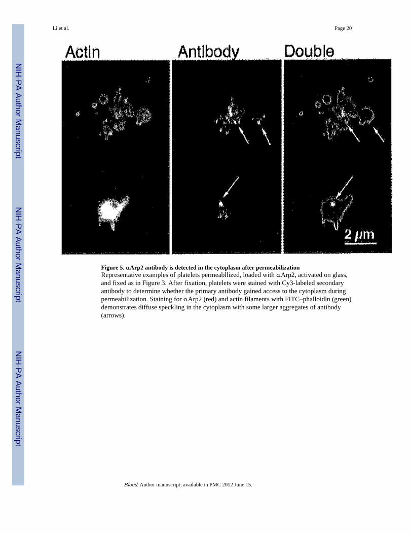
Figure 5. αArp2 antibody is detected in the cytoplasm after permeabilizationRepresentative examples of platelets permeabllized, loaded with αArp2, activated on glass,and fixed as in Figure 3. After fixation, platelets were stained with Cy3-labeled secondaryantibody to determine whether the primary antibody gained access to the cytoplasm duringpermeabilization. Staining for αArp2 (red) and actin filaments with FITC–phalloidln (green)demonstrates diffuse speckling in the cytoplasm with some larger aggregates of antibody(arrows).
Li et al. Page 20
Blood. Author manuscript; available in PMC 2012 June 15.
NIH
-PA Author Manuscript
NIH
-PA Author Manuscript
NIH
-PA Author Manuscript

Figure 6. Location of endogenous Arp2/3 in spread platelets(A) Three different spread platelets fixed in 4% paraformaldehyde containing 0.25% Tritonand stained with αArp2. Arp2/3 displays a speckled cytoplasmic staining and a row ofImmunofluorescent dots at the periphery, (B) Double label of a single platelet fixed as inpanel A and stained for Arp2/3 with αp34 (Cy3, red) and for actin filaments (FITC–phalloidin, green). Superimposition of the double label shows Arp2/3 at the periphery. Bar =2 µm. (C) Triple image of a single platelet fixed without detergent to permit imaging byphase microscopy and then stained with αArp2 (red) and phalloidin (green). Note thatArp2/3 is found in a bright arc internal to the actin frill. Arrows indicate weak staining at theoutermost edge of the lamellipodia. Also note that the platelet contours imaged by phalloidincorrespond to those imaged by phase microscopy. Bar = 2 µm.
Li et al. Page 21
Blood. Author manuscript; available in PMC 2012 June 15.
NIH
-PA Author Manuscript
NIH
-PA Author Manuscript
NIH
-PA Author Manuscript
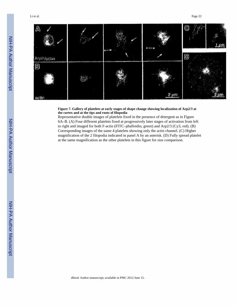
Figure 7. Gallery of platelets at early stages of shape change showing localization of Arp2/3 atthe cortex and at the tips and roots of filopodiaRepresentative double images of platelets fixed in the presence of detergent as in Figure6A–B. (A) Four different platelets fixed at progressively later stages of activation from leftto right and imaged for both F-actin (FITC–phalloidin, green) and Arp2/3 (Cy3, red). (B)Corresponding images of the same 4 platelets showing only the actin channel. (C) Highermagnification of the 2 filopodia indicated in panel A by an asterisk. (D) Fully spread plateletat the same magnification as the other platelets in this figure for size comparison.
Li et al. Page 22
Blood. Author manuscript; available in PMC 2012 June 15.
NIH
-PA Author Manuscript
NIH
-PA Author Manuscript
NIH
-PA Author Manuscript
NIH
-PA Author Manuscript
NIH
-PA Author Manuscript
NIH
-PA Author Manuscript
Li et al. Page 23
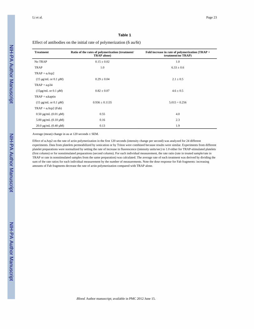
Table 1
Effect of antibodies on the initial rate of polymerization (δ au/δt)
Treatment Ratio of the rates of polymerization (treatment/TRAP alone)
Fold increase in rate of polymerization (TRAP +treatment/no TRAP)
No TRAP 0.15 ± 0.02 1.0
TRAP 1.0 6.33 ± 0.6
TRAP + αArp2
(15 µg/mL or 0.1 µM) 0.29 ± 0.04 2.1 ± 0.5
TRAP + αp34
(15µg/mL or 0.1 µM) 0.82 ± 0.07 4.6 ± 0.5
TRAP + αkaptin
(15 µg/mL or 0.1 µM) 0.936 ± 0.1135 5.015 + 0.256
TRAP + αArp2 (Fab)
0.50 µg/mL (0.01 µM) 0.55 4.0
5.00 µg/mL (0.10 µM) 0.16 2.3
20.0 µg/mL (0.40 µM) 0.13 1.9
Average (mean) change in au at 120 seconds ± SEM.
Effect of αArp2 on the rate of actin polymerization in the first 120 seconds (intensity change per second) was analyzed for 24 differentexperiments. Data from platelets permeabilized by sonication or by Triton were combined because results were similar. Experiments from differentplatelet preparations were normalized by setting the rate of increase in fluorescence (intensity units/sec) to 1.0 either for TRAP-stimulated platelets(first column) or for nonstimulated preparations (second column). For each individual measurement, the rate ratio (rate in treated sample/rate inTRAP or rate in nonstimulated samples from the same preparation) was calculated. The average rate of each treatment was derived by dividing thesum of the rate ratios for each individual measurement by the number of measurements. Note the dose response for Fab fragments: increasingamounts of Fab fragments decrease the rate of actin polymerization compared with TRAP alone.
Blood. Author manuscript; available in PMC 2012 June 15.
Related Documents