International Journal of Molecular Sciences Review Insights into the Role of Tick Salivary Protease Inhibitors during Ectoparasite–Host Crosstalk Mohamed Amine Jmel 1,† , Hajer Aounallah 2,3,† , Chaima Bensaoud 1 , Imen Mekki 1,4 , Jindˇ rich Chmelaˇ r 4 , Fernanda Faria 3 , Youmna M’ghirbi 2 and Michalis Kotsyfakis 1, * Citation: Jmel, M.A.; Aounallah, H.; Bensaoud, C.; Mekki, I.; Chmelaˇ r, J.; Faria, F.; M’ghirbi, Y.; Kotsyfakis, M. Insights into the Role of Tick Salivary Protease Inhibitors during Ectoparasite–Host Crosstalk. Int. J. Mol. Sci. 2021, 22, 892. https://doi. org/10.3390/ijms22020892 Received: 25 December 2020 Accepted: 13 January 2021 Published: 17 January 2021 Publisher’s Note: MDPI stays neutral with regard to jurisdictional claims in published maps and institutional affil- iations. Copyright: © 2021 by the authors. Licensee MDPI, Basel, Switzerland. This article is an open access article distributed under the terms and conditions of the Creative Commons Attribution (CC BY) license (https:// creativecommons.org/licenses/by/ 4.0/). 1 Laboratory of Genomics and Proteomics of Disease Vectors, Biology Centre CAS, Institute of Parasitology, Branišovská 1160/31, 37005 ˇ Ceské Budˇ ejovice, Czech Republic; [email protected] (M.A.J.); [email protected] (C.B.); [email protected] (I.M.) 2 Institut Pasteur de Tunis, Université de Tunis El Manar, LR19IPTX, Service d’Entomologie Médicale, Tunis 1002, Tunisia; [email protected] (H.A.); [email protected] (Y.M.) 3 Innovation and Development Laboratory, Innovation and Development Center, Instituto Butantan, São Paulo 05503-900, Brazil; [email protected] 4 Faculty of Science, University of South Bohemia in ˇ Ceské Budˇ ejovice, 37005 ˇ Ceské Budˇ ejovice, Czech Republic; [email protected] * Correspondence: [email protected] † These authors contributed equally. Abstract: Protease inhibitors (PIs) are ubiquitous regulatory proteins present in all kingdoms. They play crucial tasks in controlling biological processes directed by proteases which, if not tightly regulated, can damage the host organism. PIs can be classified according to their targeted proteases or their mechanism of action. The functions of many PIs have now been characterized and are showing clinical relevance for the treatment of human diseases such as arthritis, hepatitis, cancer, AIDS, and cardiovascular diseases, amongst others. Other PIs have potential use in agriculture as insecticides, anti-fungal, and antibacterial agents. PIs from tick salivary glands are special due to their pharmacological properties and their high specificity, selectivity, and affinity to their target proteases at the tick–host interface. In this review, we discuss the structure and function of PIs in general and those PI superfamilies abundant in tick salivary glands to illustrate their possible practical applications. In doing so, we describe tick salivary PIs that are showing promise as drug candidates, highlighting the most promising ones tested in vivo and which are now progressing to preclinical and clinical trials. Keywords: protease inhibitors; proteases; tick saliva; drug discovery 1. Introduction Proteases are ubiquitous enzymes in plants, animals, and microorganisms that play key roles in the majority of physiological processes [1,2]. Proteases are involved in several reversible and irreversible reaction cascades including hormone production/signaling pathways, apoptosis, inflammatory reactions, and homeostasis [3]. Depending on the active amino acid in the enzyme’s active site, proteases are classified into cysteine, serine, aspartic, glutamic, and threonine proteases [3], and metalloproteases represent another protease class containing a divalent metal ion linked to the active site residues [4]. Serine proteases are the most abundant proteolytic enzymes, followed by metallo- and cysteine proteases and finally the aspartic and threonine proteases [5]. In addition to their roles in vital biochemical processes, proteases are also implicated in various diseases such as viral diseases, cancer, inflammation, and bleeding disorders [6]. Given their important roles in diverse physiological processes, protease activity must be rigorously controlled and regulated to avoid any enzyme dysregulation that might be pathogenic to the host organism [7]. This tight regulation is usually conducted by blocking the zymogen, the Int. J. Mol. Sci. 2021, 22, 892. https://doi.org/10.3390/ijms22020892 https://www.mdpi.com/journal/ijms

Welcome message from author
This document is posted to help you gain knowledge. Please leave a comment to let me know what you think about it! Share it to your friends and learn new things together.
Transcript
International Journal of
Molecular Sciences
Review
Insights into the Role of Tick Salivary Protease Inhibitorsduring Ectoparasite–Host Crosstalk
Mohamed Amine Jmel 1,† , Hajer Aounallah 2,3,† , Chaima Bensaoud 1, Imen Mekki 1,4, Jindrich Chmelar 4,Fernanda Faria 3 , Youmna M’ghirbi 2 and Michalis Kotsyfakis 1,*
�����������������
Citation: Jmel, M.A.; Aounallah, H.;
Bensaoud, C.; Mekki, I.; Chmelar, J.;
Faria, F.; M’ghirbi, Y.; Kotsyfakis, M.
Insights into the Role of Tick Salivary
Protease Inhibitors during
Ectoparasite–Host Crosstalk. Int. J.
Mol. Sci. 2021, 22, 892. https://doi.
org/10.3390/ijms22020892
Received: 25 December 2020
Accepted: 13 January 2021
Published: 17 January 2021
Publisher’s Note: MDPI stays neutral
with regard to jurisdictional claims in
published maps and institutional affil-
iations.
Copyright: © 2021 by the authors.
Licensee MDPI, Basel, Switzerland.
This article is an open access article
distributed under the terms and
conditions of the Creative Commons
Attribution (CC BY) license (https://
creativecommons.org/licenses/by/
4.0/).
1 Laboratory of Genomics and Proteomics of Disease Vectors, Biology Centre CAS, Institute of Parasitology,Branišovská 1160/31, 37005 Ceské Budejovice, Czech Republic; [email protected] (M.A.J.);[email protected] (C.B.); [email protected] (I.M.)
2 Institut Pasteur de Tunis, Université de Tunis El Manar, LR19IPTX, Service d’Entomologie Médicale,Tunis 1002, Tunisia; [email protected] (H.A.); [email protected] (Y.M.)
3 Innovation and Development Laboratory, Innovation and Development Center, Instituto Butantan,São Paulo 05503-900, Brazil; [email protected]
4 Faculty of Science, University of South Bohemia in Ceské Budejovice, 37005 Ceské Budejovice, Czech Republic;[email protected]
* Correspondence: [email protected]† These authors contributed equally.
Abstract: Protease inhibitors (PIs) are ubiquitous regulatory proteins present in all kingdoms. Theyplay crucial tasks in controlling biological processes directed by proteases which, if not tightlyregulated, can damage the host organism. PIs can be classified according to their targeted proteasesor their mechanism of action. The functions of many PIs have now been characterized and areshowing clinical relevance for the treatment of human diseases such as arthritis, hepatitis, cancer,AIDS, and cardiovascular diseases, amongst others. Other PIs have potential use in agriculture asinsecticides, anti-fungal, and antibacterial agents. PIs from tick salivary glands are special due totheir pharmacological properties and their high specificity, selectivity, and affinity to their targetproteases at the tick–host interface. In this review, we discuss the structure and function of PIsin general and those PI superfamilies abundant in tick salivary glands to illustrate their possiblepractical applications. In doing so, we describe tick salivary PIs that are showing promise as drugcandidates, highlighting the most promising ones tested in vivo and which are now progressing topreclinical and clinical trials.
Keywords: protease inhibitors; proteases; tick saliva; drug discovery
1. Introduction
Proteases are ubiquitous enzymes in plants, animals, and microorganisms that playkey roles in the majority of physiological processes [1,2]. Proteases are involved in severalreversible and irreversible reaction cascades including hormone production/signalingpathways, apoptosis, inflammatory reactions, and homeostasis [3]. Depending on theactive amino acid in the enzyme’s active site, proteases are classified into cysteine, serine,aspartic, glutamic, and threonine proteases [3], and metalloproteases represent anotherprotease class containing a divalent metal ion linked to the active site residues [4]. Serineproteases are the most abundant proteolytic enzymes, followed by metallo- and cysteineproteases and finally the aspartic and threonine proteases [5]. In addition to their rolesin vital biochemical processes, proteases are also implicated in various diseases such asviral diseases, cancer, inflammation, and bleeding disorders [6]. Given their importantroles in diverse physiological processes, protease activity must be rigorously controlledand regulated to avoid any enzyme dysregulation that might be pathogenic to the hostorganism [7]. This tight regulation is usually conducted by blocking the zymogen, the
Int. J. Mol. Sci. 2021, 22, 892. https://doi.org/10.3390/ijms22020892 https://www.mdpi.com/journal/ijms
Int. J. Mol. Sci. 2021, 22, 892 2 of 23
inactive enzyme precursor, or through the action of protease inhibitors (PIs) [8], whichpartially or totally inhibit enzymes by forming a complex with their target proteases [4].
While the majority of PIs are proteins or peptides, some low molecular weight non-proteinaceous compounds such as polysaccharides, glycerolipids, triterpenes, and polyphe-nols are also considered PIs [9]; these are not specific to particular proteases and inhibit abroad spectrum of enzymes [10]. In contrast, proteinaceous PIs are usually more specificand can even target unique proteases. As a result, PIs can be classified according to their tar-get enzyme, although exceptions are frequently encountered, such as the α2-macroglobulinthat inhibits proteases of different classes [6]. A particularly impressive role for PIs hasbeen observed in parasite–host crosstalk; PIs found in tick saliva have been shown tomodulate host immune cells [11], mediating local immunosuppression and modulatingblood clotting at the site of infection, thereby exerting a beneficial effect to the tick (allowingattachment and feeding) at the expense of the host [12].
Given their fundamental roles and potential translational application, there havebeen significant efforts to identify new PIs from various sources and study those alreadyidentified in detail using novel technologies and methods. Indeed, with technologicaladvances, the study of PIs has substantially improved over recent years, not least theavailability of three-dimensional (3D) structural information for several PIs and theirtargeted proteases, permitting receptor-based design.
Here, we review the biochemistry and fundamental mechanisms of action of PIs. Weenumerate and discuss the different classes of PIs based on the proteases they inhibit andtheir mechanism of action. Moreover, we discuss their applications in critical fields likeagriculture and medicine. In the final section, we focus on an interesting natural source ofPIs, tick salivary glands, and their potential pharmacological applications.
2. Classification of Protease Inhibitors
The catalytic activity of proteases is regulated by different inhibition mechanismsand different PI families [13]. Despite similarities in the 3D structure of PIs, they can beclassified into over 107 families and divided into 40 clans according to their structuralsimilarities (secondary and tertiary) and their different functions [13]. Laskowski and Katofirst developed a classification scheme for PIs in 1980 [14] according to their reactive center,disulfide bond number, and amino acid sequence [14]. With advances in biotechnologyand increasing knowledge about PIs, Rawlings et al. [15] established a classification of PIsin 2004 based on amino acid sequence homology that subclassified PIs to 48 families and26 clans [5].
2.1. Target-Based Classification
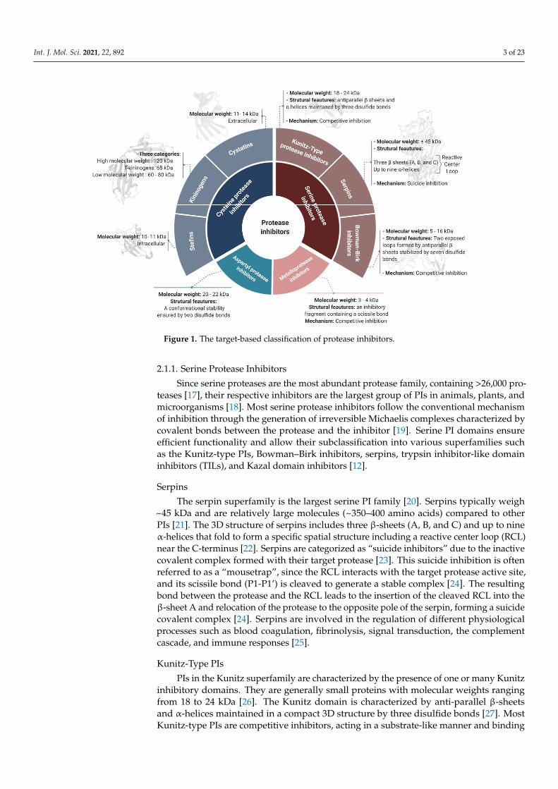
PIs can be classified according to their target protease into six groups [4]: serine,cysteine, aspartyl, glutamate, and threonine protease inhibitors. Metalloprotease inhibitorsare also contained within this classification, as they inhibit proteases with a divalent metalion in their active site [16]. A non-exhaustive list of the most common PI families, theirprincipal features, and their properties according to the target-based classification systemis illustrated in Figure 1 and discussed below.
Int. J. Mol. Sci. 2021, 22, 892 3 of 23
Int. J. Mol. Sci. 2021, 22, 892 3 of 23
Figure 1. The target-based classification of protease inhibitors.
2.1.1. Serine Protease Inhibitors Since serine proteases are the most abundant protease family, containing >26,000 pro-
teases [17], their respective inhibitors are the largest group of PIs in animals, plants, and microorganisms [18]. Most serine protease inhibitors follow the conventional mechanism of inhibition through the generation of irreversible Michaelis complexes characterized by covalent bonds between the protease and the inhibitor [19]. Serine PI domains ensure ef-ficient functionality and allow their subclassification into various superfamilies such as the Kunitz-type PIs, Bowman–Birk inhibitors, serpins, trypsin inhibitor-like domain in-hibitors (TILs), and Kazal domain inhibitors [12].
Serpins The serpin superfamily is the largest serine PI family [20]. Serpins typically weigh
~45 kDa and are relatively large molecules (~350–400 amino acids) compared to other PIs [21]. The 3D structure of serpins includes three β-sheets (A, B, and C) and up to nine α-helices that fold to form a specific spatial structure including a reactive center loop (RCL) near the C-terminus [22]. Serpins are categorized as “suicide inhibitors” due to the inac-tive covalent complex formed with their target protease [23]. This suicide inhibition is often referred to as a “mousetrap”, since the RCL interacts with the target protease active site, and its scissile bond (P1-P1’) is cleaved to generate a stable complex [24]. The result-ing bond between the protease and the RCL leads to the insertion of the cleaved RCL into the β-sheet A and relocation of the protease to the opposite pole of the serpin, forming a suicide covalent complex [24]. Serpins are involved in the regulation of different physio-logical processes such as blood coagulation, fibrinolysis, signal transduction, the comple-ment cascade, and immune responses [25].
Kunitz-Type PIs
Figure 1. The target-based classification of protease inhibitors.
2.1.1. Serine Protease Inhibitors
Since serine proteases are the most abundant protease family, containing >26,000 pro-teases [17], their respective inhibitors are the largest group of PIs in animals, plants, andmicroorganisms [18]. Most serine protease inhibitors follow the conventional mechanismof inhibition through the generation of irreversible Michaelis complexes characterized bycovalent bonds between the protease and the inhibitor [19]. Serine PI domains ensureefficient functionality and allow their subclassification into various superfamilies suchas the Kunitz-type PIs, Bowman–Birk inhibitors, serpins, trypsin inhibitor-like domaininhibitors (TILs), and Kazal domain inhibitors [12].
Serpins
The serpin superfamily is the largest serine PI family [20]. Serpins typically weigh~45 kDa and are relatively large molecules (~350–400 amino acids) compared to otherPIs [21]. The 3D structure of serpins includes three β-sheets (A, B, and C) and up to nineα-helices that fold to form a specific spatial structure including a reactive center loop (RCL)near the C-terminus [22]. Serpins are categorized as “suicide inhibitors” due to the inactivecovalent complex formed with their target protease [23]. This suicide inhibition is oftenreferred to as a “mousetrap”, since the RCL interacts with the target protease active site,and its scissile bond (P1-P1’) is cleaved to generate a stable complex [24]. The resultingbond between the protease and the RCL leads to the insertion of the cleaved RCL into theβ-sheet A and relocation of the protease to the opposite pole of the serpin, forming a suicidecovalent complex [24]. Serpins are involved in the regulation of different physiologicalprocesses such as blood coagulation, fibrinolysis, signal transduction, the complementcascade, and immune responses [25].
Kunitz-Type PIs
PIs in the Kunitz superfamily are characterized by the presence of one or many Kunitzinhibitory domains. They are generally small proteins with molecular weights rangingfrom 18 to 24 kDa [26]. The Kunitz domain is characterized by anti-parallel β-sheetsand α-helices maintained in a compact 3D structure by three disulfide bonds [27]. MostKunitz-type PIs are competitive inhibitors, acting in a substrate-like manner and binding
Int. J. Mol. Sci. 2021, 22, 892 4 of 23
reversibly to the protease [28]. Active site blocking is mediated by the RCL attachmentto the catalytic zone through a non-covalent bond. The highly exposed RCL loop ofKunitz-domain inhibitors is suitable for a wide variety of proteases, so these inhibitors arerelatively non-specific and therefore potentially useful across a range of applications [29].Indeed, Kunitz-type PIs are known to regulate inflammation and coagulation factors andhave also been implicated in tumor biology [3].
Bowman–Birk Inhibitors (BBIs)
This superfamily of PIs is characterized by small molecular weight peptides rangingfrom 5 to 16 kDa and a structure with a single or two inhibitory regions [30]. BBIs arecompetitive inhibitors and follow the classical mechanism of substrate binding to theprotease active site [31]. A single BBI inhibitor protein can act on two different targetproteases simultaneously by virtue of two opposed loops formed by antiparallel β-sheetsand stabilized by seven disulfide bonds [23,32]. Given their specific mechanism, severalresearchers have focused on BBIs for specific applications such as to inhibit cancer [33].
2.1.2. Cysteine Protease Inhibitors
Cysteine protease inhibitors (CPIs), or cystatins, are the second largest group of PIsafter serine PIs. They are divided into three main families: family-1 cystatins or stefins,family-2 cystatins or cystatins, and family-3 cystatins or kininogens. Stefins are mostlyintracellular and the smallest cystatin family in terms of molecular weight (10–11 kDa) [3].Stefins inhibit cathepsins B, L, and H and also papain. In several therapeutic investigations,they have been identified as potential diagnostic tools for cancer [34]. Like stefins, cystatinsinhibit papain and cathepsins B, L, and H, but, they are larger (11–14 kDa) and are trans-ported out of the cell to exert their action [35]. Kininogens are divided into three categories:high molecular weight kininogens (120 kDa), T-kininogens (68 kDa), and low molecularweight kininogens (60–80 kDa) [3,36,37]. They play important roles in the modulation ofinflammatory responses and are used as biomarkers of kidney disorders [37].
There are numerous documented functions of CPIs, and some have been shownto be critical for the proper functioning of important physiological pathways such ascathepsin regulation [38]. The structural features of CPIs include 4 to 5 antiparallel β-sheetssurrounding an α-helix. Their highly conserved inhibitory domain is mainly composed oftwo hairpin-like loops formed by the β-sheets and the N-terminal region [39]. Cystatinsfollow the competitive inhibition model with slight modifications, as they do not bindin a substrate-like manner. In fact, the two hairpin loops bind to the protease active siteand block the access of any substrate, while the N-terminal region maintains effectiveattachment of the inhibitor to the enzyme [40,41].
2.1.3. Metalloproteases Inhibitors
Despite their low molecular weight (only 3–4 kDa), metalloprotease inhibitors (MPIs)effectively inhibit a wide range of metalloproteases [42]. MPIs are classified as competitiveinhibitors, since they act in a substrate-like manner [43]. From a structural point of view,MPIs do not possess an inhibitory loop or a specific secondary structure for inhibition.Instead, their inhibitory fragment is located near the C-terminus and contains a scissilebond. The cleavage of this latter bond allows the fixation of the new C-terminal side to theactive site of the protease with the help of its metallic ion [23,28]. The efficiency of MPIinhibition has been reported to be enhanced by secondary interactions outside the activesite of the protease [44].
2.1.4. Aspartyl Protease Inhibitors
The natural aspartyl PIs are proteins of about 20 to 22 kDa and conformational stabilityensured by two disulfide bonds [45]. Despite aspartyl proteases including importantmembers such as cathepsins D and E, renin, pepsin A and C, and, most importantly, theHIV-1 protease, their natural inhibitors remain poorly described for several reasons. One
Int. J. Mol. Sci. 2021, 22, 892 5 of 23
main reason is probably related to the low representation of aspartyl proteases in the humangenome, with only 15 members described [46]. Regardless of their low bioavailability, thepresence of a scissile bond and their short half-life have meant that strategies to inhibitaspartyl proteases involve the development of synthetic peptides or mimics with a non-cleavable bond to replace the scissile bond [47].
2.2. Mechanism-Based Classification
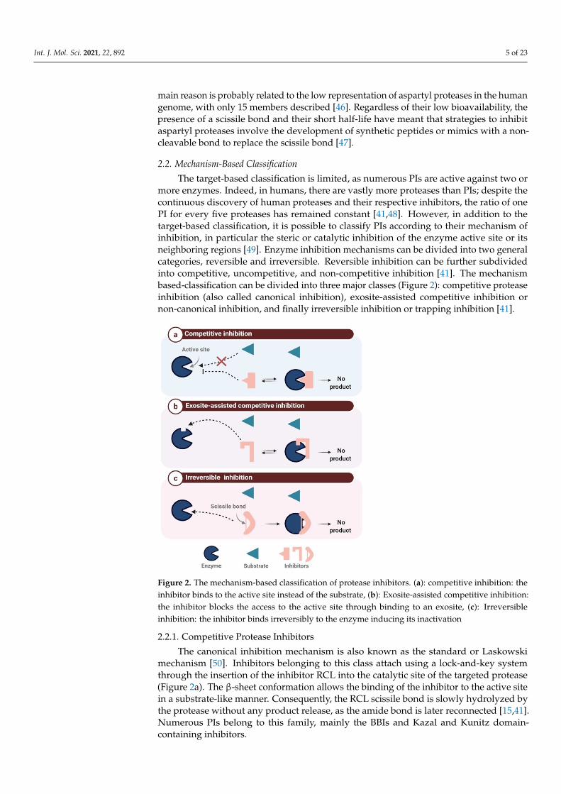
The target-based classification is limited, as numerous PIs are active against two ormore enzymes. Indeed, in humans, there are vastly more proteases than PIs; despite thecontinuous discovery of human proteases and their respective inhibitors, the ratio of onePI for every five proteases has remained constant [41,48]. However, in addition to thetarget-based classification, it is possible to classify PIs according to their mechanism ofinhibition, in particular the steric or catalytic inhibition of the enzyme active site or itsneighboring regions [49]. Enzyme inhibition mechanisms can be divided into two generalcategories, reversible and irreversible. Reversible inhibition can be further subdividedinto competitive, uncompetitive, and non-competitive inhibition [41]. The mechanismbased-classification can be divided into three major classes (Figure 2): competitive proteaseinhibition (also called canonical inhibition), exosite-assisted competitive inhibition ornon-canonical inhibition, and finally irreversible inhibition or trapping inhibition [41].
Int. J. Mol. Sci. 2021, 22, 892 5 of 23
2.1.4. Aspartyl Protease Inhibitors The natural aspartyl PIs are proteins of about 20 to 22 kDa and conformational sta-
bility ensured by two disulfide bonds [45]. Despite aspartyl proteases including important members such as cathepsins D and E, renin, pepsin A and C, and, most importantly, the HIV-1 protease, their natural inhibitors remain poorly described for several reasons. One main reason is probably related to the low representation of aspartyl proteases in the hu-man genome, with only 15 members described [46]. Regardless of their low bioavailabil-ity, the presence of a scissile bond and their short half-life have meant that strategies to inhibit aspartyl proteases involve the development of synthetic peptides or mimics with a non-cleavable bond to replace the scissile bond [47].
2.2. Mechanism-Based Classification The target-based classification is limited, as numerous PIs are active against two or
more enzymes. Indeed, in humans, there are vastly more proteases than PIs; despite the continuous discovery of human proteases and their respective inhibitors, the ratio of one PI for every five proteases has remained constant [41,48]. However, in addition to the tar-get-based classification, it is possible to classify PIs according to their mechanism of inhi-bition, in particular the steric or catalytic inhibition of the enzyme active site or its neigh-boring regions [49]. Enzyme inhibition mechanisms can be divided into two general cate-gories, reversible and irreversible. Reversible inhibition can be further subdivided into competitive, uncompetitive, and non-competitive inhibition [41]. The mechanism based-classification can be divided into three major classes (Figure 2): competitive protease in-hibition (also called canonical inhibition), exosite-assisted competitive inhibition or non-canonical inhibition, and finally irreversible inhibition or trapping inhibition [41].
Figure 2. The mechanism-based classification of protease inhibitors. (a): competitive inhibition: the inhibitor binds to the active site instead of the substrate, (b): Exosite-assisted competitive inhibi-tion: the inhibitor blocks the access to the active site through binding to an exosite, (c): Irreversible inhibition: the inhibitor binds irreversibly to the enzyme inducing its inactivation
Figure 2. The mechanism-based classification of protease inhibitors. (a): competitive inhibition: theinhibitor binds to the active site instead of the substrate, (b): Exosite-assisted competitive inhibition:the inhibitor blocks the access to the active site through binding to an exosite, (c): Irreversibleinhibition: the inhibitor binds irreversibly to the enzyme inducing its inactivation
2.2.1. Competitive Protease Inhibitors
The canonical inhibition mechanism is also known as the standard or Laskowskimechanism [50]. Inhibitors belonging to this class attach using a lock-and-key systemthrough the insertion of the inhibitor RCL into the catalytic site of the targeted protease(Figure 2a). The β-sheet conformation allows the binding of the inhibitor to the active sitein a substrate-like manner. Consequently, the RCL scissile bond is slowly hydrolyzed bythe protease without any product release, as the amide bond is later reconnected [15,41].Numerous PIs belong to this family, mainly the BBIs and Kazal and Kunitz domain-containing inhibitors.

Int. J. Mol. Sci. 2021, 22, 892 6 of 23
2.2.2. Exosite-Assisted Competitive Inhibitors
Known also as non-canonical inhibitors, this class represents inhibitors binding to asecondary site distinct from the protease active site (Figure 2b). Access to this active siteis blocked in a non-catalytic manner [51,52]. This inhibition mechanism is classified ascompetitive but differs from the standard Laskowski mechanism [50]. Inhibitor binding tothe exosite is crucial for inhibition, as it maintains the inhibitor–enzyme interaction andenhances inhibitor specificity [51]. As mentioned above, CPIs are non-canonical PIs thatfollow a slightly modified competitive inhibition mechanism, since the N-terminal regiondoes not interact with the active site in a substrate-like manner but switches to the side ofthe active site to ensure sufficient binding energy for the enzyme–inhibitor complex [41,53].
2.2.3. Irreversible Inhibition
This class of inhibition is triggered by the protease, which catalyzes the activationof its respective inhibitor (also referred to as a suicide substrate). The cleavage of theinhibitor reactive loops (Figure 2c) triggers a major conformational change, resulting inthe irreversible cross-linking of the protease to its inhibitor [23,51]. α-2-macroglobulin is a600 kDa inhibitor with four reactive loops on its surface, which plays a major role in theelimination of excessive proteases in the blood [54]. Serpins are also well-known suicideinhibitors, as described above [55].
3. Applications of Protease Inhibitors
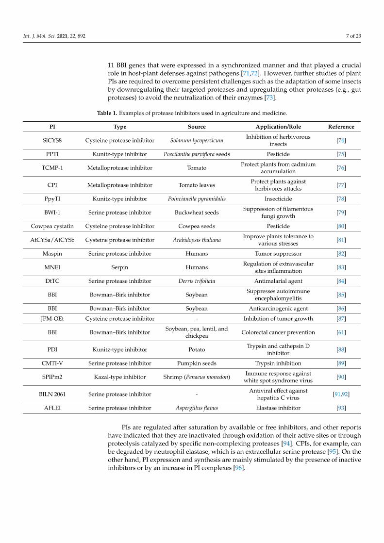
The accelerating increase in PI-related knowledge (structure, mechanism of action,functional applications) has paved the way for several biotechnological applications of PIsrelated to medicine and agriculture, as summarized in Table 1.
Biomedical research efforts have underscored the major role of these inhibitors inpathobiology, providing opportunities to exploit them for the treatment of diseases such asHIV [56] and cancer [57]. PIs targeting human proteases are implicated in protease-relatedblood coagulation disorders, cancers, immune system dysfunction, and neurodegenerativediseases [4]. In HIV infections, several combinations of PIs have been used therapeu-tically [58], and protease inhibitor-based drugs such as nucleoside/nucleotide reversetranscriptase inhibitors (NRTIs) or non-nucleoside reverse transcriptase inhibitors (NNR-TIs) have been developed as antiretroviral drugs [59]. In cancer, PIs have been tested asantitumor therapies to target the proteases implicated in tumor progression and metastasissuch as matrix metalloproteases, cathepsin B, matriptase, and kallikreins [60]; for example,a BBI caused apoptosis and cell cycle arrest in colon cancer (HT-29) and breast cancer(MCF-7) cell lines [61]. Moreover, a BBI mixture from soybean was shown to have a cancerchemo-preventive effect against neoplastic polyps and was applied to the preclinical detec-tion of prostate cancer [36]. Additionally, proteases are common in the immune system andneurodegenerative diseases [32]. Several interesting properties, such as their high targetspecificity, have made PIs interesting candidates for drug discovery [2]. Over 70 PIs havebeen commercialized as drugs, with over 150 examined in clinical trials and thousandspreclinically [23]. Among the commonly-used commercialized PI drugs, enalapril and cap-topril inhibit angiotensin-converting enzyme and are used to treat cardiovascular disease,and bivalirudin is a thrombin inhibitor and potent anticoagulant [62,63].
In agriculture, plant PIs have been thoroughly investigated as they are importantin plant defense mechanisms. With the increased use of genetic engineering, transgenicplants with genes coding for PIs have been investigated to improve agriculture. Indeed,the PI dose applied to plants was correlated with protection against phytopathogens,insects, microbes, and pests [64,65]. As a result, PIs have been employed in agricultureas insecticides [66], anti-fungal agents [67], and antibacterial agents [68]. They targetthe digestive proteases of phytophagous entities to exert anti-proliferative and growthinhibitory effects [69]. Transcriptomic studies have revealed that PIs are the main proteinsproduced during plant exposure to external pathogens [70]. A previous in silico study ofsoybean plants revealed that a multigene family coding for Bowman–Birk PIs contained
Int. J. Mol. Sci. 2021, 22, 892 7 of 23
11 BBI genes that were expressed in a synchronized manner and that played a crucialrole in host-plant defenses against pathogens [71,72]. However, further studies of plantPIs are required to overcome persistent challenges such as the adaptation of some insectsby downregulating their targeted proteases and upregulating other proteases (e.g., gutproteases) to avoid the neutralization of their enzymes [73].
Table 1. Examples of protease inhibitors used in agriculture and medicine.
PI Type Source Application/Role Reference
SlCYS8 Cysteine protease inhibitor Solanum lycopersicum Inhibition of herbivorousinsects [74]
PPTI Kunitz-type inhibitor Poecilanthe parviflora seeds Pesticide [75]
TCMP-1 Metalloprotease inhibitor Tomato Protect plants from cadmiumaccumulation [76]
CPI Metalloprotease inhibitor Tomato leaves Protect plants againstherbivores attacks [77]
PpyTI Kunitz-type inhibitor Poincianella pyramidalis Insecticide [78]
BWI-1 Serine protease inhibitor Buckwheat seeds Suppression of filamentousfungi growth [79]
Cowpea cystatin Cysteine protease inhibitor Cowpea seeds Pesticide [80]
AtCYSa/AtCYSb Cysteine protease inhibitor Arabidopsis thaliana Improve plants tolerance tovarious stresses [81]
Maspin Serine protease inhibitor Humans Tumor suppressor [82]
MNEI Serpin Humans Regulation of extravascularsites inflammation [83]
DtTC Serine protease inhibitor Derris trifoliata Antimalarial agent [84]
BBI Bowman–Birk inhibitor Soybean Suppresses autoimmuneencephalomyelitis [85]
BBI Bowman–Birk inhibitor Soybean Anticarcinogenic agent [86]
JPM-OEt Cysteine protease inhibitor - Inhibition of tumor growth [87]
BBI Bowman–Birk inhibitor Soybean, pea, lentil, andchickpea Colorectal cancer prevention [61]
PDI Kunitz-type inhibitor Potato Trypsin and cathepsin Dinhibitor [88]
CMTI-V Serine protease inhibitor Pumpkin seeds Trypsin inhibition [89]
SPIPm2 Kazal-type inhibitor Shrimp (Penaeus monodon) Immune response againstwhite spot syndrome virus [90]
BILN 2061 Serine protease inhibitor - Antiviral effect againsthepatitis C virus [91,92]
AFLEI Serine protease inhibitor Aspergillus flavus Elastase inhibitor [93]
PIs are regulated after saturation by available or free inhibitors, and other reportshave indicated that they are inactivated through oxidation of their active sites or throughproteolysis catalyzed by specific non-complexing proteases [94]. CPIs, for example, canbe degraded by neutrophil elastase, which is an extracellular serine protease [95]. On theother hand, PI expression and synthesis are mainly stimulated by the presence of inactiveinhibitors or by an increase in PI complexes [96].
Int. J. Mol. Sci. 2021, 22, 892 8 of 23
4. Tick Salivary Glands: An Attractive Source of PIs with Potential PharmacologicalApplications
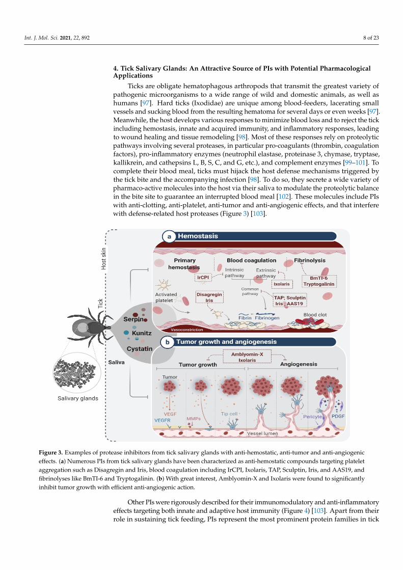
Ticks are obligate hematophagous arthropods that transmit the greatest variety ofpathogenic microorganisms to a wide range of wild and domestic animals, as well ashumans [97]. Hard ticks (Ixodidae) are unique among blood-feeders, lacerating smallvessels and sucking blood from the resulting hematoma for several days or even weeks [97].Meanwhile, the host develops various responses to minimize blood loss and to reject the tickincluding hemostasis, innate and acquired immunity, and inflammatory responses, leadingto wound healing and tissue remodeling [98]. Most of these responses rely on proteolyticpathways involving several proteases, in particular pro-coagulants (thrombin, coagulationfactors), pro-inflammatory enzymes (neutrophil elastase, proteinase 3, chymase, tryptase,kallikrein, and cathepsins L, B, S, C, and G, etc.), and complement enzymes [99–101]. Tocomplete their blood meal, ticks must hijack the host defense mechanisms triggered bythe tick bite and the accompanying infection [98]. To do so, they secrete a wide variety ofpharmaco-active molecules into the host via their saliva to modulate the proteolytic balancein the bite site to guarantee an interrupted blood meal [102]. These molecules include PIswith anti-clotting, anti-platelet, anti-tumor and anti-angiogenic effects, and that interferewith defense-related host proteases (Figure 3) [103].
Int. J. Mol. Sci. 2021, 22, 892 8 of 23
degraded by neutrophil elastase, which is an extracellular serine protease [95]. On the other hand, PI expression and synthesis are mainly stimulated by the presence of inactive inhibitors or by an increase in PI complexes [96].
4. Tick Salivary Glands: An Attractive Source of PIs with Potential Pharmacological Applications
Ticks are obligate hematophagous arthropods that transmit the greatest variety of pathogenic microorganisms to a wide range of wild and domestic animals, as well as hu-mans [97]. Hard ticks (Ixodidae) are unique among blood-feeders, lacerating small vessels and sucking blood from the resulting hematoma for several days or even weeks [97]. Meanwhile, the host develops various responses to minimize blood loss and to reject the tick including hemostasis, innate and acquired immunity, and inflammatory responses, leading to wound healing and tissue remodeling [98]. Most of these responses rely on proteolytic pathways involving several proteases, in particular pro-coagulants (thrombin, coagulation factors), pro-inflammatory enzymes (neutrophil elastase, proteinase 3, chy-mase, tryptase, kallikrein, and cathepsins L, B, S, C, and G, etc.), and complement enzymes [99–101]. To complete their blood meal, ticks must hijack the host defense mechanisms triggered by the tick bite and the accompanying infection [98]. To do so, they secrete a wide variety of pharmaco-active molecules into the host via their saliva to modulate the proteolytic balance in the bite site to guarantee an interrupted blood meal [102]. These molecules include PIs with anti-clotting, anti-platelet, anti-tumor and anti-angiogenic ef-fects, and that interfere with defense-related host proteases (Figure 3) [103].
Figure 3. Examples of protease inhibitors from tick salivary glands with anti-hemostatic, anti-tumor and anti-angiogenic effects. (a) Numerous PIs from tick salivary glands have been characterized as anti-hemostatic compounds targeting plate-let aggregation such as Disagregin and Iris, blood coagulation including IrCPI, Ixolaris, TAP, Sculptin, Iris, and AAS19, and fibrinolyses like BmTI-6 and Tryptogalinin. (b) With great interest, Amblyomin-X and Ixolaris were found to signifi-cantly inhibit tumor growth with efficient anti-angiogenic action.
Other PIs were rigorously described for their immunomodulatory and anti-inflam-matory effects targeting both innate and adaptive host immunity (Figure 4) [103]. Apart
Figure 3. Examples of protease inhibitors from tick salivary glands with anti-hemostatic, anti-tumor and anti-angiogeniceffects. (a) Numerous PIs from tick salivary glands have been characterized as anti-hemostatic compounds targeting plateletaggregation such as Disagregin and Iris, blood coagulation including IrCPI, Ixolaris, TAP, Sculptin, Iris, and AAS19, andfibrinolyses like BmTI-6 and Tryptogalinin. (b) With great interest, Amblyomin-X and Ixolaris were found to significantlyinhibit tumor growth with efficient anti-angiogenic action.
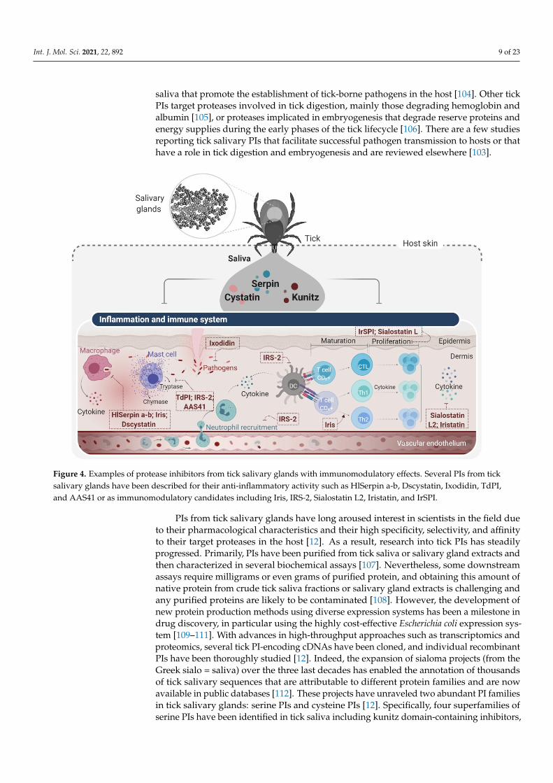
Other PIs were rigorously described for their immunomodulatory and anti-inflammatoryeffects targeting both innate and adaptive host immunity (Figure 4) [103]. Apart from theirrole in sustaining tick feeding, PIs represent the most prominent protein families in tick
Int. J. Mol. Sci. 2021, 22, 892 9 of 23
saliva that promote the establishment of tick-borne pathogens in the host [104]. Other tickPIs target proteases involved in tick digestion, mainly those degrading hemoglobin andalbumin [105], or proteases implicated in embryogenesis that degrade reserve proteins andenergy supplies during the early phases of the tick lifecycle [106]. There are a few studiesreporting tick salivary PIs that facilitate successful pathogen transmission to hosts or thathave a role in tick digestion and embryogenesis and are reviewed elsewhere [103].
Int. J. Mol. Sci. 2021, 22, 892 9 of 23
from their role in sustaining tick feeding, PIs represent the most prominent protein fami-lies in tick saliva that promote the establishment of tick-borne pathogens in the host [104]. Other tick PIs target proteases involved in tick digestion, mainly those degrading hemo-globin and albumin [105], or proteases implicated in embryogenesis that degrade reserve proteins and energy supplies during the early phases of the tick lifecycle [106]. There are a few studies reporting tick salivary PIs that facilitate successful pathogen transmission to hosts or that have a role in tick digestion and embryogenesis and are reviewed elsewhere [103].
Figure 4. Examples of protease inhibitors from tick salivary glands with immunomodulatory effects. Several PIs from tick salivary glands have been described for their anti-inflammatory activity such as HlSerpin a-b, Dscystatin, Ixodidin, TdPI, and AAS41 or as immunomodulatory candidates including Iris, IRS-2, Sialostatin L2, Iristatin, and IrSPI.
PIs from tick salivary glands have long aroused interest in scientists in the field due to their pharmacological characteristics and their high specificity, selectivity, and affinity to their target proteases in the host [12]. As a result, research into tick PIs has steadily progressed. Primarily, PIs have been purified from tick saliva or salivary gland extracts and then characterized in several biochemical assays [107]. Nevertheless, some down-stream assays require milligrams or even grams of purified protein, and obtaining this amount of native protein from crude tick saliva fractions or salivary gland extracts is chal-lenging and any purified proteins are likely to be contaminated [108]. However, the de-velopment of new protein production methods using diverse expression systems has been a milestone in drug discovery, in particular using the highly cost-effective Escherichia coli expression system [109–111]. With advances in high-throughput approaches such as tran-scriptomics and proteomics, several tick PI-encoding cDNAs have been cloned, and indi-vidual recombinant PIs have been thoroughly studied [12]. Indeed, the expansion of si-aloma projects (from the Greek sialo = saliva) over the three last decades has enabled the annotation of thousands of tick salivary sequences that are attributable to different protein families and are now available in public databases [112]. These projects have unraveled two abundant PI families in tick salivary glands: serine PIs and cysteine PIs [12]. Specifi-cally, four superfamilies of serine PIs have been identified in tick saliva including kunitz
Figure 4. Examples of protease inhibitors from tick salivary glands with immunomodulatory effects. Several PIs from ticksalivary glands have been described for their anti-inflammatory activity such as HlSerpin a-b, Dscystatin, Ixodidin, TdPI,and AAS41 or as immunomodulatory candidates including Iris, IRS-2, Sialostatin L2, Iristatin, and IrSPI.
PIs from tick salivary glands have long aroused interest in scientists in the field dueto their pharmacological characteristics and their high specificity, selectivity, and affinityto their target proteases in the host [12]. As a result, research into tick PIs has steadilyprogressed. Primarily, PIs have been purified from tick saliva or salivary gland extracts andthen characterized in several biochemical assays [107]. Nevertheless, some downstreamassays require milligrams or even grams of purified protein, and obtaining this amount ofnative protein from crude tick saliva fractions or salivary gland extracts is challenging andany purified proteins are likely to be contaminated [108]. However, the development ofnew protein production methods using diverse expression systems has been a milestone indrug discovery, in particular using the highly cost-effective Escherichia coli expression sys-tem [109–111]. With advances in high-throughput approaches such as transcriptomics andproteomics, several tick PI-encoding cDNAs have been cloned, and individual recombinantPIs have been thoroughly studied [12]. Indeed, the expansion of sialoma projects (from theGreek sialo = saliva) over the three last decades has enabled the annotation of thousandsof tick salivary sequences that are attributable to different protein families and are nowavailable in public databases [112]. These projects have unraveled two abundant PI familiesin tick salivary glands: serine PIs and cysteine PIs [12]. Specifically, four superfamilies ofserine PIs have been identified in tick saliva including kunitz domain-containing inhibitors,
Int. J. Mol. Sci. 2021, 22, 892 10 of 23
serpins, trypsin inhibitor-type cysteine-rich domain inhibitors (TIL domain inhibitors),and kazal domain inhibitors, while cysteine PIs refer to the cystatin family [12]. SeveralPIs, principally kunitz-type inhibitors, serpins, and cystatins, have now been rigorouslycharacterized in vitro and in vivo [102,113,114], but only a limited number have progressedto pre-clinical and clinical trials due to strict criteria for clinical use [11].
4.1. Tick Serine Protease Inhibitors and Their Applications
Kunitz domain inhibitors are highly abundant in tick salivary secretions and areusually associated with the inhibition of trypsin-like serine proteases [115]. As men-tioned above, members of this family can contain multiple tandem repeats of Kunitzdomains [115]. In tick salivary glands, only proteins with a single (monolaris), two (bilaris),or five (penthalaris) Kunitz inhibitory domains have been detected [116]. Proteins fromthis superfamily are considered to be the most valuable serine PIs in tick salivary glandsgiven their wide range of possible applications.
Tick salivary Kunitz domain inhibitors mostly function as anti-thrombotic factors byinhibiting various proteases in the coagulation cascade and involved in platelet aggrega-tion [117]. Most of these inhibitors target thrombin, factor Xa, factor XIIa, trypsin, andelastase [117]. For instance, Ir-CPI [118] and TAP [119] were reported as anticoagulants,and Disagregin was identified as a platelet aggregation inhibitor. Ir-CPI is a multi-targetKunitz-type inhibitor that specifically interacts with activated human contact phase factors(FXIIa, FXIa, and kallikrein), thereby inhibiting the intrinsic coagulation pathway [118].The administration of Ir-CPI in both rat and mouse venous and arterial thrombosis modelsresulted in a significant dose-dependent reduction in venous thrombus formation andrevealed a defect in the formation of arterial occlusive thrombi [118]. In addition, miceinjected with Ir-CPI were protected against collagen- and epinephrine-induced thromboem-bolism without bleeding or impaired blood coagulation parameters [118]. Recently, theantithrombotic activity of Ir-CPI was evaluated in preclinical animal models, confirmingthat Ir-CPI is an effective and safe antithrombotic agent [120]. TAP, on the other hand, is asingle target Kunitz-domain anticoagulant peptide from Ornithodoros moubata saliva [119]first identified as a slow tight-binding competitive inhibitor of FXa [119]. The recombinantform (rTAP) was shown to significantly inhibit thromboplastin-induced fibrinopeptideA generation following infusion into conscious rhesus monkeys [119]. TAP has beentested in diverse models of venous and arterial thrombosis. In a primate model of arterialthrombosis, rTAP demonstrated an antithrombotic effect and inhibited blood clotting moreeffectively than heparin, a commonly used anticoagulant in patients with cardiovasculardisease, acute thrombosis, and arrhythmias [121,122]. In another study, TAP-antibodyhad antithrombotic effects at low doses in a murine ferric chloride-induced carotid arterythrombosis model [123]. Despite its promising anti-thrombotic effects, TAP has yet to betested in humans, mainly due to its antigenicity. However, both studies hypothesizedthat direct FXa inhibitors could be used as an effective pharmacological approach for theprevention of high-shear arterial thrombosis and other diseases such as atherosclerosis oratrial fibrillation. In contrast to TAP, Disagregin, also isolated from O. moubata, is a potentKunitz-type inhibitor and selective inhibitor of fibrinogen-dependent platelet aggregationand the adhesion of platelets to fibrinogen [124]. Disagregin inhibits platelet aggregationin plasma stimulated by several agonists such as ADP, thrombin, platelet-activating factor,and collagen [125]. Apart from having a unique sequence, the authors of this study demon-strated that Disagregin binds to GPIIb-IIIa through a mechanism distinct from that used byRGD-containing disintegrins [125]. These anti-hemostatic Kunitz-type compounds may beuseful therapeutically to treat other cardiovascular or hematological diseases.
In addition to their anti-hemostatic proprieties, some tick PIs have been describedas having anti-tumor and anti-angiogenic therapeutic properties, such as Ixolaris andAmblyomin-X [126,127]. Ixolaris is a pleiotropic two-Kunitz domain PI isolated from thesalivary glands of Ixodes scapularis, with promising antithrombotic [126], anti-angiogenic,and antitumor proprieties [128]. Recombinant Ixolaris was expressed in insect cells and
Int. J. Mol. Sci. 2021, 22, 892 11 of 23
shown to behave as a fast-and-tight ligand of FXa exosites, thereby inhibiting factor VIIa(FVIIa)/tissue factor (TF)-induced factor X (FX) activation [128–130]
A recent structural study revealed that Ixolaris interacts with FXa via a noncanonicalmechanism [131]. Ixolaris also reduces heparin-catalyzed inhibition by antithrombin IIIby binding to plasmatic FX and weakens the binding of FXa to plasmatic or immobilizedheparin [129,132]. In its antithrombotic activity, Ixolaris caused a dose-dependent reductionin thrombus formation in a venous thrombosis model by inhibiting the extrinsic pathway ofthe coagulation cascade [133]. Due to its potent and long-lasting activity with no apparenthemorrhage or bleeding side-effects, Ixolaris is considered to be an effective and possiblysafe antithrombotic agent [133]. Given the mutual relationship between cancer and its bloodsupply [134], several studies have investigated the effect of Ixolaris on tumor growth andtumor vascularization. First, Ixolaris has been shown to inhibit the growth of U87-MG cellsin nude mice without visible bleeding [135], and the inhibitory effect of Ixolaris on tumorgrowth was accompanied by downregulation of vascular endothelial growth factor (VEGF)and a decrease in tumor vasculature in a human glioblastoma model [135,136]. In recentpreclinical studies, Ixolaris administered in a murine melanoma model retained inhibitoryactivity on blood coagulation and showed a significant anti-metastatic effect compared tountreated controls [126]. Taken together, these findings suggest that Ixolaris might be apromising anticancer and antiangiogenesis therapeutic, especially for metastatic melanoma.
Amblyomin-X is a well-studied Kunitz-type inhibitor from tick salivary glands thathas undergone advanced pre-clinical testing. Amblyomin-X is a non-competitive inhibitorof FXa with a unique structure that acts on prothrombinase and tenase complexes. It wasidentified in the salivary glands of Amblyomma cajennense (currently A. sculptum) [137] andwas first described as an anti-coagulant [137,138] and later intensively investigated for itsanti-tumor and anti-angiogenic effects [139,140]. Although the effects of Amblyomin-X onblood coagulation might be relevant to its antitumor effects, it has also been shown to havedirect, non-hemostatic effects on cells such as proteasome and autophagy inhibition [134].Amblyomin-X selectively acts on tumor cells and induces apoptotic cell death, inhibitingmurine melanoma growth in vivo and decreasing the number of metastatic events [141].Moreover, Amblyomin-X induced apoptosis in murine renal cell carcinoma in a dose-dependent manner, provoking mitochondrial dysfunction and stimulating the productionof reactive oxygen species (ROS) [139]. Apart from its anti-tumor effects, Amblyomin-Xinhibits VEGF-A-induced angiogenesis by modulating endothelial cell proliferation and ad-hesion in the chicken chorioallantoic membrane (CAM) [142,143]. Moreover, Amblyomin-Xmodulates Rho-GTPases and uPAR signaling and reduces the release of MMPs, thereby dis-rupting the actin cytoskeleton and decreasing the cellular migration of tumor cell lines [141].In a pre-clinical study, Amblyomin-X was administered to mice harboring orthotopic kid-ney tumors and significantly decreased lung metastasis [144]. Impressively, the injecteddose of Amblyomin-X was safe, with any symptoms of toxicity being reversible and onlyseen at higher doses. In a horse melanoma model, Amblyomin-X modulated the tumorimmune microenvironment by inducing tumor cell death and significantly reducing thetumor size [144].
Other Kunitz-type proteins have been described as immunomodulatory compounds,particularly anti-inflammatory candidates. From I. ricinus, IrSPI, a Kunitz elastase inhibitor,exhibited immunomodulatory activity by repressing the proliferation of CD4+ T lymphocytesand pro-inflammatory cytokine secretion from both splenocytes and macrophages [145]. BmTI-6 is a Kunitz-type trypsin and plasmin inhibitor isolated from Rhipicephalus (Boophilus) microplus,which attenuated the pathophysiological and inflammatory parameters of induced emphy-sema in mice [146]. Kunitz domain-containing proteins were also found to inhibit tryptasesreleased from mast cells during the inflammatory process. Tick-derived protease inhibitor(TdPI) from the salivary glands of R. appendiculatus is a tight-binding Kunitz-related in-hibitor of human tryptase β [147]. TdPI was also found to suppress the activity of trypsinand plasmin, but with lower specificity. In one model, TdPI entered mouse mast cells, accu-mulated in their cytosolic granules, and prevented the autocatalytic activation of tryptase,
Int. J. Mol. Sci. 2021, 22, 892 12 of 23
thereby suppressing inflammation [147]. Tryptogalinin from I. scapularis, in addition toinhibiting β-tryptase, also targeted other serine proteases such as α-chymotrypsin, plas-min, matriptase, and elastase involved in inflammation and tissue remodeling [148]. BothTdPI and Tryptogalinin are therefore promising candidates for the treatment of allergicinflammatory disorders like asthma [147–149].
Aside from inhibiting proteases, proteins with this domain can also target ion chan-nels [150,151]. It has been shown that a modified Kunitz domain peptide from the salivaryglands of R. appendiculatus can activate potassium channels in an in vitro system, suggest-ing a vasodilator function [152]. These properties have made Kunitz domain inhibitorsattractive potential new remedies targeting various life-threatening diseases [134].
Serpins are the largest family of serine protease inhibitors, present in all kingdomsand the second most abundant PI superfamily in tick salivary glands [153]. Like theirmammalian counterparts, tick serpins appear to execute their function through a suicidecleavage mechanism in which both the serpin and the targeted protease are permanentlyinactivated after recognition [153]. Of note, some tick serpins can inhibit multiple enzymesincluding AAmS6, AAS27, and AAS41 from A. americanum [154–156], IxscS-1E1 fromI. scapularis [157], and RmS-6 and RmS-17 from R. microplus [158]. Rather than beingconsidered promiscuous, they appear to be selective in the sense that the targeted enzymesare often part of a conserved biological mechanism. One well-studied protein to illustratethis point is AAS19 from the salivary glands of A. americanum, which inhibits blood clottingfactors Xa, IXa, XIIa and XIa, thrombin, trypsin, and plasmin, all of which are involved inthe hemostatic system [159,160].
Investigations into several tick serpins have provided a fundamental understandingof the molecular basis of their roles in tick biology, reproduction, parasitism, and bloodfeeding [153]. From this perspective, various possible applications of tick salivary serpinshave been proposed based on extensive in vitro and in vivo studies. Nevertheless, to ourknowledge, none of these serpins has reached preclinical trials, possibly due to their highmolecular weight. As serpins are relatively “big” molecules, typically ~45 kDa but upto 100 kDa due to differences in their glycosylation profiles, this could be problematicwhen designing new drugs [161]. Encouragingly, a recent study investigated the activity ofthe SA-RCL peptide derived from the RCL domain of the tick serpin HlSerpin-a, whichdisplayed similar enzymatic inhibitory activity and immunosuppressive properties tofull-length HlSerpin-a [162]. By virtue of their proprieties, various applications of ticksalivary serpins have emerged. For instance, RHS8 from the tick R. haemaphysaloides wasstudied to understand tick reproduction, given its role in vitellogenesis [163]. rSerpinfrom R. microplus [164], Iris from I. ricinus [165], and AAS19 from A. americanum havebeen proposed as candidate anti-tick vaccines. The immunization of rabbits and/or micewith recombinant rSerpin and Iris increased the mortality of feeding ticks and reducedtheir weight after engorgement [165]. The vaccination of rabbits with rAAS19 resultedin faster feeding, smaller ingested blood volumes, and impaired the ability of ticks to layeggs [159]. Of significant interest, almost 20 serpins from different tick species have nowbeen proposed for pharmacological use, since they target diverse physiological processesincluding blood coagulation, fibrinolysis, inflammation, and immunity [166]. Tick ser-pins have been shown to profoundly modulate host inflammation, and several serpinshave been proposed as potential candidates for drug development against inflammatorydiseases. For example, HlSerpin-a and HlSerpin-b, from the hard tick Hae. longicornis,displayed PI activities against multiple mammalian proteases [162]. Both suppressed theexpression of inflammatory cytokines such as TNF-α, interleukin (IL)-6, and IL-1β fromlipopolysaccharide-stimulated mouse bone marrow-derived macrophages (BMDMs) ormouse bone marrow-derived dendritic cells [162]. From A. americanum, rAAS41 inhibitedchymase-mediated inflammation in rat paw edema and vascular permeability models [154].Furthermore, some tick salivary serpins are pleiotropic, targeting both hemostatic andimmune system components. Iris was the first ectoparasite serpin shown to interfere withboth hemostasis and the immune response [167,168]. Iris modulated T cell and macrophage
Int. J. Mol. Sci. 2021, 22, 892 13 of 23
responsiveness by inducing a Th2-type response [168]. Iris also interfered with inflam-mation by inhibiting the production of pro-inflammatory cytokines by peripheral bloodmononuclear cells [167]. Iris also was noted to bind to monocytes/macrophages and alterthe secretion of TNF-α [169]. Interestingly, these activities were independent of Iris’s pro-tease inhibitory function, making it an attractive candidate for the design of therapies fordiseases with TNF-α overexpression. Finally, Iris modulates host hemostasis by targetingthrombin, FXa, and tissue plasminogen activator, thereby inhibiting platelet adhesion,blood coagulation, and fibrinolysis [168]. In contrast to Iris, the anti-inflammatory actionof a second serpin from I. ricinus, IRS-2 [170], was solely due to its function as a serineproteinase inhibitor. IRS-2 was the first ectoparasite protein that specifically inhibited boththe cathepsin G and chymase released by stimulated neutrophils and mast cells, respec-tively, during inflammation [170]. IRS-2 also selectively inhibited the production of IL-6 indendritic cells stimulated with Borrelia spirochetes, attenuating STAT-3 phosphorylationand finally impairing Th17 differentiation and maturation [171]. Its anti-inflammatoryfunction was confirmed in in vivo paw edema experiments, in which IRS-2 extensivelyinhibited edema formation and neutrophil recruitment in the inflamed tissue [170]. More-over, this serpin inhibited cathepsin G-induced and thrombin-induced platelet aggregation,suggesting a role in hemostasis [170].
TIL domain inhibitors (trypsin inhibitor-like cysteine-rich domain) are underrepre-sented in tick salivary glands compared to the other protease inhibitor superfamilies butare predicted to inhibit serine proteases [12]. Several TIL domain-containing peptideswere reported in the sialotranscriptomes and sialoproteomes of different tick species [98].However, to our knowledge, only one has so far been functionally characterized: Ixodidinfrom R. microplus [172]. Ixodidin has anti-trypsin and anti-elastase properties in additionto antimicrobial activity [172].
The Kazal-type proteinase inhibitors (KPIs) were detected in tick salivary glandsand are predicted to function as anticoagulants in blood-sucking animals such as leeches,mosquitoes, and ticks [98]. However, to our best knowledge, no protein from this super-family has been functionally characterized from tick salivary glands.
Although most of the PIs found in tick saliva can be classified into the previouslymentioned classes, some inhibitors defy classification. For instance, Sculptin was classifiedas a new thrombin inhibitor identified in a transcriptomic analysis of A. sculptum salivaryglands. It also prolonged blood clotting times in a concentration-dependent manner [173].This inhibitor is similar to hirudin, an important and widely-studied inhibitor from leeches,and molecules of the same class have been used clinically [174]. Sculptin was classified asa competitive, reversible, and specific thrombin inhibitor, because its inhibition mechanismwas slightly different to that of hirudin; however, the agents have a similar inhibitionconstant (Ki). Studies with Sculptin have shown that, during inhibition, it is degraded byserinoproteases including thrombin, so it has been suggested that this inhibitor probablydoes not require antidotes [173]. Interestingly, Sculptin diverges phylogenetically fromhirudin, and this class of inhibitors is rarely found in ticks; however, due to the importanceof molecules similar to hirudin in salivary complexes from leeches, this class may warrantcloser attention in ticks.
4.2. Tick Cysteine PIs and Their Applications
Cystatins constitute a superfamily of tight-binding inhibitors that are widely repre-sented in various organisms and reversibly interact with papain-like cysteine proteases(family C1) and legumains (family C13). In ticks, only inhibitors of papain-like cysteineproteases have been reported to date [11], all belonging to two of four subgroups of CPIs:type 1 cystatins (stefins), which are mostly involved in intracellular blood digestion ortick developmental processes; and type 2 cystatins, the most studied in ticks, which canbe secreted via their saliva to overcome host immune responses [175]. We previouslyreviewed the role of cystatins in tick physiology and blood feeding [175]. Since theirfirst identification in ticks, the functions of around 20 cystatins have been experimentally
Int. J. Mol. Sci. 2021, 22, 892 14 of 23
validated, at least in vitro, and we list and describe these in our recent review [12], so herewe only discuss those cystatins validated in vivo. Due to their immuno-pharmacologicalproperties, tick cystatins have been proposed as therapeutics for immune-related diseases.For instance, two secreted type 2 salivary cystatins from I. scapularis, Sialostatin L andSialostatin L2, have been functionally characterized and shown to have anti-inflammatoryand immunosuppressive functions in vitro and in mammalian models of immune-relateddiseases [176,177]. Sialostatin L’s proprieties are possibly due to its inhibitory activityagainst lysosomal cysteine cathepsins L, C, V, S, and X and papain, which are importantin matrix degradation by fibroblasts, and intracellularly for protein cleavage by antigen-presenting cells [176]. In a related study, Sialostatin L inhibited the proliferation of bothCD4+ and CD8+ T cells, suggesting a modulatory effect on adaptive immunity [176–178].Furthermore, Sialostatin L inhibited neutrophil migration in severe inflammation and thesecretion of cytokines by mast cells, dendritic cells, and lymphocytes [176]. Sialostatin Ldramatically reduced the secretion of IL-9 by Th9 cells, an essential inducer of asthma symp-toms [179]. Through this inhibition, Sialostatin L abrogated airway hyperresponsivenessand eosinophilia in an experimental asthma model, probably by inhibiting IRF4 [179,180].Another possible application of Sialostatin L was associated with its ability to decrease theproduction of IFNγ and IL-17 by T cells in an experimental autoimmune encephalomyelitis(EAE) mouse model of multiple sclerosis [181], in which administration of Sialostatin Lsignificantly prevented disease symptoms [181]. Similar to Sialostatin L, Sialostatin L2 hasbeen described as an anti-inflammatory compound since it inhibits cathepsins L, C, V, andS, with preferential affinity for cathepsins L and V [182]. Sialostatin L2 impaired inflamma-some formation and inhibited caspase-1 maturation, leading to a decrease in IL-1 and IL-18secretion by macrophages [182]. Moreover, Sialostatin L2 suppressed IFN-β-mediatedimmune reactions in murine dendritic cells upon infection with Borrelia burgdorferi [183].DsCystatin, from the salivary glands of Dermacentor silvarum, interacted with humancathepsins L and B and impaired their activities [184]. DsCystatin was demonstrated toinhibit the expression of inflammatory cytokines such as IL1β, IFNγ, TNFα, and IL-6from mouse BMDMs. DsCystatin also attenuated TLR4 signaling by targeting TRAF6 andrelieved inflammation in Freund’s adjuvant-induced mouse arthritis models [184]. With asimilar affinity to Sialostatins, Iristatin, a novel type 2 cystatin from I. ricinus, inhibited theproteolytic activity of cathepsins L and C [185]. It also reduced the production of several Tcell-derived cytokines including IL-2, IL-4, IL-9, and IFN-γ, mast cell pro-inflammatorycytokines (notably IL-6 and IL-9), and nitric oxide by macrophages [185]. In addition,Iristatin inhibited CD4+ T cell proliferation following OVA antigen induction and hin-dered neutrophil and myeloid cell recruitment in vivo and in vitro. With such promisingimmunosuppressive activities, these cystatins may be exploitable as immunotherapeutics.
5. Concluding Remarks
Nature has always been a constant resource for drug discovery, providing a catalogof diverse compounds with different and interesting proprieties. Given their crucialroles in diverse physiological processes, naturally-derived PIs are major drug candidatesfor the treatment of several life-threatening diseases. Here, we reviewed several PIsimplicated as therapies for diseases such as hypertension, AIDS, adult T cell leukemia,malaria, Alzheimer’s disease, hepatitis, and diabetes. Naturally-derived PIs have a hightarget specificity and selectivity and a low risk of toxicity and immunogenicity due totheir low molecular weights; consequently, they are predicted to have fewer side-effectswhen administered at the correct doses. PIs are also seen as attractive compounds inagriculture due to their pesticide, antimicrobial, insecticide, anti-fungal, and antibacterialproperties. Interestingly, some PIs from vegetable sources exhibit unique stabilities at hightemperatures and extreme pH. As reviewed here, two classification schemes are oftenused when characterizing new PIs. Despite their functional and sometimes structuralsimilarities, there is no unified design concept that is valid for all classes, and PI-deriveddrug development remains quite challenging. For efficient development of PI-derived
Int. J. Mol. Sci. 2021, 22, 892 15 of 23
drugs, new design concepts and technologies are required such as docking simulations,in silico screening, or in silico de novo design. The evolving knowledge and continuousincrease in information about their structure, mechanism of action, and function pave theway for future in-depth studies.
Addressing these challenges, PIs from tick salivary glands might be regarded as “safecompounds” given their high affinity and specificity to their target protease in the host.Apart from being specific, tick PIs target several biological systems including the immunesystem, hemostasis, inflammation, and wound healing as well as pathophysiologicalprocesses such as tumor formation and angiogenesis. Over the last three decades, hundredsof PIs from different tick species have been characterized at the biochemical and functionallevels, and some of them have been tested in advanced in vivo and preclinical trials.However, there is still no commercialized tick-derived therapy, and even the most advancedstudies are still at the preclinical stage.
Author Contributions: M.A.J. and H.A.: writing and original draft preparation; C.B.: writing,reviewing and editing; I.M.: original draft preparation; J.C., F.F. and Y.M.: reviewing and editing;M.K.: supervision, funding acquisition, reviewing, and editing. All authors have read and agreed tothe published version of the manuscript.
Funding: This work was supported by the Grant Agency of the Czech Republic (grant 19-38207247S to M.K) and by the Coordenação de Aperfeiçoamento de Pessoal de Nível Superior–Brasil(CAPES)–Finance Code 001; by FAPESP (CETICS: 2013/07467-1) and Fundação Butantan.
Acknowledgments: All the figures were created with Biorender.com.
Conflicts of Interest: The authors declare no conflict of interest.
Abbreviations
ADP Adenosine diphosphateBBI Bowman–Birk inhibitorBMDM Bone-marrow-derived macrophagesCAM Chicken chorioallantoic membraneEAE Experimental autoimmune encephalomyelitisHIV Human immunodeficiency virusIFN InterferonIL InterleukinIr-CPI Ixodes ricinus contact phase inhibitorIRF IFN-regulatory factorIrSPI Ixodes Ricinus serine protease inhibitorIRS-2 Ixodes ricinus serpin-2KPIs Kazal-type proteinase inhibitorsMMP Matrix metallopeptidaseMPI Metalloprotease inhibitorNRTI Nucleotide/nucleoside reverse transcriptase inhibitorNNRTI Non-nucleoside reverse transcriptase inhibitorOVA OvalbuminPBMC Peripheral blood mononuclear cellsPDGF Platelet-Derived Growth FactorPI Protease inhibitorRCL Reactive center loopRENCA Murine renal adenocarcinomaRGD Arginylglycylaspartic acidRHS8 Rhipicephalus haemaphysaloidesROS Reactive oxygen speciesSTAT Signal transducer and activator of transcriptionTAP Tick anticoagulant peptideTdPI Tick-derived protease inhibitor
Int. J. Mol. Sci. 2021, 22, 892 16 of 23
TIL Trypsin inhibitor likeTLR Toll-like receptorTNF Tumor necrosis factorTRAF TNF receptor-associated factorVEGF Vascular endothelial growth factor
References1. Hartl, M.; Giri, A.P.; Kaur, H.; Baldwin, I.T. The multiple functions of plant serine protease inhibitors: Defense against herbivores
and beyond. Plant Signal. Behav. 2011, 6, 1009–1011. [CrossRef] [PubMed]2. Craik, D.J.; Fairlie, D.P.; Liras, S.; Price, D. The future of peptide-based drugs. Chem. Biol. Drug Des. 2013, 81, 136–147. [CrossRef]3. Shamsi, T.N.; Parveen, R.; Fatima, S. Characterization, biomedical and agricultural applications of protease inhibitors: A review.
Int. J. Biol. Macromol. 2016, 91, 1120–1133. [CrossRef] [PubMed]4. Harish, B.S.; Uppuluri, K.B. Microbial serine protease inhibitors and their therapeutic applications. Int. J. Biol. Macromol. 2018,
107, 1373–1387. [CrossRef]5. Dunaevsky, Y.E.; Popova, V.V.; Semenova, T.A.; Beliakova, G.A.; Belozersky, M.A. Fungal inhibitors of proteolytic enzymes:
Classification, properties, possible biological roles, and perspectives for practical use. Biochimie 2014, 101, 10–20. [CrossRef][PubMed]
6. Sabotic, J.; Kos, J. Microbial and fungal protease inhibitors—Current and potential applications. Appl. Microbiol. Biotechnol. 2012,93, 1351–1375. [CrossRef]
7. Oliva, M.L.; Sampaio, M.U. Action of plant proteinase inhibitors on enzymes of physiopathological importance. An. Acad. Bras.Cienc. 2009, 81, 615–621. [CrossRef]
8. Gagaoua, M.; Hafid, K.; Boudida, Y.; Becila, S.; Ouali, A.; Picard, B.; Boudjellal, A.; Sentandreu, M.A. Caspases and ThrombinActivity Regulation by Specific Serpin Inhibitors in Bovine Skeletal Muscle. Appl. Biochem. Biotechnol. 2015, 177, 279–303.[CrossRef] [PubMed]
9. Doljak, B.; Cateni, F.; Anderluh, M.; Procida, G.; Zilic, J.; Zacchigna, M. Glycerolipids as selective thrombin inhibitors from thefungus Stereum hirsutum. Drug Dev. Ind. Pharm. 2006, 32, 635–643. [CrossRef]
10. Overall, C.M.; Blobel, C.P. In search of partners: Linking extracellular proteases to substrates. Nat. Rev. Mol. Cell Biol. 2007, 8,245–257. [CrossRef]
11. Stibraniova, I.; Bartikova, P.; Holikova, V.; Kazimirova, M. Deciphering Biological Processes at the Tick-Host Interface OpensNew Strategies for Treatment of Human Diseases. Front. Physiol. 2019, 10. [CrossRef] [PubMed]
12. Chmelar, J.; Kotal, J.; Langhansova, H.; Kotsyfakis, M. Protease Inhibitors in Tick Saliva: The Role of Serpins and Cystatins inTick-host-Pathogen Interaction. Front. Cell. Infect. Microbiol. 2017, 7, 216. [CrossRef] [PubMed]
13. Rawlings, N.D.; Alan, J.; Thomas, P.D.; Huang, X.D.; Bateman, A.; Finn, R.D. The MEROPS database of proteolytic enzymes,their substrates and inhibitors in 2017 and a comparison with peptidases in the PANTHER database. Nucleic Acids Res. 2018, 46,D624–D632. [CrossRef] [PubMed]
14. Laskowski, M.; Kato, I. Protein Inhibitors of Proteinases. Annu. Rev. Biochem. 1980, 49, 593–626. [CrossRef] [PubMed]15. Rawlings, N.D.; Tolle, D.P.; Barrett, A.J. Evolutionary families of peptidase inhibitors. Biochem. J. 2004, 378, 705–716. [CrossRef]16. Jacobson, A.R.; Adler, M.; Silvaggi, N.R.; Allen, K.N.; Smith, G.M.; Fredenburg, R.A.; Stein, R.L.; Park, J.B.; Feng, X.C.; Shoemaker,
C.B.; et al. Small molecule metalloprotease inhibitor with in vitro, ex vivo and in vivo efficacy against botulinum neurotoxinserotype A. Toxicon Off. J. Int. Soc. Toxinol. 2017, 137, 36–47. [CrossRef]
17. Page, M.J.; Di Cera, E. Serine peptidases: Classification, structure and function. Cell. Mol. Life Sci. 2008, 65, 1220–1236. [CrossRef]18. Billinger, E.; Zuo, S.S.; Johansson, G. Characterization of Serine Protease Inhibitor from Solanum tuberosum Conjugated to
Soluble Dextran and Particle Carriers. ACS Omega 2019, 4, 18456–18464. [CrossRef]19. Durvanger, Z.; Boros, E.; Hegedus, R.; Dobo, J.; Kocsis, A.; Fodor, K.; Gal, P.; Mezo, G.; Pal, G.; Harmat, V.; et al. Studying
the Structural Basis for Selectivity in Complexes of Peptide Inhibitors and Serine-Proteases of the Complement System. Acta.Crystallogr. A 2019, 75, E120. [CrossRef]
20. Shi, Y.H.; Shao, Y.N.; Lv, Z.M.; Li, C.H. Serpin-type serine protease inhibitor mediates coelomocyte apoptosis in Apostichopusjaponicus. Fish Shellfish Immunol. 2020, 104, 410–418. [CrossRef]
21. Wei, X.M.; Xu, J.; Yang, J.M.; Liu, X.Q.; Zhang, R.R.; Wang, W.J.; Yang, J.L. Involvement of a Serpin serine protease inhibitor(OoSerpin) from mollusc Octopus ocellatus in antibacterial response. Fish Shellfish Immunol. 2015, 42, 79–87. [CrossRef] [PubMed]
22. Gettins, P.G.W. Serpin structure, mechanism, and function. Chem. Rev. 2002, 102, 4751–4803. [CrossRef] [PubMed]23. Hellinger, R.; Gruber, C.W. Peptide-based protease inhibitors from plants. Drug Discov. Today 2019, 24, 1877–1889. [CrossRef]
[PubMed]24. Huntington, J.A. Serpin structure, function and dysfunction. J. Thromb. Haemost. 2011, 9, 26–34. [CrossRef] [PubMed]25. Turk, B.; Turk, D.; Turk, V. Protease signalling: The cutting edge. EMBO J. 2012, 31, 1630–1643. [CrossRef]26. Bendre, A.D.; Ramasamy, S.; Suresh, C.G. Analysis of Kunitz inhibitors from plants for comprehensive structural and functional
insights. Int. J. Biol. Macromol. 2018, 113, 933–943. [CrossRef] [PubMed]27. Blisnick, A.A.; Foulon, T.; Bonnet, S.I. Serine Protease Inhibitors in Ticks: An Overview of Their Role in Tick Biology and
Tick-Borne Pathogen Transmission. Front. Cell. Infect. Microbiol. 2017, 7, 199. [CrossRef]
Int. J. Mol. Sci. 2021, 22, 892 17 of 23
28. Gomes, M.T.; Oliva, M.L.; Lopes, M.T.; Salas, C.E. Plant proteinases and inhibitors: An overview of biological function andpharmacological activity. Curr. Protein Pept. Sci. 2011, 12, 417–436. [CrossRef]
29. Yang, X.; van der Donk, W.A. Ribosomally synthesized and post-translationally modified peptide natural products: New insightsinto the role of leader and core peptides during biosynthesis. Chemistry 2013, 19, 7662–7677. [CrossRef]
30. Armstrong, W.B.; Taylor, T.H.; Kennedy, A.R.; Melrose, R.J.; Messadi, D.V.; Gu, M.; Le, A.D.; Perloff, M.; Civantos, F.; Goodwin,W.J.; et al. Bowman birk inhibitor concentrate and oral leukoplakia: A randomized phase IIb trial. Cancer Prev. Res. 2013, 6,410–418. [CrossRef]
31. Dai, H.; Ciric, B.; Zhang, G.X.; Rostami, A. Bowman-Birk Inhibitor attenuates experimental autoimmune encephalomyelitis bydelaying infiltration of inflammatory cells into the CNS. Immunol. Res. 2011, 51, 145–152. [CrossRef] [PubMed]
32. Safavi, F.; Rostami, A. Role of serine proteases in inflammation: Bowman-Birk protease inhibitor (BBI) as a potential therapy forautoimmune diseases. Exp. Mol. Pathol. 2012, 93, 428–433. [CrossRef] [PubMed]
33. Palavalli, M.H.; Natarajan, S.S.; Wang, T.T.; Krishnan, H.B. Imbibition of soybean seeds in warm water results in the release ofcopious amounts of Bowman-Birk protease inhibitor, a putative anticarcinogenic agent. J. Agric. Food Chem. 2012, 60, 3135–3143.[CrossRef] [PubMed]
34. Zajc, I.; Sever, N.; Bervar, A.; Lah, T.T. Expression of cysteine peptidase cathepsin L and its inhibitors stefins A and B in relation totumorigenicity of breast cancer cell lines. Cancer Lett. 2002, 187, 185–190. [CrossRef]
35. Martins, L.A.; Kotal, J.; Bensaoud, C.; Chmelar, J.; Kotsyfakis, M. Small protease inhibitors in tick saliva and salivary glands andtheir role in tick-host-pathogen interactions. Biochim. Biophys. Acta Proteins Proteom. 2020, 1868, 140336. [CrossRef]
36. Srikanth, S.; Chen, Z. Plant Protease Inhibitors in Therapeutics-Focus on Cancer Therapy. Front. Pharmacol. 2016, 7, 470. [CrossRef]37. Filler, G.; Bokenkamp, A.; Hofmann, W.; Le Bricon, T.; Martinez-Bru, C.; Grubb, A. Cystatin C as a marker of GFR-history,
indications, and future research. Clin. Biochem. 2005, 38, 1–8. [CrossRef]38. Priyadarshini, M.; Khan, R.H.; Bano, B. Physicochemical properties of thiol proteinase inhibitor isolated from goat pancreas.
Biopolymers 2010, 93, 708–717. [CrossRef]39. Benchabane, M.; Schluter, U.; Vorster, J.; Goulet, M.C.; Michaud, D. Plant cystatins. Biochimie 2010, 92, 1657–1666. [CrossRef]40. Vorster, B.J.; Goulet, M.C.; Michaud, D. Plant cystatins and insect cysteine proteases: Weapons in a molecular arms race. S. Afr. J.
Bot. 2012, 79, 221–222.41. Farady, C.J.; Craik, C.S. Mechanisms of macromolecular protease inhibitors. Chembiochem A Eur. J. Chem. Biol. 2010, 11, 2341–2346.
[CrossRef] [PubMed]42. Gomis-Ruth, F.X.; Maskos, K.; Betz, M.; Bergner, A.; Huber, R.; Suzuki, K.; Yoshida, N.; Nagase, H.; Brew, K.; Bourenkov, G.P.; et al.
Mechanism of inhibition of the human matrix metalloproteinase stromelysin-1 by TIMP-1. Nature 1997, 389, 77–81. [CrossRef][PubMed]
43. Bateman, K.S.; James, M.N. Plant protein proteinase inhibitors: Structure and mechanism of inhibition. Curr. Protein Pept. Sci.2011, 12, 340–347. [CrossRef] [PubMed]
44. Turra, D.; Lorito, M. Potato type I and II proteinase inhibitors: Modulating plant physiology and host resistance. Curr. ProteinPept. Sci. 2011, 12, 374–385. [CrossRef] [PubMed]
45. Li, B.J.; Gadahi, J.A.; Gao, W.X.; Zhang, Z.C.; Ehsan, M.; Xu, L.X.; Song, X.K.; Li, X.R.; Yan, R.F. Characterization of a novelaspartyl protease inhibitor from Haemonchus contortus. Parasites Vectors 2017, 10. [CrossRef]
46. Mondal, M.; Radeva, N.; Koster, H.; Park, A.; Potamitis, C.; Zervou, M.; Klebe, G.; Hirsch, A.K.H. Structure-Based Design ofInhibitors of the Aspartic Protease Endothiapepsin by Exploiting Dynamic Combinatorial Chemistry. Angew. Chem. Int. Edit.2014, 53, 3259–3263. [CrossRef]
47. Motwani, H.V.; De Rosa, M.; Odell, L.R.; Hallberg, A.; Larhed, M. Aspartic protease inhibitors containing tertiary alcoholtransition-state mimics. Eur. J. Med. Chem. 2015, 90, 462–490. [CrossRef]
48. Jiang, L.G.; Andersen, L.M.; Andreasen, P.A.; Chen, L.Q.; Huang, M.D. Insights into the serine protease mechanism based onstructural observations of the conversion of a peptidyl serine protease inhibitor to a substrate. BBA Gen. Subj. 2016, 1860, 599–606.[CrossRef]
49. Zuchowski, J.; Grzywnowicz, K. Partial purification of proteinase K inhibitors from liquid-cultured mycelia of the white rotbasidiomycete Trametes versicolor. Curr. Microbiol. 2006, 53, 259–264. [CrossRef]
50. Laskowski, M.; Qasim, M.A. What can the structures of enzyme-inhibitor complexes tell us about the structures of enzymesubstrate complexes? BBA Protein Struct. M 2000, 1477, 324–337. [CrossRef]
51. Grosse-Holz, F.M.; van der Hoorn, R.A.L. Juggling jobs: Roles and mechanisms of multifunctional protease inhibitors in plants.New Phytol. 2016, 210, 794–807. [CrossRef] [PubMed]
52. Clemente, M.; Corigliano, M.G.; Pariani, S.A.; Sanchez-Lopez, E.F.; Sander, V.A.; Ramos-Duarte, V.A. Plant Serine ProteaseInhibitors: Biotechnology Application in Agriculture and Molecular Farming. Int. J. Mol. Sci. 2019, 20, 1345. [CrossRef] [PubMed]
53. Joshi, R.S.; Mishra, M.; Suresh, C.G.; Gupta, V.S.; Giri, A.P. Complementation of intramolecular interactions for structural-functional stability of plant serine proteinase inhibitors. BBA Gen. Subj. 2013, 1830, 5087–5094. [CrossRef] [PubMed]
54. Stoops, J.K.; Schroeter, J.P.; Kolodziej, S.J.; Strickland, D.K. Structure-Function-Relationships of Human Alpha(2)-Macroglobulin-3-Dimensional Structures of Native Alpha(2)-Macroglobulin and Its Methylamine and Chymotrypsin Derivatives. Biol. Alpha2Macroglobulin Recept. Relat. Proteins 1994, 737, 212–228. [CrossRef]

Int. J. Mol. Sci. 2021, 22, 892 18 of 23
55. Antao, C.M.; Malcata, F.X. Plant serine proteases: Biochemical, physiological and molecular features. Plant Physiol. Bioch. 2005,43, 637–650. [CrossRef]
56. Kodoth, S.M.; Chavan, S.J.; Pahwa, S.G. Immunomodulatory effect of protease inhibitor Nelfinavir on host cells: Implications fortreatment in HIV disease. J. Allergy Clin. Immun. 2001, 107, S199.
57. Sierko, E.; Wojtukiewicz, M.Z.; Zimnoch, L.; Tokajuk, P.; Ostrowska-Cichocka, K.; Kisiel, W. Co-localization of Protein Z, ProteinZ-Dependent protease inhibitor and coagulation factor X in human colon cancer tissue: Implications for coagulation regulationon tumor cells. Thromb. Res. 2012, 129, E112–E118. [CrossRef]
58. Barragan, P.; Podzamczer, D. Lopinavir/ritonavir: A protease inhibitor for HIV-1 treatment. Expert Opin. Pharmaco. 2008, 9,2363–2375. [CrossRef]
59. Ng, T.B.; Lam, S.K.; Fong, W.P. A homodimeric sporamin-type trypsin inhibitor with antiproliferative, HIV reverse transcriptase-inhibitory and antifungal activities from wampee (Clausena lansium) seeds. Biol. Chem. 2003, 384, 289–293. [CrossRef]
60. Koblinski, J.E.; Ahram, M.; Sloane, B.F. Unraveling the role of proteases in cancer. Clin. Chim. Acta 2000, 291, 113–135. [CrossRef]61. Clemente, A.; Arques, M.D. Bowman-Birk inhibitors from legumes as colorectal chemopreventive agents. World J. Gastroentero.
2014, 20, 10305–10315. [CrossRef] [PubMed]62. Mannisto, T.K.; Karvonen, K.E.; Kerola, T.V.; Ryhanen, L.J. Inhibitory effect of the angiotensin converting enzyme inhibitors
captopril and enalapril on the conversion of procollagen to collagen. J. Hypertens. 2001, 19, 1835–1839. [CrossRef] [PubMed]63. Wiggins, B.S.; Spinler, S.; Wittkowsky, A.K.; Stringer, K.A. Bivalirudin: A direct thrombin inhibitor for percutaneous transluminal
coronary angioplasty. Pharmacotherapy 2002, 22, 1007–1018. [CrossRef] [PubMed]64. Ussuf, K.K.; Laxmi, N.H.; Mitra, R. Proteinase inhibitors: Plant-derived genes of insecticidal protein for developing insect-resistant
transgenic plants. Curr. Sci. India 2001, 80, 847–853.65. Schuler, T.H.; Poppy, G.M.; Kerry, B.R.; Denholm, I. Insect-resistant transgenic plants. Trends Biotechnol. 1998, 16, 168–175.
[CrossRef]66. Ahmed, A.; Shamsi, A.; Bano, B. Deciphering the toxic effects of iprodione, a fungicide and malathion, an insecticide on thiol
protease inhibitor isolated from yellow Indian mustard seeds. Environ. Toxicol. Phar. 2018, 61, 52–60. [CrossRef]67. Braga-Silva, L.A.; Santos, A.L.S. Aspartic Protease Inhibitors as Potential Anti-Candida albicans Drugs: Impacts on Fungal
Biology, Virulence and Pathogenesis. Curr. Med. Chem. 2011, 18, 2401–2419. [CrossRef]68. Zhao, Y.R.; Xu, Y.H.; Jiang, H.S.; Xu, S.; Zhao, X.F.; Wang, J.X. Antibacterial activity of serine protease inhibitor 1 from kuruma
shrimp Marsupenaeus japonicus. Dev. Comp. Immunol. 2014, 44, 261–269. [CrossRef]69. Jongsma, M.A.; Beekwilder, J. Co-Evolution of Insect Proteases and Plant Protease Inhibitors. Curr. Protein Pept. Sci. 2011, 12,
437–447. [CrossRef]70. Zhu, J.Y.; He, Y.X.; Yan, X.M.; Liu, L.; Guo, R.; Xia, X.B.; Cheng, D.J.; Mi, X.Z.; Samarina, L.; Liu, S.R.; et al. Duplication and
transcriptional divergence of three Kunitz protease inhibitor genes that modulate insect and pathogen defenses in tea plant(Camellia sinensis). Hortic. Res. 2019, 6. [CrossRef]
71. de Almeida Barros, B.; da Silva, W.G.; Moreira, M.A.; de Barros, E.G. In silico characterization and expression analysis of themultigene family encoding the Bowman-Birk protease inhibitor in soybean. Mol. Biol. Rep. 2012, 39, 327–334. [CrossRef]
72. Odeny, D.A.; Stich, B.; Gebhardt, C. Physical organization of mixed protease inhibitor gene clusters, coordinated expressionand association with resistance to late blight at the StKI locus on potato chromosome III. Plant Cell Environ. 2010, 33, 2149–2161.[CrossRef]
73. Alvarez-Alfageme, F.; Maharramov, J.; Carrillo, L.; Vandenabeele, S.; Vercammen, D.; Van Breusegem, F.; Smagghe, G. PotentialUse of a Serpin from Arabidopsis for Pest Control. PLoS ONE 2011, 6, e0020278. [CrossRef]
74. Goulet, M.C.; Dallaire, C.; Vaillancourt, L.P.; Khalf, M.; Badri, A.M.; Preradov, A.; Duceppe, M.O.; Goulet, C.; Cloutier, C.;Michaud, D. Tailoring the specificity of a plant cystatin toward herbivorous insect digestive cysteine proteases by single mutationsat positively selected amino acid sites. Plant Physiol. 2008, 146, 1010–1019. [CrossRef] [PubMed]
75. Garcia, V.A.; Freire, M.; Novello, J.C.; Marangoni, S.; Macedo, M.L. Trypsin inhibitor from Poecilanthe parviflora seeds:Purification, characterization, and activity against pest proteases. Protein J. 2004, 23, 343–350. [CrossRef] [PubMed]
76. Manara, A.; Fasani, E.; Molesini, B.; DalCorso, G.; Pennisi, F.; Pandolfini, T.; Furini, A. The Tomato MetallocarboxypeptidaseInhibitor I, which Interacts with a Heavy Metal-Associated Isoprenylated Protein, Is Implicated in Plant Response to Cadmium.Molecules 2020, 25, 700. [CrossRef] [PubMed]
77. Diez-Diaz, M.; Conejero, V.; Rodrigo, I.; Pearce, G.; Ryan, C.A. Isolation and characterization of wound-inducible carboxypeptidaseinhibitor from tomato leaves. Phytochemistry 2004, 65, 1919–1924. [CrossRef]
78. Guimaraes, L.C.; de Oliveira, C.F.; Marangoni, S.; de Oliveira, D.G.; Macedo, M.L. Purification and characterization of a Kunitzinhibitor from Poincianella pyramidalis with insecticide activity against the Mediterranean flour moth. Pestic. Biochem. Physiol.2015, 118, 1–9. [CrossRef]
79. Dunaevsky, Y.E.; Gladysheva, I.P.; Pavlukova, E.B.; Beliakova, G.A.; Gladyshev, D.P.; Papisova, A.I.; Larionova, N.I.; Belozersky,M.A. The anionic protease inhibitor BWI-1 from buckwheat seeds. Kinetic properties and possible biological role. Physiol. Plant.1997, 101, 483–488. [CrossRef]
80. Aguiar, J.M.; Franco, O.L.; Rigden, D.J.; Bloch, C., Jr.; Monteiro, A.C.; Flores, V.M.; Jacinto, T.; Xavier-Filho, J.; Oliveira, A.E.;Grossi-de-Sa, M.F.; et al. Molecular modeling and inhibitory activity of cowpea cystatin against bean bruchid pests. Proteins 2006,63, 662–670. [CrossRef]

Int. J. Mol. Sci. 2021, 22, 892 19 of 23
81. Zhang, X.; Liu, S.; Takano, T. Two cysteine proteinase inhibitors from Arabidopsis thaliana, AtCYSa and AtCYSb, increasing thesalt, drought, oxidation and cold tolerance. Plant Mol. Biol. 2008, 68, 131–143. [CrossRef] [PubMed]
82. Maass, N.; Teffner, M.; Rosel, F.; Pawaresch, R.; Jonat, W.; Nagasaki, K.; Rudolph, P. Decline in the expression of the serineproteinase inhibitor maspin is associated with tumour progression in ductal carcinomas of the breast. J. Pathol. 2001, 195, 321–326.[CrossRef] [PubMed]
83. Cooley, J.; Takayama, T.K.; Shapiro, S.D.; Schechter, N.M.; Remold-O’Donnell, E. The serpin MNEI inhibits elastase-like andchymotrypsin-like serine proteases through efficient reactions at two active sites. Biochemistry 2001, 40, 15762–15770. [CrossRef][PubMed]
84. Bhattacharyya, A.; Babu, C.R. Purification and biochemical characterization of a serine proteinase inhibitor from Derris trifoliataLour. seeds: Insight into structural and antimalarial features. Phytochemistry 2009, 70, 703–712. [CrossRef]
85. Touil, T.; Ciric, B.; Ventura, E.; Shindler, K.S.; Gran, B.; Rostami, A. Bowman-Birk inhibitor suppresses autoimmune inflammationand neuronal loss in a mouse model of multiple sclerosis. J. Neurol. Sci. 2008, 271, 191–202. [CrossRef] [PubMed]
86. Kennedy, A.R. The Bowman-Birk inhibitor from soybeans as an anticarcinogenic agent. Am. J. Clin. Nutr. 1998, 68, 1406s–1412s.[CrossRef] [PubMed]
87. Bell-McGuinn, K.M.; Garfall, A.L.; Bogyo, M.; Hanahan, D.; Joyce, J.A. Inhibition of cysteine cathepsin protease activity enhanceschemotherapy regimens by decreasing tumor growth and invasiveness in a mouse model of multistage cancer. Cancer Res. 2007,67, 7378–7385. [CrossRef] [PubMed]
88. Guo, J.X.; Erskine, P.T.; Coker, A.R.; Wood, S.P.; Cooper, J.B. Structure of a Kunitz-type potato cathepsin D inhibitor. J. Struct. Biol.2015, 192, 554–560. [CrossRef]
89. Krishnamoorthi, R.; Gong, Y.X.; Richardson, M. A New-Protein Inhibitor of Trypsin and Activated Hageman-Factor fromPumpkin (Cucurbita-Maxima) Seeds. FEBS Lett. 1990, 273, 163–167. [CrossRef]
90. Visetnan, S.; Donpudsa, S.; Supungul, P.; Tassanakajon, A.; Rimphanitchayakit, V. Domain 2 of a Kazal serine proteinase inhibitorSPIPm2 from Penaeus monodon possesses antiviral activity against WSSV. Fish Shellfish Immunol. 2014, 41, 526–530. [CrossRef]
91. Lamarre, D.; Anderson, P.C.; Bailey, M.; Beaulieu, P.; Bolger, G.; Bonneau, P.; Bos, M.; Cameron, D.R.; Cartier, M.; Cordingley,M.G.; et al. An NS3 protease inhibitor with antiviral effects in humans infected with hepatitis C virus. Nature 2003, 426, 186–189.[CrossRef] [PubMed]
92. Hinrichsen, H.; Benhamou, Y.; Wedemeyer, H.; Reiser, M.; Sentjens, R.E.; Calleja, J.L.; Forns, X.; Erhardt, A.; Cronlein, J.; Chaves,R.L.; et al. Short-term antiviral efficacy of BILN 2061, a hepatitis C virus serine protease inhibitor, in hepatitis C genotype 1patients. Gastroenterology 2004, 127, 1347–1355. [CrossRef] [PubMed]
93. Okumura, Y.; Ogawa, K.; Uchiya, K.; Nikai, T. Isolation and characterization of a novel elastase inhibitor, AFLEI from Aspergillusflavus. Nihon Ishinkin Gakkai Zasshi 2006, 47, 219–224. [CrossRef] [PubMed]
94. Hibbetts, K.; Hines, B.; Williams, D. An overview of proteinase inhibitors. J. Vet. Intern. Med. 1999, 13, 302–308. [CrossRef][PubMed]
95. Abrahamson, M.; Mason, R.W.; Hansson, H.; Buttle, D.J.; Grubb, A.; Ohlsson, K. Human cystatin C. role of the N-terminalsegment in the inhibition of human cysteine proteinases and in its inactivation by leucocyte elastase. Biochem. J. 1991, 273, 621–626.[CrossRef]
96. Travis, J.; Bangalore, N. Biochemical mechanisms for disrupting the proteinase-proteinase inhibitor balance in tissues. AgentsActions Suppl. 1993, 42, 19–25. [CrossRef]
97. Jongejan, F.; Uilenberg, G. The global importance of ticks. Parasitology 2004, 129, S3–S14. [CrossRef]98. Francischetti, I.M.B.; Sa-Nunes, A.; Mans, B.J.; Santos, I.M.; Ribeiro, J.M.C. The role of saliva in tick feeding. Front. Biosci. A J.
Virtual Libr. 2009, 14, 2051–2088. [CrossRef]99. Mans, B.J. Evolution of Vertebrate Hemostatic and Inflammatory Control Mechanisms in Blood-Feeding Arthropods. J. Innate
Immun. 2011, 3, 41–51. [CrossRef]100. Mans, B.J.; Neitz, A.W.H. Adaptation of ticks to a blood-feeding environment: Evolution from a functional perspective. Insect
Biochem. Mol. Biol. 2004, 34, 1–17. [CrossRef]101. Zavasnik-Bergant, T.; Vidmar, R.; Sekirnik, A.; Fonovic, M.; Salat, J.; Grunclova, L.; Kopacek, P.; Turk, B. Salivary Tick Cystatin
OmC2 Targets Lysosomal Cathepsins S and C in Human Dendritic Cells. Front. Cell. Infect. Microbiol. 2017, 7. [CrossRef][PubMed]
102. Nuttall, P.A. Wonders of tick saliva. Ticks Tickborne Dis. 2019, 10, 470–481. [CrossRef] [PubMed]103. Parizi, L.F.; Ali, A.; Tirloni, L.; Oldiges, D.P.; Sabadin, G.A.; Coutinho, M.L.; Seixas, A.; Logullo, C.; Termignoni, C.; Vaz, I.D.
Peptidase inhibitors in tick physiology. Med. Vet. Entomol. 2018, 32, 129–144. [CrossRef] [PubMed]104. Liu, X.Y.; de la Fuente, J.; Cote, M.; Galindo, R.C.; Moutailler, S.; Vayssier-Taussat, M.; Bonnet, S.I. IrSPI, a Tick Serine Protease
Inhibitor Involved in Tick Feeding and Bartonella henselae Infection. PLoS Negl. Trop. Dis. 2014, 8, e2993. [CrossRef] [PubMed]105. Sojka, D.; Pytelkova, J.; Perner, J.; Horn, M.; Konvickova, J.; Schrenkova, J.; Mares, M.; Kopacek, P. Multienzyme degradation of
host serum albumin in ticks. Ticks TickBorne Dis. 2016, 7, 604–613. [CrossRef] [PubMed]106. Estrela, A.; Seixas, A.; Termignoni, C. A cysteine endopeptidase from tick (Rhipicephalus (Boophilus) microplus) larvae with
vitellin digestion activity. Comp. Biochem. Phys. B 2007, 148, 410–416. [CrossRef]107. Limo, M.K.; Voigt, W.P.; Tumbo-Oeri, A.G.; Njogu, R.M.; ole-MoiYoi, O.K. Purification and characterization of an anticoagulant
from the salivary glands of the ixodid tick Rhipicephalus appendiculatus. Exp. Parasitol. 1991, 72, 418–429. [CrossRef]
Int. J. Mol. Sci. 2021, 22, 892 20 of 23
108. Katz, L.; Baltz, R.H. Natural product discovery: Past, present, and future. J. Ind. Microbiol. Biot. 2016, 43, 155–176. [CrossRef]109. Gileadi, O. Recombinant Protein Expression in E. coli: A Historical Perspective. Methods Mol. Biol. 2017, 1586, 3–10. [CrossRef]110. Baghban, R.; Farajnia, S.; Ghasemi, Y.; Mortazavi, M.; Zarghami, N.; Samadi, N. New Developments in Pichia pastoris Expression
System, Review and Update. Curr. Pharm. Biotechnol. 2018, 19, 451–467. [CrossRef]111. Rozov, S.M.; Permyakova, N.V.; Deineko, E.V. Main Strategies of Plant Expression System Glycoengineering for Producing
Humanized Recombinant Pharmaceutical Proteins. Biochem. Biokhimiia 2018, 83, 215–232. [CrossRef] [PubMed]112. Chmelar, J.; Kotal, J.; Karim, S.; Kopacek, P.; Francischetti, I.M.B.; Pedra, J.H.F.; Kotsyfakis, M. Sialomes and Mialomes: A
Systems-Biology View of Tick Tissues and Tick-Host Interactions. Trends Parasitol. 2016, 32, 242–254. [CrossRef] [PubMed]113. Koh, C.Y.; Kini, R.M. Anticoagulants from hematophagous animals. Expert Rev. Hematol. 2008, 1, 135–139. [CrossRef] [PubMed]114. Aounallah, H.; Bensaoud, C.; M’ghirbi, Y.; Faria, F.; Chmelar, J.; Kotsyfakis, M. Tick Salivary Compounds for Targeted Im-
munomodulatory Therapy. Front. Immunol. 2020, 11. [CrossRef]115. Ranasinghe, S.; McManus, D.P. Structure and function of invertebrate Kunitz serine protease inhibitors. Dev. Comp. Immunol.
2013, 39, 219–227. [CrossRef] [PubMed]116. Ribeiro, J.M.C.; Alarcon-Chaidez, F.; Francischetti, I.M.B.; Mans, B.J.; Mather, T.N.; Valenzuela, J.G.; Wikel, S.K. An annotated
catalog of salivary gland transcripts from Ixodes scapularis ticks. Insect Biochem. Mol. Biol. 2006, 36, 111–129. [CrossRef]117. Chmelar, J.; Calvo, E.; Pedra, J.H.F.; Francischetti, I.M.B.; Kotsyfakis, M. Tick salivary secretion as a source of antihemostatics. J.
Proteom. 2012, 75, 3842–3854. [CrossRef]118. Decrem, Y.; Rath, G.; Blasioli, V.; Cauchie, P.; Robert, S.; Beaufays, J.; Frere, J.M.; Feron, O.; Dogne, J.M.; Dessy, C.; et al. Ir-CPI, a
coagulation contact phase inhibitor from the tick Ixodes ricinus, inhibits thrombus formation without impairing hemostasis. J.Exp. Med. 2009, 206, 2381–2395. [CrossRef]
119. Neeper, M.P.; Waxman, L.; Smith, D.E.; Schulman, C.A.; Sardana, M.; Ellis, R.W.; Schaffer, L.W.; Siegl, P.K.S.; Vlasuk, G.P.Characterization of Recombinant Tick Anticoagulant Peptide—A Highly Selective Inhibitor of Blood-Coagulation Factor-Xa. J.Biol. Chem. 1990, 265, 17746–17752. [CrossRef]
120. Pireaux, V.; Tassignon, J.; Demoulin, S.; Derochette, S.; Borenstein, N.; Ente, A.; Fiette, L.; Douxfils, J.; Lancellotti, P.; Guyaux, M.;et al. Anticoagulation With an Inhibitor of Factors XIa and XIIa During Cardiopulmonary Bypass. J. Am. Coll. Cardiol. 2019, 74,2178–2189. [CrossRef]
121. Schaffer, L.W.; Davidson, J.T.; Vlasuk, G.P.; Siegl, P.K.S. Antithrombotic Efficacy of Recombinant Tick Anticoagulant Peptide—APotent Inhibitor of Coagulation Factor-Xa in a Primate Model of Arterial Thrombosis. Circulation 1991, 84, 1741–1748. [CrossRef][PubMed]
122. Onishi, A.; St Ange, K.; Dordick, J.S.; Linhardt, R.J. Heparin and anticoagulation. Front. Biosci. Landmrk. 2016, 21, 1372–1392.[CrossRef]
123. Stoll, P.; Bassler, N.; Hagemeyer, C.E.; Eisenhardt, S.U.; Chen, Y.C.; Schmidt, R.; Schwarz, M.; Ahrens, I.; Katagiri, Y.; Pannen, B.;et al. Targeting ligand-induced binding sites on GPIIb/IIIa via single-chain antibody allows effective anticoagulation withoutbleeding time prolongation. Arterioscl. Throm. Vas. 2007, 27, 1206–1212. [CrossRef] [PubMed]
124. Karczewski, J.; Connolly, T.M. The interaction of disagregin with the platelet fibrinogen receptor, glycoprotein IIb-IIIa. Biochem.Biophys. Res. Commun. 1997, 241, 744–748. [CrossRef] [PubMed]
125. Barboza, T.; Gomes, T.; da Costa Medeiros, P.; Ramos, I.P.; Francischetti, I.; Monteiro, R.Q.; Gutfilen, B.; de Souza, S.A.L.Development of 131I-ixolaris as a theranostic agent: Metastatic melanoma preclinical studies. Clin. Exp. Metastasis. 2020, 37,489–497. [CrossRef]
126. Boufleur, P.; Sciani, J.M.; Goldfeder, M.; Faria, F.; Branco, V.; Chudzinski-Tavassi, A.M. Biodistribution and Pharmacokineticsof Amblyomin-X, a Novel Antitumour Protein Drug in Healthy Mice. Eur. J. Drug Metab. Pharmacokinet. 2019, 44, 111–120.[CrossRef] [PubMed]
127. Francischetti, I.M.B.; Valenzuela, J.G.; Andersen, J.F.; Mather, T.N.; Ribeiro, J.M.C. Ixolaris, a novel recombinant tissue factorpathway inhibitor (TFPI) from the salivary gland of the tick, Ixodes scapularis: Identification of factor X and factor Xa as scaffoldsfor the inhibition of factor VIIa/tissue factor complex. Blood 2002, 99, 3602–3612. [CrossRef] [PubMed]
128. Monteiro, R.Q.; Rezaie, A.R.; Ribeiro, J.M.C.; Francischetti, I.M.B. Ixolaris: A factor Xa heparin-binding exosite inhibitor. Biochem.J. 2005, 387, 871–877. [CrossRef]
129. Monteiro, R.Q.; Rezaie, A.R.; Bae, J.-S.; Calvo, E.; Andersen, J.F.; Francischetti, I.M.B. Ixolaris binding to factor X reveals aprecursor state of factor Xa heparin-binding exosite. Protein Sci. 2008, 17, 146–153. [CrossRef]
130. De Paula, V.S.; Sgourakis, N.G.; Francischetti, I.M.B.; Almeida, F.C.L.; Monteiro, R.Q.; Valente, A.P. NMR structure determinationof Ixolaris and factor X(a) interaction reveals a noncanonical mechanism of Kunitz inhibition. Blood 2019, 134, 699–708. [CrossRef]
131. Monteiro, R.Q. Targeting exosites on blood coagulation proteases. An Acad. Bras. Cienc. 2005, 77, 275–280. [CrossRef] [PubMed]132. Nazareth, R.A.; Tomaz, L.S.; Ortiz-Costa, S.; Atella, G.C.; Ribeiro, J.M.C.; Francischetti, I.M.B.; Monteiro, R.Q. Antithrombotic
properties of Ixolaris, a potent inhibitor of the extrinsic pathway of the coagulation cascade. Thromb. Haemost. 2006, 96, 7–13.[CrossRef] [PubMed]
133. Chudzinski-Tavassi, A.M.; Morais, K.L.P.; Pacheco, M.T.F.; Pasqualoto, K.F.M.; de Souza, J.G. Tick salivary gland as potentialnatural source for the discovery of promising antitumor drug candidates. Biomed. Pharmacother. 2016, 77, 14–19. [CrossRef][PubMed]
Int. J. Mol. Sci. 2021, 22, 892 21 of 23
134. Carneiro-Lobo, T.C.; Konig, S.; Machado, D.E.; Nasciutti, L.E.; Forni, M.F.; Francischetti, I.M.B.; Sogayar, M.C.; Monteiro, R.Q.Ixolaris, a tissue factor inhibitor, blocks primary tumor growth and angiogenesis in a glioblastoma model. J. Thromb. Haemost.2009, 7, 1855–1864. [CrossRef] [PubMed]
135. Carneiro-Lobo, T.C.; Schaffner, F.; Disse, J.; Ostergaard, H.; Francischetti, I.M.B.; Monteiro, R.Q.; Ruf, W. The tick-derived inhibitorIxolaris prevents tissue factor signaling on tumor cells. J. Thromb. Haemost. 2012, 10, 1849–1858. [CrossRef]
136. Batista, I.F.C.; Ramos, O.H.P.; Ventura, J.S.; Junqueira-de-Azevedo, I.L.M.; Ho, P.L.; Chudzinski-Tavassi, A.M. A new Factor Xainhibitor from Amblyomma cajennense with a unique domain composition. Arch. Biochem. Biophys. 2010, 493, 151–156. [CrossRef]
137. Branco, V.G.; Iqbal, A.; Alvarez-Flores, M.P.; Sciani, J.M.; de Andrade, S.A.; Iwai, L.K.; Serrano, S.M.T.; Chudzinski-Tavassi, A.M.Amblyomin-X having a Kunitz-type homologous domain, is a noncompetitive inhibitor of FXa and induces anticoagulationin vitro and in vivo. Biochim. Biophys. Acta 2016, 1864, 1428–1435. [CrossRef]
138. Akagi, E.M.; Júnior, P.L.d.S.; Simons, S.M.; Bellini, M.H.; Barreto, S.A.; Chudzinski-Tavassi, A.M. Pro-apoptotic effects ofAmblyomin-X in murine renal cell carcinoma “in vitro”. Biomed. Pharmacother. 2012, 66, 64–69. [CrossRef]
139. Maria, D.A.; de Souza, J.G.; Morais, K.L.P.; Berra, C.M.; Zampolli, H.d.C.; Demasi, M.; Simons, S.M.; de Freitas Saito, R.; Chammas,R.; Chudzinski-Tavassi, A.M. A novel proteasome inhibitor acting in mitochondrial dysfunction, ER stress and ROS production.Invest New Drugs 2013, 31, 493–505. [CrossRef]
140. Schmidt, M.C.B.; Morais, K.L.P.; Almeida, M.E.S.d.; Iqbal, A.; Goldfeder, M.B.; Chudzinski-Tavassi, A.M. Amblyomin-X, arecombinant Kunitz-type inhibitor, regulates cell adhesion and migration of human tumor cells. Cell. Adh. Migr. 2020, 14, 129–138.[CrossRef]
141. Drewes, C.C.; Dias, R.Y.S.; Hebeda, C.B.; Simons, S.M.; Barreto, S.A.; Ferreira, J.M.; Chudzinski-Tavassi, A.M.; Farsky, S.H.P.Actions of the Kunitz-type serine protease inhibitor Amblyomin-X on VEGF-A-induced angiogenesis. Toxicon 2012, 60, 333–340.[CrossRef]
142. Drewes, C.C.; Dias, R.Y.; Branco, V.G.; Cavalcante, M.F.; Souza, J.G.; Abdalla, D.S.P.; Chudzinski-Tavassi, A.M.; Farsky, S.H.P.Post-transcriptional control of Amblyomin-X on secretion of vascular endothelial growth factor and expression of adhesionmolecules in endothelial cells. Toxicon 2015, 101, 1–10. [CrossRef] [PubMed]
143. Maria, D.A.; Will, S.E.A.L.; Bosch, R.V.; Souza, J.G.; Sciani, J.M.; Goldfeder, M.B.; Rondon, G.G.; Chudzinski-Tavassi, A.M.Preclinical evaluation of Amblyomin-X, a Kunitz-type protease inhibitor with antitumor activity. Toxicol. Rep. 2019, 6, 51–63.[CrossRef] [PubMed]
144. Lichtenstein, F.; Iqbal, A.; de Lima Will, S.E.A.; Bosch, R.V.; DeOcesano-Pereira, C.; Goldfeder, M.B.; Chammas, R.; Trufen, C.E.M.;Morais, K.L.P.; de Souza, J.G.; et al. Modulation of Stress and Immune Response by AMBLYOMIN-X Results in Tumor Cell Deathin a Horse Melanoma Model. Available online: https://pubmed.ncbi.nlm.nih.gov/32286411/ (accessed on 15 January 2021).
145. Blisnick, A.A.; Šimo, L.; Grillon, C.; Fasani, F.; Brûlé, S.; Le Bonniec, B.; Prina, E.; Marsot, M.; Relmy, A.; Blaise-Boisseau, S.; et al.The Immunomodulatory Effect of IrSPI, a Tick Salivary Gland Serine Protease Inhibitor Involved in Ixodes ricinus Tick Feeding.Vaccines 2019, 7, 148. [CrossRef]
146. Duran, A.F.A.; Neves, L.d.P.; da Silva, F.R.S.; Machado, G.C.; Ferreira, G.C.; Lourenço, J.D.; Tanaka, A.S.; Martins, M.d.A.; Lopes,F.D.T.Q.S.; Sasaki, S.D. rBmTI-6 attenuates pathophysiological and inflammatory parameters of induced emphysema in mice. Int.J. Biol. Macromol. 2018, 111, 1214–1221. [CrossRef] [PubMed]
147. Paesen, G.C.; Siebold, C.; Harlos, K.; Peacey, M.F.; Nuttall, P.A.; Stuart, D.I. A tick protein with a modified Kunitz fold inhibitshuman tryptase. J. Mol. Biol. 2007, 368, 1172–1186. [CrossRef] [PubMed]
148. Sommerhoff, C.P.; Schaschke, N. Mast cell tryptase beta as a target in allergic inflammation: An evolving story. Curr. Pharm. Des.2007, 13, 313–332. [CrossRef] [PubMed]
149. Valdés, J.J.; Schwarz, A.; Cabeza de Vaca, I.; Calvo, E.; Pedra, J.H.F.; Guallar, V.; Kotsyfakis, M. Tryptogalinin is a tick Kunitzserine protease inhibitor with a unique intrinsic disorder. PLoS ONE 2013, 8, e62562. [CrossRef]
150. Castaneda, O.; Harvey, A.L. Discovery and characterization of cnidarian peptide toxins that affect neuronal potassium ionchannels. Toxicon 2009, 54, 1119–1124. [CrossRef]
151. Lucchesi, K.J.; Moczydlowski, E. On the Interaction of Bovine Pancreatic Trypsin-Inhibitor with Maxi Ca2+-Activated K+Channels—A Model System for Analysis of Peptide-Induced Subconductance States. J. Gen. Physiol. 1991, 97, 1295–1319.[CrossRef]
152. Paesen, G.C.; Siebold, C.; Dallas, M.L.; Peers, C.; Harlos, K.; Nuttall, P.A.; Nunn, M.A.; Stuart, D.I.; Esnouf, R.M. An Ion-channelModulator from the Saliva of the Brown Ear Tick has a Highly Modified Kunitz/BPTI Structure. J. Mol. Biol. 2009, 389, 734–747.[CrossRef]
153. Lucas, A.; Yaron, J.R.; Zhang, L.Q.; Ambadapadi, S. Overview of Serpins and Their Roles in Biological Systems. Methods Protoc.2018, 1826, 1–7. [CrossRef]
154. Kim, T.K.; Tirloni, L.; Berger, M.; Diedrich, J.K.; Yates, I.I.I.; Termignoni, C.; Vaz, I.D.; Mulenga, A. Amblyomma americanumserpin 41 (AAS41) inhibits inflammation by targeting chymase and chymotrypsin. Int. J. Biol. Macromol. 2020, 156, 1007–1021.[CrossRef] [PubMed]
155. Tirloni, L.; Kim, T.K.; Berger, M.; Termignoni, C.; Vaz, I.D.; Mulenga, A. Amblyomma americanum serpin 27 (AAS27) is a ticksalivary anti-inflammatory protein secreted into the host during feeding. PLoS Negl. Trop. Dis. 2019, 13, e7660. [CrossRef][PubMed]
Int. J. Mol. Sci. 2021, 22, 892 22 of 23
156. Mulenga, A.; Kim, T.; Ibelli, A.M.G. Amblyomma americanum tick saliva serine protease inhibitor 6 is a cross-class inhibitor ofserine proteases and papain-like cysteine proteases that delays plasma clotting and inhibits platelet aggregation. Insect Mol. Biol.2013, 22, 306–319. [CrossRef] [PubMed]
157. Ibelli, A.M.G.; Kim, T.K.; Hill, C.C.; Lewis, L.A.; Bakshi, M.; Miller, S.; Porter, L.; Mulenga, A. A blood meal-induced Ixodesscapularis tick saliva serpin inhibits trypsin and thrombin, and interferes with platelet aggregation and blood clotting. Int. J.Parasitol. 2014, 44, 369–379. [CrossRef]
158. Tirloni, L.; Kim, T.K.; Coutinho, M.L.; Ali, A.; Seixas, A.; Termignoni, C.; Mulenga, A.; Vaz, I.D. The putative role of Rhipicephalusmicroplus salivary serpins in the tick-host relationship. Insect Biochem. Mol. Biol. 2016, 71, 12–28. [CrossRef]
159. Radulovic, Z.M.; Mulenga, A. Heparan sulfate/heparin glycosaminoglycan binding alters inhibitory profile and enhancesanticoagulant function of conserved Amblyomma americanum tick saliva serpin 19. Insect Biochem. Mol. Biol. 2017, 80, 1–10.[CrossRef]
160. Kim, T.K.; Tirloni, L.; Radulovic, Z.; Lewis, L.; Bakshi, M.; Hill, C.; Vaz, I.D.; Logullo, C.; Termignoni, C.; Mulenga, A.Conserved Amblyomma americanum tick Serpin19, an inhibitor of blood clotting factors Xa and XIa, trypsin and plasmin, hasanti-haemostatic functions. Int. J. Parasitol. 2015, 45, 613–627. [CrossRef]
161. Sanrattana, W.; Maas, C.; de Maat, S. SERPINs-From Trap to Treatment. Front. Med. 2019, 6. [CrossRef]162. Wang, F.Q.; Song, Z.Y.; Chen, J.; Wu, Q.H.; Zhou, X.; Ni, X.H.; Dai, J.F. The immunosuppressive functions of two novel tick
serpins, HlSerpin-a and HlSerpin-b, from Haemaphysalis longicornis. Immunology 2020, 159, 109–120. [CrossRef]163. Xu, Z.M.; Yan, Y.J.; Zhang, H.S.; Cao, J.; Zhou, Y.Z.; Xu, Q.M.; Zhou, J.L. A serpin from the tick Rhipicephalus haemaphysaloides:
Involvement in vitellogenesis. Vet. Parasitol. 2020, 279. [CrossRef] [PubMed]164. Jittapalapong, S.; Kaewhom, P.; Pumhom, P.; Canales, M.; de la Fuente, J.; Stich, R.W. Immunization of rabbits with recombinant
serine protease inhibitor reduces the performance of adult female Rhipicephalus microplus. Transbound. Emerg. Dis. 2010, 57,103–106. [CrossRef] [PubMed]
165. Prevot, P.P.; Couvreur, B.; Denis, V.; Brossard, A.; Vanhamme, L.; Godfroid, E. Protective immunity against Ixodes ricinus inducedby a salivary serpin. Vaccine 2007, 25, 3284–3292. [CrossRef] [PubMed]
166. Lucas, A.; Yaron, J.R.; Zhang, L.Q.; Macaulay, C.; McFadden, G. Serpins: Development for Therapeutic Applications. SerpinsMethods Protoc. 2018, 1826, 255–265. [CrossRef]
167. Leboulle, G.; Crippa, M.; Decrem, Y.; Mejri, N.; Brossard, M.; Bollen, A.; Godfroid, E. Characterization of a novel salivaryimmunosuppressive protein from Ixodes ricinus ticks. J. Biol. Chem. 2002, 277, 10083–10089. [CrossRef]
168. Prevot, P.P.; Adam, B.; Boudjeltia, K.Z.; Brossard, M.; Lins, L.; Cauchie, P.; Brasseur, R.; Vanhaeverbeek, M.; Vanhamme, L.;Godfroid, E. Anti-hemostatic effects of a serpin from the saliva of the tick Ixodes ricinus. J. Biol. Chem. 2006, 281, 26361–26369.[CrossRef]
169. Prevot, P.P.; Beschin, A.; Lins, L.; Beaufays, J.; Grosjean, A.; Bruys, L.; Adam, B.; Brossard, M.; Brasseur, R.; Boudjeltia, K.Z.; et al.Exosites mediate the anti-inflammatory effects of a multifunctional serpin from the saliva of the tick Ixodes ricinus. Febs. J. 2009,276, 3235–3246. [CrossRef]
170. Chmelar, J.; Oliveira, C.J.; Rezacova, P.; Francischetti, I.M.B.; Kovarova, Z.; Pejler, G.; Kopacek, P.; Ribeiro, J.M.C.; Mares, M.;Kopecky, J.; et al. A tick salivary protein targets cathepsin G and chymase and inhibits host inflammation and platelet aggregation.Blood 2011, 117, 736–744. [CrossRef]
171. Palenkova, J.; Lieskovska, J.; Langhansova, H.; Kotsyfakis, M.; Chmelar, J.; Kopecky, J. Ixodes ricinus Salivary Serpin IRS-2 AffectsTh17 Differentiation via Inhibition of the Interleukin-6/STAT-3 Signaling Pathway. Infect. Immun. 2015, 83, 1949–1956. [CrossRef]
172. Fogaca, A.C.; Almeida, I.C.; Eberlin, M.N.; Tanaka, A.S.; Bulet, P.; Daffre, S. Ixodidin, a novel antimicrobial peptide from thehemocytes of the cattle tick Boophilus microplus with inhibitory activity against serine proteinases. Peptides 2006, 27, 667–674.[CrossRef] [PubMed]
173. Iqbal, A.; Goldfeder, M.B.; Marques-Porto, R.; Asif, H.; Souza, J.G.; Faria, F.; Chudzinski-Tavassi, A.M. Revisiting antithrombotictherapeutics; sculptin, a novel specific, competitive, reversible, scissile and tight binding inhibitor of thrombin. Sci. Rep. 2017, 7,1431. [CrossRef] [PubMed]
174. Lee, C.J.; Ansell, J.E. Direct thrombin inhibitors. Br. J. Clin. Pharmacol. 2011, 72, 581–592. [CrossRef]175. Schwarz, A.; Valdes, J.J.; Kotsyfakis, M. The role of cystatins in tick physiology and blood feeding. Ticks Tickborne Dis. 2012, 3,
117–127. [CrossRef] [PubMed]176. Kotsyfakis, M.; Horka, H.; Salat, J.; Andersen, J.F. The crystal structures of two salivary cystatins from the tick Ixodes scapularis
and the effect of these inhibitors on the establishment of Borrelia burgdorferi infection in a murine model. Mol. Microbiol. 2010,77, 456–470. [CrossRef] [PubMed]
177. Kotsyfakis, M.; Sa-Nunes, A.; Francischetti, I.M.B.; Mather, T.N.; Andersen, J.F.; Ribeiro, J.M.C. Antiinflammatory and immuno-suppressive activity of sialostatin L, a salivary cystatin from the tick Ixodes scapularis. J. Biol. Chem. 2006, 281, 26298–26307.[CrossRef] [PubMed]
178. Sa-Nunes, A.; Bafica, A.; Lucas, D.A.; Conrads, T.P.; Veenstra, T.D.; Andersen, J.F.; Mather, T.N.; Ribeiro, J.M.C.; Francischetti,I.M.B. Prostaglandin E-2 is a major inhibitor of dendritic cell maturation and function in Ixodes scapularis saliva. J. Immunol.2007, 179, 1497–1505. [CrossRef]
Int. J. Mol. Sci. 2021, 22, 892 23 of 23
179. Horka, H.; Staudt, V.; Klein, M.; Taube, C.; Reuter, S.; Dehzad, N.; Andersen, J.F.; Kopecky, J.; Schild, H.; Kotsyfakis, M.; et al. TheTick Salivary Protein Sialostatin L Inhibits the Th9-Derived Production of the Asthma-Promoting Cytokine IL-9 and Is Effectivein the Prevention of Experimental Asthma. J. Immunol. 2012, 188, 2669–2676. [CrossRef]
180. Klein, M.; Bruhl, T.J.; Staudt, V.; Reuter, S.; Grebe, N.; Gerlitzki, B.; Hoffmann, M.; Bohn, T.; Ulges, A.; Stergiou, N.; et al. TickSalivary Sialostatin L Represses the Initiation of Immune Responses by Targeting IRF4-Dependent Transcription in Murine MastCells. J. Immunol. 2015, 195, 621–631. [CrossRef]
181. Sa-Nunes, A.; Bafica, A.; Antonelli, L.R.; Choi, E.Y.; Francischetti, I.M.B.; Andersen, J.F.; Shi, G.P.; Chavakis, T.; Ribeiro,J.M.; Kotsyfakis, M. The Immunomodulatory Action of Sialostatin L on Dendritic Cells Reveals Its Potential to Interfere withAutoimmunity. J. Immunol. 2009, 182, 7422–7429. [CrossRef]
182. Chen, G.; Wang, X.W.; Severo, M.S.; Sakhon, O.S.; Sohail, M.; Brown, L.J.; Sircar, M.; Snyder, G.A.; Sundberg, E.J.; Ulland, T.K.;et al. The Tick Salivary Protein Sialostatin L2 Inhibits Caspase-1-Mediated Inflammation during Anaplasma phagocytophilumInfection. Infect. Immun. 2014, 82, 2553–2564. [CrossRef] [PubMed]
183. Lieskovska, J.; Palenikova, J.; Sirmarova, J.; Elsterova, J.; Kotsyfakis, M.; Chagas, A.C.; Calvo, E.; Ruzek, D.; Kopecky, J. Ticksalivary cystatin sialostatin L2 suppresses IFN responses in mouse dendritic cells. Parasite Immunol. 2015, 37, 70–78. [CrossRef][PubMed]
184. Sun, T.; Wang, F.Q.; Pan, W.; Wu, Q.H.; Wang, J.W.; Dai, J.F. An Immunosuppressive Tick Salivary Gland Protein DsCystatinInterferes With Toll-Like Receptor Signaling by Downregulating TRAF6. Front. Immunol. 2018, 9. [CrossRef] [PubMed]
185. Kotal, J.; Stergiou, N.; Busa, M.; Chlastakova, A.; Berankova, Z.; Rezacova, P.; Langhansova, H.; Schwarz, A.; Calvo, E.; Kopecky,J.; et al. The structure and function of Iristatin, a novel immunosuppressive tick salivary cystatin. Cell. Mol. Life Sci. 2019, 76,2003–2013. [CrossRef] [PubMed]
Related Documents