March 2018 The CAMLOG Partner Magazine 17 INNOVATIVE AND INSPIRING THE OR GLOBAL SYMPOSIUM

Welcome message from author
This document is posted to help you gain knowledge. Please leave a comment to let me know what you think about it! Share it to your friends and learn new things together.
Transcript

March 2018 The CAMLOG Partner Magazine
17INNOVATIVE AND INSPIRING THE OR GLOBAL SYMPOSIUM

logo 17 • CAMLOG Partner Magazine • March 2018EDITORIAL2
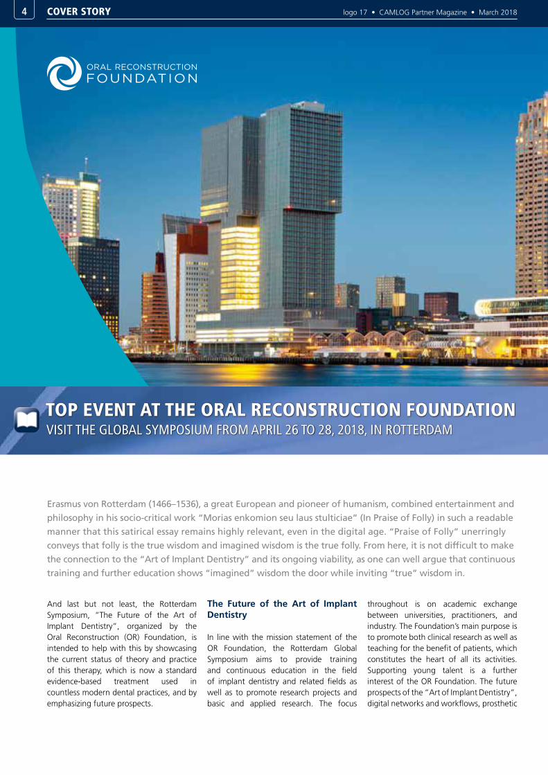
In the last issue of our partner magazine (logo 16), we reported on the renaming of the CAMLOG Foundation to the independent Oral Reconstruction (OR) Foundation. The next logical step was to rename the International CAMLOG Congress the Oral Reconstruction Global Symposium. As early as next year, we will continue the successful CAMLOG congresses with our Global Symposium “The Future of the Art of Implant Dentistry” held in Rotterdam from April 26 to 28, 2018, with more than 55 renowned speakers presenting and discussing the latest scientific findings.
After our last congress in Krakow, the bar for the OR Foundation has been set very high. The successful symbiosis of a largely unknown but very impressive venue, instructive workshops, and an exciting scientific program with first-class speakers will be continued with the Oral Reconstruction Foundation Symposium 2018 in Rotterdam.
Rotterdam is known as a port and industrial city, less as a tourist attraction or party city, and even less as a venue for an international medical and implant
dentistry congress. We will not be able to change that in 2018. However, we would like to point out – with all due modesty – that the Foundation and CAMLOG enjoy an excellent reputation in the professional world not only as implant specialists but also as congress organizers. The highlights of the 2018 Symposium’s scientific program can be found on page 4 and you can look forward to a scientific congress, international exchange, exciting best practices, the cool city of Rotterdam, and an atmospheric get-together.
See you in Rotterdam for “The Future of the Art of Implant Dentistry”!
Sincerely
Dr. Alex SchärOral Reconstruction FoundationCEO and Member of the Foundation Board
Dear readers
logo 17 • CAMLOG Partner Magazine • March 2018 3
logo – the CAMLOG Partner-Magazine • Publication frequency: Twice annually • Publisher: CAMLOG Biotechnologies AG • Margarethenstr. 38 • 4053 Basel Switzerland • www.camlog.com, Editorial team: Oliver Ehehalt (responsible), Michael Ludwig, Anela Mehic, Françoise Peters, Andrea Stix, Ingrid Strobel • Photos: CAMLOG and its licensors • Design: Kerstin Gerhardt, Duc-An Do.
Note: Named contributions express the opinion of the author and not necessarily the opinion of the publisher. Names marked with ® are registered trademarks of their respective manufacturers. CAMLOG Biotechnologies AG may market products only upon approval by the competent authorities. Not all products may be available in every country.
CONTENT
COVER STORY
• Top event at the Oral Reconstruction Foundation – April 26 to 28, 2018, in Rotterdam 4
SCIENCE / CLINICAL RESEARCH
• Synergy of the elements: The functionality and biocompatibility of biomaterials 8
CASE STUDY
• Anterior tooth rehabilitation with implants 12 • Insertion of two-piece ceramic implants with simultaneous GBR 20 • Mem-Lok® RCM in use 23 • Prosthetic restorations with iSy Implants 24
PRODUCTS
• The new R-Tx® Abutment System 30 • DEDICAM – Communicating in a network 32• New aligning tools for the COMFOUR® Abutment System 33
PRACTICE MANAGEMENT
• Assessing employees correctly 34 • Personal fulfillment in self-employment 38
EVENTS
• 2. CAMLOG start-up days 2018 40
LIFESTYLE
• Rotterdam, a bustling port metropolis with high lifestyle potential 42
logo 17 • CAMLOG Partner Magazine • March 2018COVER STORY4
And last but not least, the Rotterdam Symposium, “The Future of the Art of Implant Dentistry”, organized by the Oral Reconstruction (OR) Foundation, is intended to help with this by showcasing the current status of theory and practice of this therapy, which is now a standard evidence-based treatment used in countless modern dental practices, and by emphasizing future prospects.
The Future of the Art of Implant Dentistry
In line with the mission statement of the OR Foundation, the Rotterdam Global Symposium aims to provide training and continuous education in the field of implant dentistry and related fields as well as to promote research projects and basic and applied research. The focus
throughout is on academic exchange between universities, practitioners, and industry. The Foundation’s main purpose is to promote both clinical research as well as teaching for the benefit of patients, which constitutes the heart of all its activities. Supporting young talent is a further interest of the OR Foundation. The future prospects of the “Art of Implant Dentistry”, digital networks and workflows, prosthetic
Erasmus von Rotterdam (1466–1536), a great European and pioneer of humanism, combined entertainment and
philosophy in his socio-critical work “Morias enkomion seu laus stulticiae” (In Praise of Folly) in such a readable
manner that this satirical essay remains highly relevant, even in the digital age. “Praise of Folly” unerringly
conveys that folly is the true wisdom and imagined wisdom is the true folly. From here, it is not difficult to make
the connection to the “Art of Implant Dentistry” and its ongoing viability, as one can well argue that continuous
training and further education shows “imagined” wisdom the door while inviting “true” wisdom in.
TOP EVENT AT THE ORAL RECONSTRUCTION FOUNDATION VISIT THE GLOBAL SYMPOSIUM FROM APRIL 26 TO 28, 2018, IN ROTTERDAM

logo 17 • CAMLOG Partner Magazine • March 2018 COVER STORY 5
and surgical concepts, and the numerous research projects currently supported by the Foundation will be presented to the professional audience during the symposium.
For the first time ever, specialist dental staff will also be included in the symposium. On Friday, parallel to the main program, a professional development course on current topics will be offered.
Learning by doing
“All theory is gray”, and we all know that people learn quickest and most effectively when they actively participate and get stuck in. This key aspect of learning will be put into practice on April 26, 2018,
with a whole range of workshops. A total of twelve workshops will be offered in different languages, including German and English:• 3D implant planning and the CAMLOG®
Guide System (English)• The one crown one time concept (English)• Soft-tissue augmentation and corrections
for the prevention and management of peri-implant disease (English)
• COMFOUR® Concepts – Indications and their implementation (German and English)
• Top performance thanks to two-piece ceramic implants (German and English)
• Key factors for success with implants in the esthetic zone (German)
• The digital implant workflow in routine practice (German)
• The special workshop: successful soft-tissue grafting: a hands -on workshop presented by the Center for Advanced Dental Education. The renowned American speaker Dr. Edward P. Allen will present the tunnel technique methods using allogeneic and xenogeneic graft materials. The minimally invasive procedures demonstrated here – with practical exercises – are state-of-the-art in the microsurgical tunnel technique.
The limited number of spots for attending the workshops will be allocated according to the principle of “first come, first served”; please register as soon as possible because our experience from the international CAMLOG congresses shows that demand will be high!
THE FUTURE OF THE ART OF IMPLANT DENTISTRY

logo 17 • CAMLOG Partner Magazine • March 2018COVER STORY6
The scientific program
After the welcoming address and opening of the scientific part of the congress by President Prof. Dr. Irena Sailer and President Dr. Ben Derksen, the kaleidoscope of lectures, presentations, and discussions will begin on Friday, April 27, 2018, in Session 1 with the topic of peri-implant soft tissue management. The crucial factors for stability, aspects of soft tissue transplantation, and the management of defects using minimally invasive techniques will be covered.Session 2 is devoted to the very topical issue of digitization. Have we really already reached the stage where we can speak of complete digitization in dentistry? Or does this sound more like wishful thinking, and we are still faced with numerous challenges? And what are the tangible advantages of digital implant prosthetics?Which new surgical procedures are feasible with the CAMLOG® Implant System, the choice of optimal implantation time, and
clever treatment concepts with iSy® to achieve even better esthetic results: all this will be covered in Session 3. Session 4 is devoted entirely to science with the scholarship holders of the OR Foundation presenting the preliminary results of their ongoing research. In Session 5, the authors of the selected posters will each present their work in eight-minute short presentations.
The motto of the evening is: “Let’s celebrate King’s Day!“ – In the spectacular ambience of the Gothic Laurenskerk, the only remnant of medieval Rotterdam, the guests will be spoiled with food and drink as if they were King Willem Alexander himself, who celebrates his birthday exuberantly with the entire population on this day, “Koningsdag”. A live band plus DJ will provide the appropriate festive atmosphere.
The second day of the scientific program will start on Saturday, April 28, 2018, with Session 6 providing an informative comparison of the biological properties and tissue integration ability of “innovative”
zirconium dioxide and “classic” titanium implants. In particular, the indications for CERALOG®, the CAMLOG ceramic implant system, will be the subject of the presentations, rounded off by the presentation of the four-year results with CERALOG® Implants. The fundamental question as to when and where “white roots” have their right to exist is the final part of Session 6.As a result of demographic developments in industrialized countries, dentistry must also deal with the issue of aging populations. What are our options for edentulous patients? The “facts & fiction” of this topic, restorative concepts, and options for immediate restoration will be discussed in Session 7.This will be followed by the award ceremony of the Foundation Research Prize and the presentation of the awards for the winning posters.
At the end of the symposium, the scientific program in Session 8 will focus on complications and failures. Based on some practical cases we want to jointly learn from problem cases. To ensure that the participants learn even more effectively,

PROGRAM OVERVIEW
logo 17 • CAMLOG Partner Magazine • March 2018
The scientific program is divided into eight sessions, each of which ends with a joint discussion:
SCIENTIFIC PROGRAM – DAY 1
• SESSION 1 Soft tissue management and dental implants
• SESSION 2 Digital workflow in implant dentistry
• SESSION 3 Treatment concepts
• SESSION 4 Primary results of research supported by the Oral
Reconstruction Foundation
• SESSION 5 Poster presentations
SCIENTIFIC PROGRAM – DAY 2
• SESSION 6 Are ceramic implants an alternative to titanium?
• SESSION 7 Restorative concepts
• SESSION 8 Problems, complications and failures – what can
we learn from them?
COVER STORY 7
1 Dutch for bicycle
this last program item of the symposium is designed to be interactive, and by using smartphones the audience can actively participate in the debate, ask questions, and present arguments.
Comments on the current activities of the OR Foundation as well as a glimpse of the future will round off the symposium and help ensure that all participants can return home certain that they have experienced what can justifiably be called “The Future of the Art of Implant Dentistry” in Rotterdam.
What else needs to be said
It goes without saying that the OR Foundation in Rotterdam 2018 not only offers a high-class symposium at a favorable price-performance ratio but it also provides an attractive partner program:• A day in Delft: historic city center, idyllic
canals, the world-famous porcelain factory; all definitely worth a visit…
• Rotterdam Harbor Cruise: a harbor must be explored from the element that brings it to life, the water. A discovery cruise through Europe’s largest trading port in a specially chartered barge.
• Rotterdam by bicycle: what could be more Dutch than riding through Rotterdam on a guided tour with “fiets”1? And the boat trip by water taxi could hardly be more typical of the city.
• The architectural capital of the Netherlands: that’s what insiders call Rotterdam. Our guided tour through the city with emphasis on its architecture is a must for those interested in architectural history.
Further information can be found in the brochure enclosed with this issue of “logo” and on the Internet at: www.orfoundation.org/globalsymposium. You can register directly online. The OR Foundation and CAMLOG as Founding Partner look forward to welcoming you!

SCIENCE / CLINICAL RESEARCH logo 17 • CAMLOG Partner Magazine • March 201888
Numerous factors play a role in the selection of a suitable biomaterial. The reactions triggered by the material used are related to its biocompatibility as well as its chemical, physical, and mechanical properties. The combination of these properties and the ability of the material to augment or replace the body’s own tissues determine its quality.
SYNERGY OF THE ELEMENTS: THE FUNCTIONALITY AND BIOCOMPATIBILITY OF BIOMATERIALS
Structure and composition of the biomaterial
The contact surface of the biomaterial and both the humoral and cellular components of the body interact in many different ways that are influenced by the topography of the material (Fig. 1).
Bones and soft tissue are structures that are not easy to replicate. The functionality of a biomaterial results from the sum of its biological interactions [1]. If functionality meets biocompatibility, this leads to a biomaterial that effectively supports tissue regeneration.
When choosing a biomaterial, the central question is how the implanted material interacts with the surrounding tissues. For example, sintered bone replacement materials tend to be absorbed relatively quickly [2]. Excessively rapid resorption, however, can lead to a weakening of the newly formed bone, which can lead to complications.
In contrast, MinerOss® X and MinerOss® XP have a slow absorption rate [3]. This allows substantial new bone formation. From a biological point of view, the ideal bone graft material should promote the formation of a stable blood clot because
granulation tissue will then grow around the bone. This soft callus is subsequently converted into woven bone and then to lamellar bone. Ultimately this is referred to as remodeling [4].
The biomaterial should be both functional as well as biocompatible and promote the healing process. From a clinical point of view, the bone replacement material must therefore meet several requirements:
• Porosity, as well as pore size and inter-connectivity, are decisive factors for the use of bone replacement materials. The material has to provide a three-dimen-
Fig. 1: The inorganic porcine bone mineral matrix of MinerOss® XP in 75-fold and 25-fold SEM magnification. Image courtesy of Dr. S-T Li.

logo 17 • CAMLOG Partner Magazine • March 2018 99SCIENCE / CLINICAL RESEARCH
sional framework for the formation of new bone. The properties of the framework in turn influence the success of the bone graft material (Fig. 2).• Autologous bone is still the gold standard but it is associated with higher costs and longer treatment times, and it requires an additional surgical procedure which can lead to increased donor site morbidity. Potential complications should be reduced [2,5]. • Further aspects are the handling of the
material – the easier the better for the clinician – as well as the cost efficiency.
• Aside from these factors, it is vital to bear in mind that bone augmentation surgery is often performed as part of a dental implant surgery. Therefore, the biological potential of the bone graft material should also be taken into account [1].
Biological interactions
In two animal studies Li et al. [6] compared two bone graft materials of animal origin – isolated porcine cancellous bone (PCA/MinerOss® XP) (see Fig. 1) and commercially used carbonate apatite of bovine origin. The authors concluded that both materials in the intraoral and orthopedic bone defect functioned as an osteoconductive matrix that promoted bone regeneration. A study conducted
by Roberts et al. [1] showed a correlation between the behavior of progenitor cells and the composition of the material. The study showed that the calcium-phosphate ratio and cell attachment play
a decisive role in new bone formation (Fig. 3).
In another study, Yuen et al. [7] showed that the dense Mem-Lok® RCM
Fig. 2: MinerOss® X: the bimodal pore structure.
Fig. 3: Of the compared materials, MinerOss® X generates more new bone.
30
25
20
15
10
5
0
Num
ber (
%)
0.1 1.0 Pore diameter (μm) Pore diameter (μm)
200 600
The capillary effect is created by the mesopores and leads to fast uptake of the material in the blood.
The interconnected macropores enable the migration of cells and blood vessels and the integration of the particles, which allow effective osseointegration.
A B C D E
14
12
10
8
6
4
2
0
Bone
vol
ume
(%)

SCIENCE / CLINICAL RESEARCH logo 17 • CAMLOG Partner Magazine • March 20181010
membrane improves mechanical stability (Fig.4). Li et al´s [8] preclinical tests using Mem-Lok® Pliable have shown that the tensile strength is three times higher than with a comparable collagen membrane. Furthermore, the same tests show a lower degree of inflammation and fewer foreign body reactions.
Clinical Application
Comparative studies [3,5] have shown that choosing a bone graft material can be difficult. The needs and preferences of the clinician play as important a role as the needs of the patient.
In a histomorphometric human study, Garnieri et al. [3] compared and evaluated the bone reactions of a bovine (MinerOss® X) and porcine xenograft (MinerOss® XP) that were inserted into adjacent extraction sockets.
The histological results suggested that both materials were both biocompatible and osteoconductive.
Gonshor and Tye [5] evaluated the behavior of inorganic xenogeneic bone graft materials (MinerOss® X and MinerOss® XP) in 10 patients with biopsies performed after 6 months. The results showed that the newly formed vital bone was in close contact with the biomaterial (Fig. 5).
Conclusion
The appropriate bone graft material has to be selected based on knowledge of both its properties and also its clinical outcome. The ultimate goal is predictable results, clinical success and reproducibility. The xenogeneic bone replacement materials MinerOss® X and MinerOss® XP promise the best possible “synergy of the elements”.
Fig. 4: Biomechanical strength of the membrane.
400
350
300
250
200
150
100
50
0
Tens
ile s
tren
gth
(g)
Comparable collagen
membrane available on the
market
350
± 8
0
74 ±
10
Mem-Lok®
RCM
Histology of the alveolar ridge with bovine bone replacement material MinerOss® X (Trichrome stain X 10): blue arrow = bone replacement material; yellow arrow = vital bone; red arrow = newly formed bone (osteoid)
100 micrometers
Histology of the alveolar ridge with bovine bone replacement material MinerOss® X (Trichrome stain X 20): yellow arrow = vital bone; red arrow = newly formed bone (osteoid)
50 micrometers
Histology of the alveolar ridge with porcine bone replacement material MinerOss® X (Trichrome stain X 20): yellow arrow = vital bone; red arrow = newly formed bone (osteoid)
50 micrometers100 micrometers
Histology of the alveolar ridge with porcine bone replacement material MinerOss® XP (Trichrome stain X 10): blue arrow = bone replacement material; yellow arrow = vital bone; red arrow = newly formed bone (osteoid)
Fig. 5: Histological examinations of the alveolar ridge with the xenogeneic bone replacement materials MinerOss® X and MinerOss® XP six months postoperatively – integration and bone healing. Courtesy of Renzo Guarnieri MD DDS

logo 17 • CAMLOG Partner Magazine • March 2018 1111
[1] Roberts SJ Geris L, Kerckhofs G, Desmet E, Schrooten J, Luyten F. The combined bone forming capacity of human periosteal derived cells and calcium phosphates. Biomaterials 2011;32: 4393-405.
[2] Figueiredo M, Fernando A, Martins G, Freitas J, Judas F, Figueiredo H. Effect of the calcination temperature on the composition and microstructure of hydroxyapatite derived from human and animal bone. Ceramics International 2010;36(8): 2383–93.
[3] Guarnieri R, Devilliers P, Grande M, Stefanelli LV, Di Carlo S, Pompa G. Histologic evaluation of bone healing of adjacent
alveolar sockets grafted with bovine- and porcine-derived bone: A comparative case report in humans. Regenerative Biomaterials 2017; 1–4 doi: 10.1093/rb/rbx002.
[4] Schindeler A, McDonald MM, Bokko P, Little DG. Bone remodelling during fracture repair: The cellular picture. Semin Cell Dev Biol. 2008; 19(5): 459-66.
[5] Gonshor A, Tye C L. evaluation of anorganic bovine bone mineral in post-extraction alveolar sockets: A case series. J Osseointegration, 2010;1(2):25-30.
[6] Li ST, Chen HC, Yuen D. Isolation and characterization of a porous carbonate apatite from porcine cancellous bone. Science, Technology, Innovation, Aug. 2014: 1–13.
[7] Yuen D, Junchaya C, Zuchich G, Usreich JB, Lin HB, Li ST. A resorbable, reconstituted type I collagen membrane for guided tissue regeneration and soft tissue augmentation. Society for Biomaterials. 2000; 1228.
[8] Li ST, Yuen D, Martin D, Lee NS. A comparative study of a new porcine collagen membrane to BioGide®. Science, Technology, Innovation. 2015;February:1–5.
LITERATURE
Periodontal defects
Extraction sockets
Horizontal ridge
augmentation
Sinus augmentation
Vertical ridge augmentation
Dehiscence defects
Block grafting Immediate implantation
MinerOss® XCancellous a aa aa aaa a aa a a
MinerOss® XCortical
MinerOss® XCollagen a aa a a a
MinerOss® XPCancellous aa aa aa aaa a aa a aa
Mem-Lok® RCM aa aaa aaa aa aa aaa a
Mem-Lok® Pliable aaa aa aa aaa a* aa a
asuited aa well suited aaa very well suited
Depending on the indication, mixing with Cancellous is recommended. Strong dense structure of tissue fibers. Suitable for additional stability of the graft and for resorption protection.
Indication-related application options
SCIENCE / CLINICAL RESEARCH

logo 17 • CAMLOG Partner Magazine • March 20181212
Dr. Hajo Peters, Vienna
IMPLANT-BASED ANTERIOR TOOTH REHABILITATION WITH MAXIMUM INTERDISCIPLINARY CONCEPT
Fig. 1: Clinically normal initial situation five years after endodontic restoration of tooth 11. Fig. 2: Radiological 3D representation of the upper jaw defect with erosion in the incisive foramen (axial layer) as well as loss of the labial compact bone in sagittal reconstruction.
This case study describes the one-year surgical and prosthetic therapy, the special dental features as well as the current one-year follow-up of a 24-year-old patient who was treated with an anterior tooth implant (CAMLOG® SCREW-LINE). In order to meet the demands of the patient as a professional musician both functionally and esthetically, the Viennese esthetic protocol developed by MDT Christian Koczy, Dr. Otto Exenberger, and Dr. Hajo Peters was used, which is based on the close interdisciplinary approach between dental technician, prosthodontist, and surgeon. This protocol is a maximum concept which will be presented in all its details. The documentation describes the diagnosis and clinical procedure from anterior tooth removal in the maxilla with extensive cystectomy, via the temporary restoration, reconstruction of the alveolar process, implant positioning, and augmentation of the soft tissue through to shaping and restoration using individual ceramic prosthetics.
Case history and clinical examination
During a routine dental examination and subsequent diagnosis by means of panoramic tomography, an extensive radiological translucency around the anterior maxilla was detected in the patient, who was 22 years old at the time of the examination (2014). According to initial information provided, the patient was free of symptoms and had a normal oral situation for both soft tissue and hard tooth substance (Fig. 1).
The patient is a professional musician (saxophonist) and specific questioning with regard to the suspect anterior tooth region finally resulted in the patient mentioning
an occasional sensitivity under the upper lip, which the patient himself attributed to daily practice with the woodwind instrument and a possible overstressing of the lip musculature but he had never considered this to be due to an illness. [1–2]
Endodontic treatment of tooth 11 five years previously was revealed in the case history. The root canal treatment was necessary because of a mechanical-traumatic dislocation of the tooth, which, after initial splinting and subsequent sensitivity checks, exhibited no positive vitality and thus no re-innervation/vascularization of the damaged pulp.
To better assess the suspected apical pathology, a three-dimensional X-ray
diagnosis was performed, which showed the full extent of the bone defect: a sharply defined osseous translucency of approx. 2×3×1 cm in the frontal alveolar process of the maxilla with bony erosion to the incisive foramen and close relation to the endodontically treated tooth 11. Extensive vestibular loss of the alveolar process was dominant in the sagittal section (Fig. 2).
Surgical rehabilitation
The radiological findings correlated with the almost complete fenestration of the labial alveolar bone in the subsequent surgical therapy (Fig. 3). Due to the minimally invasive oriented tooth extraction using a piezosurgery device (mectron, Cologne) to separate the periodontal anchorage
CASE STUDY

logo 17 • CAMLOG Partner Magazine • March 2018 1313
of the tooth, a cervical bone bridge was retained coronal to the defect. The previous formation of a vestibular pedicle mucoperiosteal flap provided an overview of the pathological process on the one hand and protection for the adjacent papillae on the ohter hand. The cystectomy revealed an infected radical cyst which was later histologically confirmed, and which could be enucleated in full. The clinical dimensions of the extracted cyst tissue are shown in Figure 4. To stabilize the clot, a collagen cone (Parasorb, Resorba, Nuernberg) was inserted into the socket without applying pressure (Fig. 5). Since the volume of the bone resorption had already reached such an extent at the time of tooth extraction, adequate reossification of the defect was not expected. This applied in particular to the cancellous alveolar process of the maxilla compared to similar defects in the mandible with a broad cortical bone and higher regeneration potential. For this reason, and because of the presence of an infected cyst, immediate filling of the defect using a socket preservation technique was omitted. In primary wound care, the main objective was therefore soft tissue coverage and healing of the defect to achieve optimal
conditions for surgical re-entry for bone augmentation after about 6 weeks. The post-operative course was accompanied with systemic antibiotic treatment (amoxicillin) and a local chlorhexidine gluconate rinse (0.2%) until the sutures were removed after one week.
Soft tissue healing and temporary restoration
The clinical findings of the anterior tooth segment revealed the expected optical defects after completion of soft tissue healing. Both vertical and transversal losses were apparent when the patient returned. On the other hand, the wound healing was free of irritation, which included complete soft tissue closure of the extraction socket and an inflammation-free gingiva and socket mucosa (Fig. 6). Throughout the entire treatment, starting with tooth extraction and continuing through to the insertion of the implant crown, the patient wore the temporary thermoformed splint (Fig. 7), which was functionally and esthetically acceptable to the patient. To ensure undisturbed soft tissue healing, it is essential to use a temporary restoration
which does not apply pressure to the mucous membrane tegument. This implies the use of either splints or adhesive bridges fastened using the acid etching technique. The latter offers the advantage of a greater wearing comfort for the patient (no impairment of the occlusion), however, it does require time-consuming removal and post-operative reattachment to the adjacent teeth for all subsequent treatment steps. In this case, the patient’s needs have to be discussed and taken into account accordingly. Our patient opted for an easy-to-use temporary splint that could be adapted or replaced during the course of the therapy.
Bone grafting
As was to be expected at the time of the cystectomy and tooth extraction, the extensive bone defect was revealed when the site was reopened, and this had increased even further due to the resorption of the painstakingly preserved cervical bone bridge (Fig. 8). Once again, this shows that any expectations about bone preservation after tooth extraction in the region of the tooth-supporting
Fig. 3: Intraoperative site after tooth extraction and cystectomy. Delicate preserved cervical bone bridge.
Fig. 4: In toto enucleated radical cyst from the apical region of tooth 11.
Fig. 5: Clot stabilization in the cyst defect and the extraction socket using xenogeneic collagen cone.
Fig. 6: Clinical defect situation six weeks after tooth extraction.
Fig. 8: Expansion of the bony defect with complete resorption of the buccal bone wall prior to bone augmentation.
Fig. 7: Thermoformed splint as a long-term temporary restoration throughout the entire treatment.
CASE STUDY

logo 17 • CAMLOG Partner Magazine • March 20181414
alveolar process are uncertain and do not allow a reliable prognosis despite a careful and minimally invasive surgical technique. For this reason, it is essential to explain the necessity of bone augmentation to patients before tooth extraction and to inform them about the different clinical measures and treatment options for regeneration.
Augmentation of the bony hard tissue defect was performed using two autologous bone blocks (Fig. 9). These were harvested under local anesthesia in the region of the right ascending mandible. In order to ensure maximum adaptation of the vertical block inserted in the area of the extraction socket, the block was extracted with a trephine bur and the recipient site was reshaped using a milling machine (Fig. 10) with a corresponding diameter. Oro-vestibular oversizing of the augmentation area is necessary in order to compensate for subsequent resorption during the healing phase. Due to the insufficient coverage of the vertical defect, a second block had to be applied to the remaining apical defect in the form of a bone cover. The defect was first filled
with a mixture of autologous chips and xenogeneic particulate bone replacement material (Bio-Oss, Geistlich, Wolhusen). Both blocks were fixed to the underlying alveolar process with osteosynthesis screws (Ustomed, Tuttlingen) in a stable position. The combination of two blocks shown illustrates the legitimacy of the two established bone flap augmentation procedures with distance osteogenesis and congruent block adaptation with contact osteogenesis. In order to level the transitions between bone blocks and the outer curve of the alveolar process and as resorption protection, particulate bone graft material was coated and covered with a resorbable collagen membrane (Bio-Gide, Geistlich, Wolhusen) as part of guided bone regeneration (Fig. 11).
Implantation
The drilling template was fabricated in the laboratory in preparation for the implantation, which was performed approximately four months after bone block augmentation (Fig. 12). For this purpose, a model and a thermoform film of the initial state prior to tooth extraction
were created in which the assembled drill sleeve was inserted according to the positioning and axial direction of the original tooth 11. Wherever possible, initial models should be prepared before tooth extraction in order to provide sufficient anatomical references for the further steps. Otherwise, the drill sleeve is primarily aligned towards the later prosthetic insertion direction under consideration of the adjacent neighboring teeth. Since the technician was aware that maximum bone reconstruction of the implant bed was planned, it was possible to fabricate the drilling guide according to purely anatomical and prosthetic design features without having to take into account any bone deficits which were anyway difficult to detect in the model.
Figure 13 shows the orthopantomogram with inserted drilling template and radiopaque sleeve as well as the two osteosynthesis screws in situ. The frontal view of the clinical situation four months post-augmentation is shown in Figure 14. In this context, the improvement in the vertical alveolar ridge dimension in comparison to Figure 6 is of particular importance. The
Fig. 15: Occlusal view corresponding to Fig. 14 with rehabilitated alveolar ridge width.
Fig. 16: Opened augmentation area with osteosynthesis screws in situ.
Fig. 17: Template-guided implant pilot hole after removal of the osteosynthesis screws.
Fig. 10: Trephine drill for bone block removal and ablative bur for preparing the recipient site with a corresponding diameter.
Fig. 11: Collagen membrane covering of the augmentation site as part of guided bone regeneration.
Fig. 9: Bone blocks fixed with osteosynthesis screws for augmentation (horizontal block: bone cover method; vertical block: site-congruent block).
CASE STUDY

logo 17 • CAMLOG Partner Magazine • March 2018 1515
Fig. 12: Drilling template in thermoforming process with prefabricated sleeve with positioning and axial direction aligned with the original tooth.
Fig. 18: Occlusal view of the inserted implant with very well regenerated vestibular ridge contour.
Fig. 21: Palatal pedicle connective tissue graft for soft tissue augmentation.
Fig. 13: Orthopantomogram with drilling template before removal of osteosynthesis screws and implantation.
Fig. 19: Frontal view with paracrestal implant shoulder at maximum vertical bone regeneration.
Fig. 14: Frontal view with clinical findings after four months of healing of the block grafts.
Fig. 20: Orthopantomogram for radiological evaluation after implantation 11.
occlusal view (Fig. 15) already showed clinically a physiological alveolar ridge transversal, which gave rise to the hope of corresponding bone regeneration.The surgical field was opened using a mucosal split flap to allow the possibility of simultaneous soft tissue augmentation after implantation. After preparation of the two-layer vestibular flap, bone regeneration of the previous defect could be easily assessed (Fig. 16). The bone blocks were fully integrated and showed both excellent transverse regeneration as well as maximum vertical regeneration. After removing the osteosynthesis screws, template guided pilot drilling was performed (Fig. 17). Extension of the drilling shaft up to the diameter of final form drilling was performed freehand. After completion of the bony preparation, the drilling shaft is to be probed for an intact bony boundary on all sides with a blunt button probe in the form of bone mapping. This rules out perforations to the nasal floor and a possible need for subsequent augmentation – especially vestibular – can be determined. Subsequent augmentation was not necessary in this case. The manually inserted implant (CAMLOG®
SCREW-LINE) was surrounded in the occlusal view by a sufficiently dimensioned and well perfused vestibular bone wall. The curvature of the jaw arch was also completely restored (Fig. 18). The frontal view shows the crestal bone profile and the paracrestal implant shoulder position as well as the positioning of the implant in mesiodistal direction while maintaining the anatomical minimum distances to the adjacent teeth (1.5 to 2 mm), so that the subsequent formation of papillae could be ensured with sufficient nutrition of the interdental bone (Fig. 19). In this case, a 4.3 mm wide and 13 mm long CAMLOG® SCREW-LINE implant was inserted and radiologically documented after surgery (Fig. 20).In order to increase the volume of the periimplant gingiva, simultaneous soft tissue augmentation with a palatal pedicle connective tissue graft had already been performed with the placement of the implant. For this purpose, the paramarginal palatal, subepithelial prepared tissue was driven into the tooth gap over the anterior pedicle pole (Fig. 21) and fixed with the previously prepared periosteum of the split flap with an absorbable suture. This
CASE STUDY

logo 17 • CAMLOG Partner Magazine • March 2018
allowed the mucosal portion of the split flap to be closed without tension, resulting in a significant tissue increase with complete plastic coverage of the implant region. The connective tissue transplanted in this manner brings with it the genetic information for the long-term formation of keratinized gingiva at the implantation site. Postoperatively, there is often swelling present which, in combination with soft tissue augmentation, causes a visual reduction in the clinically visible adjacent crowns (Fig. 22). It is essential to inform the patient about this and to adjust the temporary restoration accordingly. After ten weeks, the new soft tissue situation had been fully established (Fig. 23). This also corresponded to the initial state for the subsequent implant prosthetic measures for soft tissue shaping. At this point in time, all hard and soft tissue augmentation treatment steps have been completed. It must be ensured that the soft tissue even has an oversized reconstruction if possible, to ensure sufficient reserves for soft tissue shaping.
Gingiva forming
Following the implantation, the intra-operative impression of the implant was taken with an individual tray (Fig. 24). This served as the basis for the fabrication of an individual healing cap (Fig. 25). This healing cap is milled from either plastic or zirconium oxide, based on the scanned prosthetic set-up of the tooth to be replaced, and bonded with a temporary abutment. The healing cap, inserted after surgical exposure using a displacement procedure, protrudes about 2 mm beyond the gingival margin and has its widest extension at the emergence point (Fig.26). The objective of this individual healing cap is to shape the emergence profile of the subsequent implant abutment in the gingiva. For this purpose, the axial incision must optimally replicate both the oro-vestibular and the mesiodistal dimensions of the crown anatomy at the gingival level (Fig. 27). We consider the introduction of the concept of an individual healing cap to be the decisive link in our esthetic treatment protocol between the surgical rehabilitation of the implant site and the prosthetic replica of an anatomical crown.
The retention time of the individual healing cap in the mouth for gingiva forming varies from individual to individual and between eight to twelve weeks can transpire before the implant impression can be taken.
Individual abutment and implant crown
After removing the individual healing cap, it was necessary to ensure that the delicate gingiva funnel was not deformed while the impression of the implant was being taken. For this reason, a prefabricated impression post was wrapped in low-viscosity plastic and hardened in the gingival funnel (Fig. 28). The model thus obtained reflects the implant situation with the gingival emergence profile of the future crown at the same time. On this basis, an individual abutment was fabricated that fulfils three essential tasks: color-coded individualization of the tooth core; final manipulation of the gingiva through varying subgingival design; maximum basal width comparable to a prepared natural tooth crown with approx. 1 mm subgingival crown margin (Fig. 29). Our master casts are always fabricated
Fig. 23: Ten weeks after soft tissue augmentation. Initial situation for tissue shaping.
Fig. 24: Intraoperative implant impression with individual tray.
Fig. 22: Postoperative swelling one week after soft tissue augmentation.
Fig. 28: Implant impression with customized impression post for stabilization of the gingival funnel.
Fig. 29: Individual abutment in situ. Fig. 30: Ceramic veneered zirconium oxide crown on the master cast without gingival mask.
16 CASE STUDY

logo 17 • CAMLOG Partner Magazine • March 2018
without gingiva masks, as we want to transfer the surgically and prosthetically elaborately formed gingival margin exactly as a defined limit to the model, a purpose for which resilient gingival masks are not indicated (Fig. 30). The zirconium crown framework was milled (Cercon ht light, Degudent, Hanau) and veneered with ceramic materials (Cercon ceram Kiss, Degudent, Hanau). By powdering neutral, highly-fluorescent ceramic materials onto the framework, all the necessary light properties can be imparted with minimal layer thickness. Shade selection beforehand in the laboratory is of crucial importance because up to 20 shades and mixed hues are required for the individual esthetic design of anterior tooth crowns (Fig. 31). The shade nuances were determined directly on the patient and transferred into an esthetic layering scheme (Fig. 32). [3]
The crown fabricated in this way was cemented and the clinical gingival condi-tions were documented with photos as a reference after one month in situ (Fig. 33). The clinical follow-up was performed after 15 months (Fig. 34): The result of the
esthetic rehabilitation with a single-tooth crown implant after bone and soft tissue re-generation and soft tissue forming is reliably stable and the patient is fully satisfied with the esthetics and function.
Discussion/ Conclusion
The present patient case describes the detailed course of treatment of an esthetic anterior tooth rehabilitation after tooth loss, loss of parts of the alveolar process due to an extensive cyst, and the gradual regeneration of hard and soft tissue as a prerequisite for implant surgery and prosthetic therapy. The sequence of treatment steps, which can extend over one year, must be strictly coordinated in terms of time in order to eliminate delays and to allow sufficient time for the biological regeneration processes in particular. This requires applying a comprehensive therapy plan in the form of a standardized protocol, which sets milestones for the treatment, which are then adapted to the individual patient case. For this purpose, the surgical, prosthetic and dental work steps are worked out in an interdisciplinary conference and, after
Fig. 25: Individual healing cap on the model.
Fig. 31: Individual shade guide for shade selection for the patient.
Fig. 34: Follow-up after 15 months shows perfect biological and prosthetic conditions.
Fig. 26: Frontal view of the individual healing cap in situ.
Fig. 32: The patient’s layering scheme for shade customization of the implant crown.
Fig. 27: Occlusal view of the individual healing cap with anatomically optimal emergence profile.
Fig. 33: Inserted implant crown after one month.
17CASE STUDY
logo 17 • CAMLOG Partner Magazine • March 2018
reaching a consensus, discussed in a joint meeting with the patient where they are informed about the various options. [4–7]
Our patient was informed from the very beginning about the treatment steps to be carried out. As a saxophone player, complete functional rehabilitation was of fundamental importance to him, which is why alternative treatment options were eliminated in the preliminary discussion with the patient [1–2]. The consent of the patient to such a maximum concept, as presented here, requires the treatment team to make additional planning efforts, to ensure perfect interdisciplinary communication, and to maintain a mutual understanding of the disciplines involved. Finally, the technical treatment milestones, which we consider essential for the application of the Viennese esthetic protocol, should be summarized:
• Detailed jaw models and photo documentation of the initial situation (data back-up)
• Minimally invasive tooth extraction• A pathogen-free surgical field• 3D hard tissue reconstruction• Pressure-relieved temporary restoration• Prosthetically oriented implant position
(drilling template)• Sufficient soft tissue reconstruction• Individual gingiva forming• Individual abutment• Shade selection with layering scheme• Zirconium oxide framework with veneer
My thanks to the entire team for their interdisciplinary cooperation. Special mention goes to my colleague Dr. Otto Exenberger, for his involvement in the prosthesis, as well as to the master dental technician Christian Koczy, both from Vienna.
[1] Prensky DH, Shapiro GI, Silverman SI. Dental diagnosis and treatment for musicians. Special Care in Dentistry 1986;6(5):198-202
[2] Yeo DKL, Pham TP, Baker J, Porter SAT. Specific orofacial problems experienced by musicians. Review. Australian Dental Journal. 2002;47(1):2-11 [3] Witzel T. Klinische Untersuchung computergestützter Zahnfarbbestimmung im Vergleich zu visueller Abmusterung durch das menschliche Auge. Clinical survey of computer-aided tooth color determination as compared to visual detection by the human eye. 2005. Dissertation Universität Würzburg, Deutschland, urn:nbn:de:bvb:20-opus-11549
[4] Simeone P, De Paoli C, De Paoli S, Leofreddi G, Sgró S. Interdisciplinary treatment planning for single-tooth restorations in the esthetic zone. J Esthet Restor Dent. 2007;19:79-89[5] Yao J, Li M, Tang H, Wang PL, Zhao YX, McGrath C, Mattheos N. What do patients expect from treatment with dental implants? Perceptions, expectations, and misconceptions: a multicenter study. Clin Oral Impl. Res. 2017;28:261-271
[6] Deeb G, Wheeler B, Jones M, Carrico C, Laskin D, Deeb JG. Public and patient knowledge about dental implants. J Oral Maxillofac Surg. 2017;75(7):1387-1391
[7] Jivraj S, Corrado P, Chee W. An interdisciplinary approach to treatment planning in implant dentistry. British Dental J. 2007;202(1):11-18
LITERATURE
18
Dr. Hajo Peters
Dr. med. dent. Hajo Peters is a dentist and oral surgeon with his own private practice in Vienna. His clinical specialties include the complex implant-based rehabilitation in the anterior tooth region.
After his studies at the Private University of Witten/Herdecke (state examination 1999), Dr. Hajo Peters was a research assistant in the Faculty of Dentistry for a further two years. In 2001 he obtained his doctorate with summa cum laude and moved to the Charité Berlin, where he worked for five years in the Department of Oral Surgery. In 2004, after three years of university specialist training, he was appointed an oral surgeon. From 2007 to 2010 he was a partner in a joint oral surgery practice in Filderstadt near Stuttgart. Since 2010 he has been running the specialist practice mundgerecht in Vienna. Dr. Peters is a national and international speaker and author.
Together with Prof. Jackowski from the University of Witten/Herdecke and Prof. Hölzle from the University of Aachen, Dr. Hajo Peters is the editor of a surgical clinic guide, which was published by the Springer-Verlag in August 2017 and is already an Amazon bestseller. Its predecessor (published in 2007 by Elsevier-Verlag) is considered standard literature.
Memberships:• German Association for Implant Dentistry (DGI) • German Association for Dental and Oral Medicine (DGZMK)• Working Group for Maxillofacial Surgery (AGKi)• Working Group for Forensic Odontostomatology (AKFOS)• Working Group Oral Pathology and Oral Medicine (AKOPOM)• International Association of Student Clinicians- American Dental Association (SCADA)
Contact details
mundgerechtSpecialist practice for oral healthDr. Hajo PetersWeimarer Strasse 5/21A-1180 Vienna www.mundgerecht.at
AUTHOR
CASE STUDY
logo 17 • CAMLOG Partner Magazine • March 2018
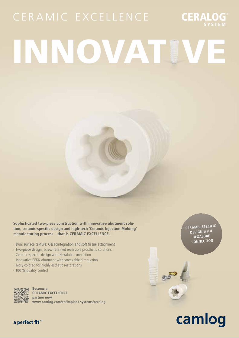
Sophisticated two-piece construction with innovative abutment solu-tion, ceramic-specific design and high-tech 'Ceramic Injection Molding' manufacturing process – that is CERAMIC EXCELLENCE.
· Dual surface texture: Osseointegration and soft tissue attachment · Two-piece design, screw-retained reversible prosthetic solutions · Ceramic-specific design with Hexalobe connection · Innovative PEKK abutment with stress shield reduction · Ivory colored for highly esthetic restorations · 100 % quality control
Become a CERAMIC EXCELLENCE partner nowwww.camlog.com/en/implant-systems/ceralog
INNOVAT VE
CERAMIC-SPECIFIC
DESIGN WITH
HEXALOBE
CONNECTION
CERALOG_Advert_Innovative_A4_EN.indd 1 07.09.2017 16:05:12
logo 17 • CAMLOG Partner Magazine • March 20182020
Dr. Alexander Volkmann, Jena
INSERTION OF TWO-PIECE CERAMIC IMPLANTS WITH SIMULTANEOUS GBR – A SUCCESSFUL PROCEDURE WITH COORDINATED COMPONENTS
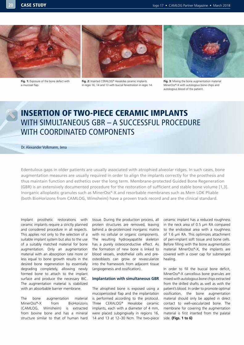
Fig. 3: Mixing the bone augmentation material MinerOss®-X with autologous bone chips and autologous blood of the patient.
Edentulous gaps in older patients are usually associated with atrophied alveolar ridges. In such cases, bone augmentation measures are usually required in order to align the implants correctly for the prosthesis and thus maintain function and esthetics over the long term. Membrane-protected Guided Bone Regeneration (GBR) is an extensively documented procedure for the restoration of sufficient and stable bone volume [1,3]. Inorganic alloplastic granules such as MinerOss®-X and resorbable membranes such as Mem LOK Pliable (both BioHorizons from CAMLOG, Wimsheim) have a proven track record and are the clinical standard.
Implant prosthetic restorations with ceramic implants require a strictly planned and considered procedure in all respects. This applies not only to the selection of a suitable implant system but also to the use of a suitably matched material for bone augmentation. Only an augmentation material with an absorption rate more or less equal to bone growth results in the desired bone regeneration by essentially degrading completely, allowing newly formed bone to attach to the implant surface and produce the necessary BIC. The augmentation material is stabilized with an absorbable barrier membrane.
The bone augmentation material MinerOss®-X from BioHorizons (CAMLOG, Wimsheim) is extracted from bovine bone and has a mineral structure similar to that of human hard
tissue. During the production process, all protein structures are removed, leaving behind a de-proteinized inorganic matrix with no cellular or organic components. The resulting hydroxyapatite skeleton has a purely osteoconductive effect. As the formation of new bone is linked to blood vessels, endothelial cells and pre-osteoblasts can grow or revascularize into the framework from adjacent tissue (angiogenesis and ossification).
Implantation with simultaneous GBR
The atrophied bone is exposed using a mucoperiosteal flap and the implantation is performed according to the protocol. Three CERALOG® Hexalobe ceramic implants, each with a diameter of 4 mm, were placed subgingivally in regions 16, 14 and 13 at 12–30 Ncm. The two-piece
ceramic implant has a reduced roughness in the neck area of 0.5 µm RA compared to the endosteal area with a roughness of 1.6 µm RA. This optimizes attachment of peri-implant soft tissue and bone cells. Before filling with the bone augmentation material MinerOss®-X, the implants are covered with a cover cap for submerged healing.
In order to fill the buccal bone deficit, MinerOss®-X cancellous bone granules are mixed with autologous bone chips extracted from the drilled shafts as well as with the patient’s blood. In order to promote optimal ossification, the bone augmentation material should only be applied in direct contact to well-vascularized bone. The membrane for covering the augmentation material is first inserted from the palatal side. (Figs. 1 to 6)
Fig. 2: Inserted CERALOG® Hexalobe ceramic implants in regio 16, 14 and 13 with buccal fenestration in regio 14.
Fig. 1: Exposure of the bone defect with a mucosal flap.
CASE STUDY
logo 17 • CAMLOG Partner Magazine • March 2018 2121
Covering with membrane
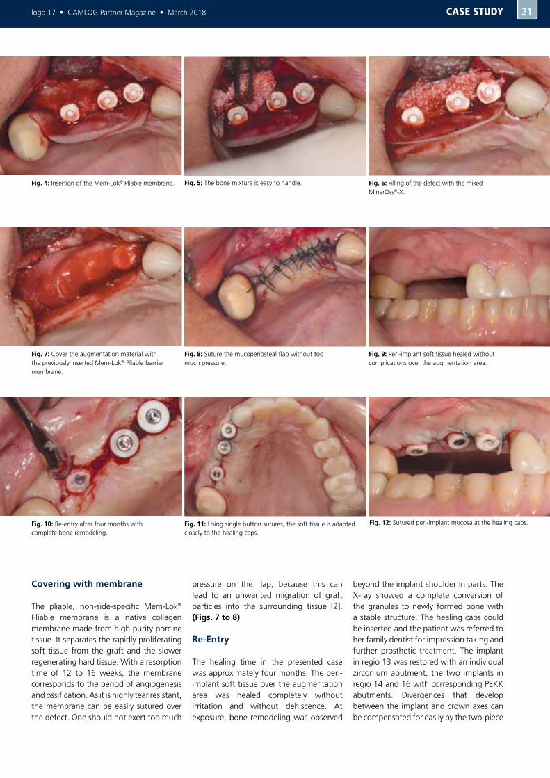
The pliable, non-side-specific Mem-Lok® Pliable membrane is a native collagen membrane made from high purity porcine tissue. It separates the rapidly proliferating soft tissue from the graft and the slower regenerating hard tissue. With a resorption time of 12 to 16 weeks, the membrane corresponds to the period of angiogenesis and ossification. As it is highly tear resistant, the membrane can be easily sutured over the defect. One should not exert too much
pressure on the flap, because this can lead to an unwanted migration of graft particles into the surrounding tissue [2]. (Figs. 7 to 8)
Re-Entry
The healing time in the presented case was approximately four months. The peri-implant soft tissue over the augmentation area was healed completely without irritation and without dehiscence. At exposure, bone remodeling was observed
beyond the implant shoulder in parts. The X-ray showed a complete conversion of the granules to newly formed bone with a stable structure. The healing caps could be inserted and the patient was referred to her family dentist for impression taking and further prosthetic treatment. The implant in regio 13 was restored with an individual zirconium abutment, the two implants in regio 14 and 16 with corresponding PEKK abutments. Divergences that develop between the implant and crown axes can be compensated for easily by the two-piece
Fig. 4: Insertion of the Mem-Lok® Pliable membrane.
Fig. 7: Cover the augmentation material with the previously inserted Mem-Lok® Pliable barrier membrane.
Fig. 10: Re-entry after four months with complete bone remodeling.
Fig. 9: Peri-implant soft tissue healed without complications over the augmentation area.
Fig. 8: Suture the mucoperiosteal flap without too much pressure.
Fig. 11: Using single button sutures, the soft tissue is adapted closely to the healing caps.
Fig. 12: Sutured peri-implant mucosa at the healing caps.
Fig. 5: The bone mixture is easy to handle. Fig. 6: Filling of the defect with the mixed MinerOss®-X.
CASE STUDY
logo 17 • CAMLOG Partner Magazine • March 2018
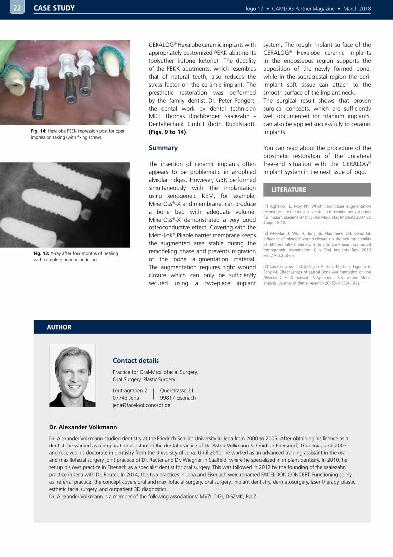
CERALOG® Hexalobe ceramic implants with appropriately customized PEKK abutments (polyether ketone ketone). The ductility of the PEKK abutments, which resembles that of natural teeth, also reduces the stress factor on the ceramic implant. The prosthetic restoration was performed by the family dentist Dr. Peter Pangert, the dental work by dental technician MDT Thomas Blochberger, saalezahn - Dentaltechnik GmbH (both Rudolstadt). (Figs. 9 to 14)
Summary
The insertion of ceramic implants often appears to be problematic in atrophied alveolar ridges. However, GBR performed simultaneously with the implantation using xenogeneic KEM, for example, MinerOss®-X and membrane, can produce a bone bed with adequate volume. MinerOss®-X demonstrated a very good osteoconductive effect. Covering with the Mem-Lok® Pliable barrier membrane keeps the augmented area stable during the remodeling phase and prevents migration of the bone augmentation material. The augmentation requires tight wound closure which can only be sufficiently secured using a two-piece implant
system. The rough implant surface of the CERALOG® Hexalobe ceramic implants in the endosseous region supports the apposition of the newly formed bone, while in the supracrestal region the peri-implant soft tissue can attach to the smooth surface of the implant neck.The surgical result shows that proven surgical concepts, which are sufficiently well documented for titanium implants, can also be applied successfully to ceramic implants.
You can read about the procedure of the prosthetic restoration of the unilateral free-end situation with the CERALOG® Implant System in the next issue of logo.
[1] Aghaloo TL, Moy PK. Which hard tissue augmentation techniques are the most successful in furnishing bony support for implant placement? Int J Oral Maxillofac Implants 2007;22 Suppl:49-70
[2] Mir-Mari J, Wui H, Jung RE, Hämmerle CH, Benic GI. Influence of blinded wound closure on the volume stability of different GBR materials: an in vitro cone-beam computed tomographic examination. Clin Oral Implants Res. 2016 Feb;27(2):258-65. [3] Sanz-Sanchez I, Ortiz-Vigon A, Sanz-Martin I, Figuero E, Sanz M. Effectiveness of Lateral Bone Augmentation on the Alveolar Crest Dimension: A Systematic Review and Meta-analysis. Journal of dental research 2015;94:128s-142s
LITERATURE
22
Fig. 14: Hexalobe PEEK impression post for open impression taking (with fixing screw).
Fig. 13: X-ray after four months of healing with complete bone remodeling.
Dr. Alexander Volkmann
Dr. Alexander Volkmann studied dentistry at the Friedrich Schiller University in Jena from 2000 to 2005. After obtaining his licence as a dentist, he worked as a preparation assistant in the dental practice of Dr. Astrid Volkmann-Schmidt in Ebersdorf, Thuringia, until 2007 and received his doctorate in dentistry from the University of Jena. Until 2010, he worked as an advanced training assistant in the oral and maxillofacial surgery joint practice of Dr. Reuter and Dr. Wiegner in Saalfeld, where he specialized in implant dentistry. In 2010, he set up his own practice in Eisenach as a specialist dentist for oral surgery. This was followed in 2012 by the founding of the saalezahn practice in Jena with Dr. Reuter. In 2014, the two practices in Jena and Eisenach were renamed FACELOOK CONCEPT. Functioning solely as referral practice, the concept covers oral and maxillofacial surgery, oral surgery, implant dentistry, dermatosurgery, laser therapy, plastic esthetic facial surgery, and outpatient 3D diagnostics. Dr. Alexander Volkmann is a member of the following associations: MVZI, DGI, DGZMK, FvdZ
Contact details
Practice for Oral-Maxillofacial Surgery, Oral Surgery, Plastic Surgery
Leutragraben 2 Querstrasse 21 07743 Jena 99817 [email protected]
AUTHOR
CASE STUDY
logo 17 • CAMLOG Partner Magazine • March 2018 23
Dr. Olaf Daum, Leimen
MEM-LOK® RCM IN USE – EXPERIENCES OF A PRACTITIONER
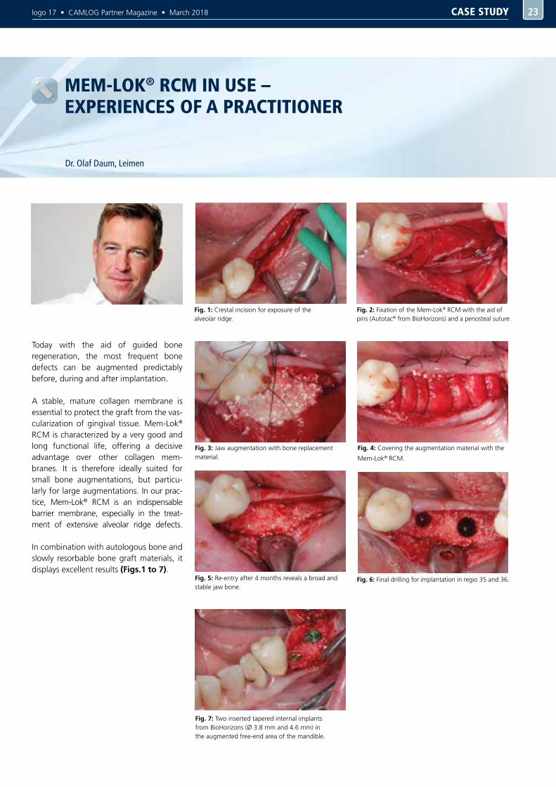
Today with the aid of guided bone regeneration, the most frequent bone defects can be augmented predictably before, during and after implantation.
A stable, mature collagen membrane is essential to protect the graft from the vas-cularization of gingival tissue. Mem-Lok® RCM is characterized by a very good and long functional life, offering a decisive advantage over other collagen mem-branes. It is therefore ideally suited for small bone augmentations, but particu-larly for large augmentations. In our prac-tice, Mem-Lok® RCM is an indispensable barrier membrane, especially in the treat-ment of extensive alveolar ridge defects. In combination with autologous bone and slowly resorbable bone graft materials, it displays excellent results (Figs.1 to 7).
Fig. 1: Crestal incision for exposure of the alveolar ridge.
Fig. 2: Fixation of the Mem-Lok® RCM with the aid of pins (Autotac® from BioHorizons) and a periosteal suture.
Fig. 3: Jaw augmentation with bone replacement material.
Fig. 4: Covering the augmentation material with the
Mem-Lok® RCM.
Fig. 5: Re-entry after 4 months reveals a broad and stable jaw bone.
Fig. 6: Final drilling for implantation in regio 35 and 36.
Fig. 7: Two inserted tapered internal implants from BioHorizons (Ø 3.8 mm and 4.6 mm) in the augmented free-end area of the mandible.
CASE STUDY

logo 17 • CAMLOG Partner Magazine • March 20182424
Dr. Jörg-Martin Ruppin, Penzberg
PROSTHETIC RESTORATIONS WITH ISY IMPLANTS ONE SYSTEM, MANY OPTIONS
Implant dentistry has been firmly established in dentistry for over twenty years. In the pioneering days of implant dentistry, efforts focused mainly on reliable osseointegration. Due to advances both in implant surfaces and shapes as well as surgical techniques, success rates of 95–99% are achieved today accompanied by excellent long-term stability [1,2].
Fig. 1: The well-formed alveolar ridge is fully
adequate for restoration with a full denture.
Fig. 3: Checking the first drill holes with directional indicators.
Fig. 2: Stabilization of the prosthesis in the mandible was only possible by using implants.
Although the patients’ needs for implant restoration are by no means satisfied, the literature shows a certain stagnation in the number of implant restorations performed. A recent, nationwide online survey in Germany [3] confirmed that the number of teeth extracted exceeds the number of implants inserted by a factor greater than 10. There is therefore an obvious imbalance between patient needs on the one hand and performed implant restorations on the other. This is due to a number of reasons, ranging from the subjective reservations of patients about implants, to the partly demanding neces-sary surgical interventions, to financial as-pects [2]. From the patient’s perspective, a modern implant system should therefore primarily be economical without sacrific-ing quality, long-term stability, and safety of use. From a user’s perspective, aspects such as “workflow”, “efficiency” and “simplicity” are also crucial, since time efficiency has a major impact on the eco-nomic efficiency of treatments in everyday practice.
An implant system should therefore satisfy the following requirements:- Safe application and long-term stability- High quality and precision- Cost-effective- A workflow that is as easy and effective
as possible.
As mentioned initially, the term “cost-effectiveness” exceeds by far the simple matter of price. Major aspects are time efficiency and workflow simplicity: only when a system is time efficient in its application to the patient and the number and duration of treatment sessions can be reduced, does a system offer economic added value beyond the mere material price of the individual components, which can be decisive for a cost-effective treatment method.
Using two patient cases as examples, the workflow and prosthetic options of the iSy System are described.
First case: the Locator® restoration
The first patient case presents the restoration of an edentulous mandible with four interforaminal implants. At the time of restoration, the patient was just under 70 years old. The general medical history was normal apart from hypertension and nicotine dependence. The maxilla was also edentulous but could be treated sufficiently with a full denture as the alveolar ridges were still well formed (Figs. 1 and 2). However, the patient complained of insufficient retention of the prosthesis in the mandible. The bone available in the mandible was well suited for restoration with implants, only the width of the crestal portion of the alveolar process was strongly atrophied (Cawood class IV) [4]. After consultation with the patient on the treatment options, the iSy System was chosen.
Implant insertion
At the time of implantation, the extraction of the residual denture in the mandible was approximately six weeks prior, in
CASE STUDY

logo 17 • CAMLOG Partner Magazine • March 2018 2525
Fig. 4: The implants were inserted using the pre-mounted implant bases.
Fig. 5: The implant bases were reversed and the screw connection loosened.
Fig. 6: The abutment screws were removed.
Fig. 7: Due to the conical inner connection, the implant base held firm, even after removal of the screw.
Fig. 10: The implants were covered. The jawbone was built up using the previously harvested crestal bone fractions.
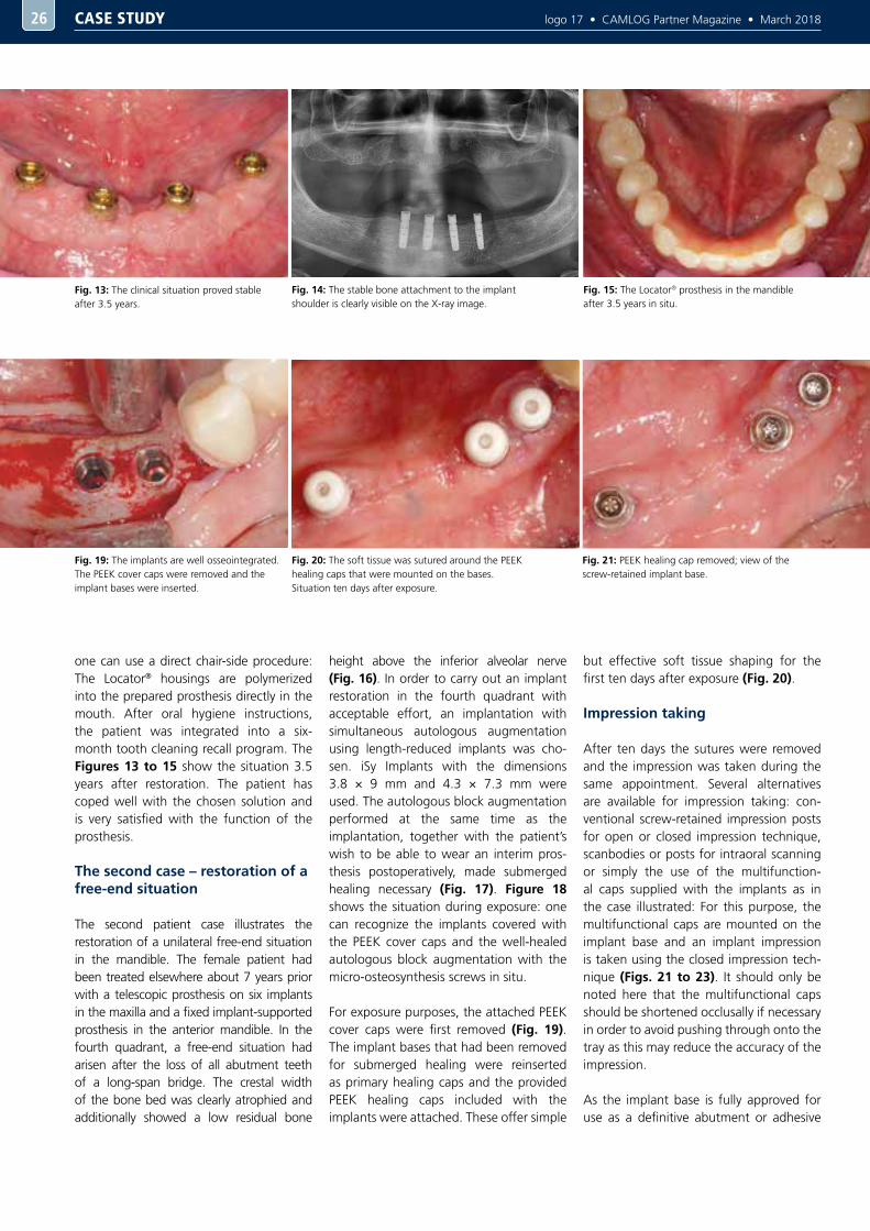
Fig. 11: For exposure purposes, the PEEK cover caps were removed. A very good bone bed was evident.
Fig. 9: The internal structure of the implant became visible after removing the implant base.
Fig. 12: The Locator® abutments were inserted directly during exposure.
Fig. 8: An abutment disconnector was used to remove them from the implant.
other words, we were dealing with a delayed immediate implantation. First, the alveolar process was exposed. The crestal atrophied ridge was removed via micro-segmental osteotomy and the autologous bone harvested in this was partially used for augmentation in regio 42. The implants were inserted on both sides in regio 34 to 44 after exposing the mental foramen. (Figs. 3 and 4). Due to of the augmentation and in order not to unnecessarily complicate the wearing of the mandibular prosthesis for the patient during the healing period, submerged healing was chosen. The iSy
Implant System is supplied with a pre-mounted implant base, which is also used to insert the implant, in readiness for a transgingival healing protocol.
It is also suitable without restrictions for submerged healing, however. To do this, the implants are covered with the PEEK cover caps included in the set after removing the implant base (Figs. 5 to 10). The slightly subcrestal implant positioning indicated for the system has proven to be advantageous, especially when an interim prosthesis is to be worn in the area upon
which was operated. This reliably prevents interference with implant healing caused by denture pressure points.
Prosthetic phase
Osseointegration of the implants is completed after three months of healing. During exposure, the PEEK cover caps can be removed and the implants restored directly with the definitive Locator® abutments (Figs. 11 and 12). Impression taking can be used optionally for processing in the dental laboratory or, as in the presented case,
CASE STUDY
logo 17 • CAMLOG Partner Magazine • March 20182626
Fig. 20: The soft tissue was sutured around the PEEK healing caps that were mounted on the bases. Situation ten days after exposure.
Fig. 19: The implants are well osseointegrated. The PEEK cover caps were removed and the implant bases were inserted.
Fig. 21: PEEK healing cap removed; view of the screw-retained implant base.
Fig. 14: The stable bone attachment to the implant shoulder is clearly visible on the X-ray image.
Fig. 15: The Locator® prosthesis in the mandible after 3.5 years in situ.
Fig. 13: The clinical situation proved stable after 3.5 years.
one can use a direct chair-side procedure: The Locator® housings are polymerized into the prepared prosthesis directly in the mouth. After oral hygiene instructions, the patient was integrated into a six-month tooth cleaning recall program. The Figures 13 to 15 show the situation 3.5 years after restoration. The patient has coped well with the chosen solution and is very satisfied with the function of the prosthesis.
The second case – restoration of a free-end situation
The second patient case illustrates the restoration of a unilateral free-end situation in the mandible. The female patient had been treated elsewhere about 7 years prior with a telescopic prosthesis on six implants in the maxilla and a fixed implant-supported prosthesis in the anterior mandible. In the fourth quadrant, a free-end situation had arisen after the loss of all abutment teeth of a long-span bridge. The crestal width of the bone bed was clearly atrophied and additionally showed a low residual bone
height above the inferior alveolar nerve (Fig. 16). In order to carry out an implant restoration in the fourth quadrant with acceptable effort, an implantation with simultaneous autologous augmentation using length-reduced implants was cho-sen. iSy Implants with the dimensions 3.8 × 9 mm and 4.3 × 7.3 mm were used. The autologous block augmentation performed at the same time as the implantation, together with the patient’s wish to be able to wear an interim pros-thesis postoperatively, made submerged healing necessary (Fig. 17). Figure 18 shows the situation during exposure: one can recognize the implants covered with the PEEK cover caps and the well-healed autologous block augmentation with the micro-osteosynthesis screws in situ.
For exposure purposes, the attached PEEK cover caps were first removed (Fig. 19). The implant bases that had been removed for submerged healing were reinserted as primary healing caps and the provided PEEK healing caps included with the implants were attached. These offer simple
but effective soft tissue shaping for the first ten days after exposure (Fig. 20).
Impression taking
After ten days the sutures were removed and the impression was taken during the same appointment. Several alternatives are available for impression taking: con-ventional screw-retained impression posts for open or closed impression technique, scanbodies or posts for intraoral scanning or simply the use of the multifunction-al caps supplied with the implants as in the case illustrated: For this purpose, the multifunctional caps are mounted on the implant base and an implant impression is taken using the closed impression tech-nique (Figs. 21 to 23). It should only be noted here that the multifunctional caps should be shortened occlusally if necessary in order to avoid pushing through onto the tray as this may reduce the accuracy of the impression.
As the implant base is fully approved for use as a definitive abutment or adhesive
CASE STUDY

logo 17 • CAMLOG Partner Magazine • March 2018 2727
Fig. 17: The clinical situation of the three iSy® Implants three months after implantation with autologous bone augmentation and submerged healing.
Fig. 23: The multifunctional caps from the basal direction in the impression.
Fig. 26: The CAD design of the individual abutments on the iSy® Implant bases.
Fig. 18: The osteosynthesis screws were removed during exposure.
Fig. 24: Soft tissue in regio 44 after removing the implant base. iSy Esthomic healing caps were used to shape the emergence profile.
Fig. 27: The position of the shoulders of the individual zirconium abutments was checked during try-in.
Fig. 25: The emergence profile created with the aid of the iSy® Esthomic healing caps.
Fig. 22: Multifunctional caps mounted on the implant base for closed impression taking.
Fig. 16: The orthopantomogram of the second patient case shows the situation directly after implantation and augmentation.
abutment, the laboratory did not have to order any additional implant parts. After taking the impression, the implant bases were removed again and enclosed with the impression for the laboratory. Now the iSy Esthomic healing caps were used. They are available in three sizes (S, M, and L) depending on the prosthetic tooth shape desired. The major advantage here is that the implant bases can be used in the laboratory for the final restoration, while
the emergence profile can be optimally shaped intraorally using the iSy Esthomic healing caps (Figs. 24 and 25). In the dental laboratory, individual CAD/CAM-fabricated zirconium oxide abut-ments were designed on the iSy implant bases (Fig. 26). When using individual abutments, it is recommended to use an abutment try-in to clinically check the exact position of the preparation margin (Fig. 27). Elastic rubber gingival masks on
the model cannot adequately imitate gin-gival resilience, so this clinical examination and a possible correction of the preparation margins is useful. A maximum 1 mm sub-gingival position of the preparation mar-gins should be maintained here in order to be able to safely remove cement residues when inserting the dental prosthesis [5].
The dental prosthesis was then completed in the laboratory. An all-ceramic construction
CASE STUDY
logo 17 • CAMLOG Partner Magazine • March 20182828
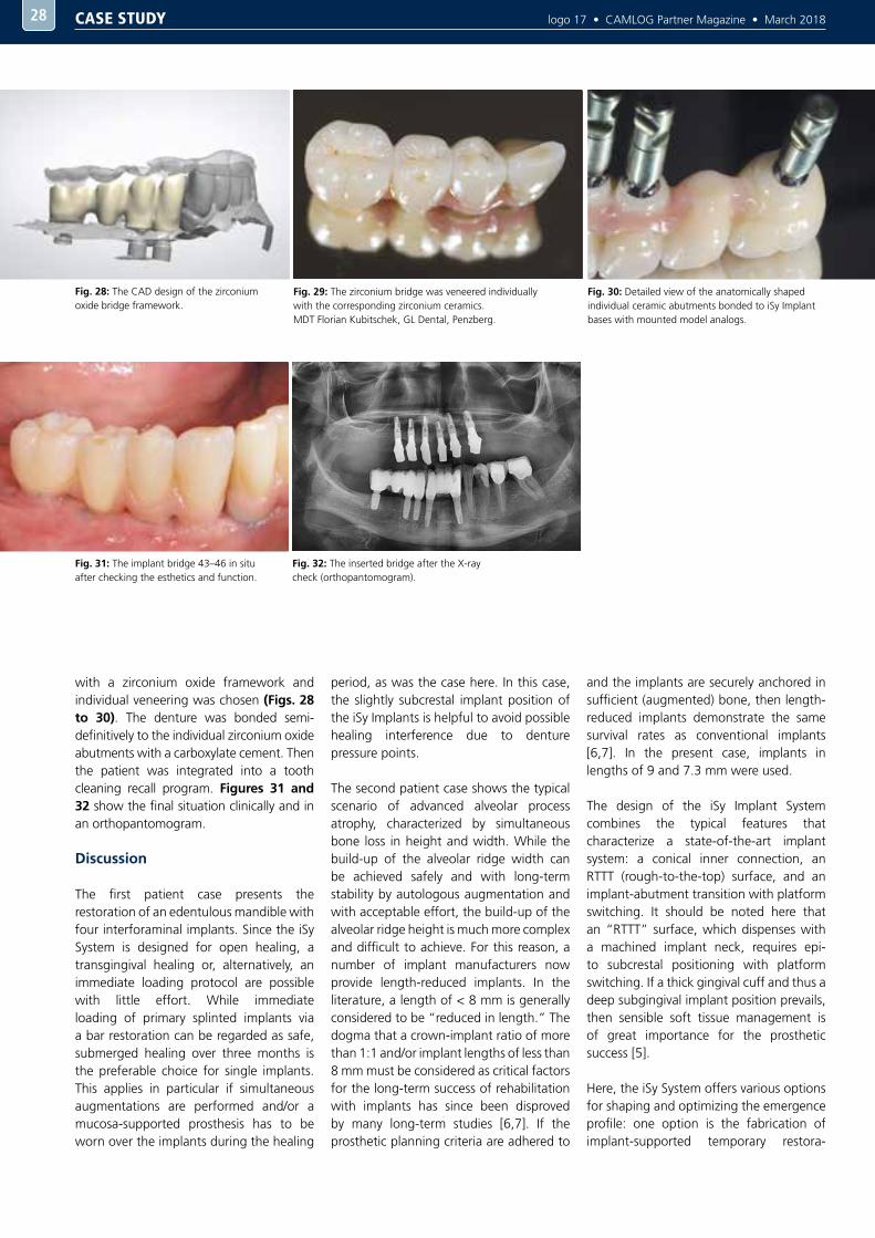
with a zirconium oxide framework and individual veneering was chosen (Figs. 28 to 30). The denture was bonded semi-definitively to the individual zirconium oxide abutments with a carboxylate cement. Then the patient was integrated into a tooth cleaning recall program. Figures 31 and 32 show the final situation clinically and in an orthopantomogram.
Discussion
The first patient case presents the restoration of an edentulous mandible with four interforaminal implants. Since the iSy System is designed for open healing, a transgingival healing or, alternatively, an immediate loading protocol are possible with little effort. While immediate loading of primary splinted implants via a bar restoration can be regarded as safe, submerged healing over three months is the preferable choice for single implants. This applies in particular if simultaneous augmentations are performed and/or a mucosa-supported prosthesis has to be worn over the implants during the healing
period, as was the case here. In this case, the slightly subcrestal implant position of the iSy Implants is helpful to avoid possible healing interference due to denture pressure points.
The second patient case shows the typical scenario of advanced alveolar process atrophy, characterized by simultaneous bone loss in height and width. While the build-up of the alveolar ridge width can be achieved safely and with long-term stability by autologous augmentation and with acceptable effort, the build-up of the alveolar ridge height is much more complex and difficult to achieve. For this reason, a number of implant manufacturers now provide length-reduced implants. In the literature, a length of < 8 mm is generally considered to be “reduced in length.” The dogma that a crown-implant ratio of more than 1:1 and/or implant lengths of less than 8 mm must be considered as critical factors for the long-term success of rehabilitation with implants has since been disproved by many long-term studies [6,7]. If the prosthetic planning criteria are adhered to
and the implants are securely anchored in sufficient (augmented) bone, then length-reduced implants demonstrate the same survival rates as conventional implants [6,7]. In the present case, implants in lengths of 9 and 7.3 mm were used.
The design of the iSy Implant System combines the typical features that characterize a state-of-the-art implant system: a conical inner connection, an RTTT (rough-to-the-top) surface, and an implant-abutment transition with platform switching. It should be noted here that an “RTTT” surface, which dispenses with a machined implant neck, requires epi- to subcrestal positioning with platform switching. If a thick gingival cuff and thus a deep subgingival implant position prevails, then sensible soft tissue management is of great importance for the prosthetic success [5].
Here, the iSy System offers various options for shaping and optimizing the emergence profile: one option is the fabrication of implant-supported temporary restora-
Fig. 28: The CAD design of the zirconium oxide bridge framework.
Fig. 32: The inserted bridge after the X-ray check (orthopantomogram).
Fig. 31: The implant bridge 43–46 in situ after checking the esthetics and function.
Fig. 30: Detailed view of the anatomically shaped individual ceramic abutments bonded to iSy Implant bases with mounted model analogs.
Fig. 29: The zirconium bridge was veneered individually with the corresponding zirconium ceramics. MDT Florian Kubitschek, GL Dental, Penzberg.
CASE STUDY

logo 17 • CAMLOG Partner Magazine • March 2018 2929
AUTHOR
tions that can be prepared on the multi-functional caps with minimal effort and cost. Alternatively, the use of shortened multifunctional caps, individually sup-plemented with plastic, is an option for individual healing caps. As shown in the case presentation, we most often use the iSy Esthomic healing caps after taking the impression with the multifunctional caps: they are available in three diameters (S, M, and L) and different gingival heights. This is a very easy and efficient way to achieve an emergence profile, analogous to the workflow of the CAMLOG System, by suc-cessively using bottleneck, cylindrical, or wide body healing caps. The implant base of the iSy Implant fulfils several functions simultaneously here: as an implant place-ment instrument, as a primary healing cap for transgingival healing or after exposure, as an impression post, a temporary abut-ment and a definitive adhesive abutment for individual abutments. This allows a very efficient workflow with minimal use of ad-ditional implant parts, which enables an efficient and cost-effective treatment.
Our thanks go to MDT Florian Kubitschek and the entire team of GL Dental, Penzberg, for the technical implementation of the case.
Contact details
Dr. Jörg-Martin RuppinSpecialist in oral surgeryMasur Implant Center PenzbergBichler Strasse 1782377 Penzberg, [email protected]
Dr. Jörg-Martin Ruppin
Dr. Jörg-Martin Ruppin studied at the Albert Ludwigs University of Freiburg and passed his examinations in dentistry in 1998. From 1999 to 2001 he worked as an assistant dentist in a specialist implant dentistry practice in Freiburg im Breisgau. He gained his doctoral degree in dentistry in 2001. From 2001 to 2007 he trained in the Department of Oral and Maxillofacial Surgery at the University Hospital of the Technical University of Aachen (RWTH) to become an oral surgeon. During this time he taught and researched in the specialist field of computer-guided implant dentistry and three-dimensional imaging. He gave scientific presentations and wrote national and international publications on computer-guided surgery. He then worked as an oral surgeon in the Practice Clinic for Implant Dentistry and Plastic Surgery Dr. Lippold in Munich. Since 2009 he has headed the Implant Center Penzberg Dr. Masur, Dr. Ruppin & Colleagues. Jörg-Martin Ruppin is a member of the societies: German Society for Dental and Oral Medicine (DGZMK), German Society for Oral Implantology (DGI), International Team for Implantology (ITI), SimPlant Academy and is active as an international speaker.
LITERATURE
[1] Semper, W., Hildebrand, D., Özyuvaci, H., Nelson, K.: Erfolgsrate von Implantaten mit sandgestrahlter und geätzter Oberfläche im Oberkiefer nach einer Einheilzeit von zwölf Wochen: Eine retrospektive Analyse. Z Zahnärztl Impl 23(3),176 ff.(2007)
[2] Von Arx, T., Hardt, N., Wallkamm, B., Kurt, B.: Die TIME Technik: Lokale Osteoplastik zur Alveolarkammaugmentation – Auswertung und Ergebnisse der ersten 15 Fälle. Implantologie 1, 33-48 (1996)
[3] Isbaner, J: Darum wird in Deutschland so wenig implantiert. Zahnarzt, Wirtschaft und Praxis ZPW online, Branchenmeldungen 16.06.2014
[4] J. I. Cawood, R. A. Howell: A classification of the edentulous jaws. In: International journal of oral and maxillofacial surgery. Band 17, Nr. 4, August 1988, ISSN 0901-5027, S. 232–236, PMID 3139793 (englisch).
[5] Wilson T.G.: The Positive Relationship Between Excess Cement and Peri-Implant Disease. J Periodontal 2009, 80: 1388-1392
[6] Lai, H.C., Si H.S., Zhuang, L.H., Shen, H., Liu, Y.L., Wismeijer, D.: Long-term outcomes of short dental implants supporting single crowns in posterior region: a clinical retrospective study of 5–10 years. Clin. Oral Impl. Res. 24 (2), 2013; 230–237
[7] Schneider D, Witt L, Hämmerle C.F.: Influence of the crown-to-implant length ratio on the clinical performance of implants supporting single crown restorations: a cross-sectional retrospective 5-year investigation. Clin. Oral Impl. Res. 23, 2012; 169–174.
LITERATURE
CASE STUDY

logo 17 • CAMLOG Partner Magazine • March 2018
Over the past decades, the LOCATOR® Removable Attachment System has experienced tremendous growth and become a market leader in the field of implant-supported prosthesis anchoring systems. It is trusted equally by dental manufacturers, users, and patients.Zest Dental Solutions®, the manufacturer of prosthesis anchorage systems, has improved the standard LOCATOR® abutment.The new abutment, named Locator R-Tx®, is based on the same type of treatment, with a different design, functional advances, and optimized esthetics. Adding all the changes together, this results in simplified product application, greater patient satisfaction, and increased confidence.
The new definition of strength – DuraTec™
Composed of several layers of titanium nitride and titanium carbonitride, the patent-pending DuraTecTM coating provides a harder, more abrasion-resistant surface. It is over 30% harder, has an abrasion resistance that is more than 25% higher, and reduces surface roughness by almost 65% compared to the titanium nitride coating of the standard LOCATOR® system. Optimization of the LOCATOR R-Tx® is completed by an esthetic gingiva shade, which at the same time ensures adequate visibility for the patient. This results in a revolutionary combination that is not available in this form with any other abutment on the market.
The new screw mechanism
The Locator R-Tx® abutments are inserted with a hexagon screwdriver (industrial standard 0.05"/1.25 mm* hexagon). The central hexagon reduces the area on which food residues and plaque could accumulate.
The new pivot technology and the new coronal shape
The nylon replacement males are pivoted within the die housing. The patented modifications, a channel on the inside of the matrix housing, allow the die housing to pivot up to 30 degrees above the nylon replacement males. Furthermore, the narrower coronal design of the R-Tx® abutments can compensate for maximum
divergences or convergences of up to 60 degrees between the implants. The pivoting technology enables patients to insert their prostheses easily and prevent damage to individual components.
The cone-like abutment, designed with a narrower, wedge-like shape with two retention rings, enables the patient to align the prosthesis more easily and insert it precisely.
PRODUCTS30
In October 2017, CAMLOG introduced the next generation of the Locator® anchoring system for removable implant-supported prostheses. The LOCATOR R-Tx® abutments are available in different gingival heights for the CAMLOG®, CONELOG®, and iSy® Implant Systems.
THE NEW LOCATOR R-Tx® ABUTMENT SYSTEM THE PORTFOLIO EXTENSION FOR CAMLOG®, CONELOG®, AND ISY®
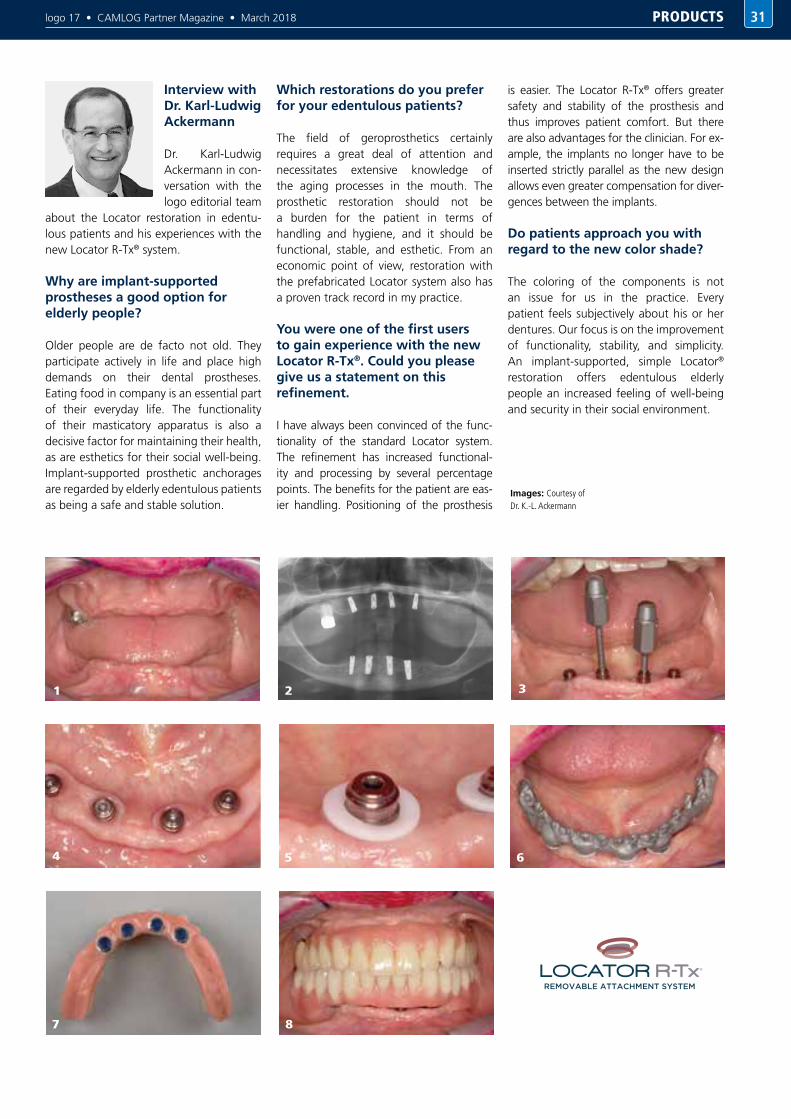
logo 17 • CAMLOG Partner Magazine • March 2018
Interview with Dr. Karl-Ludwig Ackermann
Dr. Karl-Ludwig Ackermann in con-versation with the logo editorial team
about the Locator restoration in edentu-lous patients and his experiences with the new Locator R-Tx® system.
Why are implant-supported prostheses a good option for elderly people?
Older people are de facto not old. They participate actively in life and place high demands on their dental prostheses. Eating food in company is an essential part of their everyday life. The functionality of their masticatory apparatus is also a decisive factor for maintaining their health, as are esthetics for their social well-being. Implant-supported prosthetic anchorages are regarded by elderly edentulous patients as being a safe and stable solution.
Which restorations do you prefer for your edentulous patients?
The field of geroprosthetics certainly requires a great deal of attention and necessitates extensive knowledge of the aging processes in the mouth. The prosthetic restoration should not be a burden for the patient in terms of handling and hygiene, and it should be functional, stable, and esthetic. From an economic point of view, restoration with the prefabricated Locator system also has a proven track record in my practice.
You were one of the first users to gain experience with the new Locator R-Tx®. Could you please give us a statement on this refinement.
I have always been convinced of the func-tionality of the standard Locator system. The refinement has increased functional-ity and processing by several percentage points. The benefits for the patient are eas-ier handling. Positioning of the prosthesis
is easier. The Locator R-Tx® offers greater safety and stability of the prosthesis and thus improves patient comfort. But there are also advantages for the clinician. For ex-ample, the implants no longer have to be inserted strictly parallel as the new design allows even greater compensation for diver-gences between the implants.
Do patients approach you with regard to the new color shade?
The coloring of the components is not an issue for us in the practice. Every patient feels subjectively about his or her dentures. Our focus is on the improvement of functionality, stability, and simplicity. An implant-supported, simple Locator® restoration offers edentulous elderly people an increased feeling of well-being and security in their social environment.
PRODUCTS 31
1 3
5
7
2
4 6
8
Images: Courtesy of Dr. K.-L. Ackermann

logo 17 • CAMLOG Partner Magazine • March 201832
With DEDICAM, CAMLOG has positioned itself superbly in the field of CAD/CAM prosthetics and is becoming increasingly important for practices and laboratories with its range of products for the digital workflow in routine tasks. Read here about the possibilities DEDICAM offers you, the progression from analog to digital, and the new services offered.
DEDICAM COMMUNICATING IN A NETWORK
The DEDICAM concept represents a process that has been optimized down to the last detail and guarantees reliable results, with maximum comfort and individual freedom. “We support our customers in implementing their preferred work processes with their preferred partner. We do this by providing open interfaces across the hardware and software”, explains Martin Steiner, Divisional Head of DEDICAM and adds: “This means that dental laboratories can remain independent and do not need to make any further investments beyond their existing infrastructure.”
The broad range of services offered by DEDICAM is intended to provide even better networking and integration of the treatment teams in future, that is, surgical and prosthetic dentists and dental technicians. The reason being that technological progress, and digitization in particular, opens up completely new possibilities with regard to “backward planning”. When used correctly, users not only save time and resources with
a modern restoration concept but also optimize work processes and make the results more predictable and reproducible.
Individual customized solutions
In order to make all this possible, CAMLOG and Henry Schein have started a cooperation with their established digital platforms DEDICAM and Henry Schein ConnectDental. ConnectDental is a platform for digital dentistry that offers dentists and technicians fully integrated technology solutions with optimal workflow, training, expert advice, and support. This is a sales model that has not yet been introduced in Germany and which ensures a seamless transition in workflows. Due to this cooperation, DEDICAM can now offer individually customized solutions for digital workflows.
Further innovations in the portfolio
In issue 40 of logo, a number of new products, services, and materials from the DEDICAM portfolio which pave the
way for a fully digital workflow were highlighted. DEDICAM can now also be used to create individually printed 3D models as well as implant drilling templates. Implant planning is executed via DICOM data and superimposition with STL scanning data. These and modern implant restoration concepts enable workflows to be designed so that individual healing caps and temporary restorations can be prepared and supplied along with drilling templates, guide implants, and drill sets for the surgery appointment.
DEDICAM’s very wide range of products makes almost everything possible and the focus is on offering open and solution-oriented concepts for surgeons, prosthodontists, and dental technicians. DEDICAM will continue to follow this route and successively offer new services. This includes taking the wishes and needs of users and patients into account in the development process.
PRODUCTS

logo 17 • CAMLOG Partner Magazine • March 2018 PRODUCTS 33
DEDICAM COMMUNICATING IN A NETWORK
NEW ALIGNING TOOLS FOR THE COMFOUR® ABUTMENT SYSTEM
The conversion of the CONELOG® SCREW-LINE implant systems and the CAMLOG® SCREW-LINE Promote® and Promote® plus to the new packaging has been completed. The new mounted insertion post has proven itself for the insertion of implants into narrow tooth gaps. The design change of the insertion post required an adaptation of the COMFOUR® Aligning tools. The new aligning tools (17° and 30°) now not only differ in that they are attached to the insertion posts and are slightly slimmer than the previous version, but also in that the triangular guide for the holding key attachment has been extended. The longer functional surface offers greater safety and support when fixing the holding key for fine adjustment of the inner configuration for exact cam alignment and thus for the correct positioning of the angled bar abutments of the COMFOUR® Concept. The new insertion tool also fits into the alignment tools and can also be used for adjusting the inner implant geometry.
The standard aligning tools continue to be available for the Guide implants. (Article number is J2269.0003 for the 17° aligning tool and J2269.0004 for the 30° aligning tool.)
CAMLOG SystemNew packaging concept
Guide System
from CAMLOG
Insertion post
Aligning tool
Insertion post
Aligning tool
Cam alignment distal
30°
Cam alignment distal
30°
The COMFOUR® Aligning tools for the new packaging system can be supplied under the following article numbers:
Aligning tool 17° – Art.No. J2269.005 Aligning tool 30° – Art.No. J2269.006

logo 17 • CAMLOG Partner Magazine • March 201834 PRACTICE MANAGEMENT
Management begins with self-management
Knowledge of one’s own personality is a prerequisite for being able to assess employees well. Every person perceives himself and his own set of values positively. If this were not the case, we would behave differently and this behavior would then also appear to be good, true, and correct. The well-known sociologist and philosopher Niklas Luhmann once described this as follows: “There are as many truths as there are people” and thus made it clear how singular one’s own perception is after all. This self-centeredness – in technical jargon called “self-hugging” or “value tyranny” – consequently can only represent one’s own viewpoint. Only those who self-reflect can get to know their own “view of the world” in all its facets. On the one hand, this makes it possible to understand the different effects one has on other people, and on the other hand, to better understand and question the perceptions one has of one’s fellow human beings.
Its use in the practice
A heightened awareness of one’s own motives promotes understanding and appreciation of the differences of communication partners in a practice – regardless of whether they are assistants, technicians, patients, or partners in industry, insurance companies, or professional associations. Interpersonal relationships can thus be made more harmonious and sustainably improved. Understanding people helps to avoid conflicts and misunderstandings as well as to increase one’s own individual motivation for lasting top performance. If you know your own drivers and desires, you can enhance your individual performance potential more efficiently and also present yourself more authentically, convincingly, and competently. If you know the drivers of your employees, you can also enhance their potentials more effectively and to the mutual benefit of both sides, keep job satisfaction at a permanently high level
and increase harmony within the team. Internally, this results in a high degree of identification with the practice, which in turn leads to fewer absences and reduced employee turnover. Externally, this can have positive effects on patient acquisition and loyalty as well as the analog and digital reputation of a practice.
Personality diagnostics – it is the tool that counts
In the previous article (logo 40) I introduced you to the Reiss Profiles. After the death of Prof. Steven Reiss, this renowned and scientifically based personality diagnostic tool has undergone an innovation: the Reiss Profile became the Reiss Motivation Profile (RMP) – the tried and tested system with a new look. The German license holders teamed up with the University of Luxembourg and now offer Luxx profiles: For the most part the result is an adoption of the existing Reiss Profile but with refinements based
Next to technical, business, and various other management skills, managing employees is an essential topic for successfully directing a company. However, the conditions in a dental practice differ from those in other companies. There is hardly another workplace where the interpersonal distance is so small, because supervisors and employees have to move within each other’s comfort zone. In order to work in peace, mutual agreement, and harmony despite this permanent unnatural closeness, developing an understanding of each other is an important foundation. But only those who understand themselves can learn to understand others and communicate adequately. Internal communication contributes considerably to the success of a company and should therefore be acknowledged accordingly in modern management.
ASSESSING EMPLOYEES CORRECTLY PERSONALITY DIAGNOSTICS AS A MANAGEMENT TOOL

logo 17 • CAMLOG Partner Magazine • March 2018
on scientific studies, adaptation of the questionnaire and the desires items for a more detailed analysis of the personality and a more differentiated evaluation. This allows mapping of over 8 billion different personalities. Both instruments represent the only scientifically based tools in the field of personality diagnostics. One must differentiate these tools from other helpful tools which, for example, check the perceptual and functional competence (S. C. I. L.) or depict human behavior (insights, structogram, etc.). These tools address the highest level of personality and show how a person works or behaves. They examine the question of the “what” and the “how” and provide an overview at a behavioral level. An evaluation with the “9 Levels” tool depicts the individual levels of personality development. Compared to these analyses, RMP and Luxx address the lowest level of human identity and reveal the personality. They show who we really are and deal with the question of “why.” Desires remain more or less consistent over a lifetime. Behavior can be changed at any time – also upon order.
Learning to assess correctly
There are 16 basic desires. The analysis deals with the manifestation of the individual desires and, in particular, the combination of desires. Based on the combinations, it becomes clear whether disharmonies exist within a personality
or reinforcing drivers, whether unused potentials are unexploited, where true lucky charms are hidden, and also whether self-image and external image correlate. To illustrate the point, I would like to single out the desire for “social recognition”. This desire describes whether a positive self-image is generated by the person himself or results from the opinion of others. People with a low desire for recognition are self-sufficient and self-reliant. They view criticism as an opportunity to improve themselves, like to be made aware of mistakes, and regard constructive feedback as a learning opportunity. The personal relevance of the person expressing criticism usually plays an important role.People with a high desire for recognition want to please, to be good in all areas, and usually place high demands on themselves. They experience moments of happiness when they receive praise or when their work meets with approval. These people often tend to be perfectionists, although the goal is not perfect performance, but instead praise and recognition. Everything is registered and judged personally; even factual criticism is usually assessed emotionally. Self-assessment is limited by uncertainty; self-doubt is the result. This generally leads to a very friendly behavior, not with the aim of the relationship aspect but to avoid rejection. This vulnerability strengthens the perception of others: people with a high recognition desire are therefore usually very empathetic;
35PRACTICE MANAGEMENT

logo 17 • CAMLOG Partner Magazine • March 201836
depending on the other desires, however, they can also tend towards suggestive behavior. The pursuit of perfection is often the reason for an enormous level of work and performance.
Example “Social Recognition”
In my consultancy work I met a doctor whose personality structure had a very strong desire for recognition. In the case of this physician, the high desire for recognition had a major impact on his management style: anyone who always wants to please will find it difficult to give clear instructions, to express constructive criticism, to call a spade a spade, and to make clear decisions. Unpleasant things could not be given a positive slant, overdue instructions, and even dismissals were not issued, decisions made were frequently
reversed again to escape criticism. When discussing the personality analysis, the doctor suddenly realized that he was living in wrong harmony with his team, how high his willingness was to accept the unacceptable, and how unproductive this was for all parties, including himself. It also became clear that his own perception regarding interaction in the practice was far higher than was actually the case from the employees’ point of view. The consequence of the analysis was to part company with an employed dentist, a serious critical interview with the technician, and a revision of his employment contract. Rules for constructive feedback within the team were established as were weekly fixed meetings with clear communication rules. In addition, a hierarchy was incorporated in the practice structure, so that management meetings were now was now assigned
to the practice manager, who in turn was responsible for the corresponding communication within the team. By taking this step, leadership can be embodied and true harmony created.It is not difficult to see how the perception of a superior with a high desire for recognition can influence the way they treat the employees and the entire operation of the practice. However, if an employee has a high desire for recognition, this can just as well lead to unrest. Equality within the team is quickly threatened if an employee claims too much attention for themself. It is also possible that the employee quickly feels misunderstood or that the remainder of the team is stressed by the high vulnerability of an individual. Depending on their characteristics, the employee could, however, also radiate false harmony and leave a confused team behind in the event of an overhasty dismissal.
Example “Structure“
In another case, two female practice partners were unhappy with each other. Minor disagreements kept building up which inevitably spread to the team. The analysis revealed that one of the practice partners set very high values in the areas of influence, autonomy, and structure. Self-image and external image did not correlate at first. She realized that she was an autonomous loner and that a single practice with a very structured practice management would always have been her field of choice. It could be shown that the parties involved were not necessarily to blame for retrospective problems but that the environment they had created was the contributing factor. It became clear to her why she often made unattainable demands on her practice partner and the employees, only to perform these herself in the end. People with a high desire for influence and structure tend towards control. People with a combination of high influence and high autonomy tend to make decisions themselfves. Misunderstandings and problems in the team were therefore predestined. Her conclusion from the analysis was to gradually withdraw from the operative side of the business through early retirement and to devote more of her time to administration – to the benefit of all. An alternative solution would have been a clear division of expertise and skills and the transfer of responsibility to employees
Persönlichkeitsprofil
Status
Einfluss
Soziale Anerkennung
Neugier
Besitzen
Autonomie
Sozialkontakte
Prinzipien
Soziales Engagement
Struktur
Sicherheit
Revanche
Bewegung
Essensgenuss
Familie
Sinnlichkeit
Status
Einfluss
Soziale Anerkennung
Neugier
Besitzen
Autonomie
Sozialkontakte
Prinzipien
Soziales Engagement
Struktur
Sicherheit
Revanche
Bewegung
Essensgenuss
Familie
Sinnlichkeit
0 0 1-1-2-3 2 3
34%14%2% 34% 14% 2%
Neutral HochNiedrig
0 0 1-1-2-3 2 3
2,7
1,6
1,5
-2,0
-0,4
-0,5
-1,5
1,4
0,5
0,7
0,8
1,4
0,9
1,9
-2,0
0,9
PRACTICE MANAGEMENT

logo 17 • CAMLOG Partner Magazine • March 2018
who could keep pace in terms of structure and influence. An understanding attitude for the differences of the team members was achieved after the analysis. A feeling of esteem could develop.
Who is suitable for what?
If you have an employee with a high desire for social contact or family, they are is likely to be suitable for reception or patient care. A high desire in the areas of structure could be helpful in the operating theatre, the accounting, hygiene, or practice organization departments. An ideal practice manager could have a high requirement for influence and curiosity as well as a medium need recognition and autonomy. It is therefore very helpful to recognize whether an employee is in the right posi-tion at all, or whether there is a desire for change and which support measures an employee needs in order to be able to ade-quately meet the necessary requirements.
Like with like
If the respective desires of two personalities fit, they will understand each other intuitively. However, this is rarely the case. For example, if one’s own level of curiosity tends to be rather low, the emphasis will be on practical action rather than on continuous training. If you have an employee with a high level of curiosity, the willingness to undergo continuous training can be disillusioning for both parties and misunderstandings arise. Vice versa, this can also be just as frustrating if, as a manager, you focus on continuous education and set a good example, while the employee prefers to stick to the established modus operandi and takes little interest in continuous education or shows no willingness at all to invest their free time in further education. Of course, good qualifications should be the standard in every practice. However, the question arises: which employee has the potential and also the desire to be supported and
who will possibly respond with loyalty.To ensure that a practice operates continuously at a high level of quality, a harmonious atmosphere and trustworthy working culture are essential. Successful communication lays the foundation for this as well as for health, joy, and motivation at the workplace. These factors are crucial for the well-being of the entire practice team and make everyday work in the practice much easier. This makes it all the more important to invest in human resources. Consequently, we are not only concerned with the active management of employees but also with optimizing the quality and effectiveness of leadership to create a successful business in a planned manner. For this reason, personality diagnostics should be an important consideration even when hiring employees.
In order to complete the topic of leadership and employee appraisal, I will examine the topic of “management errors” in more detail in the next article.
Andrea Stix, M.Sc., MBAConsultant for Communication Strategy and Practice Marketing
Syst. Business Coach, Reiss-Profile-Master, NLP Coach
EVENTS 37
logo 17 • CAMLOG Partner Magazine • March 201838
Dr. Hartmann opened the evening with information about the profession of female dentists. Her personal commitment to the dental profession and her passion for women’s independence are contagious. According to Dr. Hartmann, there is hardly any other profession that offers such a wide range of possibilities as dentistry. This applies to both employment and self-employment and is confirmed by the low risk of insolvency, which she states as being around 1 percent.
Mr. Wagner referred to the high number of options available to dentists, which conversely also make it much more difficult to decide what is best for themselves. Be it self-employment or employment, in a single practice or joint practice, there are plenty of opportunities to practice dentistry.
Furthermore, anyone who has already worked in different practices knows that there are different practice management styles in addition to the various medical activities. It is not easy to answer which type of work and practice culture suits one best and how one can develop these and then implement them successfully in order to combine one’s career and family life.
Often people are strongly influenced by their environment. Much of what others consider to be good, meaningful, important, and valuable can influence decisions. Far too often, however, too little attention is paid to the individual’s own wishes and needs. It is important to recognize motivation, strengths, skills, values, and goals and to use them in a targeted way for fulfilled self-reliance.
The fundamentals of one’s own personality have priority over goal-oriented self-management and leadership of employees and the control of medical and non-medical practice processes. How to recognize these for oneself are the contents of a Peak Performance Preparation Workshop (P3) by Bernd M. Wagner.
The presentation gave insights on how to determine these personal fundamentals, such as talent, strengths, values, and preferences, the most suitable working environment, commensurate responsibility, and satisfying tasks as a doctor, entrepreneur, or manager. Those interested can explore these fundamentals in his P3 workshop over three evenings and get to know themselves better in the process. This will help you gain greater
An information evening of the Dentista Regional Group Stuttgart led by Dr. Amely Hartmann and in cooperation with CAMLOG, was held at the Skyloft above the roofs of Stuttgart on Tuesday, September 26, 2017. This location provided a perfect setting for the topic of personal fulfillment in self-employment. The Dentista e. V., an independent association for dentists and dental technicians, had invited Mr. Bernd M. Wagner, Manager for strategic practice concepts at CAMLOG to join them for the evening. For more than ten years now, he has been supporting and guiding doctors with specific tasks and questions to let them identify, find, or implement the right professional environment into their own practice.
PERSONAL FULFILLMENT IN SELF-EMPLOYMENT HOW DO ECONOMIC SUCCESS AND PERSONAL FULFILLMENT CORRELATE?
PRACTICE MANAGEMENT
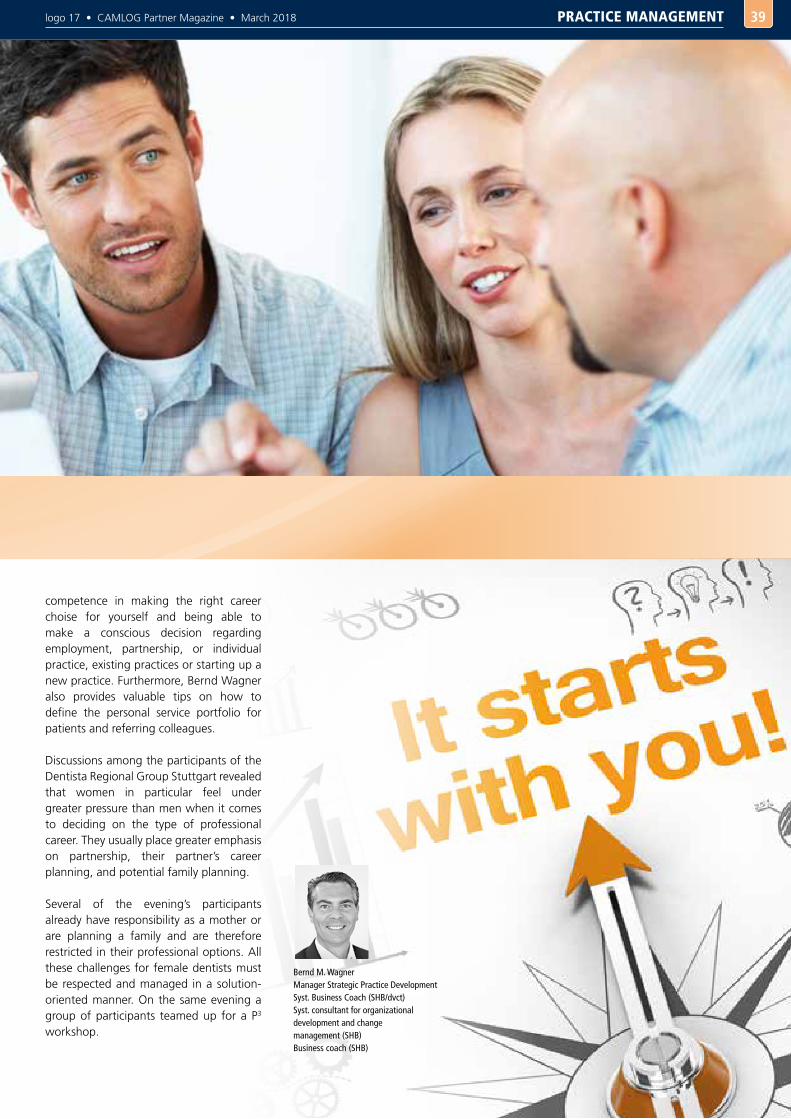
logo 17 • CAMLOG Partner Magazine • March 2018
competence in making the right career choise for yourself and being able to make a conscious decision regarding employment, partnership, or individual practice, existing practices or starting up a new practice. Furthermore, Bernd Wagner also provides valuable tips on how to define the personal service portfolio for patients and referring colleagues.
Discussions among the participants of the Dentista Regional Group Stuttgart revealed that women in particular feel under greater pressure than men when it comes to deciding on the type of professional career. They usually place greater emphasis on partnership, their partner’s career planning, and potential family planning.
Several of the evening’s participants already have responsibility as a mother or are planning a family and are therefore restricted in their professional options. All these challenges for female dentists must be respected and managed in a solution-oriented manner. On the same evening a group of participants teamed up for a P3 workshop.
39PRACTICE MANAGEMENT
Bernd M. WagnerManager Strategic Practice DevelopmentSyst. Business Coach (SHB/dvct) Syst. consultant for organizational development and change management (SHB) Business coach (SHB)

logo 17 • CAMLOG Partner Magazine • March 2018EVENTS40
2. CAMLOG START-UP-DAYS 2018
CAMLOG continues with its successful advanced training concept “Log-in-to-your-future” for young dentists. The Start-up-Days will be held for the second time in Frankfurt am Main on April 13 and 14, 2018. At the inaugural event in 2016, 230 young dentists were enthusiastic about the new advanced training concept. The innovative advanced training format offers support for career orientation and specialization as well as the challenges in volved in boththe start-up phase, take-overs.
Today, young dentists face a number of different issues. Due to changing overall conditions, such as patient needs, competitive pressure, and new types and concepts of care, the requirements for successful practice management are constantly increasing. These days, the owner of a practice is no longer primarily a dentist. As entrepreneurs, they need
to embody a wide variety of different roles. This is something they were not prepared for during their studies and were therefore not given the necessary know-how on topics such as the development of practice strategies, practice marketing, or personnel management. The speakers at the Start-up Days present a wide range of different business concepts, speak about the path they took, and the obstacles they overcame to achieve their current success. A broad spectrum of different successful concepts will be presented at this congress. This includes presentations of models for the owner-managed individual practice, the family-run business, centers and joint practices as well as positions of employment. As there is no such thing as a single success story, the experienced speakers give insights into the most diverse concepts. On the first day of the congress, they will report on their own practice concepts, talk openly about their
experiences and learning curves as well as pitfalls encountered on their road to success. The second day is devoted fully to theoretical and practical workshops. Each participant can choose two favorites in advance from the nine workshops offered on topics such as practice marketing and management, corporate and employee management, and practical courses on surgery and care concepts.The aim of the Start-up Days is to inspire and motivate young dentists for their professional future and careers. With these modules, they will be well equipped for the decision-making process. At the end of the congress, participants should be more able to answer questions such as: Am I the type to have my own practice? Can I manage this on my own or do I need a practice partner? How much responsibility do I want? Is specialization interesting for me – and if so, in which field?
LOG IN TO YOUR FUTURE.
For more information and registration please go to
www.log-in-to-your-future.de
CONGRESS LANGUAGE:
GERMAN13./14. APRIL
2018 FRANKFURT
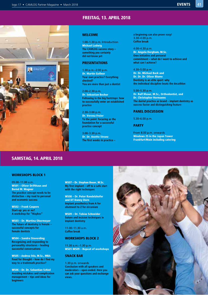
logo 17 • CAMLOG Partner Magazine • March 2018 EVENTS 41
FREITAG, 13. APRIL 2018
SAMSTAG, 14. APRIL 2018
WELCOME
1.00–1.30 p.m. IntroductionMichael LudwigThe CAMLOG success story – something you certainly did not know yet
PRESENTATIONS
1.30 p.m.–2:00 p.m.Dr. Martin GollnerYour own practice? Everything checked? You are more than just a dentist
2.00–2.30 p.m.Dr. Sebastian BecherFollowing in the big footsteps: how to successfully enter an established practice
2.30–3.00 p.m.Dr. Verena FreierTo the point: focusing as the foundation for a successful practice concept
3.00–3.30 p.m.Dr. Dr. Anette StrunzThe first weeks in practice –
a beginning can also prove easy!3.30–4.00 p.m.Coffee break
4.00–4.30 p.m.Dr. Angela Dergham, M.Sc. Own initiative and personal commitment – what do I want to achieve and what can I achieve?
4.30–5.00 p.m.Dr. Dr. Michael Back and Dr. Dr. Dr. Oliver BlumeDentistry is an elite sport: why the individual discipline beats the decathlon
5.00–5.30 p.m.Dr. Ralf Masur, M.Sc., Orthodontist, and Dr. Christopher HermannsThe dental practice as brand – implant dentistry as success factor and distinguishing feature
PANEL DISCUSSION
5.30–6.00 p.m.
PARTY
From 8.00 p.m. onwardsWindows 25 in the Japan Tower Frankfurt/Main including catering
WORKSHOPS BLOCK 1
09.00–11.00 a.m.WS01 – Oliver Drifthaus and Bernd M. WagnerThe practice concept needs to be distinctive – my road to personal and economic success
WS02 – Frank Caspers Start-up: yes or no? A workshop for “Maybes”
WS03 – Dr. Martina Obermeyer The future of dentistry is female – successful concepts for female dentists
WS04 – Sandra Steverding Recognizing and responding to personality structures – leading successful conversations
WS05 – Andrea Stix, M.Sc., MBA Food for thought – how do I find my way to a trademark practice?
WS06 – Dr. Dr. Sebastian Schiel Avoiding mistakes and complication management – tips and ideas for beginners
WS07 – Dr. Stephan Beuer, M.Sc. My first implant – off to a safe start with the right techniques
WS08 – Dr. Peter Randelzhofer and DT Danny DornImplant prosthetics from A for abutment to Z for zirconium WS09 – Dr. Tobias Schneider Suture and incision techniques in implant dentistry
11.00–11.30 a.m.Coffee break
WORKSHOPS BLOCK 2
11.30 a.m.–1.30 p.m. WS01-WS09 – Repeat of workshops
SNACK BAR
1.30 p.m. onwards Conclusion with all speakers and moderators – open ended. Here you can ask your questions and exchange views.

logo 17 • CAMLOG Partner Magazine • March 2018LIFESTYLE42
ROTTERDAM A BUSTLING PORT METROPOLIS WITH HIGH LIFESTYLE POTENTIAL
Those who paid close attention in geography classes will immediately remember that Rotterdam with its more than 600,000 inhabitants is the second largest city in the Netherlands. It is also Europe’s largest seaport, making Rotterdam the third largest seaprot in the world.
Founded in 1230, the city on the estuary of the Rhine is not only a traffic hub and a major industrial center, it is also developing into a cultural and architectural center of great appeal, whose rise to fame is evident in the impressive and continuously growing city skyline.
Architecture to please everyone’s taste
Rotterdam’s starting point for developing into an architectural jewel was the ghostly remnants of bombings from World War II. Unlike other European cities, the Dutch port city took advantage of this opportunity to create a generous and courageous reconstruction. When strolling through this city with an open mind, it becomes obvious that the basic idea was not to build a “car-friendly” metropolis but rather to create a metropolis that embraces culture and people. Probably the most futuristic are the “cube apartments”,
38 apartments in the Blaak district that are tilted by 45 degrees. What are the absolute musts for sightseeing? The Markthal is the perfect place to fortify yourself for city excursions. With more than 100 #stalls, eight restaurants, and 15 grocery stores, this, the largest market hall in the Netherlands, offers such a wealth of culinary offerings that it is advisable not to visit the Markthal too hungry. You otherwise run the risk of overeating because, as we all know, our eyes are often bigger than our stomachs.
A maritime experience at its best
Of course, one should not leave the world’s third largest port without having ventured on a proper harbor tour. The traditional “Spido” boats offer a perfect view of Rotterdam’s architecture with its partly very bold, progressive approaches to design. The Erasmus bridge, referred to by locals as the “Swan,” the imposing
Euromast, and the city’s modern sky-scrapers along the Wilhelminakade are all spectacular examples of the characteristic architecture.
If this little boat ride has given you a taste for more of the sea, you should include a visit to the Maritiem Museum Rotterdam. With more than one million marine objects and all kinds of (inter-)active experiences, the museum provides an excellent insight into the long and eventful history of the Dutch seafarers, including the dreaded “Sea Beggars”, the Dutch pirates who gave the Spaniards so much grief.
Culture – high and “alternative”
The Museum Boijmans van Beuningen showcases both contemporary art and the old masters in a remarkable concept, and the Kunsthal, which has become an icon of contemporary Dutch architecture thanks to its impressive construction, surprising

logo 17 • CAMLOG Partner Magazine • March 2018 LIFESTYLE 43
Jan PetersWriter Kaiseraugst/Switzerland
choice of materials, and innovative design elements, is also well worth a visit.If you are looking for a particularly “bizarre” museum, the Dutch Pinball Museum in Rotterdam springs to mind. Information on where this special cultural site is located is available to pinball wizards from us only on request because this kind of culture still counts as underground…
Chill-out zone(s)
There are more than enough places to relax in multi-cultural Rotterdam. The Witte de Withstraat can be recommended as a center for such activities. This is also home to De Witte Aap – the White Monkey –
which is highly praised by Vogue and even considered by some to be the best bar in the world; and if that’s not good enough for chilling out, then even we at logo are at the end of our wits´end. You’ll just have to try it out for yourself when you attend the Oral Reconstruction Global Symposium in Rotterdam from April 26 to 28.
April 27: Koningsdag
This is the day in Holland when one color and one color alone rules the land of the canals: orange – in honor of the House of Orange-Nassau and the King, the whole country immerses itself in this color. If you want to experience our otherwise
rather rational and well-controlled Dutch neighbors becoming ecstatic, you simply have to join in on April 27 in Rotterdam and toast King Willem-Alexander with a Pilsje.

iSy is the intelligent implant system: lean range, easy to handle and highly efficient. iSy
has a particularly smart solution in store for CEREC® users: scan adapter and scanbody are
simply snapped onto the pre-mounted implant base - no need for a screwdriver. This saves
several work steps and you save time..
Simply snap on, scan and save time - This is iSy. Convince yourself.
CEREC® IS A REGISTERED TRADEMARK OF DENTSPLY SIRONA.
Watch the movie now:
More information at www.isy-implant.com/cerec
iSy_Advert_CEREC_A4_EN.indd 1 08.09.2017 10:43:08
Related Documents