Innate-Like Control of Human iNKT Cell Autoreactivity via the Hypervariable CDR3b Loop Gediminas Matulis 1 , Joseph P. Sanderson 2 , Nikolai M. Lissin 3 , Maria B. Asparuhova 4 , Gopal R. Bommineni 5 , Daniel Schu ¨ mperli 4 , Richard R. Schmidt 5 , Peter M. Villiger 1 , Bent K. Jakobsen 3 , Stephan D. Gadola 1,2 * 1 Center for Experimental Rheumatology, University of Bern, Inselspital, Bern, Switzerland, 2 Division of Infection, Inflammation and Immunity, University of Southampton, School of Medicine, Sir Henry Wellcome and ‘‘Hope’’ Laboratories, United Kingdom, 3 Immunocore Ltd., Abingdon, United Kingdom, 4 Institute of Cell Biology, University of Bern, Bern, Switzerland, 5 Fachbereich Chemie, University of Konstanz, Konstanz, Germany Abstract Invariant Natural Killer T cells (iNKT) are a versatile lymphocyte subset with important roles in both host defense and immunological tolerance. They express a highly conserved TCR which mediates recognition of the non-polymorphic, lipid- binding molecule CD1d. The structure of human iNKT TCRs is unique in that only one of the six complementarity determining region (CDR) loops, CDR3b, is hypervariable. The role of this loop for iNKT biology has been controversial, and it is unresolved whether it contributes to iNKT TCR:CD1d binding or antigen selectivity. On the one hand, the CDR3b loop is dispensable for iNKT TCR binding to CD1d molecules presenting the xenobiotic alpha-galactosylceramide ligand KRN7000, which elicits a strong functional response from mouse and human iNKT cells. However, a role for CDR3b in the recognition of CD1d molecules presenting less potent ligands, such as self-lipids, is suggested by the clonal distribution of iNKT autoreactivity. We demonstrate that the human iNKT repertoire comprises subsets of greatly differing TCR affinity to CD1d, and that these differences relate to their autoreactive functions. These functionally different iNKT subsets segregate in their ability to bind CD1d-tetramers loaded with the partial agonist a-linked glycolipid antigen OCH and structurally different endogenous b-glycosylceramides. Using surface plasmon resonance with recombinant iNKT TCRs and different ligand-CD1d complexes, we demonstrate that the CDR3b sequence strongly impacts on the iNKT TCR affinity to CD1d, independent of the loaded CD1d ligand. Collectively our data reveal a crucial role for CDR3b for the function of human iNKT cells by tuning the overall affinity of the iNKT TCR to CD1d. This mechanism is relatively independent of the bound CD1d ligand and thus forms the basis of an inherent, CDR3b dependent functional hierarchy of human iNKT cells. Citation: Matulis G, Sanderson JP, Lissin NM, Asparuhova MB, Bommineni GR, et al. (2010) Innate-Like Control of Human iNKT Cell Autoreactivity via the Hypervariable CDR3b Loop. PLoS Biol 8(6): e1000402. doi:10.1371/journal.pbio.1000402 Academic Editor: Hidde L. Ploegh, Whitehead Institute, United States of America Received December 21, 2009; Accepted May 13, 2010; Published June 22, 2010 Copyright: ß 2010 Matulis et al. This is an open-access article distributed under the terms of the Creative Commons Attribution License, which permits unrestricted use, distribution, and reproduction in any medium, provided the original author and source are credited. Funding: SDG and JPS are funded by the Higher Education Funding Council for England (HEFCE). GM is a recipient of a Novartis Research Foundation student grant. This work was funded by the Max Cloetta Foundation, the Swiss National Science Foundation (3200-068070, 3200-069338), the Swiss Multiple Sclerosis Foundation, Kamillo Eisner Foundation and Kurt und Senta Herrmann Stiftung. The funders had no role in study design, data collection and analysis, decision to publish, or preparation of the manuscript. Competing Interests: The authors have declared that no competing interests exist. Abbreviations: bGC, b-glucosylceramide; CDR, complementarity determining region; iNKT, Invariant Natural Killer T-lymphocytes; K7, KRN7000 a- galactosylceramide; LacCer, Lactosylceramide; MFI, mean fluorescent intensity; PBMC, peripheral blood mononuclear cell * E-mail: [email protected] Introduction Invariant Natural Killer T (iNKT) cells are a conserved subset of highly potent and versatile T-cells which specifically recognize the non-polymorphic lipid-presenting molecule CD1d [UniprotKB P15813] [1]. iNKT cells co-express a unique T-Cell Receptor (iNKT TCR), which mediates recognition of CD1d, and the pan- NK receptor NKR-P1A (CD161). Human and mouse iNKT TCRs feature a homologous invariant TCRa chain, i.e. Va24-Ja18 in humans and Va14-Ja18 in mice. In addition, all human iNKT TCRs make use of a single TCR Vb family, Vb11, whereas mouse iNKT TCRs utilize several different TCR Vb families. The current paradox of iNKT biology lies in the fact that, despite their apparent innate-like simplicity, they can exert directly conflicting functions. On the one hand, several in vivo studies have demonstrated an essential role for iNKT cells in the induction and maintenance of immunological tolerance [2,3]. Consistent with this, iNKT cells exert a protective role in animal models of spontaneous autoimmunity [4,5], and numerical and functional defects of iNKT cells are observed in different human autoim- mune diseases [6]. In contrast to these tolerogenic functions, iNKT cells can exert potent cytotoxic functions and contribute to host defense against tumors and various infectious pathogens [7,8,9]. Whether different subsets of iNKTs are involved in these opposed roles or whether individual iNKT clones fulfill both of these functions under different conditions is unknown. Several mechanisms underpin iNKT activation during host defense, such as TLR [10,11,12] and PPAR-c activation [13], co-stimulatory molecule signaling [14], and inflammatory cytokines [15,16]. However, it is unknown how iNKT cells are induced to mediate their tolerogenic functions under non-inflammatory conditions. PLoS Biology | www.plosbiology.org 1 June 2010 | Volume 8 | Issue 6 | e1000402

Welcome message from author
This document is posted to help you gain knowledge. Please leave a comment to let me know what you think about it! Share it to your friends and learn new things together.
Transcript

Innate-Like Control of Human iNKT Cell Autoreactivityvia the Hypervariable CDR3b LoopGediminas Matulis1, Joseph P. Sanderson2, Nikolai M. Lissin3, Maria B. Asparuhova4, Gopal R.
Bommineni5, Daniel Schumperli4, Richard R. Schmidt5, Peter M. Villiger1, Bent K. Jakobsen3, Stephan D.
Gadola1,2*
1 Center for Experimental Rheumatology, University of Bern, Inselspital, Bern, Switzerland, 2 Division of Infection, Inflammation and Immunity, University of Southampton,
School of Medicine, Sir Henry Wellcome and ‘‘Hope’’ Laboratories, United Kingdom, 3 Immunocore Ltd., Abingdon, United Kingdom, 4 Institute of Cell Biology, University
of Bern, Bern, Switzerland, 5 Fachbereich Chemie, University of Konstanz, Konstanz, Germany
Abstract
Invariant Natural Killer T cells (iNKT) are a versatile lymphocyte subset with important roles in both host defense andimmunological tolerance. They express a highly conserved TCR which mediates recognition of the non-polymorphic, lipid-binding molecule CD1d. The structure of human iNKT TCRs is unique in that only one of the six complementaritydetermining region (CDR) loops, CDR3b, is hypervariable. The role of this loop for iNKT biology has been controversial, and itis unresolved whether it contributes to iNKT TCR:CD1d binding or antigen selectivity. On the one hand, the CDR3b loop isdispensable for iNKT TCR binding to CD1d molecules presenting the xenobiotic alpha-galactosylceramide ligand KRN7000,which elicits a strong functional response from mouse and human iNKT cells. However, a role for CDR3b in the recognitionof CD1d molecules presenting less potent ligands, such as self-lipids, is suggested by the clonal distribution of iNKTautoreactivity. We demonstrate that the human iNKT repertoire comprises subsets of greatly differing TCR affinity to CD1d,and that these differences relate to their autoreactive functions. These functionally different iNKT subsets segregate in theirability to bind CD1d-tetramers loaded with the partial agonist a-linked glycolipid antigen OCH and structurally differentendogenous b-glycosylceramides. Using surface plasmon resonance with recombinant iNKT TCRs and different ligand-CD1dcomplexes, we demonstrate that the CDR3b sequence strongly impacts on the iNKT TCR affinity to CD1d, independent ofthe loaded CD1d ligand. Collectively our data reveal a crucial role for CDR3b for the function of human iNKT cells by tuningthe overall affinity of the iNKT TCR to CD1d. This mechanism is relatively independent of the bound CD1d ligand and thusforms the basis of an inherent, CDR3b dependent functional hierarchy of human iNKT cells.
Citation: Matulis G, Sanderson JP, Lissin NM, Asparuhova MB, Bommineni GR, et al. (2010) Innate-Like Control of Human iNKT Cell Autoreactivity via theHypervariable CDR3b Loop. PLoS Biol 8(6): e1000402. doi:10.1371/journal.pbio.1000402
Academic Editor: Hidde L. Ploegh, Whitehead Institute, United States of America
Received December 21, 2009; Accepted May 13, 2010; Published June 22, 2010
Copyright: � 2010 Matulis et al. This is an open-access article distributed under the terms of the Creative Commons Attribution License, which permitsunrestricted use, distribution, and reproduction in any medium, provided the original author and source are credited.
Funding: SDG and JPS are funded by the Higher Education Funding Council for England (HEFCE). GM is a recipient of a Novartis Research Foundation studentgrant. This work was funded by the Max Cloetta Foundation, the Swiss National Science Foundation (3200-068070, 3200-069338), the Swiss Multiple SclerosisFoundation, Kamillo Eisner Foundation and Kurt und Senta Herrmann Stiftung. The funders had no role in study design, data collection and analysis, decision topublish, or preparation of the manuscript.
Competing Interests: The authors have declared that no competing interests exist.
Abbreviations: bGC, b-glucosylceramide; CDR, complementarity determining region; iNKT, Invariant Natural Killer T-lymphocytes; K7, KRN7000 a-galactosylceramide; LacCer, Lactosylceramide; MFI, mean fluorescent intensity; PBMC, peripheral blood mononuclear cell
* E-mail: [email protected]
Introduction
Invariant Natural Killer T (iNKT) cells are a conserved subset of
highly potent and versatile T-cells which specifically recognize the
non-polymorphic lipid-presenting molecule CD1d [UniprotKB
P15813] [1]. iNKT cells co-express a unique T-Cell Receptor
(iNKT TCR), which mediates recognition of CD1d, and the pan-
NK receptor NKR-P1A (CD161). Human and mouse iNKT TCRs
feature a homologous invariant TCRa chain, i.e. Va24-Ja18 in
humans and Va14-Ja18 in mice. In addition, all human iNKT
TCRs make use of a single TCR Vb family, Vb11, whereas mouse
iNKT TCRs utilize several different TCR Vb families.
The current paradox of iNKT biology lies in the fact that,
despite their apparent innate-like simplicity, they can exert directly
conflicting functions. On the one hand, several in vivo studies have
demonstrated an essential role for iNKT cells in the induction and
maintenance of immunological tolerance [2,3]. Consistent with
this, iNKT cells exert a protective role in animal models of
spontaneous autoimmunity [4,5], and numerical and functional
defects of iNKT cells are observed in different human autoim-
mune diseases [6].
In contrast to these tolerogenic functions, iNKT cells can exert
potent cytotoxic functions and contribute to host defense against
tumors and various infectious pathogens [7,8,9]. Whether different
subsets of iNKTs are involved in these opposed roles or whether
individual iNKT clones fulfill both of these functions under
different conditions is unknown. Several mechanisms underpin
iNKT activation during host defense, such as TLR [10,11,12] and
PPAR-c activation [13], co-stimulatory molecule signaling [14],
and inflammatory cytokines [15,16]. However, it is unknown how
iNKT cells are induced to mediate their tolerogenic functions
under non-inflammatory conditions.
PLoS Biology | www.plosbiology.org 1 June 2010 | Volume 8 | Issue 6 | e1000402

Some iNKT clones exhibit substantial activation in response to
CD1d-expressing antigen-presenting cells in the absence of
exogenous antigens. This autoreactive function is essential for
both iNKT selection [17] and tolerogenic activity [18]. While
iNKT TCR binding to CD1d is absolutely required [19], the
mechanistic basis of iNKT cell autoreactivity is largely unresolved.
In particular, the importance of specific CD1d-presented endog-
enous lipid antigens for the autoreactive interaction of the iNKT
TCR with CD1d is contentious.
Studies in mice have suggested that the iNKT repertoire
displays clonal heterogeneity with regard to recognition of weaker
stimulatory lipid antigens, such as the a-galactosylceramide
analogue OCH. These differences can be explained by the
differential Vb family usage in mouse iNKT TCRs [20,21,22].
However, human iNKT TCRs use a single Vb family and so the
short hypervariable complementarity determining region
(CDR3b) loop in human iNKT TCRs is their only truly adaptive
element. It is not known whether this is sufficient to endow the
human iNKT TCR with meaningful ability to discriminate a
diverse range of human CD1d-presented antigens.
Here we examined a large panel of human iNKT cell lines and
clones for their binding to different CD1d-ligand tetramers and related
this both to the affinity of their TCRs to different CD1d-ligand
complexes and to their functional recognition of diverse antigens. The
results presented here demonstrate that variations in the CDR3b loop
have a profound, antigen-independent, impact on the iNKT TCR’s
affinity to CD1d and on iNKT cell autoreactive function.
Results
OCH-CD1d Tetramers Reveal Broad Heterogeneity of K7-CD1d Tetramer Positive Human iNKT Cells
Previous studies have shown that the CDR3b loop is
dispensable for the ability of human iNKT cells to strongly react
to the a-galactosylceramide antigen KRN7000 (K7), a xenobiotic
glycolipid which can be presented to iNKT cells by CD1d. In fact,
K7-CD1d tetramer staining does not allow discrimination of
different human iNKT cell subsets by flow cytometry. We
hypothesized that CD1d-tetramers loaded with weaker antigens
might be better able to reveal the existence of CDR3b-dependent
variation among human iNKT cells.
Therefore, we first examined whether different human iNKT
subsets could be segregated by their binding to CD1d tetramers
that were loaded with the synthetic iNKT partial agonist antigen
OCH. For this purpose, polyclonal iNKT lines, generated from
healthy donors by in vitro stimulation with K7, were tested for
their binding to both K7- and OCH-CD1d tetramers. In all of
these lines, K7-CD1d tetramers stained a single, clearly distinct,
homogeneous, and strongly fluorescent population of iNKT
lymphocytes (Figure 1A). In contrast, staining of the same lines
with OCH-CD1d tetramers revealed a considerable degree of
variation in fluorescence, suggesting the presence of distinct iNKT
subpopulations (Figure 1A). Importantly, similar qualitative
differences between K7- and OCH-CD1d tetramer staining of
iNKT cells could also be observed ex vivo (Figure 1B), indicating
that these differences were not due to an artifact of previous in
vitro stimulation with K7. In order to examine whether the
broadly heterogeneous OCH-CD1d tetramer staining of human
iNKT cells resulted from stable clonal variation or from transient
changes in TCR expression levels, we generated a large panel of
‘‘K7/OCH-naıve’’ human iNKT cell clones and lines. For this
purpose, Va24+/Vb11+ T cells were directly sorted ex vivo from
healthy human donors and expanded using the non-specific T cell
mitogen phytohaemagglutinin. Ninety-seven different human
Va24+/Vb11+ T cell lines and 256 Va24+/Vb11+ T cell clones
Figure 1. Distinct iNKT cell subpopulations revealed by OCH-CD1d tetramer staining. OCH- and K7-CD1d tetramer stainings of (A) arepresentative K7-stimulated human iNKT line after 14 d in vitro cultureand (B) a healthy human volunteer’s PBMC ex vivo are shown. While K7-CD1d tetramer staining identifies a single homogeneous population ofiNKT cells (upper row), OCH-CD1d tetramer staining reveals the presenceof different distinct iNKT populations within these samples (lower row).doi:10.1371/journal.pbio.1000402.g001
Author Summary
Our immune system uses randomly modified T-cellreceptors (TCRs) to adapt its discriminative capacity torapidly changing pathogens. The T-cell receptor (TCR) hassix flexible, variable peptide loops that make contact withantigens presented to them on the surface of other cells.Invariant Natural Killer T-cells (iNKT) are regulatory T-cellswith a unique type of TCR (iNKT-TCR) that recognizes lipidantigens presented by specific MHC-like molecules knownas CD1d. In human iNKT-TCRs, only one of the six loops,CDR3beta, is variable. By comparing how different humaniNKT clones bind and react to different CD1d-lipidcomplexes we uncover the existence of a hierarchical orderof the human iNKT cell repertoire in which strongly CD1d-binding clones are autoreactive while weak CD1d-bindingclones are non-autoreactive. Direct measurements of iNKT-TCR binding to CD1d using surface plasmon resonancerecapitulated this hierarchy at the protein level. The datashow that variation in the CDR3beta loop conveys dramaticdifferences in human iNKT TCR affinity that are independentof the CD1d bound ligand. Thus the CDR3beta loopprovides the structural basis for the functional hierarchyof the human iNKT repertoire. We postulate that during thelife-course, CDR3beta-dependent asymmetrical activationof different human iNKT clones leads to a bias in the iNKTrepertoire, and this could result in age-dependent defects ofiNKT-mediated immune regulation in later life.
CDR3b Controls CD1d Recognition by Human iNKT
PLoS Biology | www.plosbiology.org 2 June 2010 | Volume 8 | Issue 6 | e1000402

from 13 different healthy donors were established and analyzed by
flow cytometry with K7- and OCH-CD1d tetramers.
All Va24+/Vb11+ T-cell clones and lines showed bright,
homogeneous staining with K7-tetramers (Figure 2), thereby
confirming them as iNKT cells. Individual iNKT clones showed
modest variation, up to 6-fold, in K7-CD1d tetramer mean
fluorescence intensity (MFI). In contrast, multiple iNKT cell
subpopulations with differing fluorescence intensities were re-
vealed by OCH-CD1d tetramer staining in 31 of the 97 iNKT
lines (Figure 2A), thereby mirroring the above described findings
in K7 stimulated iNKT lines. As expected, all 256 iNKT clones
stained homogeneously with OCH-CD1d tetramers. However,
substantial differences, up to 200-fold, in OCH-CD1d tetramer
MFI were observed between individual clones (Figure 2B). Based
on the observed large differences in OCH-CD1d tetramer MFI,
the 256 human iNKT clones were categorized as OCHHIGH
(MFI.300; n = 41), OCHINT (MFI.50 and ,300; n = 164), or
OCHLOW (MFI,50; n = 51).
Importantly, the differences in OCH-CD1d tetramer staining
could not be explained by differences in either TCR or CD4 co-
receptor expression. Whereas K7-CD1d tetramer binding signif-
icantly correlated with surface expression levels of the Va24 and
Vb11 TCR chains, no such association was observed for OCH-
CD1d tetramer staining (Figure 2C). Furthermore, CD4 co-
receptor usage was not related to the intensity of the iNKT clones’
OCH or K7-CD1d tetramer staining (unpublished results).
The results of these experiments revealed that the human iNKT
repertoire is broadly heterogeneous with regard to the ability of
individual clones to bind OCH-CD1d tetramers, independent of
either CD4 co-receptor or TCR expression levels.
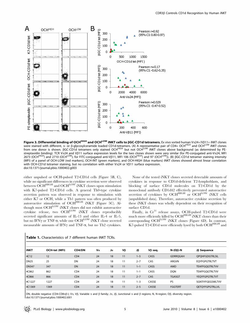
Human OCHHIGH and OCHLOW iNKT Cells ExhibitDifferential Binding to CD1d Molecules Presentingb-Glycosylceramide
The above results indicated that clonally distributed qualitative
differences in iNKT TCRs were responsible for the considerable
variation in OCH-CD1d tetramer binding. However, differences in
iNKT TCR mediated recognition of an unnatural compound like
OCH would be physiologically irrelevant if they simply reflected
random differences in OCH-specific antigen selectivity. To explore
this possibility, 18 iNKT clones of broadly varying OCH-CD1d
MFI were tested for their ability to bind CD1d tetramers loaded
with the common mammalian glycolipid b-glycosylceramide
(bGC). These 18 iNKT clones displayed significant variation, up
to 50-fold, in bGC-CD1d tetramer staining (Figure 3A). Impor-
tantly, a strong association was evident between OCH-CD1d
tetramer staining and bGC-CD1d tetramer staining, while no
correlation was seen between bGC-CD1d tetramer staining and
Va24 TCR chain surface expression (Figure 3B). These results
demonstrated that the observed broad variation in OCH-CD1d
tetramer binding between individual human iNKT clones was not
simply due to their antigen selectivity but was a reflection of a
general variability in human iNKT TCR binding to CD1d loaded
with weak antigenic lipids. Furthermore, they indicated that OCH-
CD1d tetramer binding can act as a surrogate marker for human
iNKT cell recognition of endogenous CD1d antigens.
The Hypervariable CDR3b Loop Has a Strong Effect onthe Affinity of Human iNKT TCRs to CD1d PresentingEither a- or b-Anomeric Glycolipids
Based on the above results we hypothesized that the observed
substantial differences in tetramer staining between OCHHIGH
and OCHLOW iNKT clones resulted from significant variations in
TCR:CD1d binding affinity. As expected, sequencing of the TCR
Va24 and Vb11 chains demonstrated the usage of the known
invariant Va24-Ja18 rearrangement in all clones, while Vb11 in
these clones was rearranged with several different Jb families,
resulting in highly variable CDR3b sequences. This indicated that,
in human iNKT TCRs, structural differences of the CDR3b loop
have a substantial impact on iNKT TCR binding to CD1d. To
test this in a cell-free system we cloned the extracellular domains of
the TCR Vb11 chains from a panel of seven OCHHIGH and
OCHLOW iNKT cell clones (Table 1), as well as the invariant
TCR Va24 chain from one iNKT clone, and used them to
generate soluble Va24/Vb11 iNKT TCRs. Binding of these
recombinant iNKT TCRs to K7-, OCH-, as well as bGC- and
lactosylceramide (LacCer-) loaded recombinant human CD1d
complexes was measured using surface plasmon resonance
(Figure 4A; Table 2).
The results of these experiments showed a striking variation, up
to 40-fold, between the different iNKT TCRs in their binding
affinity (KD) to a given ligand-CD1d complex (for K7-CD1d, KD:
0.24–3.67 mM; for OCH-CD1d, KD: 2.17–38.3 mM; for bGC-
CD1d, KD: 2.17–85 mM; for LacCer-CD1d, KD: 2.1–54 mM; see
Table 2). These findings clearly showed that the CDR3b loop of
human iNKT TCRs can strongly impact on their binding to
ligand-CD1d complexes.
Importantly, the binding affinities of all seven recombinant
iNKT TCRs to OCH-CD1d strongly correlated with the OCH-
CD1d tetramer staining (MFI) of their corresponding original
iNKT clones (Figure 4B). Moreover, the binding affinity of a given
iNKT TCR to OCH-CD1d also correlated closely with its affinity
to either bGC- or K7-CD1d (Figure 4C). Therefore, the wide
variation in affinity between our seven human iNKT TCRs
contrasted to the lack of variation in antigen selectivity. In other
words, the CDR3b loop of human iNKT TCRs modulated the
overall binding affinity to different human ligand-CD1d complex-
es irrespective of the bound ligand.
Based on these findings we hypothesized that the TCRs of
OCHHIGH iNKT clones could also mediate enhanced functional
recognition of endogenous ligand-CD1d complexes. We tested this
hypothesis by comparing autoreactive responses of OCHHIGH and
OCHLOW iNKT clones to CD1d-expressing antigen-presenting cells.
Autoreactive Functions of Human iNKT Cells Correlatewith Their OCH-CD1d Binding
We directly compared the extent of proliferation, cytokine
secretion, and cytotoxicity of human OCHHIGH and OCHLOW
iNKT cells in response to CD1d expressing human cell lines
presenting either endogenous or specific exogenous (‘‘pulsed’’)
glycolipids. Because functional responses of iNKT cells might
change during long term in vitro culture, we compared different
donor-matched pairs of OCHHIGH and OCHLOW iNKT cell
clones with identical in vitro history, i.e. each pair was sorted from
a given donor 3 wk prior to the experiment and kept under
identical cell culture conditions until the day of the experiment.
The selected clones were all CD4+ and were additionally matched
for TCR expression levels. For all pairs, OCHHIGH iNKT clones
exhibited significantly greater proliferation than OCHLOW iNKT
clones in response to either unpulsed or OCH-pulsed T2-CD1d
lymphoblasts. In contrast, when T2-CD1d were pulsed with the
strong agonist ligand K7, both OCHHIGH and OCHLOW iNKT
clones proliferated vigorously, and to similar extent (Figure 5A).
Next, we measured CD1d-dependent secretion of a panel of
cytokines by OCHHIGH and OCHLOW iNKT clones. The
OCHHIGH iNKT clones secreted considerably greater quantities
of cytokines than their OCHLOW counterparts in response to
CDR3b Controls CD1d Recognition by Human iNKT
PLoS Biology | www.plosbiology.org 3 June 2010 | Volume 8 | Issue 6 | e1000402
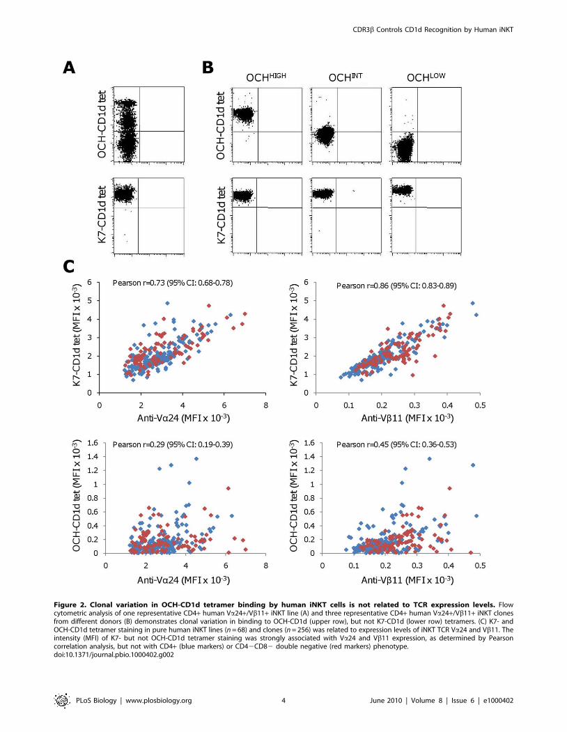
Figure 2. Clonal variation in OCH-CD1d tetramer binding by human iNKT cells is not related to TCR expression levels. Flowcytometric analysis of one representative CD4+ human Va24+/Vb11+ iNKT line (A) and three representative CD4+ human Va24+/Vb11+ iNKT clonesfrom different donors (B) demonstrates clonal variation in binding to OCH-CD1d (upper row), but not K7-CD1d (lower row) tetramers. (C) K7- andOCH-CD1d tetramer staining in pure human iNKT lines (n = 68) and clones (n = 256) was related to expression levels of iNKT TCR Va24 and Vb11. Theintensity (MFI) of K7- but not OCH-CD1d tetramer staining was strongly associated with Va24 and Vb11 expression, as determined by Pearsoncorrelation analysis, but not with CD4+ (blue markers) or CD42CD82 double negative (red markers) phenotype.doi:10.1371/journal.pbio.1000402.g002
CDR3b Controls CD1d Recognition by Human iNKT
PLoS Biology | www.plosbiology.org 4 June 2010 | Volume 8 | Issue 6 | e1000402
either unpulsed or OCH-pulsed T2-CD1d cells (Figure 5B, C),
while no significant differences in cytokine secretion were observed
between OCHHIGH and OCHLOW iNKT clones upon stimulation
with K7-pulsed T2-CD1d cells. A general Th0-type cytokine
secretion pattern was observed in response to stimulation with
either K7 or OCH, while a Th1 pattern was often produced by
autoreactive stimulation of OCHHIGH iNKT (Figure 5C). Al-
though most OCHLOW iNKT clones did not exhibit autoreactive
cytokine release, two OCHLOW iNKT clones reproducibly
secreted significant amounts of IL-13 and either IL-4 or IL-5,
but no IFNc or TNF-a, while one OCHLOW iNKT clone secreted
measurable amounts of IFNc and TNF-a, but no Th2 cytokines.
None of the tested iNKT clones secreted detectable amounts of
cytokines in response to CD1d-deficient T2-lymphoblasts, and
blocking of surface CD1d molecules on T2-CD1d by the
monoclonal antibody CD1d42 effectively prevented autoreactive
secretion of cytokines by OCHHIGH or OCHLOW iNKT cells
(unpublished data). Therefore, autoreactive cytokine secretion by
these iNKT clones was wholly dependent on their recognition of
surface CD1d.
Finally, in Cr51 release assays, OCH-pulsed T2-CD1d were
much more efficiently killed by OCHHIGH iNKT clones than their
corresponding OCHLOW iNKT clones (Figure 6D). In contrast,
K7-pulsed T2-CD1d were efficiently lysed by both OCHHIGH and
Figure 3. Differential binding of OCHHIGH and OCHLOW iNKT cells to bGC-CD1d tetramers. Ex vivo sorted human Va24+/Vb11+ iNKT cloneswere stained with different, a- or b-glycosylceramide loaded CD1d-tetramers. (A) A representative pair of CD4+ OCHHIGH and OCHLOW iNKT clonesfrom one donor is shown. bGC-CD1d tetramers only stained OCHHIGH but not OCHLOW iNKT clones above background (as determined by PE-streptavidin binding). TCR Va24 and Vb11 surface expression levels for the two clones shown were very similar (for PE-conjugated anti-Va24, MFI2673 (OCHHIGH) and 2710 (OCHLOW); for FITC-conjugated anti-Vb11, MFI 106 (OCHHIGH) and 97 (OCHLOW)). (B) bGC-CD1d tetramer staining intensity(MFI) of a panel of OCH-LOW (red markers), OCH-INT (green markers), and OCH-HIGH (blue markers) iNKT clones showed almost linear correlationwith OCH-CD1d tetramer staining, but no correlation with either Va24 or Vb11 surface expression.doi:10.1371/journal.pbio.1000402.g003
Table 1. Characteristics of 7 different human iNKT TCRs.
iNKT OCH-tet (MFI) CD4/DN Va Ja Vb Jb Vb seq. N-(Db)-N Jb Sequence
4C12 12 CD4 24 18 11 1–5 CASS GDRRQGAH QPQHFGDGTRLSIL
DN25 25 DN 24 18 11 2–7 CAS ARGVN EQYFGPGTRLTVT
DN347 347 DN 24 18 11 1–1 CASS AMD TEAFFGQGTRLTVV
4C862 862 CD4 24 18 11 1–1 CASS DQN TEAFFGQGTRLTVV
4C866 866 CD4 24 18 11 2–7 CAS TGASGT YEQYFGPGTRLTVT
4C1227 1227 CD4 24 18 11 1–3 CASSE PS SGNTIYFGEGSWLTVV
4C1369 1369 CD4 24 18 11 2–5 CASSE FGGTERT QETQYFGPGTRLLVL
DN, double negative (CD4-CD8ab-); Va, Vb, Variable a and b family; Ja, Jb, Junctional a and b regions; N, N-region; Db, diversity region.doi:10.1371/journal.pbio.1000402.t001
CDR3b Controls CD1d Recognition by Human iNKT
PLoS Biology | www.plosbiology.org 5 June 2010 | Volume 8 | Issue 6 | e1000402

Figure 4. The CDR3b loop strongly impacts on human iNKT TCR affinity to CD1d, independent of the CD1d-bound ligand. (A) Bindingof two recombinant human iNKT TCRs, one OCHHIGH (4C1369) and one OCHLOW (4C12), to K7-, OCH-, bGC-, and LacCer-CD1d at equilibrium is shown(see also panel C and Table 2). (B) The affinity of the seven recombinant iNKT TCRs to OCH-CD1d, as determined by SPR, was linearly related to thestaining intensity (MFI) of the original iNKT clone with OCH-CD1d tetramers. (C) The seven recombinant human iNKT TCRs followed a strict hierarchyof binding to ligand-CD1d complex, which was not affected by the specific CD1d-bound ligand. These iNKT TCRs differed only with regard to theirCDR3beta sequence (Table 1).doi:10.1371/journal.pbio.1000402.g004
Table 2. Binding of 7 human iNKT TCRs to different CD1d/ligand complexes.
iNKT K7-CD1d OCH-CD1d bGC-CD1d
KD (mM) TK (sec) KD (mM) TK (sec) KD (mM) TK (sec)
4C12 3.6760.85 0.9960.03 38.3161.50 0.7160.03 85.0165.96 ND
DN25 3.3460.17 0.9260.03 37.2761.62 ND 42.4361.88 ND
DN347 1.9960.17 1.0160.06 16.6460.65 0.5960.01 34.5062.74 ND
4C862 1.7560.13 1.0260.04 14.8060.59 0.6360.05 29.6563.69 ND
4C866 0.6260.03 2.2460.05 5.8260.37 1.1360.13 6.2660.91 1.4260.16
4C1227 0.6660.06 2.5560.08 7.4560.59 1.1060.07 9.1961.61 ND
4C1369 0.2460.01 12.3860.36 2.1760.16 4.7860.55 2.1760.13 4.3660.73
KD, dissociation constant; TK, dissociation half-time; ND, not determined. All values given 6 standard deviation.doi:10.1371/journal.pbio.1000402.t002
CDR3b Controls CD1d Recognition by Human iNKT
PLoS Biology | www.plosbiology.org 6 June 2010 | Volume 8 | Issue 6 | e1000402

Figure 5. Differential autoreactive functional responses by human OCHHIGH and OCHLOW iNKT clones. Matched pairs of human OCHHIGH
(red columns and markers) and OCHLOW (blue columns and markers) iNKT clones were compared for their ability to proliferate, secrete cytokines, and
CDR3b Controls CD1d Recognition by Human iNKT
PLoS Biology | www.plosbiology.org 7 June 2010 | Volume 8 | Issue 6 | e1000402

OCHLOW iNKT clones, whereas neither OCHHIGH nor
OCHLOW iNKT clones showed relevant cytotoxicity towards
unpulsed T2-CD1d lymphoblasts.
Together, these results demonstrated that OCH-CD1d tetramer
staining allows for identification of distinct human OCHHIGH and
OCHLOW iNKT clones, which exhibit differential functional
ability to respond to endogenous ligand-CD1d complexes. The
above results indicated that the autoreactive potential of human
iNKT clones is governed by the affinity of their iNKT TCR to
CD1d, and therefore the structure of their CDR3b loop.
TCRs from OCHHIGH but not OCHLOW Human iNKTSubsets Bind to Endogenous CD1d-Ligand Complexes
In order to test our hypothesis that OCHHIGH and OCHLOW
iNKT TCRs differed in their binding to endogenous ligand-CD1d
complexes, we generated soluble fluorescent iNKT TCR-tetra-
mers derived from an autoreactive OCHHIGH iNKT clone and a
non-autoreactive OCHLOW iNKT clone. As shown in Figure 6,
both iNKT TCR tetramers bound well to K7-pulsed T2-CD1d. In
contrast, only the OCHHIGH-derived iNKT TCR tetramer was
able to effectively stain unpulsed T2-CD1d. These results further
substantiated our hypothesis that autoreactive recognition of
CD1d by human iNKT cells is primarily determined by the
structure of their iNKT TCRs’ CDR3b loop.
All together, these studies demonstrated that the human iNKT
cell repertoire exhibits considerable clonally distributed CDR3b-
dependent differences in overall TCR affinity to CD1d, irrespec-
tive of the bound ligand, and that these inherent structural
differences control iNKT autoreactive activation.
Discussion
iNKT cells are a conserved subset of highly potent regulatory T
cells at the innate-adaptive interface. The hallmark of human
iNKT cells is their unique TCR, which is composed of an
invariant TCR Va24-Ja18 alpha chain and a semi-invariant TCR
Vb11 chain. The only variable, and therefore potentially adaptive,
element in human iNKT TCRs is their hypervariable CDR3bloop. The results of the present study demonstrate for the first
time, to our knowledge, that the structure of the hypervariable
CDR3b loop in human iNKT TCRs exerts a strong impact on
CD1d binding and is a key determinant of iNKT cell
autoreactivity. The magnitude of the effect of CDR3b variations
on human iNKT TCR:CD1d binding observed here was
unexpected as previous studies with mouse iNKT TCRs have
reported only minor effects of CDR3b mutations on CD1d
binding. Furthermore, they strongly suggest that CDR3b loops in
autoreactive iNKT TCRs make functionally important direct
protein-protein contacts with human CD1d, rather than contacts
with CD1d-bound ligands, thereby affecting overall affinity rather
than antigen specificity.
The role of the hypervariable CDR3b loop in human iNKT
TCRs is currently unresolved. It is dispensable for binding to
CD1d molecules that are loaded with the strong agonist ligand K7,
and hence K7-CD1d tetramers do not support subset differenti-
ation of human iNKT cells. Consistent with this, the recently
solved structures of one human and two mouse iNKT TCR:K7-
CD1d co-crystals have found no relevant contacts between
CDR3b and the K7-CD1d complex [20,23]. In contrast, recent
mutagenesis studies have indicated that the CDR3b loop of mouse
iNKT TCRs may exert some impact on the affinity to CD1d,
particularly when CD1d was loaded with weaker antigens
[24,25,26].
We found that human iNKT cells were surprisingly heteroge-
neous in their binding to CD1d tetramers loaded with the partial
agonist ligand OCH, which is a synthetic analogue of K7. Up to
200-fold differences in OCH-CD1d tetramer staining were
observed between individual iNKT clones, independent of
variations in TCR expression. The same clones exhibited only
modest differences in K7-CD1d tetramer staining, which could
largely be explained simply by variations in TCR expression.
Importantly, we found that the clonal variation in OCH-CD1d
tetramer binding was directly related to OCH-CD1d dependent
exhibit cytotoxicity in response to lipid-pulsed or endogenous lipid presenting CD1d-positive antigen presenting cells. (A) Proliferation of threerepresentative pairs of OCHHIGH and OCHLOW iNKT clones from different healthy donors in response to K7-, OCH-, or vehicle-pulsed human CD1d-expressing T2 cells (T2-CD1d) or to K7-pulsed CD1d negative T2 cells (T2-) is shown. OCHHIGH clones consistently displayed greater proliferation thanOCHLOW clones in response to OCH or vehicle pulsed T2-CD1d. cpm, counts per minute. Mean values 6 s.e.m. are shown. (B) Cytokine secretionprofiles of a representative pair of matched OCHHIGH and OCHLOW iNKT clones in response to the strong agonist ligand K7 and the partial agonistligand OCH, presented by T2-CD1d, are shown. OCHHIGH iNKT clones exhibited much stronger cytokine secretion than OCHLOW iNKT cells in responseto OCH-pulsed T2-CD1d, while cytokine secretion was similar for both in response to K7-pulsed T2-CD1d. (C) Autoreactive cytokine release inresponse to T2-CD1d in the absence of added exogenous ligands is shown for four matched pairs of OCHHIGH and OCHLOW iNKT clones. OCHHIGH butnot OCHLOW iNKT clones consistently exhibited substantial autoreactive cytokine secretion. (D) Specific lysis of K7- (filled markers) and OCH- (unfilledmarkers) pulsed T2-CD1d targets is shown for three matched pairs of OCHHIGH and OCHLOW iNKT clones from different donors.doi:10.1371/journal.pbio.1000402.g005
Figure 6. Differential binding of OCHHIGH and OCHLOW iNKTclone derived TCR tetramers to endogenous lipid presentingCD1d molecules. PE-conjugated recombinant iNKT TCR tetramersderived from OCHHIGH (4C1369; red lines) and OCHLOW (4C12; blue lines)iNKT clones, at increasing concentrations, were used to stain T2-CD1dlymphoblasts. Clear staining of vehicle-pulsed T2-CD1d (unfilledmarkers) was only seen with the OCHHIGH TCR tetramer, whereas bothiNKT TCR tetramers strongly bound to K7-pulsed T2-CD1d (filledmarkers). The black bar shows background staining of T2- cells withiNKT TCR tetramers.doi:10.1371/journal.pbio.1000402.g006
CDR3b Controls CD1d Recognition by Human iNKT
PLoS Biology | www.plosbiology.org 8 June 2010 | Volume 8 | Issue 6 | e1000402
functional responses, while no such linkage was observed between
K7-CD1d tetramer staining and K7-dependent functional iNKT
activation. These data underpinned the notion that the five
germline encoded CDR loops in human iNKT TCRs, i.e.
CDR1a-3a and CDR1b-2b, are sufficient for effective iNKT cell
interaction with K7-CD1d [26]. Importantly, they strongly
indicated that productive iNKT TCR interactions with OCH-
CD1d require additional binding energy provided by certain
CDR3b loop structures. We tested this hypothesis by directly
measuring the binding of K7- and OCH-CD1d complexes to a
panel of seven recombinant human iNKT TCRs, which were
derived from selected OCHHIGH and OCHLOW iNKT clones.
These recombinant iNKT TCRs differed only in their CDR3bstructure. The results of these experiments demonstrated that the
broad clonal heterogeneity in OCH-CD1d tetramer staining is
indeed directly determined by the iNKT clones’ TCRs binding
affinities to OCH-CD1d, and hence the structure of the CDR3bloop. Conversely, while all tested recombinant iNKT TCRs
bound approximately 10-fold better to K7-CD1d than to OCH-
CD1d, the fold-differences in affinity between the strongest and
the weakest binding iNKT TCRs were similar for binding to either
OCH- or K7-CD1d. Together with the above discussed tetramer-
based and functional studies, this indicates that the synthetic
CD1d ligand K7 pushes the interaction between human CD1d
and iNKT TCRs beyond a physiological range. This is consistent
with numerous in vivo and in vitro studies which showed that K7
induces concurrent massive iNKT cell secretion of TH1-, TH2-,
and TH17-type cytokines, whereas OCH causes a clearly TH2-
biased cytokine secretion pattern [27]. Also, addition of K7 to
mouse fetal thymic organ cultures leads to effective deletion of
iNKT cells [28], and K7 stimulation induces a prolonged anergy
in iNKT cells [29], which supports the view that K7 is not a
physiological ligand for iNKT cells. Hence, a full understanding of
the biological role of CDR3b loop polymorphism will require
more studies with weaker agonistic antigens, and the results of this
study suggest that OCH is a good surrogate for endogenous weak
agonist antigens.
There are two competing models to explain how differences in
CDR3b loop structure could translate into variations of weak
antigen recognition. In an ‘‘antigen-dependent’’ or ‘‘adaptive’’
model, the CDR3b loop bestows upon iNKT cells a degree of
lipid selectivity by controlling iNKT TCR affinity to CD1d in a
lipid antigen-specific manner. Alternatively, in an ‘‘antigen-
independent’’ or ‘‘innate-like’’ model, the CDR3b loop structure
modulates iNKT TCR binding affinity to CD1d via protein-
protein interactions. This model would allow higher, but not
lower, affinity TCR structures to recognize CD1d molecules
presenting weaker lipid antigens but, crucially, without differen-
tial patterns of lipid antigen selectivity between iNKT TCRs of
similar CD1d affinity. In other words, this model predicts that the
inherent CDR3b sequence in a given human iNKT clone would
determine its iNKT TCR’s general ability to bind to diverse
ligand-CD1d complexes. An important corollary of this would be
a fixed hierarchy of high and low affinity iNKT clones. A
prediction arising from this model would be that iNKT cells lack
the ability to develop immunological memory to specific
pathogens, which is a hallmark of adaptive immunity. Although
iNKT TCRs clearly belong to the broader family of rearranged,
and therefore ‘‘adaptive,’’ TCRs and BCRs, their limited
structural diversity and lack of antigen-selectivity, as proposed
by this model, are strongly reminiscent of innate immune
receptors.
In order to test which of the two above models best explains the
observed CDR3b-dependent variation in iNKT TCR binding to
OCH-CD1d, we examined recognition of two b-linked glucosyl-
ceramides, bGC and LacCer, by a panel of iNKT TCRs. K7 and
OCH are a-linked monosaccharide glycosylceramides and are not
expressed in mammals, whereas bGC and LacCer are natural b-
linked glycosylceramides of mammalian cell membranes. The
different configurations of a- and b-anomeric glycolipids enforce
substantial differences in the orientation of their glycosyl head-
groups when presented by CD1d [30,31]. Therefore, if the
substantial variation in iNKT TCR affinity to OCH-CD1d
observed in our study was mainly a function of clonal variation
in lipid antigen specificity, as predicted by the ‘‘adaptive’’ model,
there should be no association between an individual iNKT
TCR’s affinity to OCH-CD1d and its affinity to either bGC-
CD1d or LacCer-CD1d. However, the results of the present study
strongly support the ‘‘innate’’ model: bGC-CD1d tetramer
binding to human iNKT clones correlated in a linear fashion
with OCH-CD1d tetramer binding, and our binding studies with
several different soluble iNKT TCRs demonstrated that the
CDR3b loop of human iNKT TCRs strongly modulated the
overall binding affinity to different human ligand-CD1d complex-
es, independent of the bound ligand.
CDR3b loop hypervariability of human iNKT TCRs therefore
strongly impacts on overall affinity to CD1d but does not exert a
relevant effect on antigen selectivity. The powerful effect of natural
CDR3b variations on human iNKT TCR:CD1d affinity observed
in our study was unexpected as previous iNKT TCR mutagenesis
studies in mice have suggested only a weak impact of CDR3bstructure on iNKT TCR binding affinity [24,25,26]. Indeed,
hybridomata expressing mouse iNKT TCRs with randomized
CDR3b regions only displayed moderate variability in binding to
K7-CD1d tetramers, and only very few TCRs were capable of
interacting with CD1d presenting endogenous lipids [25].Further-
more, previously published iNKT TCR:CD1d co-crystal struc-
tures showed a negligible contribution of the CDR3b to the
interaction [20,23]. The apparent discrepancies between these
studies and the current findings could indicate relevant species
differences, as the mutagenesis studies have concentrated on
mouse iNKT binding or else might reflect differences in study
design: the only crystal structure study of human iNKT
TCR:CD1d binding was limited to a single iNKT TCR of
unknown weak antigen-CD1d affinity while the current study
systematically screened a large panel of naturally occurring human
iNKT clones. Interestingly, while the iNKT TCR used for the
human co-crystal structure study displayed very limited contacts
between its CDR3b loop and CD1d, a modeling exercise of TCR
Vb11 docking onto CD1d in the same study [23] pointed to a
significant degree of plasticity of the CDR3b conformation. In
particular, the CDR3b loop of one of our previously published
CD1d-restricted Va242 Vb11+ TCRs, TCR 5E [32], could
make significant contacts with the alpha-2 helix of human
CD1d [23]. Consistent with this, a refolded hybrid TCR of the
5E Vb11 chain and the invariant Va24-Ja18 chain binds with
high affinity to both CD1d/OCH and CD1d/bGC (unpublished
data). Therefore, certain CDR3b loop structures can potentially
facilitate the recognition of human CD1d loaded with weak
ligands by providing additional binding energy to the TCR-
CD1d interaction.
Sequence analysis of the CDR3b loops studied did not reveal
any obvious correlations between CD1d binding affinity and either
physicochemical properties of the loop as a whole or the position
of specific residues within the sequence. This is not surprising,
given the high degree of conformational flexibility of CDR loops.
The above described considerable binding affinities of some
human iNKT TCRs to naturally occurring beta-anomeric
CDR3b Controls CD1d Recognition by Human iNKT
PLoS Biology | www.plosbiology.org 9 June 2010 | Volume 8 | Issue 6 | e1000402

glycolipids, i.e. bGC and LacCer, have important implications
for the clonal distribution of iNKT autoreactivity. CD1d-
dependent autoreactivity of iNKT cells, i.e. their CD1d-
mediated activation in the absence of exogenous antigens, is
likely to play important biological roles, but the molecular
mechanisms determining iNKT autoreactivity have been
unresolved. CD1d-dependent autoreactivity is observed in
approximately 30% of mouse iNKT hybridomas[19], and
studies in iNKT deficient and autoimmune prone mice have
shown that autoreactive CD1d-recognition is required for iNKT
selection and also iNKT-mediated immunological tolerance
[15,18,33,34]. However, much less is known about the role of
CD1d-dependent iNKT autoreactivity in humans. Neonatal
human iNKT cells exhibit an activated memory phenotype,
indicating their in vivo recognition of CD1d molecules in the
absence of exogenous ligands [35].
An ‘‘adaptive’’ model has been proposed to explain autoreactive
activation of iNKT cells in mouse models of bacterial infection,
and it was postulated that autoreactive murine iNKT cells
specifically recognize de novo synthesized antigens, such as
isogloboside 3 [36]. Consistent with this model, mouse CD1d
requires endosomal trafficking to elicit autoreactive activation of
murine iNKT cells, which suggests that processing of the ligand-
CD1d complex is essential [37]. However, in contrast to mouse
iNKT cells, human iNKT cell autoreactivity is not dependent on
CD1d trafficking or endosomal acidification [38], again suggesting
important species differences between mouse and human iNKT
cell activation.
The antigen-independent ‘‘innate-like’’ model discussed above
offers a simpler explanation for the clonally distributed iNKT
autoreactivity. iNKT clones with higher overall iNKT
TCR:CD1d affinity would have an intrinsically greater autoreac-
tive potential than low affinity clones, and these differences in
autoreactive potential would be independent of de novo synthesized
CD1d-bound ligands. Autoreactive activation of iNKT clones in
this model would still be controlled by local conditions, such as
TLR signaling [12], CD1d expression [16], or cytokine expression
[39]. High affinity iNKT clones would be capable of exerting
autoreactive functions under physiological conditions, while low
affinity iNKT clones would only be recruited under more pro-
inflammatory conditions, e.g. during bacterial infections.
Our functional analyses of autoreactive activation of OCHHIGH
and OCHLOW iNKT clones support the ‘‘innate-like’’ model.
Firstly, autoreactive activation of several matched pairs of human
iNKT clones was closely associated with their OCH-CD1d
tetramer binding characteristics. Secondly, only iNKT TCR-
tetramers generated from OCHHIGH iNKT clones were able to
bind to CD1d-expressing antigen-presenting cells in the absence
of exogenous lipid. The above data therefore underpin the
‘‘innate-like’’ model, whereby the hypervariable CDR3b loop
balances TCR binding affinity to CD1d protein, and hence the
autoreactive potential of an iNKT clone, independent of the
bound ligand.
The different activation thresholds of ex vivo sorted human
OCHHIGH and OCHLOW iNKT clones shown herein suggest
different in vivo functions of these subsets. For example,
OCHHIGH and OCHLOW iNKT cells might differ in their ability
to drive the formation of immature DCs and consequently in their
capability to constitutively promote peripheral tolerance. Finally, it
is intriguing to speculate that CDR3b-dependent asymmetrical
activation of the human iNKT repertoire could, over time, skew
the balance between OCHHIGH and OCHLOW iNKT clones, with
ensuing consequences for iNKT-dependent functions in both host
defense and immunological tolerance.
Methods and Materials
Generation of Human iNKT Cell Clones and LinesPeripheral blood mononuclear cells (PBMC) were isolated from
human peripheral venous blood by density gradient centrifugation
(Ficoll-Hypaque; Amersham Pharmacia and Upjohn). The study
was approved by the local ethics committee (KEK, Bern,
Switzerland). All donors gave informed consent. Human iNKT
clones and lines were generated by FACSVantage sorting of
Va24+/Vb11+ T cells into round-bottomed 96-well plates. Sorted
cells were stimulated with 1 mg/ml phytohaemagglutinin (Remel,
USA) in the presence of autologous c-irradiated (35Gy) PBMCs.
Cells were grown in T cell growth medium (RPMI 1640, 2%
human AB serum (SRK, CH), 10% fetal bovine serum (FBS),
0.1 mg/ml kanamycin, 1 mM sodium pyruvate, 1% non-essential
amino acids, 1% L-glutamax, and 50 mM 2-mercaptoethanol (all
from Gibco Invitrogen) and IL-2 (Proleukin, Chiron) 200 IU/ml).
IL-2 concentration in the medium was gradually reduced to
20 IU/ml 3 wk after sorting.
Flow CytometryThe following fluorescent reagents were used to analyze human
iNKT cells: PE-conjugated human CD1d tetramers loaded with
either K7, OCH, bGC [40]; FITC-conjugated anti-human TCR
Vb11, PE-anti-human TCR Va24, (Serotec, UK); PerCP-anti-
CD3, FITC-anti-CD3, APC-anti-CD4, APC-anti-CD8, (BD
Pharmingen). After addition of staining reagents, cells were
incubated at 4uC for 45 min, washed twice in ice-cold PBS/1%
FBS, and acquired on a four-color FACSCalibur flow cytometer
(Becton Dickinson). Propidium iodide was used to exclude dead
cells. Data were processed using CellQuest Pro software (BD
Biosciences, USA). Staining with PE-streptavidin conjugated
iNKT-TCR tetramers (4C12 and 4C1369) were carried out in
the same way as CD1d-tetramer stainings.
Generation of Soluble Heterodimeric TCRsSoluble TCR heterodimers were generated as previously
described [41]. Briefly, the extracellular region of each TCR
chain was individually cloned in the bacterial expression vector
pGMT7 and expressed in Escherichia coli BL21-DE3 (pLysS).
Residues Thr48 and Ser57, respectively, of the a- and b-chain
TCR constant region domains were both mutated to cysteine.
Expression, refolding, and purification of the resultant disulfide-
linked iNKT TCR ab heterodimers was carried out as previously
described [32].
Surface Plasmon ResonanceStreptavidin (,5,000 RU) was linked to a Biacore CM-5 chip
(BIAcore AB, UK) using the amino-coupling kit according to
manufacturer’s instructions, and lipid-CD1d complexes or control
proteins (bGC-CD1b and HLA-A2*01-NY-Eso-1(157-165) com-
plex) were flowed over individual flow cells at ,50 mg/ml until the
response measured ,1,000 RU. Serial dilutions of recombinant
iNKT TCRs were then flowed over the relevant flow cells at a rate
of 5 ml/min (for equilibrium binding measurements) or 50 ml/min
(for kinetic measurements). Responses were recorded in real time
on a Biacore 3000 machine at 25uC, and data were analyzed using
BIAevaluation software (Biacore, Sweden). Equilibrium dissocia-
tion constants (KD values) were determined assuming a 1:1
interaction (A+B « AB) by plotting specific equilibrium binding
responses against protein concentrations followed by non-linear
least squares fitting of the Langmuir binding equation, AB =
B6ABmax/(KD+B), and were confirmed by linear Scatchard plot
analysis using Origin 6.0 software (Microcal, USA). Kinetic
CDR3b Controls CD1d Recognition by Human iNKT
PLoS Biology | www.plosbiology.org 10 June 2010 | Volume 8 | Issue 6 | e1000402

binding parameters (kon and koff) were determined using
BIAevaluation software.
Generation of Stable T2-CD1d Lymphoblast LinesStable human CD1d-expressing T2-lymphoblast lines and
clones (T2-CD1d) were generated by spin infection of T2
lymphoblasts with lentiviral particles encoding the human CD1d
gene. VSV–G pseudotyped lentiviral particles were generated as
previously described [42]. The following primers were used to
clone full-length human CD1d into the lentiviral vector
pHR’SIN18: 59-AGCGGGATCCGCCGCCACCATGGGGTG-
CCTGCTGTTTCTGCTG-39 (forward), and 59-GCGTCTCG-
AGTCACAGGACGCCCTGATAGGAAGTTTG-39 (reverse).
In brief, HEK293T cells were co-transfected with 5 mg of
pVSV-G [43], 10 mg of the packaging plasmid pCMV d8.91
[44], and 15 mg of the human CD1d-encoding transfer vector
pHR’SIN18-hCD1d by calcium phosphate method. Viral super-
natants were harvested 48–60 h post-transfection, filtered, and
concentrated by centrifugation at 25,000 rpm, 4uC for 90 min.
Viral pellets were resuspended in 1 ml fresh RPMI 1640 for
transduction. Transduced cells were maintained in growth
medium for 10 d before sorting of human CD1d-expressing T2
single cells and lines on a FACSVantage SE apparatus (Becton
Dickinson, USA), using PE-conjugated anti-human-CD1d anti-
body CD1d42 (Pharmingen, Switzerland).
Proliferation and Cytokine Secretion AssaysT2 lymphoblast cells (T2-) and CD1d-expressing T2 lympho-
blast cells (T2-CD1d) were used as antigen presenting cells (APC).
56104 iNKT cells were plated in a 96-well round-bottom plate in
triplicates with either medium alone, with 2.56104 T2-CD1d, or
with T2 lymphoblasts. Before use, T2-CD1d and T2 lymphoblasts
were treated with 0.1 mg/ml mitomycin C for 1 h at 37uC and
extensively washed with PBS. Lipid antigens (K7, OCH, and
bGC) were added at a final concentration of 100 ng/ml. Lipids
were solubilized at 200 mg/ml by sonication in vehicle (0.5%
Tween-20), which was also used as a negative control. IL-2 was
added to the culture medium at a final concentration of 10 IU/ml.
Proliferation was measured during the last 18 h of a 96 h
incubation by addition of 1 mCi [3H]-methyl-thymidine (1
Ci = 37 GBq, Amersham Pharmacia), followed by harvesting
and scintillation counting (Perkin Elmer beta counter).
Levels of IL-4, IL-5, IL-10, IL-13, GM-CSF, IFN-c, and TNF-awere measured in the cell supernatants, collected after 48 h of
incubation, by Bio-Plex suspension array system (Bio-Rad, USA),
according to manufacturer’s recommendations.
Cytotoxicity AssaysT2 lymphoblasts and T2-CD1d were cultured for 16 h either in
the presence of lipid antigens at 100 ng/ml concentration or an
equivalent quantity of vehicle. They were then labeled with
100 mCi of 51Cr (GE Healthcare, UK) for 1 h at 37uC and washed
3 times with warm RPMI 1640 supplemented with 1% FBS.
iNKT cells were added in duplicates at different effector-to-
target cell ratios and cultured for 4 h. Maximal 51Cr release was
determined from target cells lysed by hydrochloric acid. The
percentage of specific lysis was calculated by the following formula:
[(experimental cpm 2 spontaneous release cpm)/(maximum
release cpm 2 spontaneous release cpm)] 6100%. Percentage of
unspecific lysis was always ,20%.
Generation of Fluorescent iNKT TCR TetramersSoluble iNKT-TCR heterodimers were biotinylated via an
engineered BirA motif on the C-terminus of their TCR b-chain
and then conjugated to PE-streptavidin (Molecular Probes, USA).
Multimeric complexes were purified by FPLC (Pharmacia,
Sweden) on an SD200 column (Pharmacia, Sweden) and
concentrated to 1 mg/ml using Vivaspin20 concentrators (Vi-
vascience, UK).
Acknowledgments
We thank Nicolas Pumphrey for technical assistance.
Author Contributions
The author(s) have made the following declarations about their
contributions: Conceived and designed the experiments: GM SDG.
Performed the experiments: GM JPS NML MBA SDG. Analyzed the
data: GM JPS NML PMV BKJ SDG. Contributed reagents/materials/
analysis tools: MBA GRB DS RRS BKJ SDG. Wrote the paper: JPS SDG.
References
1. Kronenberg M (2005) Toward an understanding of NKT cell biology: progress
and paradoxes. Annu Rev Immunol 23: 877–900.
2. Cain JA, Smith JA, Ondr JK, Wang B, Katz JD (2006) NKT cells and IFN-
gamma establish the regulatory environment for the control of diabetogenic T
cells in the nonobese diabetic mouse. J Immunol 176: 1645–1654.
3. Sonoda KH, Stein-Streilein J (2002) CD1d on antigen-transporting APC and
splenic marginal zone B cells promotes NKT cell-dependent tolerance.
Eur J Immunol 32: 848–857.
4. Naumov YN, Bahjat KS, Gausling R, Abraham R, Exley MA, et al. (2001)
Activation of CD1d-restricted T cells protects NOD mice from developing
diabetes by regulating dendritic cell subsets. Proc Natl Acad Sci U S A 98:
13838–13843.
5. Yang JQ, Wen X, Liu H, Folayan G, Dong X, et al. (2007) Examining the role
of CD1d and natural killer T cells in the development of nephritis in a
genetically susceptible lupus model. Arthritis Rheum 56: 1219–1233.
6. van der Vliet HJ, von Blomberg BM, Nishi N, Reijm M, Voskuyl AE, et al.
(2001) Circulating V(alpha24+) Vbeta11+ NKT cell numbers are decreased in a
wide variety of diseases that are characterized by autoreactive tissue damage.
Clin Immunol 100: 144–148.
7. Amprey JL, Im JS, Turco SJ, Murray HW, Illarionov PA, et al. (2004) A subset
of liver NK T cells is activated during Leishmania donovani infection by CD1d-
bound lipophosphoglycan. J Exp Med 200: 895–904.
8. Crowe NY, Smyth MJ, Godfrey DI (2002) A critical role for natural killer T cells
in immunosurveillance of methylcholanthrene-induced sarcomas. J Exp Med
196: 119–127.
9. Kinjo Y, Wu D, Kim G, Xing GW, Poles MA, et al. (2005) Recognition of
bacterial glycosphingolipids by natural killer T cells. Nature 434: 520–525.
10. Paget C, Mallevaey T, Speak AO, Torres D, Fontaine J, et al. (2007) Activation
of invariant NKT cells by toll-like receptor 9-stimulated dendritic cells requires
type I interferon and charged glycosphingolipids. Immunity 27: 597–609.
11. Raftery MJ, Winau F, Giese T, Kaufmann SH, Schaible UE, et al. (2008) Viral
danger signals control CD1d de novo synthesis and NKT cell activation.
Eur J Immunol 38: 668–679.
12. Salio M, Speak AO, Shepherd D, Polzella P, Illarionov PA, et al. (2007)
Modulation of human natural killer T cell ligands on TLR-mediated antigen-
presenting cell activation. Proc Natl Acad Sci U S A 104: 20490–20495.
13. Szatmari I, Gogolak P, Im JS, Dezso B, Rajnavolgyi E, et al. (2004) Activation of
PPARgamma specifies a dendritic cell subtype capable of enhanced induction of
iNKT cell expansion. Immunity 21: 95–106.
14. Zheng X, Zhang H, Yin L, Wang CR, Liu Y, et al. (2008) Modulation of NKT
cell development by B7-CD28 interaction: an expanding horizon for
costimulation. PLoS One 3: e2703. doi:10.1371/journal.pone.0002703.
15. Brigl M, Bry L, Kent SC, Gumperz JE, Brenner MB (2003) Mechanism of
CD1d-restricted natural killer T cell activation during microbial infection. Nat
Immunol 4: 1230–1237.
16. Skold M, Xiong X, Illarionov PA, Besra GS, Behar SM (2005) Interplay of
cytokines and microbial signals in regulation of CD1d expression and NKT cell
activation. J Immunol 175: 3584–3593.
17. Smiley ST, Kaplan MH, Grusby MJ (1997) Immunoglobulin E production in
the absence of interleukin-4-secreting CD1-dependent cells. Science 275:
977–979.
18. Sonoda KH, Exley M, Snapper S, Balk SP, Stein-Streilein J (1999) CD1-reactive
natural killer T cells are required for development of systemic tolerance through
an immune-privileged site. J Exp Med 190: 1215–1226.
CDR3b Controls CD1d Recognition by Human iNKT
PLoS Biology | www.plosbiology.org 11 June 2010 | Volume 8 | Issue 6 | e1000402

19. Bendelac A, Lantz O, Quimby ME, Yewdell JW, Bennink JR, et al. (1995) CD1
recognition by mouse NK1+ T lymphocytes. Science 268: 863–865.
20. Pellicci DG, Patel O, Kjer-Nielsen L, Pang SS, Sullivan LC, et al. (2009)
Differential recognition of CD1d-alpha-galactosyl ceramide by the V beta 8.2
and V beta 7 semi-invariant NKT T cell receptors. Immunity 31: 47–59.
21. Schumann J, Voyle RB, Wei BY, MacDonald HR (2003) Cutting edge:
influence of the TCR V beta domain on the avidity of CD1d:alpha-
galactosylceramide binding by invariant V alpha 14 NKT cells. J Immunol
170: 5815–5819.
22. Stanic AK, Shashidharamurthy R, Bezbradica JS, Matsuki N, Yoshimura Y,
et al. (2003) Another view of T cell antigen recognition: cooperative engagement
of glycolipid antigens by Va14Ja18 natural T(iNKT) cell receptor [corrected].
J Immunol 171: 4539–4551.
23. Borg NA, Wun KS, Kjer-Nielsen L, Wilce MC, Pellicci DG, et al. (2007) CD1d-
lipid-antigen recognition by the semi-invariant NKT T-cell receptor. Nature
448: 44–49.
24. Florence WC, Xia C, Gordy LE, Chen W, Zhang Y, et al. (2009) Adaptability of
the semi-invariant natural killer T-cell receptor towards structurally diverse
CD1d-restricted ligands. EMBO J 28: 3579–3590.
25. Mallevaey T, Scott-Browne JP, Matsuda JL, Young MH, Pellicci DG, et al.
(2009) T cell receptor CDR2 beta and CDR3 beta loops collaborate functionally
to shape the iNKT cell repertoire. Immunity 31: 60–71.
26. Scott-Browne JP, Matsuda JL, Mallevaey T, White J, Borg NA, et al. (2007)
Germline-encoded recognition of diverse glycolipids by natural killer T cells. Nat
Immunol 8: 1105–1113.
27. Miyamoto K, Miyake S, Yamamura T (2001) A synthetic glycolipid prevents
autoimmune encephalomyelitis by inducing TH2 bias of natural killer T cells.
Nature 413: 531–534.
28. Chun T, Page MJ, Gapin L, Matsuda JL, Xu H, et al. (2003) CD1d-expressing
dendritic cells but not thymic epithelial cells can mediate negative selection of
NKT cells. J Exp Med 197: 907–918.
29. Parekh VV, Wilson MT, Olivares-Villagomez D, Singh AK, Wu L, et al. (2005)
Glycolipid antigen induces long-term natural killer T cell anergy in mice. J Clin
Invest 115: 2572–2583.
30. Koch M, Stronge VS, Shepherd D, Gadola SD, Mathew B, et al. (2005) The
crystal structure of human CD1d with and without alpha-galactosylceramide.
Nat Immunol 6: 819–826.
31. Zajonc DM, Savage PB, Bendelac A, Wilson IA, Teyton L (2008) Crystal
structures of mouse CD1d-iGb3 complex and its cognate Valpha14 T cell
receptor suggest a model for dual recognition of foreign and self glycolipids.
J Mol Biol 377: 1104–1116.32. Gadola SD, Koch M, Marles-Wright J, Lissin NM, Shepherd D, et al. (2006)
Structure and binding kinetics of three different human CD1d-alpha-
galactosylceramide-specific T cell receptors. J Exp Med 203: 699–710.33. Sireci G, Russo D, Dieli F, Porcelli SA, Taniguchi M, et al. (2007)
Immunoregulatory role of Jalpha281 T cells in aged mice developing lupus-like nephritis. Eur J Immunol 37: 425–433.
34. Wang B, Geng YB, Wang CR (2001) CD1-restricted NK T cells protect
nonobese diabetic mice from developing diabetes. J Exp Med 194: 313–320.35. D’Andrea A, Goux D, De Lalla C, Koezuka Y, Montagna D, et al. (2000)
Neonatal invariant Valpha24+ NKT lymphocytes are activated memory cells.Eur J Immunol 30: 1544–1550.
36. Zhou D, Mattner J, Cantu C, 3rd, Schrantz N, Yin N, et al. (2004) Lysosomalglycosphingolipid recognition by NKT cells. Science 306: 1786–1789.
37. Chiu YH, Park SH, Benlagha K, Forestier C, Jayawardena-Wolf J, et al. (2002)
Multiple defects in antigen presentation and T cell development by miceexpressing cytoplasmic tail-truncated CD1d. Nat Immunol 3: 55–60.
38. Chen X, Wang X, Keaton JM, Reddington F, Illarionov PA, et al. (2007)Distinct endosomal trafficking requirements for presentation of autoantigens and
exogenous lipids by human CD1d molecules. J Immunol 178: 6181–6190.
39. Sakuishi K, Oki S, Araki M, Porcelli SA, Miyake S, et al. (2007) Invariant NKTcells biased for IL-5 production act as crucial regulators of inflammation.
J Immunol 179: 3452–3462.40. Gadola SD, Karadimitris A, Zaccai NR, Salio M, Dulphy N, et al. (2003)
Generation of CD1 tetramers as a tool to monitor glycolipid-specific T cells.Philos Trans R Soc Lond B Biol Sci 358: 875–877.
41. Boulter JM, Glick M, Todorov PT, Baston E, Sami M, et al. (2003) Stable,
soluble T-cell receptor molecules for crystallization and therapeutics. ProteinEng 16: 707–711.
42. Zufferey R, Dull T, Mandel RJ, Bukovsky A, Quiroz D, et al. (1998) Self-inactivating lentivirus vector for safe and efficient in vivo gene delivery. J Virol
72: 9873–9880.
43. Akkina RK, Walton RM, Chen ML, Li QX, Planelles V, et al. (1996) High-efficiency gene transfer into CD34+ cells with a human immunodeficiency virus
type 1-based retroviral vector pseudotyped with vesicular stomatitis virusenvelope glycoprotein G. J Virol 70: 2581–2585.
44. Zufferey R, Nagy D, Mandel RJ, Naldini L, Trono D (1997) Multiply attenuatedlentiviral vector achieves efficient gene delivery in vivo. Nat Biotechnol 15:
871–875.
CDR3b Controls CD1d Recognition by Human iNKT
PLoS Biology | www.plosbiology.org 12 June 2010 | Volume 8 | Issue 6 | e1000402
Related Documents