Development/Plasticity/Repair Inhibition of p53 Transcriptional Activity: A Potential Target for Future Development of Therapeutic Strategies for Primary Demyelination Jiadong Li, 1 Cristina A. Ghiani, 2 Jin Young Kim, 1 Aixiao Liu, 1 Juan Sandoval, 1 Jean DeVellis, 2 and Patrizia Casaccia-Bonnefil 1 1 Department of Neuroscience and Cell Biology, Robert Wood Johnson Medical School, Piscataway, New Jersey 08854, and 2 Departments of Psychiatry and Neurobiology, Mental Retardation Research Center, David Geffen School of Medicine, University of California, Los Angeles, Los Angeles, California 90095 Oligodendrogliopathy, microglial infiltration, and lack of remyelination are detected in the brains of patients with multiple sclerosis and are accompanied by high levels of the transcription factor p53. In this study, we used the cuprizone model of demyelination, characterized by oligodendrogliopathy and microglial infiltration, to define the effect of p53 inhibition. Myelin preservation, decreased microglial recruitment, and gene expression were observed in mice lacking p53 or receiving systemic administration of the p53 inhibitor pifithrin-, compared with untreated controls. Decreased levels of lypopolysaccharide-induced gene expression were also observed in vitro, in p53 / primary microglial cultures or in pifithrin--treated microglial BV2 cells. An additional beneficial effect of lack or inhibition of p53 was observed in Sox2 multipotential progenitors of the subventricular zone that responded with increased proliferation and oligodendrogliogenesis. Based on these results, we propose transient inhibition of p53 as a potential therapeutic target for demyelinating conditions primarily characterized by oligodendrogliopathy. Key words: myelin repair; oligodendrocyte; microglia; transcription; myelin; demyelination Introduction Multiple sclerosis (MS) is a demyelinating disorder characterized by heterogeneity of clinical and neuropathological signs and dis- ease susceptibility (Lucchinetti et al., 2000). At least two broad categories have been defined based on neuropathological find- ings: one is characterized by immune-mediated demyelination occurring at perivenous locations, whereas the other one is char- acterized primarily by myelin loss consequent to oligodendrogli- opathy and is associated with extensive microglial infiltration (Barnett and Prineas, 2004). An additional difference between these two neuropathological findings is the presence of remyeli- nation attempts that are frequently detected in the immune- mediated forms and are less prominent in cases with extensive oligodendrogliopathy (Lucchinetti et al., 2000). Very little is known regarding the causes of oligodendroglial death in MS pa- tients; however, increased levels of the pro-apoptotic transcrip- tion factor p53 and of its downstream genes have been detected in cases characterized by oligodendrogliopathy, microglial infiltra- tion, and relative lack of remyelination (Kuhlmann et al., 2002; Aboul-Enein et al., 2003; Wosik et al., 2003; Stadelmann et al., 2005). Studies in cultured human oligodendrocytes further iden- tified p53 as a critical pro-apoptotic effector (Ladiwala et al., 1999; Wosik et al., 2003). We thereby hypothesized that this tran- scription factor could play a very important role in models of primary demyelination consequent to oligodendrocyte dystrophy. To test this hypothesis, we have adopted the cuprizone model of toxic demyelination, which is characterized by oligodendro- cyte apoptosis and microglial infiltration (Hiremath et al., 1998; Matsushima and Morell, 2001). In this model, a reproducible pattern of demyelination can be induced in the dorsal corpus callosum of C57BL/6 mice by administering a cuprizone diet for 6 weeks (Matsushima and Morell, 2001). After 3 weeks of contin- uous feeding, mature oligodendrocytes die by apoptosis, and this is associated with microglial infiltration in the absence of any breakage of the blood– brain barrier (Hiremath et al., 1998) and increased expression of microglial gene products (Morell et al., 1998; Jurevics et al., 2002). In this study, we demonstrate that elevated levels of p53 can be detected in the corpus callosum of mice during the first 2–3 weeks of cuprizone diet. Using genetic deletion and pharmacological inhibition of p53, we demonstrate that this transcription factor modulates three prominent events characteristic of human and animal models of primary demyeli- nation consequent to oligodendrogliopathy, including extensive oligodendrocytic apoptosis, microglial activation, and lack of re- myelination. Thus, we propose the transient inhibition of p53 Received Sept. 17, 2007; revised March 23, 2008; accepted April 29, 2008. This work was supported by National Institutes of Health (NIH)–National Insitute of Neurological Disorders and Stroke Grant RO1 NS052738, NIH–National Institute of Child Health and Human Development Grant PO1 HD06576 and HD04612 (J.D.V.), National Multiple Sclerosis Society (P.C.-B.), the Christopher and Dana Reeve Foundation, and by the Multiple Sclerosis Research Foundation (P.C.-B.). Correspondence should be addressed to Dr. Patrizia Casaccia-Bonnefil at her present address: Department of Neuroscience, Mount Sinai School of Medicine, One Gustave Levy Place, Box 1065, New York, NY 10069. E-mail: [email protected]. DOI:10.1523/JNEUROSCI.0184-08.2008 Copyright © 2008 Society for Neuroscience 0270-6474/08/286118-10$15.00/0 6118 • The Journal of Neuroscience, June 11, 2008 • 28(24):6118 – 6127

Welcome message from author
This document is posted to help you gain knowledge. Please leave a comment to let me know what you think about it! Share it to your friends and learn new things together.
Transcript

Development/Plasticity/Repair
Inhibition of p53 Transcriptional Activity: A Potential Targetfor Future Development of Therapeutic Strategies forPrimary Demyelination
Jiadong Li,1 Cristina A. Ghiani,2 Jin Young Kim,1 Aixiao Liu,1 Juan Sandoval,1 Jean DeVellis,2 andPatrizia Casaccia-Bonnefil1
1Department of Neuroscience and Cell Biology, Robert Wood Johnson Medical School, Piscataway, New Jersey 08854, and 2Departments of Psychiatry andNeurobiology, Mental Retardation Research Center, David Geffen School of Medicine, University of California, Los Angeles, Los Angeles, California 90095
Oligodendrogliopathy, microglial infiltration, and lack of remyelination are detected in the brains of patients with multiple sclerosis andare accompanied by high levels of the transcription factor p53. In this study, we used the cuprizone model of demyelination, characterizedby oligodendrogliopathy and microglial infiltration, to define the effect of p53 inhibition. Myelin preservation, decreased microglialrecruitment, and gene expression were observed in mice lacking p53 or receiving systemic administration of the p53 inhibitor pifithrin-�,compared with untreated controls. Decreased levels of lypopolysaccharide-induced gene expression were also observed in vitro, inp53 �/� primary microglial cultures or in pifithrin-�-treated microglial BV2 cells. An additional beneficial effect of lack or inhibition ofp53 was observed in Sox2� multipotential progenitors of the subventricular zone that responded with increased proliferation andoligodendrogliogenesis. Based on these results, we propose transient inhibition of p53 as a potential therapeutic target for demyelinatingconditions primarily characterized by oligodendrogliopathy.
Key words: myelin repair; oligodendrocyte; microglia; transcription; myelin; demyelination
IntroductionMultiple sclerosis (MS) is a demyelinating disorder characterizedby heterogeneity of clinical and neuropathological signs and dis-ease susceptibility (Lucchinetti et al., 2000). At least two broadcategories have been defined based on neuropathological find-ings: one is characterized by immune-mediated demyelinationoccurring at perivenous locations, whereas the other one is char-acterized primarily by myelin loss consequent to oligodendrogli-opathy and is associated with extensive microglial infiltration(Barnett and Prineas, 2004). An additional difference betweenthese two neuropathological findings is the presence of remyeli-nation attempts that are frequently detected in the immune-mediated forms and are less prominent in cases with extensiveoligodendrogliopathy (Lucchinetti et al., 2000). Very little isknown regarding the causes of oligodendroglial death in MS pa-tients; however, increased levels of the pro-apoptotic transcrip-tion factor p53 and of its downstream genes have been detected incases characterized by oligodendrogliopathy, microglial infiltra-
tion, and relative lack of remyelination (Kuhlmann et al., 2002;Aboul-Enein et al., 2003; Wosik et al., 2003; Stadelmann et al.,2005). Studies in cultured human oligodendrocytes further iden-tified p53 as a critical pro-apoptotic effector (Ladiwala et al.,1999; Wosik et al., 2003). We thereby hypothesized that this tran-scription factor could play a very important role in models ofprimary demyelination consequent to oligodendrocytedystrophy.
To test this hypothesis, we have adopted the cuprizone modelof toxic demyelination, which is characterized by oligodendro-cyte apoptosis and microglial infiltration (Hiremath et al., 1998;Matsushima and Morell, 2001). In this model, a reproduciblepattern of demyelination can be induced in the dorsal corpuscallosum of C57BL/6 mice by administering a cuprizone diet for6 weeks (Matsushima and Morell, 2001). After 3 weeks of contin-uous feeding, mature oligodendrocytes die by apoptosis, and thisis associated with microglial infiltration in the absence of anybreakage of the blood– brain barrier (Hiremath et al., 1998) andincreased expression of microglial gene products (Morell et al.,1998; Jurevics et al., 2002). In this study, we demonstrate thatelevated levels of p53 can be detected in the corpus callosum ofmice during the first 2–3 weeks of cuprizone diet. Using geneticdeletion and pharmacological inhibition of p53, we demonstratethat this transcription factor modulates three prominent eventscharacteristic of human and animal models of primary demyeli-nation consequent to oligodendrogliopathy, including extensiveoligodendrocytic apoptosis, microglial activation, and lack of re-myelination. Thus, we propose the transient inhibition of p53
Received Sept. 17, 2007; revised March 23, 2008; accepted April 29, 2008.This work was supported by National Institutes of Health (NIH)–National Insitute of Neurological Disorders and
Stroke Grant RO1 NS052738, NIH–National Institute of Child Health and Human Development Grant PO1 HD06576and HD04612 (J.D.V.), National Multiple Sclerosis Society (P.C.-B.), the Christopher and Dana Reeve Foundation, andby the Multiple Sclerosis Research Foundation (P.C.-B.).
Correspondence should be addressed to Dr. Patrizia Casaccia-Bonnefil at her present address: Department ofNeuroscience, Mount Sinai School of Medicine, One Gustave Levy Place, Box 1065, New York, NY 10069. E-mail:[email protected].
DOI:10.1523/JNEUROSCI.0184-08.2008Copyright © 2008 Society for Neuroscience 0270-6474/08/286118-10$15.00/0
6118 • The Journal of Neuroscience, June 11, 2008 • 28(24):6118 – 6127

function as a potential therapeutic target for demyelinating con-ditions characterized by primary oligodendroglial dysfunction.
Materials and MethodsAnimal model of experimental demyelination induced by dietary cuprizone.All of the experiments were performed in 8-week-old mice. For the ex-periments on p53 expression levels and pifithrin-� treatment, we usedC57BL/6J mice (n � 40). For the immunohistochemical experiments,the microarray gene expression profiling, and the validation by quanti-tative real-time PCR, we used mice obtained from Trp53 Tm1Tyj C57BL/6Jheterozygous breeding pairs (n � 24 p53�/�; n � 24 p53 �/�). Mousegenotypes were confirmed by tail clipping and PCR using the primers5�-ACAGCGTGGTGGTACCTTAT-3 (ImRo36), 5�-TATACTCAGAG-CCGGCCT-3� (ImRo37), and 5�-TCCTCGTGCTTTACGGTATC-3�(neo), yielding a fragment of 375 bp for p53�/� and 525 bp in p53 �/�
mice. Eight-week-old male p53 �/� and p53�/� sibling mice were placedon a diet of 0.2% (w/w) cuprizone (Sigma) mixed into milled chow
(Harlan Teklad Certified LM-485, code7012CM), which was available ad libitum. Shamand cuprizone-treated C57BL/6J mice (n � 40)were killed weekly for the time-course study(n � 24); the remaining 16 C57BL/6J, thep53�/� (n � 24), and p53 �/� mice (n � 24)were maintained on the cuprizone diet for thespecified time period or received additionaltreatment as indicated by the specific experi-mental paradigm. Mice were maintained insterile pathogen-free conditions according toprotocols approved by the Institutional AnimalCare and Use Committee of Robert WoodJohnson Medical School/University of Medi-cine and Dentistry of New Jersey.
Pifithrin treatment in vivo. The first group ofC57BL/6J mice was subject to DMSO (n � 7) orpifithrin-� (n � 7) (2 mg/kg body weight, i.p.)injection every other day after mice were fedwith 0.2% cuprizone diet for 2 weeks. After 10 d
of continuous injection and feeding treatment, four mice were killed forperfusion with 4% paraformaldehyde (PFA) followed by cryopreserva-tion and immunohistochemical analysis, and the remaining three micewere killed for RNA extraction. The second group of mice was subject toDMSO (n � 4) or pifithrin-� (n � 4) (2 mg/kg body weight, i.p.) injec-tion every other day after mice were fed with 0.2% cuprizone diet for 4weeks. After 10 d of continuous injection and feeding treatment, fourmice were killed for perfusion with 4% PFA for immunohistochemicalanalysis.
Gene expression profiling using Affymetrix microarrays. Total RNA wasisolated from whole brains of wild-type C57B mice or p53 knock-outmice after 4 weeks of cuprizone treatment using Trizol (Invitrogen) andcleaned up with the RNeasy Mini kit (Qiagen). RNA from each group(n � 3) was pooled in equimolar amount before hybridization withGeneChip Mouse Expression Set 430A Arrays (Affymetrix), which con-tains probes for detecting 22,600 transcripts. After RNA isolation, all thesubsequent technical procedures including quality control, measure-ment of RNA concentration, cDNA synthesis, biotin labeling of cRNA,hybridization, and scanning of the arrays were performed using standardprocedure at the Cancer Institute of New Jersey.
Quantitative real-time PCR. Total RNA was extracted from mousebrains by using Trizol reagent (Invitrogen) and cleaned using the RNeasyMini kit (Qiagen). RNA quality was assessed by electrophoresis on dena-turing MOPS (4-morpholinepropanesulfonic acid) gels and spectropho-tometry. Two micrograms of total RNA were used in 20 �l of reversetranscription (RT) reaction, using the SuperScript RT-PCR kit (Invitro-gen). Quantitative real-time PCR was performed using Applied Biosys-tems SYBR green PCR master mix in the 7900HT Sequence DetectionPCR System. Negative controls (samples in which reverse transcriptasewas omitted) were amplified individually by PCR using the differentprimer sets used in the present studies to ensure the absence of genomicDNA contamination. PCR amplification resulted in the generation ofsingle bands. The melting curve of each sample was measured to ensurethe specificity of the amplified products. Data were normalized to theinternal control glyceraldehyde-3-phosphate dehydrogenase (GAPDH )and analyzed using Pfaffl ��Ct method. For primers used in quantitativePCR, see Table 1.
Statistical analysis was performed using GraphPad Prism 4.01 software(Graph Pad Software) by ANOVA followed by Bonferroni’s multiplecomparison test or Friedman test, followed by Dunn’s multiple-comparison test. An unpaired Student’s t test was used when only twoconditions were compared.
Immunohistochemistry, confocal image acquisition, and quantitativeanalysis. For immunohistochemistry, animals were anesthetized and per-fused intracardially with 4% PFA in 0.1 M phosphate buffer, pH 7.2. Weilmyelin staining was performed by the histopathology service Neuro-Science Associates. For all the other experiments, the brains were re-moved, postfixed overnight in the same solution at 4°C, cryopreserved in30% sucrose in 0.1 M phosphate buffer, and embedded in optimal cutting
Figure 1. Upregulation of p53 levels in the corpus callosum of mice on a cuprizone diet. A,Quantitative real-time PCR of p53 mRNA levels in samples isolated from the dorsal corpuscallosum of mice treated with cuprizone for the indicated times (2wk, 3wk, 4wk, 6wk) and inmice treated with cuprizone and then allowed to recover for 2 or 3 weeks (2wk post cup, 3wkpost cup). The levels were normalized to GAPDH, and the levels detected in mice that were feda normal diet were arbitrarily set as reference point (untreated). Bar graphs represent themean of at least four independent experiments, each performed in triplicate. Error bars indi-cate SEM. *p�0.05 (one-way ANOVA followed by Bonferroni’s multiple comparison test). wk,Week; cup, cuprizone. B, Immunohistochemistry of corpus callosum of untreated wild-typemice and cuprizone-treated wild-type (p53 �/�) or p53 knock-out (p53 �/�) mice stainedwith antibodies against p53 (red) and DAPI (blue). Staining of the sections from cuprizone-treated p53 �/� mice served as control for antibody specificity. Scale bar, 80 �m.
Table 1. Sequences of primers for quantitative PCR
Gene Forward Reverse
BDNF 5�-CCATAAGGACGCGGACTTGT-3� 5�-AAGAGTAGAGGAGGCTCCAAAGG-3��2-Microglobulin 5�-GATACATACGCCTGCAGAGTTAAGC-3� 5�-GATCACATGTCTCGATCCCAGTAG-3�CHKL3 5�-ACTATTTTGAAACCAGCAGCCTTT-3� 5�-AGATCTGCCGGTTTCTCTTAGTCA-3�CGT 5�-TGGTTGACATACTGGATCACTATACTA-3� 5�-CGATCACAAATCCACACATATCATT-3�CNPase 5�-TGGTGTCCGCTGATGCTTAC-3� 5�-CCGCTCGTGGTTGGTATCAT-3�Gadd45 5�-ATTACGGTCGGCGTGTACGA-3� 5�-CAGCAGGCACAGTACCACGTTA-3�GAPDH 5�-ACCCAGAAGACTGTGGATGG-3� 5�-CACATTGGGGGTAGGAACAC-3�Lysozyme 5�-GTCACTGCTCAGGCCAAGGT-3� 5�-TAGCCAGCCATTCCATTCCT-3�MSC-2 5�-GGGTACTACTGCACAGCAGACAAC-3� 5�-GGAATTTTCACGGTGATGTTCA-3�p21 5�-GAGACAACGGCACACTTTGCT-3� 5�-CCACAGGCACCATGTCCAA-3�Sox2 5�-AACTTTTGTCCGAGACCGAGAA-3� 5�-CCTCCGGGAAGCGTGTACT-3�Tcf4 5�-GTCCTCGCTGGTCAATGAAT-3� 5�-CCCTTAAAGAGCCCTCCATC-3�TNF� 5�-GCACCACCATCAAGGACTCAA-3� 5�-TGCAGAACTCAGGAATGGACAT-3�p53 5�-CTCACTCCAGCTACCTGAAGA-3� 5�-AGAGGCAGTCAGTCAGTCTGAGTCA-3�Mouse IL1� 5�-GTGAATGCCACCTTTTGACA-3� 5�-CAAAGGTTTGGAAGCAGCC-3�
Li et al. • p53 Inhibition as Therapeutic Target J. Neurosci., June 11, 2008 • 28(24):6118 – 6127 • 6119

temperature compound. Frozen sections (20 �m) were cut, incubated inblocking buffer PGBA (0.1 M phosphate buffer, 0.1% gelatin, 1% bovineserum albumin, 0.002% sodium azide) containing 10% normal goat se-rum (0.5% Triton X-100 was added for cytoplasmic and nuclear anti-gens) for 30 min, and incubated overnight with the primary antibodydiluted in the same blocking buffer. After rinsing in PBS, sections wereincubated with the appropriate secondary antibodies conjugated to flu-orescein or rhodamine (Southern Biotechnologies, GE Healthcare, Jack-son ImmunoResearch, and Vector Laboratories). 4�,6-Diamidino-2-phenylindole (DAPI; 1:1000; Invitrogen) was used as a nuclearcounterstain. Immunostained sections were analyzed using a fluorescentmicroscope (DMRA; Leica) followed by confocal analysis (LSM 510 metaconfocal laser-scanning microscope; Carl Zeiss MicroImaging). Confo-cal images were captured at a 1 �m interval, and stacks of six slices weretypically used for the generation of the projections. For the quantificationof the in vivo experiments, a minimum of three sections per mouse andthree mice for each group were evaluated. The following primary anti-bodies were used: anti-�-tubulin III (1:500, clone Tuj1; Covance), anti-GFAP (1:1000; Dako), anti-bromodeoxyuridine (BrdU; 1:200; Dako),Sox2 (1:1000; Millipore Bioscience Research Reagents), CC1/APC(mouse monoclonal, 1:50; Oncogene Research Products), myelin basicprotein (MBP; mouse monoclonal SMI99, 1:1000 for immunohisto-chemistry and Western blot analysis; Sternberger Monoclonals), Mac1(rat monoclonal, 1:100; Millipore Bioscience Research Reagents), andKi67 (Abcam). After incubation with primary antibodies, the cells werestained with the appropriate secondary antibodies conjugated with flu-orophores (Jackson ImmunoResearch) or with biotin (Vector Laborato-ries). To stain with anti-BrdU antibodies (1:200; Dako), the sections weretreated with 2N HCl for 10 min at room temperature, followed by neu-tralization in 0.1 M borax, pH 8.6, for 10 min at room temperature. Afterwashing with 0.1 M phosphate buffer, the cells were incubated with anti-BrdU antibodies in PGBA containing 0.1% Triton X-100 for at least 3 h atroom temperature. Then, the cells were incubated with appropriate sec-ondary antibodies and DAPI. Immunoreactive cells were analyzed usinga fluorescence microscope (Leica), and images were captured using aHamamatsu CCD camera using the Image software.
In vitro experiments in primary cultures of microglial cells from p53�/�
and p53�/� mice and in the BV2 microglial cell line. Postnatal day 1cortices from p53 �/� and p53 �/� siblings were used for cell culture.Briefly, after removal of the meninges, cortical tissue was minced anddissociated by gentle trituration in NM10 medium (DMEM plus 10%FBS), using fire-polished Pasteur pipettes. Cells were plated into P75tissue culture flasks coated with 10 ng/ml poly-D-lysine and cultured at37°C in a humidified incubator with 10% CO2 supplementation. Themedium was changed every 2 d. Three weeks later, cells were harvested byshaking at 37°C at 190 rpm overnight. The collected cells were plated intouncoated 10 cm tissue culture dishes. Thirty minutes after plating, thefloating cells were discarded by replacing the medium. The following day,cells were treated with 100 ng/ml lypopolysaccharide (LPS) for 6 h, andthe RNA was harvested for additional analysis. The immortalized micro-glia cell line, BV2, was donated by V. Bocchini (University of Perugia,Perugia, Italy); it was generated by infecting primary mouse microgliacultures with a v-raf/v-myc oncogene-carrying retrovirus (J2) (Blasi etal., 1990). BV2 cells were maintained in DMEM/F-12 containing 10%FCS. Confluent cultures were passed by trypsinization. BV2 cells werecultured in DMEM/F-12 plus 10% FBS until �75% confluent.Pifithrin-� (20 �M) and/or LPS (100 ng/ml) were added at the same time,and cells were harvested in Trizol 6 h later. Control cells received thesame volume of water or DMSO. Total RNA was extracted as reportedabove. Expression levels of the genes of interest were normalized toGAPDH levels.
In vitro experiments in multipotential neural precursors derived from theadult subventricular zone. Subventricular zones (SVZs) were dissectedfrom 8-week-old C57BL/6J mice in Leibovitz’s L-15 medium (Invitro-gen). After digestion with papain (Sigma), cells were mechanically disso-ciated by pipetting and resuspended in complete medium consisting ofDMEM/F-12 (Invitrogen) supplemented with B27 (Invitrogen), 25 mM
glucose, 2 �g/ml heparin (Sigma), 20 ng/ml epidermal growth factor(EGF) and 10 ng/ml FGF. An equal number of cells was plated in 48-well
plates. Cells were incubated at 37°C in a 5% CO2 incubator for 7 d andadded fresh EGF (Peprotec) and FGF (Peprotec) every other day. To testmultipotency of neural precursors in vitro, neurospheres were rinsed in0.1 M phosphate buffer and mechanically dissociated. The same numberof cells was plated in 8-well chamber slides in differentiation medium:DMEM/F-12 supplemented with B27, 25 mM glucose, and 1% FBS (In-vitrogen). The following day, cells were pretreated with 25 �M cuprizone(Sigma) for 24 h and allowed to differentiate for 5 additional days indifferentiation medium in the presence or absence of 20 �M pifithrin-�.
Resultsp53 is expressed in the corpus callosum of mice treated withcuprizone, and its ablation and pharmacological inhibitiondecrease the extent of demyelinationElevated levels of the transcription factor p53 were previouslydescribed in the brain of MS patients characterized by demyeli-nation consequent to oligodendroglial cell death and by micro-glial infiltration (Kuhlmann et al., 2002; Wosik et al., 2003). Theanimal model that best recapitulates these neuropathologicalfindings is the cuprizone model of demyelination that is charac-terized by loss of myelin in the dorsal corpus callosum because of
Figure 2. Mice lacking p53 are less sensitive to cuprizone-induced demyelination. A, Bright-field low-power (2.5�) and high-power (20�) micrographs of coronal brain sections of wild-type mice at the level of the dorsal fornix. At 8 weeks of age, wild-type siblings (p53 �/�) werefed for 4 weeks with regular food (control diet) or with a modified diet containing 0.2% cupri-zone and then killed. Brain sections were stained with Weil’s to detect myelinating tracts. Notethe reduction of myelinating fibers in the corpus callosum of cuprizone-treated wild-type mice(arrow). B, Low- and high-power micrographs of coronal brain sections from p53-deficientsiblings (p53 �/�) either untreated (control diet) or treated with cuprizone. Note the sparing ofmyelin in the corpus callosum of cuprizone-treated mice (arrow).
6120 • J. Neurosci., June 11, 2008 • 28(24):6118 – 6127 Li et al. • p53 Inhibition as Therapeutic Target

oligodendrocytic death and microglial activation (Matsushimaand Morell, 2001). To define whether p53 levels were also up-regulated in the corpus callosum of cuprizone-fed mice, we per-formed real-time PCR on samples isolated at multiple timepoints during cuprizone treatment (Fig. 1A). In agreement withthe previous findings in the brain of MS patients, in this animalmodel we also detected a twofold increase in p53 transcripts dur-ing the first 2–3 weeks of cuprizone diet that temporally corre-lated with oligodendrocytic apoptosis and microglial infiltration(Matsushima and Morell, 2001). The increased transcripts wereaccompanied by enhanced immunoreactivity in the corpus cal-losum of cuprizone-treated mice in coronal sections stained witha commercially available antibody against p53, the specificity ofwhich was supported by the inability to stain sections fromknock-out mice (Fig. 1B). Based on these results, we hypothe-sized that p53 may play an important role in the cuprizone modelof demyelination.
To test this hypothesis, we compared the extent of myelin lossin the dorsal corpus callosum of p53�/� (n � 4) and p53�/� (n �4) mice after 4 weeks of cuprizone treatment (Fig. 2). Myelin inthe corpus callosum of p53�/� and p53�/� treated mice wasdetected by Weil staining. Loss of myelinated fibers was detectedin the dorsal corpus callosum of cuprizone-treated p53�/� mice(Fig. 2A) but not in the same brain region of cuprizone-treatedp53�/� siblings (Fig. 2B). The myelin loss detected in p53�/�
mice (n � 3) after 4 weeks of cuprizone was consistent with thedecreased levels of myelin transcripts assessed by real-time PCR(Fig. 3A). The levels of ceramide-galactosyl-transferase (cgt) were66.6 4%, and the levels of 2�,3�cyclic nucleotide-3�-phosphodiesterase (cnp) transcripts in cuprizone-treated p53�/�
mice were 61.8 5% lower than untreated controls. In contrast,in cuprizone-treated p53�/� mice, the cgt levels were only 31.2 5% lower, and those of cnp were 28 6% lower than untreatedcontrols (Fig. 3A). Additional results supporting the preservationof myelinated fibers in the corpus callosum of p53�/� mice (n �3) was provided by immunohistochemical analysis of coronalsections stained by antibodies specific for MBP (Fig. 3B). Thepersistence of MBP staining correlated with the greater numberof surviving CC1� oligodendrocytes in the corpus callosum ofcuprizone-treated p53�/� mice compared with wild-type mice(n � 3) (Fig. 3C,D). In agreement with the hypothesized role ofp53 as a pro-apoptotic gene, decreased apoptosis was detected inthe corpus callosum of cuprizone-treated p53�/� mice comparedwith p53�/� mice (Fig. 3E). Together, these data suggested animportant role for p53 function in myelin loss consequent tooligodendrogliopathy.
To further explore the potential therapeutic value of thesefindings, we asked whether pharmacological inhibition of p53with pifithrin-� (Culmsee et al., 2001) would similarly preventmyelin loss in the cuprizone model of demyelination. Weadopted two distinct protocols of pifithrin-� administration. In afirst set of experiments, wild-type C57BL/6 mice (n � 8) receivedsystemic administration of pifithrin-� (2 mg/kg body weight; n �4) or vehicle control (n � 4) during the second and third week of
Figure 3. Increased oligodendrocyte survival and myelin sparing in mice lacking p53. A,Relative RNA levels of the myelin gene transcripts for cgt and cnp detected in the dorsal corpuscallosum of p53 �/� and p53 �/� mice after 4 weeks of feeding on a control or 0.2% cuprizonediet. The RNA levels in each sample were referred to the GAPDH levels, and the levels detectedin the untreated animals were chosen as a reference point. Note the higher levels of myelin genetranscripts detected in cuprizone-treated p53 �/� mice compared with treated wild-type sib-lings. *p � 0.05; **p � 0.01. Error bars indicate SEM. B, Confocal image of coronal brainsections of wild-type ( p53�/�) and knock-out ( p53 �/�) siblings fed a control or cuprizonediet for 4 weeks and then stained with antibodies specific for MBP to identify myelinated fibers(red) and DAPI (blue) as nuclear counterstain. Scale bar, 50 �m. C, Confocal image of coronalbrain sections of wild-type ( p53�/�) and knock-out ( p53 �/�) siblings fed a control or a0.2% cuprizone diet for 4 weeks and then stained with the CC1 antibody to identify oligoden-drocytes (red) and DAPI (blue) as nuclear counterstain. D, Bar graphs indicate the relativenumber of CC1� cells detected in the dorsal corpus callosum of p53�/� and p53 �/� mice
4
receiving either control or 0.2% cuprizone diet. n � 3 animals per condition; ***p � 0.001. E,Bar graphs indicate the relative number of TUNEL� cells detected in the dorsal corpus callosumof p53�/� and p53 �/� mice after 3 weeks of control or 0.2% cuprizone diet. n � 3 animalsper condition; ***p � 0.001. Note that in p53 �/� mice the difference in the number of CC1�and TUNEL� cells between the untreated and cuprizone-treated groups is not statisticallysignificant. Un, Untreated (control); cup, cuprizone; TUNEL, terminal deoxynucleotidyltransferase-mediated biotinylated UTP nick end labeling.
Li et al. • p53 Inhibition as Therapeutic Target J. Neurosci., June 11, 2008 • 28(24):6118 – 6127 • 6121

cuprizone diet, at a time coincident with thedetection of elevated p53 levels and peak ofoligodendrocytic apoptosis in the corpuscallosum (Fig. 4A). In a second set of exper-iments, pifithrin-� (n � 4) or vehicle con-trol (n � 4) was administered after the peakof oligodendrocytic death, during thefourth and fifth week of cuprizone treat-ment (Fig. 4D). Early administration ofpifithrin-� significantly increased oligo-dendrocyte survival (Fig. 4B) while de-creasing the number of apoptotic cells (Fig.4C) detected in the corpus callosum. Incontrast, late administration of pifithrin-�had a less prominent effect on oligodendro-cyte survival (Fig. 4F), although it still pro-tected the existing myelinated fibers (Fig.4E). These results suggested that, besidesthe role in oligodendrocyte apoptosis, p53might affect the integrity of myelinated fi-bers also by a second mechanism, possiblyinvolving the activation of microglial cellsrecruited to the corpus callosum.
Genetic and pharmacological inhibitionof p53 function decreases microglialrecruitment and gene expressionIn agreement with the protective role of p53inhibition, gene expression profiling ofsamples isolated from the corpus callosumof p53�/� and p53�/� mice using Af-fymetrix gene microarrays revealed greaterlevels of myelin gene transcripts (supple-mental Table 1, available at www.jneurosci.org as supplemental material)and lower levels of activated microglial genetranscripts (supplemental Table 2, availableat www.jneurosci.org as supplemental material), in cuprizone-treated p53�/� compared with p53�/� mice. As expected,cuprizone-treated p53�/� mice did not exhibit the characteristicupregulation of p53-dependent target genes (i.e., p21, gadd45)detected in treated p53�/� mice (Fig. 5A). Previous studies hadreported the characteristic upregulation of microglial genes, in-cluding lysozyme, �2-microglobulin, and several cathepsins in thecorpus callosum of cuprizone-fed mice (Morell et al., 1998;Jurevics et al., 2002). In agreement with the hypothesis that p53function interferes with the program of microglial gene activa-tion, we detected lower levels of lysozyme, �2-microglobulin, andtnf� in the corpus callosum of cuprizone-treated p53�/� micecompared with p53�/� mice (Fig. 5B). Similar results were ob-tained in mice receiving systemic administration of pifithrin-�because systemic administration of this inhibitor significantlyimpaired the cuprizone-induced upregulation of p21 and gadd45transcripts in cuprizone-fed mice (Fig. 5C) and lowered the tran-scriptional activation of microglia in response to cuprizone (Fig.5D).
Decreased expression of genes characteristic of activated mi-croglia in p53�/� mice could result from impaired recruitment ofmicroglial cells to the corpus callosum of cuprizone-fed miceand/or from a transcriptional effect of p53 in the recruited mi-croglial cells. The presence of microglial cells was first assessed inthe corpus callosum of cuprizone-fed p53�/� mice using immu-nohistochemistry and antibodies specific for the antigen Mac1.
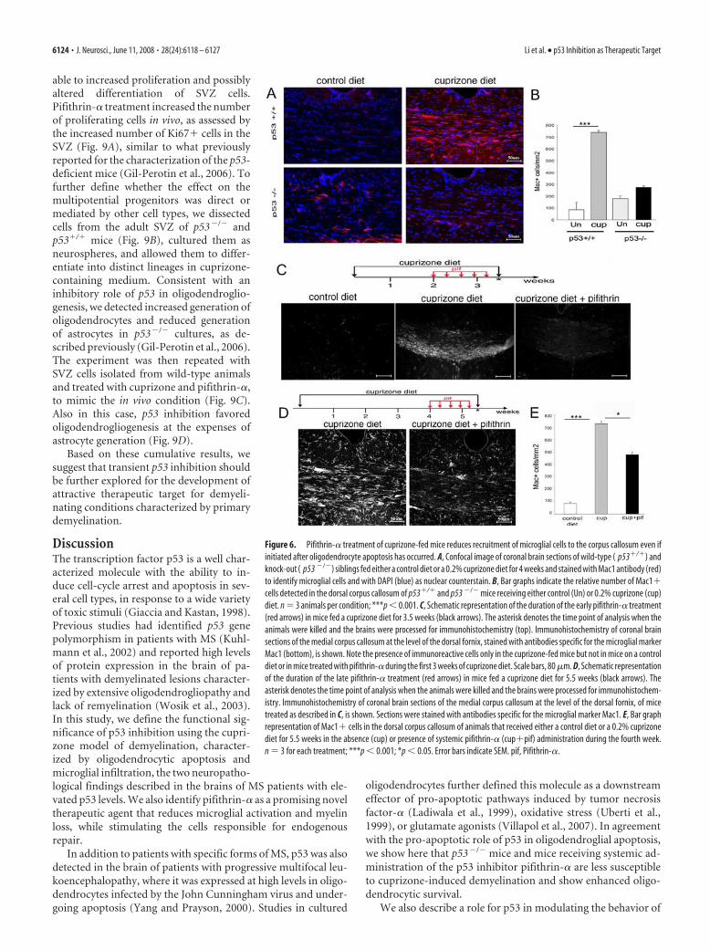
Fewer Mac1� cells were detected in the corpus callosum ofcuprizone-treated p53�/� mice compared with p53�/� mice(Fig. 6A,B). Similar results were detected in mice receivingpifithrin-� treatment either before (Fig. 6C) or after the occur-rence of oligodendroglial death (Fig. 6D,E). When pifithrin-�treatment was started during the early stages of cuprizone diet, weobserved a decline in oligodendrocytic apoptosis and the nearlycomplete absence of Mac1� cells in the corpus callosum oftreated animals (Fig. 6C). This effect could be attributable to thefact that fewer microglial cells were recruited to the corpus callo-sum if fewer oligodendrocytes died. In a second set of experi-ments, pifithrin-� treatment was initiated after the period ofmassive oligodendrocytic death had already occurred. Also in thiscase, however, pifithrin-�-treated mice showed a significantlyreduced number of Mac1�-immunoreactive cells in the corpuscallosum (Fig. 6D). Together, these results suggest that modula-tion of p53 function affects the behavior of microglial cells inmultiple ways.
To further define whether the effect of p53 inhibition on mi-croglial cells was cell intrinsic and independent of cross talks withthe oligodendrocytic population, we adopted a reductionist ap-proach and used either the BV2 microglial cell line or primarymicroglial cultures isolated from the cortices of p53�/� andp53�/� mice. The advantage of the in vitro experimental system isthe possibility to evaluate the effect of p53 on microglial geneexpression, using a homogeneous and controlled experimental
Figure 4. Systemic administration of pharmacological inhibitors of p53 is most effective in protecting oligodendrocytes andmyelin when started during the first 3 weeks of cuprizone treatment. A, Schematic representation of the early administrationprotocol of pifithrin-� treatment (red arrows) during the second and third week of cuprizone diet (black arrows). The asteriskdenotes the time point of analysis when the animals were killed and the brains were processed for immunohistochemistry. B, Bargraphs indicate the number of CC1� oligodendrocytes in the dorsal corpus callosum of animals that received either a controldiet or a 0.2% cuprizone diet for 3.5 weeks in the absence (cup) or presence of systemic pifithrin-� administration (cup�pif).n � 3 for each treatment; ***p � 0.001; **p � 0.01. C, TUNEL� cell counts in the corpus callosum of animals that receivedeither a control diet or a 0.2% cuprizone diet for 3.5 weeks in the absence (cup) or presence of systemic pifithrin-� administra-tion (cup�pif). n � 3 for each treatment; ***p � 0.001; *p � 0.05. D, Schematic representation of the late administrationprotocol of pifithrin-� treatment (red arrows) started during the fourth week of cuprizone diet (black arrows). The asteriskdenotes the time point of analysis when the animals were killed and the brains were processed for immunohistochemistry. E,Confocal image of coronal brain sections of wild-type mice on a regular diet (control) or fed a cuprizone diet for 5.5 weeks in theabsence (cuprizone 5.5 wk) or presence of systemic pifithrin-� administration (cup � pif 5.5 wk). Sections were stained withMBP antibody to identify myelinated fibers (red) and DAPI (blue) as nuclear counterstain. F, Bar graph quantification of CC1�oligodendrocytes in the dorsal corpus callosum of animals that received either a control diet or a 0.2% cuprizone diet for 5.5weeks in the absence (cup) or presence of systemic pifithrin-� (cup�pif) administration during the fourth week. n � 3 for eachtreatment; ***p � 0.001; **p � 0.01. Error bars indicate SEM. pif, Pifithrin-�; cup, cuprizone.
6122 • J. Neurosci., June 11, 2008 • 28(24):6118 – 6127 Li et al. • p53 Inhibition as Therapeutic Target

setting. Consistent with the possibility that p53 modulated theexpression of genes characteristic of activated microglia, we de-tected increased p53 levels in cultured microglial BV2 cells afterLPS (100 ng/ml) stimulation (Fig. 7A). LPS also induced theexpression of the p53 gene target p21, and this increase was
blocked by cotreatment with pifithrin-� (20 �M) (Fig. 7B). TheLPS-dependent elevation of p21 transcript levels was also de-tected in p53�/� primary cultures of microglial cells from mice,but not in cells isolated from p53�/� mice (Fig. 7C). Treatment ofBV2 cells with pifithrin-� effectively reduced the LPS-dependentactivation of the microglial genes lysozyme and �2-microglobulin,although it had no effect on the levels of cathepsin Z (Fig. 7D).Similarly, LPS stimulation or cuprizone treatment (supplementalFig. 1, available at www.jneurosci.org as supplemental material)of primary microglial cultures elicited a greater increase in thetranscript levels of �2-microglobulin and tnf� in p53�/� cellscompared with p53�/� mice (Fig. 7E). The fact that pifithrin orgenetic ablation of p53 did not completely abolish the transcrip-tional response of microglia to LPS is consistent with the impor-tance of nuclear factor-�b (NF-�b) regulation of this transcrip-tional program (Lu et al., 2007). Together, these data support arole for p53 as one of the modulators of gene expression in acti-vated microglial cells.
We have previously mentioned that primary demyelinationconsequent to oligodendrogliopathy and microglial infiltration isalso associated with defective repair. Because p53 affected botholigodendrocyte loss and microglial activation, we asked whetherthis molecule could also modulate the behavior of cells affectingrepair. Previous studies had shown that demyelination of thecorpus callosum is repaired by resident oligodendrocyte progen-itors and by multipotential cells residing in the adult SVZ (Nait-Oumesmar et al., 1999; Picard-Riera et al., 2002; Murtie et al.,2005; Trebst et al., 2007). Other studies (Billon et al., 2004), in-cluding those from our group (Gil-Perotin et al., 2006), had re-ported an important role for p53 in regulating the pool of oligo-dendrocyte progenitors in the developing brain and inmodulating the behavior of multipotential cells in the adult SVZ.However, the potential role of p53 in repair after demyelinationremained to be defined. To address this question, we tested p53expression in the SVZ of cuprizone-treated mice (supplementalFig. 2, available at www.jneurosci.org as supplemental material)and compared the responsiveness of multipotential SVZ cells inp53�/� mice with that observed in p53�/� and also in pifithrin-treated p53�/� mice after cuprizone-induced demyelination. Im-munohistochemical analysis of coronal brain sections using an-tibodies specific for the neural stem cell marker Sox 2 (Kondo andRaff, 2004) revealed an increase in immunoreactive cells in re-sponse to cuprizone-induced demyelination, a response that wasenhanced by pifithrin-treated mice (Fig. 8A). In agreement withprevious reports on the role of p53 in multipotential progenitorsof the adult SVZ (Gil-Perotin et al., 2006), p53�/� mice hadgreater resting numbers of Sox2� cells, and cuprizone treatmentsignificantly increased their number (Fig. 8B). Because SVZ cellscan be also characterized for the presence of the oligodendrocyteprecursor surface molecule polysialic acid-neural cell adhesionmolecule (PSA-NCAM) (Ben-Hur et al., 1998; Nait-Oumesmaret al., 1999, 2007; Decker et al., 2000), we conducted a similaranalysis on the adult SVZ of cuprizone-treated mice. Consistentwith the results obtained for Sox2, cuprizone treatment per seinduced a significant increase in PSA-NCAM� cells in the SVZ ofuntreated p53�/� mice (Fig. 8C). This increase was further en-hanced by either treatment with pharmacological inhibitors ofp53 (Fig. 8C) or in animals with genetic ablation (Fig. 8D). Over-all, these results suggest that p53 function is important for regu-lating the number of multipotential progenitors in the SVZ thatare involved in repair.
Because p53 is an important modulator of the cell cycle, weasked whether the increased number of Sox2� cells was attribut-
Figure 5. Decreased expression of microglial genes in mice with genetic deletion or phar-macological inhibition of p53. A, B, Quantitative real-time PCR of the mRNA levels for the p53target genes gadd45, p21, and tcf4 (A) and of �2-microglobulin, lysozyme, and tnf-� (B) insamples isolated from the corpus callosum of untreated (un) and cuprizone-treated (cup)p53�/� and p53 �/� mice. C, D, Quantitative real-time PCR of the mRNA levels for the p53target genes gadd45, p21, and tcf4 (C) and of �2-microglobulin, lysozyme, and tnf-� (D) insamples isolated from the corpus callosum of untreated controls (control diet), cuprizone-fedmice (cup), and cuprizone-fed mice receiving pifithrin-� treatment (cup�pif). The RNA levelswere referred to the GAPDH levels, and the values detected in cuprizone-treated mice in theabsence and presence of pifithrin treatment were referred in terms of percentage value relativeto the levels detected in untreated controls. *p � 0.05; **p � 0.01 . Error bars indicate SEM.
Li et al. • p53 Inhibition as Therapeutic Target J. Neurosci., June 11, 2008 • 28(24):6118 – 6127 • 6123
able to increased proliferation and possiblyaltered differentiation of SVZ cells.Pifithrin-� treatment increased the numberof proliferating cells in vivo, as assessed bythe increased number of Ki67� cells in theSVZ (Fig. 9A), similar to what previouslyreported for the characterization of the p53-deficient mice (Gil-Perotin et al., 2006). Tofurther define whether the effect on themultipotential progenitors was direct ormediated by other cell types, we dissectedcells from the adult SVZ of p53�/� andp53�/� mice (Fig. 9B), cultured them asneurospheres, and allowed them to differ-entiate into distinct lineages in cuprizone-containing medium. Consistent with aninhibitory role of p53 in oligodendroglio-genesis, we detected increased generation ofoligodendrocytes and reduced generationof astrocytes in p53�/� cultures, as de-scribed previously (Gil-Perotin et al., 2006).The experiment was then repeated withSVZ cells isolated from wild-type animalsand treated with cuprizone and pifithrin-�,to mimic the in vivo condition (Fig. 9C).Also in this case, p53 inhibition favoredoligodendrogliogenesis at the expenses ofastrocyte generation (Fig. 9D).
Based on these cumulative results, wesuggest that transient p53 inhibition shouldbe further explored for the development ofattractive therapeutic target for demyeli-nating conditions characterized by primarydemyelination.
DiscussionThe transcription factor p53 is a well char-acterized molecule with the ability to in-duce cell-cycle arrest and apoptosis in sev-eral cell types, in response to a wide varietyof toxic stimuli (Giaccia and Kastan, 1998).Previous studies had identified p53 genepolymorphism in patients with MS (Kuhl-mann et al., 2002) and reported high levelsof protein expression in the brain of pa-tients with demyelinated lesions character-ized by extensive oligodendrogliopathy andlack of remyelination (Wosik et al., 2003).In this study, we define the functional sig-nificance of p53 inhibition using the cupri-zone model of demyelination, character-ized by oligodendrocytic apoptosis andmicroglial infiltration, the two neuropatho-logical findings described in the brains of MS patients with ele-vated p53 levels. We also identify pifithrin-� as a promising noveltherapeutic agent that reduces microglial activation and myelinloss, while stimulating the cells responsible for endogenousrepair.
In addition to patients with specific forms of MS, p53 was alsodetected in the brain of patients with progressive multifocal leu-koencephalopathy, where it was expressed at high levels in oligo-dendrocytes infected by the John Cunningham virus and under-going apoptosis (Yang and Prayson, 2000). Studies in cultured
oligodendrocytes further defined this molecule as a downstreameffector of pro-apoptotic pathways induced by tumor necrosisfactor-� (Ladiwala et al., 1999), oxidative stress (Uberti et al.,1999), or glutamate agonists (Villapol et al., 2007). In agreementwith the pro-apoptotic role of p53 in oligodendroglial apoptosis,we show here that p53�/� mice and mice receiving systemic ad-ministration of the p53 inhibitor pifithrin-� are less susceptibleto cuprizone-induced demyelination and show enhanced oligo-dendrocytic survival.
We also describe a role for p53 in modulating the behavior of
Figure 6. Pifithrin-� treatment of cuprizone-fed mice reduces recruitment of microglial cells to the corpus callosum even ifinitiated after oligodendrocyte apoptosis has occurred. A, Confocal image of coronal brain sections of wild-type ( p53�/�) andknock-out ( p53 �/�) siblings fed either a control diet or a 0.2% cuprizone diet for 4 weeks and stained with Mac1 antibody (red)to identify microglial cells and with DAPI (blue) as nuclear counterstain. B, Bar graphs indicate the relative number of Mac1�cells detected in the dorsal corpus callosum of p53�/� and p53 �/� mice receiving either control (Un) or 0.2% cuprizone (cup)diet. n � 3 animals per condition; ***p � 0.001. C, Schematic representation of the duration of the early pifithrin-� treatment(red arrows) in mice fed a cuprizone diet for 3.5 weeks (black arrows). The asterisk denotes the time point of analysis when theanimals were killed and the brains were processed for immunohistochemistry (top). Immunohistochemistry of coronal brainsections of the medial corpus callosum at the level of the dorsal fornix, stained with antibodies specific for the microglial markerMac1 (bottom), is shown. Note the presence of immunoreactive cells only in the cuprizone-fed mice but not in mice on a controldiet or in mice treated with pifithrin-� during the first 3 weeks of cuprizone diet. Scale bars, 80 �m. D, Schematic representationof the duration of the late pifithrin-� treatment (red arrows) in mice fed a cuprizone diet for 5.5 weeks (black arrows). Theasterisk denotes the time point of analysis when the animals were killed and the brains were processed for immunohistochem-istry. Immunohistochemistry of coronal brain sections of the medial corpus callosum at the level of the dorsal fornix, of micetreated as described in C, is shown. Sections were stained with antibodies specific for the microglial marker Mac1. E, Bar graphrepresentation of Mac1� cells in the dorsal corpus callosum of animals that received either a control diet or a 0.2% cuprizonediet for 5.5 weeks in the absence (cup) or presence of systemic pifithrin-� (cup�pif) administration during the fourth week.n � 3 for each treatment; ***p � 0.001; *p � 0.05. Error bars indicate SEM. pif, Pifithrin-�.
6124 • J. Neurosci., June 11, 2008 • 28(24):6118 – 6127 Li et al. • p53 Inhibition as Therapeutic Target

microglial cells, which is independent of the effect on the oligo-dendrocytes. Indeed, systemic administration of pharmacologi-cal inhibitors of p53 (i.e., pifithrin-�) to cuprizone-treated micepositively impacted the course of the disease, by decreasing mi-croglial accumulation and gene expression even if started afterthe occurrence of oligodendroglial apoptosis. The effect of p53 ongene expression was also suggested by the results of the in vitrostudies in LPS-stimulated primary microglial cultures and in theBV2 microglial cell line. In agreement with previous reports onthe increased expression of p53 in microglial cells in response toinfectious or excitotoxic stimuli (Garden et al., 2004; Villapol etal., 2007), we have detected the upregulation of p53 and of itsdownstream target gene p21 in cultures stimulated with LPS andsuggested that p53 functions as a modulator of the transcriptionalresponse characteristic of activated microglia. Our results indi-cate lower levels of �2-microglobulin levels in p53�/� microglialcultures compared with controls, in response to the activatingstimulus. Treatment of the BV2 cells with pifithrin-� similarlyreduced the LPS-dependent activation of the microglial genes
lysozyme and �2-microglobulin. The effect of genetic ablation ofp53 or pharmacological treatment with pifithrin-� was selectivebecause it did not affect the levels of cathepsin Z. Our resultscannot determine whether the effect of p53 on microglial geneexpression is direct or indirect, possibly by modulating additionalcomponents of the transcriptional network. In peripheral mac-rophages, for instance, p53 has been shown to inhibit cytokineexpression (Komarova et al., 2005), by interfering with NF-�b-dependent gene expression (Dijsselbloem et al., 2007). Althoughit is becoming increasingly evident that peripheral macrophagesand resident microglial cells constitute two distinct populations(Melchior et al., 2006), our data do not address whether p53-dependent regulation of gene expression might represent an ad-ditional difference between these two cell types. However, ourresults support a p53-mediated transcriptional response duringmicroglial activation.
The role of microglia in demyelination is well established(Carson, 2002; Kim and de Vellis, 2005; Sanders and De Keyser,2007), and the studies in the cuprizone model of demyelination(Hiremath et al., 1998; Pasquini et al., 2007) have provided sup-port to the concept that microglial activation might contribute tooligodendrocytic demise and neuronal damage (Pasquini et al.,2007). These premises have led to the therapeutic use of the tet-racycline derivative minocycline, an antibiotic with the ability toinhibit the inflammatory response by modulation of the micro-glial component (Yrjanheikki et al., 1998, 1999; Yong et al.,2004). Whereas this approach has proven to be effective in limit-ing the lesion size in response to cuprizone (Pasquini et al., 2007)and in experimental autoimmune encephalomyelitis models(Popovic et al., 2002), it is also clear that it has negatively im-pacted remyelination and repair (Kotter et al., 2001; Li et al.,2005). The negative effect of minocycline treatment on remyeli-nation could be attributed to several potential reasons. One pos-sibility is that minocycline exerts a dose-dependent inhibition ofoligodendrocyte progenitor proliferation, thereby reducing thepool of cells that can form new myelin (Li et al., 2005). An alter-native possibility is that minocycline reduces the production ofimportant trophic factors (i.e., BDNF) from microglial cells,thereby affecting oligodendrocyte progenitor cell proliferationand differentiation (Dougherty et al., 2000; Du et al., 2003).Therefore, the general consensus is that it is important to limitmicroglial activation, without necessarily abolishing it, to pro-vide a source of trophic support for the repair mechanism. Ourstudy, using genetic or pharmacological inhibition of p53, sug-gests that transient inhibition of this transcription factor mightprovide multiple beneficial effects in demyelinating disordersconsequent to oligodendrogliopathy and massive microglial in-filtration. Systemic administration with pifithrin-�, for instance,decreases the extent of myelin loss and microglial accumulationand selectively reduced the levels of cytotoxic microglial geneproducts (i.e., lysozyme) without affecting the expression of tro-phic genes (i.e., BDNF). An added beneficial effect of this treat-ment was the proliferative and differentiative effect on multipo-tential progenitors in the adult SVZ that resulted in an increasednumber of Sox2-positive cells and enhanced oligodendroglio-genesis at the expenses of astrogliogenesis. We have previouslyreported that cells cultured from the SVZ of p53�/� mice havethe ability to generate more O4� oligodendrocytes than cellsisolated from p53�/� siblings (Gil-Perotin et al., 2006). Althoughwe cannot rule out cell-extrinsic mechanisms, our current studyshows that treatment with pifithrin-� results in a remarkablysimilar phenotype.
In conclusion, we propose that pharmacological compounds
Figure 7. Pharmacological inhibition of p53-dependent transcriptional activity decreasesthe transcriptional activation induced by LPS in microglial cells. A, Quantitative real-time PCR ofp53 mRNA levels in samples isolated from BV2 microglial cells either untreated (Un) or treatedwith 100 ng/ml LPS. *p � 0.01 versus untreated cells. B, Quantitative real-time PCR of the p21mRNA levels in samples extracted from BV2 cells either untreated or treated with 100 ng/ml LPSalone (LPS), 20 �M pifithrin-� alone, or cotreated for 6 h. **p � 0.001 versus LPS-treated cells;*p � 0.01 versus untreated cells. C, Real-time PCR of p21 transcript levels measured in RNAsamples from primary microglial cultures isolated from p53�/� and p53 �/� mice and stim-ulated with LPS. D, Quantitative real-time PCR of the mRNA levels in samples isolated from BV2cells as described in B. Lysozyme, *p � 0.01 versus untreated cells, **p � 0.01 versus LPS;�2-microglobulin, *p � 0.05 versus LPS. The levels of cathepsin Z were not affected bypifithrin-�. Bar graphs represent the mean SEM of four independent experiments run intriplicate (ANOVA followed by Bonferroni’s multiple comparison test). E, Effect of LPS treatmenton transcripts indicative of microglial cell activation. The bar graphs represent the results ofquantitative real-time PCR performed on RNA isolated from primary cultures of p53�/� andp53 �/� microglial cells stimulated with LPS. Note the lower level of microglial gene activationdetected in p53 �/� cells and the similarity with the in vivo data in cuprizone-fed mice, shownin Figure 5. Un, Untreated; PFT, pifithrin-�; P�L, pifithrin-� plus LPS. Error bars indicate SEM.
Li et al. • p53 Inhibition as Therapeutic Target J. Neurosci., June 11, 2008 • 28(24):6118 – 6127 • 6125

with transient inhibitory activity on p53,should be further investigated as potentialtherapeutic agents for primary demyelina-tion. The design of small peptides with theability to inactivate transiently and possiblyin a reversible manner p53 in a cell-specificmanner is highly desirable to avoid thecomplications that sustained inactivationof p53 could have on lymphocyte apoptosis(Okuda et al., 2002) and tumorigenesis(Donehower et al., 1992; Donehower, 1996;Lozano, 2007).
ReferencesAboul-Enein F, Rauschka H, Kornek B, Stadelmann
C, Stefferl A, Bruck W, Lucchinetti C, Schmid-bauer M, Jellinger K, Lassmann H (2003) Pref-erential loss of myelin-associated glycoproteinreflects hypoxia-like white matter damage instroke and inflammatory brain diseases. J Neu-ropathol Exp Neurol 62:25–33.
Barnett MH, Prineas JW (2004) Relapsing and re-mitting multiple sclerosis: pathology of thenewly forming lesion. Ann Neurol 55:458 – 468.
Ben-Hur T, Rogister B, Murray K, Rougon G,Dubois-Dalcq M (1998) Growth and fate ofPSA-NCAM� precursors of the postnatalbrain. J Neurosci 18:5777–5788.
Billon N, Terrinoni A, Jolicoeur C, McCarthy A,Richardson WD, Melino G, Raff M (2004)Roles for p53 and p73 during oligodendrocytedevelopment. Development 131:1211–1220.
Blasi E, Barluzzi R, Bocchini V, Mazzolla R, BistoniF (1990) Immortalization of murine microglialcells by a v-raf/v-myc carrying retrovirus. J Neu-roimmunol 27:229 –237.
Carson MJ (2002) Microglia as liaisons betweenthe immune and central nervous systems: func-tional implications for multiple sclerosis. Glia40:218 –231.
Culmsee C, Zhu X, Yu QS, Chan SL, Camandola S,Guo Z, Greig NH, Mattson MP (2001) A syn-thetic inhibitor of p53 protects neurons againstdeath induced by ischemic and excitotoxic in-sults, and amyloid beta-peptide. J Neurochem77:220 –228.
Decker L, Avellana-Adalid V, Nait-Oumesmar B,Durbec P, Baron-Van Evercooren A (2000)Oligodendrocyte precursor migration and dif-ferentiation: combined effects of PSA residues,growth factors, and substrates. Mol Cell Neuro-sci 16:422– 439.
Dijsselbloem N, Goriely S, Albarani V, Gerlo S, Fran-coz S, Marine JC, Goldman M, Haegeman G, Vanden Berghe W (2007) Acritical role for p53 in the control of NF-kappaB-dependent gene expressionin TLR4-stimulated dendritic cells exposed to Genistein. J Immunol178:5048–5057.
Donehower LA (1996) The p53-deficient mouse: a model for basic and ap-plied cancer studies. Semin Cancer Biol 7:269 –278.
Donehower LA, Harvey M, Slagle BL, McArthur MJ, Montgomery Jr CA,Butel JS, Bradley A (1992) Mice deficient for p53 are developmentallynormal but susceptible to spontaneous tumours. Nature 356:215–221.
Dougherty KD, Dreyfus CF, Black IB (2000) Brain-derived neurotrophicfactor in astrocytes, oligodendrocytes, and microglia/macrophages afterspinal cord injury. Neurobiol Dis 7:574 –585.
Du Y, Fischer TZ, Lee LN, Lercher LD, Dreyfus CF (2003) Regionally spe-cific effects of BDNF on oligodendrocytes. Dev Neurosci 25:116 –126.
Garden GA, Guo W, Jayadev S, Tun C, Balcaitis S, Choi J, Montine TJ, MollerT, Morrison RS (2004) HIV associated neurodegeneration requires p53in neurons and microglia. FASEB J 18:1141–1143.
Giaccia AJ, Kastan MB (1998) The complexity of p53 modulation: emergingpatterns from divergent signals. Genes Dev 12:2973–2983.
Gil-Perotin S, Marin-Husstege M, Li J, Soriano-Navarro M, Zindy F, RousselMF, Garcia-Verdugo JM, Casaccia-Bonnefil P (2006) Loss of p53 in-duces changes in the behavior of subventricular zone cells: implication forthe genesis of glial tumors. J Neurosci 26:1107–1116.
Hiremath MM, Saito Y, Knapp GW, Ting JP, Suzuki K, Matsushima GK(1998) Microglial/macrophage accumulation during cuprizone-induceddemyelination in C57BL/6 mice. J Neuroimmunol 92:38 – 49.
Jurevics H, Largent C, Hostettler J, Sammond DW, Matsushima GK, Klein-dienst A, Toews AD, Morell P (2002) Alterations in metabolism and geneexpression in brain regions during cuprizone-induced demyelination andremyelination. J Neurochem 82:126 –136.
Kim SU, de Vellis J (2005) Microglia in health and disease. J Neurosci Res81:302–313.
Komarova EA, Krivokrysenko V, Wang K, Neznanov N, Chernov MV, Kom-arov PG, Brennan ML, Golovkina TV, Rokhlin OW, Kuprash DV, Nedo-
Figure 8. Increased Sox2� and PSA-NCAM� cells in the SVZ of mice with genetic or pharmacological inhibition of p53 invivo after cuprizone-induced demyelination. A, Confocal image of brain sections from adult p53�/�mice receiving a 4 weekcontrol diet or a 0.2% cuprizone diet in the absence (cup) or presence of systemic administration of pifithrin-� (cup�pif).Sections were stained with antibodies against Sox2 (green) and DAPI (blue). The bar graphs represent the number of Sox2�cells counted in coronal sections at the level of the dorsal corpus callosum of at least three mice per group. **p � 0.01; *p �0.05. Note the wider distribution of the Sox2� cells in mice treated with cuprizone compared with controls and the greaterresponse detected in the animals treated with pifithrin-�. B, Confocal image of brain sections from untreated (control diet) orcuprizone-treated (cup) p53 �/� adult mice stained with antibodies against Sox2 (green) and DAPI (blue). The bar graphsrepresent the number of Sox2� cells. Also in this case, cuprizone treatment significantly increased the number of Sox2� cellsin the SVZ of p53 �/� mice. *p � 0.05. Note the greater number observed in these mice compared with wild type (A). C,Confocal image of PSA-NCAM� cells in the adult SVZ of p53�/� mice receiving a 4 week control diet or a 0.2% cuprizone dietin the absence (cup) or presence of systemic administration of pifithrin-� (cup�pif). Sections were stained with antibodiesagainst PSA-NCAM (red) and DAPI (blue). The bar graphs represent the number of PSA-NCAM� cells counted in the SVZ at thelevel of the dorsal corpus callosum of at least three mice per group. **p � 0.01; ***p � 0.001. Note that the combination ofcuprizone and pifithrin-� had a stronger effect on the number of PSA-NCAM� cells than cuprizone alone. D, Confocal image ofPSA-NCAM� (red) cells in the adult SVZ of p53 �/� mice untreated (control diet) or after 4 weeks of cuprizone diet (cup). Notethat the number of PSA-NCAM� cells is already elevated in untreated controls and that after cuprizone stimulation it increasesto levels that are comparable with those detected in the pifithrin-treated wild-type animals. Scale bar, 50 �m. Error barsindicate SEM. V, Ventricle.
6126 • J. Neurosci., June 11, 2008 • 28(24):6118 – 6127 Li et al. • p53 Inhibition as Therapeutic Target

spasov SA, Hazen SL, Feinstein E, Gudkov AV (2005) p53 is a suppressorof inflammatory response in mice. FASEB J 19:1030 –1032.
Kondo T, Raff M (2004) Chromatin remodeling and histone modificationin the conversion of oligodendrocyte precursors to neural stem cells.Genes Dev 18:2963–2972.
Kotter MR, Setzu A, Sim FJ, Van Rooijen N, Franklin RJ (2001) Macrophagedepletion impairs oligodendrocyte remyelination following lysolecithin-induced demyelination. Glia 35:204 –212.
Kuhlmann T, Glas M, zum Bruch C, Mueller W, Weber A, Zipp F, Bruck W(2002) Investigation of bax, bcl-2, bcl-x and p53 gene polymorphisms inmultiple sclerosis. J Neuroimmunol 129:154 –160.
Ladiwala U, Li H, Antel JP, Nalbantoglu J (1999) p53 induction by tumornecrosis factor-alpha and involvement of p53 in cell death of humanoligodendrocytes. J Neurochem 73:605– 611.
Li WW, Setzu A, Zhao C, Franklin RJ (2005) Minocycline-mediated inhibi-tion of microglia activation impairs oligodendrocyte progenitor cell re-sponses and remyelination in a non-immune model of demyelination.J Neuroimmunol 158:58 – 66.
Lozano G (2007) The oncogenic roles of p53 mutants in mouse models.Curr Opin Genet Dev 17:66 –70.
Lu DY, Tang CH, Liou HC, Teng CM, Jeng KC, Kuo SC, Lee FY, Fu WM(2007) YC-1 attenuates LPS-induced proinflammatory responses and ac-tivation of nuclear factor-kappaB in microglia. Br J Pharmacol 151:396 –405.
Lucchinetti C, Bruck W, Parisi J, Scheithauer B, Rodriguez M, Lassmann H(2000) Heterogeneity of multiple sclerosis lesions: implications for thepathogenesis of demyelination. Ann Neurol 47:707–717.
Matsushima GK, Morell P (2001) The neurotoxicant, cuprizone, as a modelto study demyelination and remyelination in the central nervous system.Brain Pathol 11:107–116.
Melchior B, Puntambekar SS, Carson MJ (2006) Microglia and the controlof autoreactive T cell responses. Neurochem Int 49:145–153.
Morell P, Barrett CV, Mason JL, Toews AD, Hostettler JD, Knapp GW, Mat-sushima GK (1998) Gene expression in brain during cuprizone-induceddemyelination and remyelination. Mol Cell Neurosci 12:220 –227.
Murtie JC, Zhou YX, Le TQ, Vana AC, Armstrong RC (2005) PDGF andFGF2 pathways regulate distinct oligodendrocyte lineage responses inexperimental demyelination with spontaneous remyelination. NeurobiolDis 19:171–182.
Nait-Oumesmar B, Decker L, Lachapelle F, Avellana-Adalid V, Bachelin C,Van Evercooren AB (1999) Progenitor cells of the adult mouse subven-tricular zone proliferate, migrate and differentiate into oligodendrocytesafter demyelination. Eur J Neurosci 11:4357– 4366.
Nait-Oumesmar B, Picard-Riera N, Kerninon C, Decker L, Seilhean D, Ho-glinger GU, Hirsch EC, Reynolds R, Baron-Van Evercooren A (2007)Activation of the subventricular zone in multiple sclerosis: evidence forearly glial progenitors. Proc Natl Acad Sci USA 104:4694 – 4699.
Okuda Y, Okuda M, Bernard CC (2002) The suppression of T cell apoptosisinfluences the severity of disease during the chronic phase but not therecovery from the acute phase of experimental autoimmune encephalo-myelitis in mice. J Neuroimmunol 131:115–125.
Pasquini LA, Calatayud CA, Bertone Una AL, Millet V, Pasquini JM, Soto EF(2007) The neurotoxic effect of cuprizone on oligodendrocytes dependson the presence of pro-inflammatory cytokines secreted by microglia.Neurochem Res 32:279 –292.
Picard-Riera N, Decker L, Delarasse C, Goude K, Nait-Oumesmar B, LiblauR, Pham-Dinh D, Evercooren AB (2002) Experimental autoimmuneencephalomyelitis mobilizes neural progenitors from the subventricularzone to undergo oligodendrogenesis in adult mice. Proc Natl Acad SciUSA 99:13211–13216.
Popovic N, Schubart A, Goetz BD, Zhang SC, Linington C, Duncan ID(2002) Inhibition of autoimmune encephalomyelitis by a tetracycline.Ann Neurol 51:215–223.
Sanders P, De Keyser J (2007) Janus faces of microglia in multiple sclerosis.Brain Res Rev 54:274 –285.
Stadelmann C, Ludwin S, Tabira T, Guseo A, Lucchinetti CF, Leel-Ossy L,Ordinario AT, Bruck W, Lassmann H (2005) Tissue preconditioningmay explain concentric lesions in Balo’s type of multiple sclerosis. Brain128:979 –987.
Trebst C, Heine S, Lienenklaus S, Lindner M, Baumgartner W, Weiss S,Stangel M (2007) Lack of interferon-beta leads to accelerated remyeli-nation in a toxic model of central nervous system demyelination. ActaNeuropathol 114:587–596.
Uberti D, Yavin E, Gil S, Ayasola KR, Goldfinger N, Rotter V (1999) Hydro-gen peroxide induces nuclear translocation of p53 and apoptosis in cells ofoligodendroglia origin. Brain Res Mol Brain Res 65:167–175.
Villapol S, Acarin L, Faiz M, Castellano B, Gonzalez B (2007) Distinct spatialand temporal activation of caspase pathways in neurons and glial cellsafter excitotoxic damage to the immature rat brain. J Neurosci Res85:3545–3556.
Wosik K, Antel J, Kuhlmann T, Bruck W, Massie B, Nalbantoglu J (2003)Oligodendrocyte injury in multiple sclerosis: a role for p53. J Neurochem85:635– 644.
Yang B, Prayson RA (2000) Expression of Bax, Bcl-2, and P53 in progressivemultifocal leukoencephalopathy. Mod Pathol 13:1115–1120.
Yong VW, Wells J, Giuliani F, Casha S, Power C, Metz LM (2004) Thepromise of minocycline in neurology. Lancet Neurol 3:744 –751.
Yrjanheikki J, Keinanen R, Pellikka M, Hokfelt T, Koistinaho J (1998) Tet-racyclines inhibit microglial activation and are neuroprotective in globalbrain ischemia. Proc Natl Acad Sci USA 95:15769 –15774.
Yrjanheikki J, Tikka T, Keinanen R, Goldsteins G, Chan PH, Koistinaho J(1999) A tetracycline derivative, minocycline, reduces inflammation andprotects against focal cerebral ischemia with a wide therapeutic window.Proc Natl Acad Sci USA 96:13496 –13500.
Figure 9. Pharmacological inhibition of p53-dependent transcriptional activity increasesthe proliferation and the generation of O4� cells from multipotential neural progenitors de-rived from the adult SVZ. A, Quantification of Ki67� cells in the SVZ of mice that received onlya 0.2% cuprizone (cup) diet or also received systemic administration of pifithrin-� (cup�pif)during the second (2wk) or fourth (4wk) week of cuprizone diet. *p � 0.05; **p � 0.01. B,Primary cultures of SVZ cells were obtained from adult p53�/� and p53 �/� mice, cultured asneurospheres, dissociated, exposed to cuprizone (25 �M), and allowed to differentiate. Red,GFAP (astrocytes); green, O4 (oligodendrocytes); blue, DAPI as nuclear counterstain. Scale bar,20 �m. C, SVZ cells isolated from adult p53 �/� mice were cultured as neurospheres, dissoci-ated, exposed to cuprizone (25 �M), and allowed to differentiate either in the absence (cupri-zone) or presence of 20 �M pifithrin-� (cuprizone�pifithrin) or 5 d. Cultures were then stainedwith antibodies specific for GFAP (red, astrocytes), O4 (green, oligodendrocytes), and DAPI(blue) as nuclear counterstain. D, Bar graphs indicate the percentage of immunoreactive cellsrelative to total DAPI� cells. *p � 0.01; **p � 0.001. Error bars indicate SEM.
Li et al. • p53 Inhibition as Therapeutic Target J. Neurosci., June 11, 2008 • 28(24):6118 – 6127 • 6127
Related Documents


