Inhibition of brain energy metabolism by the a-keto acids accumulating in maple syrup urine disease Angela M. Sgaravatti, Rafael B. Rosa, Patrı ´cia F. Schuck, Ce ´sar A.J. Ribeiro, Clo ´vis M.D. Wannmacher, Angela T.S. Wyse, Carlos S. Dutra-Filho, Moacir Wajner * Departamento de Bioquı ´mica, Instituto de Cie ˆncias Ba ´sicas da Sau ´de, Universidade Federal do Rio Grande do Sul, Rua Ramiro Barcelos, 2600-Anexo, CEP 90035-003 Porto Alegre, RS, Brazil Servic ßo de Gene ´tica Me ´dica do Hospital de Clı ´nicas de Porto Alegre, Porto Alegre, RS, Brazil Universidade Luterana do Brasil, Canoas, RS, Brazil Received 14 May 2003; received in revised form 29 August 2003; accepted 23 September 2003 Abstract Neurological dysfunction is a common finding in patients with maple syrup urine disease (MSUD). However, the mechanisms underlying the neuropathology of brain damage in this disorder are poorly known. In the present study, we investigated the effect of the in vitro effect of the branched chain a-keto acids (BCKA) accumulating in MSUD on some parameters of energy metabolism in cerebral cortex of rats. [ 14 CO 2 ] production from [ 14 C] acetate, glucose uptake and lactate release from glucose were evaluated by incubating cortical prisms from 30-day-old rats in Krebs– Ringer bicarbonate buffer, pH 7.4, in the absence (controls) or presence of 1 –5 mM of a-ketoisocaproic acid (KIC), a-keto-h-methylvaleric acid (KMV) or a-ketoisovaleric acid (KIV). All keto acids significantly reduced 14 CO 2 production by around 40%, in contrast to lactate release and glucose utilization, which were significantly increased by the metabolites by around 42% in cortical prisms. Furthermore, the activity of the respiratory chain complex I – III was significantly inhibited by 60%, whereas the other activities of the electron transport chain, namely complexes II, II–III, III and IV, as well as succinate dehydrogenase were not affected by the keto acids. The results indicate that the major metabolites accumulating in MSUD compromise brain energy metabolism by blocking the respiratory chain. We presume that these findings may be of relevance to the understanding of the pathophysiology of the neurological dysfunction of MSUD patients. D 2003 Elsevier B.V. All rights reserved. Keywords: Maple syrup urine disease; a-Ketoisocaproic acid; a-Keto-h-methylvaleric acid; a-Ketoisovaleric acid; Energy metabolism 1. Introduction Maple syrup urine disease (MSUD), or branched chain keto aciduria (BCKA), is an inborn error of metabolism caused by severe deficiency of the branched chain a-ketoacid dehydrogenase complex (BCKAD, E.C. 1.2.4.4) activity [1]. The inability of this enzyme complex to oxidize a-ketoiso- caproic acid (KIC), a-keto-h-methylvaleric acid (KMV) and a-ketoisovaleric acid (KIV) leads to tissue accumulation of these metabolites and their precursor amino acids leucine, isoleucine and valine, respectively, in the affected individu- als. Patients with MSUD present poor feeding, apnoea, ketoacidosis, convulsions, coma and psychomotor delay. Central nervous system (CNS) imaging reveals low density of white matter corresponding to hypomyelination/demye- lination and cerebral atrophy. The disease is severe enough to cause a fatal outcome in a significant number of patients if not diagnosed and treated promptly. Those who survive present a variable degree of mental retardation [1]. The reduction of branched chain amino acids (BCAA) has been the main target for treating MSUD patients and diet has been the mainstay of treatment [2], whenever the disease is not responsive to thiamine [1]. Although this approach has contributed decisively to the survival of the 0925-4439/$ - see front matter D 2003 Elsevier B.V. All rights reserved. doi:10.1016/j.bbadis.2003.09.010 Abbreviations: MSUD, maple syrup urine disease; BCAA, branched chain amino acids; BCKA, branched chain keto acids; BCKAD, branched chain L-a-keto acid dehydrogenase; KIC, a-ketoisocaproic acid; KMV, a- keto-h-methylvaleric acid; KIV, a-ketoisovaleric acid; CNS, central nervous system; GABA, gamma-aminobutyric acid * Corresponding author. Departamento de Bioquı ´mica, Instituto de Cie ˆncias Ba ´sicas da Sau ´de, Universidade Federal do Rio Grande do Sul, Rua Ramiro Barcelos, 2600-Anexo, CEP 90035-003 Po ˆrto Alegre, RS, Brazil. Tel.: +55-51-33165571; fax: +55-51-33168010. E-mail address: [email protected] (M. Wajner). www.bba-direct.com Biochimica et Biophysica Acta 1639 (2003) 232 – 238

Welcome message from author
This document is posted to help you gain knowledge. Please leave a comment to let me know what you think about it! Share it to your friends and learn new things together.
Transcript
www.bba-direct.com
Biochimica et Biophysica Acta 1639 (2003) 232–238
Inhibition of brain energy metabolism by the a-keto acids accumulating
in maple syrup urine disease
Angela M. Sgaravatti, Rafael B. Rosa, Patrıcia F. Schuck, Cesar A.J. Ribeiro,Clovis M.D. Wannmacher, Angela T.S. Wyse, Carlos S. Dutra-Filho, Moacir Wajner*
Departamento de Bioquımica, Instituto de Ciencias Basicas da Saude, Universidade Federal do Rio Grande do Sul, Rua Ramiro Barcelos, 2600-Anexo,
CEP 90035-003 Porto Alegre, RS, Brazil
Servic�o de Genetica Medica do Hospital de Clınicas de Porto Alegre, Porto Alegre, RS, Brazil
Universidade Luterana do Brasil, Canoas, RS, Brazil
Received 14 May 2003; received in revised form 29 August 2003; accepted 23 September 2003
Abstract
Neurological dysfunction is a common finding in patients with maple syrup urine disease (MSUD). However, the mechanisms
underlying the neuropathology of brain damage in this disorder are poorly known. In the present study, we investigated the effect of the in
vitro effect of the branched chain a-keto acids (BCKA) accumulating in MSUD on some parameters of energy metabolism in cerebral cortex
of rats. [14CO2] production from [14C] acetate, glucose uptake and lactate release from glucose were evaluated by incubating cortical prisms
from 30-day-old rats in Krebs–Ringer bicarbonate buffer, pH 7.4, in the absence (controls) or presence of 1–5 mM of a-ketoisocaproic acid
(KIC), a-keto-h-methylvaleric acid (KMV) or a-ketoisovaleric acid (KIV). All keto acids significantly reduced 14CO2 production by around
40%, in contrast to lactate release and glucose utilization, which were significantly increased by the metabolites by around 42% in cortical
prisms. Furthermore, the activity of the respiratory chain complex I–III was significantly inhibited by 60%, whereas the other activities of
the electron transport chain, namely complexes II, II–III, III and IV, as well as succinate dehydrogenase were not affected by the keto acids.
The results indicate that the major metabolites accumulating in MSUD compromise brain energy metabolism by blocking the respiratory
chain. We presume that these findings may be of relevance to the understanding of the pathophysiology of the neurological dysfunction of
MSUD patients.
D 2003 Elsevier B.V. All rights reserved.
Keywords: Maple syrup urine disease; a-Ketoisocaproic acid; a-Keto-h-methylvaleric acid; a-Ketoisovaleric acid; Energy metabolism
1. Introduction caproic acid (KIC), a-keto-h-methylvaleric acid (KMV) and
Maple syrup urine disease (MSUD), or branched chain
keto aciduria (BCKA), is an inborn error of metabolism
caused by severe deficiency of the branched chain a-ketoacid
dehydrogenase complex (BCKAD, E.C. 1.2.4.4) activity [1].
The inability of this enzyme complex to oxidize a-ketoiso-
0925-4439/$ - see front matter D 2003 Elsevier B.V. All rights reserved.
doi:10.1016/j.bbadis.2003.09.010
Abbreviations: MSUD, maple syrup urine disease; BCAA, branched
chain amino acids; BCKA, branched chain keto acids; BCKAD, branched
chain L-a-keto acid dehydrogenase; KIC, a-ketoisocaproic acid; KMV, a-
keto-h-methylvaleric acid; KIV, a-ketoisovaleric acid; CNS, central
nervous system; GABA, gamma-aminobutyric acid
* Corresponding author. Departamento de Bioquımica, Instituto de
Ciencias Basicas da Saude, Universidade Federal do Rio Grande do Sul,
Rua Ramiro Barcelos, 2600-Anexo, CEP 90035-003 Porto Alegre, RS,
Brazil. Tel.: +55-51-33165571; fax: +55-51-33168010.
E-mail address: [email protected] (M. Wajner).
a-ketoisovaleric acid (KIV) leads to tissue accumulation of
these metabolites and their precursor amino acids leucine,
isoleucine and valine, respectively, in the affected individu-
als. Patients with MSUD present poor feeding, apnoea,
ketoacidosis, convulsions, coma and psychomotor delay.
Central nervous system (CNS) imaging reveals low density
of white matter corresponding to hypomyelination/demye-
lination and cerebral atrophy. The disease is severe enough to
cause a fatal outcome in a significant number of patients if not
diagnosed and treated promptly. Those who survive present a
variable degree of mental retardation [1].
The reduction of branched chain amino acids (BCAA)
has been the main target for treating MSUD patients and
diet has been the mainstay of treatment [2], whenever the
disease is not responsive to thiamine [1]. Although this
approach has contributed decisively to the survival of the

A.M. Sgaravatti et al. / Biochimica et Bio
affected individuals, a considerable number of the ‘‘well-
treated’’ patients present a variable degree of developmental
delay/mental retardation accompanied by chronic brain
structural changes. This may possibly be because the
pathophysiology of the neurological dysfunction of MSUD
is poorly known. However, there is a large body of evidence
associating defective leucine metabolism and the neurolog-
ical symptoms of these patients. In fact, leucine and/or its
keto acid, a-ketoisocaproate, have been considered to be the
main neurotoxic metabolites in MSUD [1,3,4]. Accordingly,
we have previously shown that early chronic subcutaneous
administration of high doses of leucine to young rats
induces learning/memory deficits verified in the open field
and in the shuttle avoidance tasks during adult age [5],
indicating that high leucine levels during brain development
may significantly contribute to the learning and memory
deficits. Furthermore, convulsive properties for KIV have
been also demonstrated, suggesting that this metabolite is
probably involved in the genesis of the convulsions char-
acteristic of MSUD patients [6]. In addition, it has been
postulated that brain energy deficit provoked by the metab-
olites accumulating in MSUD [2,7–10], competition of
KIV, KIC and their hydroxyderivatives with L-glutamate
for decarboxylation and the consequent reduction of g-
aminobutyric acid (GABA) concentration [11], impairment
of myelin development [12–15], low plasma and brain
levels of essential amino acids [16,17] and reduced brain
uptake of essential amino acids leading to decreased neu-
rotransmitter synthesis [18] may contribute to brain injury.
A recent study observed that the BCKA that accumulate in
MSUD trigger apoptosis in glial and neuronal cells, being
more toxic than the corresponding BCAA [19]. These
investigators also found a reduction in cell respiration, as
measured by cellular oxygen consumption. Although some
of these results indicate that deficit of energy metabolism is
involved in neural cell damage, the exact mechanisms
underlying impairment of energy metabolism in brain cells
are not yet established.
Therefore, in the present study, we investigated the in vitro
effect of the a-keto acids, which primarily accumulate in
MSUD on some parameters of energy metabolism in cerebral
cortex of rats. We evaluated CO2 production, from [U-14C]
acetate, lactate release and glucose uptake, as well as the
activities of the respiratory chain complexes, in the hope to
contribute to the understanding of the mechanisms underly-
ing the neurological damage present in MSUD patients.
2. Material and methods
2.1. Reagents
All chemicals were purchased from Sigma Chemical Co.,
St. Louis, MO, USA, except for the radiolabeled compound
[U-14C] acetate, which was purchased from Amersham
International plc, UK.
2.2. Animals
Wistar rats obtained from the Central Animal House of
the Departamento de Bioquımica, ICBS, UFRGS were used.
Rats were kept with dams until weaning at 21 days of age.
The animals had free access to water and to a standard
commercial chow and were maintained on a 12:12 h light/
dark cycle in an air-conditioned constant temperature
(22F 1 jC) colony room. The ‘‘Principles of Laboratory
Animal Care’’ (NIH publication 85-23, revised 1985) were
followed in all the experiments and the experimental proto-
col was approved by the Ethics Committee for Animal
Research of the Federal University of Rio Grande do Sul,
Porto Alegre.
2.3. Tissue and homogenate preparation
Thirty-day-old rats were sacrificed by decapitation, the
brain was rapidly removed and the cerebral cortex was
isolated. Cerebral cortex was cut into two perpendicular
directions to produce 400-Am-wide prisms using a McIlwain
chopper. Prisms were pooled, weighed and used for 14CO2
production, lactate release and glucose uptake assays. For
the respiratory chain enzyme activities determination, cere-
bral cortex was homogenized (1:10, w/v) in SETH buffer,
pH 7.4 (250 mM sucrose, 2 mM EDTA, 10 mM Trizma
base, 50 UI ml� 1 heparin). The homogenates were centri-
fuged at 800� g for 10 min and the supernatants kept at
� 70 jC until used for enzyme activity determination. The
period between homogenate preparation and enzyme anal-
ysis was always less than 5 days. The various parameters of
energy metabolism were determined in the presence of
various concentrations (1.0–5.0 mM) of KIC, KMV and
KIV according to standard methods. Control groups did not
contain any acid in the incubation medium.
2.4. 14CO2 production
Cortical prisms (50 mg) were added to small flasks (11
cm3) containing 0.5 ml Krebs–Ringer bicarbonate buffer,
pH 7.4. Flasks were pre-incubated in a metabolic shaker at
37 jC for 15 min (90 oscillations min� 1). After pre-
incubation, 0.2 ACi [U-14C] acetate and 0.5 mM of the
unlabeled acetate were added to the incubation medium. In
some experiments, we measured CO2 production from
[1,5-14C] citrate (0.2 ACi) in the presence of 0.5 mM
unlabeled citrate. KIC, KMV or KIV were added to the
incubation medium at final concentrations of 1.0 or 5.0 mM.
The controls did not contain the a-keto acids. The flasks
were gassed with a O2/CO2 (95:5) mixture and sealed with
rubber stoppers and Parafilm M. Glass center wells contain-
ing a folded 65 mm/5 mm piece of Whatman 3 filter paper
were hung from the stoppers. After 60 min of incubation at
37 jC in a metabolic shaker (90 oscillations min� 1), 0.1 ml
of 50% trichloroacetic acid was added to the medium and
0.1 ml of benzethonium hydroxide was added to the center
physica Acta 1639 (2003) 232–238 233
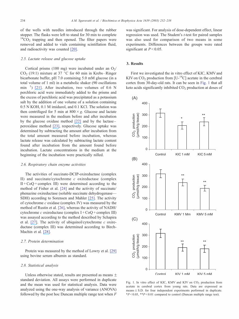
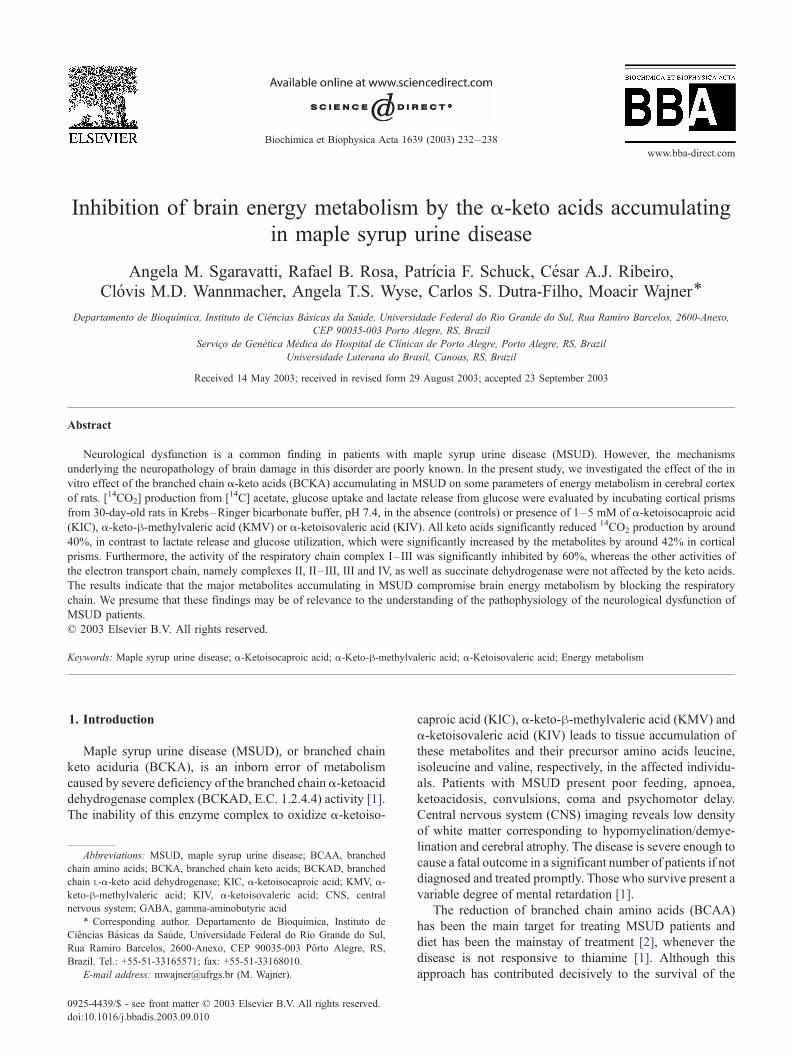
Fig. 1. In vitro effect of KIC, KMV and KIV on CO2 production from
acetate in cerebral cortex from young rats. Data are expressed as
meansF S.D. for four independent experiments performed in duplicate.
*P< 0.05, **P < 0.01 compared to control (Duncan multiple range test).
A.M. Sgaravatti et al. / Biochimica et Biophysica Acta 1639 (2003) 232–238234
of the wells with needles introduced through the rubber
stopper. The flasks were left to stand for 30 min to complete14CO2 trapping and then opened. The filter papers were
removed and added to vials containing scintillation fluid,
and radioactivity was counted [20].
2.5. Lactate release and glucose uptake
Cortical prisms (100 mg) were incubated under an O2/
CO2 (19:1) mixture at 37 jC for 60 min in Krebs–Ringer
bicarbonate buffer, pH 7.0 containing 5.0 mM glucose (in a
total volume of 1 ml) in a metabolic shaker (90 oscillations
min� 1) [21]. After incubation, two volumes of 0.6 N
perchloric acid were immediately added to the prisms and
the excess of perchloric acid was precipitated as a potassium
salt by the addition of one volume of a solution containing
0.5 N KOH, 0.1 M imidazol, and 0.1 KCl. The solution was
then centrifuged for 5 min at 800� g. Glucose and lactate
were measured in the medium before and after incubation
by the glucose oxidase method [22] and by the lactase–
peroxidase method [23], respectively. Glucose uptake was
determined by subtracting the amount after incubation from
the total amount measured before incubation, whereas
lactate release was calculated by subtracting lactate content
found after incubation from the amount found before
incubation. Lactate concentrations in the medium at the
beginning of the incubation were practically nilled.
2.6. Respiratory chain enzyme activities
The activities of succinate-DCIP-oxireductase (complex
II) and succinate/cytochrome c oxireductase (complex
II + CoQ+ complex III) were determined according to the
method of Fisher et al. [24] and the activity of succinate/
phenazine oxireductase (soluble succinate dehydrogenase—
SDH) according to Sorensen and Mahler [25]. The activity
of cytochrome c oxidase (complex IV) was measured by the
method of Rustin et al. [26], whereas the activity of NADH/
cytochrome c oxireductase (complex I +CoQ+ complex III)
was assayed according to the method described by Schapira
et al. [27]. The activity of ubiquinol/cytochrome c oxire-
ductase (complex III) was determined according to Birch-
Machin et al. [28].
2.7. Protein determination
Protein was measured by the method of Lowry et al. [29]
using bovine serum albumin as standard.
2.8. Statistical analysis
Unless otherwise stated, results are presented as means Fstandard deviation. All assays were performed in duplicate
and the mean was used for statistical analysis. Data were
analyzed using the one-way analysis of variance (ANOVA)
followed by the post hoc Duncan multiple range test when F
was significant. For analysis of dose-dependent effect, linear
regression was used. The Student’s t-test for paired samples
was also used for comparison of two means in some
experiments. Differences between the groups were rated
significant at P < 0.05.
3. Results
First we investigated the in vitro effect of KIC, KMVand
KIVon CO2 production from [U-14C] acetate in the cerebral
cortex from 30-day-old rats. It can be seen in Fig. 1 that all
keto acids significantly inhibited CO2 production at doses of
A.M. Sgaravatti et al. / Biochimica et Biophysica Acta 1639 (2003) 232–238 235
1 mM and higher with maximal inhibition around 42%
(KIC: [F(2,9) = 10.349; P < 0.01]; KMV:[F(2,9) = 16.781;
P < 0.01]; KIV:[F(2,9) = 20.146; P < 0.01]). The effect was
concentration-dependent for KMV (h =� 0.7996; P < 0.01)
and KIV (h =� 0.6408; P < 0.05). We also tested the effect
of 5.0 mM KIC on CO2 production from [1,5-14C] citrate.
We verified that the acid significantly inhibited CO2 forma-
tion [t(17) = 21.41; P < 0.001] (means: control = 1388.94;
KIC = 1161.47).
Next we investigated whether the keto acids could affect
glycolysis, by assessing the effect of these compounds on
glucose uptake and lactate release in the brain tissue. As can
be observed in Fig. 2, KIC [F(2,12) = 17.652; P < 0.01],
Fig. 2. In vitro effect of KIC, KMV and KIV on glucose utilization by
cerebral cortex from young rats. Data are expressed as meansF S.D. for
four or five independent experiments performed in duplicate. *P< 0.05,
**P < 0.01 compared to control (Duncan multiple range test).
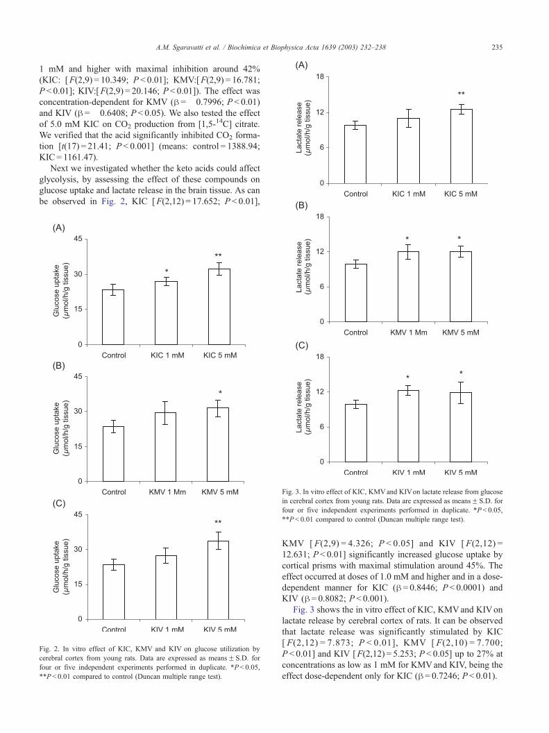
Fig. 3. In vitro effect of KIC, KMVand KIVon lactate release from glucose
in cerebral cortex from young rats. Data are expressed as meansF S.D. for
four or five independent experiments performed in duplicate. *P < 0.05,
**P < 0.01 compared to control (Duncan multiple range test).
KMV [ F(2,9) = 4.326; P < 0.05] and KIV [ F(2,12) =
12.631; P < 0.01] significantly increased glucose uptake by
cortical prisms with maximal stimulation around 45%. The
effect occurred at doses of 1.0 mM and higher and in a dose-
dependent manner for KIC (h = 0.8446; P < 0.0001) and
KIV (h = 0.8082; P < 0.001).
Fig. 3 shows the in vitro effect of KIC, KMVand KIVon
lactate release by cerebral cortex of rats. It can be observed
that lactate release was significantly stimulated by KIC
[ F(2,12) = 7.873; P < 0.01], KMV [ F(2,10) = 7.700;
P < 0.01] and KIV [F(2,12) = 5.253; P < 0.05] up to 27% at
concentrations as low as 1 mM for KMVand KIV, being the
effect dose-dependent only for KIC (h = 0.7246; P < 0.01).

Table 1
In vitro effect of KIC, KMV and KIVon the activities of the mitochondrial
respiratory chain complexes in cerebral cortex from young rats
Respiratory chain
complexes
Control KIC
(1 mM)
KMV
(1 mM)
KIV
(1 mM)
Complex I +CoQ+ III 20F 6.3 9.9F 2.1* 8.1F1.7* 9.9F 4.7*
Succinate dehydrogenase 25F 3.0 25F 2.8 24F 2.1 23F 1.1
Succinate DCIP
oxireductase (II)
12F 0.7 12F 1.8 13F 2.1 13F 2.4
Complex II +CoQ+ III 52F 1.7 52F 2.4 53F 3.2 53F 2.4
Complex III 92F 19 80F 16 73F 5.1 83F 3.7
Cytochrome c oxidase
(IV)
132F 13 148F 16 145F 15 139F 5.0
Data are expressed as meansF S.D. for three or five independent
experiments performed in triplicate.
*P< 0.05 compared to control (Duncan multiple range test).
A.M. Sgaravatti et al. / Biochimica et Biophysica Acta 1639 (2003) 232–238236
We also assessed the effect of KIC, KIV and KMVon the
respiratory chain enzyme activities in an attempt to elucidate
the biochemical defect responsible for the inhibition of
aerobic glycolysis (lower CO2 formation) and activation of
anaerobic glycolysis (increased lactate release) by the a-keto
acids accumulating inMSUD. It can be seen in Table 1 that all
a-keto acids significantly inhibited complex I–III [F(3,16) =
8.492, P < 0.001] at 1 mM concentration with maximal
inhibition around 60%, without affecting the activity of the
other respiratory chain complexes (complex II: [F(3,8) =
0.225; P>0.05], SDH: [F(3,8) = 0.253; P>0.05], complex
II– III: [F(3,8) = 0.190, P>0.05], complex III: [F(3,8) =
1.105, P>0.05], complex IV: [F(3,8) = 0.925, P>0.05]).
4. Discussion
Tissue accumulation and high urinary excretion of KIC,
KMV and KIV occurs in MSUD, an inherited metabolic
disorder caused by deficiency of the BCKAD activity.
Although neurological symptoms are predominant in this
disease, the mechanisms responsible for the brain damage in
the affected individuals are poorly established. The under-
standing of the exact biochemical alterations in brain may
possibly contribute to a better therapeutic management of
MSUD patients.
Since the BCKA, which are converted by transamination
to their respective BCAA leucine, valine and isoleucine, are
the major metabolites accumulating in MSUD, in the
present study we studied the in vitro effect of KIC, KIV
and KMV on various biochemical parameters of energy
metabolism in cerebral cortex of rats. We investigated the
activity of the Krebs cycle by measuring the CO2 generated
from acetate and the anaerobic metabolism by measuring
lactate release from glucose. We initially verified a signif-
icant reduction of CO2 formation from acetate by over 40%
in cortical prisms incubated in the presence of each a-keto
acid. Since the BCKA are monocarboxylic acids and there-
fore use the same membrane transporter as acetate (mono-
carboxylic transporter), the inhibition observed could be due
to a competition between the BCKA and acetate [30].
However, we also verified that CO2 formation from citrate
was also inhibited by KIC, the BCKA found in greater
concentrations and that easily cross cell membranes. Since
citrate uses the tricarboxylic transporter, it is feasible that the
reduction of CO2 caused by the BCKA reflects a true
inhibition of the Krebs cycle or the respiratory chain. It is
interesting to observe that all keto acids significantly
inhibited CO2 production at 1 mM concentration.
Lactate is produced in considerable amounts by the brain,
and more specifically by glial cells. Lactate release, which
reflects lactate production, was significantly stimulated in
the presence of all metabolites by around 20–30%. There-
fore, it may be concluded that the a-keto acids accumulating
in MSUD reduce the Krebs cycle activity and increase
anaerobic glycolysis, indicating that they may alter the
energy metabolism in brain cortex of rats.
We have also verified that the BCKA provoked a
significant increase of glucose uptake by around 40% in
brain cortical prisms, and this is expected once anaerobic
metabolism, which was activated by these compounds, uses
more glucose because less ATP is produced. It should be
stressed that glucose is the major substrate utilized for
neural cell metabolism.
The next experiments were performed in order to evaluate
the effect of the BCKA on the respiratory chain function by
measuring the activities of complexes I–IV in cerebral
cortex of rats. We verified that all BCKA significantly
inhibited complex I–III by around 50–60%, without altering
the activities of complexes II, II–III, III, IV and succinate
dehydrogenase. Since complex III was not affected by the
BCKA, it may be presumed that complex I activity was
blocked by the BCKA. In summary, our findings indicate
that the BCKA accumulating in MSUD impair brain energy
metabolism, possibly due to inhibition of the respiratory
chain complex I. These results confirm other reports showing
that energy metabolism is compromised by the metabolites
accumulating in MSUD [2,7–10,19]. They are also in
agreement with a recent report showing that the BCKA
markedly reduced cell respiration but did not impair the rate
of mitochondrial succinate oxidation in neuronal and glial
cells [19]. Since succinate enters the respiratory chain via
complex II, these investigators conclude that the BCKA did
not affect the function of respiratory chain complexes II, III
and IV. Taken together, these observations and our present
investigation, it is feasible that cell respiration is inhibited by
BCKA at complex I, and this is possibly the mechanism
through which the Krebs cycle is blocked and the anaerobic
glycolysis is stimulated in the presence of these metabolites.
Interestingly, isolated human complex I defects have
been identified in a number of neurodegenerative diseases,
including Parkinson’s disease, focal dystonia and Leber’s
hereditary optic neuropathy [31], a fact that suggests that the
activity of this respiratory chain complex is important for
normal CNS function. Another interesting observation was
that the administration of 1-methyl-4-phenyl 1,2,3,6 tetra-

A.M. Sgaravatti et al. / Biochimica et Biophysica Acta 1639 (2003) 232–238 237
hydropyridine (MPTP), which is an inductor of Parkinson-
ism in animals, inhibits complex I activity probably by
oxidative damage of complex I since the inhibition is
prevented by free radical scavengers [32,33]. Furthermore,
complex I inhibition induces free radical generation from
the respiratory chain, suggesting a self-amplifying cycle of
complex I deficiency that may result in progressive cell
damage [34]. In this context, we have recently demonstrated
that the BCKA elicits oxidative stress in brain [35]. There-
fore, it is possible that the inhibition of complex I by these
compounds may have occurred via oxidation of essential
subunits of this complex. The observation that the activity
of NADH-CoQ oxidoreductase (complex I) is very sensitive
to reactive oxygen species [36–38] corroborates with this
hypothesis. It seems that the reduction of complex I activity
depends either on a reversible oxidation of sulfhydryl
groups or on an irreversible oxidative modification of
[4Fe–4S] clusters of the enzyme [38].
In conclusion, we present evidence of an electron transport
chain inhibition, probably at complex I in the brain caused by
the BCKA accumulating in MSUD at the concentrations
usually found in the affected individuals [39]. We observed
that 1 mM concentration of the various BCKA compromised
brain energy metabolism. Although lower concentrations of
these compounds were not used in our assays, it is feasible
that doses less than 1 mM might also be inhibitory, and this
may possibly have pathophysiologic relevance for MSUD
patients with moderate increases in these keto acids. This
probably explains previous reports of impaired energy pro-
duction caused by these metabolites, as identified by lower
CO2 production or increased lactate release [9,40–44]. It is
difficult to extrapolate our findings to the human condition.
However, if the in vitro inhibition of brain energymetabolism
caused by the metabolites which most accumulate in MSUD
also occurs under in vivo conditions, it is conceivable that
lack of energymay be involved in the neurological symptoms
present in MSUD patients. An interesting observation is that
these patients present hypoglycemia and cerebral edema,
particularly during metabolic decompensation, when the
levels of the BCKA and BCAA dramatically increase [1],
reflecting a failure of the active ionic transport necessary to
maintain the normal volume of neural cells. Whether energy
deficit or other abnormalities, such as oxidative stress or
excitotoxicity, is mainly responsible for brain damage in
MSUD patients is a matter of future investigation.
Acknowledgements
This work was supported by Conselho Nacional de
Desenvolvimento Cientıfico e Tecnologico (CNPq), Coor-
denac�ao de Aperfeic�oamento de Pessoal de Ensino Superior
(CAPES), Financiadora de Estudos e Projetos (FINEP), and
Pro-Reitoria de Pesquisa e Pos-Graduac�ao da Universidade
Federal do Rio Grande do Sul (PROPESQ/UFRGS).
References
[1] D.T. Chuang, V.E. Shih, Maple syrup urine disease (branched-chain
ketoaciduria), in: C.R. Scriver, A.L. Beaudet, W.L. Sly, D. Valle
(Eds.), The Metabolic and Molecular Bases of Inherited Disease,
8th ed., McGraw-Hill, New York, 2001, pp. 1971–2005.
[2] D.J. Danner, L.J. Elsas II, Disorders of branched chain amino acid and
keto acid metabolism, in: C.R. Scriver, A.L. Beaudet, W.L. Sly, D.
Valle (Eds.), The Metabolic Basis of Inherited Disease,McGraw-Hill,
New York, 1989, pp. 671–692.
[3] M.L. Efron, Aminoaciduria, N. Engl. J. Med. 272 (1965) 1058–1067.
[4] S.E. Snyderman, P.M. Norton, E. Roitman, Maple syrup urine dis-
ease with particular reference to diet therapy, Pediatrics 34 (1964)
454–472.
[5] C.F. Mello, L. Feksa, A.M. Brusque, C.M. Wannmacher, M. Wajner,
Chronic early leucine administration induces behavioral deficits in
rats, Life Sci. 8 (1999) 747–755.
[6] A.S. Coitinho, C.F. de Mello, T.T. Lima, J. de Bastiani, M.R. Fighera,
M. Wajner, Pharmacological evidence that alpha-ketoisovaleric acid
induces convulsions through GABAergic and glutamatergic mecha-
nisms in rats, Brain Res. 894 (2001) 68–73.
[7] R.K. Howell, M. Lee, Influence of a-keto acids on the respiration of
brain in vitro, Proc. Soc. Exp. Biol. Med. 113 (1963) 660–663.
[8] A.P. Halestrap, M.D. Brand, R.M. Denton, Inhibition of mitochon-
drial pyruvate transport by phenylpyruvate and a-ketoisocaproate,
Biochem. Biophys. Acta 367 (1974) 102–108.
[9] J.M. Land, J. Mowbray, J.B. Clark, Control of pyruvate and h-hy-droxybutyrate utilization in rat brain mitochondria and its relevance to
phenylketonuria and maple syrup urine disease, J. Neurochem. 26
(1976) 823–830.
[10] C. Pilla, R.F.D Cardozo, C.S. Dutra, A.T.S. Wyze, M. Wajner, C.M.D.
Wannmacher, Effect of leucine administration on creatine kinase ac-
tivity in rat brain, Metab. Brain Dis. 18 (2003) 17–25.
[11] R.E. Tashian, Inhibition of brain glutamic acid decarboxylase by phe-
nylalanine, valine, and leucine derivatives: a suggestion concerning the
etiology of the neurological defect in phenylketonuria and branched-
chain ketonuria, Metabolism 10 (1961) 393–402.
[12] S.H. Appel, Inhibition of brain protein synthesis: an approach to a
biochemical basis of neurological dysfunction in the amino-acidurias,
Trans. N. Y. Acad. Sci. 29 (1966) 63–70.
[13] T. Taketomi, T. Kunishita, A. Hara, S. Mizushima, Abnormal protein
and lipid compositions of the cerebral myelin of a patient with maple
syrup urine disease, Jpn. J. Exp. Med. 53 (1983) 109–116.
[14] D. Tribble, R. Shapira, Myelin proteins: degradation in rat brain ini-
tiated by metabolites causative of maple syrup urine disease, Bio-
chem. Biophys. Res. Commun. 114 (1983) 440–446.
[15] E. Treacy, C.L. Clow, T.R. Reade, D. Chitayat, O.A. Mamer, C.R.
Scriver, Maple syrup urine disease: interrelationship between branched
chain amino-, oxo-, and hydroxyacids; implications for treatment;
association with CNS dysmelination, J. Inherit. Metab. Dis. 15
(1992) 121–135.
[16] M. Wajner, C.R. Vargas, Reduction of plasma concentrations of large
neutral amino acids in patients with maple urine disease during crises,
Arch. Dis. Child. 80 (1999) 579.
[17] M. Wajner, D.M. Coelho, A.G. Barschak, P.R. Araujo, R.F. Pires, F.L.
Lulhier, C.R. Vargas, Reduction of large neutral amino acid concen-
tration in plasma and CSF of patients with maple syrup urine disease
during crises, J. Inherit. Metab. Dis. 23 (2000) 505–512.
[18] P. Araujo, G.F. Wassermann, K. Tallini, V. Furnaletto, C.R. Vargas,
C.M. Wannmacher, C.S. Dutra-Filho, A.T. Wyse, M. Wajner, Reduc-
tion of large neutral amino acid levels in plasma and brain of hyper-
leucinemic rats, Neurochem. Int. 38 (2001) 529–537.
[19] P. Jouvet, P. Rustin, D.L. Taylor, J.M. Pocock, U. Felderhoff-Mueser,
N.D. Mazarakis, C. Sarraf, U. Joashi, M. Koszma, K. Greewood, A.D.
Mehmet, H. Mehmet, Branched chain amino acids induce apoptosis in
neural cells without mitochondrial membrane despolarization or cy-
A.M. Sgaravatti et al. / Biochimica et Biophysica Acta 1639 (2003) 232–238238
tochrome c release: implications for neurological impairment associ-
ated with maple syrup urine disease, Mol. Biol. Cell. 11 (2000)
1919–1932.
[20] C.S. Dutra-Filho, M. Wajner, E. Gassen, R. Candiago, A. Wihlems,
H. Malfussi, C.M.D. Wannmacher, Effect of organic acids on in vitro
glucose oxidation by cerebral cortex of young rats, Med. Sci. Res. 23
(1995) 25–26.
[21] J.C. Dutra, M. Wajner, C.F. Wannmacher, C.S. Dutra-Filho, C.M.D.
Wannmacher, Effects of methylmalonate and propionate on glucose
and ketone bodies uptake in vitro by brain of developing rats, Bio-
chem. Med. Metab. Res. 45 (1991) 56–64.
[22] P.A. Trinder, Determination of blood glucose using on oxidase-per-
oxidase system with a non-carcinogenic chromogen, J. Clin. Pathol.
22 (1969) 158–161.
[23] N. Shimojo, K. Naka, C. Nakajima, C. Yoshikawa, K. Okuda, K.
Okada, Test-strip method for measuring lactate in whole blood, Clin.
Chem. 35 (1989) 1992–1994.
[24] J.C. Fisher, W. Ruitenbeek, J.A. Berden, J.M. Trijbels, J.H. Veer-
kamp, M. Stadhouders, R.C. Sengers, A.J. Janssen, Differential in-
vestigation of the capacity of succinate oxidation in human skeletal
muscle, Clin. Chim. Acta 153 (1985) 23–36.
[25] R.G. Sorensen, H.R. Mahler, Localization of endogenous ATPases at
the nerve terminal, J. Bioenerg. Biomembranes 14 (1982) 527–547.
[26] P. Rustin, D. Chretien, T. Bourgeron, B. Gerard, A. Rotig, J.M. Sau-
dubray, A. Munnich, Biochemical and molecular investigations in
respiratory chain deficiencies, Clin. Chim. Acta 228 (1994) 35–51.
[27] A.H. Schapira, V.M. Mann, J.M. Cooper, D. Dexter, S.E. Daniel, P.
Jenner, J.B. Clark, C.D. Marsden, Anatomic and disease specificity of
NADH CoQ1 reductase (complex I) deficiency in Parkinson’s dis-
ease, J. Neurochem. 55 (1990) 2142–2145.
[28] M.A. Birch-Machin, H.L. Briggs, A.A. Saborido, L.A. Bindoff, D.M.
Turnbull, An evaluation of the measurement of the activities of com-
plexes I– IV in the respiratory chain of human skeletal muscle mito-
chondria, Biochem. Med. Metabol. Biol. 51 (1994) 35–42.
[29] O.H. Lowry, N.J. Rosebrough, A.L. Farr, R.J. Randall, Protein meas-
urement with the folin phenol reagent, J. Biol. Chem. 193 (1951)
265–275.
[30] G.F. Oldendorf, Carrier-mediated blood–brain barrier transport of
short-chain monocarboxylic organic acids, Am. J. Physiol. 224
(1973) 1450–1453.
[31] A.H.V. Schapira, Human complex I defects in neurodegenerative dis-
eases, Biochem. Biophys. Acta 1364 (1998) 261–270.
[32] M.W. Cleeter, J.M. Cooper, A.H. Shapira, Irreversible inhibition of
mitochondrial complex I by 1-methyl-4-phenylpyridinium: evidence
for free radical involvement, J. Neurochem. 58 (1992) 786–789.
[33] K.F. Tipton, T.P. Singer, Advances in our understanding of the mech-
anisms of neurotoxicity of MPTP and related compounds, J. Neuro-
chem. 61 (1993) 1191–1206.
[34] M.L. Genova, B. Ventura, G. Giuliano, C. Bovina, G. Formiggini,
G.P. Castelli, G. Lenaz, The site of production of superoxide radical in
mitochondrial complex I is not a bound ubisemiquinone but presum-
ably iron–sulfur cluster N2, FEBS Lett. 505 (2001) 364–368.
[35] F.U. Fontella, E. Gassen, V. Pulrolnik, C.M.D. Wannmacher, A.B.
Klein, M. Wajner, C.S. Dutra, Stimulation of lipid peroxidation in
vitro in rat brain by metabolites accumulating in maple syrup urine
disease, Metab. Brain Dis. 17 (2002) 47–54.
[36] A. Dupius, J.M Skehel, J.M. Walker, NADH: ubiquinone oxidoreduc-
tase from bovine mitochondria, Biochem. J. 277 (1991) 11–15.
[37] Y. Zhang, O. Marcillat, C. Giulivi, L. Ernster, K.J. Davies, The ox-
idative inactivation of mitochondrial electron transport chain compo-
nents and ATPase, J. Biol. Chem. 265 (1990) 16330–16336.
[38] A.P. Kudin, T.A. Kudina, J. Seyfried, S. Vielhaber, H. Beck, C.E.
Elger, W.S. Kunz, Seizure-dependent modulation of mitochondrial
oxidative phosphorylation in rat hippocampus, Eur. J. Neurosci. 15
(2002) 1105–1114.
[39] Y. Shigematsu, K. Kikuchi, T. Momoi, Organic acids and branched-
chain amino acids in body fluids before and after multiple exchange
transfusions in maple syrup urine disease, J. Inherit. Metab. Dis. 6
(1983) 183–189.
[40] R.H. Jackson, T.P. Singer, Inactivation of the 2-ketoglutarate and
pyruvate dehydrogenase complexes of beef heart by branched chain
keto acids, J. Biol. Chem. 258 (1983) 1857–1865.
[41] G.E. Gibson, J.P. Blass, Inibition of acetylcholine synthesis and of
carbohydrate utilization by maple syrup urine disease metabolites,
J. Neurochem. 26 (1976) 1073–1078.
[42] E. Walajtys-Rode, J.R. Williamson, Effects of branched chain a-ke-
toacids on the metabolism of isolated rat liver cells: III. Interactions
with pyruvate dehydrogenase, J. Biol. Chem. 255 (1980) 413–418.
[43] H.R. Zielke, Y. Huang, P.J. Baab, R.M. Collins, C.L. Zielke, J.T.
Tildon, Effect of alpha-ketoisocaproate and leucine on the in vivo
oxidation of glutamate and glutamine in the rat brain, Neurochem.
Res. 22 (1997) 1159–1164.
[44] M.C. McKenna, U. Sonnewald, X. Huang, J. Stevenson, S.F. Johnsen,
L.M. Sande, H.R. Zielke, Alpha-ketoisocaproate alters the production
of both lactate and aspartate from [U-13C] glutamate in astrocytes: a13C NMR study, J. Neurochem. 70 (1998) 1001–1008.
Related Documents