Hindawi Publishing Corporation Biochemistry Research International Volume 2010, Article ID 516704, 7 pages doi:10.1155/2010/516704 Research Article Inhaled Anesthetics Promote Albumin Dimerization through Reciprocal Exchange of Subdomains Benjamin J. Pieters, 1 Eugene E. Fibuch, 1 Joshua D. Eklund, 2 and Norbert W. Seidler 2 1 Department of Anesthesiology, University of Missouri-Kansas City School of Medicine, 4401 Wornall Road, Kansas City, MO 64111, USA 2 Biochemistry Department, Kansas City University of Medicine and Biosciences, 1750 Independence Avenue, Kansas City, MO 64106, USA Correspondence should be addressed to Norbert W. Seidler, [email protected] Received 1 October 2009; Revised 9 December 2009; Accepted 14 January 2010 Academic Editor: Vladimir Uversky Copyright © 2010 Benjamin J. Pieters et al. This is an open access article distributed under the Creative Commons Attribution License, which permits unrestricted use, distribution, and reproduction in any medium, provided the original work is properly cited. Inhaled anesthetics affect protein-protein interaction, but the mechanisms underlying these effects are still poorly understood. We examined the impact of sevoflurane and isoflurane on the dimerization of human serum albumin (HSA), a protein with anesthetic binding sites that are well characterized. Intrinsic fluorescence emission was analyzed for spectral shifting and self-quenching, and control first derivatives (spectral responses to changes in HSA concentration) were compared against those obtained from samples treated with sevoflurane or isoflurane. Sevoflurane increased dimer-dependent self-quenching and both decreased oligomer- dependent spectral shifting, suggesting that inhaled anesthetics promoted HSA dimerization. Size exclusion chromatography and polarization data were consistent with these observations. The data support the proposed model of a reciprocal exchange of subdomains to form an HSA dimer. The open-ended exchange of subdomains, which we propose occuring in HSA oligomers, was inhibited by sevoflurane and isoflurane. 1. Introduction Inhaled anesthetics affect protein-protein interaction by disrupting heteromeric [1] and homomeric [2] binding in some cases and by promoting such interactions in others [3, 4]. These observations warrant further examination of the interfacial cavities that regulate protein-protein interaction. Human serum albumin (HSA) provides a useful model in studying these processes, since it can form dimers [5] and oligomers [6] in a concentration-dependent manner [7]. HSA has three homologous domains (I–III), each with two subdomains (a, b), and inhaled anesthetics bind at several sites [8–13], one of which is located at the cleft between subdomains IIa and IIb. This high-affinity site is at the external side of a cavity that contains the protein’s only tryptophan (W214), whose properties were tracked fluorometrically. We recently proposed [14] that this site can interchange with a site on another HSA forming reciprocal intersubdomain interactions in a dimeric conformation. The current study suggests that this process is modulated by inhaled anesthetics. 2. Materials and Methods 2.1. Preparation of Samples. HSA (10 mg/mL; Sigma- Aldrich: A-1653, A-8763) was dissolved in a 10 mM sodium phosphate buffer (pH = 7.4), passed through a 0.2 μm filter, diluted to 0.5–6.0 mg/mL and allowed to equilibrate. HSA was approximately 99% pure as determined by agarose gel electrophoresis and was essentially free of globulins. Samples (1.0 mL) were treated with and without sevoflurane (12 μL; 15 minutes on rotator at 22 ◦ C) and then assayed as described below. Sevoflurane (Abbott; bp: 58.6 ◦ C; density: 1.52 g/cm 3 ) and isoflurane (Hospira; bp: 48.5 ◦ C; density: 1.50 g/cm 3 ) were layered with argon following use. In order to effectively

Welcome message from author
This document is posted to help you gain knowledge. Please leave a comment to let me know what you think about it! Share it to your friends and learn new things together.
Transcript
Hindawi Publishing CorporationBiochemistry Research InternationalVolume 2010, Article ID 516704, 7 pagesdoi:10.1155/2010/516704
Research Article
Inhaled Anesthetics Promote Albumin Dimerizationthrough Reciprocal Exchange of Subdomains
Benjamin J. Pieters,1 Eugene E. Fibuch,1 Joshua D. Eklund,2 and Norbert W. Seidler2
1 Department of Anesthesiology, University of Missouri-Kansas City School of Medicine,4401 Wornall Road, Kansas City, MO 64111, USA
2 Biochemistry Department, Kansas City University of Medicine and Biosciences,1750 Independence Avenue, Kansas City, MO 64106, USA
Correspondence should be addressed to Norbert W. Seidler, [email protected]
Received 1 October 2009; Revised 9 December 2009; Accepted 14 January 2010
Academic Editor: Vladimir Uversky
Copyright © 2010 Benjamin J. Pieters et al. This is an open access article distributed under the Creative Commons AttributionLicense, which permits unrestricted use, distribution, and reproduction in any medium, provided the original work is properlycited.
Inhaled anesthetics affect protein-protein interaction, but the mechanisms underlying these effects are still poorly understood. Weexamined the impact of sevoflurane and isoflurane on the dimerization of human serum albumin (HSA), a protein with anestheticbinding sites that are well characterized. Intrinsic fluorescence emission was analyzed for spectral shifting and self-quenching, andcontrol first derivatives (spectral responses to changes in HSA concentration) were compared against those obtained from samplestreated with sevoflurane or isoflurane. Sevoflurane increased dimer-dependent self-quenching and both decreased oligomer-dependent spectral shifting, suggesting that inhaled anesthetics promoted HSA dimerization. Size exclusion chromatographyand polarization data were consistent with these observations. The data support the proposed model of a reciprocal exchangeof subdomains to form an HSA dimer. The open-ended exchange of subdomains, which we propose occuring in HSA oligomers,was inhibited by sevoflurane and isoflurane.
1. Introduction
Inhaled anesthetics affect protein-protein interaction bydisrupting heteromeric [1] and homomeric [2] binding insome cases and by promoting such interactions in others[3, 4]. These observations warrant further examination of theinterfacial cavities that regulate protein-protein interaction.Human serum albumin (HSA) provides a useful modelin studying these processes, since it can form dimers [5]and oligomers [6] in a concentration-dependent manner[7]. HSA has three homologous domains (I–III), each withtwo subdomains (a, b), and inhaled anesthetics bind atseveral sites [8–13], one of which is located at the cleftbetween subdomains IIa and IIb. This high-affinity site isat the external side of a cavity that contains the protein’sonly tryptophan (W214), whose properties were trackedfluorometrically. We recently proposed [14] that this site caninterchange with a site on another HSA forming reciprocal
intersubdomain interactions in a dimeric conformation. Thecurrent study suggests that this process is modulated byinhaled anesthetics.
2. Materials and Methods
2.1. Preparation of Samples. HSA (10 mg/mL; Sigma-Aldrich: A-1653, A-8763) was dissolved in a 10 mM sodiumphosphate buffer (pH = 7.4), passed through a 0.2 μm filter,diluted to 0.5–6.0 mg/mL and allowed to equilibrate. HSAwas approximately 99% pure as determined by agarose gelelectrophoresis and was essentially free of globulins. Samples(1.0 mL) were treated with and without sevoflurane (12 μL;15 minutes on rotator at 22◦C) and then assayed as describedbelow. Sevoflurane (Abbott; bp: 58.6◦C; density: 1.52 g/cm3)and isoflurane (Hospira; bp: 48.5◦C; density: 1.50 g/cm3)were layered with argon following use. In order to effectively
2 Biochemistry Research International
Wavelength (nm)
300 350 400 450 500
Flu
ores
cen
ceem
issi
on(r
fu)
0
30
60
90
120
150
180
HSA (mg/mL)
0 0.1 0.2 0.3 0.4 0.5
AU
C(r
fu×
103)
0
6
12
18
24
Figure 1: Fluorescence spectra at low HSA concentrations. Trypto-phan fluorescence spectra were obtained as described in Materialsand Methods. Ascending tracings represent HSA at 0.1, 0.2, 0.3, 0.4,and 0.5 mg/mL, respectively. Inset, plot of the AUC (areas underthe curve of entire spectral region) against HSA concentration (r2 =0.997).
assess the role of agent on HSA self-association at variousprotein concentrations, it was necessary for us to use satu-rating levels of sevoflurane and isoflurane (12 μL in 1.0 mLsamples), which are immiscible in water and appear as a verysmall bolus at the base of the test vessel. Thus, all of thebinding sites that were previously identified in the literaturewould be likely saturated. By attempting to use substantiallylower volumes, a bolus would not form resulting in a coaton the surface that volatilizes rapidly, leaving an uncertaintyassociated with partial binding of agent particularly sinceprotein concentration was varied. Under the conditions usedin this study, protein denaturation does not occur [15], andHSA returned to its native state approximately 21 hours afterremoval of agent (data not shown).
2.2. Fluorescence Spectroscopy. Tryptophan emission spectra(e.g., 293 nm; em, 300–500 nm; 2.5 nm slits; 600 nm/min;290 nm em cutoff) were obtained at various HSA concen-trations using a Perkin Elmer LS50B luminescence spec-trometer. Values for the center of spectral mass (CSM =∑
(νFi)di/∑
Fi di, i = 33.3 cm−1), where (ν) is wavenumberand (Fi) is emission intensity, were calculated from theemission spectra. They indicate spectral position and reflectthe conformational events that change hydration at theW214 [16]. We calculated a parameter from the spectraldata that reflects the magnitude of concentration-dependentself-quenching [14]. Fluorescence intensities of HSA atconcentrations below 0.5 mg/mL were measured as areasunder the curve (Fo =
∑Fi di), showing a linear increase
with protein concentration (Figure 1) and demonstratingthat there was no self-quenching.
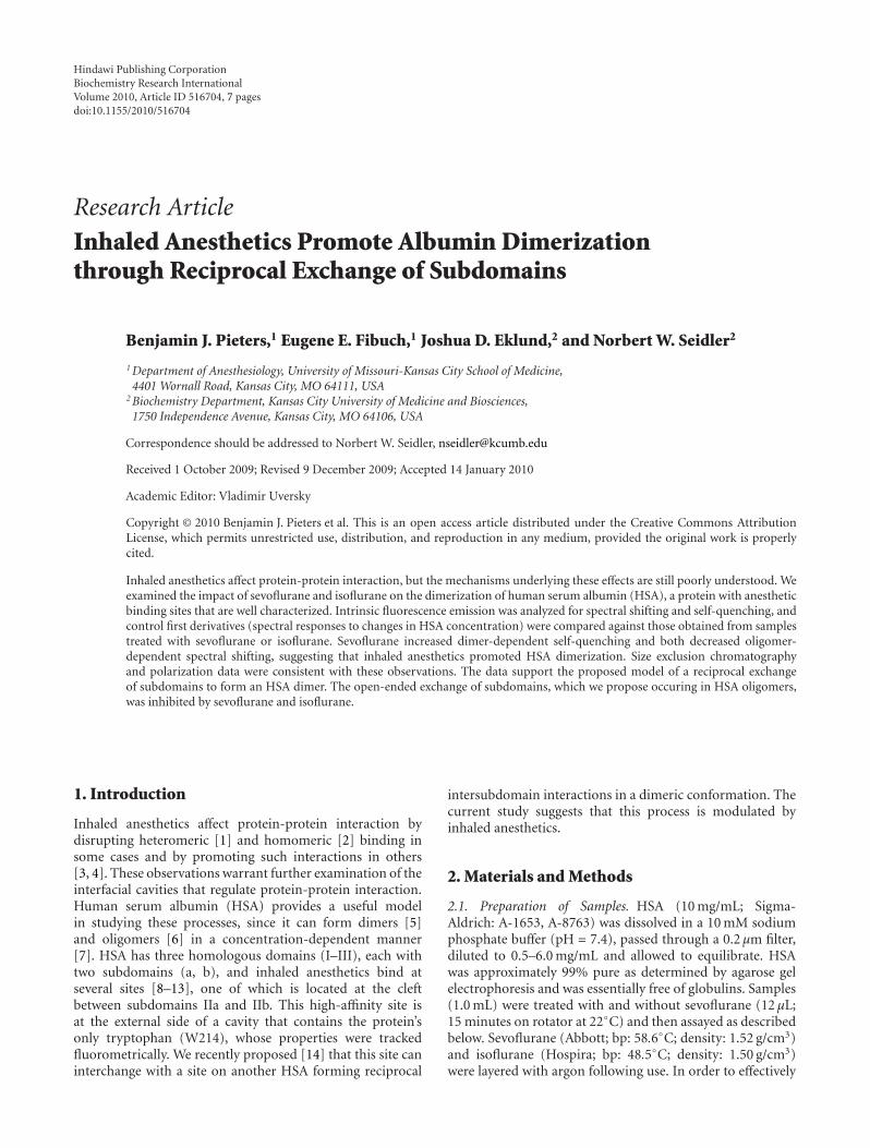
Regression lines (Figure 2) were extended to give aprojected Fo for control and for samples treated with agent.At higher HSA concentrations, fluorescence (F) intensitiesdeviated from the linear trajectory (Figure 2) similar to
[HSA] (mg/mL)
0.5 0.75 1 1.25 1.5
Are
au
nde
rcu
rve
(rfu
)
8000
12000
16000
20000
24000
28000
(a)
[HSA] (mg/mL)
0.5 0.75 1 1.25 1.5
Are
au
nde
rcu
rve
(rfu
)
8000
12000
16000
20000
24000
28000
4000
(b)
Figure 2: Observed fluorescence shown relative to projectedunquenched maxima. Extrapolation of regression lines were drawnfrom fluorescence data that were obtained at low HSA concentra-tions from control (solid line), sevoflurane-[A] and isoflurane [B]-treated (dashed line) samples. The projected unquenched maximaare compared with data (dotted line) observed at the concentrationsshown (control, closed circles; sevoflurane-[A] and isoflurane [B]-treated, open circles). The difference between the regression linesand the dotted lines represents IQU.
the self-quenching that occurs in the homomeric assemblyof protein ionophores [17]. The difference between theextrapolated linear projection (Fo) and the observed F (IQU= Fo− F) provides a useful parameter, which we termed inte-grated quench units (IQUs), that represents the magnitude ofquenching at each of the protein concentrations tested.
Solute quenching studies, which use the Stern-Volmerexpression, Fo/F = 1 + kQ, where Q is solute concentration,assume a bimolecular process [18]. This approach involvesfixing protein concentration, which acts as the fluorophore,and varies quencher concentration. In our study, we variedthe fluorophore concentration, or HSA, which acts also asthe quencher, making the term, Fo/F, inappropriate. Thedifference term, Fo− F, is commonly used with fluorophores
Biochemistry Research International 3
that self-quench [19] and with differentially quenchedfluorophores in a single protein [18]. The quenched complex(dimer/oligomer) is implied by the expression, Fo − F =IQU, which was tracked as a function of HSA concentrationand dependent upon at least two equilibria (monomer/dimerand monomer/trimer). IQU and CMS were calculated fromthe spectra obtained at various concentrations of HSA inthe presence and absence of sevoflurane or isoflurane. Fromthese discrete data, first derivatives and second derivativeswere calculated as previously described [14]. The parametersused in this study (i.e., first derivatives) did not lend them-selves well for investigation of dose-response relationships.Tryptophan polarization was also measured at ex/em of293 nm/345 nm (5 nm slits; 10 s integrations) and data givenin millipolarization (mP) units.
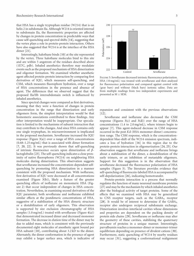
2.3. Size Exclusion Chromatography. Samples were runthrough a gel filtration column (1.5 × 12 cm; agarose-basedBioGel A 1.5 m fine) using a BioLogic LC system (BioRad).A sodium citrate (10 mM, pH = 6.4) elution buffer at a1.0 mL/min flow rate was used with UV detection (280 nmabsorbance). Quantitation of monomers and dimers wereperformed by either integration of absorbances (SigmaPlot 11.0) or peak height measurements. Comparisons toassess the effects of sevoflurane involved t-tests or one-wayANOVAs with Bonferroni posttests. Calibration standardsgamma globulin (158 kDa) and ovalbumin (44 kDa) wereobtained from BioRad.
3. Results
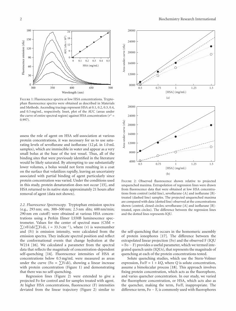
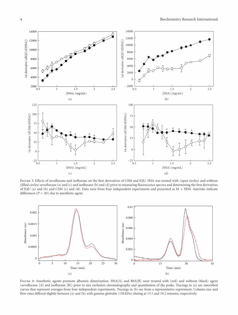
In order to assess the effects of anesthetic agent on HSAdimerization, we examined the spectral response to changesin HSA concentration. The HSA concentration range atwhich dimers and oligomers form was previously established[7, 20]; as HSA increases, the amount of oligomers increasesas well as the size of the multimer from trimer to tetra-,penta-, and hexamers. Once the trimer forms, it is likely themultimerization event is monomeric addition to a polymer.This event can be examined at lower concentrations whichallow for fluorometric analysis without interference due tohigher concentrations. The purpose of this study was toexamine the conformational transitions that are initiallyexpressed at the concentration range used, which mayprovide insight into the behavior of target CNS proteins thatexist in the low micromolar range. In untreated samples,IQU and CSM increase with HSA concentration exhibitinga biphasic response [14], suggesting that self-associationaffects the W214 and that there are different conformationalevents for dimerization and for postdimer oligomerization.With sevoflurane, first derivatives of IQU were greater thancontrol during HSA dimerization over the 0.6 to 1.3 mgHSA/mL range (Figure 3(a)), suggesting that sevofluranepromoted protein-protein interactions that enhanced self-quenching.
With isoflurane, first derivatives of IQU were decreased atall concentrations examined (Figure 3(b)). These disparateobservations may be attributed to differential occupancy
at the subdomain IIa binding site. We previously showedthat second derivatives of IQU from control samples below1 mg HSA/mL differed reproducibly from that obtained atconcentrations above 1 mg HSA/mL [14]. Interestingly, acomparison of second derivatives of IQU between controland treated samples exhibited differences only in the post-dimeric range from 1.0 to 1.75 mg HSA/mL for both agents(control: 4723 ± 465.6 versus isoflurane: 663 ± 582.3, M ±SEM, P <. 0005 and control: 5772± 359.5 versus sevoflurane:3957 ± 161.8, P < .001). All other comparisons of controlversus treated samples were found to be not significant,suggesting that anesthetic agent exhibited the greatest effectin the range thought to involve the early forms of oligomersbeginning with trimers [7]. These results were consistentwith those involving CSM. With sevoflurane, first derivativesof CSM were less than control in the postdimeric or earlyoligomeric phase over the 1.4 to 2.0 mg HSA/mL range(Figure 3(c)), suggesting a stabilization of HSA dimers. Withisoflurane, first derivatives of CSM were also less than controlin precisely the same postdimeric range from 1.4 to 2.0 mgHSA/mL (Figure 3(d)).
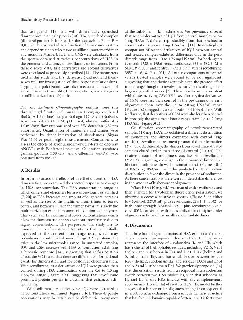
Gel filtration chromatography of sevoflurane-treatedsamples (1.0 mg HSA/mL) exhibited a different distributionof monomers and dimers compared with control (Fig-ure 4(a)). Sevoflurane treatment promoted dimer formation(P < .05). Additionally, the dimers from sevoflurane-treatedsamples eluted earlier than those of control (P < .04). Therelative amount of monomers was less with sevoflurane(P< .03), suggesting a change in the monomer-dimer equi-librium. Isoflurane showed a similar effect (Figure 4(b))at 0.675 mg BSA/mL with the predicted shift in proteindistribution to favor the dimer in the presence of isoflurane.At these concentrations there were no detectable differencesin the amount of higher-order oligomers.
When HSA (10 mg/mL) was treated with sevoflurane andthen analyzed for tryptophan fluorescence polarization, weobserved a decrease relative to control (Figure 5) in eitherlow (control: 227.0 mP; plus sevoflurane, 224.1, P < .02) orhigh ionic strength (control: 228.9; plus sevoflurane: 225.7,P < .005), consistent with a destabilization of higher-orderoligomers in favor of the smaller more mobile dimer.
4. Discussion
The three homologous domains of HSA exist in a V-shape.The apposing lobes represent domains I and III. The vertexrepresents the interface of subdomains IIa and IIb, whichhas a cluster of hydrophobic residues, including V216, V231(helix 2 and 3, subdomain IIa) and L331, L347 (helix 2 and3, subdomain IIb), and has a salt bridge between residueR209 (helix 2, subdomain IIa) and residues D324 and E354(helix 2 and 3, subdomain IIb). We previously proposed [14]that dimerization results from a reciprocal intersubdomainswitch between two HSA molecules, such that subdomainsIIa and IIb of one HSA interact with the complementarysubdomains (IIb and IIa) of another HSA. The model furthersuggests that higher-order oligomers emerge from sequentialintersubdomain exchanges from a unique trimeric structurethat has free subdomains capable of extension. It is fortuitous
4 Biochemistry Research International
[HSA] (mg/mL)
0.5 1 1.5 2 2.52000
4000
6000
8000
10000
12000
14000
1st
deri
vati
ve(d
IQU/d
[HSA
])
(a)
[ H S A ] ( m g / m L )
0 . 5 1 1 . 5 2 2 . 5
2 0 0 0
4 0 0 0
6 0 0 0
8 0 0 0
1 0 0 0 0
1 2 0 0 0
1 4 0 0 0
1 s t
d e r i
v a t i
v e (
dI Q
U /d
[ H S A
] )
0
−2 0 0 0
(b)
0
25
50
75
100
125
[HSA] (mg/mL)
0.5 1 1.5 2 2.5
1st
deri
vati
ve(d
CSM
/d[H
SA])
−25
∗ ∗ ∗ ∗
(c)
0
25
50
75
100
[HSA] (mg/mL)
0.5 1 1.5 2 2.5
1st
deri
vati
ve(d
CSM
/d[H
SA])
−25
∗ ∗ ∗ ∗ ∗
(d)
Figure 3: Effects of sevoflurane and isoflurane on the first derivatives of CSM and IQU. HSA was treated with (open circles) and without(filled circles) sevoflurane (a) and (c) and isoflurane (b) and (d) prior to measuring fluorescence spectra and determining the first derivativesof IQU (a) and (b) and CSM (c) and (d). Data were from four independent experiments and presented as M ± SEM. Asterisks indicatedifferences (P < .05) due to anesthetic agent.
Time (min)
0 5 10 15 20 25 30
Abs
orba
nce
(au
)
0
0.0005
0.001
0.0015
0.002
(a)
Time (min)
0 15 30 45
Abs
orba
nce
(au
)
0
0.002
0.004
0.006
0.008
0.01
(b)
Figure 4: Anesthetic agents promote albumin dimerization. HSA[A] and BSA[B] were treated with (red) and without (black) agent(sevoflurane [A] and isoflurane [B]) prior to size exclusion chromatography and quantitation of the peaks. Tracings in (a) are smoothedcurves that represent averages from four independent experiments. Tracings in (b) are from a representative experiment. Column size andflow rates differed slightly between (a) and (b) with gamma globulin (158 kDa) eluting at 13.1 and 19.2 minutes, respectively.
Biochemistry Research International 5
that HSA has a single tryptophan residue (W214) that is onhelix 2 in subdomain IIa. Although W214 is oriented internalto subdomain IIa, the fluorometric properties are affectedby changes in protein concentration in predictable ways thatcause self-quenching and spectral shifts [14], suggesting thatthe vertex plays a role in protein-protein interaction. Othershave also suggested that W214 is at the interface of the HSAdimer [21].
Interestingly, halothane binds [10] at the site representedby the vertex. Three halothane molecules bind to this siteand are within 5 angstrom of the residues described above(1E7C, pdb). Inhaled anesthetics therefore may modulateevents such as the proposed mechanism of HSA dimerizationand oligomer formation. We examined whether anestheticagent affected protein-protein interaction by comparing firstderivatives of IQU, which measures self-quenching, andCSM, which measures fluorophore hydration, over a rangeof HSA concentrations in the presence and absence ofagent. The differences that we observed suggest that theproposed IIa/IIb intersubdomain switch is influenced byinhaled anesthetics.
Since spectral changes were compared as first derivatives,meaning that they were a function of changes in proteinconcentration in the range that dimerization and earlyoligomers form, the simplest interpretation would be thathomomeric associations contributed to these findings. Anyother interpretation would be inappropriate. Our specula-tion is limited to the mechanisms of subunit interaction thatmay contribute to the changes. And since HSA contains onlyone single tryptophan, its microenvironment is implicatedin the proposed mechanism. Sevoflurane increased the IQUresponse (Figure 3(a)) over a range of HSA concentrations(0.68–1.25 mg/mL) that is associated with dimer formation[7, 20, 22]. It was previously shown that self-quenchingof intrinsic fluorescence occurs upon dimerization of β-lactoglobulin [23]. The self-quenching indicates a close prox-imity of native fluorophores (W214) on neighboring HSAmolecules during dimerization. This observation suggeststhat sevoflurane increased the concentration-dependent self-quenching by promoting HSA dimerization in a mannerconsistent with the proposed mechanism. With isoflurane,first derivatives of IQU were decreased at all concentrationsexamined (Figure 3(b)), likely a feature of the greaterquenching effects of isoflurane on monomeric HSA (Fig-ure 2) that occur independent of changes in HSA concen-tration. Nevertheless, in examining second derivatives of theIQU parameter, both sevoflurane and isoflurane exhibitedsimilar effects at the postdimeric concentrations of HSA,suggestive of a stabilization of the HSA dimeric structureor a destabilization of early oligomers. This observationis supported by size exclusion chromatography of HSAsamples (1.0 mg/mL) treated with sevoflurane (Figure 4(a))that demonstrated increased dimer and decreased monomerformation. The decrease in elution time of the dimer may bedue to added mass, which is consistent with the previouslydocumented eight molecules of anesthetic agent bound perHSA subunit [10], contributing about 3.2 kD to the dimer.Alternately, the dimer conformation in the presence of agentmay exhibit a larger surface area, which is indicative of
Control
Pola
riza
tion
(mP
)
222
224
226
228
230
Sevoflurane
Figure 5: Sevoflurane decreased intrinsic fluorescence polarization.HSA (10 mg/mL) was treated with sevoflurane and then analyzedfor fluorescence polarization and compared against control with(gray bars) and without (black bars) isotonic saline. Data arefrom multiple readings from two independent experiments andpresented as M ± SEM.
expansion and consistent with the previous observations[12].
Sevoflurane and isoflurane also decreased the CSMresponse (Figures 3(c) and 3(d)) over the range of HSAconcentrations (1.4 to 2.0 mg/mL), where trimers begin toappear [7]. This agent-induced decrease in CSM responseoccurred in the post Kd (HSA monomer-dimer) concentra-tion range. The CSM response, which is the concentration-dependent blue-shift of the W214 emission spectrum, indi-cates a loss of hydration [16] in this region due to theprotein-protein interaction in oligomerization [24, 25]. Ourobservation suggests that sevoflurane and isoflurane bringsabout a stabilization of the dimer, a destabilization of theearly trimers, or an inhibition of metastable oligomers.Support for this suggestion is in the observation thatsevoflurane decreased the fluorescence polarization of HSAsamples (Figure 5). The literature provides evidence thatself-quenching of fluorescein-labeled HSA is accompanied byself-depolarization [26], indicating homotransfer.
Protein-protein interaction is a process that normallyregulates the function of many neuronal membrane proteins[27] and may be the mechanism by which inhaled anestheticsalter the biological activity of target proteins. Some of theeffects that we examined with HSA may be generalizedto CNS receptor targets, such as the GABAA receptor[28]. It would be of interest to determine if the GABAA
receptor also undergoes reciprocal subdomain exchange.Dimerization involves interfacial cavities whose dimensionsand properties are dependent on the packing density ofprotein side chains [29]. Sevoflurane or isoflurane may alterthe geometry of these cavities, stabilizing the quaternarystructure of proteins in a unique manner. Interestingly,parvalbumin reaches a monomer-dimer or monomer-trimerequilibrium depending on presence of divalent cations [30].Furthermore, static quenching of W214 by nearby residuesmay occur [31], suggesting a conformational realignment
6 Biochemistry Research International
from a reciprocal exchange or swap (i.e., dimer) to an open-ended exchange of subdomains (i.e., trimer as a nucleationcenter for oligomerization).
Of the multiple sites to which anesthetic agent bind,the drug binding sites I and II at subdomain IIa andIIIa, respectively, and the intersubdomain cleft betweensubdomains IIa and IIb are the sites most studied in theliterature. The intersubdomain cleft interfacing subdomainsIIa and IIb is likely the site to which the agents examinedin the present study had their effect, as this site suggests aswapping of subdomains that would have an impact on thetryptophan signal. The concentration-independent effectsof isoflurane differed from sevoflurane and their IQU firstderivatives also differed, suggesting that these two agentslikely show different affinities to the internal cavity of thesubdomain IIa, which would directly quench the tryptophansignal. Nevertheless, the corroboration of the CSM data andthe second derivatives of IQU support the working modelthat upon binding to the interface at subdomains IIa andIIb dimerization appears favored over the early oligomericstructures.
HSA dimerizes more readily without fatty acids [5], andinhaled anesthetics displace fatty acids from this intersub-domain cleft [10], suggesting that the formation of theseinterfacial cavities may be due to acyl chain displacement.This mechanism may be applicable to neuronal membraneproteins.
Isoflurane, which binds to drug binding site II insubdomain IIIa at the Y411 residue [32], also binds to theinterdomain cleft and likely drug binding site I in subdomainIIa affecting tryptophan signal [11]. We observed a largequenching of the monomeric HSA (Figure 2) consistent with[11]. Sevoflurane and isoflurane showed identical effectson CSM suggesting similar binding profiles (Figure 3).Sevoflurane also affected W214 fluorescence in a mannerthat was independent of changes in protein concentration.Monomeric HSA (0.5 mg/mL) in the presence of sevofluraneexhibited a 13.8% decrease in emission intensity and a blue-shifted spectrum (lambda-max: 351.3 nm, control; 349.2 nm,plus sevoflurane). These direct affects indicated that sevoflu-rane did also bind to drug-binding site I, which is an internalcavity of subdomain IIa. This observation is consistent withthe binding of isoflurane and halothane in HSA [11], thoughisoflurane had a greater quenching effect on monomericHSA (Figure 2). In our study, these measurements were usedto normalize the data that assessed the effects of proteinconcentration.
We observed that inhaled anesthetics had concentration-dependent effects on HSA in that dimerization was pro-moted, as evidenced by first and second derivatives ofIQU and that postdimer oligomerization was inhibited asevidenced by a decrease in first derivatives of CSM at1.4 mg HSA/mL. The data support a dimer model thatexhibits a reciprocal intersubdomain conformation thatmust rearrange to generate oligomers. Reciprocal exchangeof subdomains was previously identified as a mechanism ofhomo- and heterodimerization of transcriptional regulatorscontaining helix-loop-helix domains [33], which show simi-larities to the behavior of helix 2 and 3 of HSA subdomains
IIa and IIb, and may be a general mechanism for proteinassembly [34].
Acknowledgments
This work was supported in part by research funds fromthe Department of Anesthesiology, University of Missouri-Kansas City/Saint Luke’s Hospital, Kansas City, Missouri.Kim Evans assisted in the preparation of the manuscript.
References
[1] B. L. Anthony, R. L. Dennison, and R. S. Aronstam, “Disrup-tion of muscarinic receptor-G protein coupling is a generalproperty of liquid volatile anesthetics,” Neuroscience Letters,vol. 99, no. 1-2, pp. 191–196, 1989.
[2] B. S. Karon and D. D. Thomas, “Molecular mechanism of Ca-ATPase activation by halothane in sarcoplasmic reticulum,”Biochemistry, vol. 32, no. 29, pp. 7503–7511, 1993.
[3] G. Ghirlanda, S. A. Hilcove, R. Pidikiti, et al., “Volatileanesthetic modulation of oligomerization equilibria in ahexameric model peptide,” FEBS Letters, vol. 578, no. 1-2, pp.140–144, 2004.
[4] G. R. Froemming, D. J. Dillane, and K. Ohlendieck, “Com-plex formation of skeletal muscle Ca2+-regulatory membraneproteins by halothane,” European Journal of Pharmacology, vol.365, no. 1, pp. 91–102, 1999.
[5] J. Rescic, V. Vlachy, A. Jamnik, and O. Glatter, “Osmoticpressure, small-angle X-ray, and dynamic light scatteringstudies of human serum albumin in aqueous solutions,”Journal of Colloid and Interface Science, vol. 239, no. 1, pp. 49–57, 2001.
[6] N. P. Sollenne, H.-L. Wu, and G. E. Means, “Disruption of thetryptophan binding site in the human serum albumin dimer,”Archives of Biochemistry and Biophysics, vol. 207, no. 2, pp.264–269, 1981.
[7] R. Zini, J. Barre, F. Bree, J.-P. Tillement, and B. Sebille,“Evidence for a concentration-dependent polymerization of acommercial human serum albumin,” Journal of Chromatogra-phy A, vol. 216, pp. 191–198, 1981.
[8] A. H. Sawas, S. N. Pentyala, and M. J. Rebecchi, “Bindingof volatile anesthetics to serum albumin: measurements ofenthalpy and solvent contributions,” Biochemistry, vol. 43, no.39, pp. 12675–12685, 2004.
[9] B. W. Dubois, S. F. Cherian, and A. S. Evers, “Volatileanesthetics compete for common binding sites on bovineserum albumin: a 19F-NMR study,” Proceedings of the NationalAcademy of Sciences of the United States of America, vol. 90, no.14, pp. 6478–6482, 1993.
[10] A. A. Bhattacharya, S. Curry, and N. P. Franks, “Binding of thegeneral anesthetics propofol and halothane to human serumalbumin: high resolution crystal structures,” The Journal ofBiological Chemistry, vol. 275, no. 49, pp. 38731–38738, 2000.
[11] R. G. Eckenhoff, C. E. Petersen, C.-E. Ha, and N. V. Bhagavan,“Inhaled anesthetic binding sites in human serum albumin,”The Journal of Biological Chemistry, vol. 275, no. 39, pp.30439–30444, 2000.
[12] R. Liu, R. Pidikiti, C.-E. Ha, C.E. Petersen, N. V. Bhagavan,and R. G. Eckenhoff, “The role of electrostatic interactionsin human serum albumin binding and stabilization byhalothane,” The Journal of Biological Chemistry, vol. 277, no.39, pp. 36373–36379, 2002.
Biochemistry Research International 7
[13] I. Petitpas, T. Grune, A. A. Bhattacharya, and S. Curry, “Cry-stal structures of human serum albumin complexed withmonounsaturated and polyunsaturated fatty acids,” Journal ofMolecular Biology, vol. 314, no. 5, pp. 955–960, 2001.
[14] J. D. Eklund and N. W. Seidler, “Computational analysis ofshifts in the fluorescence spectra of human serum albumin,”in Proceedings of the International Conference on Bioinformaticsand Computational Biology (WORLDCOMP ’09), vol. 1, pp.382–387, Las Vegas, Nev, USA, July 2009.
[15] T. A. Swearengin, E. E. Fibuch, and N. W. Seidler, “Sevoflu-rane modulates the activity of glyceraldehyde 3-phosphatedehydrogenase,” Journal of Enzyme Inhibition and MedicinalChemistry, vol. 21, no. 5, pp. 575–579, 2006.
[16] K. Ado, N. Takeda, M. Kikuchi, and Y. Taniguchi, “Thepressure effect on the structure and functions of proteindisulfide isomerase,” Biochimica et Biophysica Acta, vol. 1764,no. 3, pp. 586–592, 2006.
[17] A. Chattopadhyay, S. S. Komath, and B. Raman, “Aggregationof lasalocid A in membranes: a fluorescence study,” Biochimicaet Biophysica Acta, vol. 1104, no. 1, pp. 147–150, 1992.
[18] J. R. Lakowicz, Principles of Fluorescence Spectroscopy, KluwerAcademic/Plenum Publishers, New York, NY, USA, 2ndedition, 1999.
[19] A. Lamaziere, F. Burlina, C. Wolf, G. Chassaing, G. Trugnan,and J. Ayala-Sanmartin, “Non-metabolic membrane tubula-tion and permeability induced by bioactive peptides,” PLoSOne, vol. 2, no. 2, article e201, 2007.
[20] V. Levi and F. L. Gonzalez Flecha, “Reversible fast-dimerization of bovine serum albumin detected by fluores-cence resonance energy transfer,” Biochimica et BiophysicaActa, vol. 1599, no. 1-2, pp. 141–148, 2002.
[21] N. Chadborn, J. Bryant, A. J. Bain, and P. O’Shea, “Ligand-dependent conformational equilibria of serum albuminrevealed by tryptophan fluorescence quenching,” BiophysicalJournal, vol. 76, no. 4, pp. 2198–2207, 1999.
[22] B. M. Elmadhoun, G. Q. Wang, J. F. Templeton, and F. J.Burczynski, “Binding of [3H]palmitate to BSA,” AmericanJournal of Physiology, vol. 275, no. 4, part 1, pp. G638–G644,1998.
[23] D. Renard, J. Lefebvre, M. C. A. Griffin, and W. G. Griffin,“Effects of pH and salt environment on the association ofβ-lactoglobulin revealed by intrinsic fluorescence studies,”International Journal of Biological Macromolecules, vol. 22, no.1, pp. 41–49, 1998.
[24] J. L. Silva, E. W. Miles, and G. Weber, “Pressure dissociationand conformational drift of the β dimer of tryptophansynthase,” Biochemistry, vol. 25, no. 19, pp. 5780–5786, 1986.
[25] S. M. Green, A. Ginsburg, M. S. Lewis, and P. Hensley, “Rolesof metal ions in the maintenance of the tertiary and quaternarystructure of arginase from Saccharomyces cerevisiae,” TheJournal of Biological Chemistry, vol. 266, no. 32, pp. 21474–21481, 1991.
[26] J. R. Lakowicz, J. Malicka, S. D’Auria, and I. Gryczynski,“Release of the self-quenching of fluorescence near silvermetallic surfaces,” Analytical Biochemistry, vol. 320, no. 1, pp.13–20, 2003.
[27] S. Balasubramanian, J. A. Teissere, D. V. Raju, and R. A.Hall, “Hetero-oligomerization between GABAA and GABABreceptors regulates GABAB receptor trafficking,” The Journalof Biological Chemistry, vol. 279, no. 18, pp. 18840–18850,2004.
[28] M. Yamauchi, H. Sekiyama, S. G. Shimada, and J. G. Collins,“Halothane suppression of spinal sensory neuronal responsesto noxious peripheral stimuli is mediated, in part, by both
GABAA and glycine receptor systems,” Anesthesiology, vol. 97,no. 2, pp. 412–417, 2002.
[29] R. G. Eckenhoff and J. S. Johansson, “Molecular interactionsbetween inhaled anesthetics and proteins,” PharmacologicalReviews, vol. 49, no. 4, pp. 343–367, 1997.
[30] M. T. Henzl, H. Zhao, and C. T. Saez, “Self-association ofCPV3, an avian thymic parvalbumin,” FEBS Letters, vol. 375,no. 1-2, pp. 137–142, 1995.
[31] E. L. Gelamo, C. H. Silva, H. Imasato, and M. Tabak,“Interaction of bovine (BSA) and human (HSA) serumalbumins with ionic surfactants: spectroscopy and modelling,”Biochimica et Biophysica Acta, vol. 1594, no. 1, pp. 84–99, 2002.
[32] R. Liu and R. G. Eckenhoff, “Weak polar interactions conferalbumin binding site selectivity for haloetber anesthetics,”Anesthesiology, vol. 102, no. 4, pp. 799–805, 2005.
[33] A. Marchetti, M. Abril-Marti, B. Illi, G. Cesareni, and S. Nasi,“Analysis of the Myc and Max interaction specificity with λrepressor-HLH domain fusions,” Journal of Molecular Biology,vol. 248, no. 3, pp. 541–550, 1995.
[34] M. P. Schlunegger, M. J. Bennett, and D. Eisenberg, “Oligomerformation by 3D domain swapping: a model for proteinassembly and misassembly,” Advances in Protein Chemistry,vol. 50, pp. 61–122, 1997.

Submit your manuscripts athttp://www.hindawi.com
Hindawi Publishing Corporation http://www.hindawi.com Volume 2013Hindawi Publishing Corporation http://www.hindawi.com Volume 2013
The Scientific World Journal
Hindawi Publishing Corporationhttp://www.hindawi.com
Nucleic AcidsJournal of
Volume 2013
ArchaeaHindawi Publishing Corporationhttp://www.hindawi.com Volume 2013
ISRN Biotechnology
Hindawi Publishing Corporationhttp://www.hindawi.com Volume 2013
Hindawi Publishing Corporationhttp://www.hindawi.com
GenomicsInternational Journal of
Volume 2013
Evolutionary BiologyInternational Journal of
Hindawi Publishing Corporationhttp://www.hindawi.com Volume 2013
Hindawi Publishing Corporationhttp://www.hindawi.com Volume 2013
Advances in
Virolog y
ISRN Microbiology
Hindawi Publishing Corporationhttp://www.hindawi.com Volume 2013
Marine BiologyJournal of
Hindawi Publishing Corporationhttp://www.hindawi.com Volume 2013
BioMed Research International
Hindawi Publishing Corporationhttp://www.hindawi.com Volume 2013
ISRN Zoology
Hindawi Publishing Corporationhttp://www.hindawi.com Volume 2013
Hindawi Publishing Corporationhttp://www.hindawi.com Volume 2013
Signal TransductionJournal of
ISRN Cell Biology
Hindawi Publishing Corporationhttp://www.hindawi.com Volume 2013
Hindawi Publishing Corporationhttp://www.hindawi.com Volume 2013
BioinformaticsAdvances in
PeptidesInternational Journal of
Hindawi Publishing Corporationhttp://www.hindawi.com Volume 2013
Hindawi Publishing Corporationhttp://www.hindawi.com Volume 2013
Enzyme Research
Hindawi Publishing Corporationhttp://www.hindawi.com Volume 2013
Biochemistry Research International
ISRN Molecular Biology
Hindawi Publishing Corporationhttp://www.hindawi.com Volume 2013
Stem CellsInternational
Hindawi Publishing Corporationhttp://www.hindawi.com Volume 2013
Related Documents