Induction of antitumor cytotoxic lymphocytes using engineered human primary blood dendritic cells Long Wu a,b , Huan Zhang b , Yixing Jiang c , Robert C. Gallo b,d,e,1 , and Hua Cheng b,c,d,e,1 a School of Pharmacy, Jinan University, 510632 Guangzhou, China; b Institute of Human Virology, University of Maryland School of Medicine, Baltimore, MD 21201; c Marlene and Stewart Greenebaum Cancer Center, University of Maryland School of Medicine, Baltimore, MD 21201; d Department of Medicine, University of Maryland School of Medicine, Baltimore, MD 21201; and e Department of Microbiology and Immunology, University of Maryland School of Medicine, Baltimore, MD 21201 Contributed by Robert C. Gallo, March 26, 2018 (sent for review January 23, 2018); reviewed by Jianzhu Chen, Myron Essex, and Soldano Ferrone) Dendritic cell (DC)-based cancer immunotherapy has achieved modest clinical benefits, but several technical hurdles in DC preparation, activation, and cancer/testis antigen (CTA) delivery limit its broad applications. Here, we report the development of immortalized and constitutively activated human primary blood dendritic cell lines (ihv-DCs). The ihv-DCs are a subset of CD11c + / CD205 + DCs that constitutively display costimulatory molecules. The ihv-DCs can be genetically modified to express human telo- merase reverse transcriptase (hTERT) or the testis antigen MAGEA3 in generating CTA-specific cytotoxic T lymphocytes (CTLs). In an autologous setting, the HLA-A2 + ihv-DCs that present hTERT anti- gen prime autologous T cells to generate hTERT-specific CTLs, inducing cytolysis of hTERT-expressing target cells in an HLA-A2– restricted manner. Remarkably, ihv-DCs that carry two allogeneic HLA-DRB1 alleles are able to prime autologous T cells to prolifer- ate robustly in generating HLA-A2–restricted, hTERT-specific CTLs. The ihv-DCs, which are engineered to express MAGEA3 and high levels of 4-1BBL and MICA, induce simultaneous production of both HLA-A2–restricted, MAGEA3-specific CTLs and NK cells from HLA-A2 + donor peripheral blood mononuclear cells. These cyto- toxic lymphocytes suppress lung metastasis of A549/A2.1 lung cancer cells in NSG mice. Both CTLs and NK cells are found to in- filtrate lung as well as lymphoid tissues, mimicking the in vivo trafficking patterns of cytotoxic lymphocytes. This approach should facilitate the development of cell-based immunotherapy for human lung cancer. human blood dendritic cells | cytotoxic T lymphocytes | NK cells | cancer/testis antigen | cancer immunotherapy A mong professional antigen-presenting cells, dendritic cells (DCs) hold unique abilities to prime naïve T lymphocytes in mediating antigen-specific, adaptive immune response (1). DCs also possess the capacity to induce activation and proliferation of γσ T cells and natural killer (NK) cells, bridging innate immunity to adaptive immune response (2, 3). DCs evolve from the bone marrow and migrate to lymphoid and nonlymphoid tissues via blood circulation. In blood, a vast majority of DCs are immature but capable of engulfing foreign antigens from invading patho- gens. Upon uptake of foreign antigens, immature DCs undergo a complex maturation and activation process through activation of pathogen recognition receptors (PRRs), such as Toll-like receptors (TLRs) (4–6). Mature and activated DCs characteristically express cell surface costimulatory molecules such as CD80 and CD86, assemble the antigen peptide–MHC complex, and migrate to secondary lymphoid tissues to promote antigen-specific T cell expansion. DCs constitute 1% of immune cells in blood. There are two major types of blood DCs known: CD11c + myeloid DCs (mDCs) and CD303 + plasmacytoid DCs (pDCs) (7). While pDCs are crucial in mediating antiinflammatory and antiviral response by secreting large amounts of IFN-α upon encountering pathogens, mDCs play a major role in antigen presentation to prime naïve T cells (8, 9). The CD1c + subset of mDCs is relatively abundant, and a minor subset, the CD141 + mDCs, exhibit a potent ability to cross-present extracellular antigens on MHC class I to CD8 T cells, generating cytotoxic T lymphocytes (CTLs) (10, 11). By analyzing the transcriptional profiles of DC subsets, it is proposed that human CD141 + DCs are closely related to the counterparts of murine CD8 + DCs, the major subset of DCs in mediating antigen cross-presentation (10, 11). Since DCs can engulf foreign antigens from invading pathogens, DC-mediated cross-presentation and induction of terminally differentiated cytotoxic effector T cells are essential steps in developing adaptive immunity against pathogenic viruses and cancer cells. Because of the paucity of blood DCs, the current DC method frequently exploits antigen-loaded monocyte-derived DCs (MoDCs) through a complex maturation and activation process with a mixture of cytokines and PRR stimulating factors (12, 13). This method has been widely utilized in clinical cancer vaccine trials; however, the clinical benefit of such an approach is rather limited. There are several drawbacks with this approach. Com- pared with primary blood DCs, MoDCs are more closely related to in vitro-derived macrophages (14). Additionally, MoDCs have limited growth potential and are difficult to maintain in culture for a prolonged time. Therefore, repeated preparations of MoDCs are necessary for several rounds of vaccine delivery. To address these limitations, several human DC models have been developed to evaluate their potential application for cancer Significance The dendritic cell (DC) is the master regulator of host immunity. The results of our study bring significant technical improve- ments in DC methodology. First, a method was developed to expand primary blood DCs at unlimited amounts. Second, the established DCs are constitutively activated and readily avail- able to prime naïve T cells. Third, the DCs can be genetically modified to deliver given tumor antigens in high efficiency and to express activating molecules in driving simultaneous pro- duction of antigen-specific T cells and natural killer (NK) cells. Fourth, introducing two allogeneic DRB1 molecules into the DCs improves generation of tumor antigen-specific T cells. Further, the DC-activated cytotoxic T lymphocytes and NK cells potently suppress tumor growth and metastasis in human lung cancer mouse models. Author contributions: H.C. designed research; L.W., H.Z., and H.C. performed research; L.W., Y.J., R.C.G., and H.C. analyzed data; and R.C.G. and H.C. wrote the paper. Reviewers: J.C., Massachusetts Institute of Technology; M.E., Harvard School of Public Health; and S.F., Massachusetts General Hospital. Conflict of interest statement: H.C. filed a patent application to the US Patent and Trade- mark Office involving the method for developing human blood dendritic cell lines. The invention is in the patent pending status. Published under the PNAS license. 1 To whom correspondence may be addressed. Email: [email protected] or hua. [email protected]. This article contains supporting information online at www.pnas.org/lookup/suppl/doi:10. 1073/pnas.1800550115/-/DCSupplemental. Published online April 19, 2018. www.pnas.org/cgi/doi/10.1073/pnas.1800550115 PNAS | vol. 115 | no. 19 | E4453–E4462 IMMUNOLOGY AND INFLAMMATION PNAS PLUS Downloaded by guest on December 1, 2021

Welcome message from author
This document is posted to help you gain knowledge. Please leave a comment to let me know what you think about it! Share it to your friends and learn new things together.
Transcript

Induction of antitumor cytotoxic lymphocytes usingengineered human primary blood dendritic cellsLong Wua,b, Huan Zhangb, Yixing Jiangc, Robert C. Gallob,d,e,1, and Hua Chengb,c,d,e,1
aSchool of Pharmacy, Jinan University, 510632 Guangzhou, China; bInstitute of Human Virology, University of Maryland School of Medicine, Baltimore, MD21201; cMarlene and Stewart Greenebaum Cancer Center, University of Maryland School of Medicine, Baltimore, MD 21201; dDepartment of Medicine,University of Maryland School of Medicine, Baltimore, MD 21201; and eDepartment of Microbiology and Immunology, University of Maryland School ofMedicine, Baltimore, MD 21201
Contributed by Robert C. Gallo, March 26, 2018 (sent for review January 23, 2018); reviewed by Jianzhu Chen, Myron Essex, and Soldano Ferrone)
Dendritic cell (DC)-based cancer immunotherapy has achievedmodest clinical benefits, but several technical hurdles in DCpreparation, activation, and cancer/testis antigen (CTA) deliverylimit its broad applications. Here, we report the development ofimmortalized and constitutively activated human primary blooddendritic cell lines (ihv-DCs). The ihv-DCs are a subset of CD11c+/CD205+ DCs that constitutively display costimulatory molecules.The ihv-DCs can be genetically modified to express human telo-merase reverse transcriptase (hTERT) or the testis antigen MAGEA3in generating CTA-specific cytotoxic T lymphocytes (CTLs). In anautologous setting, the HLA-A2+ ihv-DCs that present hTERT anti-gen prime autologous T cells to generate hTERT-specific CTLs,inducing cytolysis of hTERT-expressing target cells in an HLA-A2–restricted manner. Remarkably, ihv-DCs that carry two allogeneicHLA-DRB1 alleles are able to prime autologous T cells to prolifer-ate robustly in generating HLA-A2–restricted, hTERT-specific CTLs.The ihv-DCs, which are engineered to express MAGEA3 and highlevels of 4-1BBL and MICA, induce simultaneous production ofboth HLA-A2–restricted, MAGEA3-specific CTLs and NK cells fromHLA-A2+ donor peripheral blood mononuclear cells. These cyto-toxic lymphocytes suppress lung metastasis of A549/A2.1 lungcancer cells in NSG mice. Both CTLs and NK cells are found to in-filtrate lung as well as lymphoid tissues, mimicking the in vivotrafficking patterns of cytotoxic lymphocytes. This approachshould facilitate the development of cell-based immunotherapyfor human lung cancer.
human blood dendritic cells | cytotoxic T lymphocytes | NK cells |cancer/testis antigen | cancer immunotherapy
Among professional antigen-presenting cells, dendritic cells(DCs) hold unique abilities to prime naïve T lymphocytes in
mediating antigen-specific, adaptive immune response (1). DCsalso possess the capacity to induce activation and proliferation ofγσ T cells and natural killer (NK) cells, bridging innate immunityto adaptive immune response (2, 3). DCs evolve from the bonemarrow and migrate to lymphoid and nonlymphoid tissues viablood circulation. In blood, a vast majority of DCs are immaturebut capable of engulfing foreign antigens from invading patho-gens. Upon uptake of foreign antigens, immature DCs undergo acomplex maturation and activation process through activation ofpathogen recognition receptors (PRRs), such as Toll-like receptors(TLRs) (4–6). Mature and activated DCs characteristically expresscell surface costimulatory molecules such as CD80 and CD86,assemble the antigen peptide–MHC complex, and migrate tosecondary lymphoid tissues to promote antigen-specific T cellexpansion.DCs constitute 1% of immune cells in blood. There are two
major types of blood DCs known: CD11c+ myeloid DCs (mDCs)and CD303+ plasmacytoid DCs (pDCs) (7). While pDCs arecrucial in mediating antiinflammatory and antiviral response bysecreting large amounts of IFN-α upon encountering pathogens,mDCs play a major role in antigen presentation to prime naïveT cells (8, 9). The CD1c+ subset of mDCs is relatively abundant,and a minor subset, the CD141+ mDCs, exhibit a potent ability
to cross-present extracellular antigens on MHC class I to CD8T cells, generating cytotoxic T lymphocytes (CTLs) (10, 11). Byanalyzing the transcriptional profiles of DC subsets, it is proposedthat human CD141+ DCs are closely related to the counterparts ofmurine CD8+ DCs, the major subset of DCs in mediating antigencross-presentation (10, 11). Since DCs can engulf foreign antigensfrom invading pathogens, DC-mediated cross-presentation andinduction of terminally differentiated cytotoxic effector T cells areessential steps in developing adaptive immunity against pathogenicviruses and cancer cells.Because of the paucity of blood DCs, the current DC method
frequently exploits antigen-loaded monocyte-derived DCs(MoDCs) through a complex maturation and activation processwith a mixture of cytokines and PRR stimulating factors (12, 13).This method has been widely utilized in clinical cancer vaccinetrials; however, the clinical benefit of such an approach is ratherlimited. There are several drawbacks with this approach. Com-pared with primary blood DCs, MoDCs are more closely relatedto in vitro-derived macrophages (14). Additionally, MoDCs havelimited growth potential and are difficult to maintain in culturefor a prolonged time. Therefore, repeated preparations ofMoDCs are necessary for several rounds of vaccine delivery. Toaddress these limitations, several human DC models have beendeveloped to evaluate their potential application for cancer
Significance
The dendritic cell (DC) is the master regulator of host immunity.The results of our study bring significant technical improve-ments in DC methodology. First, a method was developed toexpand primary blood DCs at unlimited amounts. Second, theestablished DCs are constitutively activated and readily avail-able to prime naïve T cells. Third, the DCs can be geneticallymodified to deliver given tumor antigens in high efficiency andto express activating molecules in driving simultaneous pro-duction of antigen-specific T cells and natural killer (NK) cells.Fourth, introducing two allogeneic DRB1 molecules into theDCs improves generation of tumor antigen-specific T cells.Further, the DC-activated cytotoxic T lymphocytes and NK cellspotently suppress tumor growth and metastasis in human lungcancer mouse models.
Author contributions: H.C. designed research; L.W., H.Z., and H.C. performed research;L.W., Y.J., R.C.G., and H.C. analyzed data; and R.C.G. and H.C. wrote the paper.
Reviewers: J.C., Massachusetts Institute of Technology; M.E., Harvard School of PublicHealth; and S.F., Massachusetts General Hospital.
Conflict of interest statement: H.C. filed a patent application to the US Patent and Trade-mark Office involving the method for developing human blood dendritic cell lines. Theinvention is in the patent pending status.
Published under the PNAS license.1To whom correspondence may be addressed. Email: [email protected] or [email protected].
This article contains supporting information online at www.pnas.org/lookup/suppl/doi:10.1073/pnas.1800550115/-/DCSupplemental.
Published online April 19, 2018.
www.pnas.org/cgi/doi/10.1073/pnas.1800550115 PNAS | vol. 115 | no. 19 | E4453–E4462
IMMUNOLO
GYAND
INFLAMMATION
PNASPL
US
Dow
nloa
ded
by g
uest
on
Dec
embe
r 1,
202
1
therapeutic vaccines (15). For instance, MUTZ-3, a myeloidleukemia cell line, can mature and differentiate into DC-likecells in vitro (16), be stably maintained at the pre-DC stage inculture, and provide an adequate amount of inducible DC-likecells for the development of cancer therapeutic vaccines (17, 18).We have developed a method to establish immortalized
human primary dendritic cell lines using the viral protein Taxfrom human T cell leukemia virus type 2 (HTLV-2), a virusrelated to its pathogenic counterpart, HTLV-1 (19). HTLV-1 primarily infects CD4+ T cells to cause their malignant trans-formation and development of adult T cell leukemia-lymphomaamong 3 to 5% of infected individuals (20). HTLV-1 also infectsDCs, allowing cell-free virus to be transmissible to CD4+ T cells(21, 22). The viral protein Tax from HTLV-1 is known to activatevarious cellular signaling molecules, including NF-κB, TRAF6,Stat3, and PI3-kinase, and these molecules are also crucial inpromoting DC maturation and activation (23, 24). We hypoth-esized that Tax might harness DCs, leading to their maturation/activation and subsequent induction of an antigen-specificT cell response. We used the Tax protein from HTLV-2, avirus that is not etiologically linked to human disease, as amolecular tool to establish human primary blood dendritic celllines. Here, we report that these established dendritic cell linesenable the generation of highly potent anticancer cytotoxiclymphocytes.
ResultsDevelopment of Human Primary Blood Dendritic Cell Lines. Weexploited HTLV-2 Tax as a molecular tool for its role in mod-ulating DC function and providing greater growth of these cells.To prevent potential loss of DCs during culture of the Tax-
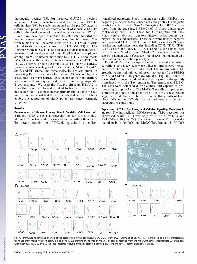
transduced peripheral blood mononuclear cells (PBMCs), wenegatively selected the transduced cells using anti-CD3 magneticbeads to deplete T cells. Two CD3-negative Tax-GFP+ cell cul-tures from the transduced PBMCs of 10 blood donors grewcontinuously over 6 mo. These two CD3-negative cell lines,which were established from two different blood donors, dis-played DC-related markers. These cells were lineage negativeand expressed CD11c, CD141, and CD205, as well as DC mat-uration and activation molecules, including CD83, CD80, CD86,CD70, CCR7, and HLA-DR (Fig. 1 A and B). We named thesetwo cell lines “ihv-DC1” and “ihv-DC2”, which represented asubset of human CD11c+/CD205+ blood DCs that maintained amaturation and activation phenotype.The ihv-DCs grew in suspension with conventional culture
conditions, and a few cells were adherent and showed typicaldendrites. To validate the ability of Tax in promoting DCgrowth, we first stimulated adherent monocytes from PBMCswith GM-CSF/IL-4 to generate MoDCs (Fig. 1C). Some ofthese MoDCs presented dendrites, and they were subsequentlytransduced with the Tax lentivirus. The transduced MoDC-Tax cells were detached during culture and capable of pro-liferating for up to 3 mo. The MoDC-Tax cells also presenteda mature and activation phenotype (Fig. 1D). These resultssuggested that Tax was able to promote the growth of bothblood DCs and MoDCs that lost cell adherence in the stan-dard culture conditions.
Expression of TLRs, Cytokines, and Cellular Signaling Molecules inihv-DCs. The intracellular dsRNA-sensing TLR3 receptor wasexpressed, while TLR4 was negative in both ihv-DCs andMoDC-Tax cells (Fig. 2A). The cleaved form of TLR7 was de-tected in both ihv-DCs and MoDC-Tax, but not in MoDCs
Fig. 1. Immunophenotyping analysis of the established ihv-DC cell lines. (A) ihv-DC1. (B) ihv-DC2. (C) Image of GM-CSF/IL-4–stimulated and differentiated DCsfrom adherent monocytes of healthy blood donors. (D) Immunophenotype of MoDC-Tax cells generated from the MoDCs that were transduced with the Tax-GFP lentivirus. In A, B, and D, red line indicates isotype antibody staining control; blue line indicates specific antibody staining.
E4454 | www.pnas.org/cgi/doi/10.1073/pnas.1800550115 Wu et al.
Dow
nloa
ded
by g
uest
on
Dec
embe
r 1,
202
1

(Fig. 2B), suggesting that Tax up-regulated TLR7 in DCs. TLR9was expressed highly in ihv-DC2, and slightly in MoDCs (Fig. 2B).Regardless of the TLR expression status, these receptors were nolonger required for inducing DC maturation and activation, sincethese established dendritic cell lines persistently expressed highlevels of costimulatory molecules such as CD80, CD86, CD70,and CD83.
The inflammatory cytokines IL-1A and TNF-α were producedin all ihv-DCs and activated MoDCs, while the immune regula-tory factor IL-10 was expressed at a negligible level (Fig. 2 C–F).IL-15, which favors the development of CD8+ cytotoxic lym-phocytes, was produced at high levels in both established den-dritic cell lines and the activated MoDCs (Fig. 2 C–F). Together,these results indicated that the expression profiles of TLRs and
Fig. 2. The expression profiles of TLRs, cytokines, and cellular signaling molecules in ihv-DCs. (A) Expression of TLR3 and TLR4 in ihv-DCs and MoDC-Tax cellsexamined by FACS. (B) Immunoblot analysis of TLR1, TLR2, TLR7, and TLR9 in the established DCs using relevant antibodies. THP-1 cells are monocytic leu-kemia cells used as control. (C–F) Cytokine expression profiles were determined by qRT-PCR in ihv-DC1 cells (C), ihv-DC2 cells (D), MoDC-Tax cells (E), and TNF-α/LPS–activated MoDCs (F). (G) Immunoblot analysis of the expressions of Tax-GFP, Bcl-2, Bcl-xL, and Mcl-1 in the established DCs. PBLs (peripheral bloodlymphocytes from normal donors) and MT4 were used for controls. (H) The phosphorylation status of Stat1, Stat3, Stat5, Rb, or cdc2 was examined usingspecific anti-phospho-specific antibodies. (I) EMSA assay for detection of the transcriptional activities of NF-κB, Stat3, and AP-1 using the nuclear extractsprepared from ihv-DCs, PBLs, and MT4 cells.
Wu et al. PNAS | vol. 115 | no. 19 | E4455
IMMUNOLO
GYAND
INFLAMMATION
PNASPL
US
Dow
nloa
ded
by g
uest
on
Dec
embe
r 1,
202
1
cytokines in ihv-DCs correlated well with the phenotypes ofmature and activated blood DCs that were generated by theconventional method. The prosurvival Bcl-2 protein, Bcl-xL, wasnotably up-regulated only in the DCs that expressed Tax (Fig.2G). The cell cycle regulators pRb and cdc2 were phosphory-lated in ihv-DCs (Fig. 2H). The phosphorylated forms of Stat1,Stat3, and Stat5 were detected in all DCs (Fig. 2H), and theactivities of the transcriptional factors, including NF-κB, Stat3,and AP-1, were also observed in ihv-DCs (Fig. 2I). Thus, itappeared that Tax immortalized blood DCs by a similar mech-anism to that of Tax-mediated immortalization of CD4+ T cellsthrough activation of growth-promoting signaling and inductionof cell cycle progression.
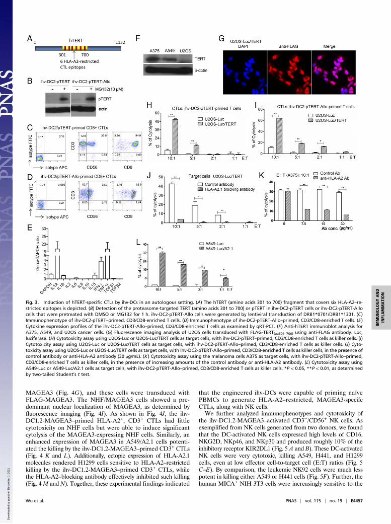
Induction of hTERT-Specific CTLs by ihv-DCs in an Autologous Setting.One of the significant advantages of the established dendritic celllines is that unlike primary DCs, the dendritic cell lines can begenetically modified to enhance their functions in antigen pre-sentation. We then selected a known universal tumor antigen,human telomerase reverse transcriptase (hTERT), and in-troduced it into HLA-A2+ ihv-DC2 cells with an HLA type (A2,B13/B40, Cw12/Cw16), with HLA-A2 being one of the mostfrequent HLA alleles. The activity of hTERT is detected inroughly 85% of human cancers and constitutes several recog-nized HLA-A2–restricted CTL epitopes (25). To facilitate anti-gen processing, we designed a fusion construct, pTERT, whichconsisted of the proteasomal target sequence of IκBα and afragment from hTERT (amino acids 301 to 700) that covered sixreported HLA-A2–restricted CTL epitopes (Fig. 3A). This de-sign was based on the mechanism that the ihv-DCs exhibitedconstitutive activities of IκB kinase/NF-κB due to Tax expres-sion. The activated IκB kinase induced serine phosphorylationand rapid turnover of IκBα. pTERT is to be degraded in thesecells, potentially allowing production of abundant amounts of theTERT peptides that form complexes with HLA molecules.pTERT was introduced into ihv-DC2 cells via lentiviral trans-duction to generate ihv-DC2-pTERT cells. We found thatpTERT was barely detected in DMSO-treated ihv-DC2-pTERT cells, and the pretreatment of these DCs with MG132, achemical inhibitor of the proteasome, induced accumulation ofpTERT (Fig. 3B).We next mixed engineered ihv-DC2-pTERT cells with naïve
PBMCs directly isolated from the autologous healthy blood do-nor at the ratio of 1:100. We observed that ihv-DC2-pTERT DCsinduced expansion of autologous T lymphocytes for 4 to 6 wk.Two weeks after cell mixing, the ihv-DC2-pTERT–primed CD8+
lymphocytes produced large amounts of IFN-γ and TNF-α (Fig.3 C and E). Importantly, the ihv-DC2-pTERT cells completelydisappeared in the mixed cell culture, as evidenced by lack of theTax mRNA examined with RT-PCR.As expected, the ihv-DC2-pTERT cells were found to pro-
mote modest expansion of autologous T cells, considering thatantigen-specific T cells were typically fewer than 1 in 100,000 inhealthy individuals. To enhance immune response to hTERT, wemodified ihv-DC2-pTERT cells by introducing two allogeneicHLA-DRB1 molecules (DRB1*0701/DRB1*1301) to generateihv-DC2-pTERT-Allo DCs. We hypothesized that the autolo-gous CD4 T cells could react to two allo-DRB1 molecules ro-bustly, thereby enhancing the development of hTERT-specific,CD8+ CTL response. Indeed, we observed that the autologousT cells vigorously responded to ihv-DC2-pTERT-Allo DCs,mediating a predominant CD8 CTL response (Fig. 3D). The ihv-DC2-pTERT-Allo–primed T cells proliferated far better thanihv-DC2-pTERT–primed T cells.ihv-DC2-pTERT–primed or ihv-DC2-pTERT-Allo–primed
CD3+/CD8+ T cells were tested for their tumor antigen-specifickilling activity on cancer cells. U2OS cells (HLA type: A*0201/A*3201, B*4402, Cw*0501/Cw*0704) were first chosen because
these cells are known to be hTERT negative (Fig. 3F). The pa-rental U2OS cells were transduced with FLAG-TERT(aa301–700),and the resulting U2OS/TERT cells showed a nuclear localiza-tion of FLAG-TERT(aa301–700) (Fig. 3G). It was shown that ihv-DC2-pTERT–primed autologous CTLs constituted a T cellpopulation that induced cytolysis only in U2OS cells that werereconstituted with TERT (Fig. 3H). Similarly, ihv-DC2-pTERT-Allo–primed CTLs exhibited a potent cytotoxicity on the TERT-expressing U2OS cells (Fig. 3I). Notably, ihv-DC2-pTERT-AlloDCs were able to prime autologous T cells to generate abundanthTERT-specific CTLs that proliferate healthily in a longer-lasting manner ex vivo. Because only HLA-A2 was matchedbetween ihv-DC2-pTERT DCs and U2OS cells in MHC class Imolecules, it was likely that the killing on the TERT-expressingU2OS cells was mediated through HLA-A2 restriction. To vali-date this notion, we applied HLA-A2–blocking antibody, whicheffectively inhibited ihv-DC2-pTERT-Allo–primed CTL-mediated killing on U2OS/TERT cells (Fig. 3J). Similarly,ihv-DC2-pTERT-Allo–primed CTLs induced cytolysis on the HLA-A2+ melanoma cells A375 (HLA type: A1/A2, B57/B44, C*16/C*6), and this killing effect was inhibited by increasing doses ofanti-HLA-A2 antibody (Fig. 3K). Further, we tested HLA-2–negative, hTERT+ A549 lung cancer cells (HLA type: A30, B44,Cw12/16). ihv-DC2-pTERT-Allo–primed CTLs had no signifi-cant cytotoxicity on the parental A549 cells but were able to killthe A549 cells that were engineered to express the HLA-A2molecule (Fig. 3L). Collectively, these results indicated that theihv-DC2-pTERT–primed or ihv-DC2-pTERT-Allo–primed CTLsrecognized hTERT antigen and had the capacity to mediate HLA-2–restricted killing on cancer cells.
Simultaneous Induction of MAGEA3-Specific CTLs and NK Cells UsingEngineered ihv-DCs. In cancer patients, cancer cells are typicallyheterogeneous, with expression of different levels of HLA mol-ecules. CTL-mediated killing mandates the presence of an HLAmolecule/CTA (cancer/testis antigen) on the surface of cancercells, while NK cells recognize target cells with down-regulatedHLA molecules. It is desirable that engineered DCs would havethe capacity to induce simultaneous production of both CTA-specific CTLs and NK cells. To test this idea, ihv-DC1 cells(HLA type: A30, B15/B40, Cw2.2) were engineered to expresshigh levels of MICA and 4-1BBL, in addition to the endoge-nously expressed CD58 (Fig. 4A), because these molecules areimportant and play a key role in activating NK cells. Thesemodified DCs were transduced with HLA-A2.1 to present HLA-A2–peptide complexes (Fig. 4A) and were engineered to over-express the testis antigen MAGEA3. MAGEA3 was expressed inmultiple types of human cancer, particularly in late stages ofmetastatic lung cancer, and the resulting DCs were named “ihv-DC1.2-MAGEA3”. HLA-A2+–matched naïve PBMCs fromvarious donors were coincubated with ihv-DC1.2-MAGEA3 DCsfor at least 2 wk. It was shown that the ihv-DC1.2-MAGEA3 cells wereable to stimulate production of both CD3+ CTLs and CD3−/CD56+
NK cells from naïve PBMCs (Fig. 4B). The DC-primed T cellswere sorted out using anti-CD3 beads, which demonstrated apredominance of CD8+ αβT cells, along with typically less than10% γσ T cells (Fig. 4C).We next selected lung cancer cells as model target cells. Three
lung cancer cell lines were chosen for analyzing their expressionsof immune-suppressive molecules including PD-L1 and TGF-β1/2, as well as CTAs (Fig. 4 D–F). All cancer cell lines expressedvariable levels of PD-L1 and high levels of TGF-β1, withH1299 cells producing abundant levels of MAGEA3, whileA549 and H441 cells produced lower levels of MAGEA3 (Fig. 4F and G). HLA-A2.1 was transduced into HLA-A2–negativeH1299 cells (HLA type: A24/A32) and A549 cells (HLA type:A30, B44, C*12/C*16) (Fig. 4H). Normal human fibroblast(NHF) cells (HLA type: A2, B51, C*7) did not express
E4456 | www.pnas.org/cgi/doi/10.1073/pnas.1800550115 Wu et al.
Dow
nloa
ded
by g
uest
on
Dec
embe
r 1,
202
1
MAGEA3 (Fig. 4G), and these cells were transduced withFLAG-MAGEA3. The NHF/MAGEA3 cells showed a pre-dominant nuclear localization of MAGEA3, as determined byfluorescence imaging (Fig. 4I). As shown in Fig. 4J, the ihv-DC1.2-MAGEA3–primed HLA-A2+, CD3+ CTLs had littlecytotoxicity on NHF cells but were able to induce significantcytolysis of the MAGEA3-expressing NHF cells. Similarly, anenhanced expression of MAGEA3 in A549/A2.1 cells potenti-ated the killing by the ihv-DC1.2-MAGEA3–primed CD3+ CTLs(Fig. 4 K and L). Additionally, ectopic expression of HLA-A2.1molecules rendered H1299 cells sensitive to HLA-A2–restrictedkilling by the ihv-DC1.2-MAGEA3–primed CD3+ CTLs, whilethe HLA-A2–blocking antibody effectively inhibited such killing(Fig. 4 M and N). Together, these experimental findings indicated
that the engineered ihv-DCs were capable of priming naïvePBMCs to generate HLA-A2–restricted, MAGEA3-specifcCTLs, along with NK cells.We further analyzed immunophenotypes and cytotoxicity of
the ihv-DC1.2-MAGEA3–activated CD3−/CD56+ NK cells. Asexemplified from NK cells generated from two donors, we foundthat the DC-activated NK cells expressed high levels of CD16,NKG2D, NKp46, and NKp30 and produced roughly 10% of theinhibitory receptor KIR2DL1 (Fig. 5 A and B). These DC-activatedNK cells were very cytotoxic, killing A549, H441, and H1299cells, even at low effector cell-to-target cell (E:T) ratios (Fig. 5C–E). By comparison, the leukemic NK92 cells were much lesspotent in killing either A549 or H441 cells (Fig. 5F). Further, thehuman MICA+ NIH 3T3 cells were increasingly sensitive to the
Fig. 3. Induction of hTERT-specific CTLs by ihv-DCs in an autologous setting. (A) The hTERT (amino acids 301 to 700) fragment that covers six HLA-A2–re-stricted epitopes is depicted. (B) Detection of the proteasome-targeted TERT (amino acids 301 to 700) or pTERT in ihv-DC2-pTERT cells or ihv-DC2-pTERT-Allocells that were pretreated with DMSO or MG132 for 1 h. ihv-DC2-pTERT-Allo cells were generated by lentiviral transduction of DRB1*0701/DRB1*1301. (C)Immunophenotype of ihv-DC2-pTERT–primed, CD3/CD8-enriched T cells. (D) Immunophenotype of ihv-DC2-pTERT-Allo–primed, CD3/CD8-enriched T cells. (E)Cytokine expression profiles of the ihv-DC2-pTERT-Allo–primed, CD3/CD8-enriched T cells as examined by qRT-PCT. (F) Anti-hTERT immunoblot analysis forA375, A549, and U2OS cancer cells. (G) Fluorescence imaging analysis of U2OS cells transduced with FLAG-TERT(aa301–700) using anti-FLAG antibody. Luc,luciferase. (H) Cytotoxicity assay using U2OS-Luc or U2OS-Luc/TERT cells as target cells, with ihv-DC2-pTERT–primed, CD3/CD8-enriched T cells as killer cells. (I)Cytotoxicity assay using U2OS-Luc or U2OS-Luc/TERT cells as target cells, with ihv-DC2-pTERT-Allo–primed, CD3/CD8-enriched T cells as killer cells. (J) Cyto-toxicity assay using U2OS-Luc or U2OS-Luc/TERT cells as target cells, with ihv-DC2-pTERT-Allo–primed, CD3/CD8-enriched T cells as killer cells, in the presence ofcontrol antibody or anti-HLA-A2 antibody (30 μg/mL). (K) Cytotoxicity assay using the melanoma cells A375 as target cells, with ihv-DC2-pTERT-Allo–primed,CD3/CD8-enriched T cells as killer cells, in the presence of increasing amounts of the control antibody or anti-HLA-A2 antibody. (L) Cytotoxicity assay usingA549-Luc or A549-Luc/A2.1 cells as target cells, with ihv-DC2-pTERT-Allo–primed, CD3/CD8-enriched T cells as killer cells. *P < 0.05, **P < 0.01, as determinedby two-tailed Student’s t test.
Wu et al. PNAS | vol. 115 | no. 19 | E4457
IMMUNOLO
GYAND
INFLAMMATION
PNASPL
US
Dow
nloa
ded
by g
uest
on
Dec
embe
r 1,
202
1

cytolysis by DC-activated NK cells at various E:T ratios (Fig. 5 Gand H).
Suppression of Lung Cancer Metastasis by ihv-DC1.2-MAGEA3–PrimedCytotoxic Lymphocytes in NSG Mice. To explore the applicationof ihv-DC–based immunotherapy, we selected the ihv-DC1.2-MAGEA3 cells for their unique ability in priming naïvePBMCs to generate abundant amounts of HLA-A2–restrictedMAGEA3-specific CTLs and NK cells, which potently inducedcytolysis of human lung cancer cells ex vivo. A549-Luc/A2.1 cellswere chosen because our previous study showed that thesecancer cells presented a desirable lung metastasis pattern wheninfused through the tail vein in NSG mice. As shown in Fig. 6A,the body weights of the mice in the control group started to drop42 d after tumor cell injection, whereas the weights of the
treatment group (Tx) mice were well maintained throughout theexperimental period. The lungs were significantly enlarged inthe control group and exhibited high levels of luciferase activity,whereas the lungs in the Tx group appeared to be normal inappearance and weight and produced only background luciferaseactivity (Fig. 6 C and D). Anti-HLA-A2.1 immunohistochemistry(IHC) staining demonstrated that the A549-Luc/A2.1 cells wereexclusively found in the lung tissues in the control mice (Fig. 6E),whereas the cancer cells were undetected in the Tx mice underthe same condition (Fig. 6F). H&E staining verified these IHCresults (Fig. S1). We next applied anti-human CD3 staining toassess the in vivo trafficking patterns of the infused CTLs. It wasshown that compared with the control mice, human CD3+ T cellswere detected in the spleen, lymph nodes, and lung tissues, and
Fig. 4. Simultaneous induction of the MAGEA3-specific CTLs and NK cells using engineered ihv-DCs. (A) ihv-DC1 cells are engineered to express high levels ofMICA, 4-1BBL, and HLA-A2.1 molecules, as shown by FACS analysis. (B). Cocultivation of ihv-DC1.2-MAGEA3 cells with HLA-A2+ naïve PBMCs produced bothCD3+ T cells and CD3−/CD56+ NK cells, as shown by FACS analysis. (C) Immunophenotype of ihv-DC1.2-MAGEA3–primed, CD3-enriched T cells from at leastfour HLA-A2+ donors. (D) PD-L1 expression in lung cancer cell lines H1299, A549, and H441, as examined by FACS. (E) IL-10 and TGF-β expression in H1299,A549, and H441, as determined by qRT-PCR. (F) CTA expression in H1299, A549, and H441 cells, as examined by qRT-PCR. (G) MAGEA3 expression in NHF,H1299, A549, and H441 cells by anti-MAGEA3 immunoblot. (H) HLA-A2 surface expression in H1299/A2.1, A549/A2.1, and H441 cell by FACS. (I) Fluorescenceimaging analysis of NHF cells and the NHF cells expressing FLAG-MAGEA3 using anti-FLAG antibody. (J) Cytotoxicity assay using NHF-Luc or NHF-Luc/MAGEA3 cells as target cells, with ihv-DC1.2-MAGEA3–primed, CD3+ T cells as killer cells. (K) Fluorescence imaging of A549/A2.1 cells and the A549/A2.1 cellsexpressing FLAG-MAGEA3 using anti-FLAG antibody. (L) Cytotoxicity assay using A549-Luc, A549-Luc/A2.1, or A549-Luc/A2.1/MAGEA3 cells as target cells, withihv-DC1.2-MAGEA3–primed, CD3+ T cells as killer cells. (M) Cytotoxicity assay using H1299-Luc or H1299-Luc/A2.1 cells as target cells, with ihv-DC1.2-MAGEA3–primed, CD3+ T cells as killer cells. (N) Cytotoxicity assay using H1299-Luc/A2.1 cells as target cells, with ihv-DC1.2-MAGEA3–primed, CD3+ T cells as killer cells,in the presence of the control antibody or anti-HLA-A2 antibody.
E4458 | www.pnas.org/cgi/doi/10.1073/pnas.1800550115 Wu et al.
Dow
nloa
ded
by g
uest
on
Dec
embe
r 1,
202
1

a few CD3+ cells were found in the liver in the Tx group mice (Fig.7 A and B). No infiltrating T cells were found in the heart andkidney in the Tx group mice (Fig. 7B). By analyzing the lung tis-sues of five mice in the Tx group, we found that both CD3+ CTLsand NKp46+ NK cells were present in the lung tissues (Fig. 7C).These experimental data, therefore, indicated their desirable invivo trafficking patterns and validated the therapeutic efficacy ofthe ihv-DC1.2-MAGEA3–primed cytotoxic lymphocytes in thelung cancer NSG model.
DiscussionWe have described a method for developing human blood den-dritic cell lines by selecting phytohemagglutinin (PHA)/IL-2–stimulated, Tax-transduced PBMCs from blood donors. Theestablished ihv-DCs can be reliably grown and maintained inculture for a prolonged time without losing their maturation andactivation phenotypes. We have also developed a method toamplify MoDCs by expressing Tax in GM-CSF/IL-4–inducedDCs. Although we are not able to establish immortalized MoDC-Tax cells, this method can still generate adequate numbers ofactivated MoDCs from several million PBMCs, which is a sig-nificant advantage over the conventional MoDC method, byeliminating undesirable apheresis procedures. In addition, unlikeprimary DCs, the established DCs can be easily modified byexpressing a given tumor antigen intracellularly, thereby pre-senting multiple CTA epitopes in priming CD8 T cells and en-hancing the development of anticancer immunity.We attribute our success to the effort of cell selection and
culture techniques at several levels. First, PHA/IL-2 stimulates
T cell proliferation, and the activated T cells secrete solublefactors that possibly enable blood immature DCs to be trans-duced by the Tax lentivirus, since primary immature DCs areresistant to lentiviral transduction. Second, because the majorityof the Tax-transduced T cells proliferate much faster than theTax-transduced DCs, depletion of T cells at the early selectionstage is an essential step that allows low numbers of DCs tothrive. Third, most Tax-transduced DCs experience a “growthcrisis” during the third month after transduction, and some ofthem regain their growth strength after passing this stage.Therefore, a continuous culture effort is necessary to aid theTax-transduced DCs to become immortal. Although the DCimmortalization efficiency is roughly 20%, this shortage can beimproved by at least two technical modifications. One approachis to develop a DC bank from healthy blood donors, which coversfrequent HLA molecules among different ethnic populations.We could select patient HLA-matched DCs from the DC bankfor cancer therapy. Another approach is that we could perform areverse-engineering technique to develop patient HLA-matchedDCs. In this method, the patient’s HLA molecules are clonedand expressed in a model dendritic cell line.A crucial phenotype of the ihv-DCs is their constitutively ac-
tivated state without need for additional stimulations. Tax ap-parently has the capacity to promote both DC maturation andactivation. We reason that Tax-induced activation of NF-κBsignaling plays a key role in the process of DC maturation andactivation. Upon encountering invading pathogens, some com-ponents of pathogens stimulate TLRs such as TLR3 andTLR4 in immature DCs. TLR3 or TLR4 engagement activates
Fig. 5. Cytotoxicity of ihv-DC1.2-MAGEA3–activated NK cells. (A and B) Immunophenotype of ihv-DC1.2-MAGEA3–activated NK cells from two HLA-A2+
donors. NK cells were negatively selected using anti-CD3 beads to deplete ihv-DC1.2-MAGEA3–primed T cells. (C–E) Cytotoxicity assay using A549-Luc/A2.1 cells (C), H441-Luc cells (D), and H1299-Luc/A2.1 cells (E) as target cells, with ihv-DC1.2-MAGEA3–activated NK cells as killer cells. (F) Cytotoxicity assayusing A549-Luc and H441-Luc cells as target cells, with NK92 cells as killer cells. (G) NIH 3T3-Luc cells were stably transduced with hMICA, and the surfaceexpression of hMICA was shown by FACS. (H) Cytotoxicity assay using 3T3-Luc and 3T3-Luc/hMICA as target cells, with ihv-DC1.2-MAGEA3–activated NK cellsas killer cells.
Wu et al. PNAS | vol. 115 | no. 19 | E4459
IMMUNOLO
GYAND
INFLAMMATION
PNASPL
US
Dow
nloa
ded
by g
uest
on
Dec
embe
r 1,
202
1

the receptor-associated TRAF6, which in turn induces the acti-vation of its downstream signaling molecules, including the IκBkinase–NF-κB complex (26, 27). It is well recognized that NF-κBis the key mediator in inducing DC maturation and activation(23, 24). TRAF6 is also necessary for activating MAP kinase/AP-1 (28). Further, the Stat3 activity is crucial for IL-15–derivedDCs to acquire their antigen-presenting capability and to medi-ate CD8+ T cell response (29). Consistent with these reports, wedemonstrate that ihv-DCs exhibit the constitutive activities ofNF-κB, AP-1, and Stat3 as induced by Tax, thereby contributingto their maturation and activation.ihv-DCs have the ability to prime naïve PBMCs to generate
antigen-specific CTLs that induce cytolysis of target cells in anHLA-restricted manner in an autologous setting. However, thenumber of tumor antigen-specific lymphocytes in healthy donorsis extremely low and, therefore, it is a challenging task to gen-erate adequate numbers of tumor antigen-specific CTLs in thecell culture condition for cancer therapy using the autologoussetting. Notably, ∼7% of peripheral lymphocytes display allor-eactive capability, which in turn promote generation of tumorantigen-specific CTLs (30–32). In the present study, we devel-oped a strategy to enhance production of the antigen-specificCTLs. By introducing two allogeneic HLA-DRB1 molecules intoihv-DCs, autologous CD4 T cells can respond to these modified
DCs robustly, in promoting generation of antigen-specific CD8CTLs. This approach may have at least two levels of significancein cancer therapy. One is that sufficient amounts of CTA-specific, autologous CTLs are readily available by cocultivatingwith engineered DCs in a cGMP facility, which can be infuseddirectly into patients. Another is that the engineered DCs withtwo allo-DRB1 molecules can serve as therapeutic cancer vac-cines that induce a better inflammatory immunity in patients.These ideas need to be investigated further in clinical trials.Furthermore, we envision that simultaneous production of bothCTA-specific CTLs and NK cells is a clear advantage in cancertherapy, considering the heterogeneous nature of cancer cells.The ihv-DC–activated NK cells are apparently of high quality, asexemplified by potent cytotoxicity and low levels of the inhibitoryreceptor KIR2DL1.In summary, the ihv-DC models offer several notable features:
(i) ihv-DCs are activated and mature DCs, thereby eliminatingthe need of the complex maturation and activation process toacquire functional DCs; (ii) ihv-DCs cells constitutively expressabundant amounts of the costimulatory receptors, includingCD80, CD86, and CD70, which stimulate antigen-specific CD8+
cytotoxic T cells to proliferate; (iii) ihv-DCs also express thechemokine receptor CCR7, the lymphoid tissue homing re-ceptor; (iv) the ihv-DC model can provide a particularly useful
Fig. 6. Suppression of metastatic lung cancer by ihv-DC1.2-MAGEA3–primed cytotoxic lymphocytes in NSG mice. (A) Body weights of NSG mice in the controlgroup and the Tx group. (B) Whole lung weights of NSG mice in the control group and the Tx group at the end point. (C) Images of whole lungs of NSG micein the control group and the Tx group at the end point. (D) The luciferase activity of the lung tissues of mice in the control group and the Tx group. (E and F)IHC staining with anti-HA (hemagglutinin) antibody to detect HLA-A2.1-HA protein in various organs of mice in the control group (E) and the Tx group (F).
E4460 | www.pnas.org/cgi/doi/10.1073/pnas.1800550115 Wu et al.
Dow
nloa
ded
by g
uest
on
Dec
embe
r 1,
202
1

tool to study DC biology; (v) these DCs can be modified geneticallyfor investigating the role of existing or putative costimulatory receptorsthat mediate or enhance a protective anticancer or antiviral immunity;(vi) the engineered ihv-DCs can promote simultaneous productionof CTA-specific CTLs and NK cells; and (vii) ihv-DCs are im-mortalized, thus allowing for their continuous availability.
Materials and MethodsAll experimental procedures are described in detail in SI Materialsand Methods.
Cell Lines, Antibodies, and Cytokines. H1299, H441, A549, and U2OS cell lineswere obtained from American Type Culture Collection. A375 and NHF celllines were kindly provided by Isaiah J. Fidler, The University of Texas, MDAnderson Cancer Center, Houston, and Jieyu Zhu, Washington State University,Spokane, WA, respectively. Recombinant IL-2 was acquired from AIDS Researchand Reference Reagent Program. Isotype IgG control antibody and variousallophycocyanin- or FITC-labeled antibodies were purchased from BioLegend.The HLA-A2–blocking antibody was purchased from GeneTex.
Lentiviral Production. The tax from HTLV-2 was fused with the fragmentencoding enhanced green fluorescence protein, and the tax2-gfp fusionfragment was cloned into the lentiviral vector in which the human elon-gation factor promoter drives the expression of transgene. To generaterecombinant lentiviruses, the lentiviral construct was cotransfected with the
packaging plasmid mix containing the expression plasmids for VSV-G, Gag-Pol, and Rev (Invitrogen) into 293 cells using SuperFect transfection reagent(Qiagen).
Generation of ihv-DC Cell Lines. Leukopaks were obtained from New YorkBlood Center. Human PBMCs were isolated from leukopaks and stimu-lated with PHA (5 μg/mL) for 24 h, followed by adding recombinant IL-2(100 units/mL). The activated PBMCs were cultured for 4 to 5 d and werethen transduced with the Tax2-GFP lentivirus in the presence of polybrene(10 μg/mL). The transduced cells were cultured continuously in the completeRPMI 1640 medium containing 10% FBS (Sigma) and 100 units/mL recom-binant IL-2. Alternatively, the transduced cells were cultured in RPMI1640 medium supplemented with 5% heat-inactivated human AB serum(Sigma). Two- to 3-wk after transduction, the transduced cells were nega-tively selected with anti-CD3 magnetic beads (Life Technologies) to depleteT cells. CD3-negative cells were maintained in culture continuously and analyzedfor their immunophenotypes about 3 mo after transduction. Two dendriticcell lines (ihv-DC1 and ihv-DC2) were established from two blood donors.The ihv-DC1 cells lost CD40 during the passages in culture, and CD40 wasrestored by the CD40 lentiviral transduction. The ihv-DCs were grown in theRPMI 1640 medium supplemented with 5% heat-inactivated human AB serumor 10% FBS in the presence of IL-2 (50 to 100 units/mL).
Induction of Tumor-Associated Antigen-Specific CTLs by ihv-DCs. The ihv-DCswere mixed with naïve PBMCs isolated from leukopaks at the ratio of 1:100.The mixed cells were kept in culture without adding exogenous cytokines for
Fig. 7. ihv-DC1.2-MAGEA3–activated CTLs and NK cells infiltrate lung tissues. (A and B) Anti-CD3 IHC was analyzed for various tissue samples of mice in thecontrol group (A) and the Tx group (B). (C) Anti-CD3 or anti-NKp46 IHC staining for the lung tissue of all five mice in the Tx group.
Wu et al. PNAS | vol. 115 | no. 19 | E4461
IMMUNOLO
GYAND
INFLAMMATION
PNASPL
US
Dow
nloa
ded
by g
uest
on
Dec
embe
r 1,
202
1
2 to 3 d, followed by adding recombinant IL-2 (100 to 200 units/mL). Theproliferation of the ihv-DC–reactive lymphocytes was monitored with FACS,and the presence of ihv-DCs in the mixed culture was monitored using fluo-rescence microscopy and qRT-PCR for detecting Tax2. Two to 3 wk after cellmixing, the ihv-DC–activated lymphocytes were analyzed with FACS and wereexamined for their cytotoxic activity on target cells.
Cytotoxicity Assay. Various cell lines that were modified to express luciferasewere used as targets, while the ihv-DC–activated lymphocytes generatedfrom 14 to 21 d after mixed lymphocyte reaction served as effectors. Cancercells were first placed in 24-well plates for 2 h for complete attachment. Theihv-DC–activated lymphocytes were then placed on cancer cells at the in-dicated E:T ratios. At the 4- or 16-h time point after coincubation of effectorand target cells, viable cells were gently washed with PBS buffer to removecellular debris and subjected to the luciferase activity assay using the kitfrom Promega. The cytotoxic activity of the ihv-DC–activated lymphocyteswas determined by comparing the luciferase activities in the target cells thatwere treated without or with the cytotoxic lymphocytes at the indicatedE:T ratios.
qRT-PCR. Total RNA was isolated using the RNeasy kit (Qiagen), and itsconcentration was determined using the NanoDrop1000 spectrophotometer(Thermo Scientific). The quality and integrity of total RNAwas assessed on 1%formaldehyde–agarose gels. cDNA was synthesized using the OmniScriptReverse Transcriptase Kit (Qiagen) following the manufacturer’s recom-mended protocol. Template samples in triplicate were subjected to qRT-PCR(Stratagene Mx3005P system) using Power SYBR Green (Applied Biosystems).
Animals and Xenograft Models. The animal study was performed using theapproved Institutional Animal Care and Use Committee protocol of the
University of Maryland School of Medicine. Male NSG mice (8 wk, 18 to 22 gbody weight) were obtained from the animal center at the University ofMaryland School of Medicine and housed in a specific pathogen-free room.A549/A2.1 cells (2 million) were collected, resuspended in Hanks buffer, andthen tail vein-injected into NSG mice. After cancer cell implantation, themice were randomized into two groups (CTL Tx and control). The treatmentbegan 5 d after cancer cell transplantation. The Tx group mice were injectedvia the tail vein with the DC-activated CTLs [q.5.d × 12 (injected every 5 d tototal of 12 injections), 20 million in 200 μL in Hanks buffer] plus 1,000 unitsrecombinant IL-2, and the control group mice were injected via the tail veinwith Hanks buffer (q.5.d × 11, iv, 200 μL plus 1,000 units recombinant IL-2).The animals’ weight was measured every 2 d.
Immunohistochemistry and H&E Staining. Tissue sections were incubated at60 °C for 10 min and then deparaffinized in xylene and subsequently inethanol (ETOH) and deionized water. After deparaffinization, tissue sectionswere immersed in a preheated antigen unmasking solution (Vector), placedinto a pressure cooker for 5 to 10 min, and then cooled to room temperatureunder cold water. The anti-human CD3 antibody or anti-NKp46 (1:100) wasused to stain the pretreated tissue sections. Next, the endogenous peroxi-dase activity was blocked by incubation of tissue sections with 3% hydrogenperoxide in PBS for 10 min. Biotinylated goat anti-mouse IgG antibody (H+L)(1:200) (Vector) was applied as the secondary antibody. A VECTASTAIN EliteABC HRP Kit (Vector) was used as chromogen, and hematoxylin counter-staining was also performed.
ACKNOWLEDGMENTS. We thank Alonso Heredia, Yiling Liu, and SuzanneGartner for technical assistance. Research reported in this publication wassupported by the University of Maryland Cancer Center and the Institute ofHuman Virology, University of Maryland School of Medicine (H.C.).
1. Palucka K, Banchereau J (2012) Cancer immunotherapy via dendritic cells. Nat Rev
Cancer 12:265–277.2. Tyler CJ, Doherty DG, Moser B, Eberl M (2015) Human Vγ9/Vδ2 T cells: Innate adaptors
of the immune system. Cell Immunol 296:10–21.3. van Beek JJ, Wimmers F, Hato SV, de Vries IJM, Sköld AE (2014) Dendritic cell cross talk
with innate and innate-like effector cells in antitumor immunity: Implications for DC
vaccination. Crit Rev Immunol 34:517–536.4. Pradere J-P, Dapito DH, Schwabe RF (2014) The yin and yang of Toll-like receptors in
cancer. Oncogene 33:3485–3495.5. Dzopalic T, Rajkovic I, Dragicevic A, Colic M (2012) The response of human dendritic
cells to co-ligation of pattern-recognition receptors. Immunol Res 52:20–33.6. Hammer GE, Ma A (2013) Molecular control of steady-state dendritic cell maturation
and immune homeostasis. Annu Rev Immunol 31:743–791.7. Mildner A, Jung S (2014) Development and function of dendritic cell subsets.
Immunity 40:642–656.8. Osugi Y, Vuckovic S, Hart DN (2002) Myeloid blood CD11c(+) dendritic cells and
monocyte-derived dendritic cells differ in their ability to stimulate T lymphocytes.
Blood 100:2858–2866.9. Kassianos AJ, Jongbloed SL, Hart DN, Radford KJ (2010) Isolation of human blood DC
subtypes. Methods Mol Biol 595:45–54.10. Jongbloed SL, et al. (2010) Human CD141+ (BDCA-3)+ dendritic cells (DCs) represent a
unique myeloid DC subset that cross-presents necrotic cell antigens. J Exp Med 207:
1247–1260.11. Bachem A, et al. (2010) Superior antigen cross-presentation and XCR1 expression
define human CD11c+CD141+ cells as homologues of mouse CD8+ dendritic cells.
J Exp Med 207:1273–1281.12. Kvistborg P, Boegh M, Pedersen AW, Claesson MH, Zocca MB (2009) Fast generation
of dendritic cells. Cell Immunol 260:56–62.13. Jolanda I, de Vries M, Adema GJ, Punt CJ, Figdor CG (2005) Phenotypical and func-
tional characterization of clinical-grade dendritic cells. Adoptive Immunotherapy:
Methods and Protocols. Methods in Molecular Medicine™, eds Ludewig B., Hoffmann
M.W. (Humana, New York), Vol 109, pp 113–125.14. Robbins SH, et al. (2008) Novel insights into the relationships between dendritic cell
subsets in human and mouse revealed by genome-wide expression profiling. Genome
Biol 9:R17.15. van Helden SF, van Leeuwen FN, Figdor CG (2008) Human and murine model cell lines
for dendritic cell biology evaluated. Immunol Lett 117:191–197.16. Masterson AJ, et al. (2002) MUTZ-3, a human cell line model for the cytokine-induced
differentiation of dendritic cells from CD34+ precursors. Blood 100:701–703.
17. Santegoets SJ, van den Eertwegh AJ, van de Loosdrecht AA, Scheper RJ, de Gruijl TD(2008) Human dendritic cell line models for DC differentiation and clinical DC vacci-nation studies. J Leukoc Biol 84:1364–1373.
18. Santegoets SJ, et al. (2006) In vitro priming of tumor-specific cytotoxic T lymphocytesusing allogeneic dendritic cells derived from the human MUTZ-3 cell line. CancerImmunol Immunother 55:1480–1490.
19. Ciminale V, Rende F, Bertazzoni U, Romanelli MG (2014) HTLV-1 and HTLV-2: Highlysimilar viruses with distinct oncogenic properties. Front Microbiol 5:398.
20. Ishitsuka K, Tamura K (2014) Human T-cell leukaemia virus type I and adult T-cellleukaemia-lymphoma. Lancet Oncol 15:e517–e526.
21. Jones KS, Petrow-Sadowski C, Huang YK, Bertolette DC, Ruscetti FW (2008) Cell-freeHTLV-1 infects dendritic cells leading to transmission and transformation of CD4(+)T cells. Nat Med 14:429–436.
22. Jain P, et al. (2009) DC-SIGN mediates cell-free infection and transmission of humanT-cell lymphotropic virus type 1 by dendritic cells. J Virol 83:10908–10921.
23. Rescigno M, Martino M, Sutherland CL, Gold MR, Ricciardi-Castagnoli P (1998) Den-dritic cell survival and maturation are regulated by different signaling pathways.J Exp Med 188:2175–2180.
24. Ardeshna KM, Pizzey AR, Devereux S, Khwaja A (2000) The PI3 kinase, p38 SAP kinase,and NF-kappaB signal transduction pathways are involved in the survival and matu-ration of lipopolysaccharide-stimulated human monocyte-derived dendritic cells.Blood 96:1039–1046.
25. Lü M-H, et al. (2012) hTERT-based therapy: A universal anticancer approach (Review).Oncol Rep 28:1945–1952.
26. Kobayashi T, et al. (2003) TRAF6 is a critical factor for dendritic cell maturation anddevelopment. Immunity 19:353–363.
27. Hull C, McLean G, Wong F, Duriez PJ, Karsan A (2002) Lipopolysaccharide signals anendothelial apoptosis pathway through TNF receptor-associated factor 6-mediatedactivation of c-Jun NH2-terminal kinase. J Immunol 169:2611–2618.
28. Bradley JR, Pober JS (2001) Tumor necrosis factor receptor-associated factors (TRAFs).Oncogene 20:6482–6491.
29. Okada S, Han S, Patel ES, Yang L-J, Chang L-J (2015) STAT3 signaling contributes tothe high effector activities of interleukin-15-derived dendritic cells. Immunol Cell Biol93:461–471.
30. Rossjohn J, McCluskey J (2007) How a home-grown T cell receptor interacts with aforeign landscape. Cell 129:19–20.
31. Gras S, Kjer-Nielsen L, Chen Z, Rossjohn J, McCluskey J (2011) The structural bases ofdirect T-cell allorecognition: Implications for T-cell-mediated transplant rejection.Immunol Cell Biol 89:388–395.
32. Morelli AE, Thomson AW (2003) Dendritic cells: Regulators of alloimmunity and op-portunities for tolerance induction. Immunol Rev 196:125–146.
E4462 | www.pnas.org/cgi/doi/10.1073/pnas.1800550115 Wu et al.
Dow
nloa
ded
by g
uest
on
Dec
embe
r 1,
202
1
Related Documents