Page 1/27 Protective Effects of Ulinastatin Combined With Thrombomodulin Against Lipopolysaccharide Induced Liver and Kidney Injury in Septic Rats Xiong Zhang Department of Basic Medicine and Clinical Pharmacy, China Pharmaceutical University Chenlin Su Department of Basic Medicine and Clinical Pharmacy, China Pharmaceutical University Shuxin Zhao Department of Basic Medicine and Clinical Pharmacy, China pharmaceutical University Jiaying Yang Department of Basic Medicine and Clinical Pharmacy, China Pharmaceutical University Etienne Empweb Anger Department of Basic Medicine and Clinical Pharmacy, China Pharmaceutical University Xiaoxing Li Department of Basic Medicine and Clinical Pharmacy, China Pharmaceutical University Chen Feng China Pharmaceutical University Qianwen Liao Department of Basic Medicine and Clinical Pharmacy, China Pharmaceutical University Ji Li Department of Basic Medicine and Clinical Pharmacy, China Pharmaceutical University Feng Yu ( [email protected] ) China Pharmaceutical University https://orcid.org/0000-0002-7021-8588 Research article Keywords: Ulinastatin, Thrombomodulin, Sepsis, lipopolysaccharide, liver injury, kidney injury Posted Date: August 30th, 2021 DOI: https://doi.org/10.21203/rs.3.rs-817705/v1 License: This work is licensed under a Creative Commons Attribution 4.0 International License. Read Full License

Welcome message from author
This document is posted to help you gain knowledge. Please leave a comment to let me know what you think about it! Share it to your friends and learn new things together.
Transcript

Page 1/27
Protective Effects of Ulinastatin Combined WithThrombomodulin Against LipopolysaccharideInduced Liver and Kidney Injury in Septic RatsXiong Zhang
Department of Basic Medicine and Clinical Pharmacy, China Pharmaceutical UniversityChenlin Su
Department of Basic Medicine and Clinical Pharmacy, China Pharmaceutical UniversityShuxin Zhao
Department of Basic Medicine and Clinical Pharmacy, China pharmaceutical UniversityJiaying Yang
Department of Basic Medicine and Clinical Pharmacy, China Pharmaceutical UniversityEtienne Empweb Anger
Department of Basic Medicine and Clinical Pharmacy, China Pharmaceutical UniversityXiaoxing Li
Department of Basic Medicine and Clinical Pharmacy, China Pharmaceutical UniversityChen Feng
China Pharmaceutical UniversityQianwen Liao
Department of Basic Medicine and Clinical Pharmacy, China Pharmaceutical UniversityJi Li
Department of Basic Medicine and Clinical Pharmacy, China Pharmaceutical UniversityFeng Yu ( [email protected] )
China Pharmaceutical University https://orcid.org/0000-0002-7021-8588
Research article
Keywords: Ulinastatin, Thrombomodulin, Sepsis, lipopolysaccharide, liver injury, kidney injury
Posted Date: August 30th, 2021
DOI: https://doi.org/10.21203/rs.3.rs-817705/v1
License: This work is licensed under a Creative Commons Attribution 4.0 International License. Read Full License

Page 2/27
AbstractBackground: Sepsis, a systemic in�ammatory disease that leads to life-threatening organ functionsdisorders, such as liver and kidney injury. Ulinastatin (UTI) and Thrombomodulin (TM) are activemacromolecules isolated from human urine. UTI and TM have been found to have therapeutic effects onin�ammatory diseases. In this study, we veri�ed protective effect of UTI combined with TM on liver andkidney injury caused by sepsis, and further explored the mechanisms.
Methods: The sepsis model was established by intravenous injection of LPS into the tail vein of rats.Blood, liver and kidney tissues were collected after injection of UTI or TM. ELISA was used to measureserum levels of pro-in�ammatory cytokines. The characteristic functional indexes of liver and kidney inserum and multiple coagulation function indexes of rats were detected via corresponding kits.Histological changes of liver and kidney tissues were investigated by HE staining. Apoptosis in liver andkidney tissues were examined by TUNEL staining, and the expression levels of apoptosis-related proteinswere also analyzed. HMGB1/TLR4/NF-κB pathway in liver and kidney tissues were examined by WesternBlot. PCNA-positive cells were detected by immunohistochemistry. The survival rate of rats in each groupwas statistically analyzed.
Results: UTI combined with TM reduced LPS-induced secretion of IL-6 and TNF-α in the serum. The drugcombination reduced the liver and kidney functional indicators ALT, AST, BUN and Cr, and amelioratedliver and kidney pathology injury of rats. It inhibited apoptosis of liver and kidney cells via down-regulating the expression of apoptotic protein Bax, Cleaved caspase-3, up-regulating the expression ofanti-apoptotic protein bcl-2, and promoted the proliferation of liver and kidney cells. The drugcombination reversed the up-regulation of HMGB1, TLR4, and phosphorylated NF-κB protein mediated byLPS. Anticoagulation test indicated UTI does not affect the anticoagulant effect of TM when they areused in combination. Moreover, the drug combination signi�cantly improved the survival rate of septicrats.
Conclusions: These results indicate that UTI combined with TM plays a key role in protecting liver andkidney injury in septic rats, which will suggest a promising treatment for sepsis-induced organ injury.
1. BackgroundSepsis is a disease with a systemic in�ammatory state with known or suspected infection (Vandewalleand Libert. 2020). The mortality rate has remained high for a long time. Pathogenic microorganisms ortoxins invade the body through the blood and activate the host immune system (Mirouse et al. 2020),thereby producing endogenous in�ammatory mediators and cytokines. At the same time, sepsis affectsvarious systems and organs of the body. It causes damage to cells and tissues and affects metabolism,which further lead to failure of various vital organs (Venet and Monneret. 2018; Tsantarliotou et al. 2019).The liver and kidney are organs vulnerable to damage from sepsis (Yan et al. 2014; Alobaidial. 2015).Acute liver and kidney injury may occur at any stage of sepsis. Sepsis patients usually have severe liver
Page 3/27
and kidney injury and often extremely critical, prone to multiple organ failure, leading to high mortality(Cecconi et al. 2018). Therefore, the repair of organ injury is particularly important in the treatment ofsepsis.
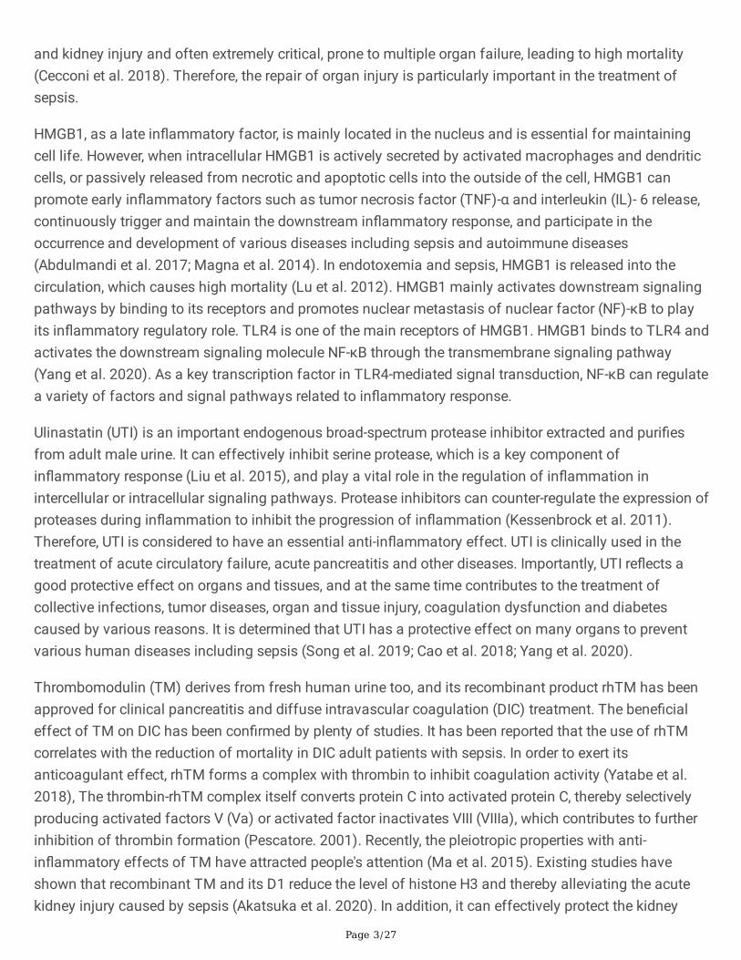
HMGB1, as a late in�ammatory factor, is mainly located in the nucleus and is essential for maintainingcell life. However, when intracellular HMGB1 is actively secreted by activated macrophages and dendriticcells, or passively released from necrotic and apoptotic cells into the outside of the cell, HMGB1 canpromote early in�ammatory factors such as tumor necrosis factor (TNF)-α and interleukin (IL)- 6 release,continuously trigger and maintain the downstream in�ammatory response, and participate in theoccurrence and development of various diseases including sepsis and autoimmune diseases(Abdulmandi et al. 2017; Magna et al. 2014). In endotoxemia and sepsis, HMGB1 is released into thecirculation, which causes high mortality (Lu et al. 2012). HMGB1 mainly activates downstream signalingpathways by binding to its receptors and promotes nuclear metastasis of nuclear factor (NF)-κB to playits in�ammatory regulatory role. TLR4 is one of the main receptors of HMGB1. HMGB1 binds to TLR4 andactivates the downstream signaling molecule NF-κB through the transmembrane signaling pathway(Yang et al. 2020). As a key transcription factor in TLR4-mediated signal transduction, NF-κB can regulatea variety of factors and signal pathways related to in�ammatory response.
Ulinastatin (UTI) is an important endogenous broad-spectrum protease inhibitor extracted and puri�esfrom adult male urine. It can effectively inhibit serine protease, which is a key component ofin�ammatory response (Liu et al. 2015), and play a vital role in the regulation of in�ammation inintercellular or intracellular signaling pathways. Protease inhibitors can counter-regulate the expression ofproteases during in�ammation to inhibit the progression of in�ammation (Kessenbrock et al. 2011).Therefore, UTI is considered to have an essential anti-in�ammatory effect. UTI is clinically used in thetreatment of acute circulatory failure, acute pancreatitis and other diseases. Importantly, UTI re�ects agood protective effect on organs and tissues, and at the same time contributes to the treatment ofcollective infections, tumor diseases, organ and tissue injury, coagulation dysfunction and diabetescaused by various reasons. It is determined that UTI has a protective effect on many organs to preventvarious human diseases including sepsis (Song et al. 2019; Cao et al. 2018; Yang et al. 2020).
Thrombomodulin (TM) derives from fresh human urine too, and its recombinant product rhTM has beenapproved for clinical pancreatitis and diffuse intravascular coagulation (DIC) treatment. The bene�cialeffect of TM on DIC has been con�rmed by plenty of studies. It has been reported that the use of rhTMcorrelates with the reduction of mortality in DIC adult patients with sepsis. In order to exert itsanticoagulant effect, rhTM forms a complex with thrombin to inhibit coagulation activity (Yatabe et al.2018), The thrombin-rhTM complex itself converts protein C into activated protein C, thereby selectivelyproducing activated factors V (Va) or activated factor inactivates VIII (VIIIa), which contributes to furtherinhibition of thrombin formation (Pescatore. 2001). Recently, the pleiotropic properties with anti-in�ammatory effects of TM have attracted people's attention (Ma et al. 2015). Existing studies haveshown that recombinant TM and its D1 reduce the level of histone H3 and thereby alleviating the acutekidney injury caused by sepsis (Akatsuka et al. 2020). In addition, it can effectively protect the kidney
Page 4/27
from injury. In the �eld of liver injury caused by sepsis, the administration of recombinant TM can alsoimprove the liver dysfunction and elevate the survival rate of septic mice (Nagato et al. 2009). In aretrospective study, recombinant human soluble TM can decrease the mortality of patients with sepsis(Yoshihiro et al. 2019). These all indicate that TM can be used in the treatment of sepsis, and has a broadresearch prospect in organ injury, but its speci�c organ protection mechanism remains unclear.
In previous studies, both UTI and TM have been found to have therapeutic effects on sepsis (Jiang et al.2018; Wu et al. 2019; Wang et al. 2019; Ashina et al. 2020), and more meaningfully, a number of studieshave shown that UTI combined with other drugs can effectively improve the treatment effect of sepsis(Meng et al. 2020; Wang et al. 2020). At present, there are many research projects on sepsis, but theresearch based on drug combination therapy is not su�cient. In summary, we use UTI combined with TMto verify its protective effect on liver and kidney injury caused by sepsis, and further explore themechanisms of action.
2. Materials And Methods
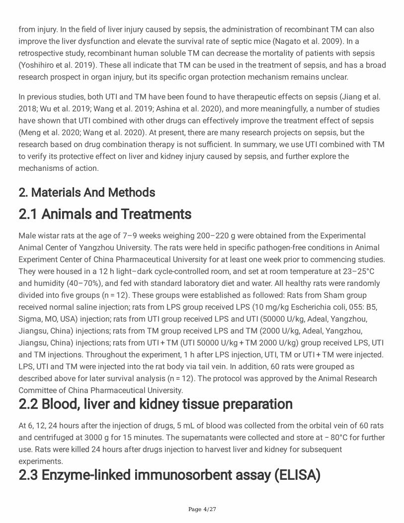
2.1 Animals and TreatmentsMale wistar rats at the age of 7–9 weeks weighing 200–220 g were obtained from the ExperimentalAnimal Center of Yangzhou University. The rats were held in speci�c pathogen-free conditions in AnimalExperiment Center of China Pharmaceutical University for at least one week prior to commencing studies.They were housed in a 12 h light–dark cycle-controlled room, and set at room temperature at 23–25°Cand humidity (40–70%), and fed with standard laboratory diet and water. All healthy rats were randomlydivided into �ve groups (n = 12). These groups were established as followed: Rats from Sham groupreceived normal saline injection; rats from LPS group received LPS (10 mg/kg Escherichia coli, 055: B5,Sigma, MO, USA) injection; rats from UTI group received LPS and UTI (50000 U/kg, Adeal, Yangzhou,Jiangsu, China) injections; rats from TM group received LPS and TM (2000 U/kg, Adeal, Yangzhou,Jiangsu, China) injections; rats from UTI + TM (UTI 50000 U/kg + TM 2000 U/kg) group received LPS, UTIand TM injections. Throughout the experiment, 1 h after LPS injection, UTI, TM or UTI + TM were injected.LPS, UTI and TM were injected into the rat body via tail vein. In addition, 60 rats were grouped asdescribed above for later survival analysis (n = 12). The protocol was approved by the Animal ResearchCommittee of China Pharmaceutical University.
2.2 Blood, liver and kidney tissue preparationAt 6, 12, 24 hours after the injection of drugs, 5 mL of blood was collected from the orbital vein of 60 ratsand centrifuged at 3000 g for 15 minutes. The supernatants were collected and store at − 80°C for furtheruse. Rats were killed 24 hours after drugs injection to harvest liver and kidney for subsequentexperiments.
2.3 Enzyme-linked immunosorbent assay (ELISA)
Page 5/27
To monitor the degree of in�ammatory response of each group, serum collected at 6 h after drugsinjection was used. Serum levels of IL-6 and TNF-α were measured by using the ELISA kits (MEILIAN,Shanghai, China) according to the manufacturer’s protocol. The reaction plates were read within 15minutes in an ELISA plate reader (Thermo Fisher,Vantaa, Finland) at 450 nm. TNF-α and IL-6concentrations were calculated relative to the appropriate standard curve and expressed as pg/ml.
2.4 Biochemical analysisThe characteristic functional indicators of liver and kidney in each group were tested, and serum wastaken at a 24 h time point. Blood urea nitrogen Kit (CO13-2-1 Jiancheng, Nanjing, China), creatinine (Cr)Determination Kit (C011-2-1, Jiancheng, Nanjing, China), Aspartate aminotransferase (AST) Assay Kit(C010-2-1 Jiancheng, Nanjing, China), and Alanine aminotransferase (ALT) Assay Kit (C009-2-1, NanjingJiangcheng, Nanjing, China) were used to assess the activity of serum BUN, Cr, AST and ALT of eachgroup.
2.5 Haematoxylin and Eosin (HE) stainingSlices of liver and kidney were �xed for 48 h in 10% neutral buffered formalin, then dehydrated in gradedconcentrations of ethanol and embedded in para�n. Samples were sectioned at 5 µm thick for HEstaining. The liver and kidney injury was observed under an optical microscope with x 400 magni�cation(Nikon, Japan) and then photographed. Pathologists perform independent quanti�cation scores for eachspecimen through blind.
2.6 Terminal-deoxynucleotidyl transferase mediated nickend labeling (TUNEL) assayKidney and liver tissues embedded in para�n were sliced at 5 µm thick for TUNEL assay. The TUNELmethod (KGA7071, Keygen, Nanjing, China) was used to detect cell apoptosis in liver tissues according tothe manufacturer's instructions. Finally, DAPI (KGA215, Keygen, Nanjing, China) was used for mounting,and positive cells in the �eld of view were observed under an optical microscope with x 400 magni�cation(Nikon, Japan).
2.7 Immunohistochemical analysis of proliferation of cellnuclear antigen (PCNA)The procedure for para�n sectioning of liver and kidney was as described above. Para�n sections werewashed for antigen retrieval, and treated with enzymatic inactivation. Then they were blocked with 1%BSA50, incubated with the primary antibody overnight at 4 ℃. Goat anti-rabbit polymer (ab92552,ABCAM) was added and incubated for 20 minutes. DAB solution (DAB-1031, Xinmai, Fuzhou, China) wasadded for color development. Hematoxylin was continually added until complete dehydration andmounting, and �nally complete dehydration and mounting. The percentage of 400 x PCNA positive cellswas analyzred in 5 random �elds in each section.
2.8 Western blot

Page 6/27
Western blot was used to detect the expression of protein. Membranes were blocked with 5% TBSTdiluted non-fat powdered milk at room temperature for 1 hour incubated with primary antibody (HMGB1,sc-56698, santa cruz; TLR4S, sc-293072 santa cruz; P-NF-κB p65, sc-166748 santa cruz; Cleavedcaspase3, #9961, Cell Signaling Technology; NF-κB p65 #8242, Cell Signaling Technology; bcl-2,WL01556, Wanlei; Bax WL01637 Wanlei; GAPDH WL01114; β-actin WL01372 Wanlei.) at 4 ℃ for 14 h. β-actin and GAPDH were used as the standard proteins. The secondary antibody was diluted with blockingsolution and incubated membranes for 90 mins at room temperature. Image J was used to analyze thegray value of the protein band.
2.9 SurvivalAfter drugs injection, the survival rate of rats in control group, LPS group, TM group, UTI group and UTI + TM group were recorded, and the whole statistics lasted for 7 days.
2.10 Rat plasma collection and processingAfter fasting overnight, blood was taken from the artery of rats and plasma was prepared. All plasmasamples were tested within 2 h at room temperature to ensure the stability. TM or UTI dissolved inphysiological saline solution were mixed with rat plasma in a volume ratio of 1 : 99. The mixture wasconducted to explore the effects of TM or UTI + TM on rat plasma coagulation indicators. At the sametime, a blank control group was set. The blank control group was prepared by mixing physiological salinewith rat plasma at a volume ratio of 1 : 99.
2.11 Coagulation index detection
The thrombin time (TT), prothrombin time (PT) and activated partial thromboplastin time (APTT)detection kits (Sun, shanghai, China) were used to test these indicators using a semi-automaticcoagulation factor analyzer (Zhongqingshidi, Taizhou, China). The instrument parameters were set, andstirring beads were added to each channel of the test cup and the drug-containing plasma to be tested(TT: 100 µL, PT: 50 µL, APTT: 50 µL and APTT reagent 50 µL) into the test cup, placed in the 37 ℃ pre-warming zone for 3 minutes. The test cup was transferred to the test zone, and detection reagents wereadded (TT: TT solution 100 µL, PT: PT solution 100 µL, APTT: CaCl2 solution 50 µL). Then, the resultscould be recorded after measuring various coagulation indexes.
2.12 Statistical analysis
All data were presented as mean ± standard deviation (SD). SPSS version 23.0 (IBM SPSS Statistics) andGraphPad Prism 8.0 (GraphPad Software) were used to analyze and graph all data. One-way analysis ofvariance (ANOVA) followed by post hoc Tukey test were used to assess the signi�cance of statisticaldifferences between groups. A P-value < 0.05 was considered statistically signi�cant.
3. Results
Page 7/27
3.1 UTI combined with TM reduced LPS-induced secretion of IL-6 and TNF-α in the serum of rats
LPS-induced sepsis lead to the secretion of pro-in�ammatory cytokines (including IL-6 and TNF-α) inserum, we assessed the effect of the combination of UTI and TM on the release of in�ammatoryfactor TNF-α and IL-6, and the systematic immune status of rat after LPS injection. ELISA analysisresults showed that, compared with the sham group, LPS could signi�cantly induced the increase inserum IL-6 and TNF-α. Both UTI and TM treatment alone reduced the serum content of IL-6 and TNF-α, but the combination of TM and UTI further reduced the levels of IL-6 and TNF-α induced by LPS(Fig. 1.A), (Fig. 1.B).
3.2 The effect of UTI combined with TM on the characteristic indexes of liver and kidney in rats
ALT and AST are two vital biomarkers of liver injury. Results of serum test showed that after the injectionof LPS, compared with sham group, ALT and AST levels of the model group increased sharply, whichcon�rmed that the liver function of the model group was hit and damaged. 24 h after using TM and UTI,the serum ALT and AST values of rats decreased. In UTI + TM group, ALT and AST values were furtherreduced(Fig. 2.A), (Fig. 2.B), indicating that UTI combined with TM effectively restored liver functioninjury. The same trend was also observed in renal function indicators. Compared with the sham group,the serum renal biomarkers BUN and Cr levels of the LPS group increased signi�cantly, while the BUN andCr values decreased in TM group and UTI group. The BUN and Cr values of drug combination groupfurther downregulated(Fig. 2.C), (Fig. 2.D), indicating that UTI combined with TM also effectively restoredthe renal function injury in septic rats.
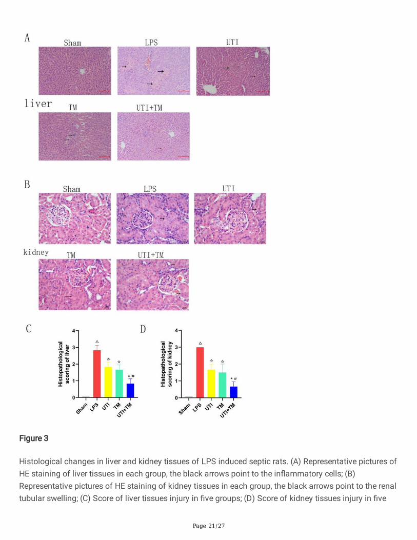
3.3 UTI combined with TM reduced liver and kidneypathology injuryHE stained liver sections showed that the model group injected with LPS caused obvious liver injury, inwhich the liver tissue cells were irregularly arranged, accompanied by a large number of necrosis andin�ammatory cell in�ltration. The liver in sham group did not suffer injury. In both TM group and UTIgroup, these changes were alleviated to a certain extent, the cells were clear and in�ammatory cellin�ltration was seen in the portal area. However, in UTI + TM group, the liver injury was further reduced,the liver cells were clear, regularly arranged, there was no obvious edema, and the necrosis was reduced(Fig. 3.A). Kidney HE stained section, it can be observed that after injection of LPS, although theglomerular structure is normal, there are renal tubular swelling and vacuolar degeneration and necrosis ofepithelial cells. A large number of in�ammatory cell in�ltration can be seen in the renal interstitium. Therenal tubules of UTI group and TM group were still swollen, but the vacuolar degeneration of renal tubularepithelial cells was reduced, and the interstitium was in�ltrated by in�ammatory cells. The pathologicalchanges in drug combination group were further improved. The swelling was further reduced, thevacuolar degeneration of renal tubular epithelial cells was reduced, and only a small amount ofin�ammatory cell in�ltration was observed (Fig. 3.B).
Page 8/27
We also scored histological damage on HE stained sections. The higher the score displayed in thehistogram, the more severe the tissue damage. A score of 0 in the sham group means that there is nodamage to the liver and kidney tissues. The liver and kidney scores of the LPS group are all greater than2.5, which means that the tissue damage is severe, liver and kidney histology scores of UTI group, TMgroup and drug combination group were all reduced, but drug combination group had the lowest score(Fig. 3.C), (Fig. 3.D), which indicated that the combination of drugs has a stronger effect on the recoveryof damaged tissues than UTI or TM treatment alone.
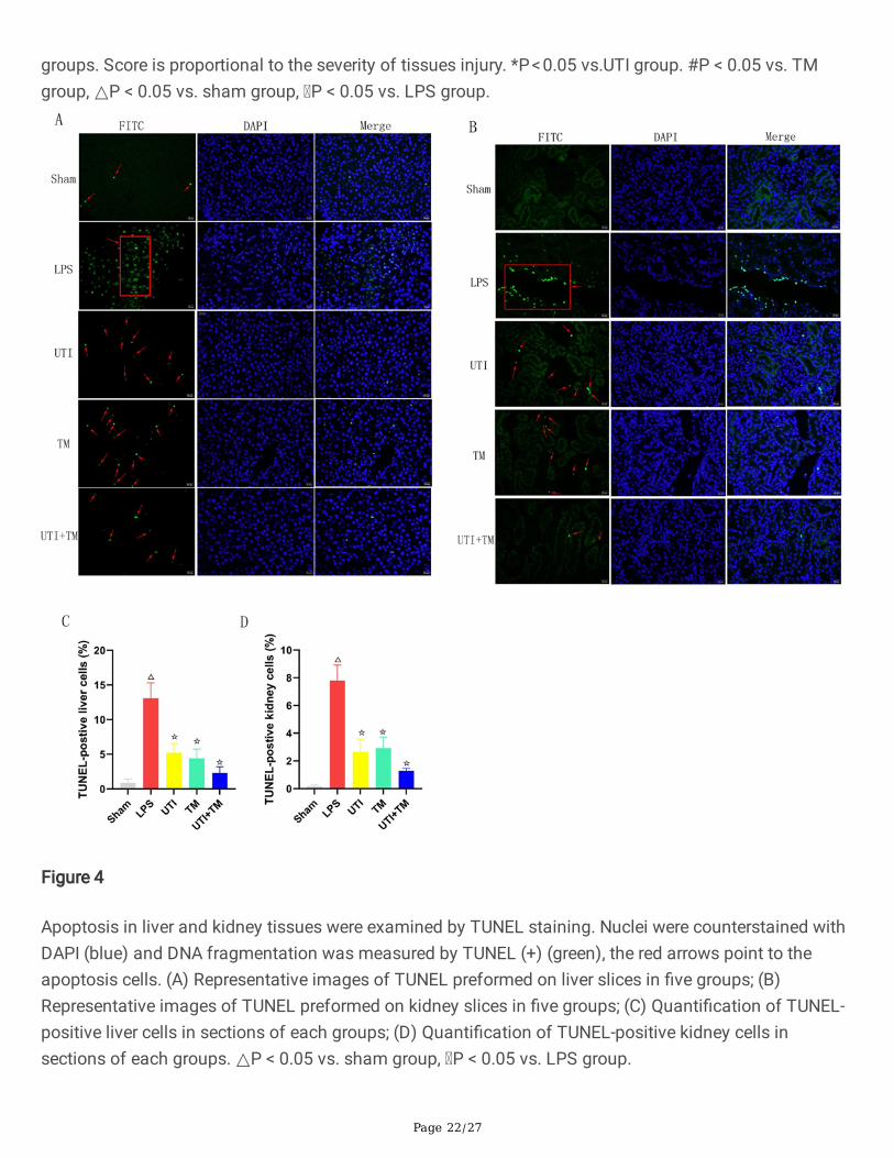
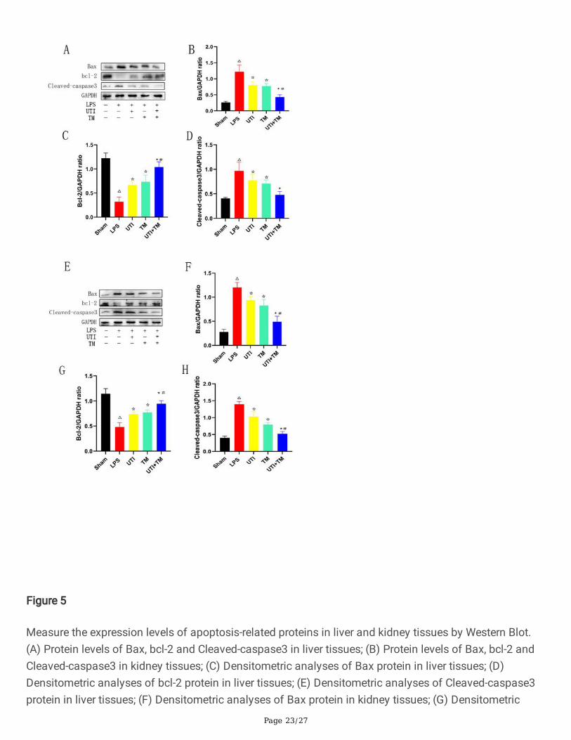
3.4 UTI combined with TM inhibited apoptosis of liver andkidney in septic ratsIn order to clarify the protective mechanism of UTI combined with TM on liver and kidney injury in septicrats, TUNEL staining was used to observe the apoptotic cells in liver (Fig. 4.A) and kidney (Fig. 4.B) slides.Compared with sham group, positive cells in the LPS group were signi�cantly increased, apoptosis wasmore likely to occur, while either UTI or TM treatment reduced the positive cells. In UTI + TM group, thenumber of apoptotic cells further decreased. We also performed statistical analysis on the positive ratesof liver (Fig. 4.C) and kidney (Fig. 4.D) cells in TUNEL staining. Whether in liver or kidney tissue, theapoptosis rate of LPS group was increased signi�cantly, while after the administration of UTI and TM, theapoptosis rate was appear to decreased, and the apoptosis rate of the combination group furtherdecreased. In addition, beside the results of TUNEL analysis, UTI and TM treatment reduced the apoptoticprotein Bax and Cleaved caspase-3. When UTI and TM were used in combination, this downward trendwas further accentuated, otherwise LPS will up-regulated the expression of Bax and cleave caspase-3.The decrease in the anti-apoptotic protein bcl-2 in LPS group was be reversed by the combination of UTIand TM (liver: Fig. 5.A-D; kidney: Fig. 5.E-H).
These data indicated that the combination of UTI and TM can protect the liver and kidney by reducing theexpression of pro-apoptotic proteins, thereby reducing cell apoptosis.
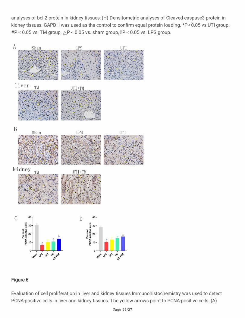
3.5 Immunohistochemistry of PCNATo investigate the effects of TM and UTI on the proliferation of hepatocytes and kidney cells, weperformed immunohistochemical analysis on PCNA (liver: Fig. 6.A), (kidney: Fig. 6.B). We counted thenumber of hepatocytes in randomly taken photomicrographs. Compared with sham group, the ratio ofpositive hepatocytes/total hepatocytes decreased signi�cantly after injection of LPS. Compared with LPSgroup, in UTI group, the positive rate increased by 3.71%, in TM group by 4.21%, the positive rate in UTI + TM group further increased by 7.39%. During the regeneration process, the proportion of positivehepatocytes of UTI + TM group was greater than that of TM group and UTI group (Fig. 6.C). We alsoanalyzed the PCNA of the kidney. During the whole process, the changes of PCNA in the kidney of eachgroup of rats were consistent with the expression of PCNA in the liver. Compared with sham group, theratio of positive kidney cells/total kidney cells decreased. Compared with LPS group, in UTI group, theratio increased by 2.68%, in UTI group by 4.60%, and further increased by 6.36% in UTI + TM group(Fig. 6.D).

Page 9/27
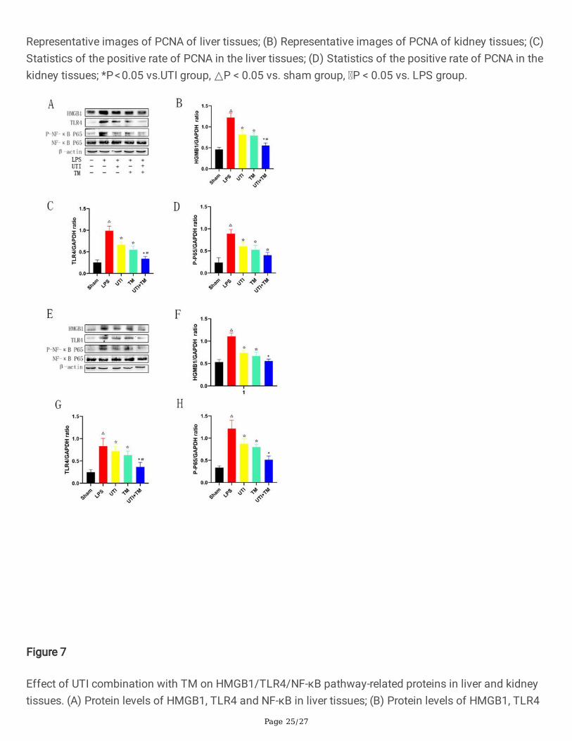
3.6 UTI combined with TM inhibited TLR4-mediated NF-κBpathwayTLR4 has been identi�ed as a receptor for LPS, and TLR4-related signal transduction pathways maymediate liver and kidney injury in septic rats. In this study, in order to explore the potential mechanism ofUTI combined with TM in protecting liver and kidney injury in septic rats, the expression of HMGB1, TLR4and the phosphorylation of NF-κB in liver and kidney tissues were evaluated. As shown in Fig. 7 (liver:Fig. 7.A-D), (kidney: Fig. 7.E-H), compared with relative to the expression of the endogenous control β-actin, HMGB1, TLR4, and P-NF-κB proteins all increased signi�cantly in liver and kidney tissues in LPSgroup. The administration of UTI and TM remarkably inhibited the protein expression of HMGB1, TLR4and P-NF-κB. As expected, the protein expression of HMGB1, TLR4 and P-NF-κB further decreased inseptic rats administered TM and UTI in combination. UTI combined with TM also signi�cantly reducedthe protein expression of downstream NF-κB.
3.7 In�uence of UTI on the anticoagulant function of TMAs shown in Table.1, as expected, in the range of 0–200 U/mL, TT, PT and APTT upregulated with theincrease of TM concentration, and all had a good linear correlation (r2 > 0.98). TT was extremely sensitiveto TM. Compared with the blank control group, 200 U/mL TM signi�cantly prolonged TT by about 3.23times (P < 0.05). However, the increase of UTI concentration in the range of 0–200 U/mL did not extendthe TT, PT and APTT of rat plasma (P < 0.05, there was no statistical difference in anticoagulant effect).According to the above results, 100 U/mL of TM was selected for subsequent experiments with UTI (100U/mL) to investigate whether UTI would reduce the anticoagulant effect of TM when combined.Importantly, when UTI was used in combination with TM, TT, PT, and APTT were not shortened comparedwith TM group, which indicated that UTI does not affect the anticoagulant effect of TM when they areused in combination (Table.2).
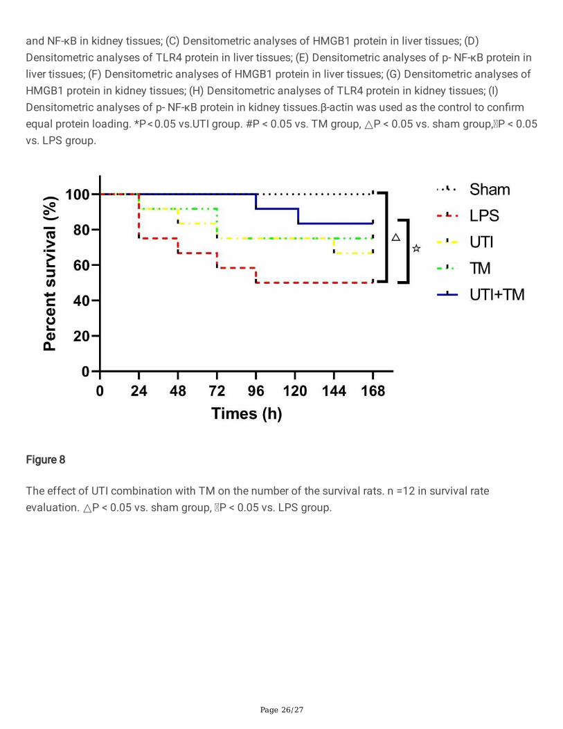
3.8 UTI combined with TM improved the Survival Rate ofLPS-Induced septic RatsThe survival period of rats was monitored every 24 hours for 7 days. The rats in Sham group showed noabnormal behavior and no death was observed. However, after inject of LPS, rats developed diarrhea,lethargy and ru�ed pelage. The number of surviving rats in LPS group decreased linearly with time,which was signi�cantly lower than in Sham group (P < 0.05). UTI and TM were administered separately toimprove the survival rate of septic rats. When UTI and TM were used in combination, the survival rate ofrats had a further improvement trend (Fig. 8). Compared with the single-drug group, UTI + TM treatmentincreased the average survival rate by 33.3% (n = 12).
4. Discussion
Page 10/27
Sepsis is a type of systemic in�ammation response syndrome, which has the characteristics of rapiddisease progression and high case fatality rate. It has always been one of the di�culties in critical illnessresearch. Society of Critical Care Medicine (SCCM) and European society of intensive medicine (ESICM)newly de�ned sepsis as the unbalanced response of the body to infection that leads to life-threateningorgan functions disorders (Vincent et al. 2016; Singer et al. 2016). The old de�nition of sepsis is asystemic in�ammatory response syndrome caused by infection, which emphasizes infection, while thenew de�nition of sepsis focuses on the body’s response to infection and imbalance with organdysfunction. This de�nition suggests that more attention should be paid to the complexpathophysiological response caused by infection during treatment. It is a return to the understanding ofthe essence of sepsis. Therefore, in the treatment of sepsis, protecting organs from damage and repairingorgan injury is particularly important. Most organ injury in sepsis is caused by the in�ammatory responseinvolving excessive and dysfunctional cytokines, and the effects of the existing treatment strategies forsepsis-induced organ injury are not satisfactory. In consequence, there is an urgent need for newtherapies or drugs for sepsis. UTI and TM are glycoproteins extracted from human urine. Several studieshave reported that these two drugs have clear anti-in�ammatory effects and a certain protective effect onliver and kidney injury. Therefore, we further tested the protective effect of UTI combined with TM onsepsis.
LPS is a component in the outer wall of gram-negative bacteria (Cavaillon. 2017). It is a classic inducer ofsepsis in medical research (de Pádua et al. 2018). It can activate mononuclear macrophages andendothelial cells through the cell signal transduction system in the body, synthesize and release a varietyof in�ammatory mediators (Plociennikowska et al. 2015), which in turn cause a series of reactions to thebody. Pro-in�ammatory cytokines such as IL-6 and TNF-α are involved in the initiation and regulation ofthe in�ammatory response (Firinu et al. 2016). Reliable studies have con�rmed that a large amount ofTNF-α and IL-6 are produced in macrophages exposed to LPS (Lee et al. 2017). In our research, Serumpro-in�ammatory factors TNF-α and IL-6 were signi�cantly increased in rats injected with LPS. However,UTI combined with TM signi�cantly inhibited the levels of TNF-α and IL-6 in serum, indicating that itsprotective effect on sepsis may be related to its anti-in�ammatory properties.
As a kind of endotoxin, LPS is directly injected into the blood by intravenous injection to cause sepsis inrats. animals infused with LPS manifest features of compensated human sepsis, including hypotension,hypermetabolism and elevated serum lactate concentrations, this model can be used to studymechanisms that contribute to the activation of in�ammatory cascades induced by bacterial antigen, andunravel interactions between distinct in�ammatory systems (such as coagulation system and cytokinenetwork), and to clarify the principle for the e�cacy of novel anti-in�ammatory compounds (Fiuza et al.2001; Lowry. 2005; Van der Poll. 2012). In addition, the infectious dose of this model can be controlledaccording to the weight of the rats, and it has good stability, repeatability and controllability (Yeh et al.2016). This method is currently widely used (Hao et al. 2017; Carty. 2019; Sun. 2018; Savio et al. 2017).
HMGB1/TLR4/NF-κB is an important in�ammatory signal pathway in LPS-induced in�ammation (Shanget al. 2019). Some reports have shown that HMGB1/TLR4 common syndrome pathway genes are

Page 11/27
expressed in the liver and kidney (Liu et al. 2020; Mohamed et al. 2020). HMGB1 is a highly conservednon-histone DNA binding protein, which is widely distributed among various organs such as lung, brain,liver, heart, and kidney. HMGB1 can be released from necrotic cells through active secretion and passiverelease, inducing in�ammation. HMGB1 is also one of the endogenous ligands of TLR4, which alsowidely expresses in liver and kidney. LPS induces tissues to release HMGB1, which mediates autophagyor triggers the initiation of in�ammation through the TLR4 signaling pathway, triggering a series ofcascade reactions. It mainly includes two pathways, including myeloid differentiation factor 88 (MYD88)dependent pathway and TRIF dependent pathway (Wang et al. 2015). Activation of MYD88 activatesdownstream IKK-α/IKK-β, leading to phosphorylation and degradation of IκB-α, and �nally NF-κB isactivated (Zhang et al. 2018). Phosphorylation of NF-κB leads to the release of pro-in�ammatorycytokines, including TNF-α, IL-1βand IL-6. Therefore, inhibiting HMGB1-TLR4 signaling pathway mayeffectively improve organ injury caused by sepsis. Our research have con�rmed that UTI and TM cansigni�cantly inhibit LPS-induced liver and kidney injury through the HMGB1-TLR4-NF-κB pathway. WhenUTI and TM were used in combination, this effect was more signi�cant, and it also upregulated thesurvival rate of rats attacked by LPS.
It is worth noting that LPS can promote apoptosis of liver and kidney cells and aggravate tissue injury(Zhang et al. 2020; Lu et al. 2020). It was veri�ed by western blot that the protein concentration ofCleaved caspase-3 and Bax increased, and the concentration of anti-apoptotic protein bcl-2 decreased. Inserum, ALT and AST, as indicators of liver characteristics, and as well as BUN and Cr, as indicators ofkidney characteristics, were both increased. It showed that the action of endotoxins led to apoptosis ofliver cells and kidney cells. With the administration of UTI and TM, the concentration of Cleaved caspase-3 and Bax decreased, the concentration of bcl-2 increased, and the values of ALT, AST, BUN and Cr beganto decrease. All this indicates that liver and kidney injury were alleviated. When UTI combined with TM,this protective effect was better. This result was further veri�ed in the TUNEL experiment, the results werefurther veri�ed. The number of positive cells in liver and kidney tissues decreased after UTI combinedwith TM, which reversed the increase in the number of positive cells caused by LPS. We also observed thechanges in the number of PCNA-positive cells in the liver and kidney tissues. The changes in PCNA wereusually closely related to tissue regeneration (Lee et al. 2019). After LPS injection, the PCNA of rat liverand kidney tissues decreased signi�cantly. LPS exposure would affect these proliferating cells and causethe decrease of their number due to cell death. After the administration, PCNA began to increase, and thecombination group’s level was higher than LPS group. Interestingly, we observed that at the concentrationwe set, the single-drug groups increased in proliferation, but not signi�cantly compared to LPS group.This indicated that the combination of UTI and TM may have a limited effect on promoting regeneration,in contrast, inhibiting apoptosis has a stronger protective effect.
The blood coagulation system plays an important role in the pathogenesis of sepsis. It promotes eachother with in�ammation, and together constitutes a key factor in the occurrence and development ofsepsis (Levi et al. 2010). Endotoxin can activate the exogenous coagulation pathway by inducing therelease of tissue factor of macrophages and endothelial cells. The coagulation factor XII activated byendotoxin can also further activate the endogenous coagulation pathway, which ultimately leads to
Page 12/27
diffuse DIC (Park et al. 2016; Latour. 1983). Therefore, we envision that in the treatment of DIC induced bysepsis, TM can not only effectively treat sepsis, but may inhibit the symptoms of DIC by activating theanticoagulation system. Our experiments proved that compared to using TM alone, UTI combined withTM did not reduce the anticoagulant effect of TM. We concluded that combined use of UTI and TM waseffective in the treatment of sepsis, and UTI did not affect the anti-DIC effect of TM. Therefore, whenchoosing medication in such special populations of DIC induced by sepsis, UTI and TM combinedadministration is a good strategy.
It is undeniable that this study still has certain limitations. First, our experiments did not conduct furtherstudies into the damage and repair of other important organs such as the heart and lungs. In addition,existing studies have shown that UTI and TM have multiple pharmacological effects and may also triggerother mechanisms, the protective mechanism of drug combination on liver and kidney is worthy of furtherexploration.
5. Conclusion5.Conclusions
In conclusion, our research showed that the combined use of UTI and TM protected liver and kidney ofLPS-treated rats. These protective effects can be attributed to UTI and TM reducing certain in�ammatorymediators such as TNF-α, IL-1β and IL-6, inhibiting apoptosis and HMGB1/TLR4/NF-κB signaling, andpromoting the proliferation of liver and kidney cells (Fig. 9). The protective effect of septic liver andkidney injury and in�ammation provided new insights into the treatment of liver and kidney injury causedby sepsis. Therefore, considering these results, the combination of TM and UTI may be a promisingstrategy for the treatment of sepsis.
AbbreviationsUTI Ulinastatin
TM Thrombomodulin
DIC Diffuse intravascular coagulation
LPS Lipopolysaccharide
TNF Tumor necrosis factor
IL Interleukin
NF Nuclear factor
HMGB1 High mobility group protein 1

Page 13/27
BUN Blood urea nitrogen
Cr Creatinine
AST Aspartate aminotransferase
ALT Alanine aminotransferase
HE Haematoxylin and Eosin
TUNEL Terminal-deoxynucleotidyl transferase mediated nick end labeling
PCNA Proliferation of cell nuclear antigen
TT Thrombin time
PT Prothrombin time
APTT Activated partial thromboplastin time
SCCM Society of Critical Care Medicine
ESICM European society of intensive medicine
MYD Myeloid differentiation factor
DeclarationsEthics approval and consent to participate
Our experiment complies with the Basel Declaration, which outlines the basic principles to be followedwhen conductingresearch in animals. The protocol was approved by the Animal Research Committee ofChina Pharmaceutical University.
Consent for publication
Not applicable
Availability of data and materials
The datasets used and/or analysed during the current study are available from the corresponding authoron reasonable request.
Competing interests
The authors declare that they have no competing interests
Page 14/27
Funding
This project was supported by the National Natural Science Fund of China (No. 81503133).
Authors' contributions
FY JL and XZ designed experimental scheme and prepared the manuscript. X Z CS SZ carried out theexperiment. XZ and EEA wrote the paper. JY and FY revised the manuscript critically and supplementedthe experiment. XL CF QL performed experimental data analysis.
Acknowledgements
We thank the Heliang Fu of Yangzhou Adeal pharmaceutical co., Ltd. for providing UTI and TM.
References1. Vandewalle J, Libert C. Glucocorticoids in Sepsis: To Be or Not to Be. Frontiers in immunology.
2020;11:1318.
2. Mirouse A, Vigneron C, Llitjos JF, Chiche JD, Mira JP, Mokart D, et al. Sepsis and Cancer: An Interplayof Friends and Foes. Am J Respir Crit Care Med. 2020;202:1625–35.
3. Venet F, Monneret G. Advances in the understanding and treatment of sepsis-inducedimmunosuppression. Nat Rev Nephrol. 2018;14:121–37.
4. Tsantarliotou MP, Lavrentiadou SN, Psalla DA, Margaritis IE, Kritsepi MG, Zervos IA, et al.Suppression of plasminogen activator inhibitor-1 (PAI-1) activity by crocin ameliorateslipopolysaccharide-induced thrombosis in rats. Food chemical toxicology: an international journalpublished for the British Industrial Biological Research Association. 2019;125:190–7.
5. Yan J, Li S, Li S. The role of the liver in sepsis. Int Rev Immunol. 2014;33:498–510.
�. Alobaidi R, Basu RK, Goldstein SL, Bagshaw SM. Sepsis-associated acute kidney injury. SeminNephrol. 2015;35:2–11.
7. Cecconi M, Evans L, Levy M, Rhodes A. Sepsis and septic shock. Lancet. 2018;392:75–87.
�. Abdulmahdi W, Patel D, Rabadi MM, Azar T, Jules E, Lipphardt M, et al. HMGB1 redox during sepsis.Redox Biol. 2017;13:600–7.
9. Magna M, Pisetsky DS. The role of HMGB1 in the pathogenesis of in�ammatory and autoimmunediseases. Molecular medicine (Cambridge, Mass.). 2014;20:138–146.
10. Lu B, Nakamura T, Inouye K, Li J, Tang Y, Lundbäck P, et al. Novel role of PKR in in�ammasomeactivation and HMGB1 release. Nature. 2012;488:670–4.
11. Yang H, Wang H, Andersson U. Targeting In�ammation Driven by HMGB1. Frontiers in immunology.2020a;11:484.
12. Liu B, Huang W, Xiao X, Xu Y, Ma S, Xia Z. Neuroprotective Effect of Ulinastatin on Spinal CordIschemia-Reperfusion Injury in Rabbits. Oxidative Med Cell Longev. 2015;2015:624819.

Page 15/27
13. Kessenbrock K, Dau T, Jenne DE. Tailor-made in�ammation: how neutrophil serine proteasesmodulate the in�ammatory response. J Mol Med. 2011;89:23–8.
14. Song Y, Miao S, Li Y, Fu H. Ulinastatin attenuates liver injury and in�ammation in a cecal ligation andpuncture induced sepsis mouse model. Journal of cellular biochemistry. 2019;120:417–24.
15. Cao C, Yin C, Shou S, Wang J, Yu L, Li X, et al. Ulinastatin Protects Against LPS-Induced Acute LungInjury By Attenuating TLR4/NF-κB Pathway Activation and Reducing In�ammatory Mediators.Shock. 2018;50:595–605.
1�. Yang XY, Song J, Hou SK, Fan HJ, Lv Q, Liu ZQ, et al. Ulinastatin ameliorates acute kidney injuryinduced by crush syndrome in�ammation by modulating Th17/Treg cells. Int Immunopharmacol.2020b;81:106265.
17. Yatabe T, Inoue S, Sakamoto S, Sumi Y, Nishida O, Hayashida K, et al. The anticoagulant treatmentfor sepsis induced disseminated intravascular coagulation; network meta-analysis. Thrombosisresearch. 2018;171:136–42.
1�. Pescatore SL. Clinical management of protein C de�ciency. Expert opinion on pharmacotherapy.2001;2:431–9.
19. Ma CY, Chang WE, Shi GY, Chang BY, Cheng SE, Shih YT, et al. Recombinant thrombomodulin inhibitslipopolysaccharide-induced in�ammatory response by blocking the functions of CD14. Journal ofimmunology (Baltimore, Md.: 1950). 2015;194:1905–1915.
20. Akatsuka M, Masuda Y, Tatsumi H, Yamakage M. Recombinant human soluble thrombomodulin isassociated with attenuation of sepsis-induced renal impairment by inhibition of extracellular histonerelease. PloS one. 2020;15:e0228093.
21. Nagato M, Okamoto K, Abe Y, Higure A, Yamaguchi K. Recombinant human soluble thrombomodulindecreases the plasma high-mobility group box-1 protein levels, whereas improving the acute liverinjury and survival rates in experimental endotoxemia. Critical care medicine. 2009;37:2181–6.
22. Yoshihiro S, Sakuraya M, Hayakawa M, Ono K, Hirata A, Takaba A, et al. Recombinant Human-Soluble Thrombomodulin Contributes to Reduced Mortality in Sepsis Patients With SevereRespiratory Failure: A Retrospective Observational Study Using a Multicenter Dataset. Shock.2019;51:174–9.
23. Jiang W, Yu X, Sun T, Chai Y, Chang P, Chen Z, et al. ADJunctive Ulinastatin in Sepsis Treatment inChina (ADJUST study): study protocol for a randomized controlled trial. Trials. 2018;19:133.
24. Wu J, Yan X, Jin G. Ulinastatin protects rats from sepsis-induced acute lung injury by suppressing theJAK-STAT3 pathway. Journal of cellular biochemistry. 2018.
25. Wang H, Liu B, Tang Y, Chang P, Yao L, Huang B, et al. Improvement of Sepsis Prognosis byUlinastatin: A Systematic Review and Meta-Analysis of Randomized Controlled Trials. FrontPharmacol. 2019;10:1370.
2�. Ashina M, Fujioka K, Nishida K, Okubo S, Ikuta T, Shinohara M, et al. Recombinant humanthrombomodulin attenuated sepsis severity in a non-surgical preterm mouse model. Scienti�creports. 2020;10:333.
Page 16/27
27. Meng F, Du C, Zhang Y, Wang S, Zhou Q, Wu L, et al. Protective effect of rhubarb combined withulinastatin for patients with sepsis. Medicine. 2020;99:e18895.
2�. Wang J, Zhou J, Bai S. Combination of Glutamine and Ulinastatin Treatments Greatly ImprovesSepsis Outcomes. Journal of in�ammation research. 2020;13:109–15.
29. Vincent JL, Mira JP, Antonelli M. Sepsis: older and newer concepts. The Lancet Respiratory medicine.2016;4:237–40.
30. Singer M, Deutschman CS, Seymour CW, Shankar-Hari M, Annane D, Bauer M, et al. The ThirdInternational Consensus De�nitions for Sepsis and Septic Shock (Sepsis-3). Jama. 2016;315:801–10.
31. Cavaillon JM. Exotoxins and endotoxins: Inducers of in�ammatory cytokines. Toxicon: o�cialjournal of the International Society on Toxinology. 2018;149:45–53.
32. de Pádua Lúcio K, Rabelo ACS, Araújo CM, Brandão GC, de Souza GHB, da Silva RG, et al. Anti-In�ammatory and Antioxidant Properties of Black Mulberry (Morus nigra L.) in a Model of LPS-Induced Sepsis. Oxidative medicine and cellular longevity. 2018;2018:5048031.
33. Płóciennikowska A, Hromada-Judycka A, Borzęcka K, Kwiatkowska K. Co-operation of TLR4 and raftproteins in LPS-induced pro-in�ammatory signaling. Cell Mol Life Sci. 2015;72:557–81.
34. Firinu D, Garcia-Larsen V, Manconi PE, Del Giacco SR. SAPHO Syndrome: Current Developments andApproaches to Clinical Treatment. Curr Rheumatol Rep. 2016;18:35.
35. Lee SB, Lee WS, Shin JS, Jang DS, Lee KT. Xanthotoxin suppresses LPS-induced expression of iNOS,COX-2, TNF-α, and IL-6 via AP-1, NF-κB, and JAK-STAT inactivation in RAW 264.7 macrophages. IntImmunopharmacol. 2017;49:21–9.
3�. Fiuza C, Suffredini AF. Human models of innate immunity: local and systemic in�ammatoryresponses. J Endotoxin Res. 2001;7:385–8.
37. Lowry SF. Human endotoxemia: a model for mechanistic insight and therapeutic targeting. Shock.2005;24(Suppl 1):94–100.
3�. van der Poll T. Experimental human sepsis models. Drug Discovery Today: Disease Models.2012;9:e3–9.
39. Yeh YC, Wu CY, Cheng YJ, Liu CM, Hsiao JK, Chan WS, et al. Effects of Dexmedetomidine onIntestinal Microcirculation and Intestinal Epithelial Barrier in Endotoxemic Rats. Anesthesiology.2016;125:355–67.
40. Hao H, Cao L, Jiang C, Che Y, Zhang S, Takahashi S, et al. Farnesoid X Receptor Regulation of theNLRP3 In�ammasome Underlies Cholestasis-Associated Sepsis. Cell Metabol. 2017;25:856–67.e855.
41. Carty M, Kearney J, Shanahan KA, Hams E, Sugisawa R, Connolly D, et al. Cell Survival and CytokineRelease after In�ammasome Activation Is Regulated by the Toll-IL-1R Protein SARM. Immunity.2019;50:1412–24.e1416.

Page 17/27
42. Sun Y, Yao X, Zhang QJ, Zhu M, Liu ZP, Ci B, et al. Beclin-1-Dependent Autophagy Protects the HeartDuring Sepsis. Circulation. 2018;138:2247–62.
43. Savio LEB, de Andrade Mello P, Figliuolo VR, de Avelar Almeida TF, Santana PT, Oliveira SDS, et al.CD39 limits P2X7 receptor in�ammatory signaling and attenuates sepsis-induced liver injury. Journalof hepatology. 2017;67:716–26.
44. Shang J, Liu W, Yin C, Chu H, Zhang M. Cucurbitacin E ameliorates lipopolysaccharide-evoked injury,in�ammation and MUC5AC expression in bronchial epithelial cells by restraining the HMGB1-TLR4-NF-κB signaling. Molecular immunology. 2019;114:571–7.
45. Liu X, Huang K, Zhang RJ, Mei D, Zhang B. Isochlorogenic Acid A Attenuates the Progression of LiverFibrosis Through Regulating HMGB1/TLR4/NF-κB Signaling Pathway. Front Pharmacol.2020;11:582.
4�. Mohamed ME, Abduldaium YS, Younis NS. Ameliorative Effect of Linalool in Cisplatin-InducedNephrotoxicity: The Role of HMGB1/TLR4/NF-κB and Nrf2/HO1 Pathways. Biomolecules. 2020;10.
47. Wang J, He GZ, Wang YK, Zhu QK, Chen W, Guo T. TLR4-HMGB1-, MyD88- and TRIF-dependentsignaling in mouse intestinal ischemia/reperfusion injury. World journal of gastroenterology.2015;21:8314–25.
4�. Zhang Z, Liu Q, Liu M, Wang H, Dong Y, Ji T, et al. Upregulation of HMGB1-TLR4 in�ammatorypathway in focal cortical dysplasia type II. J Neuroin�amm. 2018;15:27.
49. Zhang Y, Jia H, Jin Y, Liu N, Chen J, Yang Y, et al. Glycine Attenuates LPS-Induced Apoptosis andIn�ammatory Cell In�ltration in Mouse Liver. J Nutr. 2020;150:1116–25.
50. Lu QB, Du Q, Wang HP, Tang ZH, Wang YB, Sun HJ. Salusin-β mediates tubular cell apoptosis inacute kidney injury: Involvement of the PKC/ROS signaling pathway. Redox Biol. 2020;30:101411.
51. Lee GS, Yang HG, Kim JH, Ahn YM, Han MD, Kim WJ. Pine (Pinus densi�ora) needle extract couldpromote the expression of PCNA and Ki-67 after partial hepatectomy in rat. Acta cirurgica brasileira.2019;34:e201900606.
52. Levi M, van der Poll T. In�ammation and coagulation. Critical care medicine. 2010;38:26–34.
53. Park HS, Gu J, You HJ, Kim JE, Kim HK. Factor XII-mediated contact activation related to poorprognosis in disseminated intravascular coagulation. Thrombosis research. 2016;138:103–7.
54. Latour JG. Modulation of disseminated intravascular coagulation (DIC) by steroidal and non-steroidal anti-in�ammatory drugs. Agents actions. 1983;13:487–95.
TablesTable.1 Effects of UTI and TM on coagulation indicators of rat plasma.

Page 18/27
Indicator c/(U/mL) TT/s PT/s APTT/s
UTI 0 41.52±0.66 18.10±0.29 37.75±0.58
50 41.52±0.66 18.10±0.29 37.75±0.58
100 41.33±1.25 18.85±0.46 38.18±0.94
150 42.17±0.66 18.83±0.65 39.35±0.77
TM 0 41.52±0.66 18.10±0.29 37.75±0.58
50 81.57±1.14* 22.03±0.52* 51.32±0.97*
100 114.23±1.97* 24.02±0.59* 69.33±1.07*
150 147.10±2.18* 29.18±0.62* 79.45±1.31*
*P<0.05 vs. 0 μg/mL group. TT: thrombin time; PT: prothrombin time; APTT: activated partial thrombintime
Table. 2 After combined use of UTI, the effect on anticoagulation index of TM
Group c/(U/mL) TT/s PT/s APTT/s
Control 0 41.52±0.66 18.10±0.29 37.75±0.58
UTI 100 42.17±0.66 18.83±0.65 39.35±0.77
TM 100 114.23±1.97* 24.02±0.59* 69.33±1.07*
UTI+TM 100+100 119.21±3.31* 24.20±0.82* 70.78±0.87*
*P<0.05 vs. Control group. TT: thrombin time; PT: prothrombin time; APTT: activated partial thrombintime
Figures

Page 19/27
Figure 1
Changes of serum cytokines in rats after LPS injuction or sham by ELISA kit. (A) Changes in �ve groupsof TNF-α; (B) Changes in �ve groups of IL-6. *P < 0.05 vs.UTI group. #P < 0.05 vs. TM group, △P < 0.05 vs.Sham group, P < 0.05 vs. LPS group.

Page 20/27
Figure 2
Changes of serum ALT, AST, BUN and Cr levels in rats. (A) serum ALT; (B) serum AST; (C)serum BUN; (D)serum Cr. *P < 0.05 vs.UTI group. #P < 0.05 vs. TM group, △P < 0.05 vs. sham group, P < 0.05 vs. LPSgroup.
Page 21/27
Figure 3
Histological changes in liver and kidney tissues of LPS induced septic rats. (A) Representative pictures ofHE staining of liver tissues in each group, the black arrows point to the in�ammatory cells; (B)Representative pictures of HE staining of kidney tissues in each group, the black arrows point to the renaltubular swelling; (C) Score of liver tissues injury in �ve groups; (D) Score of kidney tissues injury in �ve
Page 22/27
groups. Score is proportional to the severity of tissues injury. *P < 0.05 vs.UTI group. #P < 0.05 vs. TMgroup, △P < 0.05 vs. sham group, P < 0.05 vs. LPS group.
Figure 4
Apoptosis in liver and kidney tissues were examined by TUNEL staining. Nuclei were counterstained withDAPI (blue) and DNA fragmentation was measured by TUNEL (+) (green), the red arrows point to theapoptosis cells. (A) Representative images of TUNEL preformed on liver slices in �ve groups; (B)Representative images of TUNEL preformed on kidney slices in �ve groups; (C) Quanti�cation of TUNEL-positive liver cells in sections of each groups; (D) Quanti�cation of TUNEL-positive kidney cells insections of each groups. △P < 0.05 vs. sham group, P < 0.05 vs. LPS group.
Page 23/27
Figure 5
Measure the expression levels of apoptosis-related proteins in liver and kidney tissues by Western Blot.(A) Protein levels of Bax, bcl-2 and Cleaved-caspase3 in liver tissues; (B) Protein levels of Bax, bcl-2 andCleaved-caspase3 in kidney tissues; (C) Densitometric analyses of Bax protein in liver tissues; (D)Densitometric analyses of bcl-2 protein in liver tissues; (E) Densitometric analyses of Cleaved-caspase3protein in liver tissues; (F) Densitometric analyses of Bax protein in kidney tissues; (G) Densitometric
Page 24/27
analyses of bcl-2 protein in kidney tissues; (H) Densitometric analyses of Cleaved-caspase3 protein inkidney tissues. GAPDH was used as the control to con�rm equal protein loading. *P < 0.05 vs.UTI group.#P < 0.05 vs. TM group, △P < 0.05 vs. sham group, P < 0.05 vs. LPS group.
Figure 6
Evaluation of cell proliferation in liver and kidney tissues Immunohistochemistry was used to detectPCNA-positive cells in liver and kidney tissues. The yellow arrows point to PCNA-positive cells. (A)
Page 25/27
Representative images of PCNA of liver tissues; (B) Representative images of PCNA of kidney tissues; (C)Statistics of the positive rate of PCNA in the liver tissues; (D) Statistics of the positive rate of PCNA in thekidney tissues; *P < 0.05 vs.UTI group, △P < 0.05 vs. sham group, P < 0.05 vs. LPS group.
Figure 7
Effect of UTI combination with TM on HMGB1/TLR4/NF-κB pathway-related proteins in liver and kidneytissues. (A) Protein levels of HMGB1, TLR4 and NF-κB in liver tissues; (B) Protein levels of HMGB1, TLR4
Page 26/27
and NF-κB in kidney tissues; (C) Densitometric analyses of HMGB1 protein in liver tissues; (D)Densitometric analyses of TLR4 protein in liver tissues; (E) Densitometric analyses of p- NF-κB protein inliver tissues; (F) Densitometric analyses of HMGB1 protein in liver tissues; (G) Densitometric analyses ofHMGB1 protein in kidney tissues; (H) Densitometric analyses of TLR4 protein in kidney tissues; (I)Densitometric analyses of p- NF-κB protein in kidney tissues.β-actin was used as the control to con�rmequal protein loading. *P < 0.05 vs.UTI group. #P < 0.05 vs. TM group, △P < 0.05 vs. sham group, P < 0.05vs. LPS group.
Figure 8
The effect of UTI combination with TM on the number of the survival rats. n =12 in survival rateevaluation. △P < 0.05 vs. sham group, P < 0.05 vs. LPS group.

Page 27/27
Figure 9
UTI combined with TM protected liver and kidney injury caused by sepsis. UTI combined with TMreversed the outbreak of serum in�ammatory factors (IL-6 and TNF-α), reduced serum ALT, AST, BUN andCr levels, inhibited liver and kidney cells apoptosis by regulating the protein expression of Bax, Cleavedcaspase-3, and Bcl-2, promoted the proliferation of liver and kidney cells, and inhibited the signaltransduction ability of HMGB1/TLR4/NF-κB in liver and injury tissues, which is useful in treating liver andkidney injury due to sepsis.
Related Documents