and elbows. Shoulders, hips, spine and sacroiliac joints are rarely involved. 5 e arthritis remits completely within few days with or without therapy and then recurs with increasing frequency in coming days. Recovery is typically associated with desquamation of the skin overlying the joint. Gout usually has three distinct stages: (1) acute gouty arthritis, (2) intercritical gout (asymptomatic phases between acute gouty flares which may last several months to years) and (3) chronic tophaceous gout. After approximately 10 years of recurrent gouty arthritis, patient develops into chronic gout with appearance of tophi. Chronic gouty arthritis is usually polyarticular with deposition of MSU crystals on tendons (e.g. tendo Achilles), ligaments, bursae, tibial tuberosity, and it may have concomitant tophi formation in fingers, toes, ears or upper part of forearm (Figures 2 to 4). e younger the patient is at the onset of the disease, the higher the tendency to have a progressive course. In many of the patients, the first attack may not be followed by second attack any more. A presumptive diagnosis of gout (monoarticular; old name podagra) can be made by the classical clinical presentation. A rapid response to nonsteroidal anti-inflammatory drugs (NSAIDs) or colchicine is often diagnostic but the gold standard for the diagnosis of acute gout is the demonstration of strongly negative birefringent needle- and rod-shaped crystals of MSU in the synovial fluid under compensated polarizing light microscopy. Gram stain and culture of the aspirated fluid are often performed concomitantly to exclude septic arthritis and cellulitis, both of which may mimic acute gout. 6 SUA is usually raised (> 10 mg/dL) but it should be kept in mind that up to 40% patients may have normal or low SUA level during the acute attack as stress-induced liberation of adrenocorticotropic Gout in Indian Scenario Arup Kumar Kundu Chapter 98 Figure 1: Red, hot and swollen first metatarsophalangeal joint (black arrow) of left foot in acute gout INTRODUCTION Gout is derived from the Latin word “gutta” , meaning drop, as it was believed in the past that poison falling in drops into the affected joints caused gout. Sir Alfred Garrod linked gout with hyperuricemia in 1848. 1 We now know that hyperuricemia is a biochemical precursor of gout, and not everybody with high serum uric acid (SUA) will develop gout. Gout, as we understand it today, is an inflammatory response to monosodium urate (MSU) monohydrate crystal deposition in the joints due to alteration in body urate metabolism. Gout is the most common crystal arthropathy occurring in men above 40 years of age. EPIDEMIOLOGY Gout is the most common cause of inflammatory arthritis in men aged more than 50, affecting approximately 1–2% of adult men in the Western world; they usually present in the form of “podagra” , i.e. acute onset pain in joint, erythema and swelling of the first metatarsophalangeal joint. Gout develops in men more than women (10:1) and seldom occurs in premenopausal women. Globally, the incidence of gout has increased doubly in last two decades because of multiple factors like increased longevity, increased prevalence of hypertension and rampant use of diuretics, more use of low-dose aspirin, epidemic of obesity and metabolic syndrome, dietary trends, increased alcohol consumption, increased chronic kidney disease (CKD) and major organ transplantation with use of cyclosporine or tacrolimus. Incidence of gout in India is not very clear. e prevalence is 0.12% as per International League of Nations Against Rheumatism, Community Oriented Program for Control of Rheumatic Diseases (ILAR COPCORD) study in Bhigwan village of India. 2 A study from Vellore revealed that 15.8% of the affected patients are less than 30 years of age; urban Indian population is involved more than the rural population and due to increased prevalence of metabolic syndrome in younger population, the first attack of gout occurs a decade earlier to them. 3 Another Indian study showed that high uric acid level is associated with laboratory and anthropometric parameters of metabolic syndrome. 4 GOUT: CLINICAL SPECTRUM Most cases of gout are characterized by the sudden onset of severe acute monoarthritis in a peripheral joint, mostly in the lower limb. e affected joint becomes dusky red, hot, swollen and extremely tender (Figure 1). Approximately 75% have first attack in great toe and 90% patients experience acute attack in great toe at sometime during the disease. In 3–14%, the first attack is polyarticular; the order of involvement is: insteps, ankles, heels, knees, wrists, fingers

Welcome message from author
This document is posted to help you gain knowledge. Please leave a comment to let me know what you think about it! Share it to your friends and learn new things together.
Transcript
and elbows. Shoulders, hips, spine and sacroiliac joints are rarely involved.5 The arthritis remits completely within few days with or without therapy and then recurs with increasing frequency in coming days. Recovery is typically associated with desquamation of the skin overlying the joint. Gout usually has three distinct stages: (1) acute gouty arthritis, (2) intercritical gout (asymptomatic phases between acute gouty flares which may last several months to years) and (3) chronic tophaceous gout. After approximately 10 years of recurrent gouty arthritis, patient develops into chronic gout with appearance of tophi. Chronic gouty arthritis is usually polyarticular with deposition of MSU crystals on tendons (e.g. tendo Achilles), ligaments, bursae, tibial tuberosity, and it may have concomitant tophi formation in fingers, toes, ears or upper part of forearm (Figures 2 to 4). The younger the patient is at the onset of the disease, the higher the tendency to have a progressive course. In many of the patients, the first attack may not be followed by second attack any more. A presumptive diagnosis of gout (monoarticular; old name podagra) can be made by the classical clinical presentation. A rapid response to nonsteroidal anti-inflammatory drugs (NSAIDs) or colchicine is often diagnostic but the gold standard for the diagnosis of acute gout is the demonstration of strongly negative birefringent needle- and rod-shaped crystals of MSU in the synovial fluid under compensated polarizing light microscopy. Gram stain and culture of the aspirated fluid are often performed concomitantly to exclude septic arthritis and cellulitis, both of which may mimic acute gout.6 SUA is usually raised (> 10 mg/dL) but it should be kept in mind that up to 40% patients may have normal or low SUA level during the acute attack as stress-induced liberation of adrenocorticotropic
Gout in Indian Scenario
Arup Kumar Kundu
Chapter 98
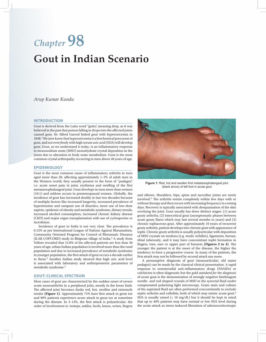
Figure 1: Red, hot and swollen first metatarsophalangeal joint (black arrow) of left foot in acute gout
INTRODUCTIONGout is derived from the Latin word “gutta”, meaning drop, as it was believed in the past that poison falling in drops into the affected joints caused gout. Sir Alfred Garrod linked gout with hyperuricemia in 1848.1 We now know that hyperuricemia is a biochemical precursor of gout, and not everybody with high serum uric acid (SUA) will develop gout. Gout, as we understand it today, is an inflammatory response to monosodium urate (MSU) monohydrate crystal deposition in the joints due to alteration in body urate metabolism. Gout is the most common crystal arthropathy occurring in men above 40 years of age.
EPIDEMIOLOGYGout is the most common cause of inflammatory arthritis in men aged more than 50, affecting approximately 1–2% of adult men in the Western world; they usually present in the form of “podagra”, i.e. acute onset pain in joint, erythema and swelling of the first metatarsophalangeal joint. Gout develops in men more than women (10:1) and seldom occurs in premenopausal women. Globally, the incidence of gout has increased doubly in last two decades because of multiple factors like increased longevity, increased prevalence of hypertension and rampant use of diuretics, more use of low-dose aspirin, epidemic of obesity and metabolic syndrome, dietary trends, increased alcohol consumption, increased chronic kidney disease (CKD) and major organ transplantation with use of cyclosporine or tacrolimus. Incidence of gout in India is not very clear. The prevalence is 0.12% as per International League of Nations Against Rheumatism, Community Oriented Program for Control of Rheumatic Diseases (ILAR COPCORD) study in Bhigwan village of India.2 A study from Vellore revealed that 15.8% of the affected patients are less than 30 years of age; urban Indian population is involved more than the rural population and due to increased prevalence of metabolic syndrome in younger population, the first attack of gout occurs a decade earlier to them.3 Another Indian study showed that high uric acid level is associated with laboratory and anthropometric parameters of metabolic syndrome.4
GOUT: CLINICAL SPECTRUMMost cases of gout are characterized by the sudden onset of severe acute monoarthritis in a peripheral joint, mostly in the lower limb. The affected joint becomes dusky red, hot, swollen and extremely tender (Figure 1). Approximately 75% have first attack in great toe and 90% patients experience acute attack in great toe at sometime during the disease. In 3–14%, the first attack is polyarticular; the order of involvement is: insteps, ankles, heels, knees, wrists, fingers

445
Chapter 98 Gout in Indian ScenarioSection 13
hormone (ACTH) and disease-induced secretion of cytokines are uricosuric. Serum urea and creatinine should be monitored for signs of renal impairment. Radiographs contribute little to the diagnosis of acute gout but ultrasonography of the affected joint may be helpful. Differential diagnoses of acute gout include septic arthritis, cellulitis, pseudogout, hemarthrosis and palindromic rheumatoid
arthritis. Even though septic arthritis and cellulitis can be easily excluded by joint aspiration and bacteriological workup, palindromic rheumatism sometimes causes lots of confusion. Relief of pain and inflammation is the primary aim of treatment in acute gout and this is achieved by:7
• Nonsteroidal anti-inflammatory drugs: Drugs like coxibs, naproxen, diclofenac, ibuprofen, piroxicam or indomethacin may be used. A note of caution is given for patients with peptic ulcer or renal impairment. Initial 24–48 hours may need a higher dose while the dose may be reduced afterward and continued for next few days. Indomethacin is regarded as “gold standard” NSAID for acute gout by some clinicians though its side effects are unacceptably high.
• Colchicine: If NSAIDs are contraindicated, oral colchicine may be started with 0.5–0.6 mg, three to four times per day to control acute flare. It is known that colchicine interferes with phagocytosis of neutrophils and chemotaxis, and its efficacy progressively declines with delay in initiation of treatment. Common side effects are nausea, vomiting and diarrhea. Administration of intravenous (IV) colchicine is no longer recommended. Few clinicians prefer to initiate treatment of acute gout with colchicine rather than NSAIDs.
• Glucocorticoids and corticotropin: Intramuscular or intra-articular (for single joint affection) methylprednisolone depot preparation 40–120 mg is useful. ACTH can be used effectively. Recent studies revealed that oral prednisolone, starting with doses of 20–40 mg/day, tapered over 2 weeks are often very effective and dramatic in relief of pain.
The choice is among colchicine, NSAID or a corticosteroid preparation. The time of initiation of treatment is more important than the choice of the drug. The sooner the drug is started, the more rapidly a complete response is attained. Generally, NSAIDs are preferred when the diagnosis is confirmed, whereas colchicine is used when one is not totally sure about the diagnosis of acute gout.Note: As polarizing microscopy is not possible everywhere in India, ordinary light microscope can be adapted to serve as a polarizing microscope using first order red filter made of ordinary glass slides and transparent cellophane tapes to see MSU crystals.
Hyperuricemia and Gout8
Hyperuricemia is defined as urate level greater than 6.8 mg/dL as measured by the automated enzymatic (uricase) method, a level at which MSU crystals remain soluble in serum at 37°C; this serum level has been rounded off to 7 mg/dL. All hyperuricemic patients do not suffer from gout and only one-tenth exhibit gout in the long run. Conversely, a small percentage of acute gouty arthritis patients may have normal or low SUA level (discussed earlier).
Molecular Basis of Inflammation in Gout8
Turning “on” the acute gouty attack:
Uncoated crystals engulfed by monocytes or synoviocytes, activates through Toll-like receptor 2 and 4, and their adaptor protein, MyD88
↓
Release TNF-a (cytokine), and IL-1b, IL-6, IL-8, IL-10 (interleukins) and chemokines
↓Activate endothelial cell adhesion molecules (E-selectin, ICAM-1, VCAM-1)
↓Neutrophil recruitment (emigration, attraction and activation)
Figure 2: Gouty tophi over proximal interphalangeal joint (black arrows) of right thumb along with deformed hand in chronic gout
Figure 3: Gouty tophi over helix of right ear (black arrow)
Figure 4: Chronic gout with discharging sinus liberating monosodium urate (MSU) crystals over fourth toe (white arrow) of left foot

446
Rheumatology Section 13
Turning “off” the acute gouty attack:
Monocytes mature into macrophages and continue to ingest crystals but release anti-inflammatory TGF-b1
↓
Autolimitation of active inflammation is brought out by neutrophil necrosis and apoptosis
↓
With the increased vascular permeability caused by the inflammatory response, large proteins (apolipoprotein B) enter synovial space and coat the crystals
Urate Transporters9
Gout is essentially due to decreased excretion (90%) and increased production (10%) uric acid. Renal excretion of uric acid is coordinated by a group of renal tubular urate transport molecules and a complex process of glomerular filtration, proximal tubular reabsorption via urate transporter 1 (URAT1; a novel transporter expressed at the apical brush border of the proximal nephron) and active secretion. Probenecid and benzbromarone increase urate excretion by inhibiting URAT1 and other organic acid transporter family. Glucose transporter 9 (GLUT9) transports uric acid along with glucose and fructose, and thus plays a role in dietary influences of glucose and fructose on hyperuricemia and gout. Low-dose aspirin blocks uric acid secretion while insulin resistance increases uric acid reabsorption.
Gout in Different Sectors
Petite GoutIt is the milder variety of gout with subtle episodic pain lasting for minutes to hours, which may recur for several years before landing into first attack of classical gout.
Polyarticular Gout7
Chronic tophaceous gout, gout in transplant patients and the initial acute gouty attack may be polyarticular (3–14%). Polyarticular disease is also seen in elderly and in postmenopausal women on diuretics.
Gout in the Elderly10
Usually occurs above 65 years of age (Male:Female = 1:1). The classical attack may be absent. It may be polyarticular with involvement of both lower and upper extremity with affection of fingers. History of diuretic overuse with or without renal insufficiency is not uncommon; comorbidities (hypertension, obesity, alcohol abuse) are usually absent. Tophi tend to occur early.
Gout in the WomenEstrogens increase urate clearance, and levels of urate in premenopausal women are generally well below solubility limit. After the menopause, reduced estrogen will reduce excretion of uric acid thereby putting women at equal risk of gout as men. Of gout patients over 60 years of age, 50% are women. The declining use of hormone replacement therapy (HRT) may further increase the frequency of gout in women at an earlier age too. The age of onset of the first attack is about a decade later than men with more involvement of the upper extremity. There is higher incidence of comorbidities (hypertension, renal failure). A review on “gout and menopause” was published from Jammu and Kashmir showed that women after menopause suffer more with gout with a subtle different clinical pattern.11
Gout in Transplant Patients7
The patients who have undergone cardiac or kidney transplant have to be treated by antirejection therapy by cyclosporine, often along with a diuretic. Gout develops in up to 10% of patients, and the presentation is atypical with polyarticular involvement which is resistant to therapy with extensive tophi formation, more involvement of upper extremity joints and occasional involvement of the spine. Glucocorticoid, which is present in the immunosuppressive regimen, may delay overt inflammation of gout in this setting. Allopurinol (a xanthine oxidase inhibitor), has potential severe interaction with azathioprine, which is metabolized by xanthine oxidase. Recent studies report that amlodipine may reduce cyclosporine-induced hyperuricemia.
Gout and Renal Disease12
The direct renal involvement of gout is responsible for uric acid nephropathy (precipitation of uric acid in the renal tubules or collecting ducts), urate nephropathy (deposition of urate crystals in the renal interstitium and pyramids) and uric acid stones (urolithiasis). Urate nephropathy may lead to chronic renal failure (CRF) and end-stage renal disease (ESRD). The current opinion is that gout per se does not result in overt renal failure, but it may happen to occur with other associated comorbidities like hypertension, diabetes mellitus, nephrolithiasis and obstructive uropathy, and long-continued use of NSAIDs.
Hyperuricemia, Gout and Coronary Artery Disease/Hypertension/Insulin Resistance8
Though uric acid is the most abundant natural antioxidant in human body, hyperuricemia may be a true independent risk factor for coronary artery disease (CAD). Hyperuricemia is a strong marker of endothelial dysfunction and insulin resistance, and an independent risk factor for hypertension.
Gout in Indian ScenarioOriginal works on gout are lacking in India. As there are scanty data in our hand, we do not know properly how Indian patients with gout behave in different stages (i.e. acute or chronic) or with different drugs. In one hand, many physicians and basic doctors are not well-trained to manage gout properly, while in the other hand lots of patients are taking allopurinol or NSAIDs by their whims. It has been seen that biochemists, gynecologists and even anatomists are treating gout here and there. Till today, pamphlets on different unnecessary “diet on gout” are distributed by doctors from their clinics. Gout is basically treated by general physicians and very few patients can reach up to rheumatologists. So, training of physicians, orthopedicians, physiatrists is an urgent need of the time along with the training and education of the patients.
Asymptomatic Hyperuricemia8
One has to remember that asymptomatic hyperuricemia is common in the community and does not itself constitute a disease entity. Hyperuricemia is present in approximately 5% of the population and in up to 25% of the hospitalized patients, and the vast majority of these individuals are at no clinical risk. Though controversial, treatment of asymptomatic hyperuricemia is not justified in most of the patients because lifelong hypouricemic therapy entails inconvenience, cost and potential toxicity of drugs.When to consider treatment:13
• Patientstoreceivechemotherapyorradiotherapy• Patientswithahistoryofrenalstone• Patientswithgoutytophi,moderaterenalfunctionalimpairment
(binge alcohol drinkers may also be candidates for prophylactic treatment)

447
Chapter 98 Gout in Indian ScenarioSection 13
• Very strong family history of gout, urolithiasis or uric acidnephropathy
• Patientswithveryhigh levelsofuricacid (at least12mg/dL inmen or 10 mg/dL in women).
Treatment options for asymptomatic hyperuricemia—a challenging task:• Specificurate-loweringdrugsarenotrecommended• Thatdoesnotmeantheconditionisignored:
– Determine the cause– Address contributing factors: Hypertension, obesity,
alcoholism, hyperlipidemia– Rethink over dietary issues– Consider losartan, fenofibrate and sulfinpyrazone in
metabolic syndrome.
INDIAN STUDY WITH SOME POSITIVE INFORMATION14
There is a marked increase in incidence of gout in certain parts of India like Kerala in the recent years, and is due to rapid urbanization and lifestyle changes. This study from Kerala revealed that there was a significant male preponderance. The first attack of gout was in the fifth decade. First metatarsophalangeal joint involvement occurred in greater than 90% while ankle joint was commonly involved in the initial attack. High incidence of renal calculi was noted.
FOLLOW-UPThe second attack may be separated from the first attack by many months or years, and in the meantime the patient should know the “yes-no” regarding “diet”. He should avoid alcohol (especially beer), red meat and sea foods. As the person is overweight, he must avoid calorie and cholesterol. Protein restriction is not necessary. Milk and milk products (dairy products), vitamin C and coffee reduce SUA level.15 Lifestyle modification should be strictly followed. If these are followed, vegetable source of purine adds very little to uric acid pool. Long-continued urate-lowering therapy is indicated in patients who had more than two to three acute gouty flares within a year keeping the fact in mind that lifelong urate-lowering treatment may be cost-effective after second or third attacks. The therapeutic goal of urate-lowering therapy is to prevent MSU crystal formation, and to promote its dissolution, if there is any; this is achieved by maintaining SUA below the saturation point for MSU, i.e. below 6 mg/dL (goal of therapy). Treatment for uric acid-lowering drug therapy should be delayed until 2–4 weeks after an acute gouty flare has resolved. Urate-lowering therapy should be instituted together with low-dose colchicine (0.6–1.2 mg/day) or an NSAID to reduce the chances of “breakthrough flares” of gout (because the rapid lowering of SUA mobilizes deposits in tissues and facilitates shedding of preformed crystals, which reprecipitate acute attack). Colchicine prophylaxis may be continued for 3–6 months until the patient is in a flare-free state with a stable SUA below 6 mg/dL (prophylactic use of low-dose NSAID is usually limited to 1 month). Urate-lowering therapy is usually continued lifelong. These drugs are used in chronic gout and in intercritical period where two or three acute gouty flares occur within a year. They are classified into three groups as: 1. Uricostatic (xanthine oxidase inhibitor): Allopurinol, oxipurinol,
febuxostat2. Uricosuric: Probenecid, sulfinpyrazone, benzbromarone,
losartan, fenofibrate3. Uricolytic: Uricase, rasburicase, pegloticase.
Further Outpatient Care and MonitoringThe patients should be monitored for recurrent attacks, the development of tophi and radiographic changes. If uric acid-lowering therapy is begun, patients should be seen every 1–3 months while adjusting the dose of medications to achieve the target uric acid level of less than 6 mg/dL. Once this level is achieved and maintained, patients can be seen every 6–12 months. Patient’s education regarding lifestyle modification, diet and side effects of drugs are mandatory.
Urate-Lowering DrugsThe commonly used drug is allopurinol; recently febuxostat has been added in the armamentarium of urate-lowering drugs. Losartan is preferred in gout with hypertension, fenofibrate in gout with dyslipidemia and sulfinpyrazone in gout with past history of cerebral infarction (sulfinpyrazone prevents platelet aggregation too). At present, sulfinpyrazone, benzbromarone, oxipurinol and uricolytics are not available in India. Allopurinol (100–800 mg/day given once daily) is used in overproducers of uric acid, stone formers and patients with advanced renal failure. In renal failure, the dose is reduced much and even in patients with dialysis, the dose is 50–100 mg/week. Allopurinol is purely a urate-lowering agent and it has no role in acute attack. Side effects are hypersensitivity reactions and Stevens-Johnson syndrome (most serious), rash, fever, hepatitis, progressive renal failure (rare) and bone marrow suppression (rare). Allopurinol, the cornerstone of present day urate-lowering therapy, has several limitations. Febuxostat (a new oral nonpurine selective xanthine oxidase inhibitor, which is metabolized in liver) is used in a dose of 40–120 mg/day in allopurinol hypersensitivity or intolerance, failure of allopurinol to normalize SUA, very high dose of allopurinol (e.g. > 800 mg/day) needed to normalize SUA, or CKD where the reduced allopurinol dosage suboptimally controls SUA. A study has shown resorption of tophi (MRI-proved) after continuous treatment with febuxostat for 2 years. Side effects are gastrointestinal symptoms, headache and abnormal liver function tests (e.g. elevation of transaminases). Probenecid (500 mg–2 g) is used in low excretor of uric acid with normal renal function (estimation of 24-hour urinary uric acid excretion is helpful in identifying under excretors or overproducers). Side effects are renal calculi formation, rash, gastrointestinal symptoms, nephritic syndrome and blood dyscrasias. Uricase is used in chemotherapy-induced hyperuricemia, lymphoproliferative and myeloproliferative disorders and tumor lysis syndrome. The drug is very costly. Choice and starting of urate-lowering drug is of paramount importance. This depends on frequency of acute flare per year, uric acid status, comorbidities, availability of drugs and its side effects, and discretion of the physician.Note: During the evolutionary process humans and the great apes lost the enzyme uricase, which converts uric acid (insoluble) to allantoin (soluble) and carbon dioxide, and this is why humans and apes get gout.
RECENT ADVANCES IN TREATMENT16
Proinflammatory cytokines like IL-1 and TNF-a are also being targeted in the new drug development program in gout. IL-1 receptor antagonist is anakinra, and TNF-a inhibitors are infliximab and etanercept. Rilonacept (also known as IL-1 trap) is also being used with success; rilonacept binds to IL-1a, IL-1b and IL-1Ra, and thus preventing IL-1 from binding to IL-1R1. Anakinra is used at a dose of 100 mg/day subcutaneously for 3 days, while the dose of rilonacept is 320 mg subcutaneous loading, followed by 160 mg subcutaneous weekly for 16 weeks. These drugs are basically useful in chronic active

448
Rheumatology Section 13
gouty arthritis resistant to other modalities of treatment. Nizam’s Institute of Medical Sciences (NIMS) at Hyderabad has used anakinra in few patients with some success (personal communication with Dr Narsimulu, Professor and Ex-HOD in Rheumatology). Canakinumab, a human monoclonal antibody targeted at IL-1b, is recently being used in acute gout and chronic gout with acute flare.
CONCLUSIONWith the rapid expansion of knowledge, physicians have realized that it is time to take uric acid seriously. The incidence of gout is increasing in urban India. Gout, the second harmful metabolic disease after diabetes mellitus, is handled haphazardly in India by different category of doctors. There is scanty data regarding incidence and prevalence, behavior of different category of patients (women, elders, with comorbidities, etc.) with disease, and effects of different drugs on patients with gout. At present, patient and physician’s education regarding management of gout are important priorities and need of the society.
REFERENCES 1. Pande I. An update on gout. Indian J Rheumatol. 2006;1:60-5. 2. Chopra A, Patil J, Billempelly V, et al. Prevalence of rheumatic diseases
in rural population in western India: a WHO-ILAR COPCORD study. J Assoc Physicians India. 2001;49:240-6.
3. Matthew A, Danda D. Clinical profile of young onset gout in India. J Ind Rheum Assoc. 2004. pp. 12-8.
4. Misra A, Khurana L. Obesity and the metabolic syndrome in developing countries. J Clin Endocrinol Metab. 2008;93:S9-30.
5. Wortmann RL. Gout and hyperuricemia. In: Firestein GS, Budd RC, Harris ED, McInnes IB, Ruddy S, Sergent JS (Eds). Kelley’s Textbook of Rheumatology, 8th edition. Philadelphia, Pa: Saunders Elsevier; 2009. pp. 1481-806.
6. Paul BJ. Crystal arthritis - past, present and future. JK Science. 2006;8:232-4.
7. Kundu AK. Gout: index case, treatment strategy, follow up and outcome. CME proceedings in IRACON at Bhubaneswar. 2010. pp. 18-22.
8. Kundu AK. Hyperuricemia revisited. Postgraduate Medicine. 2009;XXIII:257-63.
9. Enomoto A, Kimura H, Chairoungdua A, et al. Molecular identification of a renal urate anion exchanger that regulates blood urate levels. Nature. 2002;417:447-52.
10. Hanly JG, Skedgel C, Sketris I, et al. Gout in the elderly: a population health study. J Rheumatol. 2009;36:822-30.
11. Mahajan A, Tandon VR, Sharma S, et al. Gout and menopause. JK Science. 2007;9:50-1.
12. Madero M, Sarnak MJ, Wang X, et al. Uric acid and long-term outcomes in CKD. Am J Kidney Dis. 2009;53:796-803.
13. Dincer HE, Dincer AP, Levinson DJ. Asymptomatic hyperuricemia: to treat or not to treat. Cleve Clin J Med. 2002;69:594-608.
14. Paul BJ, Rahman TM, Sudheesh T. Clinical study of gout in North Kerala. Ind J Rheum. 2009;4:149-52.
15. Choi HK, Liu S, Curhan G. Intake of purine-rich foods, proteins, and dairy products and relationship to serum levels of uric acid: the Third National Health and Nutrition Examination Survey. Arthritis Rheum. 2005;52:283-9.
16. Sundy J, Terkeltaub R, Schumacher HR. Placebo-controlled pilot study of rilonacept (IL-1 Trap), a long acting IL-1 inhibitor, in refractory chronic gouty arthritis [abstract]. Ann Rheum Dis. 2008; 67(Suppl II):97.
Related Documents