Independent-Trajectories Thermodynamic-Integration Free-Energy Changes for Biomolecular Systems: Determinants of H5N1 Avian Influenza Virus Neuraminidase Inhibition by Peramivir Morgan Lawrenz, †,‡ Riccardo Baron,* ,†,‡ and J. Andrew McCammon †,‡,§,| Department of Chemistry & Biochemistry, Center for Theoretical Biological Physics, Department of Pharmacology, and Howard Hughes Medical Institute, UniVersity of California San Diego, La Jolla, California 92093-0365 Received December 17, 2008 Abstract: Free-energy changes are essential physicochemical quantities for understanding most biochemical processes. Yet, the application of accurate thermodynamic-integration (TI) computa- tion to biological and macromolecular systems is limited by finite-sampling artifacts. In this paper, we employ independent-trajectories thermodynamic-integration (IT-TI) computation to estimate improved free-energy changes and their uncertainties for (bio)molecular systems. IT-TI aids sampling statistics of the thermodynamic macrostates for flexible associating partners by ensemble averaging of multiple, independent simulation trajectories. We study peramivir (PVR) inhibition of the H5N1 avian influenza virus neuraminidase flexible receptor (N1). Binding site loops 150 and 119 are highly mobile, as revealed by N1-PVR 20-ns molecular dynamics. Due to such heterogeneous sampling, standard TI binding free-energy estimates span a rather large free-energy range, from a 19% underestimation to a 29% overestimation of the experimental reference value (-62.2 ( 1.8 kJ mol -1 ). Remarkably, our IT-TI binding free-energy estimate (-61.1 ( 5.4 kJ mol -1 ) agrees with a 2% relative difference. In addition, IT-TI runs provide a statistics-based free-energy uncertainty for the process of interest. Using ∼800 ns of overall sampling, we investigate N1-PVR binding determinants by IT-TI alchemical modifications of PVR moieties. These results emphasize the dominant electrostatic contribution, particularly through the N1 E277-PVR guanidinium interaction. Future drug development may be also guided by properly tuning ligand flexibility and hydrophobicity. IT-TI will allow estimation of relative free energies for systems of increasing size, with improved reliability by employing large-scale distributed computing. Introduction The free-energy change upon binding is the fundamental thermodynamic quantity to evaluate inhibitor affinity for a target protein. Reliable free-energy changes can be estimated by computer simulations via thermodynamic- integration (TI) methods. 1-5 In practice, such calculations are highly accurate for small compounds within the force field and model resolution employed. 6,7 In principle, TI approaches should also provide accurate binding free energies for large biological systems. 8,9 However, TI approaches require a sufficient sampling of the phase-space regions where the Hamiltonians corresponding to two states of the system differ significantly. 1,10,11 Therefore, the practical use of TI-based approaches in the context of macromolecular processes is still rather limited. * Corresponding author phone: +1-858-534-2913; e-mail: rbaron@ mccammon.ucsd.edu. † Department of Chemistry & Biochemistry. ‡ Center for Theoretical Biological Physics. § Department of Pharmacology. | Howard Hughes Medical Institute. J. Chem. Theory Comput. 2009, 5, 1106–1116 1106 10.1021/ct800559d CCC: $40.75 2009 American Chemical Society Published on Web 03/25/2009

Welcome message from author
This document is posted to help you gain knowledge. Please leave a comment to let me know what you think about it! Share it to your friends and learn new things together.
Transcript

Independent-Trajectories Thermodynamic-Integration
Free-Energy Changes for Biomolecular Systems:
Determinants of H5N1 Avian Influenza Virus
Neuraminidase Inhibition by Peramivir
Morgan Lawrenz,†,‡ Riccardo Baron,*,†,‡ and J. Andrew McCammon†,‡,§,|
Department of Chemistry & Biochemistry, Center for Theoretical Biological Physics,
Department of Pharmacology, and Howard Hughes Medical Institute, UniVersity of
California San Diego, La Jolla, California 92093-0365
Received December 17, 2008
Abstract: Free-energy changes are essential physicochemical quantities for understanding most
biochemical processes. Yet, the application of accurate thermodynamic-integration (TI) computa-
tion to biological and macromolecular systems is limited by finite-sampling artifacts. In this paper,
we employ independent-trajectories thermodynamic-integration (IT-TI) computation to estimate
improved free-energy changes and their uncertainties for (bio)molecular systems. IT-TI aids
sampling statistics of the thermodynamic macrostates for flexible associating partners by
ensemble averaging of multiple, independent simulation trajectories. We study peramivir (PVR)
inhibition of the H5N1 avian influenza virus neuraminidase flexible receptor (N1). Binding site
loops 150 and 119 are highly mobile, as revealed by N1-PVR 20-ns molecular dynamics. Due
to such heterogeneous sampling, standard TI binding free-energy estimates span a rather large
free-energy range, from a 19% underestimation to a 29% overestimation of the experimental
reference value (-62.2 ( 1.8 kJ mol-1). Remarkably, our IT-TI binding free-energy estimate
(-61.1 ( 5.4 kJ mol-1) agrees with a 2% relative difference. In addition, IT-TI runs provide a
statistics-based free-energy uncertainty for the process of interest. Using ∼800 ns of overall
sampling, we investigate N1-PVR binding determinants by IT-TI alchemical modifications of
PVR moieties. These results emphasize the dominant electrostatic contribution, particularly
through the N1 E277-PVR guanidinium interaction. Future drug development may be also guided
by properly tuning ligand flexibility and hydrophobicity. IT-TI will allow estimation of relative free
energies for systems of increasing size, with improved reliability by employing large-scale
distributed computing.
Introduction
The free-energy change upon binding is the fundamentalthermodynamic quantity to evaluate inhibitor affinity fora target protein. Reliable free-energy changes can be
estimated by computer simulations via thermodynamic-integration (TI) methods.1-5 In practice, such calculationsare highly accurate for small compounds within the forcefield and model resolution employed.6,7 In principle, TIapproaches should also provide accurate binding freeenergies for large biological systems.8,9 However, TIapproaches require a sufficient sampling of the phase-spaceregions where the Hamiltonians corresponding to twostates of the system differ significantly.1,10,11 Therefore,the practical use of TI-based approaches in the context ofmacromolecular processes is still rather limited.
* Corresponding author phone: +1-858-534-2913; e-mail: [email protected].
† Department of Chemistry & Biochemistry.‡ Center for Theoretical Biological Physics.§ Department of Pharmacology.|Howard Hughes Medical Institute.
J. Chem. Theory Comput. 2009, 5, 1106–11161106
10.1021/ct800559d CCC: $40.75 2009 American Chemical SocietyPublished on Web 03/25/2009
Finite sampling problems for a given equilibrium ther-modynamic state can be alleviated by multiple independentsimulations.12-14 This enhances phase-space sampling andallows distribution of the computation into a number ofindependent runs, which is particularly appealing in consid-eration of the rapid and steady increase of computationalpower in the form of multiple CPU clusters vs single CPUsupercomputers (e.g., http://www.sdsc.edu; http://www.nccs.gov; http://www.bsc.es).
Here, we present the independent-trajectories thermody-namic-integration (IT-TI) approach to calculate free-energychanges for (bio)molecular systems. IT-TI employs multiple,independent TI calculations to calculate a free-energy changeof interest, while incorporating both soft-core potentials15,16
and ligand translational restraints17,18 to effectively improvethe extent of phase-space accessed. Our results show thatIT-TI allows significantly increased accuracy compared withstandard TI. Using IT-TI in the context of protein-ligandbinding and macromolecular association seems particularlymotivated for highly flexible binding partners. This is thecase for the H5N1 avian influenza neuraminidase receptorstudied in this work (Figures 1 and 2).
The avian influenza virus type A, particularly its H5N1form, is becoming a worldwide pandemic threat due to itshigh virulence and lethality in birds, rapidly expanding hostreservoir, and exceptionally elevated mutation rate (http://www.who.int/csr/disease/avian_influenza). Extraordinary re-search efforts are devoted to understanding the molecularbasis of inhibitor susceptibility to avian influenza viralenzyme neuraminidase (NA) mutations, particularly for thelethal and drug-resistant group 1 NA enzymes that includeH5N1.19-21 The inhibitor peramivir (PVR, also known asBCX-1812 or RWJ-270201; developed by BioCryst Phar-maceuticals, Birmingham, AL; see Scheme 1) is demon-strated to be active in vitro and in vivo against both group1 and 2 viral NA.22,23 Therefore, PVR constitutes a promisingcandidate for further drug-design research.24
In this paper, we explore the changes of conformationaldynamics and hydration of PVR upon binding to avian
influenza virus H5N1 NA (Figure 1). We perform IT-TIcalculations that yield an accurate estimate for the N1-PVRfree energy of binding, within ∼1 kJ mol-1 of experiment.Then, we investigate N1-PVR binding determinants andquantify their thermodynamic role in the binding processthrough IT-TI alchemical modifications of selected PVRmoieties. This work represents a first step in the computer-based development of a putative novel class of N1 inhibitorsfrom accurate free-energy calculations. We anticipate thatIT-TI will allow, in general, the estimation of relative freeenergies for systems of increasing size, with improvedreliability, by employing large-scale distributed computing.25
Materials and Methods
Molecular Models. The initial coordinates for the N1neuraminidase monomer bound to the PVR inhibitor (N1-PVR) were taken from the X-ray crystal structure26 of N1bound to oseltamivir (PDB ID: 2HU4; chain A), because noN1-PVR structure has been deposited to date. Atom posi-tional coordinates for PVR were taken from the correspond-ing N8-PVR structure26 (PDB ID: 2HTU; chain A) andsuperimposed onto 2HU4 using the protein backbone CR
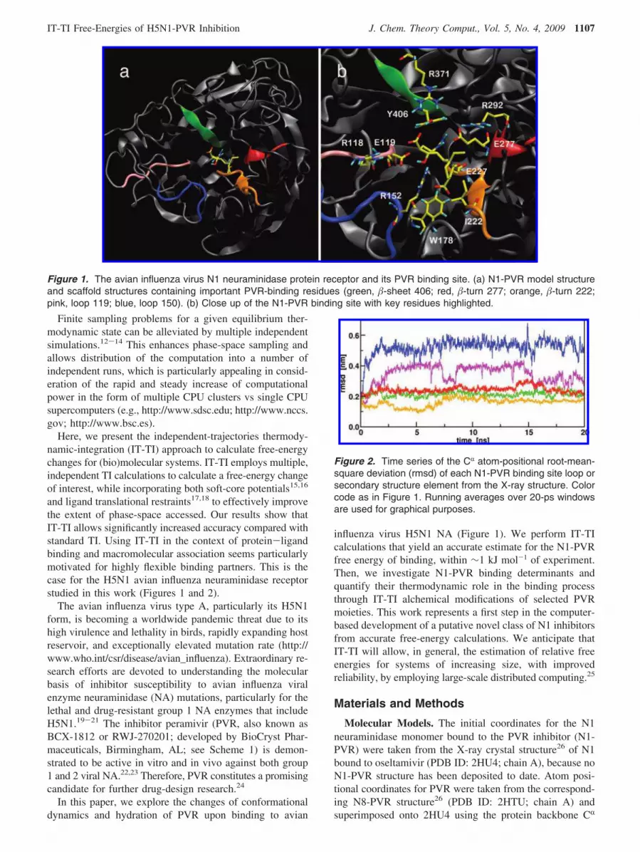
Figure 1. The avian influenza virus N1 neuraminidase protein receptor and its PVR binding site. (a) N1-PVR model structure
and scaffold structures containing important PVR-binding residues (green, �-sheet 406; red, �-turn 277; orange, �-turn 222;
pink, loop 119; blue, loop 150). (b) Close up of the N1-PVR binding site with key residues highlighted.
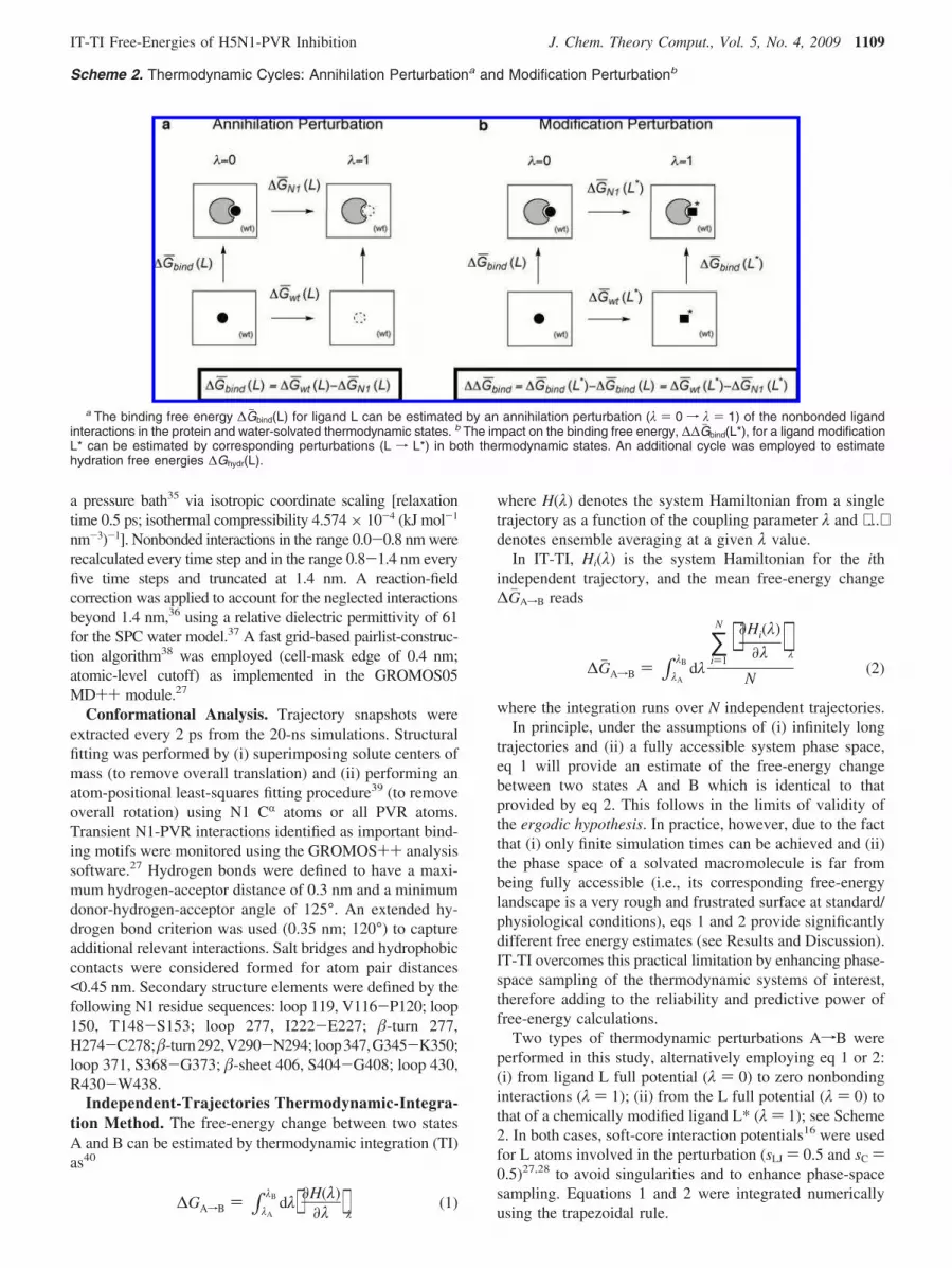
Figure 2. Time series of the CR atom-positional root-mean-
square deviation (rmsd) of each N1-PVR binding site loop or
secondary structure element from the X-ray structure. Color
code as in Figure 1. Running averages over 20-ps windows
are used for graphical purposes.
IT-TI Free-Energies of H5N1-PVR Inhibition J. Chem. Theory Comput., Vol. 5, No. 4, 2009 1107

atoms; this results in superimposition of the oseltamivir andPVR ring atoms. The N1-PVR complex was solvated in (pre-equilibrated) cubic boxes large enough (∼8.3 nm3) to avoidany interactions between mirror images under rectangularperiodic boundary conditions. Three randomly chosen watermolecules (minimum ion-ion distances of 1.0 nm) werereplaced with Na+ ions to neutralize the system. Initialconfigurations for the water (wt-PVR) and vacuum (vc-PVR)reference states were defined using the same PVR coordi-nates. For a summary of N1-PVR, wt-PVR, and vc-PVRsimulated systems, see Table 1.
Molecular Dynamics Simulations. All simulations wereperformed using the GROMOS05 software for biomolecularsimulation27 and the GROMOS force field28 (45A3 parameterset7). Amino acid charges were defined to reproduce an apparentpH 7. GROMOS PVR force-field parameters were derived fromexisting building blocks28-30 (Supporting Information, TableS1). The GROMOS compatible SPC water model31 andpreviously reported SPC-water compatible parameters for ions32
were employed. For ligand simulations in vacuo (vc-PVR) thecorresponding 45B3 parameter set was employed.
For N1-PVR, a first steepest-descent energy minimization(EM) was performed to relax solvent and ions, while proteinatom positions were restrained by using a harmonic potential(force constant k ) 2.5 × 10³ kJ mol-1 nm-2). A secondEM run without restraints eliminated any residual strain. AllEM runs were extended until the energy change per stepbecame <0.5 kJ mol-1. The system was then brought to thereference temperature (T ) 300 K) in six consecutive MDperiods of 25 ps (50 K increments). During the heating ofN1-PVR, protein atom positions were restrained with aharmonic potential, using a k from 104 to 0 (decreased insteps of 2.5 × 10³ kJ mol-1 nm-2). In addition, fourindependent MD runs were initialized by reassigning randomvelocities from Maxwell-Boltzmann distributions at 5 K.All five independent trajectories were extended (at least 2ns) to reach equilibration of the separate system Hamiltoniancomponents. Independent trajectories for the wt-PVR andvc-PVR systems were similarly prepared. One MD run foreach system was extended for 20 ns and used for confor-mational analysis (see below).
Newton’s equations of motion were integrated using theleapfrog algorithm33 with a 2-fs time step. The SHAKEalgorithm34 was applied to constrain all bond lengths (relativegeometric tolerance of 10-4). All simulations were carried outin the N,p,T ensemble (reference pressure 1 atm) by separatelycoupling the temperature of solute and solvent degrees offreedom to a 300 K heat bath35 (relaxation time 0.1 ps) and bycoupling the pressure (estimated based on an atomic virial) to
Scheme 1. Summary of the Modification Perturbations (COO-, NR3+, TAIL1, and TAIL1) for PVR Moleculea
a The IT-TI free-energy changes due to PVR hydration ∆Gj hydr(L) and N1-PVR binding ∆Gj bind(L) are shown as well as PVR alchemicalmodification ∆∆Gj hydr(L*) and ∆∆Gj bind(L*). All values are given in kJ mol-1 with corresponding σ∆Gj uncertainties between parentheses.
Table 1. System Setup for MD Simulations of PVR Bound
to the N1 Active Site (N1-PVR), Free in Water (wt-PVR),
and in Vacuum (vc-PVR)
N1-PVR wt-PVR vc-PVR
T [K] 300 300 300no. Na+ ions 3 0 0total system charge [e] 0 0 0no. solute atoms 3863 30 30no. water molecules 17046 1748 0no. atoms in system 55004 5274 30
1108 J. Chem. Theory Comput., Vol. 5, No. 4, 2009 Lawrenz et al.
a pressure bath35 via isotropic coordinate scaling [relaxationtime 0.5 ps; isothermal compressibility 4.574 × 10⁻⁴ (kJ mol-1
nm-3)-1]. Nonbonded interactions in the range 0.0-0.8 nm wererecalculated every time step and in the range 0.8-1.4 nm everyfive time steps and truncated at 1.4 nm. A reaction-fieldcorrection was applied to account for the neglected interactionsbeyond 1.4 nm,36 using a relative dielectric permittivity of 61for the SPC water model.37 A fast grid-based pairlist-construc-tion algorithm38 was employed (cell-mask edge of 0.4 nm;atomic-level cutoff) as implemented in the GROMOS05MD++ module.27
Conformational Analysis. Trajectory snapshots wereextracted every 2 ps from the 20-ns simulations. Structuralfitting was performed by (i) superimposing solute centers ofmass (to remove overall translation) and (ii) performing anatom-positional least-squares fitting procedure39 (to removeoverall rotation) using N1 CR atoms or all PVR atoms.Transient N1-PVR interactions identified as important bind-ing motifs were monitored using the GROMOS++ analysissoftware.27 Hydrogen bonds were defined to have a maxi-mum hydrogen-acceptor distance of 0.3 nm and a minimumdonor-hydrogen-acceptor angle of 125°. An extended hy-drogen bond criterion was used (0.35 nm; 120°) to captureadditional relevant interactions. Salt bridges and hydrophobiccontacts were considered formed for atom pair distances<0.45 nm. Secondary structure elements were defined by thefollowing N1 residue sequences: loop 119, V116-P120; loop150, T148-S153; loop 277, I222-E227; �-turn 277,H274-C278;�-turn292,V290-N294;loop347,G345-K350;loop 371, S368-G373; �-sheet 406, S404-G408; loop 430,R430-W438.
Independent-Trajectories Thermodynamic-Integra-
tion Method. The free-energy change between two statesA and B can be estimated by thermodynamic integration (TI)as40
where H(λ) denotes the system Hamiltonian from a singletrajectory as a function of the coupling parameter λ and ⟨ ...⟩denotes ensemble averaging at a given λ value.
In IT-TI, Hi(λ) is the system Hamiltonian for the ithindependent trajectory, and the mean free-energy change∆GjAfB reads
where the integration runs over N independent trajectories.In principle, under the assumptions of (i) infinitely long
trajectories and (ii) a fully accessible system phase space,eq 1 will provide an estimate of the free-energy changebetween two states A and B which is identical to thatprovided by eq 2. This follows in the limits of validity ofthe ergodic hypothesis. In practice, however, due to the factthat (i) only finite simulation times can be achieved and (ii)the phase space of a solvated macromolecule is far frombeing fully accessible (i.e., its corresponding free-energylandscape is a very rough and frustrated surface at standard/physiological conditions), eqs 1 and 2 provide significantlydifferent free energy estimates (see Results and Discussion).IT-TI overcomes this practical limitation by enhancing phase-space sampling of the thermodynamic systems of interest,therefore adding to the reliability and predictive power offree-energy calculations.
Two types of thermodynamic perturbations AfB wereperformed in this study, alternatively employing eq 1 or 2:(i) from ligand L full potential (λ ) 0) to zero nonbondinginteractions (λ ) 1); (ii) from the L full potential (λ ) 0) tothat of a chemically modified ligand L* (λ ) 1); see Scheme2. In both cases, soft-core interaction potentials16 were usedfor L atoms involved in the perturbation (sLJ ) 0.5 and sC )
0.5)27,28 to avoid singularities and to enhance phase-spacesampling. Equations 1 and 2 were integrated numericallyusing the trapezoidal rule.
Scheme 2. Thermodynamic Cycles: Annihilation Perturbationa and Modification Perturbationb
a The binding free energy ∆Gj bind(L) for ligand L can be estimated by an annihilation perturbation (λ ) 0 f λ ) 1) of the nonbonded ligandinteractions in the protein and water-solvated thermodynamic states. b The impact on the binding free energy, ∆∆Gj bind(L*), for a ligand modificationL* can be estimated by corresponding perturbations (L f L*) in both thermodynamic states. An additional cycle was employed to estimatehydration free energies ∆Ghydr(L).
∆GAfB ) ∫λA
λB dλ⟨ ∂H(λ)∂λ ⟩λ
(1)
∆GjAfB ) ∫λA
λB dλ
∑i)1
N ⟨ ∂Hi(λ)
∂λ ⟩λ
N(2)
IT-TI Free-Energies of H5N1-PVR Inhibition J. Chem. Theory Comput., Vol. 5, No. 4, 2009 1109

Statistical Analysis of Uncertainties. Two alternativestatistical procedures were employed to evaluate the uncer-tainty σ for ∆GAfB or ∆GjAfB free-energy estimates.
First, a simulation standard error σsim(t) of the time-varyingHamiltonian derivative at a given λ can be calculated as
with T being the total number of block averages41 throughoutthe single ith trajectory or all N concatenated independenttrajectories. (∂Ht(λ)/∂λ)λ denotes the Hamiltonian derivative,block-averaged at time t, and ⟨∂HT(λ)/∂λ⟩λ is the ensembleaverage over the entire simulation time at a given λ. As anexample, σsim(t) uncertainties are reported as error bars for⟨∂HT(λ)/∂λ⟩λ vs λ in Figure 3 (solid black curve). Then, acorresponding free-energy uncertainty can be obtained as
This follows from the standard assumption that (∂Ht(λ)/∂λ)λ values are statistically uncorrelated along the time over
different values of the coupling parameter λ. However, theσ∆Gi
uncertainty includes the physically based fluctuationsof (∂Ht(λ)/∂λ)λ, though corresponding noise is typicallyreduced by block-averaging.41 Therefore, despite its wideuse in the literature, σ∆Gi
is a questionable measure ofuncertainty for a free-energy change of interest. For example,considering that overlap of phase space at neighboring λ
values is a requirement for smooth ⟨∂HT(λ)/∂λ⟩λ vs λ curves(Figure 3), one could claim that (∂Ht(λ)/∂λ)λ time series arestatistically correlated. Nonetheless, the abovementioneduncertainty defined in eq 4 is representing the lowest possibleuncertainty for a free-energy-change estimate from standardTI. Thus, it seems the fairest choice for this study comparingTI vs IT-TI results.
Second, for IT-TI, a statistics-based uncertainty σ∆Gj on agiven free-energy change ∆GjAfB from eq 2 can be calculatedas the standard deviation from the mean (standard error) ofthe N ∆Gi results
where σ∆G is the standard deviation of the free-energy changeover the N IT-TI trajectories employed. Importantly, σ∆Gj hasa clear statistical validity,42 because of its explicit dependenceon the repeated independent estimates.
Similarly, for a general overall free-energy change ∆GjAfB,calculated as the difference between two free-energy changes∆Gj B and ∆GjA, a corresponding uncertainty can be obtainedby propagating the respective uncertainties as42
Then, the relative uncertainty for a given free-energychange A f B reads
In this study, IT-TI runs were extended to obtain suf-ficiently smooth curves of ⟨∂Hi(λ)/∂λ⟩λ vs λ (Figure 3). IT-TI trajectories were independently equilibrated (0.5 ns) foreach of the 26 λ points (from five initial equilibrated λ ) 0configurations), followed by independent sampling periods(0.5 ns) used for free-energy estimation. Increased sampling(up to 2.5 ns) times were required in the ranges 0.12 e λ e
0.24 and 0.76 e λ e 0.92. A summary of these calculationsis given in Supporting Information, Table S2. All annihilationand modification perturbations fulfilled the criterion σ∆Gj(%)< 6%. Only N1-TAIL1 and N1-TAIL2 modification per-turbations had larger σ∆Gj(%) values (up to 52%) due to thecorresponding small ∆GjAfB values (Supporting Information,Table S3).
Separation of Thermodynamic States. For ∆Gj N1(L)(Scheme 2a), the potential U(rL) ) -
1/2k
(rL - r0)2 was applied to harmonically restrain ligandtranslation and ensure its sampling of a finite phase-spacevolume VΙ. An optimal k value of 246.5 kJ mol-1 nm-2
was estimated from the ensemble-averaged L root-mean-
Figure 3. ⟨∂H(λ)/∂λ⟩λ values and corresponding uncertainties
σsim(t) vs λ. (a) Annihilation of PVR in N1 binding site (∆Gj N1(L);
Scheme 2a). (b) Annihilation of PVR in water (∆Gj wt(L);
Scheme 2a). Black lines: average values over all N individual
trajectories. Gray lines: individual trajectory TI curves. Inset
panels highlight λ regions where IT-TI averages outperform
standard individual TI calculations.
σsim(t) ) � 1T - 1 ∑
t)1
T [(∂Ht(λ)
∂λ )λ- ⟨ ∂HT(λ)
∂λ ⟩λ]2
/√T
(3)
σ∆Gi
) (∫λA
λBσsim
2(t) dλ)1/2(4)
σ∆Gj )
σ∆G
√N(5)
σ∆GjAfB
) √(σ∆GjA
)2+ (σ
∆GjB)2 (6)
σ∆GjAfB
(%) )σ
∆GjAfB
∆GjAfB
× 100 (7)
1110 J. Chem. Theory Comput., Vol. 5, No. 4, 2009 Lawrenz et al.
square deviation (rmsd) by applying the equipartitiontheorem as18
where rL is the position of L during 20-ns of unrestrainedsimulation at λ ) 0, r0 is the initial position of L in thepre-equilibrated starting configuration, P(rL) is the 3-Dpositional probability distribution of L, R is the molar gasconstant, and T ) 300 K. In this study, rL and r0 are theinstantaneous and initial positions of the PVR C4 ringcarbon.
The phase-space volume VΙ can be defined as18
where � ) 1/RT. Thus, the correction17 to the restrainingpotential bias for ∆GjN1(L) in Scheme 2 reads
Results and Discussion
Conformational Analysis of the N1-PVR Binding
Site. Figure 1 shows the six-bladed �-propeller structure ofN1 neuraminidase bound to PVR and important active siteresidues, monitored throughout 20 ns of N1-PVR MDsimulation. To investigate N1-PVR receptor flexibility andconformational changes, the CR atom-positional rmsd fromthe X-ray crystal structure was monitored for the secondarystructure elements forming the N1-PVR binding site (Figure2). Loop 150 deviates the most (up to ∼0.7 nm) to samplestable open-loop configurations (∼0.5 units) throughout theMD simulation (Figures 1 and 2, blue). This observation issimilar to that recently reported for both apo and oseltamivir-bound N1 neuraminidase (up to ∼0.6 nm).43 Additionally,we find that loop 119 demonstrates significant flexibility andsamples conformations with rmsd of ∼0.4 and ∼0.3 nm inthe N1-PVR complex (Figures 1 and 2, pink). The dynamicsof loops 150 and 119 in water indicate a significant relaxationfrom the crystal-packed conformation captured in X-rayexperiments. The �-sheet 406 and �-turns 277 and 222 showcomparatively lower rmsd deviations (0.1-0.3 nm) andsmaller fluctuations on the 20-ns time scale.
Table 2 summarizes the N1-PVR intermolecular hydrogenbonding. Scheme 1 defines PVR atom nomenclature. Adominant multicenter hydrogen bond between N1 E277 andthe PVR guanidinium group is stable for the entire simulationtime, with PVR NR3 and NR1 atoms alternating as hydrogendonors to E277 carboxyl oxygen atoms OE1 and OE2(50-69% occurrences). E277 also transiently interacts withthe PVR methyl acetamide polar hydrogen (HP; 57%). Yetanother hydrogen bond (54%) is observed between the PVRcarboxyl oxygen (OD1) and the N1 Y406 hydroxyl (O-H).
Interestingly, the Y406 residue is homologous to other keycatalytic tyrosine residues found in the avian influenza virusfamily of glycosidases (Carbohydrate Active Enzymesdatabase; http://www.cazy.org/). Thus, targeting Y406 couldbe important for drug design, as suggested by a computa-tional solvent mapping analysis.44 An extended hydrogenbond between N1 R152 on loop 150 and PVR (O-H group)occurs for 44% of the simulation time (Table 2 and Figure1) and contributes to the stability of open loop 150 ensembleof configurations.
Figure 4 describes the conformational sampling of PVRin the N1 binding site. N1 protein atoms that maintain stronginteractions with either (i) charged ligand moieties (saltbridges) or (ii) hydrophobic groups (hydrophobic packing)display distance probability distributions with one sharp,high-intensity peak. Those residues experiencing variedinteractions have broader distributions and/or multiple peaks.Throughout the 20-ns simulation, the PVR guanidiniumgroup (CR atom, Scheme 1) maintains a well-defined salt-bridging interaction with the N1 E277 carboxyl group (0.38nm average distance). The broad distribution for the PVRCR-N1 E119 distance (0.52 nm) and the doubly peakeddistribution for the PVR CR-N1 E227 distance (0.47 and0.65 nm for first and second peak) correspond to transient
⟨rmsd2⟩ ) ⟨(rL - r0)2⟩ ) ∫ (rL - r0)
2P(rL) drL )
3RT
k(8)
VI ) ∫V
exp[-�U(rL)] dr (9)
VI ) ∫V
exp[ 12RT
k(rL - r0)2]dr ) (2πRT
k )3/2(10)
∆GjN1(L) ) ∫λ)0
λ)1dλ
∑i)1
N ⟨ ∂Hi(λ)
∂λ ⟩λ
N+ RT ln(CV
Ι) (11)
Table 2. N1-PVR Intermolecular Hydrogen Bonds from 20
ns of Molecular Dynamics Simulationa
donor acceptor occurrence (%)
PVR (NR3-HR3) E277 (OE1) 69PVR (NR3-HR3) E277 (OE2) 69PVR (NR1-HR1) E277 (OE1) 50PVR (NR1HR1) E277 (OE2) 57PVR (NP-HP) E277 (OE2) 57Y406 (O-H) PVR (OD1) 54R152 (NH2-H22)b PVR (O5) 44
a PVR atom nomenclature as in Scheme 1. OE refers toglutamate carboxyl, O-H to tyrosine hydroxyl, and NH2-H22 toarginine guanidinium. All N1-PVR intermolecular hydrogen bondsoccurring >5% are shown. b Using extended hydrogen-bondingcriterion. See also Materials and Methods.
Figure 4. Conformational sampling of PVR moieties in the
N1 neuraminidase binding site from 20-ns N1-PVR molecular
dynamics simulation. Distance pairs are labeled according to
the atoms monitored: PVR CR to N1 E277 (blue), E119
(green), and E227 (red) carboxyl group carbon; PVR CD to
N1 R118 (pink), R292 (yellow), and R371 (violet); PVR C11
to N1 I222 side chain carbon (black) and W178 aromatic ring
carbon (cyan). See Scheme 1 for PVR atom nomenclature.
IT-TI Free-Energies of H5N1-PVR Inhibition J. Chem. Theory Comput., Vol. 5, No. 4, 2009 1111
salt bridges formed by these N1 glutamate residues to thePVR guanidinium group.
Stable long-range interactions between PVR and thearginine triad of R118, R371, and R292 residues (Figure 1)are observed along our 20-ns N1-PVR simulation, withdistribution peaks at 0.68, 0.8, and 0.96 nm, respectively(Figure 4). This triad of positively charged residues alsocontributes to binding via the conserved ligand carboxylgroup in both the natural ligand, sialic acid, and othersynthetic neuraminidases inhibitors (e.g., DANA, oseltamivir,and zanamivir).26,45 Our results support the fundamental roleof R118 and R371 in N1-PVR binding. The distal positionof R292 indicates less reliance of PVR on this residue andcould account for the retained PVR affinity for the N2resistance mutation R292K.23,46
The PVR aliphatic tail (Scheme 1) has been designed45
to fill a small hydrophobic subpocket, comprised of W178and I222 residues, which is conserved among both group 1and 2 NA (Figure 1). Our MD simulation confirms that PVRtail atoms interact with the branched I222 side chain (C11peak at 0.40 nm average distance; Figure 4). The same PVRtail atoms are also stably close to the W178 aromatic carbons(C11 peak at 0.42 nm; Figure 4). These results highlight theoccurrence of important hydrophobic-packing interactions inthe N1 subpocket.
We note that the majority of the conserved residuesdescribed above have been suggested to also participate inkey interactions between sialic acid and inhibitors DANA,zanamivir, and oseltamivir, as well as PVR, in both group 1and 2 NA receptors.26,44-48
Changes of PVR Hydration upon N1 Binding. PVRhydration and its changes upon N1 binding were alsoanalyzed. Figure 5 shows the radial distribution functions(rdf) for water oxygen atoms from wt-PVR and N1-PVRsimulations. The PVR guanidinium group (CR atom; Scheme1) undergoes ∼0.5 units decrease in its first peak intensityupon N1-PVR binding. We can explain this desolvationeffect by considering the tight interaction of this bulky,positively charged PVR group with the negatively chargedN1 E277 carboxyl (Figure 4 and Table 2). Desolvation uponN1 binding is also observed for the PVR aliphatic tail bythe intensity decrease of its first solvation shell peaks (C10and C11 atoms, ∼0.5-0.9 units; Figure 5). This is consistentwith the formation of more favorable interactions betweenPVR and N1 residues I222 and W178.
A different hydration behavior can be noticed for the PVRcarboxyl group (CD atom), with a similar strong intensityfor its first peak at ∼0.35 nm in both wt-PVR and N1-PVRsimulations. Its second peak intensity diminishes onlymarginally upon N1-PVR binding (∼0.3 units). Thus, thePVR carboxyl group is still solvated in the N1 binding siteby dynamic water molecules on an ensemble averaged basis.The presence of water molecules in the N1-PVR bindingsite and the lack of persistent ligand-solvent hydrogen bondsconfirm this point. A PVR carboxyl group-N1 Y406hydroxyl hydrogen bond is transiently formed (54% occur-rence; Table 2), allowing this PVR moiety to still repeatedlyinteract in the N1-PVR binding site with water molecules.Water exchange in charged protein cavities49 and water-mediated interactions in flexible carbohydrate-protein bind-ing50 have been previously reported.
The PVR hydroxyl group displays a first peak with reducedintensity when N1-bound (O5 atom, ∼0.5 units; Figure 5).This moiety forms a competing hydrogen bond to N1 R152(44%; Table 2), yet water molecules are maintained in thefirst solvation shell upon binding. The PVR acetamide group(NP atom) has limited solvent accessibility due to theadjacent hydrophobic tail. Its solvation is further decreased(∼0.5 units) in the bound state upon formation of a hydrogenbond to N1 E277 (57% occurrence; Table 2).
Overall, the interactions of the PVR guanidinium groupwith N1 E277, E227, and E119; the PVR carboxyl groupwith catalytic N1 Y406; and the PVR aliphatic tail with N1W178 and I222 in the hydrophobic subpocket appear mostrelevant to drive N1-PVR binding based on conformationaland hydration analyses.
IT-TI Free-Energy Change upon N1-PVR Binding. Thefree-energy change upon N1-PVR binding was estimatedusing the IT-TI method and compared with standard TIvalues from single trajectories, as well as with experiment.
Examples of ⟨∂Hi(λ)/∂λ⟩λ vs λ curves are shown forstandard TI and the improved IT-TI calculations of the N1protein and water reference states (Figure 3). The IT-TIcurves are smoother than those obtained from individualstandard TI runs, because of increased sampling and im-proved overall statistics obtained through N ) 5 independentensembles. Their integration (eq 2) provides ∆Gjwt(L) and∆GjN1(L) values to estimate ∆Gj bind (Supporting Information,Table S3). This is summarized in the thermodynamic cycleof Scheme 2a.
Table 3 reports the IT-TI results and their comparison withthe available experimental data. Our IT-TI ∆Gj bind estimate(-61.1 ( 5.4 kJ mol-1) matches the ∆Gj bind
exp value derivedfrom multiple IC50 measurements21,51,52 (-62.2 ( 1.8 kJmol-1; see Supporting Information, Table S4). A free-energydifference of 1.1 kJ mol-1 (i.e., 2% relative difference) hasno statistical significance within the above uncertainties.Remarkably, such an IT-TI prediction of the experimentalvalue relies on individual ∆Gj bind(N) estimates that span arather large free-energy range (see Table 3). In fact, thesestandard TI estimates are at variance with the independentcalculation performed, ranging from a substantial underes-timation (i ) 4; 19% relative difference) to a substantialoverestimation (i ) 5; 29% relative difference) of the ∆Gj bind
exp
Figure 5. Ensemble-averaged solvation of PVR moieties
when free in solution (left panel) or bound to the N1 protein
binding site (right panel). Radial distribution functions of the
water oxygen atoms from 20-ns molecular dynamics simula-
tions are shown centered on PVR atoms CR (blue), C10
(black solid), C11 (black dashed), CD (red), O5 (green), and
NP (cyan). See Scheme 1 for PVR atom nomenclature.
1112 J. Chem. Theory Comput., Vol. 5, No. 4, 2009 Lawrenz et al.
value. Only one of the individual standard TI results is ingood agreement with the ∆Gj bind
exp value (-63.7 ( 11.0 kJmol-1, i ) 1; 2% relative difference). The remaining fourTI estimates have relative differences >9%. A significantlydifferent N1-PVR binding free-energy estimate of -1180.9( 31.8 kJ mol-1 has been reported based on MM-PBSAcalculations.48
The IT-TI free-energy estimate also has a lower σ∆Gj(%)relative uncertainty compared to the σ∆Gi
(%) from standardTI, i.e. 9% of the calculated free-energy difference (Table3). σ∆Gi
(%) values associated with independent ∆Gbind(N)estimates are larger, ranging between 12% (i ) 4) and 19%(i ) 5). We stress that the uncertainties σ∆Gi
typicallyevaluated for single standard TI trajectories are not statisticalindicators of the ∆Gbind accuracy (see Materials and Methods,eq 4). Instead, repeated IT-TI runs allow calculation of themore representative free-energy uncertainty σ∆Gj (see Materi-als and Methods, eq. 5). In addition, for this study, the latteris directly comparable to the standard error of ∆Gj bind
exp
determined from N experimental values (Supporting Infor-mation, Table S4).
Overall, our results underscore the improved predictivepower of IT-TI vs standard TI, due to the increased statisticalreliability. The large deviations observed among standardTI estimates can be explained, in part, by the flexibility ofthe N1 binding site. Loops 119 and 150 demonstrateheterogeneous conformational sampling among different λ
regions; their rmsd from the initial equilibrated structurereach values up to 0.4 and 0.7 nm, respectively (data notshown). This is consistent with both the dynamic loopbehavior from the longer 20-ns MD simulation (Figure 2)and with the large σ∆Gi
values for standard TI estimates(Table 3). We conclude that the IT-TI method significantlyaids sampling of thermodynamic macrostates for flexiblereceptors by ensemble averaging of independent trajectories.
IT-TI Free-Energy Changes for PVR Alchemical
Modifications. N1-PVR binding determinants were alsoinvestigated using IT-TI free-energy changes upon computeralchemical modifications and their underlying thermody-
namic cycle (Scheme 2b). Scheme 1 summarizes the cor-responding ∆∆Gj bind values together with the PVR free energyof binding ∆Gj bind. A positive or negative value of ∆∆Gj bind
indicates thermodynamically unfavorable or favorable al-chemical modifications of the ligand L.
Neutralizing COO- and NR3+ charges has large but
opposite effects on ligand binding (∆∆Gj bind of +55.1 ( 3.1and -79.7 ( 4.2 kJ mol-1, respectively). The TAIL1 andTAIL2 modifications both have small, favorable impacts onligand binding (∆∆Gj bind of -5.8 ( 2.4 and -1.5 ( 2.0 kJmol-1, respectively). To understand these results, one mustlook at the effects on receptor-ligand interactions and ligandhydration free-energy changes (∆∆Gj hydr) upon alchemicalperturbation. In other words, a given ∆∆Gj bind change canarise from different compensating effects. For example, apositive ∆∆Gj bind value may be driven by (i) unfavorable(enthalpic or entropic) N1-ligand interactions, (ii) a morefavorable ∆∆Gj hydr, or (iii) a thermodynamically unfavorablecombination of the previous effects. Similarly, a negative∆∆Gj bind value may be driven by (i) favorable (enthalpic orentropic) N1-ligand interactions, (ii) a more unfavorable∆∆Gj hydr, or (iii) a thermodynamically favorable combinationof the previous effects. In this section, we address the impactof different IT-TI modification perturbations on N1-ligandbinding; in the next section, we consider the ligand hydrationfree energy. Both are needed to fully describe a given∆∆Gj bind binding free-energy change.
Throughout the λ ) 0 f λ ) 1 N1-COO- modificationperturbation, R292 and R371 residues move on average apartfrom the ligand scaffold. The closest arginine, R118, reducesits average distance to the ligand CD atom (SupportingInformation, Figure S1a). The important ligand carboxyl-Y406 interaction (Table 2) is partially disrupted, whileguanidinium interactions with E119, E227, and E277 aremaintained.
During the λ ) 0 f λ ) 1 N1-NR3+ modification, N1
R371 samples a more stable conformation close to the ligandCD atom, as revealed by a sharper distance distribution peak(cf. λ ) 0 vs λ ) 1; Supporting Information, Figure S1b).The aforementioned ligand carboxyl-Y406 interaction isdestabilized. Moreover, the ligand guanidinium loses itsfavorable electrostatic interaction with N1 E277, which shiftsaway from the perturbed moiety. However, the ligandacetamide group is pushed closer to residue R156, whichforms a hydrogen bond with atom OP (data not shown). Thisresidue is not observed to closely interact with the unper-turbed PVR molecule.
Following the λ ) 0 f λ ) 1 TAIL1 perturbation, I222and W178 distance distributions for the modified PVRaliphatic tail (C8 and C9 atoms) transition to sharper andfewer peaks (cf. λ ) 0 vs λ ) 1; Supporting Information,Figure S1c,d). On the other hand, the λ ) 0f λ ) 1 TAIL2
perturbation has a limited effect, as the distributions of tailatoms C8 and C10 with I222 and W178 residues remainpredominantly broad (0.4-0.9 nm; Supporting Information,Figure S1e,f).
Role of Ligand Hydration in N1 Binding Thermody-
namics. Scheme 1 summarizes the hydration free energy forPVR, ∆Gj hydr, and the changes of this quantity for its
Table 3. Free-Energy Change upon N1-PVR Bindinga
free energy (kJ mol-1)
uncertainties
change σ∆Gjd σ∆Gj (%)e
∆Gj bindexp b
-62.2 1.8 3∆Gj bind
c-61.1 5.4 9
∆Gbind(N)f σ∆Gi
g σ∆Gi(%)
i ) 1 -63.7 11.0 17i ) 2 -55.3 9.0 16i ) 3 -55.8 10.1 18i ) 4 -80.6 9.9 12i ) 5 -50.1 9.7 19
a The average ∆Gj bindexp value from repeated experiments can be
compared with the IT-TI ∆Gj bind estimate. Corresponding ∆Gj bind(N)values from individual standard TI trajectories are also reported.b Derived using data in refs 21, 51, and 52 (see SupportingInformation, Table S4). c Equation 2. d Equation 5, withpropagated uncertainties as in eq 6. e Equation 7. f Equation 1, N) 5. g Equation 4.
IT-TI Free-Energies of H5N1-PVR Inhibition J. Chem. Theory Comput., Vol. 5, No. 4, 2009 1113
alchemical modifications, ∆∆Gj hydr. The corresponding bind-ing free-energy, ∆Gj bind, and changes of this quantity,∆∆Gj bind, are also reported. Figure 6 illustrates the relativecomponents involved for PVR alchemical modifications.
We estimate an IT-TI ∆Gj hydr value of -107.8 ( 1.2 kJmol-1 for the PVR molecule. No experimental data isavailable to date for a direct comparison of this result. Wenote that this value is in qualitative agreement with hydrationfree energies of a large variety of compounds used tocalibrate the force field used in this study.6,7,30 The value of-820.5 ( 40.2 kJ mol-1 reported based on MM-PBSAcalculations significantly overestimates the favorable ther-modynamic effect of PVR hydration.48
The COO- perturbation gives a positive ∆∆Gj hydr valueof 15.7 ( 5.1 kJ mol-1. The PVR carboxyl group givesdistinct primary and secondary rdf peaks in water. Whendeleting its charge, water structure is reduced (λ ) 0 vs λ )
1; wt-COO-; Supporting Information, Figure S2). Ligandhydration around this moiety is maintained in the N1 bindingsite, yet its first solvation shell is displaced (cf. λ ) 0 vs λ
) 1; N1-COO-; Supporting Information, Figure S2). Asufficiently unfavorable ligand hydration free energy in theunbound state would drive hydrophobic binding. However,this effect is not large enough to compensate for the loss offavorable N1-ligand interactions (see the previous section).The COO- charge perturbation has the largest unfavorableimpact in the N1-bound state, leading to a ∆∆Gj bind value of55.1 ( 3.1 kJ mol-1.
A different thermodynamic compensation occurs betweenthe bound and unbound states for the NR3
+ charge perturba-tion, with a large unfavorable ∆∆Gj hydr value of +124.9 (
1.8 kJ mol-1. The single solvation peak of the PVRguanidinium group in water shifts to larger distances (cf. λ
) 0 vs λ ) 1; wt-NR3+; Supporting Information, Figure S2)
due to hydrophobic desolvation of this bulky charge group.The limited hydration of the charged PVR guanidinium inthe N1 active site is almost unaffected by the perturbation(cf. λ ) 0 vs λ ) 1; N1-NR3
+; Supporting Information,
Figure S2). The NR3+ charge perturbation has the largest
unfavorable impact on the unbound state, overcompensatingfor the loss of N1-ligand favorable interactions (see previoussection). This leads to a net ∆∆Gj bind change of -79.7 ( 4.2kJ mol-1, significantly more favorable than the PVR ∆Gj bind
value. The experimentally observed, improved binding ofinhibitors to N2 neuraminidase by hydrophobic substitutionat the PVR guanidinium position supports these results.53
In water, TAIL1 and TAIL2 modifications increase thesolvation around the unperturbed atoms (cf. λ ) 0 vs λ ) 1;Supporting Information, Figure S2) and correlate to the∆∆Gj hydr values of -1.4 ( 0.6 and 7.3 ( 0.3 kJ mol-1,respectively. In the N1 binding site, a distinct decrease (∼0.5units) for TAIL1 solvation (C9 atom; Supporting Information,Figure S2) agrees with the rearrangement of N1 hydrophobicresidues I222 and W178 (see above). The opposite signs of∆∆Gj hydr offset the changes observed in the protein as wellas entropic changes to the aliphatic tail, resulting in similar∆∆Gj bind values of -5.8 ( 2.4 and -1.5 ( 2.0 kJ mol-1 forTAIL1 and TAIL2 alchemical modifications (Figure 6).
Overall, these results emphasize the dominant electrostaticcontribution to the free energy of N1 binding for PVR andits alchemically modified variants and suggest that futuredrug development may also be guided by conveniently tuningligand flexibility and hydrophobicity.
Conclusion
The independent-trajectories thermodynamic-integration (IT-TI) approach was presented. It allows for estimation ofimproved free-energy changes for biomolecular systemsbased on multiple independent simulations. Our resultsunderscore the improved predictive power of IT-TI vsstandard TI, due to the increased statistical reliability.Standard TI estimates from individual trajectories span arather large free-energy estimate range, from a 19% under-estimation to a 29% overestimation of the experimentalreference value (-62.2 ( 1.8 kJ mol-1). Remarkably, ourIT-TI binding free-energy estimate (-61.1 ( 5.4 kJ mol-1)is in excellent agreement, i.e. 2% relative difference. Ageneral formulation is proposed to evaluate correspondingIT-TI free-energy uncertainties that rely on a statisticaltreatment of error analysis. Overall, IT-TI seems particularlypromising in the case of highly flexible protein receptors,ligands, and macromolecular binding partners in general.
Using 20-ns molecular dynamics simulation of the N1-PVR complex, we find a number of key binding interactions.The interactions of the PVR guanidinium group with N1E277, E227, and E119; the PVR carboxyl group withcatalytic N1 Y406; and the PVR aliphatic tail with N1 W178and I222 in the hydrophobic subpocket appear most relevantto drive N1-PVR binding, based on conformational andhydration analyses. This dynamic, atomistic description wascorrelated with key thermodynamic contributions to binding.
Furthermore, IT-TI was applied to explore the bindingdeterminants of avian influenza N1 neuraminidase inhibi-tion using alchemical modification of the PVR molecule.Charge annihilation of its carboxyl and guanidiniumgroups has the largest unfavorable impact in the N1-boundand unbound states, respectively. These results emphasize
Figure 6. Binding free energies (∆∆Gj bind, gray solid bars)
and corresponding hydration free energies (∆∆Gj hydr, diagonal
lined bars) for all modification perturbations in this study.
Vertical bars display the corresponding uncertainties σ∆Gj .
Corresponding free-energy values and uncertainties can be
found in Scheme 1 and in Supporting Information, Table S3.
1114 J. Chem. Theory Comput., Vol. 5, No. 4, 2009 Lawrenz et al.

the dominant electrostatic contribution to N1-PVR bindingfree energy. Alchemical modifications of the PVR aliphatictail suggest that future drug development may also beguided by conveniently tuning ligand flexibility andhydrophobicity.
Finally, this study allows us more general conclusions onfree-energy calculations in the context of protein-ligandbinding. The key to designing improved inhibitors for a giventarget relies on an accurate thermodynamic description ofboth ligand-bound and ligand-unbound receptor and ligandstates. Consequently, we suggest that the most reliable andpredictive free-energy calculations will likely rely on the useof explicit solvent simulations and MD force fields basedalso on a direct and general parametrization of solvationthermodynamics. We anticipate the application of the IT-TIapproach to develop improved and potent drugs to inhibitflexible macromolecular receptors.
Acknowledgment. The authors thank the members ofthe McCammon group for useful discussions. This work wassupported, in part, by the National Science Foundation grantPHY-0822283, Center for Theoretical Biological Physics,for the computing resources. Additional support has beenprovided by NSF, NIH, HHMI, and NBCR.
Supporting Information Available: GROMOS force-field parameters for the PVR molecule, Table S1; summaryof IT-TI calculations, Table S2; summary of the IT-TI freeenergies calculated, Table S3; experimental observables usedto estimate N1-PVR binding free energy, Table S4; PVRdistance distributions during modification perturbations,Figure S1; PVR hydration analysis during modificationperturbations, Figure S2. This material is available free ofcharge via the Internet at http://pubs.acs.org
References
(1) van Gunsteren, W. F.; Beutler, T. C.; Fraternali, F.; King,P. M.; Mark, A. E.; Smith, P. E. Computation of Free Energyin Practice: Choice of Approximations and AccuracyLimiting Factors; ESCOM Science Publishers: Leiden, 1993;Vol. 2.
(2) Jorgensen, W. L. Science 2004, 303, 1813–1818.
(3) McCammon, J. A. Curr. Opin. Struct. Biol. 1998, 8, 245–249.
(4) Gilson, M. K.; Zhou, H. X. Annu. ReV. Biophys. Biomol.Struct. 2007, 36, 21–42.
(5) Beveridge, D. L.; DiCapua, F. M. Annu. ReV. Biophys.Biophys. Chem. 1989, 18, 431–492.
(6) Baron, R.; Trzesniak, D.; de Vries, A. H.; Elsener, A.; Marrink,S. J.; van Gunsteren, W. F. ChemPhysChem 2007, 8, 452–461.
(7) Schuler, L. D.; Daura, X.; van Gunsteren, W. F. J. Comput.Chem. 2001, 22, 1205–1218.
(8) Lybrand, T. P.; McCammon, J. A.; Wipff, G. Proc. Natl.Acad. Sci. U.S.A. 1986, 83, 833–835.
(9) Hunenberger, P. H.; Helms, V.; Narayana, N.; Taylor, S. S.;McCammon, J. A. Biochemistry 1999, 38, 2358–2366.
(10) Reinhardt, W. P.; Miller, M. A.; Amon, L. M. Acc. Chem.Res. 2001, 34, 607–614.
(11) Chipot, C.; Pohorille, A. Free Energy Calculations; Springer:New York, 2007; Vol. 86.
(12) Zagrovic, B.; van Gunsteren, W. F. J. Chem. Theory Comput.2007, 3, 301–311.
(13) Adcock, S. A.; McCammon, J. A. Chem. ReV. 2006, 106,1589–1615.
(14) Fujitani, H.; Tanida, Y.; Ito, M.; Jayachandran, G.; Snow,C. D.; Shirts, M. R.; Sorin, E. J.; Pande, V. S. J. Chem. Phys.2005, 123, 084108.
(15) Zacharias, M.; Straatsma, T. P.; McCammon, J. A. J. Chem.Phys. 1994, 100, 9025–9031.
(16) Beutler, T. C.; Mark, A. E.; van Schaik, R. C.; Gerber, P. R.;van Gunsteren, W. F. Chem. Phys. Lett. 1994, 222, 529–539.
(17) Gilson, M. K.; Given, J. A.; Bush, B. L.; McCammon, J. A.Biophys. J. 1997, 72, 1047–1069.
(18) Hamelberg, D.; McCammon, J. A. J. Am. Chem. Soc. 2004,126, 7683–7689.
(19) Skeik, N.; Jabr, F. I. Int. J. Infect. Dis. 2007, 12, 233–238.
(20) Le, Q. M.; Kiso, M.; Someya, K.; Sakai, Y. T.; Nguyen, T. H.;Nguyen, K. H.; Pham, N. D.; Ngyen, H. H.; Yamada, S.;Muramoto, Y.; Horimoto, T.; Takada, A.; Goto, H.; Suzuki,T.; Suzuki, Y.; Kawaoka, Y. Nature 2005, 437, 1108.
(21) Hurt, A. C.; Selleck, P.; Komadina, N.; Shaw, R.; Brown, L.;Barr, I. G. AntiViral Res. 2007, 73, 228–31.
(22) Chand, P.; Bantia, S.; Kotian, P. L.; El-Kattan, Y.; Lin, T. H.;Babu, Y. S. Bioorg. Med. Chem. 2005, 13, 4071–4077.
(23) Gubareva, L. V.; Webster, R. G.; Hayden, F. G. Antimicrob.Agents Chemother. 2001, 45, 3403–3408.
(24) De Clercq, E. Expert Opin. Emerg. Drugs 2008, 13, 393–416.
(25) Shirts, M.; Pande, V. S. Science 2000, 290, 1903–1904.
(26) Russell, R. J.; Haire, L. F.; Stevens, D. J.; Collins, P. J.; Lin,Y. P.; Blackburn, G. M.; Hay, A. J.; Gamblin, S. J.; Skehel,J. J. Nature 2006, 443, 45–49.
(27) Christen, M.; Hunenberger, P. H.; Bakowies, D.; Baron, R.;Burgi, R.; Geerke, D. P.; Heinz, T. N.; Kastenholz, M. A.;Krautler, V.; Oostenbrink, C.; Peter, C.; Trzesniak, D.; vanGunsteren, W. F. J. Comput. Chem. 2005, 26, 1719–1751.
(28) van Gunsteren, W. F.; Billeter, S. R.; Eising, A. A.; Hunen-berger, P. H.; Kruger, P.; Mark, A. E.; Scott, W. R. P.; Tironi,I. G. Biomolecular Simulation: The GROMOS96 Manualand User Guide; vdf Hochschulverlag AG an der ETH Zurichand BIOMOS b.v. Groningen: Zurich, Groningen, 1996.
(29) Baron, R.; Bakowies, D.; van Gunsteren, W. F. J. Pept. Sci.2005, 11, 74–84.
(30) Lins, R. D.; Hunenberger, P. H. J. Comput. Chem. 2005,26, 1400–1412.
(31) Berendsen, H. J. C. Interaction Models for Water in Relationto Protein Hydration; Pullman, B. E., Ed.; Reidel: Dordrecht,1981.
(32) Åqvist, J. J. Phys. Chem. 1990, 94, 8021–8024.
(33) Hockney, R. W. Methods Comput. Phys. 1970, 9, 136–211.
(34) Ryckaert, J. P.; Ciccotti, G.; Berendsen, H. J. C. J. Comput.Phys. 1977, 23, 327–341.
(35) Berendsen, H. J. C.; Postma, J. P. M.; van Gunsteren, W. F.;di Nola, A.; Haak, J. R. J. Chem. Phys. 1984, 81, 3684–3690.
IT-TI Free-Energies of H5N1-PVR Inhibition J. Chem. Theory Comput., Vol. 5, No. 4, 2009 1115

(36) Tironi, I. G.; Sperb, R.; Smith, P. E.; van Gunsteren, W. F.J. Chem. Phys. 1995, 102, 5451–5459.
(37) Heinz, T. N.; van Gunsteren, W. F.; Hunenberger, P. H.J. Chem. Phys. 2001, 115, 1125–1136.
(38) Heinz, T. N.; Hunenberger, P. H. J. Comput. Chem. 2004,25, 1474–1486.
(39) McLachlan, A. D. J. Mol. Biol. 1979, 128, 49–79.
(40) Kirkwood, J. G. J. Chem. Phys. 1935, 3, 300–313.
(41) Allen, M. P.; Tildesley, D. J. Computer Simulations ofLiquids; Oxford University Press: New York, 1989.
(42) Taylor, J. R. An Introduction to Error Analysis. The Studyof Uncertainties in Physical Measurements, 2nd ed.;University Science Books: Sausalito, CA, 1997.
(43) Amaro, R. E.; Minh, D. D.; Cheng, L. S.; Lindstrom, W. M.,Jr.; Olson, A. J.; Lin, J. H.; Li, W. W.; McCammon, J. A.J. Am. Chem. Soc. 2007, 129, 7764–7765.
(44) Landon, M. R.; Amaro, R. E.; Baron, R.; Ngan, C. H.;Ozonoff, D.; McCammon, J. A.; Vajda, S. Chem. Biol. DrugDes. 2008, 71, 106–116.
(45) Babu, Y. S.; Chand, P.; Bantia, S.; Kotian, P.; Dehghani, A.;El-Kattan, Y.; Lin, T. H.; Hutchison, T. L.; Elliott, A. J.;Parker, C. D.; Ananth, S. L.; Horn, L. L.; Laver, G. W.;Montgomery, J. A. J. Med. Chem. 2000, 43, 3482–3486.
(46) Chachra, R.; Rizzo, R. C. J. Chem. Theory Comput. 2008,4, 1526–1540.
(47) Yen, H. L.; Hoffmann, E.; Taylor, G.; Scholtissek, C.; Monto,A. S.; Webster, R. G.; Govorkova, E. A. J. Virol. 2006, 80,8787–8795.
(48) Malaisree, M.; Rungrotmongkol, T.; Decha, P.; Intharathep,P.; Aruksakunwong, O.; Hannongbua, S. Proteins 2008, 71,1908–1918.
(49) Baron, R.; McCammon, J. A. Biochemistry 2007, 46, 10629–10642.
(50) Clarke, C.; Woods, R. J.; Gluska, J.; Cooper, A.; Nutley,M. A.; Boons, G. J. J. Am. Chem. Soc. 2001, 123, 12238–12247.
(51) Cheng, Y.; Prusoff, W. H. Biochem. Pharmacol. 1973, 22,3099–3108.
(52) Collins, P. J.; Haire, L. F.; Lin, Y. P.; Liu, J.; Russell, R. J.;Walker, P. A.; Skehel, J. J.; Martin, S. R.; Hay, A. J.; Gamblin,S. J. Nature 2008, 453, 1258–1261.
(53) Maring, C. J.; Stoll, V. S.; Zhao, C.; Sun, M.; Krueger, A. C.;Stewart, K. D.; Madigan, D. L.; Kati, W. M.; Xu, Y.; Carrick,R. J.; Montgomery, D. A.; Kempf-Grote, A.; Marsh, K. C.;Molla, A.; Steffy, K. R.; Sham, H. L.; Laver, W. G.; Gu,Y. G.; Kempf, D. J.; Kohlbrenner, W. E. J. Med. Chem. 2005,48, 3980–3990.
CT800559D
1116 J. Chem. Theory Comput., Vol. 5, No. 4, 2009 Lawrenz et al.
Related Documents