The IIOAB Journal ISSN: 0976-3104 ©IIOAB-India Vol. 1; Issue 3; 2010 17 RESEARCH: BIOINFORMATICS IN SILICO ANALYSIS OF HEMAGGLUTININ, NEURAMINIDASE, AND MATRIX2 OF H5N1 VIRUS INDONESIAN STRAIN RELATED TO ITS HIGH PATHOGENICITY Usman Sumo Friend Tambunan*, Agus Limanto, and Arli Aditya Parikesit Department of Chemistry, Faculty of Mathematics and Science, University of Indonesia, Depok 16424, INDONESIA Received on: 27 th -June-2010; Revised on: 7 th -Sept-2010; Accepted on: 15 th -Sept-2010; Published on: 1 st -Oct-2010. * Corresponding author: Email: [email protected] Tel: +6221 727 00 27; Fax: +6221 786 34 32 _____________________________________________________ ABSTRACT In the year of 2007, avian influenza outbreak which occurred in Indonesia caused mortality of almost 85% from detected avian influenza cases. Comparing the mortality rate in Indonesia to other countries with avian influenza outbreak, WHO announced that HPAI H5N1 Indonesia has the highest pathogenicity. Mutations with either antigenic shift or antigenic drift can influence the pathogenicity of influenza virus. Studies on hemagglutinin (HA), neuraminidase (NA), and matrix2 (M2) have been carried out because these three proteins have important roles in the infection process of avian influenza virus. In silico analysis was done by multiple alignment and phylogenetic tree construction. Hemagglutinin mutation was observed at the cleavage site and at the active site, while neuraminidase mutation and matrix2 mutation was observed at the active site. The amino acid character shift from hydrophilic to hydrophobic influenced the virus pathogenicity. The mutation analysis result was utilized for hemagglutinin cleavage by pro-P prediction, 3-D structure prediction, molecular docking simulation, and molecular dynamics simulation. Based on mutation analysis on hemagglutinin cleavage site, a R-X-K/R-R pattern was obtained for H5N1 Indonesia and H5N1 HongKong. Pro-P prediction results showed that the pattern which causes hemagglutinin HPAI H5N1 could be easily cut by Furin. 3-D structure analysis using molecular docking and molecular dynamics also showed that hemagglutinin and neuraminidase H5N1 Indonesia bind better with human sialic acid receptor. Meanwhile H5N1 virus's matrix2 protein gave resistance to amantadine and rimantadine. Results from the analysis revealed a relation between hemagglutinin, neuraminidase, and matrix2 mutation with the pathogenicity of H5N1 in Indonesia. . _____________________________________________________ Keywords: H5N1; hemagglutinin; neuraminidase; matrix2; molecular docking [I] INTRODUCTION Influenza virus is contagious for humans and a number of animals with specific contagiousness towards certain species. That means, if the virus infects one species, it would rarely infect another species. The general symptoms of this disease are fever, headache, throat-ache, and cough. Some influenza cases further caused pneumonia resulting in a number of deaths [1]. Influenza virus is a part of Mononegavirales order, Orthomyxoviridae family, which has single segmented genome. Based on its genus, there are three types of Influenza virus. They are type A, B, and C. Influenza virus A and B have 8 RNA segments, while Influenza virus C has 7 RNA segments. The nucleic acid of influenza virus is translated into approximately 10 proteins, namely hemagglutinin (HA), neuraminidase (NA), matrix protein (M1 and M2), non structural protein (NS1 and NS2), nucleocapsid protein (NP), polymerase basic (PB1 and PB2), and polymerase acidic (PA) [2]. Influenza A virus is classified based on its hemagglutinin and neuraminidase antigens, which are located on the viral coats. Until today, scientists have found 16 types of HA and 9 types of NA. Influenza virus A H5N1, which is widely known as avian influenza, is one of the influenza A subtype that could cause infection of poultry. However, over the course of time, it could infect humans as well. Only four strains of avian

Welcome message from author
This document is posted to help you gain knowledge. Please leave a comment to let me know what you think about it! Share it to your friends and learn new things together.
Transcript

The IIOAB Journal ISSN: 0976-3104
©IIOAB-India Vol. 1; Issue 3; 2010 17
RESEARCH: BIOINFORMATICS
IN SILICO ANALYSIS OF HEMAGGLUTININ, NEURAMINIDASE, AND MATRIX2 OF H5N1 VIRUS INDONESIAN STRAIN RELATED TO ITS HIGH PATHOGENICITY
Usman Sumo Friend Tambunan*, Agus Limanto, and Arli Aditya Parikesit
Department of Chemistry, Faculty of Mathematics and Science, University of Indonesia, Depok 16424, INDONESIA
Received on: 27th
-June-2010; Revised on: 7th
-Sept-2010; Accepted on: 15th
-Sept-2010; Published on: 1st-Oct-2010.
*Corresponding author: Email: [email protected] Tel: +6221 727 00 27; Fax: +6221 786 34 32
_____________________________________________________
ABSTRACT
In the year of 2007, avian influenza outbreak which occurred in Indonesia caused mortality of almost 85%
from detected avian influenza cases. Comparing the mortality rate in Indonesia to other countries with avian
influenza outbreak, WHO announced that HPAI H5N1 Indonesia has the highest pathogenicity. Mutations with
either antigenic shift or antigenic drift can influence the pathogenicity of influenza virus. Studies on
hemagglutinin (HA), neuraminidase (NA), and matrix2 (M2) have been carried out because these three
proteins have important roles in the infection process of avian influenza virus. In silico analysis was done by
multiple alignment and phylogenetic tree construction. Hemagglutinin mutation was observed at the
cleavage site and at the active site, while neuraminidase mutation and matrix2 mutation was observed at the
active site. The amino acid character shift from hydrophilic to hydrophobic influenced the virus
pathogenicity. The mutation analysis result was utilized for hemagglutinin cleavage by pro-P prediction, 3-D
structure prediction, molecular docking simulation, and molecular dynamics simulation. Based on mutation
analysis on hemagglutinin cleavage site, a R-X-K/R-R pattern was obtained for H5N1 Indonesia and H5N1
HongKong. Pro-P prediction results showed that the pattern which causes hemagglutinin HPAI H5N1 could
be easily cut by Furin. 3-D structure analysis using molecular docking and molecular dynamics also showed
that hemagglutinin and neuraminidase H5N1 Indonesia bind better with human sialic acid receptor.
Meanwhile H5N1 virus's matrix2 protein gave resistance to amantadine and rimantadine. Results from the
analysis revealed a relation between hemagglutinin, neuraminidase, and matrix2 mutation with the
pathogenicity of H5N1 in Indonesia.
. _____________________________________________________
Keywords: H5N1; hemagglutinin; neuraminidase; matrix2; molecular docking
[I] INTRODUCTION
Influenza virus is contagious for humans and a number of animals with specific contagiousness towards certain species. That means, if the virus infects one species, it would rarely infect another species. The general symptoms of this disease are fever, headache, throat-ache, and cough. Some influenza cases further caused pneumonia resulting in a number of deaths [1]. Influenza virus is a part of Mononegavirales order, Orthomyxoviridae family, which has single segmented genome. Based on its genus, there are three types of Influenza virus. They are type A, B, and C. Influenza virus A and B have 8 RNA segments, while Influenza virus C has 7 RNA segments. The nucleic acid of influenza virus is translated into
approximately 10 proteins, namely hemagglutinin (HA), neuraminidase (NA), matrix protein (M1 and M2), non structural protein (NS1 and NS2), nucleocapsid protein (NP), polymerase basic (PB1 and PB2), and polymerase acidic (PA) [2]. Influenza A virus is classified based on its hemagglutinin and neuraminidase antigens, which are located on the viral coats. Until today, scientists have found 16 types of HA and 9 types of NA. Influenza virus A H5N1, which is widely known as avian influenza, is one of the influenza A subtype that could cause infection of poultry. However, over the course of time, it could infect humans as well. Only four strains of avian
The IIOAB Journal ISSN: 0976-3104
©IIOAB-India Vol. 1; Issue 3; 2010 18
influenza A could cause infection in humans. They are H5N1, H7N7, H7N3, and H9N2 [2].
Avian influenza A has two types of pathogenicity: Highly Pathogenic Avian Influenza (HPAI) and Low Pathogenic Avian Influenza (LPAI). Pathogenicity means the ability of a virus to cause disease. HPAI H5N1 is called 'Asian' H5N1, which attracted worldwide attention, while LPAI H5N1 is called 'North American' H5N1 [3].
During the 20th century, Influenza A virus became a frightening pandemic disease. Three occurrences of influenza pandemic have caused mortality for millions. First pandemic (Spanish Flu) in 1918-1919 was caused by H1N1 subtype and caused 50 million deaths. Second pandemic (Asian Flu) in 1957-1958 was caused by H2N2 subtype and caused 1 million deaths. Third pandemic (Hong Kong Flu) in 1967-1968 was caused by H3N2 subtype and caused 1 million deaths as well [1]. The HPAI H5N1 was isolated from a swan ranch in China in 1996. Moreover, HPAI H5N1 has occurred in poultry market in Hong Kong. Besides that, H5N1 had caused 6 deaths out of 18 infected patients [2]. Since 1997, the HPAI H5N1 virus has caused massive mortality on poultry and human. The Asian pandemic area of this virus comprises of Japan (north) and Indonesia (south). Until now, research has proved that H5N1 infection to human occurred because of direct contact between human and infected poultry. Although a few possible human-to-human transmissions of H5N1 influenza have been reported, there is still no evidence of efficient person-to-person spread [4, 5]. In the year of 2004, H5N1 virus reached certain proportion as an Asian pandemic. There are HPAI epidemics in China, Japan, South Korea, Thailand, Vietnam, Indonesia, Cambodia, and Laos. The H5N1 epidemic in Indonesia occurred in 2005 for the first time. The epidemiological data have shown that Avian Influenza A cases have resulted in 141 human infections in the period 2005-2009. Among them, 115 lead to certain deaths. Henceforth, WHO has declared that H5N1 virus Indonesian strain is the most pathogenic avian influenza A virus [6]. The changing infection specificity of H5N1 from poultry to human was caused by single amino acid substitution on position of 226 and 228 at hemagglutinin [5]. It could change the poultry receptor binding site from its specific position of α-2,3 linked sialic acid for poultry, into specific position on human of α-2,6 linked sialic acid [4]. This change was caused by the ability of influenza A virus to mutate by antigenic drift and shift means. This feature made the virus more pathogenic and increased its ability to infect human effectively [7]. Intensive research on this disease has been done, especially for identification, diagnostic development, and prevention. The results from in silico study on H5N1 virus from Banten Province clearly show that there are amino acid substitutions
and modification of secondary structure. This has been determined based on several type of H5N1 virus comparison [8].
This research was conducted for observing whether the mutation on HA, NA, and M2 are related on its high pathogenicity on H5N1 virus in Banten, Indonesia, in the year of 2007.The HA glycoprotein forms spikes at the surface of virions, mediating attachment to host cell sialoside receptors and subsequent entry by membrane fusion and the cleavage of HA is required for viral infectivity and is a critical determinant of viral pathogenicity [1]. The NA forms knoblike structures on the surface of virus particles and catalyzes their release from infected cells, allowing virus spread [1]. The M2 is a transmembrane protein that forms an ion channel required for the uncoating process that precedes viral gene expression [1]. These three have important roles in H5N1 infection process and this process develops the pathogenicity of a virus. The objective of this research is to conduct in silico analysis of HA, NA, and M2 mutation on H5N1 virus in Indonesia, which has certain influence on its high pathogenicity towards human. The general steps are the construction of phylogenetic trees, HA cut-out prediction by furin, the search for 3D structure, molecular docking, and molecular dynamics.
[II] MATERIALS AND METHODS The following steps were conducted using Microsoft Windows XP based PC.
2.1. Search and choose the sequences The hemagglutinin, neuraminidase, and matrix2 from H5N1 subtype were downloaded from the Influenza Virus Resource database of the National Center for Biotechnology Information (NCBI) (http://www.ncbi.nlm.nih.gov.html). The 37 H5N1 virus genomes that have HA, NA, and M2 full length sequences were used in this study.
2.2. Multiple alignments
This step was conducted by using ClustalW online program
(www.ebi.ac.uk/Tools/clustralw2/index.html). The alignment result was
interpreted to pinpoint the position of different amino acid on the region
of the receptor binding domain between H5N1 Indonesian virus and
H5N1 virus from other countries. The alignment data will be utilized for
mutation analysis of H5N1 Indonesian virus.
2.3. Construction of Phylogenetic tree The construction was meant to find the sequences with high homology
with HPAI. The process was conducted by using CLC Main Workbench
5.0 software, with sequence alignment as its input.
The IIOAB Journal ISSN: 0976-3104
©IIOAB-India Vol. 1; Issue 3; 2010 19
2.4. HA0 cleave cutting prediction by pro-protein (Furin)
One of the important factors for pathogenicity is the HA cleaving
pattern by furin intracellular protein. The prediction was done by using
pro-protein (furin) online server (http://cbs.dtu.dk/services/ProP).
2.5. Building 3D model The SWISS MODEL service was utilized to build 3D models for H5N1
virus protein, by finding the exact templates. Chosen 3D structures
were the ones with a sequence homology near 100% compared with
the virus sequences. The PDB files were exposed by using Molecular
Operating Environment (MOE 2008.10).
2.6. Molecular Docking The found 3D structure was docked with its ligand by using molecular
docking software. Before the docking, preparation steps must be done.
This was done by removing water molecules, addition of hydrogen
atoms and charges. Further minimization was done using MOE
2008.10(MMFF94x). The utilized parameters for analyzing the complex
between protein and ligand are ΔGo binding and inhibition constant.
They are as follows:
The simulation on protein/ligand complex was done after molecular
docking steps with MOE 2008.10. Before molecular dynamics was
computed preparation steps were done for molecular docking, followed
by inserting the ligand in order to form the protein-ligand complex.
Then, the complex was minimized by force field MMFF94x and
solvable in Born form. The parameters were utilized in accordance with
MOE default, which is ensemble NVT (N, total atom; V, Volume; T,
Temperature) by using the NPA algorithm.
[III] RESULTS This research was only focusing on comparing H5N1
Indonesian virus strain (A/Indonesia/CDC1047/2007) as
reported to NCBI, with the H5N1 Hong Kong virus strain
(A/Hong Kong/482/97). The reason is to determine the
molecular difference between the two. The alignment was
done twice. First, the Indonesian virus sequence toward the
other 36 viruses. Second, the Indonesian virus sequence
toward the (A/Hong Kong/482/97) virus. Table: 1. HA Docking data with alpha-2,6 sialic acid
HA (PDB ID) ΔG (kkal/mol) pKi Ki (μM)
Indonesia (2IBX) -9,22 6,72 0,19
Hong Kong (2FK0) -8,4613 6,16 0,69
Table: 2. NA Docking data with alpha-2,6 sialic acid
NA (PDB ID) ΔG (kkal/mol) pKi Ki (μM)
Indonesia (2HTY) -9,1216 6,64 0,23
Hong Kong (2HU0) -7,8524 5,72 1,89
Table: 3. M2 Docking data with amantadine
M2 (PDB ID) ΔG (kkal/mol) pKi Ki (mM)
Indonesia (2RLF) -2,8202 2,05 8,79
Hong Kong (2KIH) -5,4129 3,94 0,11
Table: 4. M2 Docking data with Rimantadine
M2 (PDB ID) ΔG (kkal/mol) pKi Ki (mM)
Indonesia (2RLF) -4,6186 3,36 0,42
Hong Kong (2KIH) -5,1680 3,76 0,17
The protein sequences of HA, NA, and M2 from the influenza
virus were the sequences of H5N1 subtype. H5N1 was chosen
because this subtype caused avian influenza epidemic in
various countries, Indonesia was included from epidemic time
range of 2003-2007 and Indonesian H5N1 had the highest
pathogenicity. The search was conducted by accessing the
National Center of Biotechnology Information website
(http://ncbi.nlm.nih.gov/genomes/FLU/Database.html). It has
influenza database advanced data search in accordance with
the desired influenza virus specification. They are the
influenza type, viral host, the country of origin, type of protein
sequences, and influenza virus subtype. This research used
Influenza A virus with HPAI H5N1 subtype from human host
and H5N1 LPAI from avian host. HPAI H5N1 subtype
sequences were selected from Hong Kong, China, Vietnam,
Thailand, Laos, and Indonesia, whereas LPAI H5N1 subtype
sequences were selected from United States and Canada. The
downloaded protein sequences were HA, NA, and M2 full
length.
The downloaded HA, NA, and M2 FASTA sequences were
loaded into ClustalW2 program for Multiple Sequence
alignment process
(http://www.ebi.ac.uk/Tools/clustalw2/index.html). ClustalW2
was used to align the amino acid sequences, in order to
analyze the occurred mutation on HA, NA, and M2 HPAI
H5N1 Indonesian virus on the last reported case to WHO
(A/Indonesia/CDC1047/2007) with HPAI H5N1(A/Hong
Kong/482/97) which has been crystallized and deposited at the
Protein Data Bank (PDB). The conducted mutation analysis
between A/Indonesia/CDC1047/2007 with A/Hong
Kong/482/97 was limited only to the amino acids with
important role in active site binding, because this is the
determinant factor of the viral intrusion into the host cell. The
role of the active site on HA and NA is to bind the Sia (α-2,6)
Gal, while on M2 it's acting as ion proton channel and the
inhibition target of amantadine and rimantadine. The receptor
binding site of HA are in position 190, 225, 226, and 228
[Table-5]; on NA are in position 118, 152, 276, 292, and 371
[Table-6]; and on M2 are in position 27, 30, 31, 34, 37, and
41 [Table-7]. The positions 226 and 228 are specifically
affected for HA [9].
The IIOAB Journal ISSN: 0976-3104
©IIOAB-India Vol. 1; Issue 3; 2010 20
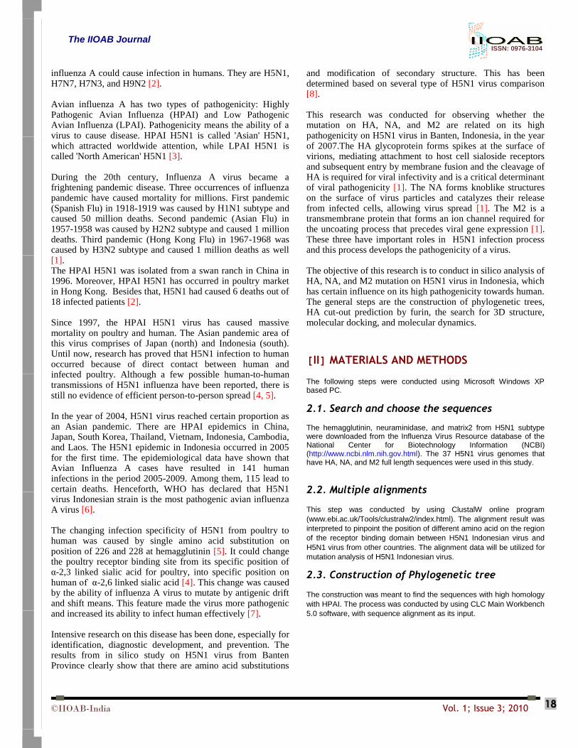
Table: 5. HA sequence alignment analysis result
HA Sequence
Positions
190 225 226 228
A/Indonesia/CDC1047/2007 (2IBX) V L V K
A/Hong Kong/482/97 (2FK0) E G Q G
Researchers reported that changes from hydrophilic to
hydrophobic on HA and NA results in an increase stability in
binding to sialic acid. This caused the increasing level of
pathogenicity in H5N1 Indonesian virus. Besides making
alignments toward H5N1 Indonesian virus and H5N1 PDB,
the alignment was done against all the chosen sequences. The
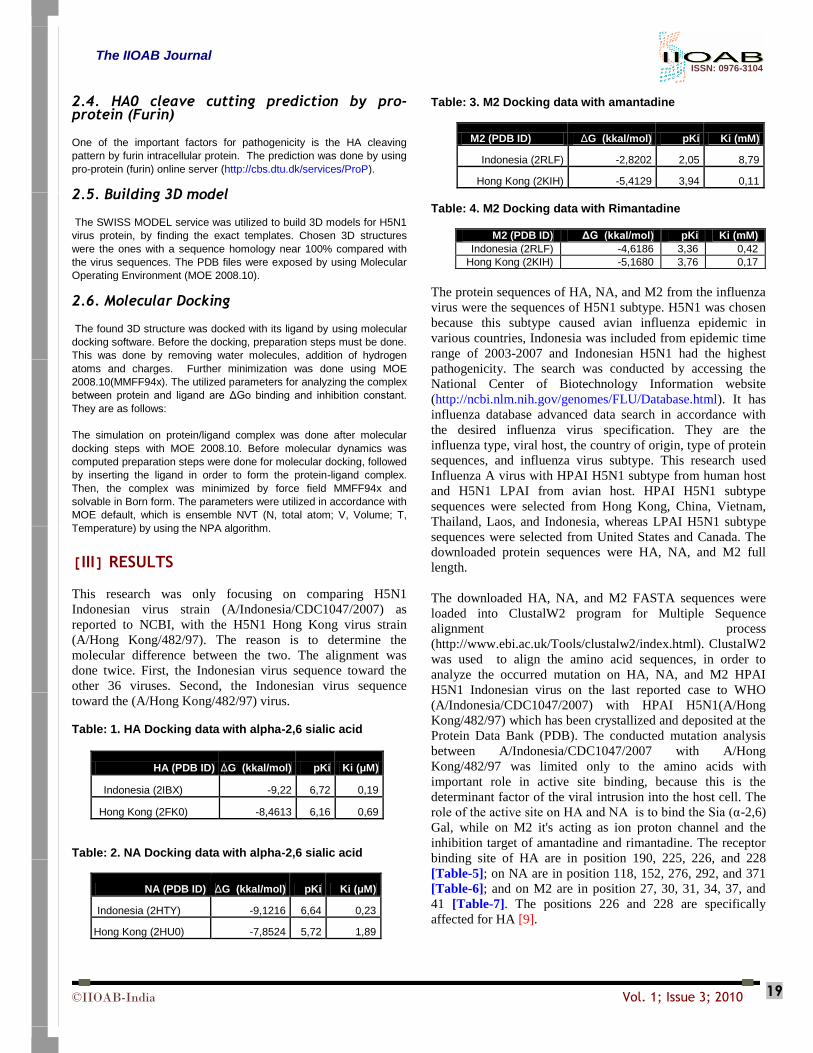
alignment results were used for building phylogenetic trees
[Figures 1, 2, and 3]. It would be useful to determine the
homology relation between Indonesian HPAI H5N1 and HPAI
H5N1 from other countries [10].
Table: 6. NA sequence alignment analysis result
Position
NA Sequence 118 152 276 292 371
A/Indonesia/CDC1047/2007
A S W Y G (2HY)
A/Hong Kong/482/97
R R E R R (2HU0)
Table: 7. M2 sequence alignment analysis result
Position
M2 Sequence 27
30
31
34
37
41
A/Indonesia/CDC1047/2007 (2RLF) A A N G H W
A/Hong Kong/482/97 (2KIH) V A N G H W
Fig: 1. Phylogenetic tree of HA H5N1
The IIOAB Journal ISSN: 0976-3104
©IIOAB-India Vol. 1; Issue 3; 2010 21
Fig: 2. Phylogenetic tree of NA H5N1
Fig: 3. Phylogenetic tree of M2 H5N1
The HA protein is useful for the binding process toward host
cell sialic acid receptor. After proteolitic process activation of
HA precursor into HA1 and HA2, the virus starts to fuse with
host cell. Previous research had shown that the HA cleaving
robustness is an important factor of avian influenza virus
virulence. LPAI has one arginine residue at the cleaving area.
Henceforth, it could only be cleaved by extra-cellular protease
like trypsin. Therefore, viral infection only occurred at the
host cell, while HPAI having poly-basic amino acids could be
cleaved by intracellular protease like furin. This allowed viral
infection to spread systemically to the whole host cell tissue
[11].
The computational results from online server furin pro-protein clearly show that A/Indonesia/CDC1047/2007 virus has R-X-K/R-R pattern, which is similar to A/Hong Kong/482/97 [Table-8]. This result clearly grouped Indonesian strain along with Highly Pathogenic Virus.
The 3D structure search was done by RSCB server (http://www.rcsb.org/pdb/static.do?p=search/index.html). The
method of this research utilized homology modeling on HA, NA, and M2 matrix of H5N1 Indonesian virus by using SWISS-MODEL workspace server. The process was conducted by giving sequence file input for browsing the 3D structure in FASTA format. The chosen mode is automated.
Table: 8. HA cleavage result by Furin
Sample of H5N1 Furin-type Cleavage Site
Prediction
A/Indonesia/CDC1047/2007 (ABM90533) ESRRKKR|GL
A/Hong Kong/482/97 (AAC32100) ERRRKKR|GL
A/Shanghai/1/2006 (BAH10637) RERRRKR|GL
A/mallard/ON/499/2005 (ABQ43787) none
The downloaded 3D structure was utilized to a molecular docking simulation toward Sia(α-2,6)Gal. Molecular docking simulation was conducted by using MOE-dock 2008.10 software. Ligand structure was drawn by using MOE2008.10 builder feature. Before starting the molecular docking, ligand was drawn and prepared. Ligand preparation was conducted
The IIOAB Journal ISSN: 0976-3104
©IIOAB-India Vol. 1; Issue 3; 2010 22
by using wash function and gas phase MMFF94x (i.e. no salvation was considered). The default parameters of MOE were utilized. One of the MOE features utilized was protonate-3D. It is a feature to solve the macromolecular protonation state assignment problem by selecting a protonation state for each chemical group that minimizes the total free energy of the system. The important parameter for protonate-3D is the repair of partial charge, which means substitution of the solvation mode while computing the force field of the molecule. The repair of partial charge and hydrogen atom was done to have the optimum state of the ligand, by using default parameter as well. The optimum state of the ligand was reached when the optimum minimization energy of protein-ligand conformation was attained. After the preparation, the same steps were done for the 3D structures of HA, NA, and M2. These steps were necessary to secure the optimum state of HA, NA, and M2 protein. The start configuration of the HA, NA, and M2 was initiated after the protonate-3D procedure [13].
The molecular dynamic simulations on HA, NA, and M2 of H5N1 Indonesian virus from its crystal structure on PDB was done to validate the interaction between protein and its ligand. The dynamic simulation was conducted on initialization step. The protein-ligand complex was optimized with partial charges and minimized with force field MMFF94x. However, this solvation was utilizing Esol calculation on the system. This process was conducted by using solvent.
The utilized statistics for conformation simulation was computed on the ensemble of structures. It was using MOE default, which is ensemble NVT with constant temperature of 300K and 101kPa pressure, while using NPA algorithm for adjusting the whole parameters. The position, velocity, and acceleration results were saved every 0.5 pico second [13].
[IV] DISCUSSION
The alignment of HA H5N1 Indonesian virus toward HA of
PDB (2FK0) shows, that mutation is close to H5N1
Indonesian virus receptor binding site. It was observed that in
position 190 and 226 a changing amino acid property, from
hydrophilic to hydrophobic, occurred. However, on position
225 and 228, the changed amino acid still retains its
hydrophobicity. The alignment of NA H5N1 Indonesian virus
toward NA of PDB (2HU0) clearly shows that mutation is
imminent on receptor binding site. It was observed, that in
position 118, 276, 292, and 371 occurs a changing property of
amino acid, from hydrophilic to hydrophobic. However, the
mutation on position 152 still retains its hydrophilicity. The
alignment of M2 H5N1 Indonesian virus with M2 of PDB
(2KIH) clearly shows mutation of H5N1 on active site
position. It is occurred on active site position 27. However, the
mutation still retains its hydrophilicity property.
The phylogenetic tree indicated that Indonesian HPAI H5N1
is closer with a HPAI H5N1 branch from other countries. It
could be inferred, that they have a close homology relation.
The close relationship is shown in HA, NA, and M2 sequences
[Figures-1 to 3].
Information for [Table 1-4]: It is a parameter from MOE. The
complete formula which is necessary for comprehending the
table is as follows:
G RT lnKp (1)
Kp 1
Ki (2)
Fig: 4. Docking visualization of 2ibx with sialic acid: A is
Ile_225, B is Val_226, C is Lys_228, D is Arg_232
Fig: 5. Docking visualization of 2fk0 with sialic acid: A is
Asn_186, B is Ser_227, C is Lys_232, D is Gly_228, E is Gln_226, F is Gly_225, H is Lys_193, I is Ser_137
Fig: 6. Docking visualization of 2hty with sialic acid: A is
Lys_245, B is Glu_291, C is Tyr_292, D is Val_247, E is Glu_248, F is Asp_250
Fig: 7. Docking visualization of 2hu0 with sialic acid: A is
Tyr_347, B is Arg_371, C is Pro_431, D is Arg_152, E is Asp_151, F is Pro_431, G is Arg_430, H is Thr_439, I is Arg_118
The IIOAB Journal ISSN: 0976-3104
©IIOAB-India Vol. 1; Issue 3; 2010 23
HA docking result with α-2,6 sialic acid [Figure-4], [Figure-
5] and respective deltaG data [Table-1] clearly show that HA
H5N1 Indonesian virus has higher binding affinity towards
sialic acid.
HA docking results with alpha-2,6 sialic acid [Figure 6],
[Figure 7] and respective deltaG data [Table 2] clearly show
that NA H5N1 Indonesian virus has higher binding affinity
towards sialic acid.
M2 docking result with Amantadine [Figure-8], [Figure-9]
and respective deltaG data [Table-3] clearly show that M2
H5N1 Indonesian virus has a lower binding affinity toward
amantadine.
M2 docking result with Rimantadine [Figure-10], [Figure-11]
and respective deltaG data [Table-4] clearly show that M2
H5N1 Indonesian virus has a lower binding affinity toward
Rimantadine.
Fig: 8. Visual Simulation Docking of 2RLF toward amantadine: A is Ala_30
Fig: 9. Visual Simulation Docking of 2KIH toward amantadine: A is Gly_34, B is Ile_35
Fig: 10. Visual Docking Simulation of 2RLF toward rimantadine: A is Ala_30, B is Gly_34
Fig: 11. Visual Docking Simulation of 2KIH toward rimantadine: A is His_37, B is Trp_41, C is Leu_38
The visual data from the docking simulation show that HA
and NA from H5N1 Indonesian virus binds better than
crystallized H5N1 virus from PDB. This clearly shows that
H5N1 Indonesian virus has higher infection efficiency
compared with other H5N1. It made the pathogenicity level of
Indonesian H5N1 higher than the others. The M2 visual
docking simulation rimantadine and amantadine show that
H5N1 Indonesian virus has higher resistance toward both
drugs, compared with crystallized H5N1 virus from Hong
Kong.
The dynamic simulation analysis was done by observing the
protein-ligand complex interaction among their atoms. The
dynamic simulation shows that H5N1 virus has resistance
towards both drugs, because they didn't bind with M2 active
site. However, this step is not conclusive, and requires further
computation (data not shown).
[V] CONCLUSION
The phylogenetic tree analysis shows that H5N1 Indonesian
virus has a close relationship with HPAI H5N1 from other
countries. However, it belongs to its own cluster, which differs
by its pathogenicity.
The sequence alignment analysis has shown that HA, NA, and
M2 of H5N1 Indonesian virus has amino acid mutation on its
active site, and it is followed with the attribute change from
hydrophilic to hydrophobic. It rendered the H5N1 Indonesian
virus more pathogenic.
The molecular docking analysis shows that HA and NA H5N1
Indonesian virus has a better ability to bind sialic acid
receptor, and the activity of amantadine and rimantadine did
not give any inhibition toward active site of M2 H5N1
Indonesian virus. It caused the Indonesian H5N1 to have a
higher pathogenicity.
We suggest conducting further research on molecular
dynamics of HA, NA, and M2 H5N1 mutation.
ACKNOWLEDGEMENT
The authors would like to express their gratitude to Ridla Bakrie, PhD, the head of Chemistry Department, Faculty of Mathematics and Science, University of Indonesia, for his support toward this research. Authors also thank Dr. Lalit Ponnala of Cornell University, USA for his copy editing of the article.
REFERENCES
[1] Kamps SB, Hoffman C, Preiser W. [2006] Influenza report
2006. Flying Publisher. Paris, Cagliari, Sevilla.
The IIOAB Journal ISSN: 0976-3104
©IIOAB-India Vol. 1; Issue 3; 2010 24
[2] Peiris JSM, De Jong MD, Guan Yi. [2007] Avian Influenza
Virus (H5N1): a Threat to Human Health. Clin Microbiol 20:
243–267.
[3] Taha FA. [2007] How Highly Pathogenic Avian Influenza
H5N1 has affected World Poultry-Meat Trade. USDA
Economic Research Service. http://www.ers.usda.gov
[4] Ungchusak K, Auewarakul P, Dowell SF. et al. [2005]
Probable Person to Person Transmision of Avian Influenza A
(H5N1). The New England J Medicine 352 (4): 333–340.
[5] Gambotto A, Barratt-Boyes SM, de Jong MD, Neumann G,
Kawaoka Y. [2008] Human Infection with Highly Pathogenic
H5N1 Influenza Virus. The Lancet 371 (9622): 1464–1475.
[6] WHO [2009] Avian Influenza Disease Outbreak News
http://www.who.int/csr/don/2009_09_31/en/index.html.
[7] Bouvier NM, Palese P. [2008] The Biology of Influenza
Viruses. Vaccine 26: D49–53.
[8] Tambunan SF, Hikmawan O, Theofilus A. [2008] In Silico
Mutation Study of Hemagglutinin and Neuraminidase on
Banten Province Strain Influenza A H5N1 Virus. Trends in
Bioinformatics 1(1): 18–24.
[9] Larkin MA, Blackshields G, Brown NP, et al. [2007] Clustal
W and Clustal X version 2.0. Bioinformatics 23: 2947–2948.
[10] Anwar T, Lal SK, Khan AU. [2006] In Silico Analysis of
Genes Nucleoprotein, Neuraminidase, and Hemagglutinin : A
Comparative Study On Different Strains of Influenza A (Bird
Flu) Virus Sub-type H5N1. In Silico Biology 6: 0015.
[11] Klenk HD. [2007] Molecular Mechanisms of Pathogenicity
and Interspecies Transmission of Avian Influenza Virus.
Germany: Institut für Virologie Philipps Universität Marburg.
Vizier Conference. Marseille, 27.04.2007.
[12] Bock JR, Gough DA. [2002] A New Method to Estimate
Ligand-Receptor Energetics. Mol Cell Proteomics 1(11): 904–
910.
[13] Kitchen DB, Decornez H, Furr JR, Bajorath J. [2004] Docking
and Scoring in Virtual Screening for Drug Discovery:
Methods and Application. Nat Rev Drug Discov 3(11): 935-
949.
ABOUT AUTHORS
Prof. Usman Sumo Friend Tambunan is currently working as a permanent professor in the chair of Bioinformatics, Department of Chemistry, Faculty of Mathematics and Science, University of Indonesia. Previously, he worked at the Indonesian Agency of Technology Assessment and Application, as a senior scientist, and graduated his Phd in Tohoku University, Japan. He was the former vice dean of Faculty of Mathematics and Science, University of Indonesia. He has conducted research on Bioinformatics for biomedics, in the topics of HPV, Avian Influenza, and Dengue Virus. He had secured a copyright patent for HPV vaccine design and published many bioinformatics-related articles in peer-reviewed international scientific journals. He is elected several times as the best lecturer and researcher in University of Indonesia. His research is currently supported by Indonesian ministry of national education grant.
Agus Limanto, Bsc is currently working as junior assistant and researcher of Professor Usman Sumo Friend Tambunan at Bioinformatics Laboratory, Department of Chemistry, Faculty of Mathematics and Science, University of Indonesia. Now, he is involved in bioinformatics research, especially in dengue and
influenza type A drug design.
Arli Aditya Parikesit, Msc is currently working as assistant of Prof Usman Sumo Friend Tambunan, lecturer, and researcher in the chair of Bioinformatics, Department of Chemistry, Faculty of Mathematics and Science, University of Indonesia. He has conducted research on Bioinformatics for biomedics, in the topics of HPV, Avian Influenza, and Dengue Virus. He finished and published his master thesis about HPV, with the support of Indonesian ministry of Education Graduate grant (hibah pasca). Now, he is a doctorate/Phd candidate at the chair of Bioinformatics, Department of Computer Science, Faculty of Computer Science and Mathematics, University of Leipzig, Germany with the support of DAAD fellowship. The theme of his doctorate research is 'Domain Cooccurence Distribution of Genetic Regulators from an Evolutionary
Perspective'.
Related Documents