ORIGINAL ARTICLE Increased presence of effector lymphocytes during Helicobacter hepaticus -induced colitis Sarah J McCaskey, Elizabeth A Rondini, Jonathan F Clinthorne, Ingeborg M Langohr, Elizabeth M Gardner, Jenifer I Fenton Sarah J McCaskey, Jenifer I Fenton, Department of Food Sci- ence and Human Nutrition, College of Osteopathic Medicine, Michigan State University, East Lansing, MI 48824, United States Elizabeth A Rondini, Jonathan F Clinthorne, Elizabeth M Gardner, Department of Food Science Human Nutrition, Mich- igan State University, East Lansing, MI 48824, United States Ingeborg M Langohr, Department of Pathobiology and Diag- nostic Investigation, Michigan State University, East Lansing, MI 48824, United States Author contributions: McCaskey SJ and Rondini EA con- tributed equally to this work; McCaskey SJ and Rondini EA co-wrote this manuscript and McCaskey SJ, Rondini EA and Clinthorne JF performed the research; Langohr IM analyzed tis- sues for histopathology; Gardner EM assisted in experimental design; Fenton JI conceived the experimental design, assisted in experimental methods, and helped prepare the manuscript. Supported by AgBio Research Center at Michigan State Uni- versity Correspondence to: Jenifer I Fenton, PhD, MPH, Department of Food Science and Human Nutrition, College of Osteopathic Medicine, Michigan State University, 208B G.M. Trout Building, East Lansing, MI 48824, United States. [email protected] Telephone: +1-517-3558474 Fax: +1-517-3538963 Received: June 13, 2011 Revised: November 12, 2011 Accepted: December 31, 2011 Published online: April 7, 2012 Abstract AIM: To identify and characterize drosophila mothers against decapentaplegic (SMAD)3-dependent changes in immune cell populations following infection with He- licobacter hepaticus ( H. hepaticus ). METHODS: SMAD3 -/- ( n = 19) and colitis-resistant SMAD3 +/- ( n = 24) mice (8-10 wk of age) were in- fected with H. hepaticus and changes in immune cell populations [T lymphocytes, natural killer (NK) cells, T regulatory cells] were measured in the spleen and mesenteric lymph nodes (MsLNs) at 0 d, 3 d, 7 d and 28 d post-infection using flow cytometry. Genotype- dependent changes in T lymphocytes and granzyme B + cells were also assessed after 28 d in proximal colon tissue using immunohistochemistry. RESULTS: As previously observed, SMAD3 -/- , but not SMAD3 +/- mice, developed colitis, peaking at 4 wk post-infection. No significant changes in T cell subsets were observed in the spleen or in the MsLNs between genotypes at any time point. However, CD4 + and CD8 + / CD62L lo cells, an effector T lymphocyte population, as well as NK cells (NKp46/DX5 + ) were significantly higher in the MsLNs of SMAD3 -/- mice at 7 d and 28 d post-in- fection. In the colon, a higher number of CD3 + cells were present in SMAD3 -/- compared to SMAD3 +/– mice at base- line, which did not significantly change during infection. However, the number of granzyme B + cells, a marker of cytolytic lymphocytes, significantly increased in SMAD3 -/- mice 28 d post-infection compared to both SMAD3 +/- mice and to baseline values. This was consistent with more severe colitis development in these animals. CONCLUSION: Data suggest that defects in SMAD3 signaling increase susceptibility to H. hepaticus -induced colitis through aberrant activation and/or dysregulation of effector lymphocytes. © 2012 Baishideng. All rights reserved. Key words: Transforming growth factor-β; Colitis; Dro- sophila mothers against decapentaplegic; Colon cancer; T lymphocytes Peer reviewers: Scott Steele, MD, FACS, FASCRS, Chief, Colon and Rectal Surgery, Department of Surgery, Madigan Army Medical Center, Fort Lewis, WA 98431, United States; Dr. Timothy Koch, Professsor, Department of Gastroenterology, Washington Hospital Center, 106 Irving Street, Washington, NW 20010, United States; Hang Nguyen, PhD, University of Auvergne, Pathogénie Bactérienne Intestinale, Centre Biomédical de Recherche et Valorisation, 28 Place Henri- Dunant, 63000 Clermont-Ferrand, France World J Gastroenterol 2012 April 7; 18(13): 1459-1469 ISSN 1007-9327 (print) ISSN 2219-2840 (online) © 2012 Baishideng. All rights reserved. Online Submissions: http://www.wjgnet.com/1007-9327office [email protected] doi:10.3748/wjg.v18.i13.1459 1459 April 7, 2012|Volume 18|Issue 13| WJG|www.wjgnet.com

Welcome message from author
This document is posted to help you gain knowledge. Please leave a comment to let me know what you think about it! Share it to your friends and learn new things together.
Transcript
ORIGINAL ARTICLE
Increased presence of effector lymphocytes during Helicobacter hepaticus-induced colitis
Sarah J McCaskey, Elizabeth A Rondini, Jonathan F Clinthorne, Ingeborg M Langohr, Elizabeth M Gardner, Jenifer I Fenton
Sarah J McCaskey, Jenifer I Fenton, Department of Food Sci-ence and Human Nutrition, College of Osteopathic Medicine, Michigan State University, East Lansing, MI 48824, United StatesElizabeth A Rondini, Jonathan F Clinthorne, Elizabeth M Gardner, Department of Food Science Human Nutrition, Mich-igan State University, East Lansing, MI 48824, United States Ingeborg M Langohr, Department of Pathobiology and Diag-nostic Investigation, Michigan State University, East Lansing, MI 48824, United States Author contributions: McCaskey SJ and Rondini EA con-tributed equally to this work; McCaskey SJ and Rondini EA co-wrote this manuscript and McCaskey SJ, Rondini EA and Clinthorne JF performed the research; Langohr IM analyzed tis-sues for histopathology; Gardner EM assisted in experimental design; Fenton JI conceived the experimental design, assisted in experimental methods, and helped prepare the manuscript.Supported by AgBio Research Center at Michigan State Uni-versityCorrespondence to: Jenifer I Fenton, PhD, MPH, Department of Food Science and Human Nutrition, College of Osteopathic Medicine, Michigan State University, 208B G.M. Trout Building, East Lansing, MI 48824, United States. [email protected] Telephone: +1-517-3558474 Fax: +1-517-3538963 Received: June 13, 2011 Revised: November 12, 2011 Accepted: December 31, 2011 Published online: April 7, 2012
AbstractAIM: To identify and characterize drosophila mothers against decapentaplegic (SMAD)3-dependent changes in immune cell populations following infection with He-licobacter hepaticus (H. hepaticus ).
METHODS: SMAD3-/- (n = 19) and colitis-resistant SMAD3+/- (n = 24) mice (8-10 wk of age) were in-fected with H. hepaticus and changes in immune cell populations [T lymphocytes, natural killer (NK) cells, T regulatory cells] were measured in the spleen and mesenteric lymph nodes (MsLNs) at 0 d, 3 d, 7 d and 28 d post-infection using flow cytometry. Genotype-
dependent changes in T lymphocytes and granzyme B+ cells were also assessed after 28 d in proximal colon tissue using immunohistochemistry.
RESULTS: As previously observed, SMAD3-/-, but not SMAD3+/- mice, developed colitis, peaking at 4 wk post-infection. No significant changes in T cell subsets were observed in the spleen or in the MsLNs between genotypes at any time point. However, CD4+ and CD8+/CD62Llo cells, an effector T lymphocyte population, as well as NK cells (NKp46/DX5+) were significantly higher in the MsLNs of SMAD3-/- mice at 7 d and 28 d post-in-fection. In the colon, a higher number of CD3+ cells were present in SMAD3-/- compared to SMAD3+/– mice at base-line, which did not significantly change during infection. However, the number of granzyme B+ cells, a marker of cytolytic lymphocytes, significantly increased in SMAD3-/- mice 28 d post-infection compared to both SMAD3+/- mice and to baseline values. This was consistent with more severe colitis development in these animals.
CONCLUSION: Data suggest that defects in SMAD3 signaling increase susceptibility to H. hepaticus -induced colitis through aberrant activation and/or dysregulation of effector lymphocytes.
© 2012 Baishideng. All rights reserved.
Key words: Transforming growth factor-β; Colitis; Dro-sophila mothers against decapentaplegic; Colon cancer; T lymphocytes
Peer reviewers: Scott Steele, MD, FACS, FASCRS, Chief, Colon and Rectal Surgery, Department of Surgery, Madigan Army Medical Center, Fort Lewis, WA 98431, United States; Dr. Timothy Koch, Professsor, Department of Gastroenterology, Washington Hospital Center, 106 Irving Street, Washington, NW 20010, United States; Hang Nguyen, PhD, University of Auvergne, Pathogénie Bactérienne Intestinale, Centre Biomédical de Recherche et Valorisation, 28 Place Henri-Dunant, 63000 Clermont-Ferrand, France
World J Gastroenterol 2012 April 7; 18(13): 1459-1469 ISSN 1007-9327 (print) ISSN 2219-2840 (online)
© 2012 Baishideng. All rights reserved.
Online Submissions: http://www.wjgnet.com/[email protected]:10.3748/wjg.v18.i13.1459
1459 April 7, 2012|Volume 18|Issue 13|WJG|www.wjgnet.com
McCaskey SJ et al . Immune response following H. hepaticus infection
McCaskey SJ, Rondini EA, Clinthorne JF, Langohr IM, Gard-ner EM, Fenton JI. Increased presence of effector lymphocytes during Helicobacter hepaticus-induced colitis. World J Gas-troenterol 2012; 18(13): 1459-1469 Available from: URL: http://www.wjgnet.com/1007-9327/full/v18/i13/1459.htm DOI: http://dx.doi.org/10.3748/wjg.v18.i13.1459
INTRODUCTIONIndividuals with inflammatory bowel disease (IBD), par-ticularly ulcerative colitis (UC), are at a higher risk of developing colon cancer than the general population[1]. A meta-analysis of 116 studies indicated that the prevalence of colon cancer in patients with UC is approximately 3.7% (95% CI: 3.2-4.2), with the cumulative probability reaching 18% by 30 years regardless of disease severity[2]. Although the etiology of UC is poorly understood, there are indications that the immune system of individuals with UC reacts abnormally to bacteria in the digestive tract. This altered immune response leads to the inflam-mation-associated pathology of IBD[3-5].
Imbalances in both innate and adaptive immune cells, such as natural killer (NK) cells and T cell subsets, includ-ing CD4+ and CD8+ T cells and CD4/CD25/Foxp3+ T regulatory (Treg) cells, are associated with the pathogene-sis of IBD[2]. The inflammation and damage caused by in-creased secretion of inflammatory cytokines during an ac-tive disease state is thought to be triggered by cytotoxicity against the commensal bacteria[6]. For example, levels of NK cytotoxicity in UC are related to the clinical stage of the disease[7]. In active disease states, NK cells are present in normal numbers, but are functionally defective, whereas NK cells exhibit normal cytotoxic activity in an inactive disease state[7]. Induction of inflammatory cytokines can also result from the disruption of the homeostatic balance between Treg and effector T helper (Th) cells. Elevated levels of pro-inflammatory CD4+ T cells lead to excess cytokine/chemokine production, thereby recruiting ad-ditional leukocytes and influencing the severity of the inflammatory response[2]. CD8+ T cells are also important in the pathogenesis of UC in humans, as demonstrated by extensive CD8+ T cell infiltration within intestinal lesions contributing to mucosal damage[8,9].
Transforming growth factor (TGF)-β is a multifunc-tional cytokine that plays an important role in epithelial and immune cell homeostasis[10,11]. TGF-β mediates many diverse biological functions on different cell types through receptor-mediated phosphorylation and activation of the drosophila mothers against decapentaplegic homolog (SMAD) family proteins, notably SMAD2 and SMAD3, which migrate to the nucleus and induce transcription of a targeted set of genes[12,13]. Dysfunctions in one or more components of TGF-β signaling are commonly observed in human IBD and during colon cancer development. For example, loss of expression of the TGF receptor type Ⅱ is observed in 90% of microsatellite instable colon
cancers, leading to loss of growth regulation in epithelial cells[14]. Additionally, although the TGF-β1 isoform is overexpressed in the colon of individuals with IBD[15], nuclear signaling is impaired due to increased levels of SMAD7[16]. SMAD7 normally inhibits TGF-β signaling by blocking activation of SMAD2/3 in response to receptor-ligand binding. Normalizing SMAD7 expression restores TGF-β signaling through SMAD3 and inhibits proinflam-matory cytokine production by lamina propria mono-nuclear cells[16].
Impairments in one or more components of the TGF-β signaling pathway are implicated in intestinal inflammation in rodent models. For example, homologous knockout of the TGF-β1 gene in mice causes an excessive inflam-matory response in multiple organs, including the heart, lungs, and intestinal tract leading to premature death[17,18]. Additionally, Maggio-Price et al[19] have demonstrated that disruption of the transcription factor SMAD3 modulates colitis susceptibility following infection with certain Helico-bacter spp. Among these, Helicobacter hepaticus (H. hepaticus) is a Gram-negative spiral bacterium that colonizes the lower intestine and the hepatobiliary tract of mice[20]. Although generally asymptomatic, infection can lead to hepatic and intestinal inflammation in certain strains of immunode-ficient mice[21-24]. In the complete absence of SMAD3 signaling, H. hepaticus induces a moderate inflammatory response in the cecum and colon, eventually leading to mucinous adenocarcinoma formation after 15-30 wk[19]. It is generally accepted that chronic low levels of inflam-mation lead to cancer promotion and progression[25-28], therefore, the SMAD3 mouse model is very similar to the development of specific human cancers where pathogen-induced inflammation is necessary (but not sufficient) to cause dysplasia and tumor formation.
Using this model, the focus of the current study was to investigate the effect of SMAD3 deficiency on changes in local and systemic immune cell populations following infection with H. hepaticus. We hypothesized that colitis susceptibility in SMAD3-/- mice induced by H. hepaticus is associated with altered immune cell populations compared to colitis resistant SMAD3+/- mice. The aims of this study were to: (1) characterize the colitis induced by H. hepaticus in colitis-sensitive SMAD3-/- vs resistant SMAD3+/- mice; (2) compare the immune cell population changes in the spleen and mesenteric lymph nodes (MsLNs); and (3) compare local immune cell changes by immunohisto-chemistry in the colon.
MATERIALS AND METHODSMurine modelSMAD3+/- and SMAD3-/- (129-Smad3tm1Par/J) mice were bred in-house. Homozygous males and heterozygous females were mated to obtain both SMAD3+/- and SMAD3-/- pups. Genotypes were confirmed by poly-merase chain reaction (PCR). Animals were housed un-der specific pathogen-free (SPF) conditions in 60 square
1460 April 7, 2012|Volume 18|Issue 13|WJG|www.wjgnet.com
inch plastic cages (maximum of five adult mice per cage) with microisolator lids in an Association for Assessment and Accreditation of Laboratory Animal Care-approved facility at Michigan State University. SPF conditions were assured through quarterly serology testing by Charles Rivers (Wilmington, MA, United States) and in-house testing for ectoparasites, endoparasites and fecal Helico-bacter species (PCR). Full necropsies (including culture and sensitivity) were performed at least yearly on rodent breeding colonies. Animal rooms were maintained at 23.3 ± 2.2 ℃ with a 12-h light/dark cycle. Mice were fed Har-lan Teklad 7913 rodent chow and sterile water ad libitum. Animal protocols were approved by the Michigan State University Institutional Animal Care and Use Committee.
Bacterial culture and infectionThe wild-type H. hepaticus strain 3B1 (ATCC 51488) was utilized for these experiments. Isolates were asepti-cally streaked onto sheep blood agar and incubated at 37 ℃ for 24-48 h inside GasPak™ gas generating pouch systems (BD Diagnostic Systems, Sparks, MD, United States). Mice were infected as previously described[19]. Briefly, bacteria were collected and resuspended in tryptic soy broth at A600 nm ≥ 1.8. Animals were then gavaged with 0.3 mL doses of fresh bacterial suspension on two consecutive days. Previously, Maggio-Price et al[19] have shown that Helicobacter infection is localized primarily in the cecum and proximal colon, and that bacterial DNA is still present in the tissue and luminal contents of the ce-cum at 12 wk post-infection. Bacterial presence was con-firmed in the current study via DNA isolation at 3 d post-infection using a commercial kit (QIAGEN tissue kit; Valencia, CA, United States) as previously described[24].
Experimental designIn study 1, SMAD3-/- mice (n = 30) at 8-10 wk of age were infected with H. hepaticus to determine onset and duration of colitis. At the time of necropsy, mice were asphyxiated with CO2 and exsanguinated via cardiac puncture. Intestinal tissue was collected and processed for histopathology at 2-8 wk post-infection. In study 2, SMAD3+/- (n = 24) and SMAD3-/- mice (n = 19) at 8-10 wk of age were infected with H. hepaticus once per day for two consecutive days. At select time points after infection (0, 3, 7 and 28 d), the spleen and MsLNs were collected and processed for lymphocyte isolation as described below. Colon and cecum tissue was collected, fixed, and processed for immunohistochemistry.
HistopathologyThe colon and cecum were removed and flushed with phosphate-buffered saline (PBS). Tissues were fixed in 10% formalin overnight, embedded in paraffin, then sectioned and stained with hematoxylin and eosin (HE). Longitudinal sections were graded for inflammation and epithelial dysplasia/neoplasia by a pathologist us-ing a blinded scoring system adapted from Maggio-Price et al[29]. Cecum and colons were scored on a 1 to 4
scale both for inflammation (1, no inflammation; 2, mild inflammation; 3, moderate inflammation; 4, marked in-flammation) and dysplasia (1, no dysplasia; 2, low-grade dysplasia; 3, high-grade dysplasia; 4, high-grade dysplasia with invasion/adenocarcinoma). The two scores for co-lon and two scores for cecum tissue in each animal were combined such that a score of 4 indicated no inflam-mation or dysplasia and a score of 16 reflected maximal inflammation and neoplasia.
Immunohistochemistry was performed on paraffin-embedded colon sections. Antibodies specific for CD3 and granzyme B were purchased from Abcam (Cam-bridge, MA, United States). Colons were sectioned at 5 µm, mounted on coated slides, deparaffinized in xylene, and rehydrated through graded ethanol-water baths. Antigen retrieval was performed using citrate buffer (10 mmol/L, pH 6.0) and a vegetable steamer. Tissues were incubated in 3% hydrogen peroxide to block endog-enous peroxidase activity and then incubated overnight at 4 ℃ in primary antibody. On the following day, tissues were washed in Tris-buffered saline containing Tween-20 (0.05%), then incubated with biotinylated secondary an-tibodies followed by streptavidin horseradish peroxidase for 45 min each at room temperature (Dako, Carpentar-ia, CA, United States). After extensive washing, antigen-bound horseradish peroxidase was detected using the chromagen 3,3’-diaminobenzidine (0.5 mg/mL; Sigma-Aldrich, St. Louis, MO, United States) dissolved in PBS (10 mmol/L, pH 7.2). Identification of cellular infiltrate in the colons of mice was performed by a pathologist. CD3+ and granzyme B+ cells were identified under a light microscope using a 20 × objective. The occurrence of positively stained cells was scored in proximal colons of mice in five fields using a 1-cm2 grid reticle as fol-lows: 0 = average of 0 cells/grid, 1 = average of ≤ 1 cell/grid, 2 = average of 2-10 cells/grid, 3 = average of 11-20 cells/grid, 4 = average of > 21 cells/grid. Final values represent mean ± SE per group (n = 3-5/group).
Lymphocyte isolationSpleens and MsLNs were removed and placed in ice-cold RPMI medium at the time of necropsy. Spleens were processed with a dounce homogenizer, pelleted, and washed in RPMI. Cells were resuspended in ACK lysing buffer (Invitrogen, Carlsbad, CA, United States) and washed twice in RPMI. MsLNs were treated with 5 mL enzymatic digest [5% fetal bovine serum (FBS), 0.5 mg/mL collagenase, 0.05 mg/mL DNaseⅠ) for 30 min at 37 ℃. Cells were passed through 70-µm filters and washed with RPMI. Cell counts were performed with a hemocytometer using trypan blue exclusion and resus-pended to a concentration of one million cells per mil-liliter of medium.
Flow cytometryLymphocytes were resuspended in fluorescence-activated cell sorting (FACS) buffer (0.1% sodium azide, 1% FBS, in dPBS) blocked with anti-Fc receptor RⅡ/Ⅲ [CD16/
1461 April 7, 2012|Volume 18|Issue 13|WJG|www.wjgnet.com
McCaskey SJ et al . Immune response following H. hepaticus infection

CD32 (purified from clone 2.4G2 hybridoma; ATCC, Manassas, VA, United States)] for 10 min on ice, and subsequently incubated with combinations of the follow-ing fluorochrome-conjugated antibodies (E-bioscience, San Diego, CA, United States; or BD Bioscience, San José, CA, United States) at concentrations ranging from 1:100 to 1:300 in FACS buffer: CD3 (PerCP-Cy5.5), CD4 (eFluor450), CD8 (PE-Cy7), CD25 (PE), FoxP3 (FITC or Alexa Fluor488), CD62 (APC), Nkp46 (FITC) and DX5 (APC). Cells were incubated in staining cocktails (one million cells per cocktail) on ice in the dark for 30 min. Intracellular staining was performed using FoxP3 staining buffer set as per the manufacturer’s instructions (E-bioscience). Briefly, after surface staining, cells were washed twice in FACS buffer, fixed in 4% paraformalde-hyde for 25 min, and permeabilized for 30 min. Permea-bilization was followed by incubation for 30 min with the appropriate antibodies diluted in permeabilization dilu-ent. Samples were then acquired on a LSR Ⅱ (BD Biosci-ence) and analyzed using FlowJo software (Tree Star Inc., Ashland, OR, United States). The number of cells in each population of interest was determined by multiplying cell percentages by the total cell number.
Statistical analysisData for the colitis and immunohistochemistry scores were analyzed using the nonparametric Kruskal-Wallis test and Dunn’s post-test for specific comparisons. Flow cytometric data was analyzed using a two-way analysis of variance in GraphPad Prism (GraphPad Software, La Jolla, CA, United States). When statistical differences were detected, Tukey’s multiple comparison test was used to determine differences between the two groups. P < 0.05 was considered significant. All data are repre-sented as mean ± SE.
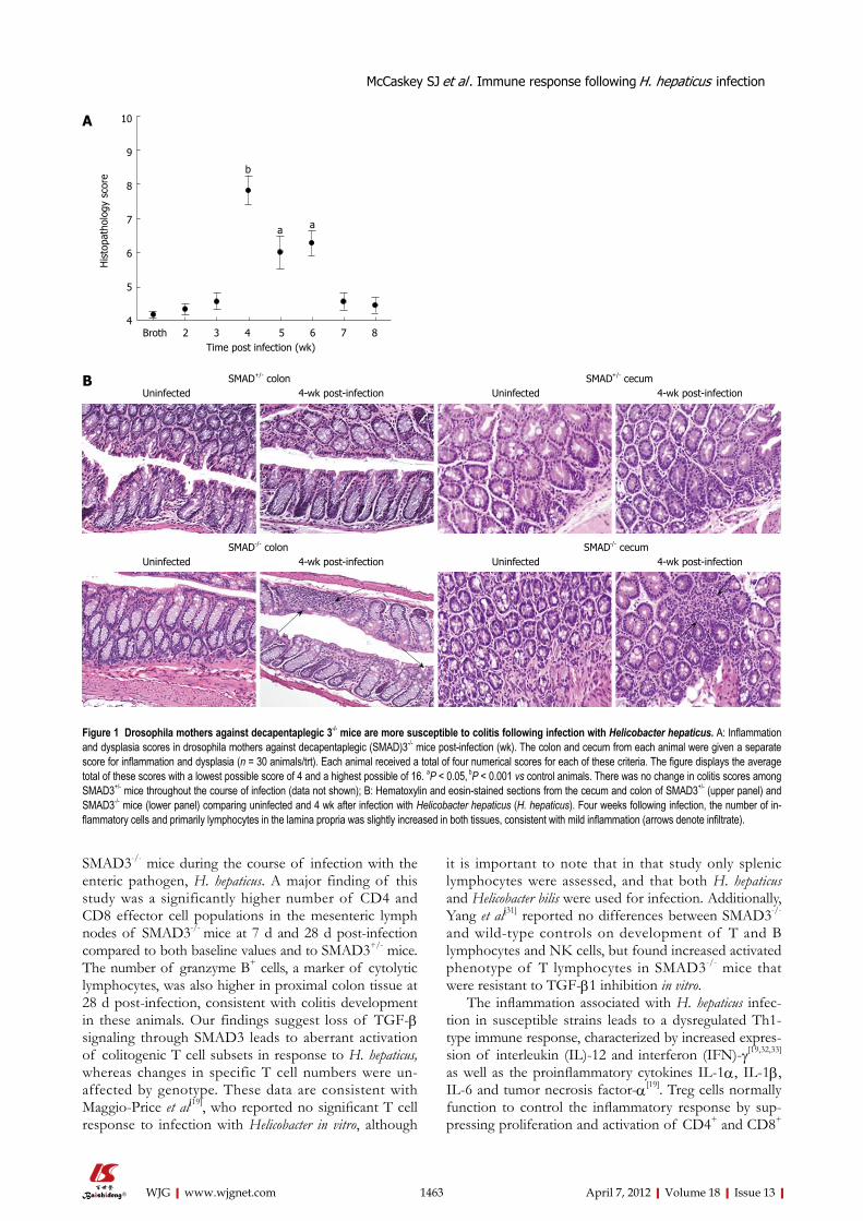
RESULTSSMAD3-deficient mice are susceptible to colitis 4 wk post-infectionColitis severity in SMAD3-/- mice (Figure 1A) peaked at 4 wk post-infection, with an average colitis score of 7.8 ± 0.4. This value was significant compared to samples taken at all other time points (P < 0.05). Colitis resolved to baseline levels in SMAD3-/- mice by 8 wk post-infec-tion. In comparison, SMAD3+/- mice were resistant to colitis development at all time points (data not shown). There was no statistically significant change in colitis scores in SMAD3+/- mice compared to baseline at any time point post-infection. Representative HE images from SMAD3+/- and SMAD3-/- mice prior to and 4 wk following infection are presented in Figure 1B.
SMAD3-dependent changes in lymphocyte populations following H. hepaticus infectionWe next evaluated genotype- and time-dependent chang-es in lymphocyte populations in the spleen and MsLNs using flow cytometry. There were no significant changes
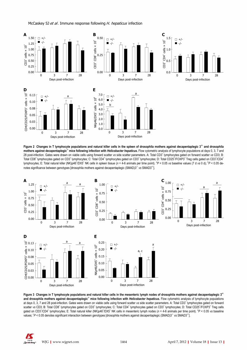
in total CD3+, CD4+ or CD8+ lymphocytes in the spleen at baseline or at any time point following infection (Fig-ure 2A-C). Tregs (FoxP3+/CD25+/CD4+) and NK cells (NKp46+/DX5+) increased in both genotypes following infection but returned to baseline by 28 d (Figure 2D and E).
In the MsLNs, CD3+, CD4+ and Treg cells were sig-nificantly higher in both genotypes at 7 d and 28 d post-infection (Figure 3A, C, and D), whereas there were no significant changes in CD8+ cells at any time point examined (Figure 3B). NK cells increased in SMAD3-deficient mice by 7 d post-infection, and were signifi-cantly different from baseline values at 28 d (Figure 3E). Comparably, NK cells were not significantly altered at any time point in SMAD3+/- mice (Figure 3E).
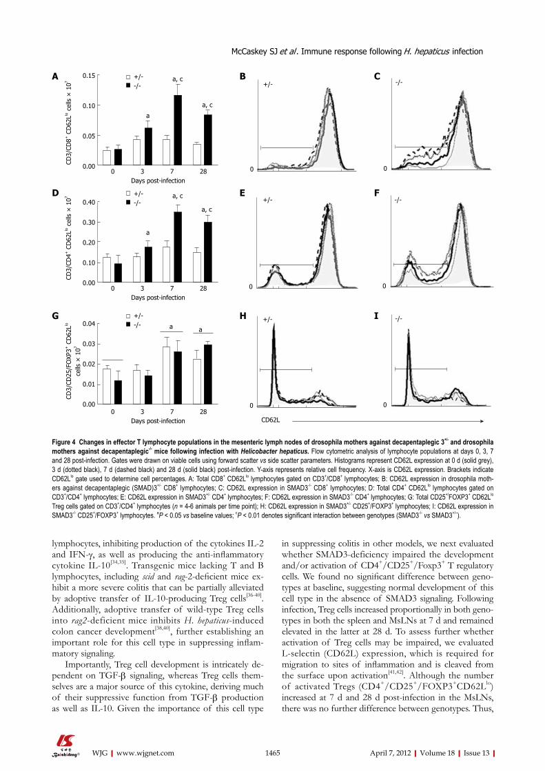
To determine activation status of the different T lymphocyte populations, we next evaluated surface ex-pression of CD62L. L-Selectin (CD62L) is an adhesion marker expressed at high levels in naïve T cells and is cleaved from the surface (CD62Llo) in activated and/or in memory T cells. There were no statistically significant changes or observable trends in the proportion or total number of activated T cells in the spleen at any time point after infection (data not shown). However, the proportion of CD3+, CD8+, CD62Llo and CD3+, CD4+, CD62Llo cells was significantly higher in SMAD3-/-
mice at 7 d and 28 d compared to baseline values and to SMAD3+/- mice (Figure 4A and D). Effector Treg cells increased in both strains at 7 d and 28 d compared to baseline values (Figure 4G). CD62L expression became dimmer at later time points in the SMAD3-/- mice for both CD8+ and CD4+ populations (Figure 4C and F) in the MsLNs, however, the intensity of CD62L expression was maintained consistently in SMAD3+/- mice through all time points (Figure 4B and E). No significant dif-ferences were observed in the percentage of Treg cells expressing reduced levels of CD62L between genotypes at any time point (Figure 4H and I).
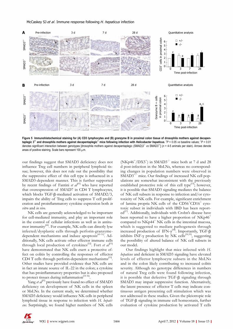
Immunohistochemical analysis of colon sections 28 d post-infectionWe next evaluated local changes in CD3+ cells and the serine protease, granzyme B, in the proximal colons of SMAD3+/- and SMAD3-/- mice 4 wk post-infection. The lamina propria in SMAD3-/- mice was moderately expand-ed by lymphocytic cells. Based on morphology and immu-nohistochemistry, these cells consisted primarily of CD3+ lymphocytes (Figure 5A). Additionally, numerous gran-zyme B+ cells were noted in the intestine of SMAD3-/- infected mice, primarily within the villous epithelium, but sometimes also within the lamina propria (Figure 5B).
DISCUSSIONFunctional TGF-β signaling is crucial for maintaining immune cell homeostasis[30]. In the present study, we evaluated changes in local and systemic immune cell populations in colitis resistant SMAD3+/- and sensitive
1462 April 7, 2012|Volume 18|Issue 13|WJG|www.wjgnet.com
McCaskey SJ et al . Immune response following H. hepaticus infection
SMAD3-/- mice during the course of infection with the enteric pathogen, H. hepaticus. A major finding of this study was a significantly higher number of CD4 and CD8 effector cell populations in the mesenteric lymph nodes of SMAD3-/- mice at 7 d and 28 d post-infection compared to both baseline values and to SMAD3+/- mice. The number of granzyme B+ cells, a marker of cytolytic lymphocytes, was also higher in proximal colon tissue at 28 d post-infection, consistent with colitis development in these animals. Our findings suggest loss of TGF-β signaling through SMAD3 leads to aberrant activation of colitogenic T cell subsets in response to H. hepaticus, whereas changes in specific T cell numbers were un-affected by genotype. These data are consistent with Maggio-Price et al[19], who reported no significant T cell response to infection with Helicobacter in vitro, although
it is important to note that in that study only splenic lymphocytes were assessed, and that both H. hepaticus and Helicobacter bilis were used for infection. Additionally, Yang et al[31] reported no differences between SMAD3-/- and wild-type controls on development of T and B lymphocytes and NK cells, but found increased activated phenotype of T lymphocytes in SMAD3-/- mice that were resistant to TGF-β1 inhibition in vitro.
The inflammation associated with H. hepaticus infec-tion in susceptible strains leads to a dysregulated Th1-type immune response, characterized by increased expres-sion of interleukin (IL)-12 and interferon (IFN)-γ[19,32,33] as well as the proinflammatory cytokines IL-1α, IL-1β, IL-6 and tumor necrosis factor-α[19]. Treg cells normally function to control the inflammatory response by sup-pressing proliferation and activation of CD4+ and CD8+
1463 April 7, 2012|Volume 18|Issue 13|WJG|www.wjgnet.com
Broth 2 3 4 5 6 7 8 Time post infection (wk)
10
9
8
7
6
5
4
His
topa
thol
ogy
scor
e
b
aa
A
Figure 1 Drosophila mothers against decapentaplegic 3-/- mice are more susceptible to colitis following infection with Helicobacter hepaticus. A: Inflammation and dysplasia scores in drosophila mothers against decapentaplegic (SMAD)3-/- mice post-infection (wk). The colon and cecum from each animal were given a separate score for inflammation and dysplasia (n = 30 animals/trt). Each animal received a total of four numerical scores for each of these criteria. The figure displays the average total of these scores with a lowest possible score of 4 and a highest possible of 16. aP < 0.05, bP < 0.001 vs control animals. There was no change in colitis scores among SMAD3+/- mice throughout the course of infection (data not shown); B: Hematoxylin and eosin-stained sections from the cecum and colon of SMAD3+/- (upper panel) and SMAD3-/- mice (lower panel) comparing uninfected and 4 wk after infection with Helicobacter hepaticus (H. hepaticus). Four weeks following infection, the number of in-flammatory cells and primarily lymphocytes in the lamina propria was slightly increased in both tissues, consistent with mild inflammation (arrows denote infiltrate).
B SMAD+/- colon SMAD+/- cecumUninfected 4-wk post-infection Uninfected 4-wk post-infection
SMAD-/- colon SMAD-/- cecumUninfected 4-wk post-infection Uninfected 4-wk post-infection
McCaskey SJ et al . Immune response following H. hepaticus infection
1464 April 7, 2012|Volume 18|Issue 13|WJG|www.wjgnet.com
0 3 7 28 Days post-infection
1.50
1.25
1.00
0.75
0.50
0.25
0.00
+/--/-
CD3+
cel
ls ×
107
0 3 7 28 Days post-infection
0.50
0.25
0.00
CD3+
CD
8+ c
ells
× 1
07 +/--/-
0 3 7 28 Days post-infection
1.5
1.0
0.5
0.0
CD3+
CD
4+ c
ells
× 1
07 +/--/-
0 3 7 28 Days post-infection
7.0
6.0
5.0
4.0
3.0
2.0
1.0
0.0
NKp
46/D
X5+ c
ells
× 1
06a
+/--/-
0 3 7 28 Days post-infection
0.13
0.10
0.08
0.05
0.03
0.00
CD4/
CD25
/FO
XP3+
cel
ls ×
107
a+/--/-
A B C
D E
Figure 2 Changes in T lymphocyte populations and natural killer cells in the spleen of drosophila mothers against decapentaplegic 3+/- and drosophila mothers against decapentaplegic-/- mice following infection with Helicobacter hepaticus. Flow cytometric analysis of lymphocyte populations at days 0, 3, 7 and 28 post-infection. Gates were drawn on viable cells using forward scatter vs side scatter parameters. A: Total CD3+ lymphocytes gated on forward scatter vs CD3; B: Total CD8+ lymphocytes gated on CD3+ lymphocytes; C: Total CD4+ lymphocytes gated on CD3+ lymphocytes; D: Total CD25+/FOXP3+ Treg cells gated on CD3+/CD4+ lymphocytes; E: Total natural killer (NK)p46+/DX5+ NK cells in spleen tissue (n = 4-6 animals per time point). aP < 0.05 vs baseline values (7 d vs 0 d); bP < 0.05 de-notes significance between genotypes [drosophila mothers against decapentaplegic (SMAD)3-/- vs SMAD3+/-].
0 3 7 28 Days post-infection
1.25
1.00
0.75
0.50
0.25
0.00
CD3+
cel
ls ×
107
a+/--/-
a
0 3 7 28 Days post-infection
1.00
0.75
0.50
0.25
0.00
CD3+
CD
8+ c
ells
× 1
07
+/--/-
0 3 7 28 Days post-infection
1.00
0.75
0.50
0.25
0.00
CD3+
CD
4+ c
ells
× 1
07 a+/--/-
aA B C
0 3 7 28 Days post-infection
0.13
0.10
0.08
0.05
0.03
0.00CD4/
CD25
/FO
XP3+
cel
ls ×
107 a
+/--/-
a
D
0 3 7 28 Days post-infection
0.25
0.20
0.15
0.10
0.05
0.00
NKp
46/D
X5+ c
ells
× 1
06
a, c
+/--/-
a
E
Figure 3 Changes in T lymphocyte populations and natural killer cells in the mesenteric lymph nodes of drosophila mothers against decapentaplegic 3+/- and drosophila mothers against decapentaplegic-/- mice following infection with Helicobacter hepaticus. Flow cytometric analysis of lymphocyte populations at days 0, 3, 7 and 28 post-infection. Gates were drawn on viable cells using forward scatter vs side scatter parameters. A: Total CD3+ lymphocytes gated on forward scatter vs CD3; B: Total CD8+ lymphocytes gated on CD3+ lymphocytes; C: Total CD4+ lymphocytes gated on CD3+ lymphocytes; D: Total CD25+/FOXP3+ Treg cells gated on CD3+/CD4+ lymphocytes; E: Total natural killer (NK)p46+/DX5+ NK cells in mesenteric lymph nodes (n = 4-6 animals per time point). aP < 0.05 vs baseline values; cP < 0.05 denotes significant interaction between genotypes [drosophila mothers against decapentaplegic (SMAD)3-/- vs SMAD3+/-].
McCaskey SJ et al . Immune response following H. hepaticus infection
b
lymphocytes, inhibiting production of the cytokines IL-2 and IFN-γ, as well as producing the anti-inflammatory cytokine IL-10[34,35]. Transgenic mice lacking T and B lymphocytes, including scid and rag-2-deficient mice ex-hibit a more severe colitis that can be partially alleviated by adoptive transfer of IL-10-producing Treg cells[36-40]. Additionally, adoptive transfer of wild-type Treg cells into rag2-deficient mice inhibits H. hepaticus-induced colon cancer development[38,40], further establishing an important role for this cell type in suppressing inflam-matory signaling.
Importantly, Treg cell development is intricately de-pendent on TGF-β signaling, whereas Treg cells them-selves are a major source of this cytokine, deriving much of their suppressive function from TGF-β production as well as IL-10. Given the importance of this cell type
in suppressing colitis in other models, we next evaluated whether SMAD3-deficiency impaired the development and/or activation of CD4+/CD25+/Foxp3+ T regulatory cells. We found no significant difference between geno-types at baseline, suggesting normal development of this cell type in the absence of SMAD3 signaling. Following infection, Treg cells increased proportionally in both geno-types in both the spleen and MsLNs at 7 d and remained elevated in the latter at 28 d. To assess further whether activation of Treg cells may be impaired, we evaluated L-selectin (CD62L) expression, which is required for migration to sites of inflammation and is cleaved from the surface upon activation[41,42]. Although the number of activated Tregs (CD4+/CD25+/FOXP3+CD62Llo) increased at 7 d and 28 d post-infection in the MsLNs, there was no further difference between genotypes. Thus,
1465 April 7, 2012|Volume 18|Issue 13|WJG|www.wjgnet.com
+/--/-
0 3 7 28 Days post-infection
a, c
CD3/
CD8+
CD
62Llo
cel
ls ×
107
0.15
0.10
0.05
0.00
a, c
a
+/- -/-
0 0
+/--/-
0 3 7 28 Days post-infection
a, c
CD3/
CD4+
CD
62Llo
cel
ls ×
107
0.40
0.30
0.20
0.10
0.00
a, c
a
+/--/-
0 3 7 28 Days post-infection
a
CD3/
CD25
/FO
XP3+
CD
62Llo
ce
lls ×
107
0.04
0.03
0.02
0.01
0.00
a
+/- -/-
0 0
+/- -/-
0 0
CD62L
A B C
D E F
H IG
Figure 4 Changes in effector T lymphocyte populations in the mesenteric lymph nodes of drosophila mothers against decapentaplegic 3+/- and drosophila mothers against decapentaplegic-/- mice following infection with Helicobacter hepaticus. Flow cytometric analysis of lymphocyte populations at days 0, 3, 7 and 28 post-infection. Gates were drawn on viable cells using forward scatter vs side scatter parameters. Histograms represent CD62L expression at 0 d (solid grey), 3 d (dotted black), 7 d (dashed black) and 28 d (solid black) post-infection. Y-axis represents relative cell frequency. X-axis is CD62L expression. Brackets indicate CD62Llo gate used to determine cell percentages. A: Total CD8+ CD62Llo lymphocytes gated on CD3+/CD8+ lymphocytes; B: CD62L expression in drosophila moth-ers against decapentaplegic (SMAD)3+/- CD8+ lymphocytes; C: CD62L expression in SMAD3-/- CD8+ lymphocytes; D: Total CD4+ CD62Llo lymphocytes gated on CD3+/CD4+ lymphocytes; E: CD62L expression in SMAD3+/- CD4+ lymphocytes; F: CD62L expression in SMAD3-/- CD4+ lymphocytes; G: Total CD25+/FOXP3+ CD62Llo Treg cells gated on CD3+/CD4+ lymphocytes (n = 4-6 animals per time point); H: CD62L expression in SMAD3+/- CD25+/FOXP3+ lymphocytes; I: CD62L expression in SMAD3-/- CD25+/FOXP3+ lymphocytes. aP < 0.05 vs baseline values; cP < 0.01 denotes significant interaction between genotypes (SMAD3-/- vs SMAD3+/–).
McCaskey SJ et al . Immune response following H. hepaticus infection
our findings suggest that SMAD3 deficiency does not influence Treg cell numbers in peripheral lymphoid tis-sue; however, this does not rule out the possibility that the suppressive effect of this cell type is influenced in a SMAD3-dependent manner. This is further supported by recent findings of Fantini et al[43] who have reported that overexpression of SMAD7 in CD4 T lymphocytes, which blocks TGF-β-mediated activation of SMAD2/3, impairs the ability of Treg cells to suppress T cell prolif-eration and proinflammatory cytokine expression both in vitro and in vivo.
NK cells are generally acknowledged to be important for cell-mediated immunity, and play an important role in the control of cellular infections as well as in antitu-mor immunity[44]. For example, NK cells can directly lyse infected/dysplastic cells through perforin-granzyme-dependent mechanisms and induce apoptosis[45-51]. Ad-ditionally, NK cells activate other effector immune cells through local production of cytokines[52]. Fort et al[53] have demonstrated that NK cells exert a protective ef-fect on colitis by controlling the responses of effector CD4 T cells through perforin-dependent mechanisms[53]. Other studies have provided evidence that NK cells are in fact an innate source of IL-22 in the colon; a cytokine that has proinflammatory properties but is also proposed to protect tissues during inflammation[54-56].
Yang et al[31] previously have found no effect of SMAD3 deficiency on development of NK cells in the spleen or MsLNs. In the current study, we determined whether SMAD3 deficiency would influence NK cells in peripheral lymphoid tissue in response to infection with H. hepati-cus. Surprisingly, we found higher numbers of NK cells
(NKp46+/DX5+) in SMAD3-/- mice both at 7 d and 28 d post-infection in the MsLNs, whereas no correspond-ing changes in population numbers were observed in SMAD3+/- mice. Our findings of increased NK cell pop-ulations are somewhat inconsistent with the previously established protective role of this cell type[53]; however, it is possible that SMAD3 signaling mediates the balance of NK cell subsets in response to infection and/or cyto-toxicity of NK cells. For example, significant enrichment of lamina propria NK cells of the CD56+CD16+ cyto-toxic subset in individuals with IBD has been report-ed[57]. Additionally, individuals with Crohn’s disease have been reported to have a higher proportion of NKp46+ compared to NKp44+ NK cells in the intestinal mucosa, which is suggested to mediate pathogenesis through increased production of IFN-γ[58]. Importantly, TGF-β inhibits INF-γ production by NK cells[30,59], suggesting the possibility of altered balance of NK cell subsets in our model.
Our findings highlight that mice infected with H. hepaticus and deficient in SMAD3 signaling have elevated levels of effector lymphocyte subsets in the MsLNs and in the colon likely contributing to increased colitis severity. Although no genotype differences in numbers of natural Treg cells were found following infection, it is possible that defective TGF-β signaling through SMAD3 may impair suppressive function. Alternatively, the latent presence of effector T cells may indicate con-tinuous antigen presenting cell stimulation which was not addressed in these studies. Given the pleiotropic role of TGF-β signaling in immune cell homeostasis, further evaluation of cytokine production by activated T cells
1466 April 7, 2012|Volume 18|Issue 13|WJG|www.wjgnet.com
+/--/-
ac
0 3 7 28 Time post-infection
6543210
CD3+
cel
ls in
pro
xim
al c
olon
Pre-infection 3 d 7 d 28 d Quantitative analysis
SMAD
3-/-
SM
AD3+
/-
+/--/-
a
c
0 3 7 28 Time post-infection
6543210
Gra
nzym
e B
+ c
ells
in
pro
xim
al c
olon
Pre-infection 3 d 7 d 28 d Quantitative analysis
SMAD
3-/-
SM
AD3+
/-
a a
a, c
A
B
Figure 5 Immunohistochemical staining for (A) CD3 lymphocytes and (B) granzyme B in proximal colon tissue of drosophila mothers against decapen-taplegic 3+/- and drosophila mothers against decapentaplegic-/- mice following infection with Helicobacter hepaticus. aP < 0.05 vs baseline values; cP < 0.01 denotes significant interaction between genotypes [drosophila mothers against decapentaplegic (SMAD)3-/- vs SMAD3+/-] (n = 4-5 animals per stain). Arrows denote areas of positive staining. Scale bars represent 100 µm.
McCaskey SJ et al . Immune response following H. hepaticus infection
derived from infected SMAD3-deficient mice would lead to a more thorough understanding of SMAD3 in colitis susceptibility. Additionally, very little is known about the role of SMAD3 in NK cell function, however, the higher presence of NKp46+/DX5+ NK cells in the MsLNs of SMAD3-deficient mice might indicate al-tered NK subsets present in the MsLNs due to different chemokines being released throughout the course of the infection. The signaling pathways involved in initiating the inflammatory response to H. hepaticus in susceptible mouse strains has also not been well characterized. H. hepaticus activates nuclear factor-κB and extracellular signal-regulated kinase signaling in bone-marrow-derived macrophages[60], which can induce both pro- and anti-inflammatory pathways[33,36,60,61]. Given the importance of TGF-β signaling in both IBD and colon cancer develop-ment in humans, further identifying innate targets in-volved in initiating the SMAD3-dependent inflammatory response to pathogenic stimuli would prove highly useful in understanding the pathogenesis of IBD as well as for designing interventions that may alter immune cell popu-lations and/or activation. Future studies addressing some of these possibilities are currently under investigation.
ACKNOWLEDGMENTSThe authors would like to thank Dr. Lillian Maggio-Price for providing SMAD3 knockout and heterozygous mice, Dr. Vince Young for donating the H. hepaticus strain, and Eric Gurzell, Dr. David Duriancik, Hillary Woodworth and Anita Gopalakrishnan for technical assistance with these experiments.
COMMENTSBackgroundIndividuals with inflammatory bowel disease (IBD) are at an increased risk of developing colon cancer. Imbalances in immune cells, such as natural killer (NK) cells and many T cell subsets, are important in the pathogenesis of IBD. These imbalances, in addition to an abnormal reaction to natural gut bacteria, lead to increased inflammation and create an environment favorable for tumor forma-tion in the colon. Research frontiersPrevious studies using the drosophila mothers against decapentaplegic 3 (SMAD3) mouse model, in which SMAD3-/- but not SMAD3+/+ mice develop coli-tis and colon cancer after infection with Helicobacter bacteria, highlight similari-ties to the development of specific human cancers in which pathogen-induced inflammation is necessary (but not sufficient) to cause dysplasia and tumor formation. This study used this model to investigate the effect of a SMAD3 defi-ciency on changes in both tissue-specific and systemic immune cell populations after bacterial infection. Innovations and breakthroughsNovel findings from this study illustrate that changes in immune response, due to genetic alteration and/or specific susceptibility, can affect the severity of coli-tis and potentially contribute to the development of colon tumors. ApplicationsThese data also suggest potential targets for prevention and treatment of chronic IBD-related inflammation. Furthermore, the SMAD3 model may also prove useful in identifying dietary and/or other interventions that alter immune cell functionality, thereby reducing inflammation and cancer. TerminologySMAD3 is an intracellular protein that functions as a signal transducer and tran-
scription factor for the transforming growth factor β superfamily.Peer reviewThis is an interesting manuscript. Overall, the topic is complicated and the au-thors present it well. There of course was a great deal of interest at one time in the treatment of ulcerative colitis with anti-Helicobacter antibiotics.
REFERENCES1 Clevers H. At the crossroads of inflammation and cancer.
Cell 2004; 118: 671-6742 Eaden JA, Abrams KR, Mayberry JF. The risk of colorectal
cancer in ulcerative colitis: a meta-analysis. Gut 2001; 48: 526-535
3 Monteleone I, Pallone F, Monteleone G. Interleukin-23 and Th17 cells in the control of gut inflammation. Mediators In-flamm 2009; 2009: 297645
4 Yeung MM, Melgar S, Baranov V, Oberg A, Danielsson A, Hammarström S, Hammarström ML. Characterisation of mucosal lymphoid aggregates in ulcerative colitis: immune cell phenotype and TcR-gammadelta expression. Gut 2000; 47: 215-227
5 Tlaskalová-Hogenová H, Tucková L, Stepánková R, Hud-covic T, Palová-Jelínková L, Kozáková H, Rossmann P, San-chez D, Cinová J, Hrncír T, Kverka M, Frolová L, Uhlig H, Powrie F, Bland P. Involvement of innate immunity in the development of inflammatory and autoimmune diseases. Ann N Y Acad Sci 2005; 1051: 787-798
6 Baumgart DC, Carding SR. Inflammatory bowel disease: cause and immunobiology. Lancet 2007; 369: 1627-1640
7 Manzano L, Alvarez-Mon M, Abreu L, Antonio Vargas J, de la Morena E, Corugedo F, Duràntez A. Functional impair-ment of natural killer cells in active ulcerative colitis: rever-sion of the defective natural killer activity by interleukin 2. Gut 1992; 33: 246-251
8 Ueyama H, Kiyohara T, Sawada N, Isozaki K, Kitamura S, Kondo S, Miyagawa J, Kanayama S, Shinomura Y, Ishikawa H, Ohtani T, Nezu R, Nagata S, Matsuzawa Y. High Fas ligand expression on lymphocytes in lesions of ulcerative colitis. Gut 1998; 43: 48-55
9 Yonamine Y, Watanabe M, Kinjo F, Hibi T. Generation of MHC class I-restricted cytotoxic T cell lines and clones against colonic epithelial cells from ulcerative colitis. J Clin Immunol 1999; 19: 77-85
10 Siegel PM, Massagué J. Cytostatic and apoptotic actions of TGF-beta in homeostasis and cancer. Nat Rev Cancer 2003; 3: 807-821
11 Gorelik L, Flavell RA. Transforming growth factor-beta in T-cell biology. Nat Rev Immunol 2002; 2: 46-53
12 Massagué J. TGF-beta signal transduction. Annu Rev Biochem 1998; 67: 753-791
13 Lin Y, Martin J, Gruendler C, Farley J, Meng X, Li BY, Lechleider R, Huff C, Kim RH, Grasser WA, Paralkar V, Wang T. A novel link between the proteasome pathway and the signal transduction pathway of the bone morphogenetic proteins (BMPs). BMC Cell Biol 2002; 3: 15
14 Grady WM, Myeroff LL, Swinler SE, Rajput A, Thiagalin-gam S, Lutterbaugh JD, Neumann A, Brattain MG, Chang J, Kim SJ, Kinzler KW, Vogelstein B, Willson JK, Markowitz S. Mutational inactivation of transforming growth factor beta receptor type II in microsatellite stable colon cancers. Cancer Res 1999; 59: 320-324
15 Stadnicki A, Machnik G, Klimacka-Nawrot E, Wolanska-Karut A, Labuzek K. Transforming growth factor-beta1 and its receptors in patients with ulcerative colitis. Int Immuno-pharmacol 2009; 9: 761-766
16 Monteleone G, Kumberova A, Croft NM, McKenzie C, Steer HW, MacDonald TT. Blocking Smad7 restores TGF-beta1 signaling in chronic inflammatory bowel disease. J Clin In-vest 2001; 108: 601-609
17 Kulkarni AB, Huh CG, Becker D, Geiser A, Lyght M, Flan-
COMMENTS
1467 April 7, 2012|Volume 18|Issue 13|WJG|www.wjgnet.com
McCaskey SJ et al . Immune response following H. hepaticus infection
ders KC, Roberts AB, Sporn MB, Ward JM, Karlsson S. Transforming growth factor beta 1 null mutation in mice causes excessive inflammatory response and early death. Proc Natl Acad Sci USA 1993; 90: 770-774
18 Shull MM, Ormsby I, Kier AB, Pawlowski S, Diebold RJ, Yin M, Allen R, Sidman C, Proetzel G, Calvin D. Targeted dis-ruption of the mouse transforming growth factor-beta 1 gene results in multifocal inflammatory disease. Nature 1992; 359: 693-699
19 Maggio-Price L, Treuting P, Zeng W, Tsang M, Bielefeldt-Ohmann H, Iritani BM. Helicobacter infection is required for inflammation and colon cancer in SMAD3-deficient mice. Cancer Res 2006; 66: 828-838
20 Fox JG, Dewhirst FE, Tully JG, Paster BJ, Yan L, Taylor NS, Collins MJ, Gorelick PL, Ward JM. Helicobacter hepaticus sp. nov., a microaerophilic bacterium isolated from livers and intestinal mucosal scrapings from mice. J Clin Microbiol 1994; 32: 1238-1245
21 Foltz CJ, Fox JG, Cahill R, Murphy JC, Yan L, Shames B, Schauer DB. Spontaneous inflammatory bowel disease in multiple mutant mouse lines: association with colonization by Helicobacter hepaticus. Helicobacter 1998; 3: 69-78
22 Fox JG, Yan L, Shames B, Campbell J, Murphy JC, Li X. Per-sistent hepatitis and enterocolitis in germfree mice infected with Helicobacter hepaticus. Infect Immun 1996; 64: 3673-3681
23 Ward JM, Anver MR, Haines DC, Melhorn JM, Gorelick P, Yan L, Fox JG. Inflammatory large bowel disease in immu-nodeficient mice naturally infected with Helicobacter hepati-cus. Lab Anim Sci 1996; 46: 15-20
24 Pratt JS, Sachen KL, Wood HD, Eaton KA, Young VB. Mod-ulation of host immune responses by the cytolethal distend-ing toxin of Helicobacter hepaticus. Infect Immun 2006; 74: 4496-4504
25 Hagemann T, Balkwill F, Lawrence T. Inflammation and cancer: a double-edged sword. Cancer Cell 2007; 12: 300-301
26 Wilson J, Balkwill F. The role of cytokines in the epithelial cancer microenvironment. Semin Cancer Biol 2002; 12: 113-120
27 Balkwill F, Coussens LM. Cancer: an inflammatory link. Na-ture 2004; 431: 405-406
28 Balkwill F, Mantovani A. Inflammation and cancer: back to Virchow? Lancet 2001; 357: 539-545
29 Maggio-Price L, Bielefeldt-Ohmann H, Treuting P, Iritani BM, Zeng W, Nicks A, Tsang M, Shows D, Morrissey P, Vin-ey JL. Dual infection with Helicobacter bilis and Helicobacter hepaticus in p-glycoprotein-deficient mdr1a-/- mice results in colitis that progresses to dysplasia. Am J Pathol 2005; 166: 1793-1806
30 Kriegel MA, Li MO, Sanjabi S, Wan YY, Flavell RA. Trans-forming growth factor-beta: recent advances on its role in immune tolerance. Curr Rheumatol Rep 2006; 8: 138-144
31 Yang X, Letterio JJ, Lechleider RJ, Chen L, Hayman R, Gu H, Roberts AB, Deng C. Targeted disruption of SMAD3 results in impaired mucosal immunity and diminished T cell re-sponsiveness to TGF-beta. EMBO J 1999; 18: 1280-1291
32 Kullberg MC, Ward JM, Gorelick PL, Caspar P, Hieny S, Cheever A, Jankovic D, Sher A. Helicobacter hepaticus trig-gers colitis in specific-pathogen-free interleukin-10 (IL-10)-deficient mice through an IL-12- and gamma interferon-dependent mechanism. Infect Immun 1998; 66: 5157-5166
33 Tomczak MF, Erdman SE, Poutahidis T, Rogers AB, Hol-combe H, Plank B, Fox JG, Horwitz BH. NF-kappa B is required within the innate immune system to inhibit micro-flora-induced colitis and expression of IL-12 p40. J Immunol 2003; 171: 1484-1492
34 Thornton AM, Shevach EM. CD4+CD25+ immunoregula-tory T cells suppress polyclonal T cell activation in vitro by inhibiting interleukin 2 production. J Exp Med 1998; 188: 287-296
35 Piccirillo CA, Shevach EM. Cutting edge: control of CD8+ T cell activation by CD4+CD25+ immunoregulatory cells. J Im-
munol 2001; 167: 1137-114036 Tomczak MF, Erdman SE, Davidson A, Wang YY, Nambiar
PR, Rogers AB, Rickman B, Luchetti D, Fox JG, Horwitz BH. Inhibition of Helicobacter hepaticus-induced colitis by IL-10 requires the p50/p105 subunit of NF-kappa B. J Immunol 2006; 177: 7332-7339
37 von Freeden-Jeffry U, Davidson N, Wiler R, Fort M, Bur-dach S, Murray R. IL-7 deficiency prevents development of a non-T cell non-B cell-mediated colitis. J Immunol 1998; 161: 5673-5680
38 Erdman SE, Rao VP, Poutahidis T, Ihrig MM, Ge Z, Feng Y, Tomczak M, Rogers AB, Horwitz BH, Fox JG. CD4(+)CD25(+) regulatory lymphocytes require interleukin 10 to interrupt colon carcinogenesis in mice. Cancer Res 2003; 63: 6042-6050
39 Maloy KJ, Salaun L, Cahill R, Dougan G, Saunders NJ, Pow-rie F. CD4+CD25+ T(R) cells suppress innate immune pa-thology through cytokine-dependent mechanisms. J Exp Med 2003; 197: 111-119
40 Erdman SE, Poutahidis T, Tomczak M, Rogers AB, Cormier K, Plank B, Horwitz BH, Fox JG. CD4+ CD25+ regulatory T lymphocytes inhibit microbially induced colon cancer in Rag2-deficient mice. Am J Pathol 2003; 162: 691-702
41 Venturi GM, Conway RM, Steeber DA, Tedder TF. CD25+-CD4+ regulatory T cell migration requires L-selectin expres-sion: L-selectin transcriptional regulation balances constitu-tive receptor turnover. J Immunol 2007; 178: 291-300
42 Grailer JJ, Kodera M, Steeber DA. L-selectin: role in regulat-ing homeostasis and cutaneous inflammation. J Dermatol Sci 2009; 56: 141-147
43 Fantini MC, Rizzo A, Fina D, Caruso R, Sarra M, Stolfi C, Becker C, Macdonald TT, Pallone F, Neurath MF, Monte-leone G. Smad7 controls resistance of colitogenic T cells to regulatory T cell-mediated suppression. Gastroenterology 2009; 136: 1308-1316, 1308-1316
44 Lünemann A, Lünemann JD, Münz C. Regulatory NK-cell functions in inflammation and autoimmunity. Mol Med 2009; 15: 352-358
45 Byrne P, McGuirk P, Todryk S, Mills KH. Depletion of NK cells results in disseminating lethal infection with Bordetella pertussis associated with a reduction of antigen-specific Th1 and enhancement of Th2, but not Tr1 cells. Eur J Immunol 2004; 34: 2579-2588
46 Dunn PL, North RJ. Early gamma interferon production by natural killer cells is important in defense against murine listeriosis. Infect Immun 1991; 59: 2892-2900
47 Harty JT, Bevan MJ. Specific immunity to Listeria mono-cytogenes in the absence of IFN gamma. Immunity 1995; 3: 109-117
48 Huang S, Hendriks W, Althage A, Hemmi S, Bluethmann H, Kamijo R, Vilcek J, Zinkernagel RM, Aguet M. Immune response in mice that lack the interferon-gamma receptor. Science 1993; 259: 1742-1745
49 Le-Barillec K, Magalhaes JG, Corcuff E, Thuizat A, Sanson-etti PJ, Phalipon A, Di Santo JP. Roles for T and NK cells in the innate immune response to Shigella flexneri. J Immunol 2005; 175: 1735-1740
50 Spörri R, Joller N, Albers U, Hilbi H, Oxenius A. MyD88-dependent IFN-gamma production by NK cells is key for control of Legionella pneumophila infection. J Immunol 2006; 176: 6162-6171
51 Way SS, Borczuk AC, Dominitz R, Goldberg MB. An essen-tial role for gamma interferon in innate resistance to Shigella flexneri infection. Infect Immun 1998; 66: 1342-1348
52 Lanier LL. NK cell recognition. Annu Rev Immunol 2005; 23: 225-274
53 Fort MM, Leach MW, Rennick DM. A role for NK cells as regulators of CD4+ T cells in a transfer model of colitis. J Im-munol 1998; 161: 3256-3261
54 Zenewicz LA, Yancopoulos GD, Valenzuela DM, Murphy
1468 April 7, 2012|Volume 18|Issue 13|WJG|www.wjgnet.com
McCaskey SJ et al . Immune response following H. hepaticus infection
1469 April 7, 2012|Volume 18|Issue 13|WJG|www.wjgnet.com
AJ, Stevens S, Flavell RA. Innate and adaptive interleukin-22 protects mice from inflammatory bowel disease. Immunity 2008; 29: 947-957
55 Radaeva S, Sun R, Pan HN, Hong F, Gao B. Interleukin 22 (IL-22) plays a protective role in T cell-mediated murine hepatitis: IL-22 is a survival factor for hepatocytes via STAT3 activation. Hepatology 2004; 39: 1332-1342
56 Zenewicz LA, Yancopoulos GD, Valenzuela DM, Murphy AJ, Karow M, Flavell RA. Interleukin-22 but not interleu-kin-17 provides protection to hepatocytes during acute liver inflammation. Immunity 2007; 27: 647-659
57 Steel AW, Mela CM, Lindsay JO, Gazzard BG, Goodier MR. Increased proportion of CD16(+) NK cells in the colonic lam-ina propria of inflammatory bowel disease patients, but not after azathioprine treatment. Aliment Pharmacol Ther 2011; 33: 115-126
58 Takayama T, Kamada N, Chinen H, Okamoto S, Kitazume MT, Chang J, Matuzaki Y, Suzuki S, Sugita A, Koganei K, Hisa-
matsu T, Kanai T, Hibi T. Imbalance of NKp44(+)NKp46(-) and NKp44(-)NKp46(+) natural killer cells in the intestinal mucosa of patients with Crohn’s disease. Gastroenterology 2010; 139: 882-892, 892.e1-3
59 Laouar Y, Sutterwala FS, Gorelik L, Flavell RA. Transform-ing growth factor-beta controls T helper type 1 cell develop-ment through regulation of natural killer cell interferon-gamma. Nat Immunol 2005; 6: 600-607
60 Tomczak MF, Gadjeva M, Wang YY, Brown K, Maroulakou I, Tsichlis PN, Erdman SE, Fox JG, Horwitz BH. Defective acti-vation of ERK in macrophages lacking the p50/p105 subunit of NF-kappaB is responsible for elevated expression of IL-12 p40 observed after challenge with Helicobacter hepaticus. J Immunol 2006; 176: 1244-1251
61 Wang Y, Rickman BH, Poutahidis T, Schlieper K, Jackson EA, Erdman SE, Fox JG, Horwitz BH. c-Rel is essential for the development of innate and T cell-induced colitis. J Immu-nol 2008; 180: 8118-8125
S- Editor Gou SX L- Editor Kerr C E- Editor Li JY
McCaskey SJ et al . Immune response following H. hepaticus infection
Related Documents


