THE YALE JOURNAL OF BIOLOGY AND MEDICINE 62 (1989), 595-609 Inhibition of Antiskin Allograft Immunity Induced by Infusions with Photoinactivated Effector T Lymphocytes (PET Cells) MARITZA I. PEREZ, M.D.,a RICHARD L. EDELSON, M.D.,a LORI JOHN,a LILIANE LAROCHE, M.D.,b AND CAROLE L. BERGER, Ph.D." aDepartment of Dermatology, Yale University School of Medicine, New Haven, Connecticut; bDepartment of Dermatology, Columbia University, New York, New York Received June 9, 1989 Induction of tolerance for skin allotransplantation requires selective suppression of the host response to foreign histocompatibility antigens. This report describes a new approach which employs pre-treatment with 8-methoxypsoralen (8-MOP) and ultraviolet A light (UVA) to render the effector cells of graft rejection immunogenic for the syngeneic recipient. Eight days after BALB/c mice received CBA/j skin grafts, their splenocytes were treated with 100 ng/ml 8-MOP and 1 J/cm2 UVA prior to reinfusion into naive BALB/c recipients. Recipient mice were tested for tolerance to alloantigens in mixed leukocyte culture (MLC), cytotoxicity (CTL), delayed-type hypersensitivity assays (DTH), and challenge with a fresh CBA/j graft. Splenocytes from BALB/c recipients of photoinactivated splenocytes containing the effector cells of CBA/j alloantigen rejection proliferated poorly in MLC and generated lower cytotoxic T-cell responses to CBA/j alloantigens in comparison with sensitized and naive controls and suppressed the MLC and CTL response to alloantigen from sensitized and naive BALB/c mice. In vivo, the DTH response was specifically suppressed to the relevant alloantigen in comparison with controls. BALB/c mice treated in this fashion retained a CBA/j skin graft for up to 42 days post-transplantation without visual evidence of rejection. These results showed that reinfusion of photoinactivated effector cells resulted in an immunosuppressive host response which specifically inhibited in vitro and in vivo responses that correlate with allograft rejection and permitted prolonged retention of histoincompatible skin grafts. The capacity selectively to down-regulate aberrant T-cell populations is an impor- tant goal of immunotherapy. Efforts in that direction have been made in a laboratory model of autoreactive disease in which cloned anti-myelin basic protein T cells induce experimental autoimmune encephalomyelitis (EAE) in susceptible strains of rodents [I]. Cohen et al. [2,3] have shown that, if these pathogenic cloned T cells are extracorporeally inactivated with methods that alter membrane fluidity (such as hydrostatic pressure) and then intravenously reinfused, they can immunoprotect syngeneic animals in a clonotypic manner against the same autoreactive disease. Among the obstacles that currently preclude application of that approach to 595 Abbreviations: AMLC: autologous mixed leukocyte culture Con A: concanavalin A CTCL: cutaneous T-cell lymphoma CTL: cytotoxicity assay DTH: delayed-type hypersensitivity response EAE: experi- mental autoimmune encephalomyelitis 8-MOP: 8-methoxypsoralen MLC: mixed leukocyte cul- ture PBS: phosphate saline buffer PET: photoinactivated effector T cells Tc: effector T cells of cytotoxic response TCR: T-cell receptor Tdth: effector T cells of delayed-type hypersensitivity response UVA: ultraviolet A light Address reprint requests to: Maritza 1. Perez, M.D., Dept. of Dermatology Yale University School of Medicine, 333 Cedar St., New Haven, CT 06510 Copyright © 1989 by The Yale Journal of Biology and Medicine, Inc. All rights of reproduction in any form reserved.

Welcome message from author
This document is posted to help you gain knowledge. Please leave a comment to let me know what you think about it! Share it to your friends and learn new things together.
Transcript
THE YALE JOURNAL OF BIOLOGY AND MEDICINE 62 (1989), 595-609
Inhibition of Antiskin Allograft Immunity Inducedby Infusions with Photoinactivated Effector
T Lymphocytes (PET Cells)
MARITZA I. PEREZ, M.D.,a RICHARD L. EDELSON, M.D.,a LORI JOHN,aLILIANE LAROCHE, M.D.,b AND CAROLE L. BERGER, Ph.D."
aDepartment of Dermatology, Yale University School of Medicine,New Haven, Connecticut; bDepartment of Dermatology,
Columbia University, New York, New York
Received June 9, 1989
Induction of tolerance for skin allotransplantation requires selective suppression of the hostresponse to foreign histocompatibility antigens. This report describes a new approach whichemploys pre-treatment with 8-methoxypsoralen (8-MOP) and ultraviolet A light (UVA) torender the effector cells of graft rejection immunogenic for the syngeneic recipient. Eight daysafter BALB/c mice received CBA/j skin grafts, their splenocytes were treated with 100 ng/ml8-MOP and 1 J/cm2 UVA prior to reinfusion into naive BALB/c recipients. Recipient mice weretested for tolerance to alloantigens in mixed leukocyte culture (MLC), cytotoxicity (CTL),delayed-type hypersensitivity assays (DTH), and challenge with a fresh CBA/j graft.
Splenocytes from BALB/c recipients of photoinactivated splenocytes containing the effectorcells of CBA/j alloantigen rejection proliferated poorly in MLC and generated lower cytotoxicT-cell responses to CBA/j alloantigens in comparison with sensitized and naive controls andsuppressed the MLC and CTL response to alloantigen from sensitized and naive BALB/c mice.In vivo, the DTH response was specifically suppressed to the relevant alloantigen in comparisonwith controls. BALB/c mice treated in this fashion retained a CBA/j skin graft for up to 42 dayspost-transplantation without visual evidence of rejection. These results showed that reinfusion ofphotoinactivated effector cells resulted in an immunosuppressive host response which specificallyinhibited in vitro and in vivo responses that correlate with allograft rejection and permittedprolonged retention of histoincompatible skin grafts.
The capacity selectively to down-regulate aberrant T-cell populations is an impor-tant goal of immunotherapy. Efforts in that direction have been made in a laboratorymodel of autoreactive disease in which cloned anti-myelin basic protein T cells induceexperimental autoimmune encephalomyelitis (EAE) in susceptible strains of rodents[I]. Cohen et al. [2,3] have shown that, if these pathogenic cloned T cells areextracorporeally inactivated with methods that alter membrane fluidity (such ashydrostatic pressure) and then intravenously reinfused, they can immunoprotectsyngeneic animals in a clonotypic manner against the same autoreactive disease.Among the obstacles that currently preclude application of that approach to
595
Abbreviations: AMLC: autologous mixed leukocyte culture Con A: concanavalin A CTCL: cutaneousT-cell lymphoma CTL: cytotoxicity assay DTH: delayed-type hypersensitivity response EAE: experi-mental autoimmune encephalomyelitis 8-MOP: 8-methoxypsoralen MLC: mixed leukocyte cul-ture PBS: phosphate saline buffer PET: photoinactivated effector T cells Tc: effector T cells ofcytotoxic response TCR: T-cell receptor Tdth: effector T cells of delayed-type hypersensitivityresponse UVA: ultraviolet A light
Address reprint requests to: Maritza 1. Perez, M.D., Dept. of Dermatology Yale University School ofMedicine, 333 Cedar St., New Haven, CT 06510
Copyright © 1989 by The Yale Journal of Biology and Medicine, Inc.All rights of reproduction in any form reserved.
PEREZ ET AL.
treatment of human diseases reflecting increased numbers or activity of pathogenic Tcells are the difficulty in isolation, in vitro expansion, and in identifying such aberrantclones. Finally, it would be necessary that the mode of cell treatment be lesssystemically toxic than standard therapies currently available.
Recently, we have reported that cutaneous T-cell lymphoma (CTCL) patients whoreceived intravenous reinfusion of mononuclear cells which have been extracorporeallyexposed to 8-methoxypsoralen in the presence of ultraviolet A light (UVA), demon-strated a profound and prolonged clinical response which appears to be immunologi-cally mediated [4]. It is noteworthy that systemic toxicity was quite limited. If it can bedemonstrated that this clinically practical therapy leads to a specific regulatory T-cellresponse, a potentially significant therapeutic principle may have been recognized:unfractionated populations of autologous mononuclear cells containing an expandedclone(s) of pathogenic T cells can immunize against the activity of the same clone(s) ifextracorporeally altered in an appropriate fashion.
After the introduction of a histoincompatible tissue transplant, populations ofdelayed-type hypersensitivity (Tdth) and cytotoxic effector T cells (Tc) are expanded,antibody directed against the graft antigens is produced. This immune response leadsto the destruction and rejection of the transplanted tissue [5]. Class I and Class IIcell-surface molecules encoded by genes within the major histocompatibility complexserve as the main targets of allogeneic responses between genetically unrelatedindividuals [6]. Tdth responses are largely directed at Class II antigens [7], and Tc aremainly directed at Class I molecules [8]. The antigen-binding receptor for target ClassI and 11 molecules is the relevant T-cell receptor (TCR) [9].
Prolonged survival of skin allografts is difficult to achieve due to the particularlyvigorous host immune response to histoincompatible antigens associated with thisorgan. Therefore, the efficacy of protocols intended to inhibit the effector T cellsmediating rejection of skin allografts can be demonstrated in this sensitive system.Andersson, Binz, and Wigzell [10] have reported that specific immune unresponsive-ness can be induced in vivo in rodents, by immunizing the animal with autologousantigen-specific lymphoblasts obtained after in vitro sensitization, fractionation, andemulsification in Freund's complete adjuvant. Thus, autoimmunization against clono-typic determinants, presumably of the TCR for a given potent antigen, may constitutean efficient way of producing specific immune tolerance in immunocompetent adultanimals.To inhibit selectively the response to alloantigen, we exploited the observation that
the antigen receptor of effector T cells can be immunogenic and that specific reactionsagainst TCR can suppress its function [ I1]. In order to inhibit the proliferation of theeffector cells of allogeneic responses, we selected a pharmacologic method of photo-inactivation by treating the cells with 8-methoxypsoralen (8-MOP) and ultraviolet Alight (UVA), which would permit retention of an intact cell membrane. 8-MOP, anaturally occurring furocoumarin, has the remarkable capacity of being activated byUVA from a biologically inert form to one capable of forming covalent bonds withpyrimidine bases of DNA [ 12]. The DNA effect induced by 8-MOP is analogous to thechemical impact of a bifunctional alkylating agent like mitomycin C. Psoralenphotoaddition can be more exquisitely titrated, however, by altering both drug leveland UVA intensity. We have previously established conditions for UVA-/8-MOP-induced DNA cross-links to inhibit mitosis by murine mononuclear cells maximallywhile simultaneously sparing the lymphocytes' capacity to serve as stimulators [ 1 3], in
596
INHIBITION OF ANTISKIN ALLOGRAFT IMMUNITY
MLC. This procedure requires an irradiance of 1 joule/cm2 UVA in the presence of100 ng/ml of 8-MOP.The following experiments were conducted to determine whether pre-exposure of
BALB/c mice immune system to splenocytes obtained from other BALB/c mice,undergoing rejection of CBA/j skin grafts and inactivated with 8-MOP photoactivatedby UVA light, would render the syngeneic recipients unresponsive to the CBA/jalloantigens as tested by in vitro and in vivo assays, which correlate with allograftrejection. In this report, we show that pre-exposure of the mouse immune system tocells generated during skin transplant rejection and photoinactivated with 8-MOP andUVA can be one way to render the secondary syngeneic recipients relatively tolerant tothe alloantigen carried by the original skin transplant.
MATERIALS AND METHODS
Mice
BALB/cBYJ (H-2d, Mlsb), CBA/j (H-2k, Mlsd), and C57BL/10 (H-2b, Mlsb) micewere purchased from The Jackson Laboratory, Bar Harbor, ME. All mice weremaintained in a specific pathogen-free facility. Female CBA/j mice four to six weeksof age were used as skin transplant donors. Female BALB/c mice four to six weeks ofage were the skin transplant recipients. C57BL/10 (H-2b, Mlsb) female mice were thedonors of splenocytes used as the irrelevant alloantigen control in in vitro and in vivoexperiments.
Skin Transplantation and Preparation of Cell Suspension
The skin grafting was performed following the procedure of Billigham and Meda-war [14]. The truncal skin of donor mice (CBA/j) was shaved and the animals weresacrificed. Full-thickness skin was obtained by sharp dissection of truncal skin,followed by removal of fat and panniculus carnosus by blunt dissection. Skin graftrecipient mice (BALB/c) were shaved and anesthetized intramuscularly with .01 ml ofKetamine hydrochloride (100 mg/ml), diluted 10:1 with Xylasine (20 mg/ml). Acircular piece of skin measuring approximately 1 x 1 cm was removed from eachrecipient mouse down to fascia from the grafting site. The grafts were applied to thefascia and sutured with Vycril 3-0 suture (Ethicon, Johnson & Johnson Co.,Somerville, NJ). Rejection was allowed to take place. Eight, 12, and 15 days aftertransplant, spleens were removed from the mice undergoing allograft rejection.Single-cell suspensions were prepared, and red cells were lysed with 0.83 percentNH4Cl. After two washings, cells were adjusted to the appropriate concentration inphosphate saline buffer (PBS) for injections (see immunization protocol) or media(culture media consisting of RPMI 1640 supplemented with 20 mM Hepes buffer, 2mM glutamine, 5 percent heat-inactivated fetal calf serum (FCS) (all from Gibco,Grand Island, NY), 5 x 10⁻⁵ M 2-mercaptoethanol, 100,ug/ml streptomycin, and 100U/ml penicillin for assays (see MLC, Cytotoxicity Assay, and so on). The cellsuspension was 89 percent viable as determined by trypan blue exclusion. Mean spleencell yield was calculated for all splenocytes obtained from BALB/c mice undergoingCBA/j skin allograft rejection, and cytotoxicity assays were performed at the differenttime intervals mentioned (see cytotoxicity assay). Degree of rejection was defined asthe product of mean cell yield and the highest cytotoxic response obtained. By thisdefinition, the peak of rejection was considered to be eight days post-transplant.
597
PEREZ ET AL.
Splenocytes obtained from these transplanted (sensitized) mice served as a source ofthe immunizing population of cells.
Photoinactivation
Optimal conditions for murine splenocyte photoinactivation, as determined by theirproliferative response to nonspecific mitogens, has been reported elsewhere [13].8-MOP (Elder Pharmaceutical, Costa Mesa, CA) was dissolved in 100 percent ethanoland subsequently in PBS. Splenocytes from BALB/c mice rejecting CBA/j skin graftwere incubated in 100 ng/ml of 8-MOP in shielded tubes for 20 minutes. The cellsuspension was dispensed into Petri dishes (Falcon, Becton Dickinson & Co., Oxnard,CA) and exposed to 1 j/cm2 of UVA. The irradiation unit consisted of six black lightbulb fluorescent tubes (40 Sylvania), emitting broad-band UVA energy (320-400nm). Emission in the UVB range was filtered out by a sheet of window glass. Light dosewas monitored by a photometer (International Light, IL 700A), equipped with a UVAprobe. The cell suspension was aseptically collected and washed twice with PBS toremove the excess 8-MOP.
Immunization Protocol
Recipient mice were injected intravenously in the tail vein with 200 ,ul of PBScontaining 30-50 x 1o6 cells. Among these cells was the in vivo expanded population ofphotoinactivated effector T (PET) cells mediating CBA/j skin allograft rejection.BALB/c recipients were injected once every week for three months. Recipient micewere then tested for T-cell unresponsiveness to CBA/j alloantigens in mixed leukocyteculture (MLC), cytotoxicity (CTL), and delayed-type hypersensitivity (DTH) assaysand challenged with fresh CBA/j skin transplants.
MLC
MLC responses were performed following modifications of the methods of Rock etal. [1 5] and Kruisbeek et al. [16] and represent the in vitro proliferative response tohistoincompatible Class 11 alloantigens [17]. Erythrocyte-free X-irradiated (1,800rads) skin donor stimulator splenocytes (CBA/j) were suspended in media andaliquoted at a concentration of 2 x 10⁵ cells/well in a volume of 50 ,l. Increasingnumbers of responder cells (1, 2, 4, and 8 x 10⁵) from either naive, sensitized, orPET-treated BALB/c mice were cultured in triplicate with 2 x 10⁵ irradiatedstimulator cells at a final volume of 200 ,l in wells of round-bottomed microtiter plates(Corning, Corning, NY). Plates were incubated at 370C, in humidified atmospherecontaining 5 percent CO2. After four days in culture [ 18], the wells were pulsed with I,uCi of 3[H] thymidine for the final 18 hours of incubation. Cultures were harvestedwith automated culture harvester (MASH, Cambridge). Incorporation of radioactiv-ity was measured by liquid scintillation counting. Data were presented as delta cpm,which represents the allogeneic stimulation response minus the response to autologousirradiated cells. Mean values of six experiments in triplicates are presented withstandard error (SE). In the three-party inhibition experiments, increasing numbers ofresponder cells (1, 2, 4, and 5 x 10⁵) from either naive, sensitized, or PET-treatedBALB/c mice were suspended in media and co-cultured with stimulator cells. Anequivalent amount of cells from either PET-treated or sensitized mice was the third
598
INHIBITION OF ANTISKIN ALLOGRAFT IMMUNITY
population of cells added to the wells. Cultures were incubated and harvested aspreviously described. Data were presented as delta cpm, which represents theallogeneic stimulation response minus the value obtained from the addition of theresponse to autologous irradiated cells from both populations of responder cells. Meanvalues of two to three experiments done in triplicate are given plus or minus SE.Percentage of inhibition was calculated by the formula: %Inhibition = [1 - (delta cpmof sensitized or naive + PET-treated responder cells/delta cpm of sensitized+ naiveresponder cells)] x 100.
Cytotoxicity Assay
Cytotoxicity assays were performed to detect the presence of alloreactive cellsprimarily responsive to Class I alloantigen disparity [8]. Skin graft recipient, naive,and PET-treated BALB/c mice were sacrificed, and a mononuclear cell suspension ofspleen cells was prepared. Splenocytes from graft recipient mice were restimulated invitro, following the methods of Buracoff et al. [19] and Granstein et al. [20]. Briefly,8x 106 responder spleen cells were co-cultured with 3 x 10⁶ erythrocyte-free
8-MOP-/UVA-inactivated skin donor strain (CBA/j) splenocytes in 24-well, flat-bottomed microtiter plates (Falcon). Cultures were incubated for five days at 370C in ahumidified atmosphere containing 5 percent CO2. Cytotoxicity of effector cells wasassessed by testing concanavalin A-(Con A) stimulated splenocytes from skin donormice (CBA/j) or syngeneic mice (BALB/c) as targets. Con A blasts were prepared bytwo-day culture of 3 x 10⁷ spleen cells in the presence of 2.5,ug/ml of Con A (SigmaChemical Co., St. Louis, MO). Con A blasts were separated from non-viable cells byLympholyte M (Accurate Chemical, Westbury, NY) gradient flotation. Viable Con Ablasts were washed twice and used for5"chromium labeling. Labeling of the blast cellswas performed by incubating 3 x 10⁷ recovered cells with 200,u of Na5"CrO4(5 mCi/ml; Amersham Corp., Arlington Heights, IL) for 90 minutes, shaking thesuspension every 15 minutes.The chromium release assay was performed in flat-bottomed microtiter plates
(Falcon, Becton Dickinson & Company, Oxnard, CA) by the addition of 1 x I045"Cr-labeled target cells to 1, 2, 4, and 8 x 10⁵ effector cells in a total volume of 200,ulof media. The plates were centrifuged (300 g, one minute) at the beginning of theassay, incubated for four to five hours at 370C, and centrifuged again (500 g, 1 50C, tenminutes). A volume of 100 ,ul of media was removed from each well and dissolved in 3cc of Biocount (Research Products International Corp., Mt. Prospect, IL) for liquidscintillation counting. Percentage of specific 5"Cr release was calculated according tothe formula E-S/M-S x 100, in which E is the isotope release in the presence ofeffector cells, S is the spontaneous isotope release in the presence of medium, and M isthe maximal release obtained by adding to target cells an equal volume of 1 percentTriton X-100. The results are reported as the mean percentage of specific release ofthree experiments performed in triplicate with spontaneous release of the isotope nothigher than 20 percent. Inhibition experiments were performed by culturing 4 x 10⁶splenocytes from either naive or sensitized mice mixed with 4 x 10⁶ splenocytes fromPET-treated mice in the presence of 3 x 10⁶ 8-MOP/UVA-inactivated splenocytesfrom skin donor mice (CBA/j). Chromium-labeled targets were prepared as describedabove. Determination of the 5'Cr specific release was performed and reported aspreviously described.
599
PEREZ ET AL.
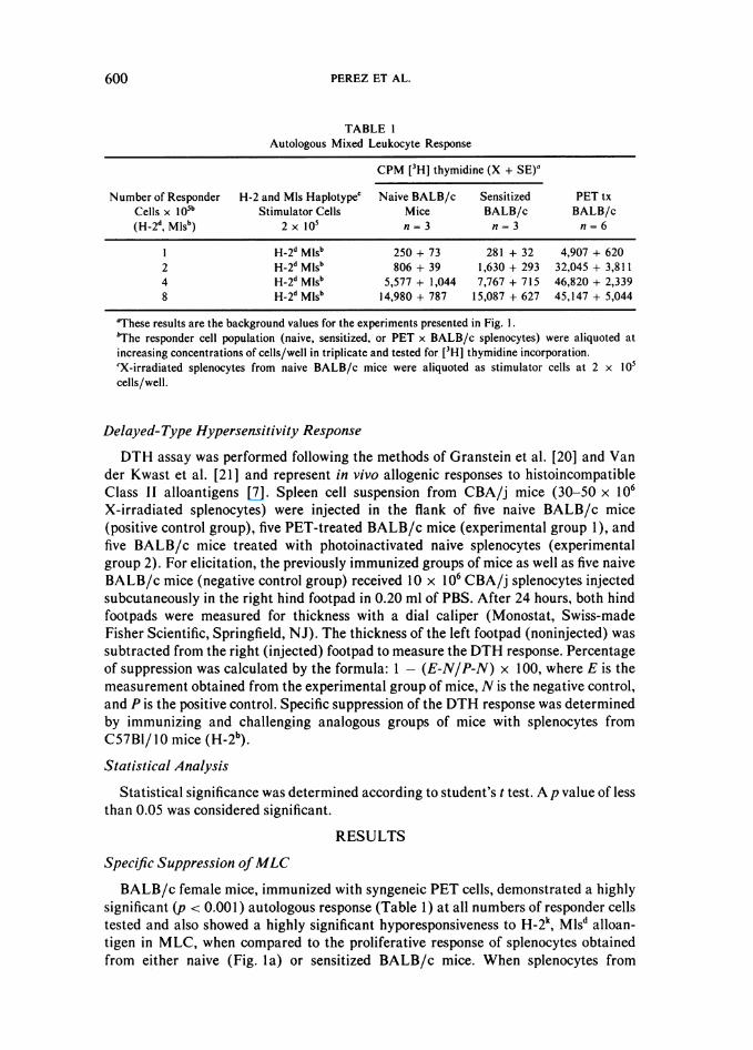
TABLE 1Autologous Mixed Leukocyte Response
CPM [3H] thymidine (X + SE)'
Number of Responder H-2 and Mls Haplotypec Naive BALB/c Sensitized PET txCells x 105b Stimulator Cells Mice BALB/c BALB/c(H2d, Mlsb) 2 x 10 n = 3 n = 3 n = 6
I HH2d Mlsb 250 + 73 281 + 32 4,907 + 6202 H-2d Mlsb 806 + 39 1,630 + 293 32,045 + 3,8114 H-2d Mlsb 5,577 + 1,044 7,767 + 715 46,820 + 2,3398 H-2d Mlsb 14,980 + 787 15,087 + 627 45,147 + 5,044
aThese results are the background values for the experiments presented in Fig. 1.'The responder cell population (naive, sensitized, or PET x BALB/c splenocytes) were aliquoted atincreasing concentrations of cells/well in triplicate and tested for [3H] thymidine incorporation.cX-irradiated splenocytes from naive BALB/c mice were aliquoted as stimulator cells at 2 x 10⁵cells/well.
Delayed-Type Hypersensitivity Response
DTH assay was performed following the methods of Granstein et al. [20] and Vander Kwast et al. [21] and represent in vivo allogenic responses to histoincompatibleClass II alloantigens [7]. Spleen cell suspension from CBA/j mice (30-50 x 106X-irradiated splenocytes) were injected in the flank of five naive BALB/c mice(positive control group), five PET-treated BALB/c mice (experimental group 1), andfive BALB/c mice treated with photoinactivated naive splenocytes (experimentalgroup 2). For elicitation, the previously immunized groups of mice as well as five naiveBALB/c mice (negative control group) received 10 x 106 CBA/j splenocytes injectedsubcutaneously in the right hind footpad in 0.20 ml of PBS. After 24 hours, both hindfootpads were measured for thickness with a dial caliper (Monostat, Swiss-madeFisher Scientific, Springfield, NJ). The thickness of the left footpad (noninjected) wassubtracted from the right (injected) footpad to measure the DTH response. Percentageof suppression was calculated by the formula: 1 - (E-N/P-N) x 100, where E is themeasurement obtained from the experimental group of mice, N is the negative control,and P is the positive control. Specific suppression of the DTH response was determinedby immunizing and challenging analogous groups of mice with splenocytes fromC57BI/10 mice (H-2b).
Statistical Analysis
Statistical significance was determined according to student's t test. A p value of lessthan 0.05 was considered significant.
RESULTS
Specific Suppression ofMLC
BALB/c female mice, immunized with syngeneic PET cells, demonstrated a highlysignificant (p < 0.001) autologous response (Table 1) at all numbers of responder cellstested and also showed a highly significant hyporesponsiveness to H-2k, Mlsd alloan-tigen in MLC, when compared to the proliferative response of splenocytes obtainedfrom either naive (Fig. la) or sensitized BALB/c mice. When splenocytes from
600
INHIBITION OF ANTISKIN ALLOGRAFT IMMUNITY
g ~ ~~~~*sEmsinZED | 0
O~~~~~NAIVE4
o o
o 4 a a 4 8
222552522 22 TSMZED5 . 5. 'd. t022 1n5
222822 OF DS5POSOC CEUSs 0w XOcUa±wOFRsPoe
6 NIV + SEDSZED NADV
N PE TETTED
4 2 ~~ 3/
NUMBER~ ~ ~ NU8EOFRESPONERCELLS X0 1U0 O5 SOMCEL
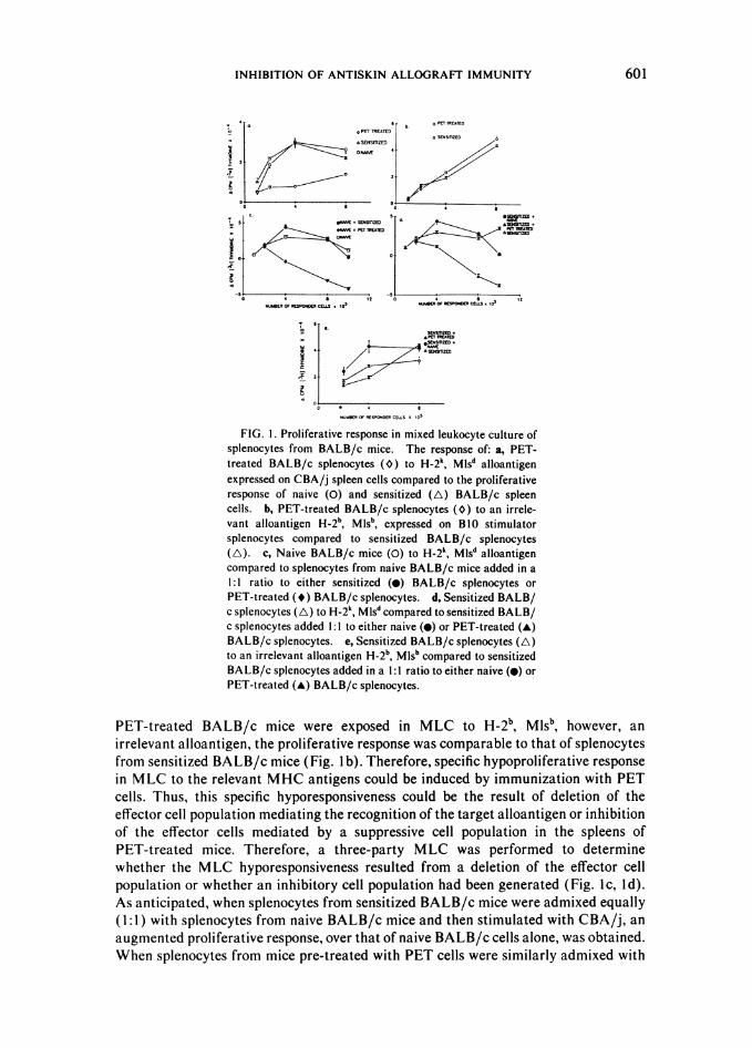
FIG. 1. Proliferative response in mixed leukocyte culture ofsplenocytes from BALB/c mice. The response of: a, PET-treated BALB/c splenocytes (O~) to H-2k, Mlsd alloantigenexpressed on CBA/j spleen cells compared to the proliferativeresponse of naive (0) and sensitized (A) BALB/c spleencells. h, PET-treated BALB/c splenocytes (O) to an irrele-vant alloantigen H-2b, Mlsb, expressed on BIO stimulatorsplenocytes compared to sensitized BALB/c splenocytes(A). c, Naive BALB/c mice (0) to H-2k, Mlsd alloantigencompared to splenocytes from naive BALB/c mice added in a1:1 ratio to either sensitized (-) BALB/c splenocytes orPET-treated (*) BALB/c splenocytes. d, Sensitized BALB/c splenocytes (A) to H-2k, Mlsd compared to sensitized BALB/c splenocytes added 1:1 to either naive (-) or PET-treated (A)BALB/c splenocytes. e, Sensitized BALB/c splenocytes (A)to an irrelevant alloantigen H-2b, Mlsb compared to sensitizedBALB/c splenocytes added in a 1:1 ratio to either naive (@) orPET-treated (-) BALB/c splenocytes.
PET-treated BALB/c mice were exposed in MLC to H-2b, Mlsb, however, anirrelevant alloantigen, the proliferative response was comparable to that of splenocytesfrom sensitized BALB/c mice (Fig. 1 b). Therefore, specific hypoproliferative responsein MLC to the relevant MHC antigens could be induced by immunization with PETcells. Thus, this specific hyporesponsiveness could be the result of deletion of theeffector cell population mediating the recognition of the target alloantigen or inhibitionof the effector cells mediated by a suppressive cell population in the spleens ofPET-treated mice. Therefore, a three-party MLC was performed to determinewhether the MLC hyporesponsiveness resulted from a deletion of the effector cellpopulation or whether an inhibitory cell population had been generated (Fig. ic, id).As anticipated, when splenocytes from sensitized BALB/c mice were admixed equally(1:1 ) with splenocytes from naive BALB/c mice and then stimulated with CBA/j, anaugmented proliferative response, over that of naive BALB/c cells alone, was obtained.When splenocytes from mice pre-treated with PET cells were similarly admixed with
601
PEREZ ET AL.
w(' 80
0.0 NAIVEU SENSIlIZED
,.&EE60 PETTREATEDIn
a.40
0.
ui 20
w
10:1 20:1 40:1 80:1
EFFECTOR TO TARGET RATIO
cn 80b. NAIVE
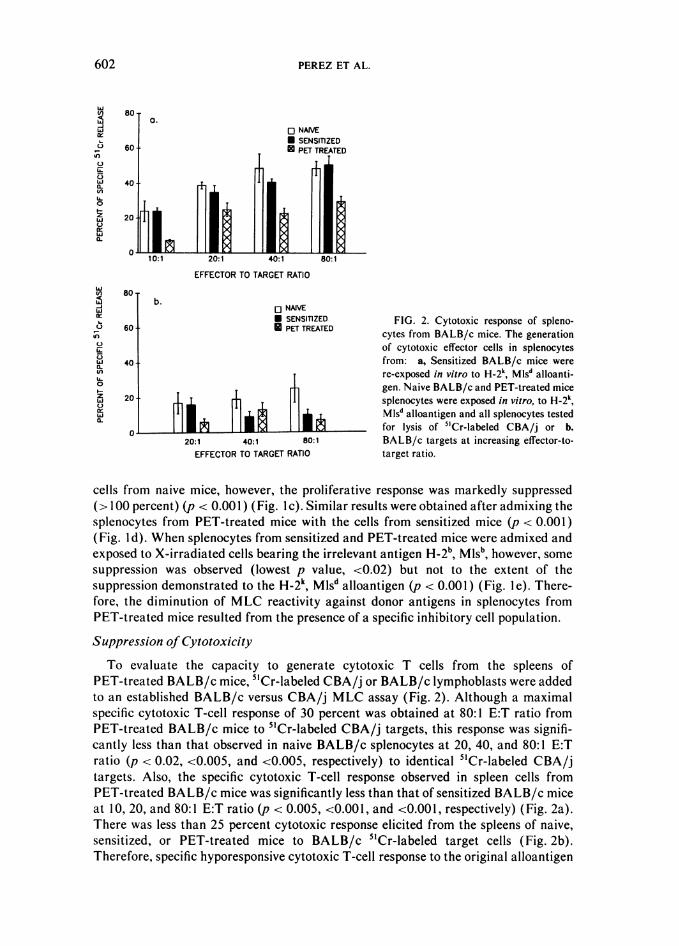
U SENSITIZED FIG. 2. Cytotoxic response of spleno-O 60 0 PET TREATED cytes from BALB/c mice. The generation
of cytotoxic effector cells in splenocytes
Cx 40 from: a, Sensitized BALB/c mice werere-exposed in vitro to H-2k, Mlsd alloanti-
2 gen. Naive BALB/c and PET-treated miceZ 20 +tT i i .splenocytes were exposed in vitro, to H-2k,
III. ^ i ^ ,$, Mlsd alloantigen and all splenocytes tested
0 __ *__ ___ __ *__ _ for lysis of "Cr-labeled CBA/j or b.20:1 40:1 80:1 BALB/c targets at increasing effector-to-
EFFECTOR TO TARGET RATIO target ratio.
cells from naive mice, however, the proliferative response was markedly suppressed(>100 percent) (p < 0.001 ) (Fig. Ic). Similar results were obtained after admixing thesplenocytes from PET-treated mice with the cells from sensitized mice (p < 0.001)(Fig. Id). When splenocytes from sensitized and PET-treated mice were admixed andexposed to X-irradiated cells bearing the irrelevant antigen H-2b, Mlsb, however, somesuppression was observed (lowest p value, <0.02) but not to the extent of thesuppression demonstrated to the H-2k, Mlsd alloantigen (p < 0.001) (Fig. I e). There-fore, the diminution of MLC reactivity against donor antigens in splenocytes fromPET-treated mice resulted from the presence of a specific inhibitory cell population.
Suppression of Cytotoxicity
To evaluate the capacity to generate cytotoxic T cells from the spleens ofPET-treated BALB/c mice, 5'Cr-labeled CBA/j or BALB/c lymphoblasts were addedto an established BALB/c versus CBA/j MLC assay (Fig. 2). Although a maximalspecific cytotoxic T-cell response of 30 percent was obtained at 80:1 E:T ratio fromPET-treated BALB/c mice to 5'Cr-labeled CBA/j targets, this response was signifi-cantly less than that observed in naive BALB/c splenocytes at 20, 40, and 80:1 E:Tratio (p < 0.02, <0.005, and <0.005, respectively) to identical 5'Cr-labeled CBA/jtargets. Also, the specific cytotoxic T-cell response observed in spleen cells fromPET-treated BALB/c mice was significantly less than that of sensitized BALB/c miceat 10, 20, and 80:1 E:T ratio (p < 0.005, <0.001, and <0.001, respectively) (Fig. 2a).There was less than 25 percent cytotoxic response elicited from the spleens of naive,sensitized, or PET-treated mice to BALB/c 5'Cr-labeled target cells (Fig. 2b).Therefore, specific hyporesponsive cytotoxic T-cell response to the original alloantigen
602
INHIBITION OF ANTISKIN ALLOGRAFT IMMUNITY 603
o a. ~~~~~~~~NAIVEi NAIVE + PET TREATED
o 80:152
40:1 45 o
S 20:1 21
10:1 59
0 20 40 60 80 100
PERCENT OF SPECIFIC 51Cr RELEASE
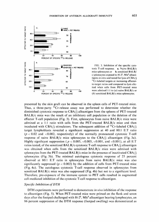
o b.<: g SENSIlIZED FIG. 3. Inhibition of the specific cyto-
O 80:1 SENSIliZED 4-PET TREATED toxic T-cell response. a. Naive BALB/ct) 80:1. 80 S mice splenocytes or b. sensitized BALB/40:1 C splenocytes exposed to H-2k, MIsd alloan-
o7- tigens in vitro and tested for lysis of CBA/j& 20:1 63 5'Cr-labeled targets at increasing effector-Li to-target ratios and compared to lysis elic-
10:1 50 ited when cells from PET-treated mice
0 280 4§0 680 8@0 100 were admixed 1:1 to (a) naive BALB/c orPERCENT OF SPECIFIC 51Cr RELEASE (b) sensitized BALB/c mice splenocytes.
presented by the skin graft can be observed in the spleen cells of PET-treated mice.Thus, a three-party 5'Cr-release assay was performed to determine whether thediminished cytotoxic response to CBA/j alloantigen from the spleens of PET-treatedBALB/c mice was the result of an inhibitory cell population or the deletion of theeffector T-cell population (Fig. 3). First, splenocytes from naive BALB/c mice wereadmixed at a 1:1 ratio with cells from the PET-treated BALB/c mice and thenincubated with CBA/j stimulators. The subsequent addition of 5'Cr-labeled CBA/jtarget lymphoblasts revealed a significant suppression at 40 and 80:1 E:T ratio(p < 0.02 and <0.002, respectively) of the normally pronounced cytotoxic T-cellresponse of naive BALB/c mice splenocytes to the CBA/j alloantigen (Fig. 3a).Highly significant suppression ( p < 0.005, <0.005, <0.001, and <0.001), at all E:Tratios tested, of the sensitized BALB/c cytotoxic T-cell response to CBA/j alloantigenwas obtained when cells from the sensitized BALB/c mice were admixed withsplenocytes from the PET-treated BALB/c mice in the presence of inactivated CBA/jsplenocytes (Fig. 3b). The minimal autologous cytotoxic response of 25 percentobserved at 80:1 E:T ratio in splenocytes from naive BALB/c mice was alsosignificantly suppressed (p < 0.002) by the addition of cells from PET-treated mice(Fig. 4a). The autologous cytotoxic T-cell response observed in splenocytes fromsensitized BALB/c mice was also suppressed (Fig. 4b) but not to a significant level.Therefore, pre-exposure of the immune system to PET cells resulted in augmentedcell-mediated inhibition of the cytotoxic T-cell response to alloantigen.
Specific Inhibition ofDTH
DTH experiments were performed to demonstrate in vivo inhibition of the responseto alloantigen (Fig. 5). When PET-treated mice were primed on the flank and sevendays after the footpad challenged with H-2k, Mlsd alloantigen bearing lymphocytes, an86 percent suppression of the DTH response (footpad swelling) was demonstrated as
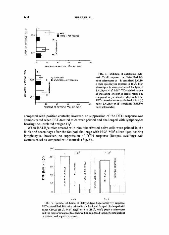
PEREZ ET AL.
a.
o NAIVE* NAIVE + PET TREATED
76
c
umLn
C
-448
PERCENT OF SPECIFIC 51Cr RELEASE
b.
lUo SENSITIZED
0 * SENSlTIZED + PET TREATED
c
0 20 40 60 80PERCENT OF SPECIFIC 51Cr RELEASE
FIG. 4. Inhibition of autologous cyto-toxic T-cell response. a. Naive BALB/cmice splenocytes or b. sensitized BALB/c mice splenocytes exposed to H-2k, Mlsdalloantigen in vitro and tested for lysis ofBALB/c (H-2d, Mlsb) 5'Cr-labeled targetsat increasing effector-to-target ratios andcompared to lysis elicited when cells fromPET-treated mice were admixed 1:1 to (a)
100 naive BALB/c or (b) sensitized BALB/cmice splenocytes.
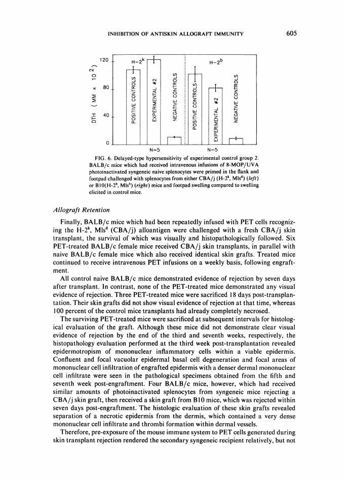
compared with positive controls; however, no suppression of the DTH response wasdemonstrated when PET-treated mice were primed and challenged with lymphocytesbearing the unrelated antigen H2b.When BALB/c mice treated with photoinactivated naive cells were primed in the
flank and seven days after the footpad challenge with H-2k, Mlsd alloantigen-bearinglymphocytes, however, no suppression of DTH response (footpad swelling) wasdemonstrated as compared with controls (Fig. 6).
0
x
-
a
H~k H~b80~ ~~~~-
80~~~~~~~~~~~0~~~~~~~
60~~~~~-iz0w u~~~~~~~~
z z40 0 0
20 cr- zi
N=5 N=5
FIG. 5. Specific inhibition of delayed-type hypersensitivity response.PET-treated BALB/c mice primed in the flank and footpad challenged witheither CBA/j (H-2k, MISd) (left) or B1O (H-2b, Mlsb) (right) splenocytesand the measurements of footpad swelling compared to the swelling elicitedin positive and negative controls.
604
0
Er
lY
LoId
LU
80:1
20:1
0
x
Lo0
0
LUIUL.LUI
80:1
20:1
E. 50_50
LA
INHIBITION OF ANTISKIN ALLOGRAFT IMMUNITY 605
120
(N0
x 80
I 40
0
0
N=5 N=5
FIG. 6. Delayed-type hypersensitivity of experimental control group 2.BALB/c mice which had received intravenous infusions of 8-MOP/UVAphotoinactivated syngeneic naive splenocytes were primed in the flank andfootpad challenged with splenocytes from either CBA/j (H-2k, Mlsd) (left)or BI0(H-2b, Mlsb) (right) mice and footpad swelling compared to swellingelicited in control mice.
Allograft Retention
Finally, BALB/c mice which had been repeatedly infused with PET cells recogniz-ing the H-2k, Mlsd (CBA/j) alloantigen were challenged with a fresh CBA/j skintransplant, the survival of which was visually and histopathologically followed. SixPET-treated BALB/c female mice received CBA/j skin transplants, in parallel withnaive BALB/c female mice which also received identical skin grafts. Treated micecontinued to receive intravenous PET infusions on a weekly basis, following engraft-ment.
All control naive BALB/c mice demonstrated evidence of rejection by seven daysafter transplant. In contrast, none of the PET-treated mice demonstrated any visualevidence of rejection. Three PET-treated mice were sacrificed 18 days post-transplan-tation. Their skin grafts did not show visual evidence of rejection at that time, whereas100 percent of the control mice transplants had already completely necrosed.The surviving PET-treated mice were sacrificed at subsequent intervals for histolog-
ical evaluation of the graft. Although these mice did not demonstrate clear visualevidence of rejection by the end of the third and seventh weeks, respectively, thehistopathology evaluation performed at the third week post-transplantation revealedepidermotropism of mononuclear inflammatory cells within a viable epidermis.Confluent and focal vacuolar epidermal basal cell degeneration and focal areas ofmononuclear cell infiltration of engrafted epidermis with a denser dermal mononuclearcell infiltrate were seen in the pathological specimens obtained from the fifth andseventh week post-engraftment. Four BALB/c mice, however, which had receivedsimilar amounts of photoinactivated splenocytes from syngeneic mice rejecting a
CBA/j skin graft, then received a skin graft from B1O mice, which was rejected withinseven days post-engraftment. The histologic evaluation of these skin grafts revealedseparation of a necrotic epidermis from the dermis, which contained a very densemononuclear cell infiltrate and thrombi formation within dermal vessels.
Therefore, pre-exposure of the mouse immune system to PET cells generated duringskin transplant rejection rendered the secondary syngeneic recipient relatively, but not
H~k H~b
H2 H H
Lu 0 4&
o z z L0= Liu C
ui ui~~~~~~~L
a- J1
PEREZ ET AL.
necessarily absolutely, tolerant to the alloantigen carried by the transplanted donorused to generate the immunizing effector cell population. In spite of these in vivolymphoid infiltrates suggesting a cellular response to the graft, substantial suppressionofMLC, CTL, and DTH was demonstrated, and prolongation of specific allograftsurvival was observed.
DISCUSSION
urine skin transplantation across H-2 barriers results in transplant rejectionprincipally mediated by activated T lymphocytes of the recipient [5], which have beenidentified as LytI +2+ [15] and L3T4+ positive [22].
In our study, CBA/j mice (H-2k, Mlsd) were selected as skin transplant donorsbecause of their disparity in the H-2 locus with BALB/c (H-2d, Misb) and theirstimulatory phenotype in the Mls locus, theoretically maximizing the expansion ofeffector cells. The Mls locus is of interest because it is the only known genetic locusother than the MHC in which strong proliferative response is observed betweenunprimed individuals displaying disparity on it [23]. The simplest view of the Mlssystem at present is that it is composed of a silent allele (Mlsb) and two independentstimulatory alleles (Mls-a and c) [24] and the co-expression of both stimulatorydeterminants (Mlsa+c=d) results in the Mlsd phenotype. By transplanting CBA/j skinonto BALB/c mice, the allografted mice should develop Tc effector cells whichrecognize Class I antigens and Tdth lymphocytes, possibly of the Thl [25] helper cellswhich recognized ClassII antigens, and possibly, L3T4+ cells responding to theMls-modified products of the MHC [26]. We appear to have inhibited this responsesignificantly with our PET immunization protocol.Our data demonstrate that this novel immunization protocol leads to the develop-
ment of significantly diminished responsiveness to alloantigen and that this inhibitionreflects the presence of cells capable of inhibiting the specific response to H-2k, Mlsdalloantigen in MLC, CTL, and DTH reactions. Moreover, a substantial increase in thelength of skin graft survival was obtained. Our findings extend those reported in othersystems by various investigators [27-29]. Rohowsky and Suciu-Foca [27] have shownthat idiotype-like receptors for alloantigens on autologous T lymphoblasts stimulatethe autologous MLC (AMLC) response in resting lymphocytes. In our studies, asubstantial increase in the AMLC response has been observed in the splenocytes ofPET-treated animals (Table 1). Dorsch and Roser [28] demonstrated a rapidlyrecirculating suppressor T-cell population in animals rendered tolerant to alloantigenby transfusion of allogeneic bone marrow cells during the neonatal period of life. Thesecells are capable of adoptively transferring tolerance, in irradiated syngeneic recip-ients, to skin transplants bearing the alloantigens to which the original tolerance wasinduced, without interfering with the rejection of an unrelated skin graft. In oursystem, we have demonstrated the presence of a cell population capable of specificallyameliorating the response in MLC, CTL, and DTH to the antigen used to generate theeffector cell population, while sparing the response to an irrelevant antigen. Bellgrau etal. [29] induced tolerance to parental lymphocytes, in F, rats, by immunizing themwith lymphocytes bearing the MHC receptors for specific parental strains of lympho-cytes. In an analogous fashion, we induced hyporesponsiveness to alloantigen byimmunizing syngeneic mice with splenocytes containing cells bearing the MHCreceptor for a specific histoincompatible antigen carried by the skin graft.
In this report, we describe a method of inducing tolerance that may be practical evenin a clinical setting. In our studies, the host selects the appropriate immunoreactive cell
606
INHIBITION OF ANTISKIN ALLOGRAFT IMMUNITY 607
type from a mixed splenocyte pool which includes an expanded population of effectorBALB/c anti CBA/j lymphocytes. These anti H-2k, MISd lymphocytes would be thecells most likely to induce a response in the secondary recipient naive BALB/c mice, ona mere numerical basis. Moreover, Ben-Nun and Cohen [30] have demonstrated thatthe induction of a suppressive clonotypic response is more efficiently attained if theEAE-inducive T-cell clones used for vaccination have been activated in vitro with theantigen and attenuated either with X-irradiation or mytomicin C prior to reinfusion.If, however, viable EAE-inducing T-cell clones are intravenously infused, acute onsetparalysis characteristic of EAE develops in the recipient animals. In our protocol, wehave used in vivo activation by skin grafting to expand effector cells and use these cellsobtained at the peak of rejection for immunization. The anti-H-2k, Mlsd T-cell-specificcontribution to these results may be better ascertained after we have cloned, photoinac-tivated, and infused them (either alone or in varying ratios with bystander spleno-cytes). Under these conditions, it will also be of interest to determine whethernonspecific bystander splenocytes exert either an adjuvant or interference effect. Thesestudies are in progress.The identity of these cells remains to be established. It will be necessary to determine
whether they are Ly2+ suppressor T cells [31] or Ly2+ anti-receptor anti-MHCcytotoxic T cells described by Kosmatopoulos et al. [32] or the CD8+ cells whichspecifically respond to determinants on the disease-mediating T-cell clones [33].
In our system, photoinactivation with 8-MOP might have been so effective inmaximizing the development of a specific inhibitory response to alloantigen because itreflects the ability to control two variables (drug dose and UVA energy), which limitthe chemical reaction to microseconds [34], and thus makes possible meticulouscontrol of the dose-response curve of cellular inactivation to a level not previouslyattainable. Significantly, 8-MOP-UVA pre-treatment of cloned anti-basic myelinprotein T cells [35] was more effective than X-irradiation of the same cells forvaccinating rats against the induction of EAE. Second, our studies on the suppressionof DTH response to sheep red blood cells [36] have demonstrated that 8-MOP-UVAtreatment of naive population of splenocytes is ineffective in inducing suppression,indicating that the presence of expanded population(s) of specific effector T cells arenecessary. Third, Laskin et al. [37] have identified specific, saturable, high-affinitybinding sites for 8-MOP on mice and human cell lines which might be a cell surfacebinding site that becomes covalently modified by exposure to UVA-modifying cellularantigenicity. It must be emphasized, however, that 8-MOP-UVA pre-treatment ofeffector cells may be merely one means of inducing the observed results.
In summary, we conclude that it is possible to induce a state of hyporeactivityagainst a specific set of MHC antigens. Syngeneic effector cells with specificity forthese antigens can be photoinactivated and used as immunogens to induce thishyporesponsive state. Since a method for photoinactivating effector T cells inanalogous fashion with that used here has been successful in the treatment of CTCLpatients [4] and is currently being tested in other T-cell-mediated disorders, ourprotocol may have potential application in the field of transplantation in humans.
REFERENCES
1. Holoshitz J, Frenkel A, Ben-Nun A, Cohen IR: Autoimmune encephalomyelitis (EAE) mediated orprevented by T lymphocyte lines directed against diverse antigenic determinants of myelin basic protein.Vaccination is determinant specific. J Immunol 131:2810-2813, 1983
2. Cohen IR: Autoimmunity: Physiologic and pernicious. Adv Inter Med 29:147-165, 1984
608 PEREZ ET AL.
3. Holoshitz J, Naparstek Y, Ben-Nun A, Cohen IR: Lines of T lymphocytes induce or vaccinate againstautoimmune arthritis. Science 219:56-58, 1983
4. Edelson RL, Berger C, Gasparro F, Jegasothy B, Heald PW, Wintroub B, Vonderheid E, Knobler R,Wolff K, Plewig G, McKiernan G, Christensen 1, Oster M, Honigsmann H, Wilford H, Kokoschka H,Rehle T, Perez M, Stingle G, Laroche L: Treatment of cutaneous T cell lymphoma by extracorporealphotochemotherapy. Preliminary results. N Engl J Med 316:297-303, 1987
5. Wood PJ, Streilein JW: Immunogenetic basis of acquired transplantation tolerance. Transplant37:223-226, 1984
6. Rosenberg AS, Mizuouchi T, Sharrow SO, Singer A: Phenotype, specificity and function of T cellsubsets and T cell interactions involved in skin allograft rejection. J Exp Med 165:1296-1315, 1987
7. Sprent J, Schaefer M: Properties of purified T cell subsets. In vitro responses to class I vs. class II H-2differences. J Exp Med 162:2068-2082, 1985
8. Buller RML, Holmes KL, Hugin A, Frederickson TH, Morse HC III: Induction of cytotoxic T-cellresponse in vivo in the absence of CD4 helper cells. Nature 328:77-79, 1987
9. Webb S, Sprent J: Downregulation of T cell responses by antibodies to the T cell receptor. J Exp Med165:584-589, 1987
10. Andersson LC, Binz H, Wigzell H: Specific unresponsiveness to transplantation antigens induced byautoimmunization with syngeneic, antigen specific T lymphoblasts. Nature 264:778-779, 1976
11. Mohagheghpour N, Damle DK, Takada S, Engleman EG: Generation of antigen receptor-specificsuppressor T cell clones in man. J Exp Med 164:950-955, 1986
12. Scott BR, Patak MA, Mohn GR: Molecular and genetic basis of furocoumarin reactions. Mutat Resear39:29-74, 1976
13. Berger CL, Canton C, Welsh J, Dervan P, Begley T, Grant S, Gasparro FP, Edelson RL: Comparison ofsynthetic psoralen derivatives and 8-MOP in the inhibition of lymphocyte proliferation. Ann NY AcadSci 453:80-90, 1985
14. Billigham RE, Medawar PB: The technique of free skin grafting in mammals. J Ext Biol 28:385-399,1951
15. Rock KL, Barnes MC, Germain RN, Benacerraf B: The role of Ia molecules in the activation of Tlymphocytes. Ia-restricted recognition of allo K/D antigens is required for Class I MHC-stimulatedmixed lymphocyte responses. J Immunol 130:457-462, 1983
16. Kruisbeek AM, Bridges S, Carmen J, Longo DL, Mond JJ: In vitro treatment of neonatal mice withanti-la antibodies interferes with the development of the class I, Class II, and Mls-reactive proliferatingT cell subset. J Immunol 134:3597-3604, 1985
17. Pawelec G: Allogeneically primed T cell clones in the analysis of lymphocyte stimulatory determinants.Human Immunology 8:239-247, 1983
18. Sprent J, Schaefer JM, Lo D, Korngold R: Properties of purified T cell subsets. In vivo responses to classI vs. class 11 H-2 differences. J Exp Med 163:998-1011, 1986
19. Buracoff SJ, Germain RN, Dorf ME, Benacerraf B: Inhibition of cell mediated cytolysis of trinitrophe-nyl-derivatized target cells by alloantisera directed to the products of the K and D loci of the H-2complex. Proc Natl Acad Sci USA 73:625-629, 1986
20. Granstein RD, Goulston C, Gaulton GN: Prolongation of murine skin allograft survival by immunologicmanipulation with anti-interleukin 2 receptor antibody. J Immunol 136:898-902, 1986
21. Van der Kwast TH, Bianchi TJ, Bril H, Benner R: Suppression of anti-graft immunity by preimmuniza-tion. Transplant 31:79-85, 1981
22. Mizouchi TS, Ono S, Malek TR, Singer A: Characterization of two distinct primary T cell populationsthat secrete IL-2 upon recognition of class I or class 11 major histocompatibility antigens. J Exp Med163:603-615, 1986
23. Janeway CA, Conrad PJ, Tite J, Jones B, Murphy DB: Efficiency of antigen presentation differs in micediffering at the Mls locus. Nature 306:80-82, 1983
24. Abe R, Ryan JJ, Hodes RJ: Mls is not a single gene allelic system. Different stimulatory Mlsdeterminants are the products of at least two non-allelic unlinked genes. J Exp Med 166:1150-1155,1987
25. Cher DJ, Mossman TR: Two types of murine helper T cell clone. Delayed-type hypersensitivity ismediated by THI clones. J Immunol 138:3688-3694, 1987
26. Kapler JW, Sfaerz W, White J, Manack PC: Self-tolerance eliminates T cells specific for Mls modifiedproducts of the major histocompatibility complex. Nature 332:35-40, 1988
27. Rohowsky C, Suciu-Foca N, Kung P, Tang TF, Reed E, King DW: Suppressor T cells in the autologousMLR with MLC-activated T lymphoblasts. Transplant Proceed XV:765-770, 1983
INHIBITION OF ANTISKIN ALLOGRAFT IMMUNITY 609
28. Dorsh S, Roser B: Recirculating suppressor T cells in transplantation tolerance. J Exp Med 145:1144-1157, 1977
29. Bellgrau D, Smilek D, Wilson DB: Induced tolerance in F1 rats to anti-major histocompatibilitycomplex receptors on parental T cells. Implication of self tolerance. J Exp Med 153:1660-1665, 1981
30. Ben-Nun A, Cohen IR: Vaccination against autoimmune encephalomyelitis (EAE): Attenuatedautoimmune T lymphocytes confer resistance to the induction of active EAE but not to EAE mediatedby the intact T lymphocyte line. Eur J Immunol 11:949-952, 1981
31. Sy MS, Dietz MH, Germain RN, Benacerraf B, Greene MI: Antigen and receptor driven regulatorymechanisms. IV. Idiotype bearing I-J + suppressor T cell factors (TsF) induce second order suppressorT cells (TsF2) which express anti-idiotypic receptors. J Exp Med 151:1183-1195, 1980
32. Kosmatopoulos K, Algara DS, Orbach-Arbouys S: Anti-receptor anti-MHC cytotoxic T lymphocytes:Their role in the resistance to graft vs host reaction. J Immunol 138:1038-1041, 1987
33. Sun D, Qin Y, Chluba J, Epplen JT, Wekerle H: Suppression of experimentally induced autoimmuneencephalomyelitis by cytolytic T-T cell interactions. Nature 322:843-845, 1988
34. Song PS, Tapley KJ Jr: Photochemistry and photobiology of psoralens. Photochem Photobiol 29:1177-1197, 1979
35. Khavari PA, Edelson RL, Lider 0, Gasparro FP, Weiner HL, Cohen IR: Specific vaccination againstphotoinactivated cloned T cells. Clin Research 36:662a, 1988
36. Laroche L, Perez MI, Edelson RL, Berger C: Induction of tolerance in mice treated with photoinacti-vated effector cells. Clin Research 35:698a, 1987
37. Laskin JD, Lee E, Yurkow EJ, Laskin DL, Gallo MA: A possible mechanism of psoralen phototoxicitynot involving direct interaction with DNA. Proc Natl Acad Sci USA 82:6158-6162, 1985
Related Documents