Increased Neural Activity of a Mushroom Body Neuron Subtype in the Brains of Forager Honeybees Taketoshi Kiya*, Takekazu Kunieda, Takeo Kubo* Department of Biological Sciences, Graduate School of Science, The University of Tokyo, Tokyo, Japan Honeybees organize a sophisticated society, and the workers transmit information about the location of food sources using a symbolic dance, known as ‘dance communication’. Recent studies indicate that workers integrate sensory information during foraging flight for dance communication. The neural mechanisms that account for this remarkable ability are, however, unknown. In the present study, we established a novel method to visualize neural activity in the honeybee brain using a novel immediate early gene, kakusei, as a marker of neural activity. The kakusei transcript was localized in the nuclei of brain neurons and did not encode an open reading frame, suggesting that it functions as a non-coding nuclear RNA. Using this method, we show that neural activity of a mushroom body neuron subtype, the small-type Kenyon cells, is prominently increased in the brains of dancer and forager honeybees. In contrast, the neural activity of the two mushroom body neuron subtypes, the small-and large-type Kenyon cells, is increased in the brains of re-orienting workers, which memorize their hive location during re-orienting flights. These findings demonstrate that the small-type Kenyon cell-preferential activity is associated with foraging behavior, suggesting its involvement in information integration during foraging flight, which is an essential basis for dance communication. Citation: Kiya T, Kunieda T, Kubo T (2007) Increased Neural Activity of a Mushroom Body Neuron Subtype in the Brains of Forager Honeybees. PLoS ONE 2(4): e371. doi:10.1371/journal.pone.0000371 INTRODUCTION A variety of animals, from nematode to human, show social behavior [1,2]. The social behaviors allow for individuals to create an entity greater than the sum of the individuals and provide the key to successful adaptation to the environment. One of the most striking features of the highly-ordered animal society is the ability to share information among individuals. In turn, higher commu- nicative ability is a fundamental basis that enables animals to maintain a more evolved society [3]. Honeybees (Apis mellifera L.) organize a highly-ordered society and have a sophisticated communicative ability known as the ‘dance communication’ [4–7]. Worker honeybees that find a rich food source return to the hive and might transmit the information on the location of the food source to their nestmates using a symbolic dance. The dance information is decoded into the spatial information of the food source by the other worker bees (followers) that follow the dancers [4–7]. During foraging flights, worker honeybees integrate the incoming sensory information: they estimate the distance of food sources based on the amount of optic flow they perceive, and direction based on the position of the sun [5,8,9], which are the essential bases for the expression of dance communication. Although there is a considerable amount of research concerning the sensory basis of these remarkable abilities [8–11], almost nothing is known about the underlying neural mechanisms. As a first step in elucidating the neural mechanisms of these remarkable abilities, it is important to identify active brain regions in dancing and foraging honeybees that might be involved in dance communication and/or information integration during foraging flight. Although methods to detect the expression of immediate early genes (IEGs) as markers of neural activity are widely used in vertebrates [12–15], neural IEGs have not yet been identified in insects. In the present study, we identified a novel IEG that can be used as a neural activity marker and found that the neural activity of a mushroom body (MB) neuron subtype is preferentially increased in foraging honeybees, suggesting its involvement in information integration during foraging flight. RESULTS A novel non-coding IEG, kakusei, can be used as a marker to visualize neural activity in the honeybee brain To identify IEGs, we used the differential display method to search for honeybee genes that are immediately induced in the brain by neural activity. To evoke strong neural activity in the brain, seizures were induced by awakening workers from ice-cold induced anesthesia, because some of the IEGs were identified by inducing seizures in the animals [16,17]. When the workers are awoken from anesthesia, they show seizure-like movement with their legs and body shaking. Using differential display screening of approximately 6500 bands, which were derived from mRNAs extracted from the brains of seizure-induced and non-treated bees, 49 candidate bands were identified. Among them, we selected nine candidates that showed a pronounced difference in band intensity between the seizure-induced and non-treated bees. After pre- Academic Editor: Martin Giurfa, Centre de Recherches su la Cognition Animale- Centre National de la Recherche Scientifique and Universite ´ Paul Sabatier, France Received August 21, 2006; Accepted March 26, 2007; Published April 18, 2007 Copyright: ß 2007 Kiya et al. This is an open-access article distributed under the terms of the Creative Commons Attribution License, which permits unrestricted use, distribution, and reproduction in any medium, provided the original author and source are credited. Funding: This work was supported by the Program for Promotion of Basic Research Activities for Innovative Bioscience (PROBRAIN). T. Kiya is the recipient of a Grant-in-Aid for Japan Society for the Promotion of Science (JSPS) Fellows. Competing Interests: This work was supported by the Program for Promotion of Basic Research Activities for Innovative Bioscience (PROBRAIN). T. Kiya is the recipient of a Grant-in-Aid for Japan Society for the Promotion of Science (JSPS) Fellows. * To whom correspondence should be addressed. E-mail: [email protected]. jp (TK); [email protected] (TK) PLoS ONE | www.plosone.org 1 April 2007 | Issue 4 | e371

Welcome message from author
This document is posted to help you gain knowledge. Please leave a comment to let me know what you think about it! Share it to your friends and learn new things together.
Transcript
Increased Neural Activity of a Mushroom Body NeuronSubtype in the Brains of Forager HoneybeesTaketoshi Kiya*, Takekazu Kunieda, Takeo Kubo*
Department of Biological Sciences, Graduate School of Science, The University of Tokyo, Tokyo, Japan
Honeybees organize a sophisticated society, and the workers transmit information about the location of food sources usinga symbolic dance, known as ‘dance communication’. Recent studies indicate that workers integrate sensory information duringforaging flight for dance communication. The neural mechanisms that account for this remarkable ability are, however,unknown. In the present study, we established a novel method to visualize neural activity in the honeybee brain using a novelimmediate early gene, kakusei, as a marker of neural activity. The kakusei transcript was localized in the nuclei of brainneurons and did not encode an open reading frame, suggesting that it functions as a non-coding nuclear RNA. Using thismethod, we show that neural activity of a mushroom body neuron subtype, the small-type Kenyon cells, is prominentlyincreased in the brains of dancer and forager honeybees. In contrast, the neural activity of the two mushroom body neuronsubtypes, the small-and large-type Kenyon cells, is increased in the brains of re-orienting workers, which memorize their hivelocation during re-orienting flights. These findings demonstrate that the small-type Kenyon cell-preferential activity isassociated with foraging behavior, suggesting its involvement in information integration during foraging flight, which is anessential basis for dance communication.
Citation: Kiya T, Kunieda T, Kubo T (2007) Increased Neural Activity of a Mushroom Body Neuron Subtype in the Brains of Forager Honeybees. PLoSONE 2(4): e371. doi:10.1371/journal.pone.0000371
INTRODUCTIONA variety of animals, from nematode to human, show social
behavior [1,2]. The social behaviors allow for individuals to create
an entity greater than the sum of the individuals and provide the
key to successful adaptation to the environment. One of the most
striking features of the highly-ordered animal society is the ability
to share information among individuals. In turn, higher commu-
nicative ability is a fundamental basis that enables animals to
maintain a more evolved society [3].
Honeybees (Apis mellifera L.) organize a highly-ordered society
and have a sophisticated communicative ability known as the
‘dance communication’ [4–7]. Worker honeybees that find a rich
food source return to the hive and might transmit the information
on the location of the food source to their nestmates using a
symbolic dance. The dance information is decoded into the spatial
information of the food source by the other worker bees (followers)
that follow the dancers [4–7]. During foraging flights, worker
honeybees integrate the incoming sensory information: they
estimate the distance of food sources based on the amount of
optic flow they perceive, and direction based on the position of the
sun [5,8,9], which are the essential bases for the expression of
dance communication. Although there is a considerable amount of
research concerning the sensory basis of these remarkable abilities
[8–11], almost nothing is known about the underlying neural
mechanisms.
As a first step in elucidating the neural mechanisms of these
remarkable abilities, it is important to identify active brain regions
in dancing and foraging honeybees that might be involved in
dance communication and/or information integration during
foraging flight. Although methods to detect the expression of
immediate early genes (IEGs) as markers of neural activity are
widely used in vertebrates [12–15], neural IEGs have not yet been
identified in insects. In the present study, we identified a novel IEG
that can be used as a neural activity marker and found that the
neural activity of a mushroom body (MB) neuron subtype is
preferentially increased in foraging honeybees, suggesting its
involvement in information integration during foraging flight.
RESULTS
A novel non-coding IEG, kakusei, can be used as
a marker to visualize neural activity in the honeybee
brainTo identify IEGs, we used the differential display method to search
for honeybee genes that are immediately induced in the brain by
neural activity. To evoke strong neural activity in the brain,
seizures were induced by awakening workers from ice-cold
induced anesthesia, because some of the IEGs were identified by
inducing seizures in the animals [16,17]. When the workers are
awoken from anesthesia, they show seizure-like movement with
their legs and body shaking. Using differential display screening of
approximately 6500 bands, which were derived from mRNAs
extracted from the brains of seizure-induced and non-treated bees,
49 candidate bands were identified. Among them, we selected nine
candidates that showed a pronounced difference in band intensity
between the seizure-induced and non-treated bees. After pre-
Academic Editor: Martin Giurfa, Centre de Recherches su la Cognition Animale-Centre National de la Recherche Scientifique and Universite Paul Sabatier, France
Received August 21, 2006; Accepted March 26, 2007; Published April 18, 2007
Copyright: � 2007 Kiya et al. This is an open-access article distributed under theterms of the Creative Commons Attribution License, which permits unrestricteduse, distribution, and reproduction in any medium, provided the original authorand source are credited.
Funding: This work was supported by the Program for Promotion of BasicResearch Activities for Innovative Bioscience (PROBRAIN). T. Kiya is the recipient ofa Grant-in-Aid for Japan Society for the Promotion of Science (JSPS) Fellows.
Competing Interests: This work was supported by the Program for Promotion ofBasic Research Activities for Innovative Bioscience (PROBRAIN). T. Kiya is therecipient of a Grant-in-Aid for Japan Society for the Promotion of Science (JSPS)Fellows.
* To whom correspondence should be addressed. E-mail: [email protected] (TK); [email protected] (TK)
PLoS ONE | www.plosone.org 1 April 2007 | Issue 4 | e371
liminary Reverse transcription-polymerase chain reaction (RT-
PCR) analysis of these candidates, we finally focused on a single
candidate that showed the most prominent and reproducible
seizure-induced transcript increase. As a result, we identified
a novel IEG that we named kakusei after the word ‘awakening’ in
Japanese (the whole sequence of kakusei was deposited as DDBJ
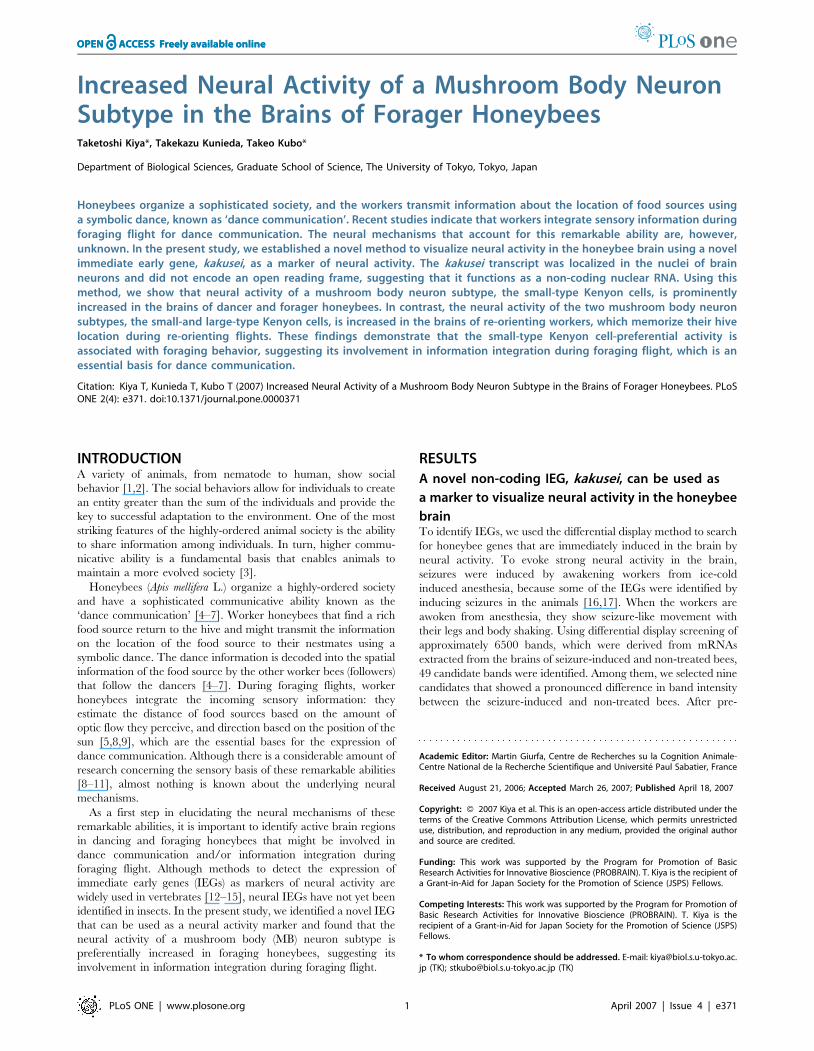
accession number AB252862). To examine the size of the kakusei
transcript, we performed Northern blot analysis using total RNA
isolated from whole brains of bees anesthetized with CO2 and bees
awakened from CO2-induced anesthesia. The results indicated
that the induced kakusei transcript was approximately 7 kb long
(Figure 1A). There was no significant open reading frame in any of
the three possible reading frames of the kakusei cDNA sequence,
suggesting that the kakusei transcript functions as a non-coding
RNA (Figure 1B). RT-PCR experiments and sequence analysis
also confirmed that the contig kakusei sequence is expressed as
continuous transcripts (Figure 1C).
Kakusei expression was transiently induced in the whole brain
after awakening the workers from anesthesia induced by either
CO2 [Figure 1D; Sz (seizure)-induced] or ice-cold (data not
shown). Real-time RT-PCR revealed that kakusei is expressed
predominantly in the brain, suggesting a brain-specific function
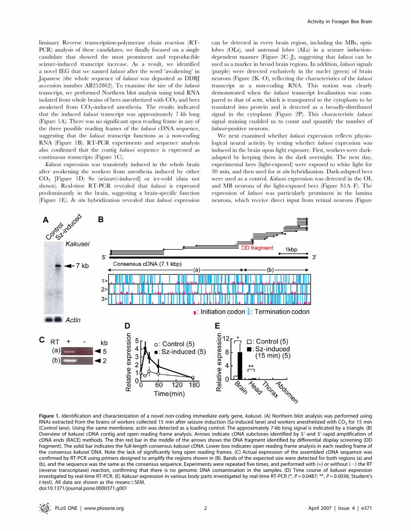
(Figure 1E). In situ hybridization revealed that kakusei expression
can be detected in every brain region, including the MBs, optic
lobes (OLs), and antennal lobes (ALs) in a seizure induction-
dependent manner (Figure 2C–J), suggesting that kakusei can be
used as a marker in broad brain regions. In addition, kakusei signals
(purple) were detected exclusively in the nuclei (green) of brain
neurons (Figure 2K–O), reflecting the characteristics of the kakusei
transcript as a non-coding RNA. This notion was clearly
demonstrated when the kakusei transcript localization was com-
pared to that of actin, which is transported to the cytoplasm to be
translated into protein and is detected as a broadly-distributed
signal in the cytoplasm (Figure 2P). This characteristic kakusei
signal staining enabled us to count and quantify the number of
kakusei-positive neurons.
We next examined whether kakusei expression reflects physio-
logical neural activity by testing whether kakusei expression was
induced in the brain upon light exposure. First, workers were dark-
adapted by keeping them in the dark overnight. The next day,
experimental bees (light-exposed) were exposed to white light for
30 min, and then used for in situ hybridization. Dark-adapted bees
were used as a control. Kakusei expression was detected in the OL
and MB neurons of the light-exposed bees (Figure S1A–F). The
expression of kakusei was particularly prominent in the lamina
neurons, which receive direct input from retinal neurons (Figure
Figure 1. Identification and characterization of a novel non-coding immediate early gene, kakusei. (A) Northern blot analysis was performed usingRNAs extracted from the brains of workers collected 15 min after seizure induction (Sz-induced lane) and workers anesthetized with CO2 for 15 min(Control lane). Using the same membrane, actin was detected as a loading control. The approximately 7-kb long signal is indicated by a triangle. (B)Overview of kakusei cDNA contig and open reading frame analysis. Arrows indicate cDNA subclones identified by 59-and 39-rapid amplification ofcDNA ends (RACE) methods. The thin red bar in the middle of the arrows shows the DNA fragment identified by differential display screening (DDfragment). The solid bar indicates the full-length consensus kakusei cDNA. Lower box indicates open reading frame analysis in each reading frame ofthe consensus kakusei DNA. Note the lack of significantly long open reading frames. (C) Actual expression of the assembled cDNA sequence wasconfirmed by RT-PCR using primers designed to amplify the regions shown in (B). Bands of the expected size were detected for both regions (a) and(b), and the sequence was the same as the consensus sequence. Experiments were repeated five times, and performed with (+) or without (2) the RT(reverse transcriptase) reaction, confirming that there is no genomic DNA contamination in the samples. (D) Time course of kakusei expressioninvestigated by real-time RT-PCR. (E) Kakusei expression in various body parts investigated by real-time RT-PCR (*, P = 0.0487; **, P = 0.0036; Student’st-test). All data are shown as the means6SEM.doi:10.1371/journal.pone.0000371.g001
Activity in Forager Bee Brain
PLoS ONE | www.plosone.org 2 April 2007 | Issue 4 | e371
S1C and D). In contrast, there was no strong kakusei expression in
the AL neurons (Figure S1G and H). These results indicate that
kakusei can be used as a marker to visualize physiological neural
activity in the honeybee brain.
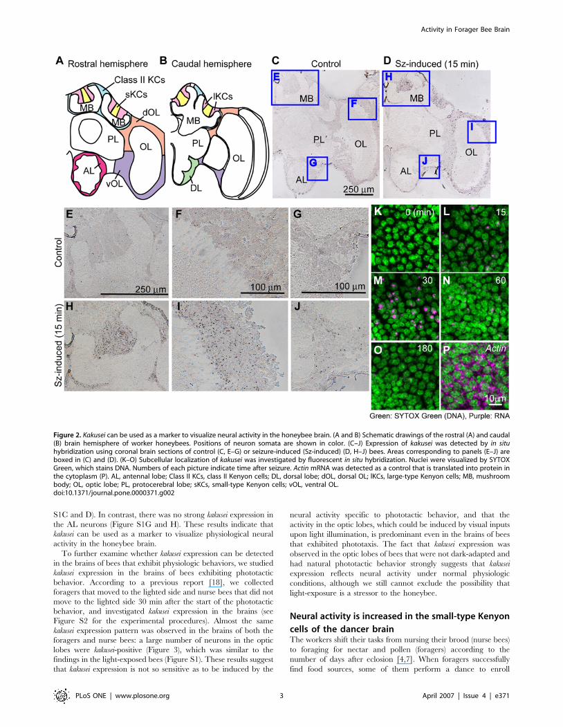
To further examine whether kakusei expression can be detected
in the brains of bees that exhibit physiologic behaviors, we studied
kakusei expression in the brains of bees exhibiting phototactic
behavior. According to a previous report [18], we collected
foragers that moved to the lighted side and nurse bees that did not
move to the lighted side 30 min after the start of the phototactic
behavior, and investigated kakusei expression in the brains (see
Figure S2 for the experimental procedures). Almost the same
kakusei expression pattern was observed in the brains of both the
foragers and nurse bees: a large number of neurons in the optic
lobes were kakusei-positive (Figure 3), which was similar to the
findings in the light-exposed bees (Figure S1). These results suggest
that kakusei expression is not so sensitive as to be induced by the
neural activity specific to phototactic behavior, and that the
activity in the optic lobes, which could be induced by visual inputs
upon light illumination, is predominant even in the brains of bees
that exhibited phototaxis. The fact that kakusei expression was
observed in the optic lobes of bees that were not dark-adapted and
had natural phototactic behavior strongly suggests that kakusei
expression reflects neural activity under normal physiologic
conditions, although we still cannot exclude the possibility that
light-exposure is a stressor to the honeybee.
Neural activity is increased in the small-type Kenyon
cells of the dancer brainThe workers shift their tasks from nursing their brood (nurse bees)
to foraging for nectar and pollen (foragers) according to the
number of days after eclosion [4,7]. When foragers successfully
find food sources, some of them perform a dance to enroll
Figure 2. Kakusei can be used as a marker to visualize neural activity in the honeybee brain. (A and B) Schematic drawings of the rostral (A) and caudal(B) brain hemisphere of worker honeybees. Positions of neuron somata are shown in color. (C–J) Expression of kakusei was detected by in situhybridization using coronal brain sections of control (C, E–G) or seizure-induced (Sz-induced) (D, H–J) bees. Areas corresponding to panels (E–J) areboxed in (C) and (D). (K–O) Subcellular localization of kakusei was investigated by fluorescent in situ hybridization. Nuclei were visualized by SYTOXGreen, which stains DNA. Numbers of each picture indicate time after seizure. Actin mRNA was detected as a control that is translated into protein inthe cytoplasm (P). AL, antennal lobe; Class II KCs, class II Kenyon cells; DL, dorsal lobe; dOL, dorsal OL; lKCs, large-type Kenyon cells; MB, mushroombody; OL, optic lobe; PL, protocerebral lobe; sKCs, small-type Kenyon cells; vOL, ventral OL.doi:10.1371/journal.pone.0000371.g002
Activity in Forager Bee Brain
PLoS ONE | www.plosone.org 3 April 2007 | Issue 4 | e371
followers to forage [7]. Therefore, we next examined kakusei
expression in the brains of the dancers, followers, and nurse bees to
identify the brain regions involved in dance communication
(Figure 4). The individual workers were caught immediately after
confirming their behaviors in the observation hives, and used for in
situ hybridization. The bees caught from the observation hives
were immediately anesthetized by ice-cold water and kept on ice
until use for in situ hybridization to maintain the current state of
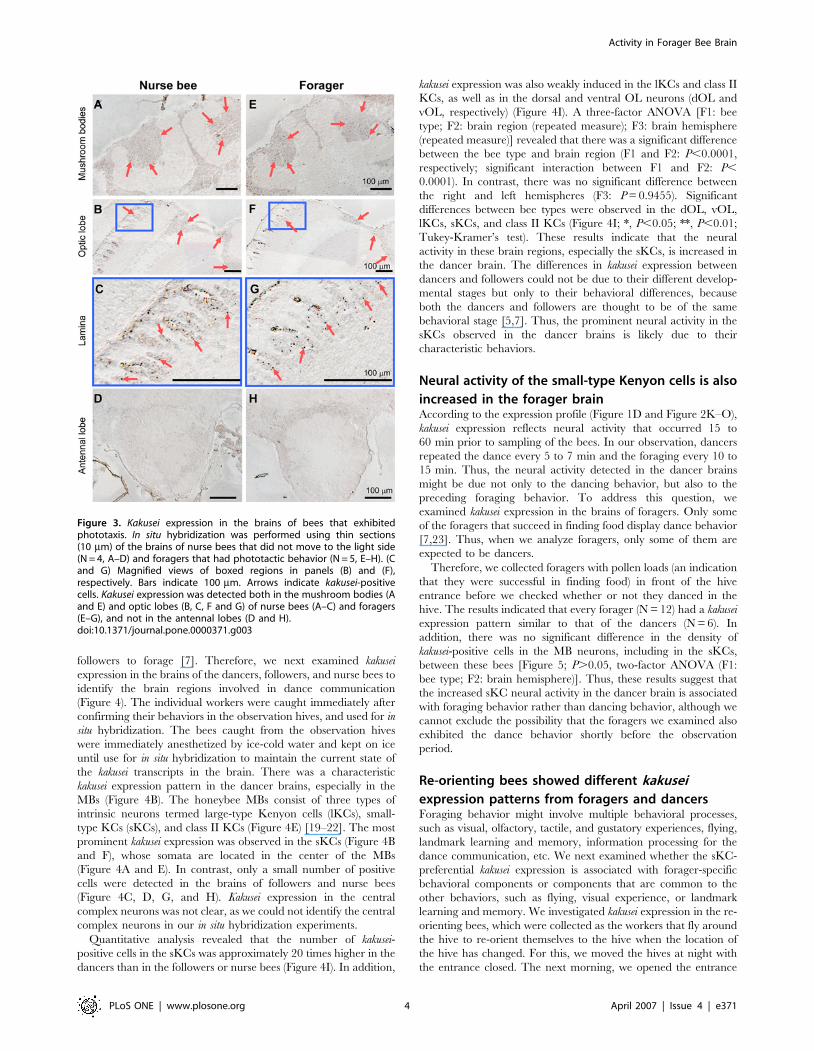
the kakusei transcripts in the brain. There was a characteristic
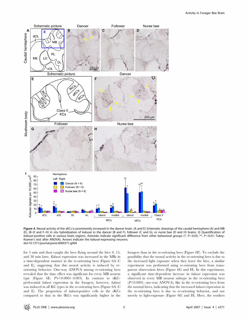
kakusei expression pattern in the dancer brains, especially in the
MBs (Figure 4B). The honeybee MBs consist of three types of
intrinsic neurons termed large-type Kenyon cells (lKCs), small-
type KCs (sKCs), and class II KCs (Figure 4E) [19–22]. The most
prominent kakusei expression was observed in the sKCs (Figure 4B
and F), whose somata are located in the center of the MBs
(Figure 4A and E). In contrast, only a small number of positive
cells were detected in the brains of followers and nurse bees
(Figure 4C, D, G, and H). Kakusei expression in the central
complex neurons was not clear, as we could not identify the central
complex neurons in our in situ hybridization experiments.
Quantitative analysis revealed that the number of kakusei-
positive cells in the sKCs was approximately 20 times higher in the
dancers than in the followers or nurse bees (Figure 4I). In addition,
kakusei expression was also weakly induced in the lKCs and class II
KCs, as well as in the dorsal and ventral OL neurons (dOL and
vOL, respectively) (Figure 4I). A three-factor ANOVA [F1: bee
type; F2: brain region (repeated measure); F3: brain hemisphere
(repeated measure)] revealed that there was a significant difference
between the bee type and brain region (F1 and F2: P,0.0001,
respectively; significant interaction between F1 and F2: P,
0.0001). In contrast, there was no significant difference between
the right and left hemispheres (F3: P = 0.9455). Significant
differences between bee types were observed in the dOL, vOL,
lKCs, sKCs, and class II KCs (Figure 4I; *, P,0.05; **, P,0.01;
Tukey-Kramer’s test). These results indicate that the neural
activity in these brain regions, especially the sKCs, is increased in
the dancer brain. The differences in kakusei expression between
dancers and followers could not be due to their different develop-
mental stages but only to their behavioral differences, because
both the dancers and followers are thought to be of the same
behavioral stage [5,7]. Thus, the prominent neural activity in the
sKCs observed in the dancer brains is likely due to their
characteristic behaviors.
Neural activity of the small-type Kenyon cells is also
increased in the forager brainAccording to the expression profile (Figure 1D and Figure 2K–O),
kakusei expression reflects neural activity that occurred 15 to
60 min prior to sampling of the bees. In our observation, dancers
repeated the dance every 5 to 7 min and the foraging every 10 to
15 min. Thus, the neural activity detected in the dancer brains
might be due not only to the dancing behavior, but also to the
preceding foraging behavior. To address this question, we
examined kakusei expression in the brains of foragers. Only some
of the foragers that succeed in finding food display dance behavior
[7,23]. Thus, when we analyze foragers, only some of them are
expected to be dancers.
Therefore, we collected foragers with pollen loads (an indication
that they were successful in finding food) in front of the hive
entrance before we checked whether or not they danced in the
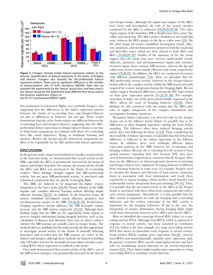
hive. The results indicated that every forager (N = 12) had a kakusei
expression pattern similar to that of the dancers (N = 6). In
addition, there was no significant difference in the density of
kakusei-positive cells in the MB neurons, including in the sKCs,
between these bees [Figure 5; P.0.05, two-factor ANOVA (F1:
bee type; F2: brain hemisphere)]. Thus, these results suggest that
the increased sKC neural activity in the dancer brain is associated
with foraging behavior rather than dancing behavior, although we
cannot exclude the possibility that the foragers we examined also
exhibited the dance behavior shortly before the observation
period.
Re-orienting bees showed different kakusei
expression patterns from foragers and dancersForaging behavior might involve multiple behavioral processes,
such as visual, olfactory, tactile, and gustatory experiences, flying,
landmark learning and memory, information processing for the
dance communication, etc. We next examined whether the sKC-
preferential kakusei expression is associated with forager-specific
behavioral components or components that are common to the
other behaviors, such as flying, visual experience, or landmark
learning and memory. We investigated kakusei expression in the re-
orienting bees, which were collected as the workers that fly around
the hive to re-orient themselves to the hive when the location of
the hive has changed. For this, we moved the hives at night with
the entrance closed. The next morning, we opened the entrance
Figure 3. Kakusei expression in the brains of bees that exhibitedphototaxis. In situ hybridization was performed using thin sections(10 mm) of the brains of nurse bees that did not move to the light side(N = 4, A–D) and foragers that had phototactic behavior (N = 5, E–H). (Cand G) Magnified views of boxed regions in panels (B) and (F),respectively. Bars indicate 100 mm. Arrows indicate kakusei-positivecells. Kakusei expression was detected both in the mushroom bodies (Aand E) and optic lobes (B, C, F and G) of nurse bees (A–C) and foragers(E–G), and not in the antennal lobes (D and H).doi:10.1371/journal.pone.0000371.g003
Activity in Forager Bee Brain
PLoS ONE | www.plosone.org 4 April 2007 | Issue 4 | e371
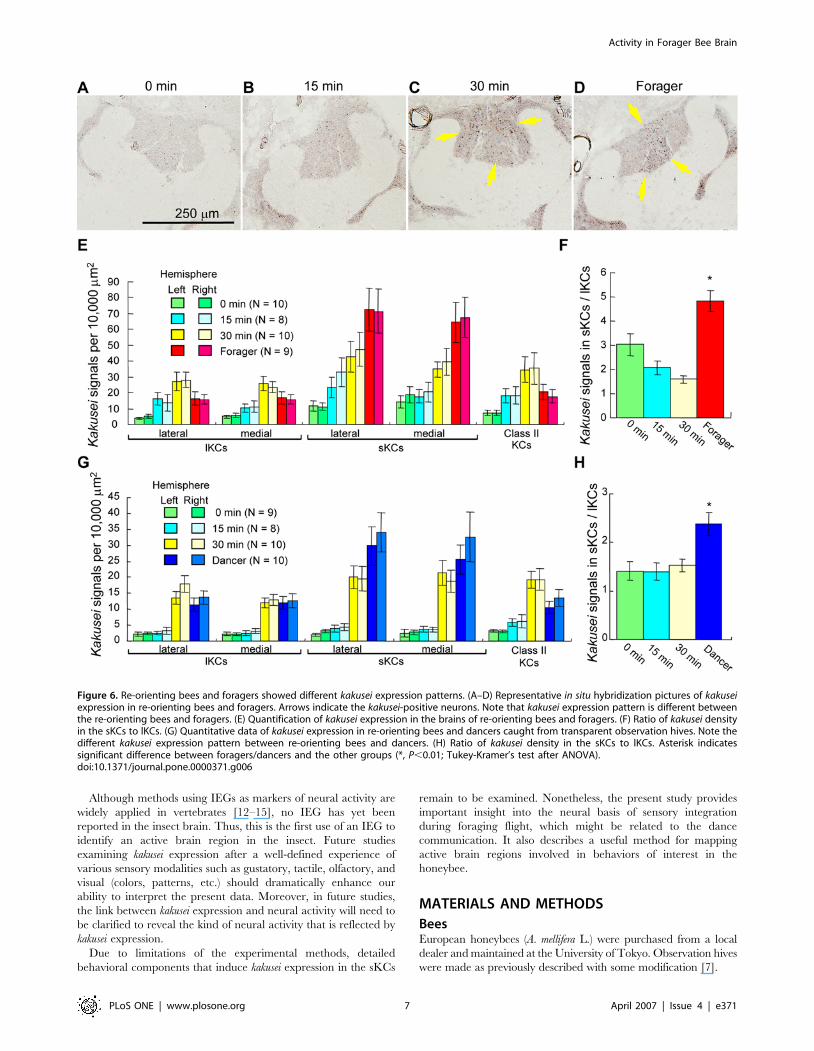
for 5 min and then caught the bees flying around the hive 0, 15,
and 30 min later. Kakusei expression was increased in the MBs in
a time-dependent manner in the re-orienting bees (Figure 6A–C
and E), suggesting that this neural activity is induced by re-
orienting behavior. One-way ANOVA among re-orienting bees
revealed that the time effect was significant for every MB neuron
type (Figure 6E: P’s,0.0001–0.003). In contrast to sKC-
preferential kakusei expression in the foragers, however, kakusei
was induced in all KC types in the re-orienting bees (Figure 6A–C
and E). The proportion of kakusei-positive cells in the sKCs
compared to that in the lKCs was significantly higher in the
foragers than in the re-orienting bees (Figure 6F). To exclude the
possibility that the neural activity in the re-orienting bees is due to
the increased light exposure when they leave the hive, a similar
experiment was performed using re-orienting bees from trans-
parent observation hives (Figure 6G and H). In this experiment,
a significant time-dependent increase in kakusei expression was
observed in every MB neuron subtype in the re-orienting bees
(P,0.0001, one-way ANOVA), like in the re-orienting bees from
the normal hives, indicating that the increased kakusei expression in
the re-orienting bees is due to re-orienting behavior, and not
merely to light-exposure (Figure 6G and H). Here, the workers
Figure 4. Neural activity of the sKCs is prominently increased in the dancer brain. (A and E) Schematic drawings of the caudal hemisphere (A) and MB(E). (B–D and F–H) In situ hybridization of kakusei in the dancer (B and F), follower (C and G), or nurse bee (D and H) brains. (I) Quantification ofkakusei-positive cells in various brain regions. Asterisks indicate significant difference from other behavioral groups (*, P,0.05; **, P,0.01; Tukey-Kramer’s test after ANOVA). Arrows indicate the kakusei-expressing neurons.doi:10.1371/journal.pone.0000371.g004
Activity in Forager Bee Brain
PLoS ONE | www.plosone.org 5 April 2007 | Issue 4 | e371
that performed re-orientation flights were probably foragers, also
suggesting that the differences in the kakusei expression pattern
between the brains of re-orienting bees and foragers/dancers
are due to differences in behavior, but not age. These results
demonstrate that the active brain regions are different between the
re-orienting bees and foragers/dancers, suggesting that the sKC-
preferential kakusei expression in forager/dancer brains is not due
to behavioral components in common with those of re-orienting
bees, like visual experience, flying, or landmark learning and
memory. Rather, the forager-specific behavioral components are
likely to be responsible for the sKC-preferential kakusei expression.
DISCUSSIONIn the present study, using novel methods to visualize neural activity
in the honeybee brain, we demonstrated that neural activity in the
MBs, especially the sKCs, is prominently increased in the brains of
dancer and forager honeybees. In contrast, the neural activity of both
the sKCs and lKCs was increased in the brains of re-orienting
workers. These findings strongly suggest that sKC-preferential
activity, but not mere MB-preferential activity, is associated with
behavioral components that are specific to foraging flight.
The MBs are believed to be important for higher sensory
integration in the insect brain [24,25]. Partial ablation of the MBs
impairs only complex olfactory learning without affecting simple
olfactory learning [26,27]. In addition, in the honeybee brain, all
sensory modalities investigated (visual, olfactory, gustatory, and
mechanosensory) project to the MBs [19,20,28–32]. Furthermore,
foraging experience greatly influence the MB neuropile volume
increase and the MB neuron outgrowth [33,34]. Although these
findings imply that the MBs are the appropriate brain regions to
process complex information during foraging behavior, such as the
calculation of distance and direction, there was no direct evidence
whether the MB neurons are actually active in forager brains. The
methods that we established in this study provide the first opportunity
to investigate neural activity in the brains of naturally behaving
honeybees and revealed that the MB neurons are active in the
dancers and foragers, although these methods are limited to labeling
only cell bodies and not the neuropile because kakusei encodes a non-
coding RNA whose expression is confined to the nuclei.
Our study demonstrated that neural activity of the sKCs, one of
the MB neuron subtypes, was prominently increased in the dancer
and forager brains. Although the inputs and outputs of the sKCs
have been well investigated, the role of the neural circuitry
composed by the sKCs is unknown [19,20,28,29,32]. The calyx
(input region) of the honeybee MB is divided into three zones: lip,
collar, and basal ring. The sKCs project dendrites to the basal ring
zone, whereas the lKCs project to the lip or collar zone [19]. On
the other hand, all sensory modalities investigated (visual, olfac-
tory, gustatory, and mechanosensory) project to both the basal ring
and lip/collar zones, which are then relayed to both sKCs and
lKCs [19,20,28–32]. Studies of the anatomy of the bee brain
suggest that the basal ring zone receives multi-modal (visual,
olfactory, gustatory, and mechanosensory) inputs and extensive
recurrent inputs from extrinsic MB neurons, whereas the lip and
collar zone receive mono-modal (olfactory and visual, respectively)
inputs [19,30,31]. In addition, the sKCs are composed of neurons
with different morphologies [19]. Thus, we speculate that the
sKC-preferential neural activity observed in the forager/dancer
brains reflects the complex activity within the MB neural networks
required for sensory integration during the foraging flight. Recent
studies suggest functional differences among the KC types based
on their gene expression patterns [21,22,35–38]. For example,
activation of Amfor, one of the genes preferentially expressed in the
sKCs, affects the onset of foraging behavior [18,38]. These
findings are also consistent with our notion that the sKCs play
roles in higher integration of the complex information that
accompanies foraging experience.
Prominent kakusei expression was detected only in the forager
brains, not in the follower brains (Figure 4), possibly due to the
differences in their foraging experience during the hour before
sampling. The followers often show reduced foraging activity
before they start following the dance [7,23]. Thus, considering the
short half-life of kakusei expression, it is possible that this behavioral
difference is reflected in the kakusei expression in the follower
brains. In addition, there were strikingly different kakusei
expression patterns in the MBs between the re-orienting and
foraging workers. Because the re-orienting bees fly around to form
spatial memory regarding their hive location [39], they have
several behavioral components in common with the foragers. How
then are the differences in kakusei-expression between re-orienting
and forager/dancer bees explained? Foraging behavior is different
from orienting behavior in some ways: for example, foragers need
to calculate the distance and direction of food sources, memorize
them in association with food information, and recall them
repetitively to repeat foraging, which involves much broader and
multi-modal sensory integration than just orienting [39–41]. Thus,
it is possible that the increased activity in the sKCs in the forager
brains is associated with these behavioral components and reflects
such sensory integration. Alternatively, it is also possible that the
sKC activity is important for both the foraging and re-orienting
behaviors and the relative repression of the lKC activity is
important for the foraging behaviors. If this is the case, the
integration of sensory information during foraging flight might
result from interactions between active sKCs and inactive lKCs.
Here we identified the transcript of novel IEG, kakusei, as a non-
coding nuclear RNA. Although microRNA, which is expressed in
response to neural activity, has been reported in vertebrates
[42,43], kakusei is the first example of a long non-coding nuclear
RNA that shows an immediate early response to neural activity.
Long nuclear RNAs regulate gene expression [44,45], whereas
microRNA post-transcriptionally regulates gene expression [46].
In general, vertebrate IEGs encode transcription factors and have
roles in modulating neural functions in an activity-dependent
manner [47]. Thus, kakusei might regulate gene expression as a long
non-coding RNA to modulate neural function.
Figure 5. Foragers showed similar kakusei expression pattern to thedancers. Quantification of kakusei expression in the brains of foragersand dancers. Foragers also showed the sKC-preferential kakuseiexpression pattern. There was no significant difference in the numberof kakusei-positive cells (P.0.05; two-factor ANOVA). Note that werepeated the experiments for the ‘dancer’ group here, and bees used inthe ‘dancer’ group for this experiment were different from those used inthe previous experiment (Figure 4).doi:10.1371/journal.pone.0000371.g005
Activity in Forager Bee Brain
PLoS ONE | www.plosone.org 6 April 2007 | Issue 4 | e371
Although methods using IEGs as markers of neural activity are
widely applied in vertebrates [12–15], no IEG has yet been
reported in the insect brain. Thus, this is the first use of an IEG to
identify an active brain region in the insect. Future studies
examining kakusei expression after a well-defined experience of
various sensory modalities such as gustatory, tactile, olfactory, and
visual (colors, patterns, etc.) should dramatically enhance our
ability to interpret the present data. Moreover, in future studies,
the link between kakusei expression and neural activity will need to
be clarified to reveal the kind of neural activity that is reflected by
kakusei expression.
Due to limitations of the experimental methods, detailed
behavioral components that induce kakusei expression in the sKCs
remain to be examined. Nonetheless, the present study provides
important insight into the neural basis of sensory integration
during foraging flight, which might be related to the dance
communication. It also describes a useful method for mapping
active brain regions involved in behaviors of interest in the
honeybee.
MATERIALS AND METHODS
BeesEuropean honeybees (A. mellifera L.) were purchased from a local
dealer and maintained at the University of Tokyo. Observation hives
were made as previously described with some modification [7].
Figure 6. Re-orienting bees and foragers showed different kakusei expression patterns. (A–D) Representative in situ hybridization pictures of kakuseiexpression in re-orienting bees and foragers. Arrows indicate the kakusei-positive neurons. Note that kakusei expression pattern is different betweenthe re-orienting bees and foragers. (E) Quantification of kakusei expression in the brains of re-orienting bees and foragers. (F) Ratio of kakusei densityin the sKCs to lKCs. (G) Quantitative data of kakusei expression in re-orienting bees and dancers caught from transparent observation hives. Note thedifferent kakusei expression pattern between re-orienting bees and dancers. (H) Ratio of kakusei density in the sKCs to lKCs. Asterisk indicatessignificant difference between foragers/dancers and the other groups (*, P,0.01; Tukey-Kramer’s test after ANOVA).doi:10.1371/journal.pone.0000371.g006
Activity in Forager Bee Brain
PLoS ONE | www.plosone.org 7 April 2007 | Issue 4 | e371
Differential displayWorker honeybee brains were dissected out from each of 10 bees
anesthetized on ice and bees awakened from ice-induced anesthesia,
which showed a seizure-like phenotype. Total RNA was isolated with
TRIzol (Invitrogen), treated with DNase I, and reverse transcribed
with SuperScript II (Invitrogen). The differential display method was
performed as described previously using a Fluorescent Differential
Display Kit and LA Taq polymerase with a combined total of 216
primer sets (Takara) [21,22]. Bands of interest were excised,
reamplified, and subcloned into a pGEM-T vector (Promega).
Northern blottingWhole brain total RNA was isolated from each of 10 bees
anesthetized by CO2 and bees awakened from CO2-induced
anesthesia. RNA was subjected to 1% formaldehyde-agarose gel
electrophoresis, transferred to a nylon membrane, and hybridized
with 32P-labeled riboprobes. 32P-labeled riboprobes were synthe-
sized by T7 polymerase with Strip-EZTM RNA Kit (Ambion) from
a template containing the fragment isolated by differential display
(DD fragment; from+4511 to+5159).
cDNA cloningTo identify the whole length of kakusei cDNA, 59-and 39-rapid
amplification of cDNA ends (RACE) methods were performed
repeatedly using the SMART RACE cDNA Amplification Kit
(Clontech).
RT-PCRRT-PCR experiments were performed using LA Taq (Takara)
according to the manufacturer’s protocol and the SMART RACE
cDNA as templates. Primers were designed to amplify the regions
shown in Figure 1B; (a) 59-CACGCTCGTCGTCGTGCCTTG-
CTCAGATAA-39 and 59-TTCAGAGCACGTTGGAACTAATC-
TCGCG-39, (b) 59-ACCTTGGAACGTGAAAGCGCATTTTC-
GA-39 and 59-AACCGTGTCCTTCTGCAGACACCTGACA-39
Quantification of the kakusei transcriptThe expression of kakusei was induced by awakening bees from
CO2-induced anesthesia. Control bees were kept in CO2. Total
RNA was extracted from three to five bees for each sample. Real-
time RT-PCR was performed with Light Cycler-DNA master
hybridization probes (Roche) according to the manufacturer’s
protocol, using gene-specific primers (kakusei; 59-GGAAACAGG-
TGGTTTGATGACCATTG and 59-CACGTTCCAAGGTTT-
AACGATGCG, actin; 59-GAAATGGCAACTGCTGCATC and
59-TCCACATCTGTTGGAAGGTG) and fluorescent probes
(kakusei; fluorescein isothiocyanate (FITC) probe, 59-CGCTG-
TAGTGCGTTTTCACTCGGATCGA, and LC-Red640 probe,
59-TCCGAGGAAATCCGAGCAAAGTTCGTTC, actin; FITC
probe, 59-CCATGAAAATTAAGATCATCGCGCCAC, and
LC-Red640 probe, 59-CGAGAAGAAATATTCCGTATGGAT-
TGGTG). The amount of kakusei transcript was normalized with
that of actin and is shown as relative to the value of control bees at
0 min or to the whole brain. There was no significant difference in
the levels of actin expression between control and seizure-induced
bees.
In situ hybridization and image analysisIn situ hybridization was performed as described previously with
some modification [22,48]. Frozen coronal brain sections (10 mm
thick) were fixed in 4% paraformaldehyde in phosphate buffered
saline, pretreated, and hybridized with digoxigen (DIG)-labeled
riboprobes. The DIG-labeled riboprobes were synthesized by T7
or SP6 polymerase with a DIG labeling mix (Roche) from
a template containing the fragment isolated by differential display
(from+4511 to+5159). After stringent washes, DIG-labeled ribop-
robes were detected immunocytochemically with peroxidase-
conjugated anti-DIG antibody (1:500; Roche) and TSA Biotin
System (Perkin Elmer). Sense probes were used as negative
controls and the signals were confirmed to be antisense probe-
specific in every experiment. Micrographs of fluorescent in situ
hybridization were taken using an IX71 confocal microscope
(Olympus). SYTOX Green (Molecular Probes) was used to stain
the nuclear DNA [22]. Intensity and brightness of the micrographs
were processed with Photoshop software (Adobe).
For quantification, the brain regions were defined as shown in
Figure 2A and B. The rostral section was defined as the section
containing the AL, and the caudal section was defined as the
section containing the DL. The numbers of kakusei-positive cells
were manually counted from rostral and caudal sections. For each
animal, one rostral and one caudal section were analyzed. The
area of each brain region was measured using ImageJ analysis
software (NIH, http://rsb.info.nih.gov/ij). The amount of signal
was divided by the area and the values from two sections were
averaged, when there were two data points for one individual (e.g.,
MB and OL neurons). Sections to be counted were randomly
selected from many sections. The density of kakusei-positive cells
was shown as a value relative to 100000 mm2. Micrographs were
numbered and signals were counted by an investigator blind to the
bee type. The number of examined bees is shown in the figure.
Statistical analyses were conducted by F-test and Student’s t-test
using Microsoft Excel (Microsoft) or JMP (SAS) software. Multiple
comparisons were performed using one-way analysis of variance
(ANOVA) and Tukey-Kramer’s test. Statistical comparisons were
made within the same brain hemisphere. Data are shown
means6standard error (SEM) throughout this paper.
SUPPORTING INFORMATION
Figure S1 Kakusei expression in response to light-exposure. (A–
H) In situ hybridization of kakusei in the brains of dark-adapted (A,
C, E, and G) or light-exposed after dark-adaptation (B, D, F, and H)
bees. (C and D) Magnified view of the boxed region in panels (A) and
(B), respectively. Kakusei expression was detected only in the OL
neurons (B, D, and H) and MB neurons (F). Arrows indicate the
kakusei-positive neurons. LA, lamina; ME, medulla; RE, retina.
Found at: doi:10.1371/journal.pone.0000371.s001 (6.90 MB TIF)
Figure S2 Schematic drawings of the phototaxis experiments.
First, approximately 20 foragers and 20 nurse bees were fed honey
and kept separately in transparent boxes under room light
conditions for 24 h (A and D: Here, only 4 bees are drawn for
simplicity.). Then, a light (100 W lamp) was set in the one side of
the box and kept for 30 min (B and E). During 30 min radiation,
almost all of the foragers and a part of nurse bees (approximately
10 bees) moved to the light side (C and F). We therefore collected
foragers that moved to the light side and nurse bees that did not
move to the light side, and investigated kakusei expression.
Found at: doi:10.1371/journal.pone.0000371.s002 (0.54 MB TIF)
ACKNOWLEDGMENTS
Author Contributions
Conceived and designed the experiments: T. Kiya. Performed the
experiments: T. Kiya. Analyzed the data: T. Kiya. Wrote the paper: T.
Kiya, T. Kunieda, T. Kubo.
Activity in Forager Bee Brain
PLoS ONE | www.plosone.org 8 April 2007 | Issue 4 | e371
REFERENCES1. Fitzpatrick MJ, Ben-Shahar Y, Smid HM, Vet LEM, Robinson GE, et al. (2005)
Candidate genes for behavioural ecology. TRENDS in Ecology and Evolution20: 96–104.
2. Robinson GE, Grozinger CM, Whitfield CW (2005) Sociogenomics: social life in
molecular terms. Nat Rev Genet 6: 257–270.3. Wilson EO (1975) Sciobiology: The New Synthesis. Cambridge, Massachusetts:
Harvard Univ. Press.4. Winston ML (1987) The Biology of the Honey Bee. Cambridge, Massachusetts:
Harvard Univ. Press.5. von Frisch K (1993) The Dance Language and Orientation of Bees; Seeley TD,
ed. Cambridge, Massachusetts: Harvard Univ. Press.
6. Dyer FC (2002) The biology of the dance language. Annu Rev Entomol 47:917–949.
7. Seeley TD (1995) The Wisdom of the Hive: The Social Physiology of Honey BeeColonies. Cambridge, Massachusetts: Harvard Univ. Press.
8. Esch HE, Zhang S, Srinivasan MV, Tautz J (2001) Honeybee dances
communicate distances measured by optic flow. Nature 411: 581–583.9. Srinivasan MV, Zhang S, Altwein M, Tautz J (2000) Honeybee navigation:
nature and calibration of the ‘‘odometer’’. Science 287: 851–853.10. Nieh JC, Tautz J (2000) Behaviour-locked signal analysis reveals weak 200–
300 Hz comb vibrations during the honeybee waggle dance. J Exp Biol 203:1573–1579.
11. Sandeman D, Tautz J, Lindauer M (1996) Transmission of vibration across
honeycombs and its detection by bee leg receptors. J Exp Biol 199: 2585–2594.12. Guzowski JF, McNaughton BL, Barnes CA, Worley PF (1999) Environment-
specific expression of the immediate-early gene Arc in hippocampal neuronalensembles. Nat Neurosci 2: 1120–1124.
13. Guzowski JF, Setlow B, Wagner EK, McGaugh JL (2001) Experience-dependent
gene expression in the rat hippocampus after spatial learning: a comparison ofthe immediate-early genes Arc, c-fos, and zif268. J Neurosci 21: 5089–5098.
14. Sakata S, Kitsukawa T, Kaneko T, Yamamori T, Sakurai Y (2002) Task-dependent and cell-type-specific Fos enhancement in rat sensory cortices during
audio-visual discrimination. Eur J Neurosci 15: 735–743.15. Mello CV, Vicario DS, Clayton DF (1992) Song presentation induces gene
expression in the songbird forebrain. Proc Natl Acad Sci U S A 89: 6818–6822.
16. Brakeman PR, Lanahan AA, O’Brien R, Roche K, Barnes CA, et al. (1997)Homer: a protein that selectively binds metabotropic glutamate receptors.
Nature 386: 284–288.17. Kato A, Ozawa F, Saitoh Y, Fukazawa Y, Sugiyama H, et al. (1998) Novel
members of the Vesl/Homer family of PDZ proteins that bind metabotropic
glutamate receptors. J Biol Chem 273: 23969–23975.18. Ben-Shahar Y, Leung HT, Pak WL, Sokolowski MB, Robinson GE (2003)
cGMP-dependent changes in phototaxis: a possible role for the foraging gene inhoney bee division of labor. J Exp Biol 206: 2507–2515.
19. Mobbs P (1982) The brain of the honeybee Apis mellifera. I. The connections
and spatial organization of the mushroom bodies. Phil Trans R Soc Lond B 298:309–354.
20. Strausfeld NJ (2002) Organization of the honey bee mushroom body:representation of the calyx within the vertical and gamma lobes. J Comp
Neurol 450: 4–33.21. Takeuchi H, Kage E, Sawata M, Kamikouchi A, Ohashi K, et al. (2001)
Identification of a novel gene, Mblk-1, that encodes a putative transcription
factor expressed preferentially in the large-type Kenyon cells of the honeybeebrain. Insect Mol Biol 10: 487–494.
22. Sawata M, Yoshino D, Takeuchi H, Kamikouchi A, Ohashi K, et al. (2002)Identification and punctate nuclear localization of a novel noncoding RNA, Ks-
1, from the honeybee brain. Rna 8: 772–785.
23. Seeley T, Visscher PK (1988) Assessing the benefits of cooperation in honeybeeforaging: search costs, forage quality, and competitive ability. Behavioral
Ecology and Sociobiology 22: 229–237.24. Menzel R (2001) Searching for the memory trace in a mini-brain, the honeybee.
Learn Mem 8: 53–62.25. Heisenberg M (2003) Mushroom body memoir: from maps to models. Nat Rev
Neurosci 4: 266–275.
26. Komischke B, Sandoz JC, Malun D, Giurfa M (2005) Partial unilateral lesions of
the mushroom bodies affect olfactory learning in honeybees Apis mellifera L.Eur J Neurosci 21: 477–485.
27. Malun D, Giurfa M, Galizia CG, Plath N, Brandt R, et al. (2002) Hydroxyurea-
induced partial mushroom body ablation does not affect acquisition andretention of olfactory differential conditioning in honeybees. J Neurobiol 53:
343–360.
28. Ehmer B, Gronenberg W (2002) Segregation of visual input to the mushroombodies in the honeybee (Apis mellifera). J Comp Neurol 451: 362–373.
29. Gronenberg W (2001) Subdivisions of hymenopteran mushroom body calyces by
their afferent supply. J Comp Neurol 435: 474–489.
30. Grunewald B (1999) Morphology of feedback neurons in the mushroom body ofthe honeybee, Apis mellifera. J Comp Neurol 404: 114–126.
31. Rybak J, Menzel R (1993) Anatomy of the mushroom bodies in the honey bee
brain: the neuronal connections of the alpha-lobe. J Comp Neurol 334:444–465.
32. Schroter U, Menzel R (2003) A new ascending sensory tract to the calyces of the
honeybee mushroom body, the subesophageal-calycal tract. J Comp Neurol 465:168–178.
33. Farris SM, Robinson GE, Fahrbach SE (2001) Experience-and age-related
outgrowth of intrinsic neurons in the mushroom bodies of the adult workerhoneybee. J Neurosci 21: 6395–6404.
34. Ismail N, Robinson GE, Fahrbach SE (2006) Stimulation of muscarinic
receptors mimics experience-dependent plasticity in the honey bee brain. ProcNatl Acad Sci U S A 103: 207–211.
35. Paul RK, Takeuchi H, Matsuo Y, Kubo T (2005) Gene expression of
ecdysteroid-regulated gene E74 of the honeybee in ovary and brain. Insect MolBiol 14: 9–15.
36. Takeuchi H, Fujiyuki T, Shirai K, Matsuo Y, Kamikouchi A, et al. (2002)
Identification of genes expressed preferentially in the honeybee mushroombodies by combination of differential display and cDNA microarray. FEBS Lett
513: 230–234.
37. Takeuchi H, Yasuda A, Yasuda-Kamatani Y, Kubo T, Nakajima T (2003)Identification of a tachykinin-related neuropeptide from the honeybee brain
using direct MALDI-TOF MS and its gene expression in worker, queen anddrone heads. Insect Mol Biol 12: 291–298.
38. Ben-Shahar Y, Robichon A, Sokolowski MB, Robinson GE (2002) Influence of
gene action across different time scales on behavior. Science 296: 741–744.
39. Menzel R, Greggers U, Smith A, Berger S, Brandt R, et al. (2005) Honey beesnavigate according to a map-like spatial memory. Proc Natl Acad Sci U S A 102:
3040–3045.
40. Riley JR, Greggers U, Smith AD, Reynolds DR, Menzel R (2005) The flightpaths of honeybees recruited by the waggle dance. Nature 435: 205–207.
41. Giurfa M (2003) Cognitive neuroethology: dissecting non-elemental learning in
a honeybee brain. Curr Opin Neurobiol 13: 726–735.
42. Vo N, Klein ME, Varlamova O, Keller DM, Yamamoto T, et al. (2005) A
cAMP-response element binding protein-induced microRNA regulates neuronal
morphogenesis. Proc Natl Acad Sci U S A 102: 16426–16431.
43. Schratt GM, Tuebing F, Nigh EA, Kane CG, Sabatini ME, et al. (2006) A
brain-specific microRNA regulates dendritic spine development. Nature 439:
283–289.
44. Brown CJ, Hendrich BD, Rupert JL, Lafreniere RG, Xing Y, et al. (1992) The
human XIST gene: analysis of a 17 kb inactive X-specific RNA that contains
conserved repeats and is highly localized within the nucleus. Cell 71: 527–542.
45. Amrein H, Axel R (1997) Genes expressed in neurons of adult male Drosophila.
Cell 88: 459–469.
46. Klein ME, Impey S, Goodman RH (2005) Role reversal: the regulation ofneuronal gene expression by microRNAs. Curr Opin Neurobiol 15: 507–513.
47. West AE, Griffith EC, Greenberg ME (2002) Regulation of transcription factors
by neuronal activity. Nat Rev Neurosci 3: 921–931.
48. Yang H, Wanner IB, Roper SD, Chaudhari N (1999) An optimized method forin situ hybridization with signal amplification that allows the detection of rare
mRNAs. J Histochem Cytochem 47: 431–446.
Activity in Forager Bee Brain
PLoS ONE | www.plosone.org 9 April 2007 | Issue 4 | e371
Related Documents