Increased NADPH oxidase activity mediates spontaneous aortic tone in genetically hypertensive rats Federica Lodi a , Angel Cogolludo a , Juan Duarte b , Laura Moreno a , Alfredo Coviello c , Maria Peral De Bruno c , Rocio Vera b , Milagros Galisteo b , Rosario Jiménez b , Juan Tamargo a , Francisco Perez-Vizcaino a, ⁎ a Department of Pharmacology, Institute of Pharmacology and Toxicology, School of Medicine, Universidad Complutense, 28040 Madrid, Spain b Department of Pharmacology, School of Pharmacy, Universidad de Granada, Spain c Department of Physiology, INSIBIO-Universidad Nacional de Tucuman, Tucuman, Argentina Received 6 February 2006; received in revised form 22 May 2006; accepted 12 June 2006 Available online 18 June 2006 Abstract NADPH oxidase is critically involved in increased blood pressure, vascular hypertrophy, inflammation and endothelial dysfunction in experimental and clinical hypertension. We hypothesized that NADPH oxidase might also play a role in the development of spontaneous aortic tone in spontaneously hypertensive rats (SHR). Wistar Kyoto rats (WKY) were used as normotensive controls. Tone was recorded under isometric conditions. NADPH oxidase activity was measured by both lucigenin luminescence and dihydroethidium fluorescence. p47 phox protein was localized by immunohistochemistry. SHR (but not WKY rat) aortae showed spontaneous tone in the absence of exogenous vasoconstrictors as evidenced by a stronger relaxant effect of Ca 2+ -free sodium nitroprusside solution. This tone was enhanced in endothelium-denuded arteries and was inhibited by superoxide dismutase, apocynin, diphenylene iodonium and quercetin. Aortic NADPH oxidase activity, measured by both lucigenin luminescence and dihydroethidium fluorescence, was increased in SHR compared with WKY rats. Immunohistochemical analysis revealed a strong increase in p47 phox expression in the medial layer in SHR. Taken together, the present results indicate that enhanced NADPH oxidase activity and, hence, NADPH driven O 2 − production, is involved in the spontaneous aortic tone in SHR. This was associated with an increased expression of p47 phox in the medial layer of the aorta. © 2006 Elsevier B.V. All rights reserved. Keywords: p47 phox ; SHR (spontaneously hypertensive rat); Spontaneous tone; Superoxide 1. Introduction Spontaneous arterial tone, also referred to as myogenic, intrinsic, basal or resting tone, is a state of partial contraction of isolated arteries when stretched or pressurized without exposure to exogenous vasoconstrictor stimuli and is a typical feature of resistance arteries (Folkow, 1989). Spontaneous tone is an intrinsic property of vascular smooth muscle cells (i.e. myogenic) even though it can be modulated by endothelial factors (Sunano et al., 1996; Veerareddy et al., 2004) and involves depolarization and subsequent opening of voltage- operated Ca 2+ channels (Laher and Bevan, 1989) as well as changes in Ca 2+ sensitivity (Matchkov et al., 2002). Conductance arteries from healthy individuals do not show spontaneous tone. However, spontaneous tone can be drastically altered by hypertension and diabetes mellitus (Sunano et al., 1996; Falcone et al., 1993; Frisbee et al., 2002). Thus, several studies have demonstrated enhanced spontaneous tone in aortae from different rodent models of experimental hypertension, including spontaneously hypertensive rats (SHR) (Sunano et al., 1996), deoxycorticosterone salt hypertensive rats (Ghosh et al., 2004) and hypertensive rats with aortic coarctation (Peral de Bruno et al., 1999; Pucci et al., 1994). Although the aorta does not contribute to the control of blood pressure, it is not just a passive vessel, and changes in aortic structure and function are essential features of hypertension (Laurent et al., 2001). The European Journal of Pharmacology 544 (2006) 97 – 103 www.elsevier.com/locate/ejphar ⁎ Corresponding author. Tel.: +34 913941477; fax: +34 913941470. E-mail address: [email protected] (F. Perez-Vizcaino). 0014-2999/$ - see front matter © 2006 Elsevier B.V. All rights reserved. doi:10.1016/j.ejphar.2006.06.028

Welcome message from author
This document is posted to help you gain knowledge. Please leave a comment to let me know what you think about it! Share it to your friends and learn new things together.
Transcript
ogy 544 (2006) 97–103www.elsevier.com/locate/ejphar
European Journal of Pharmacol
Increased NADPH oxidase activity mediates spontaneous aortictone in genetically hypertensive rats
Federica Lodi a, Angel Cogolludo a, Juan Duarte b, Laura Moreno a, Alfredo Coviello c,Maria Peral De Bruno c, Rocio Vera b, Milagros Galisteo b, Rosario Jiménez b,
Juan Tamargo a, Francisco Perez-Vizcaino a,⁎
a Department of Pharmacology, Institute of Pharmacology and Toxicology, School of Medicine, Universidad Complutense, 28040 Madrid, Spainb Department of Pharmacology, School of Pharmacy, Universidad de Granada, Spain
c Department of Physiology, INSIBIO-Universidad Nacional de Tucuman, Tucuman, Argentina
Received 6 February 2006; received in revised form 22 May 2006; accepted 12 June 2006Available online 18 June 2006
Abstract
NADPH oxidase is critically involved in increased blood pressure, vascular hypertrophy, inflammation and endothelial dysfunction inexperimental and clinical hypertension. We hypothesized that NADPH oxidase might also play a role in the development of spontaneous aortictone in spontaneously hypertensive rats (SHR). Wistar Kyoto rats (WKY) were used as normotensive controls. Tone was recorded under isometricconditions. NADPH oxidase activity was measured by both lucigenin luminescence and dihydroethidium fluorescence. p47phox protein waslocalized by immunohistochemistry. SHR (but not WKY rat) aortae showed spontaneous tone in the absence of exogenous vasoconstrictors asevidenced by a stronger relaxant effect of Ca2+-free sodium nitroprusside solution. This tone was enhanced in endothelium-denuded arteries andwas inhibited by superoxide dismutase, apocynin, diphenylene iodonium and quercetin. Aortic NADPH oxidase activity, measured by bothlucigenin luminescence and dihydroethidium fluorescence, was increased in SHR compared with WKY rats. Immunohistochemical analysisrevealed a strong increase in p47phox expression in the medial layer in SHR. Taken together, the present results indicate that enhanced NADPHoxidase activity and, hence, NADPH driven O2
− production, is involved in the spontaneous aortic tone in SHR. This was associated with anincreased expression of p47phox in the medial layer of the aorta.© 2006 Elsevier B.V. All rights reserved.
Keywords: p47phox; SHR (spontaneously hypertensive rat); Spontaneous tone; Superoxide
1. Introduction
Spontaneous arterial tone, also referred to as myogenic,intrinsic, basal or resting tone, is a state of partial contraction ofisolated arteries when stretched or pressurized without exposureto exogenous vasoconstrictor stimuli and is a typical feature ofresistance arteries (Folkow, 1989). Spontaneous tone is anintrinsic property of vascular smooth muscle cells (i.e.myogenic) even though it can be modulated by endothelialfactors (Sunano et al., 1996; Veerareddy et al., 2004) andinvolves depolarization and subsequent opening of voltage-
⁎ Corresponding author. Tel.: +34 913941477; fax: +34 913941470.E-mail address: [email protected] (F. Perez-Vizcaino).
0014-2999/$ - see front matter © 2006 Elsevier B.V. All rights reserved.doi:10.1016/j.ejphar.2006.06.028
operated Ca2+ channels (Laher and Bevan, 1989) as well aschanges in Ca2+ sensitivity (Matchkov et al., 2002).
Conductance arteries from healthy individuals do not showspontaneous tone. However, spontaneous tone can be drasticallyaltered by hypertension and diabetes mellitus (Sunano et al.,1996; Falcone et al., 1993; Frisbee et al., 2002). Thus, severalstudies have demonstrated enhanced spontaneous tone in aortaefrom different rodent models of experimental hypertension,including spontaneously hypertensive rats (SHR) (Sunano et al.,1996), deoxycorticosterone salt hypertensive rats (Ghosh et al.,2004) and hypertensive rats with aortic coarctation (Peral deBruno et al., 1999; Pucci et al., 1994). Although the aorta doesnot contribute to the control of blood pressure, it is not just apassive vessel, and changes in aortic structure and function areessential features of hypertension (Laurent et al., 2001). The
98 F. Lodi et al. / European Journal of Pharmacology 544 (2006) 97–103
development of tone in large arteries from hypertensives is thusexpected to impact on systemic hemodynamics.
Increased superoxide anion (O2−) produced under conditions
of high oxidative stress is known to produce significant changesin vascular function (Cai and Harrison, 2000). O2
− inactivates thepotent vasodilator nitric oxide (NO) impairing endothelium-dependent vasorelaxation (Cai andHarrison, 2000) andmay alsoproduce a direct vasoconstrictor effect (Jin et al., 2004).Increased myogenic tone has also been associated recentlywith an increased oxidative stress (Frisbee et al., 2002). In fact,small mesenteric arteries from superoxide dismutase (SOD)knockout mice (SOD−/−) exhibited enhanced myogenic tone(Veerareddy et al., 2004). NADPH oxidase is a multi-subunitenzymatic complex which comprises membrane-bound flavo-cytochrome b558 (formed by gp91phox (nox 2) or gp91phox
homologues (nox 1 and nox 4) and p22phox) and 3 cytoplasmicsubunits, p47phox, p67phox, and p40phox (Babior, 1999). Thiscomplex is considered to be the most important source of O2
− inthe vessel wall (Griendling et al., 2000; Souza et al., 2001).Interestingly, excess O2
− synthesized by NADPH oxidase iscritically involved in increased blood pressure, vascular hyper-trophy, inflammation and endothelial dysfunction in ex-perimental and clinical hypertension (Cai and Harrison, 2000;Calo et al., 2005; Li et al., 2004; Virdis et al., 2004; Zalba et al.,2000).
In the present study, we hypothesized that O2− derived from
NADPH oxidase might play a role in the development of spon-taneous aortic tone in SHR.Wistar Kyoto (WKY) rats were usedas normotensive controls.
2. Materials and methods
The investigation conforms with the Guide for the Care andUse of Laboratory Animals published by the US National In-stitutes of Health (NIH Publication No. 85-23, revised 1996) andapproved by our Institutional Committee for the ethical care ofanimals. Twenty week old male SHR and WKY rats were ob-tained from Harlan Laboratories (Barcelona, Spain) and killedwith a lethal dose of pentobarbitone. The descending thoracic andthe suprarenal segment of the abdominal aortae were dissected.
2.1. Contractile tension recording
Thoracic aortic rings (∼3 mm) were mounted in organchambers for isometric force recording (Duarte et al., 1993;Lopez-Lopez et al., 2004). Rings were stretched to 2 g of tensionand equilibrated for 90–120 min. During this period, Krebssolution was changed every 30 min and rings were re-stretchedas needed tomaintain a final tension of 2 g. In some experiments,the endothelium was removed by rubbing the luminal surfacewith a metal rod (Lopez-Lopez et al., 2004). After equilibration,the bathing Krebs solution was replaced with a Ca2+-free Krebssolution containing 2 mM EGTA and 10−5 M sodium nitro-prusside and changes in contractile tone were recorded for20 min. Parallel experiments in which the solution was changedto a Ca2+-containing Krebs solution served as time controls. Inother sets of experiments, after equilibration, rings were exposed
to apocynin (3×10⁻⁴ M), diphenylene iodonium (DPI, 10−5 M),quercetin (10−5 M), SOD (100 U/ml), catalase (100 U/ml), theprostanoid TP receptor antagonist 1S-[1alpha,2alpha(Z),3alpha,4alpha]]-7-[3[[2-[(phenylamino)carbonyl[hydrazino]methyl]-7-oxabicyclo[2.2.1]hept-2-yl]-5-heptenoic acid(SQ29548, 10−6 M) or vehicle (0.1% dimethylsulfoxide,DMSO) and tension was recorded for 30 min.
2.2. NADPH oxidase activity
NADPH enhanced O2− release in intact thoracic aortic rings
and in homogenates from cultured aortic smooth muscle cellswas quantified by lucigenin-enhanced chemiluminiscence aspreviously described (Lopez-Lopez et al., 2004). Cell culturewas obtained from aortic explants. Briefly, aortas were cleared ofconnective tissue, cut into pieces and cultured in Dulbecco'smodified Earle's medium (DMEM) containing 10% fetal calfserum, penicillin (100U/ml), streptomycin (100 μg/ml) and non-essential amino acids. At subconfluence, cells (passages 5–10)were placed in serum-free medium overnight, washed in ice-coldPBS, and incubated in lysis buffer (20 mmol/L monobasicpotassium phosphate, 1 mmol/L EGTA, 10 μg/ml aprotinin,0.5 μg/ml leupeptin, 0.75 μg/ml pepstatin and 0.5 mmol/Lphenylmethylsulfonyl fluoride) for 10–20 min at 4 °C. Proteincontent was measured by using the bicinchoninic acid (BCA)method. NADPH (100 μmol/L) was added to the buffer con-taining the aortic rings or to the aortic homogenate suspension(30 μg protein in 500 μL) and lucigenin was injected auto-matically, at 5 μM to avoid known artifacts when used at higherconcentrations (Janiszewski et al., 2002). NADPH oxidaseactivity was calculated by subtracting the basal values fromthose in the presence of NADPH. In order to analyze the effectsof quercetin on NADPH oxidase-derived O2
−, NADPH-stimu-lated lucigenin luminescence was analyzed in aortic rings fromWistar rats.
2.3. In situ detection of vascular O2− production
Dihydroethidium (DHE) enters the cells and is oxidized byO2− to yield ethidium, which binds to DNA to produce bright red
fluorescence. Unfixed abdominal aortic rings were cryopre-served (phosphate buffer solution 0.1 M [PBS], plus 30%sucrose for 1–2 h), placed in OCT, frozen (−80 °C), and 10 μmcross sections were obtained in a cryostat (Microm InternationalModel HM500 OM). Sections were incubated in a humidifiedchamber for 30 min in Hepes buffered solution (in mM: NaCl130, KCl 5, MgCl2 1.2, glucose 10, and HEPES 10, pH 7.3 withNaOH) at 37 °C, and then incubated for 30min in Hepes solutioncontaining DHE (10−5 M) in the dark in the absence or presenceofDPI (10−5M) and/orNAPDH (10−4M), the preferredNADPHoxidase substrate in vascular smooth muscle. Then preparationswere counterstained with the nuclear stain 4,6-diamidin-2-phen-ylindol dichlorohydrate (DAPI, 3×10⁻⁷ M) for 5 min at 37 °Cand mounted with a coverslip. Four sections of each preparationwere examined on a fluorescence microscope (Leica DM IRB,Wetzlar, Germany) using a 40× objective with the following pairsof excitation/emission filters: 340–380 nm/425 nm long pass to
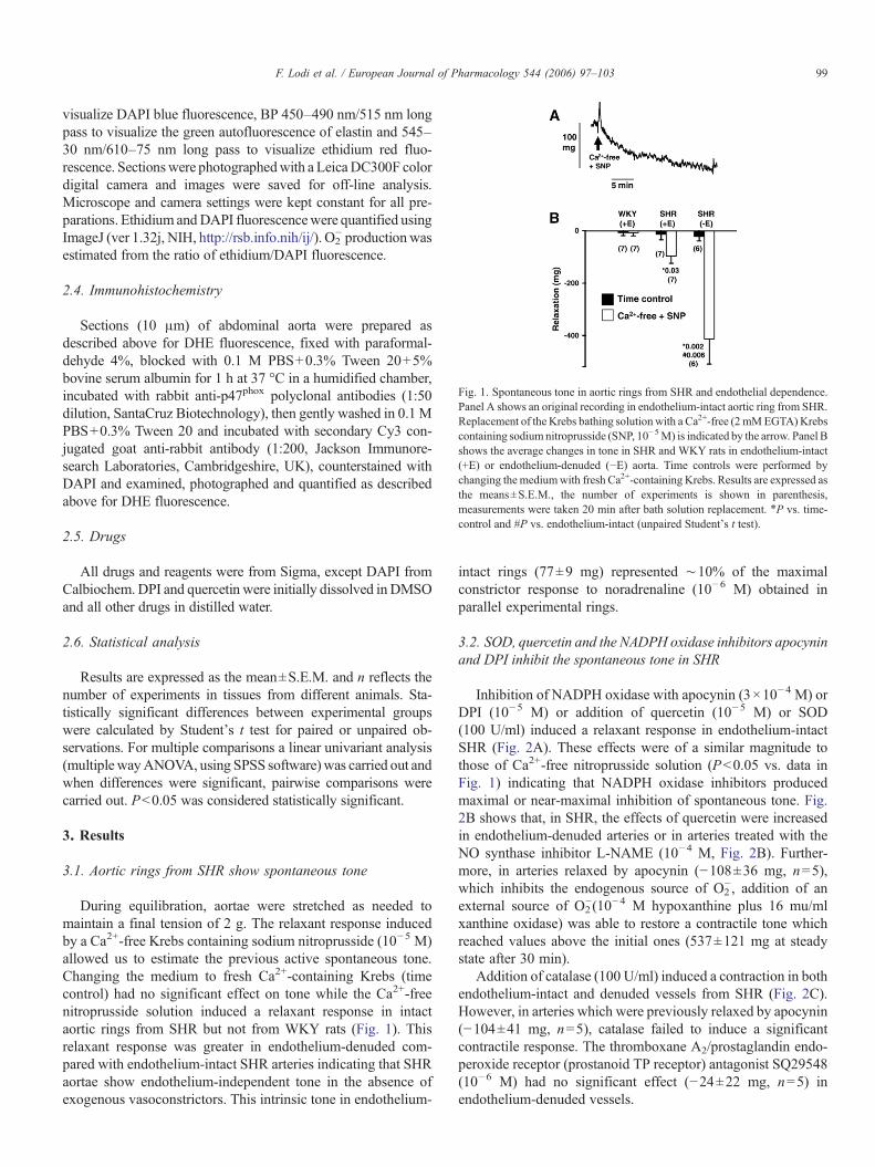
Fig. 1. Spontaneous tone in aortic rings from SHR and endothelial dependence.Panel A shows an original recording in endothelium-intact aortic ring from SHR.Replacement of the Krebs bathing solution with a Ca2+-free (2 mMEGTA) Krebscontaining sodiumnitroprusside (SNP, 10−5M) is indicated by the arrow. Panel Bshows the average changes in tone in SHR and WKY rats in endothelium-intact(+E) or endothelium-denuded (−E) aorta. Time controls were performed bychanging the mediumwith fresh Ca2+-containing Krebs. Results are expressed asthe means±S.E.M., the number of experiments is shown in parenthesis,measurements were taken 20 min after bath solution replacement. ⁎P vs. time-control and #P vs. endothelium-intact (unpaired Student's t test).
99F. Lodi et al. / European Journal of Pharmacology 544 (2006) 97–103
visualize DAPI blue fluorescence, BP 450–490 nm/515 nm longpass to visualize the green autofluorescence of elastin and 545–30 nm/610–75 nm long pass to visualize ethidium red fluo-rescence. Sectionswere photographedwith a LeicaDC300F colordigital camera and images were saved for off-line analysis.Microscope and camera settings were kept constant for all pre-parations. Ethidium andDAPI fluorescencewere quantified usingImageJ (ver 1.32j, NIH, http://rsb.info.nih/ij/). O2
− productionwasestimated from the ratio of ethidium/DAPI fluorescence.
2.4. Immunohistochemistry
Sections (10 μm) of abdominal aorta were prepared asdescribed above for DHE fluorescence, fixed with paraformal-dehyde 4%, blocked with 0.1 M PBS+0.3% Tween 20+5%bovine serum albumin for 1 h at 37 °C in a humidified chamber,incubated with rabbit anti-p47phox polyclonal antibodies (1:50dilution, SantaCruz Biotechnology), then gently washed in 0.1 MPBS+0.3% Tween 20 and incubated with secondary Cy3 con-jugated goat anti-rabbit antibody (1:200, Jackson Immunore-search Laboratories, Cambridgeshire, UK), counterstained withDAPI and examined, photographed and quantified as describedabove for DHE fluorescence.
2.5. Drugs
All drugs and reagents were from Sigma, except DAPI fromCalbiochem. DPI and quercetin were initially dissolved in DMSOand all other drugs in distilled water.
2.6. Statistical analysis
Results are expressed as the mean±S.E.M. and n reflects thenumber of experiments in tissues from different animals. Sta-tistically significant differences between experimental groupswere calculated by Student's t test for paired or unpaired ob-servations. For multiple comparisons a linear univariant analysis(multiple wayANOVA, using SPSS software) was carried out andwhen differences were significant, pairwise comparisons werecarried out. P<0.05 was considered statistically significant.
3. Results
3.1. Aortic rings from SHR show spontaneous tone
During equilibration, aortae were stretched as needed tomaintain a final tension of 2 g. The relaxant response inducedby a Ca2+-free Krebs containing sodium nitroprusside (10−5 M)allowed us to estimate the previous active spontaneous tone.Changing the medium to fresh Ca2+-containing Krebs (timecontrol) had no significant effect on tone while the Ca2+-freenitroprusside solution induced a relaxant response in intactaortic rings from SHR but not from WKY rats (Fig. 1). Thisrelaxant response was greater in endothelium-denuded com-pared with endothelium-intact SHR arteries indicating that SHRaortae show endothelium-independent tone in the absence ofexogenous vasoconstrictors. This intrinsic tone in endothelium-
intact rings (77±9 mg) represented ∼10% of the maximalconstrictor response to noradrenaline (10−6 M) obtained inparallel experimental rings.
3.2. SOD, quercetin and the NADPH oxidase inhibitors apocyninand DPI inhibit the spontaneous tone in SHR
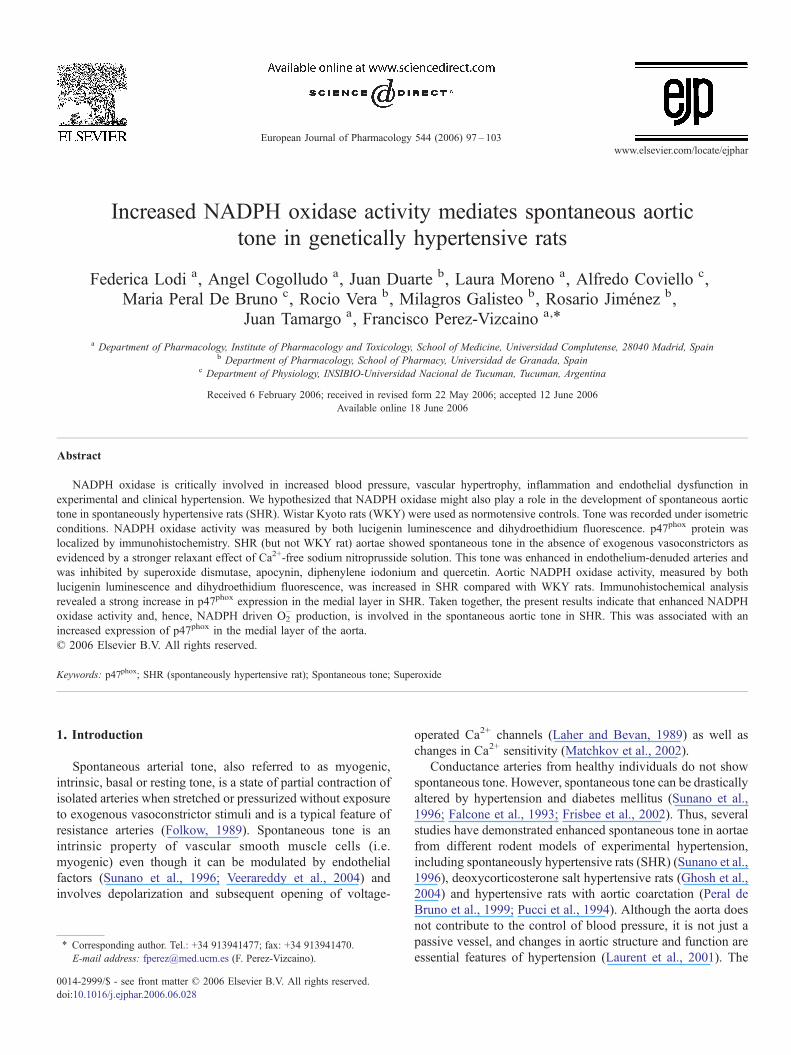
Inhibition of NADPH oxidase with apocynin (3×10⁻⁴ M) orDPI (10−5 M) or addition of quercetin (10−5 M) or SOD(100 U/ml) induced a relaxant response in endothelium-intactSHR (Fig. 2A). These effects were of a similar magnitude tothose of Ca2+-free nitroprusside solution (P<0.05 vs. data inFig. 1) indicating that NADPH oxidase inhibitors producedmaximal or near-maximal inhibition of spontaneous tone. Fig.2B shows that, in SHR, the effects of quercetin were increasedin endothelium-denuded arteries or in arteries treated with theNO synthase inhibitor L-NAME (10−4 M, Fig. 2B). Further-more, in arteries relaxed by apocynin (−108±36 mg, n=5),which inhibits the endogenous source of O2
−, addition of anexternal source of O2
−(10−4 M hypoxanthine plus 16 mu/mlxanthine oxidase) was able to restore a contractile tone whichreached values above the initial ones (537±121 mg at steadystate after 30 min).
Addition of catalase (100 U/ml) induced a contraction in bothendothelium-intact and denuded vessels from SHR (Fig. 2C).However, in arteries which were previously relaxed by apocynin(−104±41 mg, n=5), catalase failed to induce a significantcontractile response. The thromboxane A2/prostaglandin endo-peroxide receptor (prostanoid TP receptor) antagonist SQ29548(10−6 M) had no significant effect (−24±22 mg, n=5) inendothelium-denuded vessels.
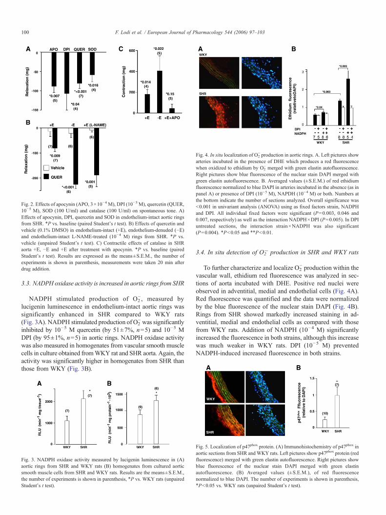
Fig. 2. Effects of apocynin (APO, 3×10⁻⁴ M), DPI (10−5 M), quercetin (QUER,10−5 M), SOD (100 U/ml) and catalase (100 U/ml) on spontaneous tone. A)Effects of apocynin, DPI, quercetin and SOD in endothelium-intact aortic ringsfrom SHR. ⁎P vs. baseline (paired Student's t test). B) Effects of quercetin andvehicle (0.1% DMSO) in endothelium-intact (+E), endothelium-denuded (−E)and endothelium-intact L-NAME-treated (10−4 M) rings from SHR. ⁎P vs.vehicle (unpaired Student's t test). C) Contractile effects of catalase in SHRaorta +E, −E and +E after treatment with apocynin. ⁎P vs. baseline (pairedStudent's t test). Results are expressed as the means±S.E.M., the number ofexperiments is shown in parenthesis, measurements were taken 20 min afterdrug addition.
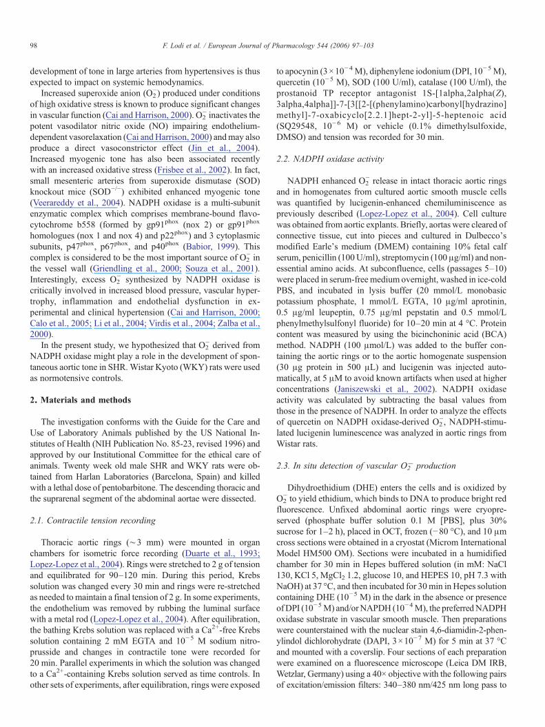
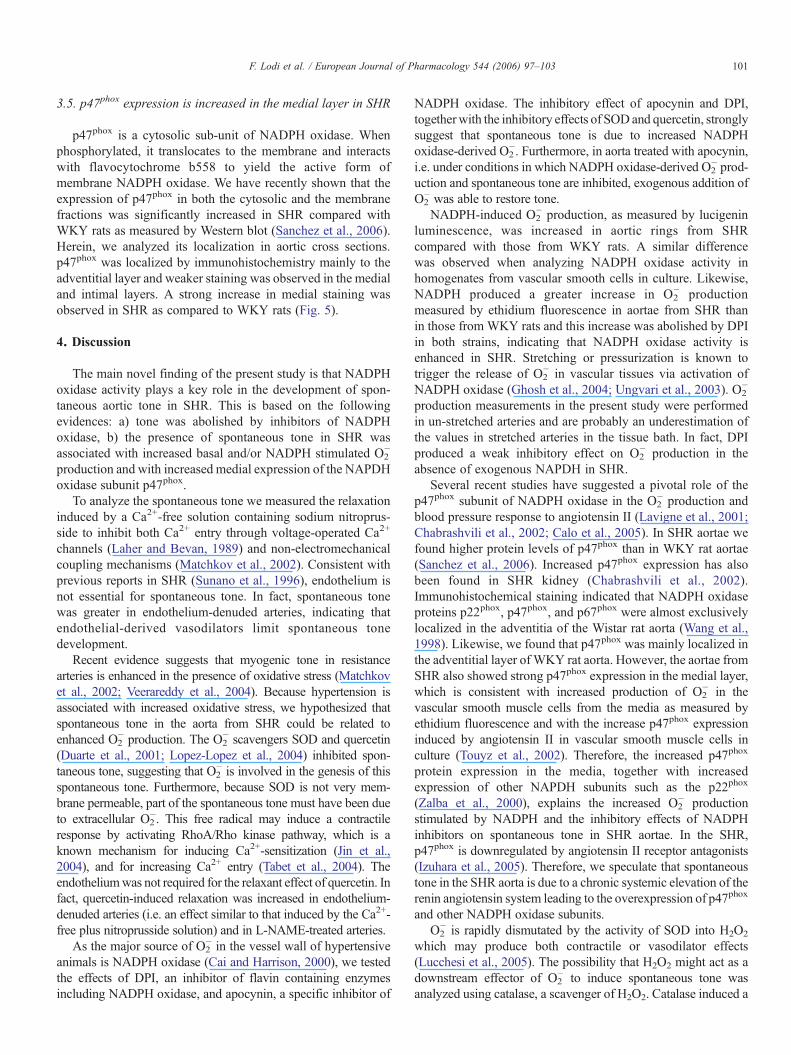
Fig. 4. In situ localization of O2− production in aortic rings. A. Left pictures show
arteries incubated in the presence of DHE which produces a red fluorescencewhen oxidized to ethidium by O2
− merged with green elastin autofluorescence.Right pictures show blue fluorescence of the nuclear stain DAPI merged withgreen elastin autofluorescence. B. Averaged values (±S.E.M.) of red ethidiumfluorescence normalized to blue DAPI in arteries incubated in the absence (as inpanel A) or presence of DPI (10−5 M), NAPDH (10−4 M) or both. Numbers atthe bottom indicate the number of sections analyzed. Overall significance was<0.001 in univariant analysis (ANOVA) using as fixed factors strain, NADPHand DPI. All individual fixed factors were significant (P=0.003, 0.046 and0.007, respectively) as well as the interaction NADPH×DPI (P=0.005). In DPIuntreated sections, the interaction strain×NADPH was also significant(P=0.004). ⁎P<0.05 and ⁎⁎P<0.01.
100 F. Lodi et al. / European Journal of Pharmacology 544 (2006) 97–103
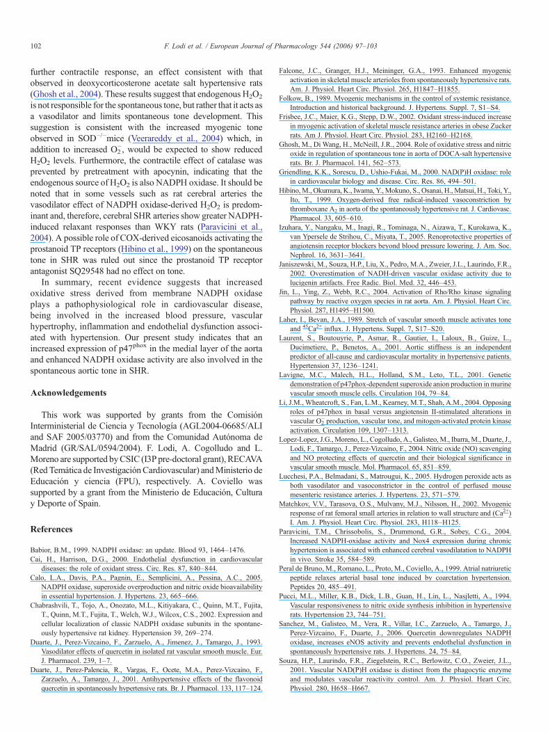
3.3. NADPH oxidase activity is increased in aortic rings from SHR
NADPH stimulated production of O2−, measured by
lucigenin luminescence in endothelium-intact aortic rings wassignificantly enhanced in SHR compared to WKY rats(Fig. 3A). NADPH stimulated production of O2
−was significantlyinhibited by 10−5 M quercetin (by 51±7%, n=5) and 10−5 MDPI (by 95±1%, n=5) in aortic rings. NADPH oxidase activitywas also measured in homogenates from vascular smooth musclecells in culture obtained fromWKY rat and SHR aorta. Again, theactivity was significantly higher in homogenates from SHR thanthose from WKY (Fig. 3B).
Fig. 3. NADPH oxidase activity measured by lucigenin luminescence in (A)aortic rings from SHR and WKY rats (B) homogenates from cultured aorticsmooth muscle cells from SHR and WKY rats. Results are the means±S.E.M.,the number of experiments is shown in parenthesis, ⁎P vs. WKY rats (unpairedStudent's t test).
3.4. In situ detection of O2− production in SHR and WKY rats
To further characterize and localize O2− production within the
vascular wall, ethidium red fluorescence was analyzed in sec-tions of aorta incubated with DHE. Positive red nuclei wereobserved in adventitial, medial and endothelial cells (Fig. 4A).Red fluorescence was quantified and the data were normalizedby the blue fluorescence of the nuclear stain DAPI (Fig. 4B).Rings from SHR showed markedly increased staining in ad-ventitial, medial and endothelial cells as compared with thosefrom WKY rats. Addition of NADPH (10−4 M) significantlyincreased the fluorescence in both strains, although this increasewas much weaker in WKY rats. DPI (10−5 M) preventedNADPH-induced increased fluorescence in both strains.
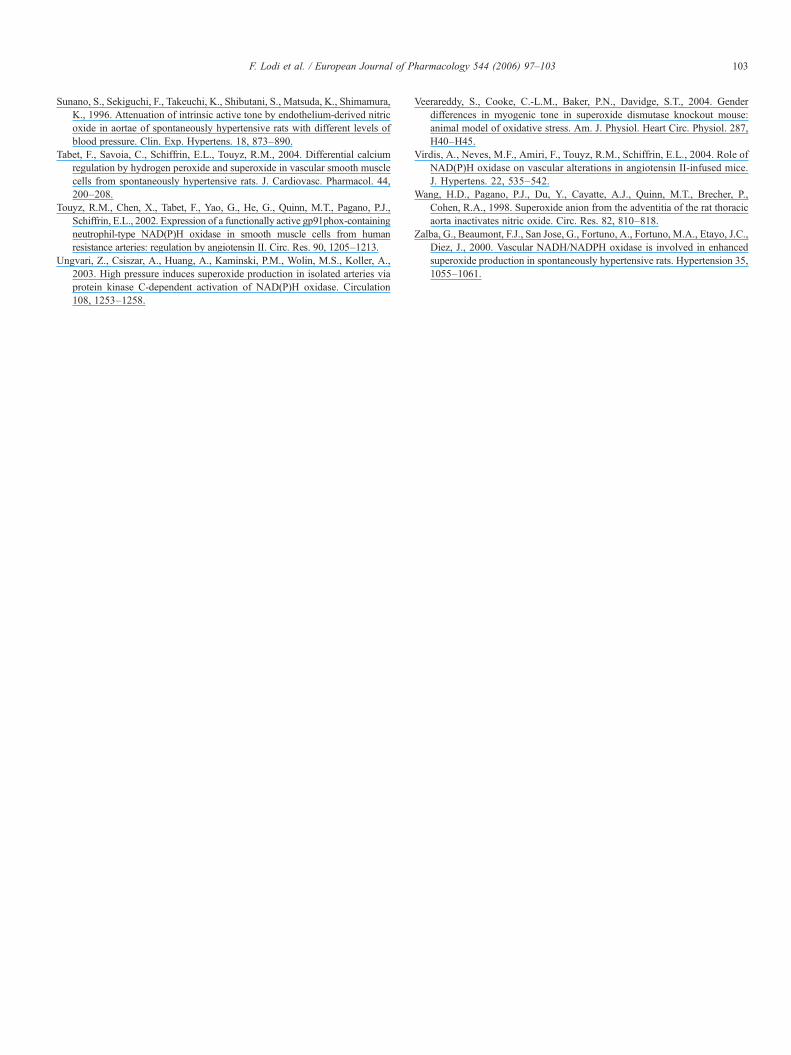
Fig. 5. Localization of p47phox protein. (A) Immunohistochemistry of p47phox inaortic sections from SHR andWKY rats. Left pictures show p47phox protein (redfluorescence) merged with green elastin autofluorescence. Right pictures showblue fluorescence of the nuclear stain DAPI merged with green elastinautofluorescence. (B) Averaged values (±S.E.M.), of red fluorescencenormalized to blue DAPI. The number of experiments is shown in parenthesis,⁎P<0.05 vs. WKY rats (unpaired Student's t test).
101F. Lodi et al. / European Journal of Pharmacology 544 (2006) 97–103
3.5. p47phox expression is increased in the medial layer in SHR
p47phox is a cytosolic sub-unit of NADPH oxidase. Whenphosphorylated, it translocates to the membrane and interactswith flavocytochrome b558 to yield the active form ofmembrane NADPH oxidase. We have recently shown that theexpression of p47phox in both the cytosolic and the membranefractions was significantly increased in SHR compared withWKY rats as measured by Western blot (Sanchez et al., 2006).Herein, we analyzed its localization in aortic cross sections.p47phox was localized by immunohistochemistry mainly to theadventitial layer and weaker staining was observed in the medialand intimal layers. A strong increase in medial staining wasobserved in SHR as compared to WKY rats (Fig. 5).
4. Discussion
The main novel finding of the present study is that NADPHoxidase activity plays a key role in the development of spon-taneous aortic tone in SHR. This is based on the followingevidences: a) tone was abolished by inhibitors of NADPHoxidase, b) the presence of spontaneous tone in SHR wasassociated with increased basal and/or NADPH stimulated O2
−
production and with increased medial expression of the NAPDHoxidase subunit p47phox.
To analyze the spontaneous tone we measured the relaxationinduced by a Ca2+-free solution containing sodium nitroprus-side to inhibit both Ca2+ entry through voltage-operated Ca2+
channels (Laher and Bevan, 1989) and non-electromechanicalcoupling mechanisms (Matchkov et al., 2002). Consistent withprevious reports in SHR (Sunano et al., 1996), endothelium isnot essential for spontaneous tone. In fact, spontaneous tonewas greater in endothelium-denuded arteries, indicating thatendothelial-derived vasodilators limit spontaneous tonedevelopment.
Recent evidence suggests that myogenic tone in resistancearteries is enhanced in the presence of oxidative stress (Matchkovet al., 2002; Veerareddy et al., 2004). Because hypertension isassociated with increased oxidative stress, we hypothesized thatspontaneous tone in the aorta from SHR could be related toenhanced O2
− production. The O2− scavengers SOD and quercetin
(Duarte et al., 2001; Lopez-Lopez et al., 2004) inhibited spon-taneous tone, suggesting that O2
− is involved in the genesis of thisspontaneous tone. Furthermore, because SOD is not very mem-brane permeable, part of the spontaneous tone must have been dueto extracellular O2
−. This free radical may induce a contractileresponse by activating RhoA/Rho kinase pathway, which is aknown mechanism for inducing Ca2+-sensitization (Jin et al.,2004), and for increasing Ca2+ entry (Tabet et al., 2004). Theendotheliumwas not required for the relaxant effect of quercetin. Infact, quercetin-induced relaxation was increased in endothelium-denuded arteries (i.e. an effect similar to that induced by the Ca2+-free plus nitroprusside solution) and in L-NAME-treated arteries.
As the major source of O2− in the vessel wall of hypertensive
animals is NADPH oxidase (Cai and Harrison, 2000), we testedthe effects of DPI, an inhibitor of flavin containing enzymesincluding NADPH oxidase, and apocynin, a specific inhibitor of
NADPH oxidase. The inhibitory effect of apocynin and DPI,togetherwith the inhibitory effects of SODand quercetin, stronglysuggest that spontaneous tone is due to increased NADPHoxidase-derived O2
−. Furthermore, in aorta treated with apocynin,i.e. under conditions in which NADPH oxidase-derived O2
− prod-uction and spontaneous tone are inhibited, exogenous addition ofO2− was able to restore tone.NADPH-induced O2
− production, as measured by lucigeninluminescence, was increased in aortic rings from SHRcompared with those from WKY rats. A similar differencewas observed when analyzing NADPH oxidase activity inhomogenates from vascular smooth cells in culture. Likewise,NADPH produced a greater increase in O2
− productionmeasured by ethidium fluorescence in aortae from SHR thanin those from WKY rats and this increase was abolished by DPIin both strains, indicating that NADPH oxidase activity isenhanced in SHR. Stretching or pressurization is known totrigger the release of O2
− in vascular tissues via activation ofNADPH oxidase (Ghosh et al., 2004; Ungvari et al., 2003). O2
−
production measurements in the present study were performedin un-stretched arteries and are probably an underestimation ofthe values in stretched arteries in the tissue bath. In fact, DPIproduced a weak inhibitory effect on O2
− production in theabsence of exogenous NAPDH in SHR.
Several recent studies have suggested a pivotal role of thep47phox subunit of NADPH oxidase in the O2
− production andblood pressure response to angiotensin II (Lavigne et al., 2001;Chabrashvili et al., 2002; Calo et al., 2005). In SHR aortae wefound higher protein levels of p47phox than in WKY rat aortae(Sanchez et al., 2006). Increased p47phox expression has alsobeen found in SHR kidney (Chabrashvili et al., 2002).Immunohistochemical staining indicated that NADPH oxidaseproteins p22phox, p47phox, and p67phox were almost exclusivelylocalized in the adventitia of the Wistar rat aorta (Wang et al.,1998). Likewise, we found that p47phox was mainly localized inthe adventitial layer of WKY rat aorta. However, the aortae fromSHR also showed strong p47phox expression in the medial layer,which is consistent with increased production of O2
− in thevascular smooth muscle cells from the media as measured byethidium fluorescence and with the increase p47phox expressioninduced by angiotensin II in vascular smooth muscle cells inculture (Touyz et al., 2002). Therefore, the increased p47phox
protein expression in the media, together with increasedexpression of other NAPDH subunits such as the p22phox
(Zalba et al., 2000), explains the increased O2− production
stimulated by NADPH and the inhibitory effects of NADPHinhibitors on spontaneous tone in SHR aortae. In the SHR,p47phox is downregulated by angiotensin II receptor antagonists(Izuhara et al., 2005). Therefore, we speculate that spontaneoustone in the SHR aorta is due to a chronic systemic elevation of therenin angiotensin system leading to the overexpression of p47phox
and other NADPH oxidase subunits.O2− is rapidly dismutated by the activity of SOD into H2O2
which may produce both contractile or vasodilator effects(Lucchesi et al., 2005). The possibility that H2O2 might act as adownstream effector of O2
− to induce spontaneous tone wasanalyzed using catalase, a scavenger of H2O2. Catalase induced a
102 F. Lodi et al. / European Journal of Pharmacology 544 (2006) 97–103
further contractile response, an effect consistent with thatobserved in deoxycorticosterone acetate salt hypertensive rats(Ghosh et al., 2004). These results suggest that endogenous H2O2
is not responsible for the spontaneous tone, but rather that it acts asa vasodilator and limits spontaneous tone development. Thissuggestion is consistent with the increased myogenic toneobserved in SOD−/−mice (Veerareddy et al., 2004) which, inaddition to increased O2
−, would be expected to show reducedH2O2 levels. Furthermore, the contractile effect of catalase wasprevented by pretreatment with apocynin, indicating that theendogenous source of H2O2 is also NADPH oxidase. It should benoted that in some vessels such as rat cerebral arteries thevasodilator effect of NADPH oxidase-derived H2O2 is predom-inant and, therefore, cerebral SHR arteries show greater NADPH-induced relaxant responses than WKY rats (Paravicini et al.,2004). A possible role of COX-derived eicosanoids activating theprostanoid TP receptors (Hibino et al., 1999) on the spontaneoustone in SHR was ruled out since the prostanoid TP receptorantagonist SQ29548 had no effect on tone.
In summary, recent evidence suggests that increasedoxidative stress derived from membrane NADPH oxidaseplays a pathophysiological role in cardiovascular disease,being involved in the increased blood pressure, vascularhypertrophy, inflammation and endothelial dysfunction associ-ated with hypertension. Our present study indicates that anincreased expression of p47phox in the medial layer of the aortaand enhanced NADPH oxidase activity are also involved in thespontaneous aortic tone in SHR.
Acknowledgements
This work was supported by grants from the ComisiónInterministerial de Ciencia y Tecnología (AGL2004-06685/ALIand SAF 2005/03770) and from the Comunidad Autónoma deMadrid (GR/SAL/0594/2004). F. Lodi, A. Cogolludo and L.Moreno are supported byCSIC (I3P pre-doctoral grant), RECAVA(Red Temática de InvestigaciónCardiovascular) andMinisterio deEducación y ciencia (FPU), respectively. A. Coviello wassupported by a grant from the Ministerio de Educación, Culturay Deporte of Spain.
References
Babior, B.M., 1999. NADPH oxidase: an update. Blood 93, 1464–1476.Cai, H., Harrison, D.G., 2000. Endothelial dysfunction in cardiovascular
diseases: the role of oxidant stress. Circ. Res. 87, 840–844.Calo, L.A., Davis, P.A., Pagnin, E., Semplicini, A., Pessina, A.C., 2005.
NADPH oxidase, superoxide overproduction and nitric oxide bioavailabilityin essential hypertension. J. Hypertens. 23, 665–666.
Chabrashvili, T., Tojo, A., Onozato, M.L., Kitiyakara, C., Quinn, M.T., Fujita,T., Quinn, M.T., Fujita, T., Welch, W.J., Wilcox, C.S., 2002. Expression andcellular localization of classic NADPH oxidase subunits in the spontane-ously hypertensive rat kidney. Hypertension 39, 269–274.
Duarte, J., Perez-Vizcaino, F., Zarzuelo, A., Jimenez, J., Tamargo, J., 1993.Vasodilator effects of quercetin in isolated rat vascular smooth muscle. Eur.J. Pharmacol. 239, 1–7.
Duarte, J., Perez-Palencia, R., Vargas, F., Ocete, M.A., Perez-Vizcaino, F.,Zarzuelo, A., Tamargo, J., 2001. Antihypertensive effects of the flavonoidquercetin in spontaneously hypertensive rats. Br. J. Pharmacol. 133, 117–124.
Falcone, J.C., Granger, H.J., Meininger, G.A., 1993. Enhanced myogenicactivation in skeletal muscle arterioles from spontaneously hypertensive rats.Am. J. Physiol. Heart Circ. Physiol. 265, H1847–H1855.
Folkow, B., 1989. Myogenic mechanisms in the control of systemic resistance.Introduction and historical background. J. Hypertens. Suppl. 7, S1–S4.
Frisbee, J.C., Maier, K.G., Stepp, D.W., 2002. Oxidant stress-induced increasein myogenic activation of skeletal muscle resistance arteries in obese Zuckerrats. Am J. Physiol. Heart Circ. Physiol. 283, H2160–H2168.
Ghosh, M., Di Wang, H., McNeill, J.R., 2004. Role of oxidative stress and nitricoxide in regulation of spontaneous tone in aorta of DOCA-salt hypertensiverats. Br. J. Pharmacol. 141, 562–573.
Griendling, K.K., Sorescu, D., Ushio-Fukai, M., 2000. NAD(P)H oxidase: rolein cardiovascular biology and disease. Circ. Res. 86, 494–501.
Hibino,M., Okumura, K., Iwama, Y.,Mokuno, S., Osanai, H.,Matsui, H., Toki, Y.,Ito, T., 1999. Oxygen-derived free radical-induced vasoconstriction bythromboxane A2 in aorta of the spontaneously hypertensive rat. J. Cardiovasc.Pharmacol. 33, 605–610.
Izuhara, Y., Nangaku, M., Inagi, R., Tominaga, N., Aizawa, T., Kurokawa, K.,van Ypersele de Strihou, C., Miyata, T., 2005. Renoprotective properties ofangiotensin receptor blockers beyond blood pressure lowering. J. Am. Soc.Nephrol. 16, 3631–3641.
Janiszewski, M., Souza, H.P., Liu, X., Pedro, M.A., Zweier, J.L., Laurindo, F.R.,2002. Overestimation of NADH-driven vascular oxidase activity due tolucigenin artifacts. Free Radic. Biol. Med. 32, 446–453.
Jin, L., Ying, Z., Webb, R.C., 2004. Activation of Rho/Rho kinase signalingpathway by reactive oxygen species in rat aorta. Am. J. Physiol. Heart Circ.Physiol. 287, H1495–H1500.
Laher, I., Bevan, J.A., 1989. Stretch of vascular smooth muscle activates toneand 45Ca2+ influx. J. Hypertens. Suppl. 7, S17–S20.
Laurent, S., Boutouyrie, P., Asmar, R., Gautier, I., Laloux, B., Guize, L.,Ducimetiere, P., Benetos, A., 2001. Aortic stiffness is an independentpredictor of all-cause and cardiovascular mortality in hypertensive patients.Hypertension 37, 1236–1241.
Lavigne, M.C., Malech, H.L., Holland, S.M., Leto, T.L., 2001. Geneticdemonstration of p47phox-dependent superoxide anion production inmurinevascular smooth muscle cells. Circulation 104, 79–84.
Li, J.M., Wheatcroft, S., Fan, L.M., Kearney, M.T., Shah, A.M., 2004. Opposingroles of p47phox in basal versus angiotensin II-stimulated alterations invascular O2
− production, vascular tone, and mitogen-activated protein kinaseactivation. Circulation 109, 1307–1313.
Lopez-Lopez, J.G., Moreno, L., Cogolludo, A., Galisteo,M., Ibarra,M., Duarte, J.,Lodi, F., Tamargo, J., Perez-Vizcaino, F., 2004. Nitric oxide (NO) scavengingand NO protecting effects of quercetin and their biological significance invascular smooth muscle. Mol. Pharmacol. 65, 851–859.
Lucchesi, P.A., Belmadani, S., Matrougui, K., 2005. Hydrogen peroxide acts asboth vasodilator and vasoconstrictor in the control of perfused mousemesenteric resistance arteries. J. Hypertens. 23, 571–579.
Matchkov, V.V., Tarasova, O.S., Mulvany, M.J., Nilsson, H., 2002. Myogenicresponse of rat femoral small arteries in relation to wall structure and (Ca2+)I. Am. J. Physiol. Heart Circ. Physiol. 283, H118–H125.
Paravicini, T.M., Chrissobolis, S., Drummond, G.R., Sobey, C.G., 2004.Increased NADPH-oxidase activity and Nox4 expression during chronichypertension is associated with enhanced cerebral vasodilatation to NADPHin vivo. Stroke 35, 584–589.
Peral de Bruno, M., Romano, L., Proto, M., Coviello, A., 1999. Atrial natriureticpeptide relaxes arterial basal tone induced by coarctation hypertension.Peptides 20, 485–491.
Pucci, M.L., Miller, K.B., Dick, L.B., Guan, H., Lin, L., Nasjletti, A., 1994.Vascular responsiveness to nitric oxide synthesis inhibition in hypertensiverats. Hypertension 23, 744–751.
Sanchez, M., Galisteo, M., Vera, R., Villar, I.C., Zarzuelo, A., Tamargo, J.,Perez-Vizcaino, F., Duarte, J., 2006. Quercetin downregulates NADPHoxidase, increases eNOS activity and prevents endothelial dysfunction inspontaneously hypertensive rats. J. Hypertens. 24, 75–84.
Souza, H.P., Laurindo, F.R., Ziegelstein, R.C., Berlowitz, C.O., Zweier, J.L.,2001. Vascular NAD(P)H oxidase is distinct from the phagocytic enzymeand modulates vascular reactivity control. Am. J. Physiol. Heart Circ.Physiol. 280, H658–H667.
103F. Lodi et al. / European Journal of Pharmacology 544 (2006) 97–103
Sunano, S., Sekiguchi, F., Takeuchi, K., Shibutani, S., Matsuda, K., Shimamura,K., 1996. Attenuation of intrinsic active tone by endothelium-derived nitricoxide in aortae of spontaneously hypertensive rats with different levels ofblood pressure. Clin. Exp. Hypertens. 18, 873–890.
Tabet, F., Savoia, C., Schiffrin, E.L., Touyz, R.M., 2004. Differential calciumregulation by hydrogen peroxide and superoxide in vascular smooth musclecells from spontaneously hypertensive rats. J. Cardiovasc. Pharmacol. 44,200–208.
Touyz, R.M., Chen, X., Tabet, F., Yao, G., He, G., Quinn, M.T., Pagano, P.J.,Schiffrin, E.L., 2002. Expression of a functionally active gp91phox-containingneutrophil-type NAD(P)H oxidase in smooth muscle cells from humanresistance arteries: regulation by angiotensin II. Circ. Res. 90, 1205–1213.
Ungvari, Z., Csiszar, A., Huang, A., Kaminski, P.M., Wolin, M.S., Koller, A.,2003. High pressure induces superoxide production in isolated arteries viaprotein kinase C-dependent activation of NAD(P)H oxidase. Circulation108, 1253–1258.
Veerareddy, S., Cooke, C.-L.M., Baker, P.N., Davidge, S.T., 2004. Genderdifferences in myogenic tone in superoxide dismutase knockout mouse:animal model of oxidative stress. Am. J. Physiol. Heart Circ. Physiol. 287,H40–H45.
Virdis, A., Neves, M.F., Amiri, F., Touyz, R.M., Schiffrin, E.L., 2004. Role ofNAD(P)H oxidase on vascular alterations in angiotensin II-infused mice.J. Hypertens. 22, 535–542.
Wang, H.D., Pagano, P.J., Du, Y., Cayatte, A.J., Quinn, M.T., Brecher, P.,Cohen, R.A., 1998. Superoxide anion from the adventitia of the rat thoracicaorta inactivates nitric oxide. Circ. Res. 82, 810–818.
Zalba, G., Beaumont, F.J., San Jose, G., Fortuno, A., Fortuno, M.A., Etayo, J.C.,Diez, J., 2000. Vascular NADH/NADPH oxidase is involved in enhancedsuperoxide production in spontaneously hypertensive rats. Hypertension 35,1055–1061.
Related Documents