Increased Expression of Cdc2 Inhibits Transport Function of RLIP76 and Promotes Apoptosis Sharad S. Singhal * , Sushma Yadav, Rit Vatsyayan, Pankaj Chaudhary, Jozef Borvak, Jyotsana Singhal, and Sanjay Awasthi Department of Molecular Biology and Immunology, University of North Texas Health Science Center, Fort Worth, TX 76107 Abstract RLIP76 is a stress-responsive glutathione-electrophile-conjugates (GS-E) and drugs transporter which is over-expressed in different types of cancers. Cdc2 is a cell-cycle check point control kinase which has been shown to bind to RLIP76 during mitosis, such that endocytosis is inhibited. In present studies, we have purified cdc2 and examined its effect on the transport-activity of RLIP76 reconstituted into artificial liposomes. Both doxorubicin (DOX) and dinitro-phenyl S-glutathione (DNP-SG) transport were inhibited by cdc2 in a concentration dependent manner. Liposomal delivery of cdc2 to H358 cells caused apoptosis, resulted in an increased intracellular doxorubicin- accumulation and decreased rate of efflux from the cells. In the present communication, we propose that the accumulation-deficient drug-resistance mediated by RLIP76 can be modulated by inhibition of RLIP76 transport-activity by cdc2. Keywords RLIP76; Cdc2; apoptosis; glutathione-conjugate; doxorubicin; transport-inhibition; drug-resistance 1. Introduction An incomplete picture of transport mechanisms responsible for drug-resistance due to accumulation-defects has led to a wide array of opinions due to inconsistent or conflicting data regarding the relative significance of a multitude of transport mechanisms. Animal and human clinical data indicate that the ABC-transporters P-glycoprotein (Pgp), MRP1 and related transporters are clearly able to mediate drug-accumulation defects in cultured malignant cells, but correlations with pathology, clinical resistance and outcomes in lung cancer are poor, and attempts at improving therapeutic efficacy by targeting these have not been successful [1,2]. Our findings have supplied a missing piece of the puzzle to the understanding of multi-specific transport mechanisms, a stress-responsive non-ABC, high capacity transporter, which must have had significant confounding effect in studies of ABC-transporters. Taken together with studies by others [3–6] linking RLIP76 with key stress-response, cell-cycling, and endocytosis *Address correspondence to: Sharad S. Singhal, Ph.D., Associate Professor, Department of, Molecular Biology and Immunology, 3500 Camp Bowie Blvd., University of North Texas, Health Science Center, Fort Worth, TX 76107-2699, Phone: 817-735-0459, Fax: 817-735-2118, E-mail: E-mail: [email protected]. Conflict of interest statement: NONE (Authors declare no conflicts of interest). Publisher's Disclaimer: This is a PDF file of an unedited manuscript that has been accepted for publication. As a service to our customers we are providing this early version of the manuscript. The manuscript will undergo copyediting, typesetting, and review of the resulting proof before it is published in its final citable form. Please note that during the production process errors may be discovered which could affect the content, and all legal disclaimers that apply to the journal pertain. NIH Public Access Author Manuscript Cancer Lett. Author manuscript; available in PMC 2010 October 8. Published in final edited form as: Cancer Lett. 2009 October 8; 283(2): 152–158. doi:10.1016/j.canlet.2009.03.033. NIH-PA Author Manuscript NIH-PA Author Manuscript NIH-PA Author Manuscript

Welcome message from author
This document is posted to help you gain knowledge. Please leave a comment to let me know what you think about it! Share it to your friends and learn new things together.
Transcript

Increased Expression of Cdc2 Inhibits Transport Function ofRLIP76 and Promotes Apoptosis
Sharad S. Singhal*, Sushma Yadav, Rit Vatsyayan, Pankaj Chaudhary, Jozef Borvak,Jyotsana Singhal, and Sanjay AwasthiDepartment of Molecular Biology and Immunology, University of North Texas Health ScienceCenter, Fort Worth, TX 76107
AbstractRLIP76 is a stress-responsive glutathione-electrophile-conjugates (GS-E) and drugs transporterwhich is over-expressed in different types of cancers. Cdc2 is a cell-cycle check point control kinasewhich has been shown to bind to RLIP76 during mitosis, such that endocytosis is inhibited. In presentstudies, we have purified cdc2 and examined its effect on the transport-activity of RLIP76reconstituted into artificial liposomes. Both doxorubicin (DOX) and dinitro-phenyl S-glutathione(DNP-SG) transport were inhibited by cdc2 in a concentration dependent manner. Liposomal deliveryof cdc2 to H358 cells caused apoptosis, resulted in an increased intracellular doxorubicin-accumulation and decreased rate of efflux from the cells. In the present communication, we proposethat the accumulation-deficient drug-resistance mediated by RLIP76 can be modulated by inhibitionof RLIP76 transport-activity by cdc2.
KeywordsRLIP76; Cdc2; apoptosis; glutathione-conjugate; doxorubicin; transport-inhibition; drug-resistance
1. IntroductionAn incomplete picture of transport mechanisms responsible for drug-resistance due toaccumulation-defects has led to a wide array of opinions due to inconsistent or conflicting dataregarding the relative significance of a multitude of transport mechanisms. Animal and humanclinical data indicate that the ABC-transporters P-glycoprotein (Pgp), MRP1 and relatedtransporters are clearly able to mediate drug-accumulation defects in cultured malignant cells,but correlations with pathology, clinical resistance and outcomes in lung cancer are poor, andattempts at improving therapeutic efficacy by targeting these have not been successful [1,2].Our findings have supplied a missing piece of the puzzle to the understanding of multi-specifictransport mechanisms, a stress-responsive non-ABC, high capacity transporter, which musthave had significant confounding effect in studies of ABC-transporters. Taken together withstudies by others [3–6] linking RLIP76 with key stress-response, cell-cycling, and endocytosis
*Address correspondence to: Sharad S. Singhal, Ph.D., Associate Professor, Department of, Molecular Biology and Immunology, 3500Camp Bowie Blvd., University of North Texas, Health Science Center, Fort Worth, TX 76107-2699, Phone: 817-735-0459, Fax:817-735-2118, E-mail: E-mail: [email protected] of interest statement: NONE (Authors declare no conflicts of interest).Publisher's Disclaimer: This is a PDF file of an unedited manuscript that has been accepted for publication. As a service to our customerswe are providing this early version of the manuscript. The manuscript will undergo copyediting, typesetting, and review of the resultingproof before it is published in its final citable form. Please note that during the production process errors may be discovered which couldaffect the content, and all legal disclaimers that apply to the journal pertain.
NIH Public AccessAuthor ManuscriptCancer Lett. Author manuscript; available in PMC 2010 October 8.
Published in final edited form as:Cancer Lett. 2009 October 8; 283(2): 152–158. doi:10.1016/j.canlet.2009.03.033.
NIH
-PA Author Manuscript
NIH
-PA Author Manuscript
NIH
-PA Author Manuscript
proteins, a novel integrated signaling model emerges in which RLIP76 plays a key effectorrole in different signaling pathways though its GS-E transport activity [7–12].
RLIP76, is a protein cloned previously by others as a Ral-effector and Ral-GAP that bridgedthe Ras and Ral pathways, and displayed GAP activity towards Rho/Rac G-proteins [3–5].Concomitantly, recent studies by other investigators indicate that RLIP76 plays a crucial rolein clathrin-coated pit mediated receptor/ligand pair endocytosis, particularly as related to TGF-β, EGF, and insulin [3–6,13,14]. Other investigators have shown that binding of cdc2 to RLIP76is essential to shut off endocytosis during mitosis [6]. A direct implication of our findings isthat the execution of endocytosis, mitosis, and apoptosis is regulated by signaling pathwayssuch as Ral, Ras, Rho/Rac and cdc2 by regulation of the rate of efflux of physiological pro-apoptotic GS-E by RLIP76, which couples ATP-hydrolysis, with GS-E transport [6–8].Additional results by other investigators have also shown that dissociation of RLIP76 frommembrane upon binding with cdc2 results in its translocation to the mitotic spindle where it ispurported to function as a motor for spindle movement [6,15]. These studies suggest a moregeneral hypothesis that RLIP76 functions as a modular ATPase that provides energy todifferent cellular protein by binding to different adaptor proteins.
The role of RLIP76 in intracellular signaling, through removal of intracellular GS-E, appearsto be as a signal terminating mechanism. Recently, we have shown that POB1 binds to andinhibits the transport activity of RLIP76, and enhances DOX accumulation and cytotoxicity aswell as inhibition of endocytosis [12]. The central importance of RLIP76 in critical cancer-related signaling pathways is emphasized by recent findings that the termination of endocytosisduring mitosis occurs as a result of formation of a complex between RLIP76, cdc2 and cyclinB1. This complex results in dissociation of RLIP76 from the membrane and its translocationto the mitotic spindle where it may play a role as a molecular motor for spindle movementduring anaphase [6].
Cdc2 (cdk1), a catalytic subunit of protein kinase complex, M-phase promoting factor (MPF)has critical regulatory functions during mitosis as well as in apoptosis [6,16]. These propertiesof cdc2 are of potential interest, because it has recently been shown to bind with RLIP76,regulate endocytosis, and translocate RLIP76 to the mitotic spindle, where it may bind to sitesknown to bind ATPase which provide energy for this process. Cdc2 is known to bephosphorylated by PKC as well as tyrosine-kinase, which is important for its catalytic activity[6,17]. The RLIP76-binding domain of cdc2 has not been identified, but it is known to bind toRLIP76 at aa 481–625 [6]. Our present studies have demonstrated that cdc2 inhibits RLIP76transport activity in concentration dependent manner and enhances DOX accumulation andcytotoxicity. In summary, our findings indicate that RLIP76 is a common effector protein forregulating cellular GS-E levels, and that multiple signaling proteins may regulate cellproliferation, differentiation, motility, and apoptosis in part through modulating RLIP76transport activity.
2. Materials and methods2.1. Materials
14C-DOX (specific activity 44.8 Ci/mmol) was purchased from NEN Life Sciences (Boston,MA). Polyclonal anti-cdc2 IgG was purchased from the EMD Biosciences (San Diego, CA).Polyclonal rabbit-anti-human rec-RLIP76 IgG as well as pre-immune IgG were prepared andpurified as described previously [7]. FITC-labeled Annexin V conjugate and TUNELfluorescence detection kit were purchased from Invitrogen (Carlsbad, CA) and Promega(Madison, WI), respectively.
Singhal et al. Page 2
Cancer Lett. Author manuscript; available in PMC 2010 October 8.
NIH
-PA Author Manuscript
NIH
-PA Author Manuscript
NIH
-PA Author Manuscript
2.2. Cell lines and CulturesHuman NSCLC H358 (bronchioalveolar) from ATCC (Manassas, VA), was used in thesestudies. Cells were cultured at 37 °C in a humidified atmosphere of 5 % CO2 in RPMI-1640medium supplemented with 10 % (v/v) heat-inactivated FBS and 1 % (v/v) P/S solution.
2.3. Cloning, Prokaryotic Expression, and Purification of cdc2The 925 bp full-length cDNA of cdc2 was a gift from Dr. van den Heuvel, MassachusettsGeneral Hospital Cancer Center, MA. Techniques for restriction enzyme digestion, ligation,transformation and other standard molecular biology manipulations were based on methodsdescribed by Sambrook et al [18]. The sequence of the cdc2 was confirmed by DNAsequencing. Following verification of the sequence of cdc2, the pET30a(+) plasmid containingthe full-length cdc2 was used to transform E. coli strain BL21(DE3) and protein was expressedin E. coli BL21(DE3) grown at 37 °C after induction with 0.4 mM IPTG. Rec-cdc2 was purifiedby metal-affinity chromatography over Ni-NTA super-flow resin (Qiagen) with slightmodifications as described previously for POB1 [12].
2.4. Cloning, Prokaryotic Expression, and Purification of RLIP76Purified RLIP76 protein (1965 bp; 655 aa) was obtained from E. coli BL21(DE3) expressingthe pET30a(+) plasmid containing full-length cDNA corresponding to the sequence of RLIP76.The purification was carried out using DNPSG-affinity resin as described previously and puritywas confirmed by SDS-PAGE and Western blot analyses [7].
2.5. Functional reconstitution of purified rec-RLIP76 into artificial liposomes and transportstudies
Purified RLIP76 was dialyzed against reconstitution buffer (10 mM Tris-HCl, pH 7.4, 2 mMMgCl2, 1 mM EGTA, 100 mM KCl, 40 mM sucrose, 2.8 mM BME, 0.05 mM BHT, and 0.025%polidocanol) and reconstituted into artificial liposomes according to our published method[7]. ATP-dependent transport of 14C-DOX and 3H-DNPSG in the rec-RLIP76 proteoliposomeswas performed by rapid-filtration technique using the protocol described by us [7]. Functionalreconstitution of purified rec-cdc2 protein into artificial-liposomes was also performed in amanner similar to rec-RLIP76. Efficiency of delivery for proteoliposomes has been establishedpreviously [12].
2.6. Transport of 14C-DOX and 3H-DNPSG by RLIP76 and its Inhibition by cdc2For these experiments, fixed amount of purified rec-RLIP76 (250 ng) was reconstituted intoproteoliposomes along with varying amounts (0–300 ng) of cdc2, and transport of 14C-DOXand 3H-DNPSG were measured. In one control, cdc2 protein was excluded while equivalentamount of BSA was reconstituted in liposomes (i.e. control liposomes), and transport of 14C-DOX and 3H-DNPSG was measured by using a 96 well-plate filtration manifold to separatethe extra-vesicular drug from that taken up by the vesicles. Uptake was measured in parallelin RLIP76-proteoliposomes and control liposomes, in absence or presence of 4 mM ATP at afixed time point of 5 min, at 37 °C.
2.7. Effect of cdc2 on apoptosis by FITC-labeled annexin V conjugate assayH358 cells (1 × 10⁵ cells/ml) were grown on cover slips. The cells were treated with controlor cdc2-proteoliposomes (40 μg/ml final conc.). After 24 h incubation, the medium wasremoved and cells were washed with cold PBS. Cells were treated with 100 μl of annexin-binding buffer (10 mM HEPES, 140 mM NaCl and 2.5 mM CaCl2, pH 7.4) containing 10 μlof annexin V conjugate and incubated for 15 min at room temperature. The cells were washedwith the annexin-binding buffer and mounted on the slides using Vectashield mounting media.
Singhal et al. Page 3
Cancer Lett. Author manuscript; available in PMC 2010 October 8.
NIH
-PA Author Manuscript
NIH
-PA Author Manuscript
NIH
-PA Author Manuscript
Fluorescence micrographs were taken using Zeiss LSM 510 META (Germany) laser scanningfluorescence microscope at 400 × magnification.
2.8. Effect of cdc2 on apoptosis by TUNEL assayH358 cells (1 × 10⁵ cells/ml) were grown on cover slips. The cells were treated with controlor cdc2-proteoliposomes (40 μg/ml final conc.). After 24 h incubation, the medium wasremoved, and cells were washed with PBS. TdT-mediated dUTP nick end labeling (TUNEL)assay was performed using Promega Fluorescence detection kit [12]. Slides were analyzedunder a fluorescence microscope using a standard fluorescein filter set to view the greenfluorescence at 520 nm and red fluorescence of propidium iodide at > 620 nm. Fluorescencemicrographs were taken using Zeiss LSM 510 META laser scanning fluorescence microscopeat 400 × magnification.
2.9. Effect of cdc2-liposomes on 14C-DOX accumulation and effluxDrug accumulation and efflux studies were performed in H358 cells treated with either controlor cdc2-proteoliposomes (40 μg/ml), according to the protocol described by us [9]. The back-added curves of cellular residual DOX vs. time were constructed as described previously [9].
2.10. Drug-Sensitivity AssayCell density during the log phase was determined by counting trypan blue excluding cells in ahemacytometer, and 20,000 cells were plated into each well of 96 well flat-bottomed micro-titer plates. After 24 hours of incubation, the cells were treated with control or cdc2-proteoliposomes (final conc. 40 μg/ml). DOX was added and IC50 was measured by performingMTT-assay 96 h later as described previously [9]. Eight-replicate wells were used for eachpoint in each of three separate measurements of IC50.
2.11. Colony-Forming AssayH358 cells (1 × 10⁵ cells/500 μL) were incubated with control or cdc2-proteoliposomes (40μg/ml final conc.) for 24 h and then aliquots of 50 and 100 μL in 60 mm size Petri-dishes,separately, in a total volume of 4 ml of medium. After 10 days, control and cdc2-proteoliposomes treated cells were stained with methylene-blue for 30 min. and colonies werecounted using Innotech Alpha-Imager HP [12].
2.12. Statistical AnalysisAll data were evaluated with a two-tailed unpaired student’s t test or compared by one-wayANOVA and are expressed as the mean ± SD. A value of P < 0.05 was considered statisticallysignificant.
3. Results3.1. Purification of recombinant RLIP76 and cdc2
Recombinant human RLIP76 protein was purified by DNPSG-affinity chromatography [7].The SDS-PAGE and Western blot against anti-RLIP76 IgG demonstrated largely intact andpure protein at 95 kDa position, confirmed by N-terminal sequencing. In contrast, 38 kDapeptide band, is a truncated C-terminal peptide, could also be detected in SDS-PAGE and this38 kDa band was recognized by anti-RLIP76 antibodies in Western blot (Fig. 1A and 1B). Ni-NTA affinity purified cdc2 was similarly examined, found free of significant impurities bySDS-PAGE, and recognition by the specific antibodies demonstrated the identity of the purifiedprotein. The purified cdc2 protein was seen in both SDS-PAGE and Western-blots at theexpected molecular weight of 32 kDa (Fig. 1C and 1D). No significant contaminant protein
Singhal et al. Page 4
Cancer Lett. Author manuscript; available in PMC 2010 October 8.
NIH
-PA Author Manuscript
NIH
-PA Author Manuscript
NIH
-PA Author Manuscript
bands were observed in SDS-PAGE and the pattern of bands in Western blot was identical tothat seen in SDS-PAGE (Fig. 1).
3.2. Cdc2 inhibits the DOX and DNPSG transport activity of purified rec-RLIP76 in artificial-liposomes
Other investigators have reported specific binding of cdc2 to RLIP76 [6], but any effect of thisbinding on RLIP76 transport-activity was not known. In previous studies, we had demonstratedthat POB1 and Hsf-1 inhibit the DOX transport activity of RLIP76 in a saturable manner andthat a maximum of about 50 and 40 % inhibition was achievable with POB1 and Hsf-1,respectively [12]. Because cdc2 has been shown to bind RLIP76 in a similar region (aa 481–625), we hypothesized that cdc2 would also inhibit the transport activity of RLIP76. To testthis postulate, we used purified rec-RLIP76 reconstituted into artificial asolectin:cholesterolliposomes for measurement of ATP-dependent transport activity using 14C-DOX and 3H-DNPSG, without or with addition of purified cdc2 protein to the transport medium. 14C-DOXand 3H-DNPSG uptake into the proteoliposomes in the absence of ATP was subtracted fromthat in the presence of ATP to obtain ATP-dependent uptake. Increasing ratio of albumin addedto the transport reaction did not affect ATP-dependent 14C-DOX and 3H-DNPSG transportcatalyzed by RLIP76. In contrast, cdc2 caused a saturable inhibition of 14C-DOX and 3H-DNPSG transport with maximal inhibition of about 35 % (Fig. 2A–D). The nature of transportinhibition is consistent with distinct but perhaps overlapping binding sites for POB1 (aa 499–655), Hsf-1 (aa 440–655) and cdc2 (aa 481–625), all being present in the C-terminal region[6,8]. These striking findings predict that simultaneous augmentation of POB1, Hsf-1 and cdc2in cells would very effectively inhibit RLIP76 transport activity in cells.
3.3. Effect of cdc2 on accumulation, efflux, and cytotoxicity of DOX in H358 NSCLCBecause inhibition or depletion of RLIP76 has been shown previously to selectively targetcertain cancer cell types for apoptosis and sensitize them to chemotherapy drug-toxicity [10,11,19], the present observations suggested that augmenting the level of cdc2 could increasethe efficacy of chemotherapeutic drugs by increasing cellular accumulation through inhibitedefflux. We tested for this possibility using the well characterized H358 cell line that is typicalfor other lung cancer cell lines and is relatively resistant to DOX [20]. H358 cells wereincubated with 14C-DOX for varying time intervals and drug-uptake in cells was quantified.Cells were pre-treated for 24 h with control or cdc2-proteoliposomes. Result of these drugaccumulation studies showed that the uptake was increased substantially when cdc2 proteinwas added (Fig. 3A). To confirm that the increased accumulation was due to inhibited transport,drug efflux studies were carried out in cells pre-treated in the same manner. The drug effluxcurve was steepest with control liposomes, affected markedly slowed when cdc2 protein waspresent (Fig. 3B). The decreased efflux and increased accumulation caused by cdc2, translatedinto increased sensitivity to DOX in MTT cytotoxicity assay done at 96 h after drug exposure(Fig. 3C). Taken together, these findings demonstrated that cdc2 is effective inhibitor of DOXefflux in these cells known to express RLIP76, and that the greater drug accumulation translatesto greater cytotoxicity.
3.4. Effect of cdc2 on apoptosis and colony-forming activityIn previous studies, we have demonstrated that RLIP76 inhibition, in the absence of cytotoxicdrugs, causes apoptosis in lung and several other types of cancer cells, while sparing non-malignant cell types [10]. The mechanism of apoptosis has been postulated to be through thedirect toxicity of lipid-hydroperoxide metabolites that accumulate upon the inhibition ofRLIP76, as well as through effects on stress-defense pathway proteins including JNK and AP1,and that apoptosis proceeds though a caspase dependent mechanism [21]. In present studies,we tested the effects of cdc2 in causing apoptosis as determined by appearance of annexin V
Singhal et al. Page 5
Cancer Lett. Author manuscript; available in PMC 2010 October 8.
NIH
-PA Author Manuscript
NIH
-PA Author Manuscript
NIH
-PA Author Manuscript
on the cell surface (an early event in apoptosis), and by the TUNEL assay which reports theappearance of DNA-fragmentation (a late event). Cdc2 caused the appearance of apoptosis byboth assays (Fig. 4A and B). Results of Annexin V and TUNEL studies demonstrated thatcdc2-proteoliposomes did cause apoptosis whereas the control liposomes caused no apoptosis.These studies show that RLIP76 is required for the majority of total apoptotic effect of cdc2,and in context of the effect of cdc2 on transport activity, these finding are most consistent withthe idea that the apoptotic activity of cdc2 is mediated through its effects on inhibition oftransport. Because of the relatively subjective nature of such measurements, we quantifiedthese effects by colony forming assays on cells treated with no liposomes, control liposomesor cdc2-proteoliposomes. The control-liposomes did not significantly affect colony formingcapacity, in contrast, cdc2-proteoliposomes had a significant (P <0.005) effect (~ 40 %reduction in colony formation) as compared with controls (Fig. 4C).
4. DiscussionPresent studies demonstrate for the first time that cdc2 inhibits the transport activity of RLIP76.Taken in context of known functions and well defined binding interactions of RLIP76 withcdc2, these findings have particular significance with respect to the view of the signalingmechanisms involved. RLIP76 is found both in the cytosol and in the plasma membrane, andthat specific cdc2-binding to RLIP76 in membrane fraction inhibits the transport activity ofRLIP76. The consequences of this inhibition were demonstrated clearly in terms of drug efflux,drug accumulation, cytotoxicity, apoptosis and colony-forming activity. These data suggestthat cdc2 interactions with RLIP76 may also function to modulate RLIP76 activity. Studies inother’s labs and our, indicate that stable transfection and constitutive expression of cdc2 is notpossible in cancer cell lines because of apoptosis caused by cdc2 over-expression [16]. In theH358 cells, augmentation of cdc2 caused increased apoptosis. The occurrence of apoptosiswere documented and quantified by immuno-histochemical TUNEL and annexin V assays(Fig. 4). These results show for the first time that cdc2 can regulate the transport function ofRLIP76 and are consistent with our previous studies showing that inhibition of RLIP76 inducesapoptosis in cancer cells through the accumulation of endogenously formed GS-E [12].Relevance of these findings generally to cancer, and specifically to lung cancer drug resistanceare based not only in observations by others of the seminal roles of RLIP76 interactingpathways in carcinogenesis and cancer pharmacology, but also because of the relativelypromiscuous nature of RLIP76 with respect to substrate specificity such that numerousamphiphilic chemotherapy drugs can function as transported substrates, as well as competitiveinhibitors of its physiological function, GS-E efflux.
Our recent studies showing significantly higher expression of RLIP76 in cancer cells ascompared with normal cells, and a good correlation between RLIP76 expression, total transportactivity and resistance to DOX, and relative sensitivity to apoptosis triggered by RLIP76depletion through siRNA or antisense, suggest that there may be sufficient cancer-specificexpression and cancer-specific toxicity to warrant consideration of RLIP76 as a therapeutictarget for cancer therapy [10,11]. Identification of a cell surface domain (aa171–185) which,when targeted at the cell surface results in inhibition of transport activity, increased cellulardrug-accumulation, and apoptosis, may considerably ease the task of development of specificsmall molecule inhibitors through either molecular modeling or library screening for ligandbinding this epitope. Cdc2 and RLIP76 are involved in clathrin coated pit-mediated receptor/ligand endocytosis, a mechanism necessary to terminate signaling initiated by binding ofvarious ligands including TGF, EGF and insulin to their corresponding receptors [6,8]. Cdc2confers drug accumulation and sensitivity through inhibiting the transport activity of RLIP76.Present studies have demonstrated that inhibitory effects of cdc2 on RLIP76 transport activitytranslates to increase in apoptosis, drug accumulation, drug sensitivity and decrease in drugefflux. The marked apoptotic effect of cdc2 augmentation in lung cancer cells suggests a novel
Singhal et al. Page 6
Cancer Lett. Author manuscript; available in PMC 2010 October 8.
NIH
-PA Author Manuscript
NIH
-PA Author Manuscript
NIH
-PA Author Manuscript
targeted therapy in which liposomally encapsulated cdc2 could be used clinically as atherapeutic agent.
AcknowledgmentsThis study was supported in part by NIH Grants CA 77495 and CA 104661, Cancer Research Foundation of NorthTexas, Institute for Cancer Research and the Joe & Jessie Crump Fund for Medical Education.
AbbreviationsRLIP76 (RALBP1)
Ral-interacting protein
POB1 partner of RLIP76
DOX doxorubicin
GSH glutathione
GS-E glutathione-electrophile conjugates
NSCLC non-small cell lung cancer
References1. Galimberti S, Testi R, Guerrini F, Fazzi R, Petrini M. The clinical relevance of the expression of several
multidrug-resistant-related genes in patients with primary acute myeloid leukemia. J Chemother2003;15:374–379. [PubMed: 12962366]
2. Kourti M, Vavatsi N, Gombakis N, Sidi V, Tzimagiorgis G, Papageorgiou T, Koliouskas D,Athanassiadou F. Expression of multidrug resistance 1 (MDR1), multidrug resistance-related protein1 (MRP1), lung resistance protein (LRP), and breast cancer resistance protein (BCRP) genes andclinical outcome in childhood acute lymphoblastic leukemia. Int J Hematol 2007;86:166–173.[PubMed: 17875533]
3. Jullien-Flores V, Dorseuil O, Romero F, Letourneur F, Saragosti S, Berger R, Tavitian A, Gacon G,Camonis JH. Bridging Ral GTPase to Rho pathways. RLIP76, a Ral effector with CDC42/Rac GTPase-activating protein activity. J Biol Chem 1995;270:22473–22477. [PubMed: 7673236]
4. Cantor SB, Urano T, Feig LA. Identification and characterization of Ral-binding protein 1, a potentialdownstream target of Ral GTPases. Mol Cell Biol 1995;15:4578–4584. [PubMed: 7623849]
5. Park SH, Weinberg RA. A putative effector of Ral has homology to Rho/Rac GTPase-activatingproteins. Oncogene 1995;11:2349–2355. [PubMed: 8570186]
6. Rosse C, Hoste S, Offner N, Picar A, Camonis J. RLIP76, an effector of the Ral GTPases, is a platformfor cdk1 to phosphorylate Epsin during switch-off of endocytosis in mitosis. J Biol Chem2003;278:30597–30604. [PubMed: 12775724]
7. Awasthi S, Cheng J, Singhal SS, Saini MK, Pandya U, Pikula S, Pikula J, Singh SV, Zimniak P, AwasthiYC. Novel function of human RLIP76: ATP-dependent transport of glutathione-conjugates anddoxorubicin. Biochemistry 2000;39:9327–9334. [PubMed: 10924126]
8. Awasthi S, Singhal SS, Sharma R, Zimniak P, Awasthi YC. Transport of glutathione-conjugates andchemotherapeutic drugs by RLIP76: a novel link between G-protein and tyrosine kinase signaling anddrug-resistance. Int J Cancer 2003;106:635–646. [PubMed: 12866021]
Singhal et al. Page 7
Cancer Lett. Author manuscript; available in PMC 2010 October 8.
NIH
-PA Author Manuscript
NIH
-PA Author Manuscript
NIH
-PA Author Manuscript
9. Stuckler D, Singhal J, Singhal SS, Yadav S, Awasthi YC, Awasthi S. RLIP76 transports vinorelbineand mediates drug-resistance in non-small cell lung cancer. Cancer Res 2005;65:991–998. [PubMed:15705900]
10. Singhal SS, Awasthi YC, Awasthi S. Regression of melanoma in a murine model by RLIP76 depletion.Cancer Res 2006;66:2354–2360. [PubMed: 16489041]
11. Singhal SS, Singhal J, Yadav S, Dwivedi S, Boor PJ, Awasthi YC, Awasthi S. Regression of lungand colon cancer xenografts by depleting or inhibiting RLIP76. Cancer Res 2007;67:4382–4389.[PubMed: 17483352]
12. Singhal SS, Yadav S, Drake K, Singhal J, Awasthi S. Hsf-1 and POB1 induce drug-sensitivity andapoptosis by inhibiting Ralbp1. J Biol Chem 2008;283:19714–19729. [PubMed: 18474607]
13. Hu Y, Mivechi NF. HSF-1 interacts with Ral-binding protein 1 in a stress-responsive, multi-proteincomplex with HSP90 in vivo. J Biol Chem 2003;278:17299–17306. [PubMed: 12621024]
14. Morinaka K, Koyama S, Nakashima S, Hinoi T, Okawa K, Iwamatsu A. Epsin binds to the EH domainof POB1 and regulates receptor-mediated endocytosis. Oncogene 1999;18:5915–5922. [PubMed:10557078]
15. Quaroni A, Paul EC. Cytocentrin is a Ral-binding protein involved in the assembly and function ofthe mitotic apparatus. J Cell Sci 1999;112:707–718. [PubMed: 9973605]
16. Golsteyn RM. Cdk1 and Cdk2 complexes (cyclin dependent kinases) in apoptosis: a role beyond thecell cycle. Cancer Lett 2005;217:129–138. [PubMed: 15617830]
17. Mueller PR, Coleman TR, Kumagai A, Dunphy WG. Myt1: a membrane-associated inhibitory kinasethat phosphorylates Cdc2 on both threonine-14 and tyrosine-15. Science 1995;270:86–90. [PubMed:7569953]
18. Sambrook, J.; Fritsch, EF.; Miniatis, T. Molecular cloning: A laboratory manual, cold spring harboruniversity. Cold spring harbor; New York: 1989.
19. Singhal SS, Yadav S, Singhal J, Zajac E, Awasthi YC, Awasthi S. Depletion of RLIP76 sensitizeslung cancer cells to doxorubicin. Biochem Pharmacol 2005;70:481–488. [PubMed: 15950949]
20. Awasthi S, Singhal SS, Singhal J, Yang Y, Zimniak P, Awasthi YC. Role of RLIP76 in lung cancerdoxorubicin-resistance: Anti-RLIP76 antibodies trigger apoptosis in lung cancer cells andsynergistically increase doxorubicin-cytotoxicity. Int J Oncol 2003;22:721–732. [PubMed:12632061]
21. Cheng J, Sharma R, Yang Y, Singhal SS, Sharma A, Saini MK, Singh SV, Zimniak P, Awasthi S,Awasthi YC. Accelerated metabolism and exclusion of 4-HNE through induction of RLIP76 andhGST5.8 is an early adaptive response of cells to heat and oxidative-stress. J Biol Chem2001;276:41213–41223. [PubMed: 11522795]
Singhal et al. Page 8
Cancer Lett. Author manuscript; available in PMC 2010 October 8.
NIH
-PA Author Manuscript
NIH
-PA Author Manuscript
NIH
-PA Author Manuscript
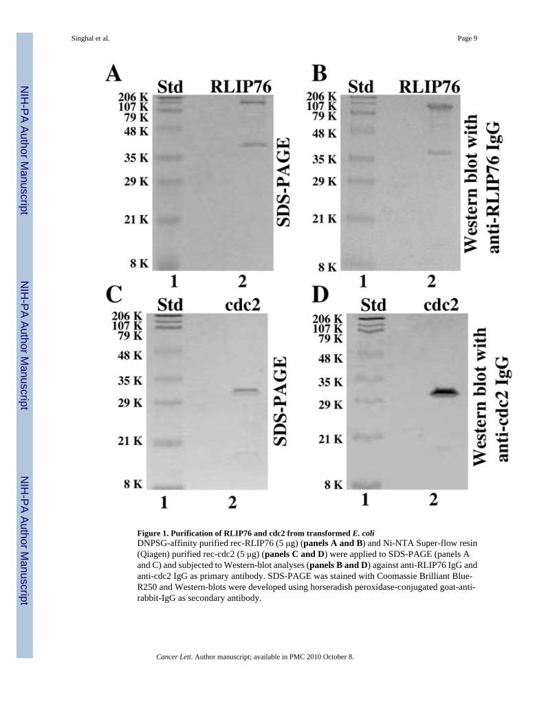
Figure 1. Purification of RLIP76 and cdc2 from transformed E. coliDNPSG-affinity purified rec-RLIP76 (5 μg) (panels A and B) and Ni-NTA Super-flow resin(Qiagen) purified rec-cdc2 (5 μg) (panels C and D) were applied to SDS-PAGE (panels Aand C) and subjected to Western-blot analyses (panels B and D) against anti-RLIP76 IgG andanti-cdc2 IgG as primary antibody. SDS-PAGE was stained with Coomassie Brilliant Blue-R250 and Western-blots were developed using horseradish peroxidase-conjugated goat-anti-rabbit-IgG as secondary antibody.
Singhal et al. Page 9
Cancer Lett. Author manuscript; available in PMC 2010 October 8.
NIH
-PA Author Manuscript
NIH
-PA Author Manuscript
NIH
-PA Author Manuscript
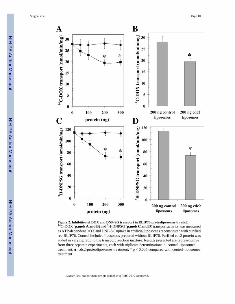
Figure 2. Inhibition of DOX and DNP-SG transport in RLIP76-proteoliposomes by cdc214C-DOX (panels A and B) and 3H-DNPSG (panels C and D) transport activity was measuredas ATP-dependent DOX and DNP-SG uptake in artificial liposomes reconstituted with purifiedrec-RLIP76. Control included liposomes prepared without RLIP76. Purified cdc2 protein wasadded in varying ratio to the transport reaction mixture. Results presented are representativefrom three separate experiments, each with triplicate determinations. •, control-liposomestreatment; ■, cdc2-proteoliposomes treatment; * p < 0.005 compared with control-liposomestreatment
Singhal et al. Page 10
Cancer Lett. Author manuscript; available in PMC 2010 October 8.
NIH
-PA Author Manuscript
NIH
-PA Author Manuscript
NIH
-PA Author Manuscript
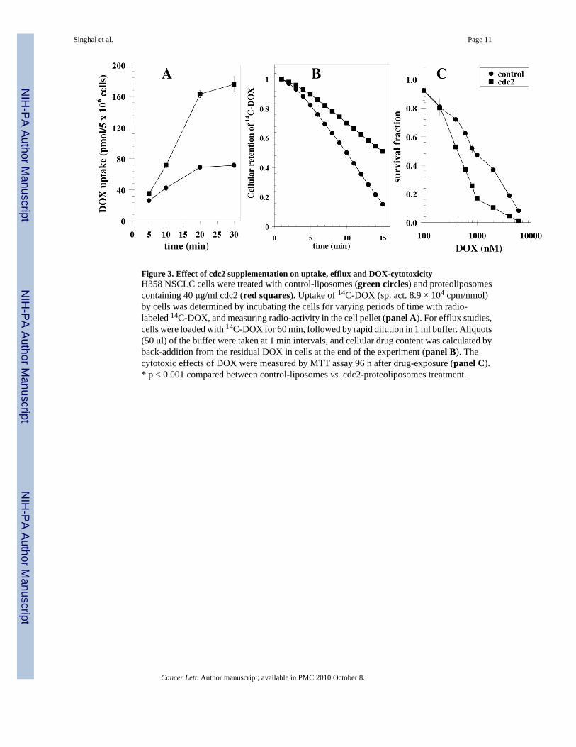
Figure 3. Effect of cdc2 supplementation on uptake, efflux and DOX-cytotoxicityH358 NSCLC cells were treated with control-liposomes (green circles) and proteoliposomescontaining 40 μg/ml cdc2 (red squares). Uptake of 14C-DOX (sp. act. 8.9 × 10⁴ cpm/nmol)by cells was determined by incubating the cells for varying periods of time with radio-labeled 14C-DOX, and measuring radio-activity in the cell pellet (panel A). For efflux studies,cells were loaded with 14C-DOX for 60 min, followed by rapid dilution in 1 ml buffer. Aliquots(50 μl) of the buffer were taken at 1 min intervals, and cellular drug content was calculated byback-addition from the residual DOX in cells at the end of the experiment (panel B). Thecytotoxic effects of DOX were measured by MTT assay 96 h after drug-exposure (panel C).* p < 0.001 compared between control-liposomes vs. cdc2-proteoliposomes treatment.
Singhal et al. Page 11
Cancer Lett. Author manuscript; available in PMC 2010 October 8.
NIH
-PA Author Manuscript
NIH
-PA Author Manuscript
NIH
-PA Author Manuscript
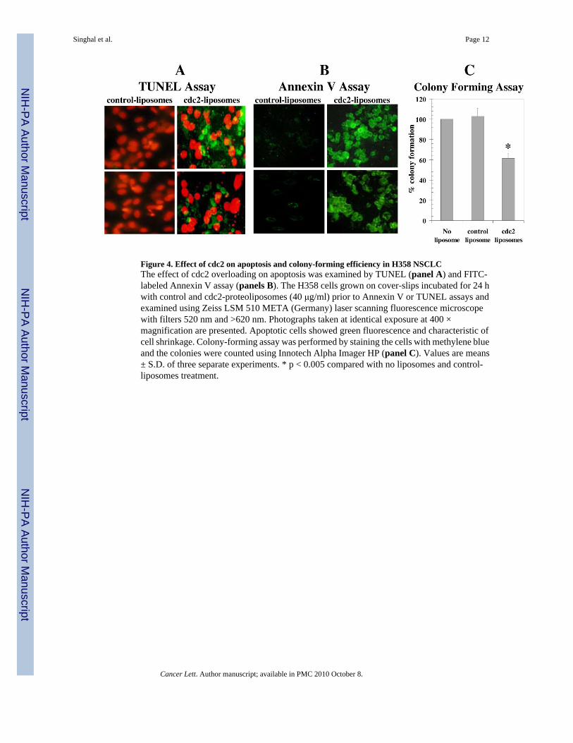
Figure 4. Effect of cdc2 on apoptosis and colony-forming efficiency in H358 NSCLCThe effect of cdc2 overloading on apoptosis was examined by TUNEL (panel A) and FITC-labeled Annexin V assay (panels B). The H358 cells grown on cover-slips incubated for 24 hwith control and cdc2-proteoliposomes (40 μg/ml) prior to Annexin V or TUNEL assays andexamined using Zeiss LSM 510 META (Germany) laser scanning fluorescence microscopewith filters 520 nm and >620 nm. Photographs taken at identical exposure at 400 ×magnification are presented. Apoptotic cells showed green fluorescence and characteristic ofcell shrinkage. Colony-forming assay was performed by staining the cells with methylene blueand the colonies were counted using Innotech Alpha Imager HP (panel C). Values are means± S.D. of three separate experiments. * p < 0.005 compared with no liposomes and control-liposomes treatment.
Singhal et al. Page 12
Cancer Lett. Author manuscript; available in PMC 2010 October 8.
NIH
-PA Author Manuscript
NIH
-PA Author Manuscript
NIH
-PA Author Manuscript
Related Documents