Leptin Inhibits Apoptosis in Thymus through a Janus Kinase-2-Independent, Insulin Receptor Substrate-1/ Phosphatidylinositol-3 Kinase-Dependent Pathway Eli Mansour, Fernanda G. Pereira, Eliana P. Arau ´ jo, Maria E. C. Amaral, Joseane Morari, Natasha R. Ferraroni, Diogenes S. Ferreira, Irene Lorand-Metze, and Lı ´cio A. Velloso Department of Internal Medicine, State University of Campinas, 13083-970 Campinas SP, Brazil The cytokine-like hormone leptin is known to exert important functions on the modulation of immune responses. Some of these effects are dependent on the property of leptin to mod- ulate the apoptosis of thymic cells. In the present study, we used Wistar rats to investigate the molecular mechanisms in- volved in leptin-dependent control of apoptosis in thymus. Apoptosis was evaluated by flow cytometry and ELISA for nucleosome determination, whereas signal transduction was evaluated by immunoprecipitation, immunoblot, and confo- cal microscopy. The Ob receptor (ObR) was expressed in most thymic cells and its relative amount reduced progressively during thymocyte maturation. ObR expression was colocal- ized with Janus kinase (JAK)-2 and signal transducer and activator of transcription-3, and an acute, in vivo, injection of leptin promoted the tyrosine phosphorylation of JAK-2 and the engagement of signal transducer and activator of tran- scription-3. The treatment with leptin also led to the tyrosine phosphorylation of insulin receptor substrate (IRS)-1 and serine phosphorylation of Akt. Chronic treatment with leptin reduced thymic apoptosis, an effect that was not inhibited by the JAK inhibitor AG 490 but was significantly inhibited by the phosphatidylinositol 3-kinase inhibitor LY 294002 and an anti- sense oligonucleotide to IRS-1. Thus, leptin inhibits the apo- ptosis of thymic cells through a mechanism that is indepen- dent of the activation of JAK-2 but depends on the engagement of the IRS-1/phosphatidylinositol 3-kinase pathway. (Endocri- nology 147: 5470 –5479, 2006) L EPTIN, THE PRODUCT of the ob gene, is a cytokine-like hormone, produced mostly by the adipose tissue in direct proportion to whole-body fat mass (1). In recent years a role for leptin in the regulation of immune response has been uncovered (2). Apparently leptin acts as a link between the nutritional status and the control of immune system activity (2). Under physiological conditions, the increase in body fat mass that follows a period of overeating, leads to an increased leptin activity in specialized neurons of the hypo- thalamus, inducing energy expenditure and reducing food intake (1, 3). In addition, leptin regulates different facets of the immune response, such as the promotion of an enhance- ment of peripheral T cell activity and proliferation (4, 5), activation of monocyte response (6, 7), regulation of cytokine production (8), and modulation of immune response during autoimmunity (9). Most leptin actions are delivered through the activation of the IL-6/gp 130-like Ob receptor (ObR) (10). Like other mem- bers of the class I cytokine family, the ObR lacks intrinsic tyrosine kinase activity and depends on the activation of an intracellular kinase to achieve full engagement of its intra- cellular signal transduction pathway (10). Upon leptin bind- ing, the ObR engages Janus kinase (JAK)-2, inducing its au- tophosphorylation at tyrosine residues, which is followed by tyrosine phosphorylation of the ObR and subsequent re- cruitment, tyrosine phosphorylation, and induction of dimerization of signal transducer and activator of transcrip- tion (STAT)-3, which migrates to the nucleus and finally regulates gene transcription (10 –12). Besides its effects on the classical JAK/STAT signaling pathway, which provides a direct access to the nucleus, leptin activates several other intracellular signaling pathways such as the MAPK cascade (13–15), phosphatidylinositol 3-kinase (PI 3-kinase)/Akt (16), SH2-B (17) and insulin receptor substrate (IRS)-1 (18). Through these pathways, leptin may be integrated to a com- plex intracellular cross talk system that regulates functions such as cell growth, mitogenesis, metabolism, and apoptosis (1, 2, 19). One of the most remarkable aspects of malnutrition is the atrophy of the thymus and the development of immuno- suppression (20, 21). Starvation causes a loss of normal thy- mic architecture and reduces the number of cortical thymo- cytes by increasing the rate of apoptosis (22). In addition, starvation and chronic malnutrition promotes a fall in leptin levels (23), and this phenomenon has been proposed to play a role in the anomalous thymic morphology and function observed during nutritional deprivation states (22). This hy- pothesis has been further supported by the fact that both humans and rodents with defective leptin production present a significant restoration of different aspects of the immune response when treated with exogenous leptin (8, 22, 24) and that the treatment of leptin-deficient ob/ob mice with exogenous leptin reduces thymic atrophy by increasing its cellularity (22). First Published Online July 27, 2006 Abbreviations: FITC, Fluorescein isothiocyanate isomer 1; RPE, phy- coerythrin; IRS, insulin receptor substrate; JAK, Janus kinase; ObR, Ob receptor; PI 3-kinase, phosphatidylinositol 3-kinase; RT, reverse tran- scription; STAT, signal transducer and activator of transcription. Endocrinology is published monthly by The Endocrine Society (http:// www.endo-society.org), the foremost professional society serving the endocrine community. 0013-7227/06/$15.00/0 Endocrinology 147(11):5470 –5479 Printed in U.S.A. Copyright © 2006 by The Endocrine Society doi: 10.1210/en.2006-0223 5470

Welcome message from author
This document is posted to help you gain knowledge. Please leave a comment to let me know what you think about it! Share it to your friends and learn new things together.
Transcript
Leptin Inhibits Apoptosis in Thymus through a JanusKinase-2-Independent, Insulin Receptor Substrate-1/Phosphatidylinositol-3 Kinase-Dependent Pathway
Eli Mansour, Fernanda G. Pereira, Eliana P. Araujo, Maria E. C. Amaral, Joseane Morari,Natasha R. Ferraroni, Diogenes S. Ferreira, Irene Lorand-Metze, and Lıcio A. Velloso
Department of Internal Medicine, State University of Campinas, 13083-970 Campinas SP, Brazil
The cytokine-like hormone leptin is known to exert importantfunctions on the modulation of immune responses. Some ofthese effects are dependent on the property of leptin to mod-ulate the apoptosis of thymic cells. In the present study, weused Wistar rats to investigate the molecular mechanisms in-volved in leptin-dependent control of apoptosis in thymus.Apoptosis was evaluated by flow cytometry and ELISA fornucleosome determination, whereas signal transduction wasevaluated by immunoprecipitation, immunoblot, and confo-cal microscopy. The Ob receptor (ObR) was expressed in mostthymic cells and its relative amount reduced progressivelyduring thymocyte maturation. ObR expression was colocal-ized with Janus kinase (JAK)-2 and signal transducer andactivator of transcription-3, and an acute, in vivo, injection of
leptin promoted the tyrosine phosphorylation of JAK-2 andthe engagement of signal transducer and activator of tran-scription-3. The treatment with leptin also led to the tyrosinephosphorylation of insulin receptor substrate (IRS)-1 andserine phosphorylation of Akt. Chronic treatment with leptinreduced thymic apoptosis, an effect that was not inhibited bythe JAK inhibitor AG490 but was significantly inhibited by thephosphatidylinositol 3-kinase inhibitor LY294002 and an anti-sense oligonucleotide to IRS-1. Thus, leptin inhibits the apo-ptosis of thymic cells through a mechanism that is indepen-dent of the activation of JAK-2 but depends on the engagementof the IRS-1/phosphatidylinositol 3-kinase pathway. (Endocri-nology 147: 5470–5479, 2006)
LEPTIN, THE PRODUCT of the ob gene, is a cytokine-likehormone, produced mostly by the adipose tissue in
direct proportion to whole-body fat mass (1). In recent yearsa role for leptin in the regulation of immune response hasbeen uncovered (2). Apparently leptin acts as a link betweenthe nutritional status and the control of immune systemactivity (2). Under physiological conditions, the increase inbody fat mass that follows a period of overeating, leads to anincreased leptin activity in specialized neurons of the hypo-thalamus, inducing energy expenditure and reducing foodintake (1, 3). In addition, leptin regulates different facets ofthe immune response, such as the promotion of an enhance-ment of peripheral T cell activity and proliferation (4, 5),activation of monocyte response (6, 7), regulation of cytokineproduction (8), and modulation of immune response duringautoimmunity (9).
Most leptin actions are delivered through the activation ofthe IL-6/gp 130-like Ob receptor (ObR) (10). Like other mem-bers of the class I cytokine family, the ObR lacks intrinsictyrosine kinase activity and depends on the activation of anintracellular kinase to achieve full engagement of its intra-cellular signal transduction pathway (10). Upon leptin bind-ing, the ObR engages Janus kinase (JAK)-2, inducing its au-
tophosphorylation at tyrosine residues, which is followed bytyrosine phosphorylation of the ObR and subsequent re-cruitment, tyrosine phosphorylation, and induction ofdimerization of signal transducer and activator of transcrip-tion (STAT)-3, which migrates to the nucleus and finallyregulates gene transcription (10–12). Besides its effects on theclassical JAK/STAT signaling pathway, which provides adirect access to the nucleus, leptin activates several otherintracellular signaling pathways such as the MAPK cascade(13–15), phosphatidylinositol 3-kinase (PI 3-kinase)/Akt(16), SH2-B (17) and insulin receptor substrate (IRS)-1 (18).Through these pathways, leptin may be integrated to a com-plex intracellular cross talk system that regulates functionssuch as cell growth, mitogenesis, metabolism, and apoptosis(1, 2, 19).
One of the most remarkable aspects of malnutrition is theatrophy of the thymus and the development of immuno-suppression (20, 21). Starvation causes a loss of normal thy-mic architecture and reduces the number of cortical thymo-cytes by increasing the rate of apoptosis (22). In addition,starvation and chronic malnutrition promotes a fall in leptinlevels (23), and this phenomenon has been proposed to playa role in the anomalous thymic morphology and functionobserved during nutritional deprivation states (22). This hy-pothesis has been further supported by the fact that bothhumans and rodents with defective leptin productionpresent a significant restoration of different aspects of theimmune response when treated with exogenous leptin (8, 22,24) and that the treatment of leptin-deficient ob/ob mice withexogenous leptin reduces thymic atrophy by increasing itscellularity (22).
First Published Online July 27, 2006Abbreviations: FITC, Fluorescein isothiocyanate isomer 1; RPE, phy-
coerythrin; IRS, insulin receptor substrate; JAK, Janus kinase; ObR, Obreceptor; PI 3-kinase, phosphatidylinositol 3-kinase; RT, reverse tran-scription; STAT, signal transducer and activator of transcription.Endocrinology is published monthly by The Endocrine Society (http://www.endo-society.org), the foremost professional society serving theendocrine community.
0013-7227/06/$15.00/0 Endocrinology 147(11):5470–5479Printed in U.S.A. Copyright © 2006 by The Endocrine Society
doi: 10.1210/en.2006-0223
5470
Although the functional effects of leptin on thymic cellu-larity and apoptosis have been well characterized, the mo-lecular mechanisms involved in this control are poorly un-derstood. Therefore, the objective of the present study wasto evaluate the intracellular transduction pathways that par-ticipate in leptin-induced inhibition of apoptosis in the thy-mus of Wistar rats.
Materials and MethodsExperimental animals
Four-week-old male Wistar rats and Lepdb (db/db) and C57BLKS/Jmice were obtained from the University of Campinas Breeding Center.The Lepdb (db/db) mice were originally purchased from the JacksonLaboratory (Bar Harbor, ME) and are currently established as a colonyat the University of Campinas Breeding Center. The animals were al-lowed access to standard rodent chow and water ad libitum. All exper-iments involving animals were in accordance with the guidelines of theBrazilian College for Animal Experimentation and approved by theUniversity of Campinas Ethical Committee. Room temperature wasmaintained between 21 and 23 C with 12-h light, 12-h dark cycle. Theanimals were age matched for individual experiments and randomlydistributed into treatment or control groups.
Materials
Antibodies against JAK-2 (sc-278), STAT-3 (sc-483), phosphotyrosine(sc-508), IRS-1 (sc-559) ObR (sc-8325), and phospho-(Ser473) Akt (sc-9271)were from Santa Cruz Biotechnology (Santa Cruz, CA). Conjugatedmouse antirat CD3-fluorescein isothiocyanate isomer 1 (FITC), CD4-phycoerythrin (RPE), and CD8-RPE-Cy5 were from Serotec, Ltd. (Ox-ford, UK). 125I-protein A Sepharose and nitrocellulose paper (HybondECL, 0.45 �m) were from Amersham (Buckinghamshire, UK). Protein ASepharose 6 MB was from Pharmacia (Uppsala, Sweden), and protein AAgarose (AG-Plus) (sc-2003) was from Santa Cruz Biotechnology. Lep-tin, the JAK inhibitor AG490 (tyrphostin B42), and the PI 3-kinase in-hibitor LY294002 were acquired from Calbiochem (La Jolla, CA). Sodiumamobarbital was purchased from Eli Lilly & Co. (Indianapolis, IN). Trisbase, phenylmethylsulfonylfluoride, aprotinin, dithiothreitol, TritonX-100, Tween 20, glycerol, affinity-purified rabbit antimouse IgG, andBSA (fraction V) were obtained from Sigma (St. Louis, MO). RPMI 1640and reagents for cell culture were purchased from Invitrogen Corp.(Carlsbad, CA). Sense (5�-ACC CAC TCC TAT CCC G-3�) (IRS-1SO) andantisense (5�-CGG GAT AGG AGT GGG T-3�) (IRS-1ASO) phosphor-thioate oligonucleotides specific for IRS-1 were produced by Invitrogen(25–27). Sequence was selected among three unrelated pairs of oligo-nucleotides on the basis of their ability to block IRS-1 protein expressionas evaluated by immunoblot of total protein extracts of thymus tissueusing specific anti-IRS-1 antibodies. The oligonucleotide sequences weresubmitted to BLAST analyses (www.ncbi.nlm.nih.gov) and matchedonly for the Rattus norvegicus IRS-1 coding sequence (NCBI/NM 012969).The nucleosome ELISA kit (catalog no. QIA25) was acquired from On-cogene Research Products (Boston, MA), and the flow cytometry an-nexin-V apoptosis detection kit (Apoptest K2350) was purchased fromDako Corp. (Carpinteria, CA). The leptin ELISA kit was purchased fromLinco Research Inc. (St. Charles, MO). TRIzol reagent was purchasedfrom Invitrogen, and Moloney murine leukemia virus reverse transcrip-tase was from CLONTECH (Mountain View, CA).
Protocols for acute treatment with leptin and proteinanalysis by immunoprecipitation and immunoblotting
Rats were anesthetized by ip injection of sodium amobarbital (15mg/kg body weight) and submitted to the surgical procedure as soonas the anesthesia was assured by the loss of pedal and corneal reflexes.The abdominal cavity was opened, the portal vein was exposed, and invivo stimulation was obtained by the injection of 400 �l saline (0.9%NaCl), insulin (10�6 m), or leptin (10�6, 10�8, or 10�10 m). After thepredetermined elapsed time, the thymus was removed after a thora-cotomy. The tissue was minced coarsely and homogenized immediatelyin extraction buffer [1% Triton X-100 and 100 mm Tris (pH 7.4) con-
taining 100 mm sodium pyrophosphate, 100 mm sodium fluoride, 10 mmEDTA, 10 mm sodium vanadate, 2 mm phenylmethylsulfonylfluoride,and 0.1 mg aprotinin/ml] at 4 C with a Polytron PTA 20S generator(model PT 10/35; Brinkmann Instruments, Inc., Westbury, NY) operatedat maximum speed for 30 sec. The extracts were centrifuged at 9000 �g and 4 C in a 70.1 Ti rotor (Beckman, Palo Alto, CA) for 20 min to removeinsoluble material, and the supernatants were used for immunoprecipi-tation with anti-JAK-2, -STAT-3, or -IRS-1 antibodies, and the technicalprocedures were performed as previously described (28, 29). Proteinquantification in the supernatants was determined by the Bradfordmethod (30). Immunoprecipitates were separated by SDS-PAGE, trans-ferred to nitrocellulose membranes, and blotted with antibodies an-tiphosphotyrosine. In direct immunoblotting experiments, total proteinextracts were separated by SDS-PAGE, transferred to nitrocellulosemembranes, and antiphospho-(Ser473) Akt and anti-IRS-1 antibodiesused for blotting. Visualization of specific protein bands was performedby incubating membranes with 125 I-protein A followed by exposure tox-ray films (Kodak, Rochester, NY). In some experiments, the rats werepretreated with 100 �l AG490 10�4 m or 400 �l LY294002 5.0 �m and thensubmitted to acute treatment with leptin.
Protocols for chronic treatment with leptin and evaluationof apoptosis
For these experiments, the rats were randomly divided into eightgroups and treated by ip injection according to one of the followingprotocols: 400 �l saline; 400 �l leptin (10�6 m); 100 �l AG 490 (10�4 m)� 400 �l leptin; 400 �l LY294002 (5 �m) � 400 �l leptin; 100 �l IRS-1SO(4.0 nmol) � 400 �l leptin; 100 �l IRS-1SO (4.0 nmol) � 400 �l salinesolution; 100 �l IRS-1ASO (4.0 nmol) � 400 �l leptin; or 100 �l IRS-1ASO(4.0 nmol) � 400 �l saline solution. The treatment consisted of two doseseach day for 3 consecutive days. On the morning of the fourth day, theanimals were anesthetized as described earlier, and the thymus wasremoved by thoracotomy. Suspensions of thymocytes in PBS/2% fetalcalf serum (Cult Lab, Campinas, Brazil) were obtained using a Potterglass and used for apoptosis detection by flow cytometry. In someexperiments fragments of thymus were used for nucleosome determi-nation by ELISA. To evaluate the efficiency of the treatments with thethree inhibitors to maintain continuous inhibition upon their respectivetargets, rats treated according to the protocols above were used 1.0, 4.0,8.0, or 12.0 h after the dose in the morning of the third day of treatmentto determine JAK-2, STAT-3, and IRS-1 tyrosine phosphorylation afterAG490 treatment; Akt serine phosphorylation after LY294002 treatment;and IRS-1 protein expression after IRS-1ASO treatment.
Short-term culture of thymocytes
To prepare isolated thymocytes, rats were anesthetized; thymuseswere obtained and gently passed through a steel net. Cells were washedwith and resuspended in ice-cold RPMI 1640 containing penicillin-strep-tomycin, l-glutamine, and 0.5% fetal calf serum. Twenty-four groups of5.0 � 106 cells were placed in 2.0-ml culture dishes and treated withleptin (10�8 m), AG490 (10�6 m) � leptin (10�8 m), LY294002 (10�8 m) �leptin (10�8 m), IRS-1ASO (4.0 nmol) � leptin (10�8 m), or IRS-1ASO (4.0nmol) alone. Apoptosis was evaluated after 12 h using the ELISA nu-cleosome method.
Determination of blood leptin levels on a time course afterexogenous leptin injection
Forty rats were randomly divided into five groups of eight rats each.The first group received no treatment. The second group was subdividedinto two groups of four rats; the animals were anesthetized and receiveda dose of 400 �l leptin 10�6 m or an equal volume of saline through thecava vein. Blood was collected from the tail vein at 3.0 min. The third,fourth, and fifth groups were also subdivided into groups of four ratsand treated with an ip injection of 400 �l leptin 10�6 m or an equalvolume of saline, and blood was collected from the tail vein at 1, 6, and12 h, respectively. The rats were anesthetized before blood collection.Leptin was determined in the samples using a commercially availableELISA kit following the protocol suggested by the manufacturer (LincoResearch).
Mansour et al. • Leptin and Apoptosis in Thymus Endocrinology, November 2006, 147(11):5470–5479 5471
Detection of apoptosis by flow cytometry
The thymocytes, prepared as described above, were tested for apo-ptosis using the annexin-V technique (31). Ninety-six microliters of a cellsuspension at the final concentration of 1.0 � 106 cells/ml were incu-bated with 1.0 �l FITC-conjugated annexin-V and 2.5 �l propidiumiodide, as suggested by the manufacturer. Cells were kept on ice andincubated in the dark for 10 min before diluting the cells to 250 �l withbinding buffer and analyzing using a FACScalibur flow cytometry an-alyzer and CellQuest software (Becton Dickinson, San Jose, CA). A totalof 10,000 cells were acquired. Unlabeled cells suspended in PBS wereused as a negative control and for the determination of gates to be usedin the apoptosis assays. Apoptotic cells were measured in the annexin-V-positive propidium iodide-negative quadrant and divided by the totalnumber of cells in the gated region.
Detection of apoptosis by nucleosome ELISA
Rats were treated by ip injection as described for the chronic protocolwith saline, leptin, AG490, IRS-1ASO, and/or IRS-1SO. On the morning ofthe fourth day, the animals were anesthetized, and the thymus wasremoved by thoracotomy. Isolated thymocytes were treated as describedabove and used 12 h after treatment. Cells were lysed and centrifuged,and cytoplasmatic histone-associated DNA fragments were determinedin the supernatant by ELISA (32), as suggested by the manufacturer.
Flow cytometry for determination of cell markers
Rats were anesthetized as described earlier and the thymus removedby thoracotomy. Suspensions (1.0 � 106 cells/ml) of thymocytes inPBS/2% fetal bovine serum were obtained using a Potter glass. Samplesof 100 �l of the freshly prepared cell suspensions were incubated for 20min in the dark, at room temperature, with 10 �l of the following panelsof antibodies: CD3FITC/CD4RPE/CD8RPE-Cy5 or CD4RPE/CD8RPE-Cy5/ObR (the ObR antibody was sc-8325 from Santa Cruz). Thereafterthe cells were washed and incubated with FITC-conjugated secondaryantibody. A three-color analysis was made using a FACScalibur, and theCellQuest software (Becton Dickinson) was used for quantitative anal-ysis. In addition a cell volume analysis was used to evaluate the ex-pression of cell markers in maturating cells.
RT-PCR
Total RNA was extracted from isolated thymocytes (�107 cells/an-imal) and hypothalami of Wistar rats, Lepdb (db/db), and C57BLKS/Jmice using TRIzol reagent according to the instructions of the manu-facturer. Reverse transcription (RT) was carried out using 1.0 �g totalRNA using SuperScript reverse transcriptase (200 U/�l) and oligo (dT)(50 mm) in a 30-�l reaction volume (5 � RT buffer, 10 mm deoxynucle-otide triphosphate, and 40 U/�l Rnase-free inhibitor). The RTs involveda 50-min incubation at 42 C and a 15-min incubation at 70 C. The PCRproducts were submitted to 1.5% agarose gel electrophoresis containingethidium bromide and visualized by excitation under UV light. Pho-todocumentation was performed using the Nucleovision system (Nucle-oTech, San Mateo, CA) and band quantification was performed using theGel Expert software (NucleoTech). First-strand cDNA was PCR ampli-fied using a primer from the transmembrane region of the ObR sequence(NCBI/NM 012596) opposed with two primers capable of determiningthe short or the long forms of the receptor. The sequence of the trans-membrane region primer was 5�-CAG GGC TGT ATG TCA TTG-3�; thesequence of the primer for the short form was 5�-GTG CCC AGG AACAAT TCT-3�; and the sequence of the primer for the long form was5�-CCA GAG AAG TTA GCA CTG-3�. Control amplifications werecarried out in the presence of RNA template and polymerase but in theabsence of reverse transcriptase. The �-actin mRNA was amplified in allsamples as control for quality and amount of RNA using the primers5�-CGT AAA GAC CTC TAT TGC CAA-3� and 5�-AGC CAT GCC AAATGT GTC AT-3�, based on the sequence NCBI/NM 031144. Reactionswere carried out at 94 C for 2 min, followed by 50 cycles, each consistingof 30 sec at 92 C, 30 sec at 50 C, and 1 min at 72 C, followed by single-cycleextension for 10 min at 72 C. �-Actin was amplified by a similar protocolexcept that the reaction was limited to 30 cycles. This method, withminor modifications, has been used previously (33).
Immunohistochemistry
Thymus fragments were obtained from three control rats. Hydrated,5-�m sections of paraformaldehyde-fixed, paraffin-embedded tissuewere stained by the double-staining fluorescence method. Sections wereincubated for 30 min with 2% normal rabbit or normal goat sera at roomtemperature and then exposed for 12 h in a moister chamber at 4 C tothe panel of primary antibodies against JAK-2 (1:20)/ObR (1:20), STAT-3(1:20)/ObR (1:20), or Akt (1:50)/ObR (1:20) followed by incubation withFITC-conjugated and rhodamine-conjugated secondary antibodies. Im-ages were obtained with a laser confocal microscope (LSM510; Zeiss,New York, NY). Secondary antibody specificity was tested in a series ofpositive and negative control measurements. Complete description ofthe method has been published elsewhere (25).
Statistical analysis
All numerical results are expressed as the mean � sem of the indicatednumber of experiments. The results of blots are presented as directcomparisons of bands in autoradiographs and quantified by densitom-etry using the Scion Image software (Scion Corp., Frederick, MD). Datawere analyzed by the two-tailed unpaired Student’s t test or repeat-measures ANOVA (one-way or two-way ANOVA) followed by post hocanalysis of significance (Bonferroni test) when appropriate, comparingexperimental and control groups. The level of significance was set at P �0.05.
ResultsTime course of blood leptin levels after exogenous leptinadministration
The treatment of rats with a single dose of exogenousleptin promoted a variation of blood levels of the hormonethat reached a peak at 3.0 min and returned to basal levelsafter 12 h (Table 1).
Leptin inhibits thymic apoptosis
The treatment of rats with two daily doses of leptin (10�6
m) promoted a significant reduction of apoptosis of thymiccells as determined by the nucleosome-ELISA detectionmethod (�30% inhibition of apoptosis, P � 0.05) (Fig. 1A)and the annexin-V/flow cytometry method (�15% inhibi-tion of apoptosis, P � 0.05) (Fig. 1B).
Leptin activates JAK-2/STAT-3 and IRS-1/Akt signalingin thymus
The acute injection of a single dose of leptin (10�6 m)induced the rapid tyrosine phosphorylation of the intracel-lular kinase JAK-2 in thymic tissue (Fig. 2A, first blot). Thiseffect was detected at 1.0 min and lasted for at least 5.0 min.To evaluate the dose dependency of this phenomenon, ratswere acutely treated with a single dose of leptin (10�6, 10�8,or 10�10 m), and the thymus was obtained after 3 min (datanot shown). The highest tyrosine phosphorylation of JAK-2was obtained with the dose of 10�6 m (3.5-fold increase vs.
TABLE 1. Time course of blood leptin levels after exogenousleptin administration
0.0 min 3.0 min 1.0 h 6.0 h 12.0 h
Saline treated 2.33 2.12 2.52 2.02 2.27SEM (�) 0.33 0.12 0.54 0.45 0.44Leptin treated 2.32 52.13a 29.88a 3.34a 2.89SEM (�) 0.23 4.56 3.12 0.25 0.40
n � 4.a P � 0.05 vs. saline treated.
5472 Endocrinology, November 2006, 147(11):5470–5479 Mansour et al. • Leptin and Apoptosis in Thymus
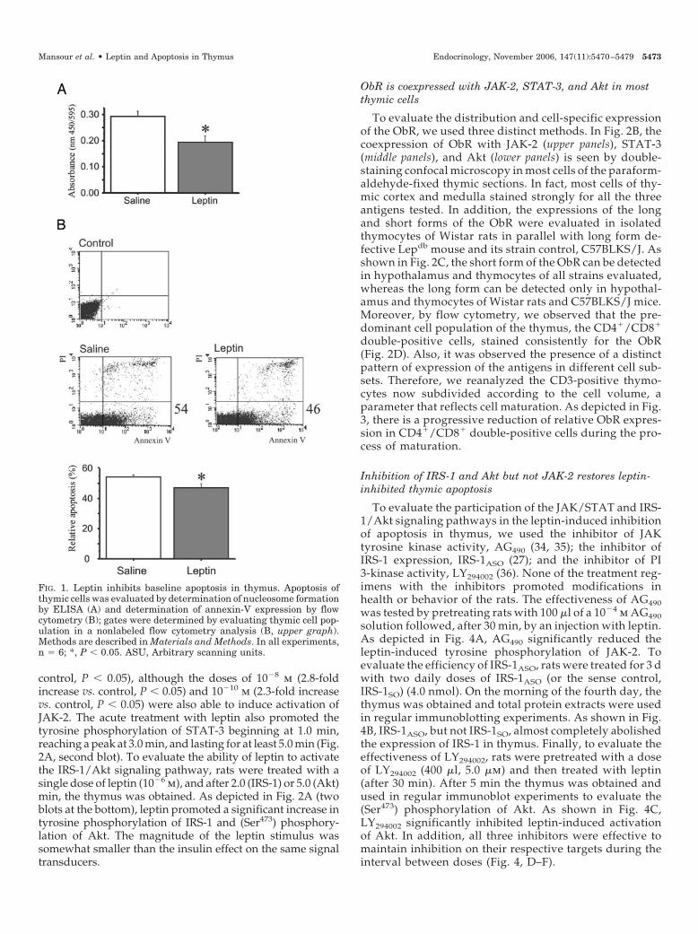
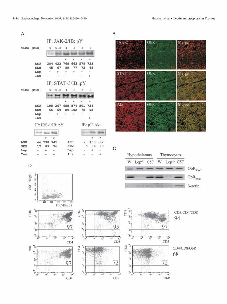
control, P � 0.05), although the doses of 10�8 m (2.8-foldincrease vs. control, P � 0.05) and 10�10 m (2.3-fold increasevs. control, P � 0.05) were also able to induce activation ofJAK-2. The acute treatment with leptin also promoted thetyrosine phosphorylation of STAT-3 beginning at 1.0 min,reaching a peak at 3.0 min, and lasting for at least 5.0 min (Fig.2A, second blot). To evaluate the ability of leptin to activatethe IRS-1/Akt signaling pathway, rats were treated with asingle dose of leptin (10�6 m), and after 2.0 (IRS-1) or 5.0 (Akt)min, the thymus was obtained. As depicted in Fig. 2A (twoblots at the bottom), leptin promoted a significant increase intyrosine phosphorylation of IRS-1 and (Ser473) phosphory-lation of Akt. The magnitude of the leptin stimulus wassomewhat smaller than the insulin effect on the same signaltransducers.
ObR is coexpressed with JAK-2, STAT-3, and Akt in mostthymic cells
To evaluate the distribution and cell-specific expressionof the ObR, we used three distinct methods. In Fig. 2B, thecoexpression of ObR with JAK-2 (upper panels), STAT-3(middle panels), and Akt (lower panels) is seen by double-staining confocal microscopy in most cells of the paraform-aldehyde-fixed thymic sections. In fact, most cells of thy-mic cortex and medulla stained strongly for all the threeantigens tested. In addition, the expressions of the longand short forms of the ObR were evaluated in isolatedthymocytes of Wistar rats in parallel with long form de-fective Lepdb mouse and its strain control, C57BLKS/J. Asshown in Fig. 2C, the short form of the ObR can be detectedin hypothalamus and thymocytes of all strains evaluated,whereas the long form can be detected only in hypothal-amus and thymocytes of Wistar rats and C57BLKS/J mice.Moreover, by flow cytometry, we observed that the pre-dominant cell population of the thymus, the CD4�/CD8�
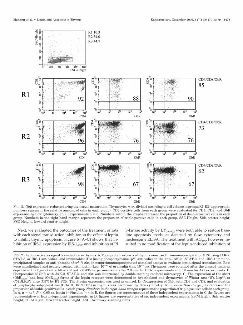
double-positive cells, stained consistently for the ObR(Fig. 2D). Also, it was observed the presence of a distinctpattern of expression of the antigens in different cell sub-sets. Therefore, we reanalyzed the CD3-positive thymo-cytes now subdivided according to the cell volume, aparameter that reflects cell maturation. As depicted in Fig.3, there is a progressive reduction of relative ObR expres-sion in CD4�/CD8� double-positive cells during the pro-cess of maturation.
Inhibition of IRS-1 and Akt but not JAK-2 restores leptin-inhibited thymic apoptosis
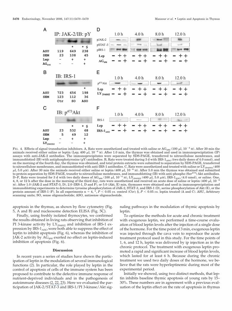
To evaluate the participation of the JAK/STAT and IRS-1/Akt signaling pathways in the leptin-induced inhibitionof apoptosis in thymus, we used the inhibitor of JAKtyrosine kinase activity, AG490 (34, 35); the inhibitor ofIRS-1 expression, IRS-1ASO (27); and the inhibitor of PI3-kinase activity, LY294002 (36). None of the treatment reg-imens with the inhibitors promoted modifications inhealth or behavior of the rats. The effectiveness of AG490was tested by pretreating rats with 100 �l of a 10�4 m AG490solution followed, after 30 min, by an injection with leptin.As depicted in Fig. 4A, AG490 significantly reduced theleptin-induced tyrosine phosphorylation of JAK-2. Toevaluate the efficiency of IRS-1ASO, rats were treated for 3 dwith two daily doses of IRS-1ASO (or the sense control,IRS-1SO) (4.0 nmol). On the morning of the fourth day, thethymus was obtained and total protein extracts were usedin regular immunoblotting experiments. As shown in Fig.4B, IRS-1ASO, but not IRS-1SO, almost completely abolishedthe expression of IRS-1 in thymus. Finally, to evaluate theeffectiveness of LY294002, rats were pretreated with a doseof LY294002 (400 �l, 5.0 �m) and then treated with leptin(after 30 min). After 5 min the thymus was obtained andused in regular immunoblot experiments to evaluate the(Ser473) phosphorylation of Akt. As shown in Fig. 4C,LY294002 significantly inhibited leptin-induced activationof Akt. In addition, all three inhibitors were effective tomaintain inhibition on their respective targets during theinterval between doses (Fig. 4, D–F).
FIG. 1. Leptin inhibits baseline apoptosis in thymus. Apoptosis ofthymic cells was evaluated by determination of nucleosome formationby ELISA (A) and determination of annexin-V expression by flowcytometry (B); gates were determined by evaluating thymic cell pop-ulation in a nonlabeled flow cytometry analysis (B, upper graph).Methods are described in Materials and Methods. In all experiments,n � 6; *, P � 0.05. ASU, Arbitrary scanning units.
Mansour et al. • Leptin and Apoptosis in Thymus Endocrinology, November 2006, 147(11):5470–5479 5473
5474 Endocrinology, November 2006, 147(11):5470–5479 Mansour et al. • Leptin and Apoptosis in Thymus
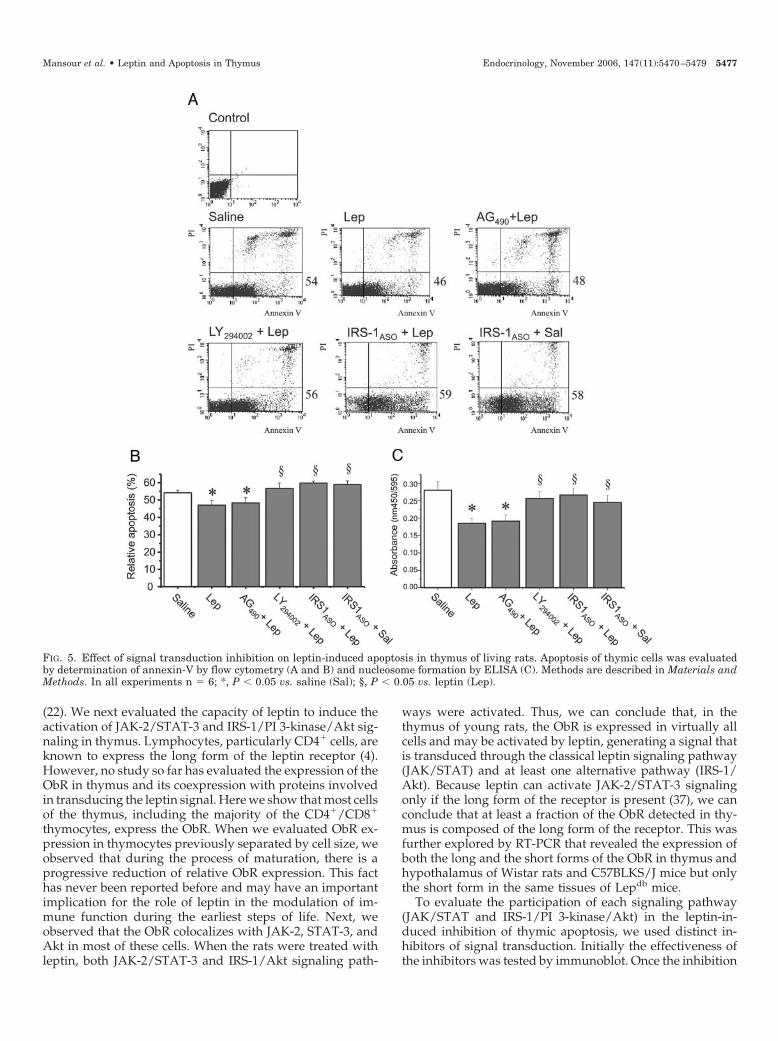
Next, we evaluated the outcomes of the treatment of ratswith each signal transduction inhibitor on the effect of leptinto inhibit thymic apoptosis. Figure 5 (A–C) shows that in-hibition of IRS-1 expression by IRS-1ASO and inhibition of PI
3-kinase activity by LY294002 were both able to restore base-line apoptosis levels, as detected by flow cytometry andnucleosome ELISA. The treatment with AG490, however, re-sulted in no modification of the leptin-induced inhibition of
FIG. 2. Leptin activates signal transduction in thymus. A, Total protein extracts of thymus were used in immunoprecipitation (IP) (using JAK-2,STAT-3, or IRS-1 antibodies) and immunoblot (IB) [using phosphotyrosine (pY) antibodies in the anti-JAK-2, -STAT-3, and -IRS-1 immuno-precipitated samples or anti-phospho-(Ser473) Akt, in nonpreimmunoprecipitated samples] assays to evaluate leptin signal transduction. Ratswere anesthetized and acutely treated with leptin (Lep; 10�6 M) or insulin (Ins; 10�6 M). Thymuses were obtained after the elapsed times asdepicted in the figure (anti-JAK-2 and anti-STAT-3 experiments) or after 2.0 min for IRS-1 experiments and 5.0 min for Akt experiments. B,Coexpression of ObR with JAK-2, STAT-3, and Akt was determined by double-staining confocal microscopy. C, The expression of the short(ObRshort) and long (ObRlong) forms of the leptin receptor were determined in hypothalami and thymocytes of Wistar rats (W), Lepdb, orC57BLKS/J mice (C57) by RT PCR. The �-actin expression was used as control. D, Coexpression of ObR with CD4 and CD8; and evaluationof lymphocyte subpopulations (CD4�/CD8�/CD3�) in thymus was performed by flow cytometry. Numbers within the graphs represent theproportion of double-positive cells in each group. Numbers in the right-hand margin represent the proportion of triple-positive cells in each group.In A, n � 4; *, P � 0.05 vs. leptin (�)/insulin (�); in B, the figures are representative of three independent experiments; in C the figures arerepresentative of four independent experiments; in D, figures are representative of six independent experiments. SSC-Height, Side scatterheight; FSC-Height, forward scatter height. ASU, Arbitrary scanning units.
FIG. 3. ObR expression reduces during thymocyte maturation. Thymocytes were divided according to cell volume in groups R1-R3 (upper graph,numbers represent the relative amount of cells in each group). CD3-positive cells from each group were evaluated for CD4, CD8, and ObRexpression by flow cytometry. In all experiments n � 6. Numbers within the graphs represent the proportion of double-positive cells in eachgroup. Numbers in the right-hand margin represent the proportion of triple-positive cells in each group. SSC-Height, Side scatter-height;FSC-Height, forward scatter height.
Mansour et al. • Leptin and Apoptosis in Thymus Endocrinology, November 2006, 147(11):5470–5479 5475
apoptosis in the thymus, as shown by flow cytometry (Fig.5, A and B) and nucleosome detection ELISA (Fig. 5C).
Finally, using freshly isolated thymocytes, we confirmedthe results obtained in living rats observing that inhibition ofPI 3-kinase activity by LY294002 and inhibition of IRS-1 ex-pression by IRS-1ASO were both able to suppress the effect ofleptin to inhibit apoptosis (Fig. 6), whereas the inhibition ofJAK-2 activity by AG490 exerted no effect on leptin-inducedinhibition of apoptosis (Fig. 6).
Discussion
In recent years a series of studies have shown the partic-ipation of leptin in the modulation of several immunologicalfunctions (2). In particular, the role played by leptin in thecontrol of apoptosis of cells of the immune system has beenproposed to contribute to the defective immune response ofnutrient-deprived individuals and in the pathogenesis ofautoimmune diseases (2, 22, 23). Here we evaluated the par-ticipation of JAK-2/STAT-3 and IRS-1/PI 3-kinase/Akt sig-
naling pathways in the modulation of thymic apoptosis byleptin.
To optimize the methods for acute and chronic treatmentwith exogenous leptin, we performed a time-course evalu-ation of blood leptin levels after the injection of a single doseof the hormone. For the time point of 3 min, exogenous leptinwas injected through the cava vein to reproduce the acutetreatment protocol used in this study. For the time points of1, 6, and 12 h, leptin was delivered by ip injection as in thechronic protocol. The treatment with exogenous leptin pro-moted a rapid and significant increase of blood leptin levels,which lasted for at least 6 h. Because during the chronictreatment we used two daily doses of the hormone, we be-lieve that the rats were hyperleptinemic during most of theexperimental period.
Initially we showed, using two distinct methods, that lep-tin inhibits baseline thymic apoptosis of young rats by 15–30%. These numbers are in agreement with a previous eval-uation of the leptin effect on the rate of apoptosis in thymus
FIG. 4. Effects of signal transduction inhibitors. A, Rats were anesthetized and treated with saline or AG490 (100 �l, 10�4 M). After 30 min theanimals received either saline or leptin (Lep; 400 �l, 10�6 M). After 1.0 min, the thymus was obtained and used in immunoprecipitation (IP)assays with anti-JAK-2 antibodies. The immunoprecipitates were separated by SDS-PAGE, transferred to nitrocellulose membranes, andimmunoblotted (IB) with antiphosphotyrosine (pY) antibodies. B, Rats were treated during 3 d with IRS-1ASO (two daily doses of 4.0 nmol), andon the morning of the fourth day, the thymus was obtained, and total protein extracts were submitted to separation by SDS-PAGE, transferredto nitrocellulose membranes, and immunoblotted with anti-IRS-1 antibodies. C, Rats were anesthetized and treated with saline or LY294002 (400�l, 5.0 �M). After 30 min the animals received either saline or leptin (400 �l, 10�6 M). After 5.0 min the thymus was obtained and submittedto protein separation by SDS-PAGE, transfer to nitrocellulose membranes, and immunoblotting (IB) with anti-phospho (Ser473) Akt antibodies.D–F, Rats were treated for 3 d with two daily doses of AG490 (100 �l, 10�4 M), LY294002 (400 �l, 5.0 �M), IRS-1ASO (4.0 nmol), or saline. One,4, 8, or 12 h after the dose in the morning of the third day, rats were anesthetized and received an acute dose of saline or leptin (400 �l, 10�6
M). After 1.0 (JAK-2 and STAT-3, D), 2.0 (IRS-1, D and F), or 5.0 (Akt, E) min, thymuses were obtained and used in immunoprecipitation andimmunoblotting experiments to determine tyrosine phosphorylation of JAK-2, STAT-3, and IRS-1 (D), serine phosphorylation of Akt (E), or theprotein amount of IRS-1 (F). In all experiments n � 4, *, P � 0.05 vs. control (Ctr); §, P � 0.05 vs. leptin treated (A and C). ASU, Arbitraryscanning units; SO, sense oligonucleotide; ASO, antisense oligonucleotide.
5476 Endocrinology, November 2006, 147(11):5470–5479 Mansour et al. • Leptin and Apoptosis in Thymus
(22). We next evaluated the capacity of leptin to induce theactivation of JAK-2/STAT-3 and IRS-1/PI 3-kinase/Akt sig-naling in thymus. Lymphocytes, particularly CD4� cells, areknown to express the long form of the leptin receptor (4).However, no study so far has evaluated the expression of theObR in thymus and its coexpression with proteins involvedin transducing the leptin signal. Here we show that most cellsof the thymus, including the majority of the CD4�/CD8�
thymocytes, express the ObR. When we evaluated ObR ex-pression in thymocytes previously separated by cell size, weobserved that during the process of maturation, there is aprogressive reduction of relative ObR expression. This facthas never been reported before and may have an importantimplication for the role of leptin in the modulation of im-mune function during the earliest steps of life. Next, weobserved that the ObR colocalizes with JAK-2, STAT-3, andAkt in most of these cells. When the rats were treated withleptin, both JAK-2/STAT-3 and IRS-1/Akt signaling path-
ways were activated. Thus, we can conclude that, in thethymus of young rats, the ObR is expressed in virtually allcells and may be activated by leptin, generating a signal thatis transduced through the classical leptin signaling pathway(JAK/STAT) and at least one alternative pathway (IRS-1/Akt). Because leptin can activate JAK-2/STAT-3 signalingonly if the long form of the receptor is present (37), we canconclude that at least a fraction of the ObR detected in thy-mus is composed of the long form of the receptor. This wasfurther explored by RT-PCR that revealed the expression ofboth the long and the short forms of the ObR in thymus andhypothalamus of Wistar rats and C57BLKS/J mice but onlythe short form in the same tissues of Lepdb mice.
To evaluate the participation of each signaling pathway(JAK/STAT and IRS-1/PI 3-kinase/Akt) in the leptin-in-duced inhibition of thymic apoptosis, we used distinct in-hibitors of signal transduction. Initially the effectiveness ofthe inhibitors was tested by immunoblot. Once the inhibition
FIG. 5. Effect of signal transduction inhibition on leptin-induced apoptosis in thymus of living rats. Apoptosis of thymic cells was evaluatedby determination of annexin-V by flow cytometry (A and B) and nucleosome formation by ELISA (C). Methods are described in Materials andMethods. In all experiments n � 6; *, P � 0.05 vs. saline (Sal); §, P � 0.05 vs. leptin (Lep).
Mansour et al. • Leptin and Apoptosis in Thymus Endocrinology, November 2006, 147(11):5470–5479 5477

of JAK signaling, IRS-1 expression, and PI 3-kinase activitywas assured, rats were submitted to different treatment pro-tocols to test the participation of each signaling cascade in thecontrol of apoptosis. Using these approaches, we found thatthe inhibition of JAK signaling did not interfere with thecapacity of leptin to inhibit apoptosis. However, inhibition ofIRS-1 expression and inhibition of PI 3-kinase activity wereboth equally effective in abolishing the inhibitory effect ofleptin on apoptosis in thymus.
Activation of Akt by an array of different signals has beenknown for a long time to exert an important effect on thecontrol of apoptosis in different cell types (38). Activated Aktcatalyzes the phosphorylation of the Bcl-2 family memberBad, inhibiting its activity and therefore inhibiting apoptosis(39). With respect to the antiapoptotic effects of leptin, theparticipation of Akt has been reported in neuroblastoma cells(40), liver (41), and prostate cancer cells (42). Conversely, nostudy has provided undisputed evidence for the requirementof the JAK/STAT signaling pathway in leptin-induced in-hibition of apoptosis. In fact, according to Dunn et al. (14),leptin, acting in thymocytes through the JAK-2/STAT-3pathway, regulates negative feedback of the signal but notthymic apoptosis.
The aim of this study was to evaluate the effect of exog-enous leptin on the basal rate of thymic apoptosis duringearly steps of life. However, because most of the experimentswere performed in living animals, we cannot discard thepossibility that some of the effects herein described were dueto indirect mechanisms. One possible indirect mechanismthat could play a role in this scenario is the production ofadrenocortical hormones. Glucocorticoids are well knownfor their role in the induction of T cell apoptosis (43). Becauseleptin is capable of inhibiting adrenocortical function (44),one could argue that defective glucocorticoid action in thy-mus of leptin-treated rats could explain the inhibition ofapoptosis described here. However, there are at least threepoints that may oppose this possibility. The first one refersto the fact that glucocorticoid-induced T cell apoptosis,which depends on the mitochondria/Apaf-1/caspase-9
pathway (45, 46), cannot be inhibited by the activation of PI3-kinase/Akt signaling (47). The second point refers to thefact that previous reports have observed the antiapoptoticeffects of leptin in isolated cell systems (4, 48), thus occurringindependently of the systemic effects of the hormone onadrenocortical function. Finally, in at least one report, it wasshown that exogenous leptin is capable of overcoming theproapoptotic effects of glucocorticoids (48). At this time, wecannot reject the possibility of the interaction of leptin withother mechanisms involved in the control of thymic cellularapoptosis. In the near future, it will be of great interest toevaluate whether leptin is able to modulate any other mech-anism involved in the control of thymocyte survival and alsodetermine whether leptin inhibits specific types of pro-gramed cell death in thymus, such as death by neglect ornegative selection.
Thus, taking together the results of the present study andthe data of the literature, we conclude that leptin exerts anantiapoptotic effect in the thymus of young rats. This effectis mediated by an IRS-1/PI 3-kinase signaling cascade and isindependent on JAK activation. Because the ObR has nointrinsic tyrosine kinase activity, we suspect that an intra-cellular kinase, other than JAK family members, acts as anintermediary between the leptin receptor and the dockingprotein IRS-1.
Acknowledgments
We thank Dr. N. Conran for English grammar editing.
Received February 22, 2006. Accepted July 17, 2006.Address all correspondence and requests for reprints to: Dr. Licio A.
Velloso, Departamento de Clınica Medica, Faculdade de Ciencias Medi-cas-State University of Campinas, 13083-970, Campinas SP, Brazil. E-mail: [email protected].
This work was supported by grants from Conselho Nacional deDesenvolvimento Cientıfico e Tecnologico and Fundacao de Amparo aPesquisa do Estado de Sao Paulo.
Disclosure summary: all authors have nothing to declare.
References
1. Friedman JM 2002 The function of leptin in nutrition, weight, and physiology.Nutr Rev 60:S1–S14; discussion S68–S84, S85–S87
2. La Cava A, Matarese G 2004 The weight of leptin in immunity. Nat RevImmunol 4:371–379
3. Flier JS 2004 Obesity wars: molecular progress confronts an expanding epi-demic. Cell 116:337–350
4. Lord GM, Matarese G, Howard JK, Baker RJ, Bloom SR, Lechler RI 1998Leptin modulates the T-cell immune response and reverses starvation-inducedimmunosuppression. Nature 394:897–901
5. Martin-Romero C, Santos-Alvarez J, Goberna R, Sanchez-Margalet V 2000Human leptin enhances activation and proliferation of human circulating Tlymphocytes. Cell Immunol 199:15–24
6. Zarkesh-Esfahani H, Pockley AG, Wu Z, Hellewell PG, Weetman AP, RossRJ 2004 Leptin indirectly activates human neutrophils via induction of TNF-�.J Immunol 172:1809–1814
7. Zarkesh-Esfahani H, Pockley G, Metcalfe RA, Bidlingmaier M, Wu Z, AjamiA, Weetman AP, Strasburger CJ, Ross RJ 2001 High-dose leptin activateshuman leukocytes via receptor expression on monocytes. J Immunol 167:4593–4599
8. Farooqi IS, Matarese G, Lord GM, Keogh JM, Lawrence E, Agwu C, SannaV, Jebb SA, Perna F, Fontana S, Lechler RI, DePaoli AM, O’Rahilly S 2002Beneficial effects of leptin on obesity, T cell hyporesponsiveness, and neu-roendocrine/metabolic dysfunction of human congenital leptin deficiency.J Clin Invest 110:1093–1103
9. Matarese G, Sanna V, Lechler RI, Sarvetnick N, Fontana S, Zappacosta S, LaCava A 2002 Leptin accelerates autoimmune diabetes in female NOD mice.Diabetes 51:1356–1361
10. Tartaglia LA 1997 The leptin receptor. J Biol Chem 272:6093–6096
FIG. 6. Effect of signal transduction inhibition on leptin-induced ap-optosis in isolated thymocytes. Apoptosis of isolated thymocytes wasevaluated by determination of nucleosome formation by ELISA.Methods are described in Materials and Methods. In all experimentsn � 4; *, P � 0.05 vs. saline (Sal); §, P � 0.05 vs. leptin (Lep).
5478 Endocrinology, November 2006, 147(11):5470–5479 Mansour et al. • Leptin and Apoptosis in Thymus
11. Zabeau L, Lavens D, Peelman F, Eyckerman S, Vandekerckhove J, TavernierJ 2003 The ins and outs of leptin receptor activation. FEBS Lett 546:45–50
12. Kloek C, Haq AK, Dunn SL, Lavery HJ, Banks AS, Myers Jr MG 2002Regulation of Jak kinases by intracellular leptin receptor sequences. J BiolChem 277:41547–41555
13. Bjorbaek C, Elmquist JK, Michl P, Ahima RS, van Bueren A, McCall AL, FlierJS 1998 Expression of leptin receptor isoforms in rat brain microvessels. En-docrinology 139:3485–3491
14. Dunn SL, Bjornholm M, Bates SH, Chen Z, Seifert M, Myers Jr MG 2005Feedback inhibition of leptin receptor/Jak2 signaling via Tyr1138 of the leptinreceptor and suppressor of cytokine signaling 3. Mol Endocrinol 19:925–938
15. Banks AS, Davis SM, Bates SH, Myers Jr MG 2000 Activation of downstreamsignals by the long form of the leptin receptor. J Biol Chem 275:14563–14572
16. Vecchione C, Maffei A, Colella S, Aretini A, Poulet R, Frati G, Gentile MT,Fratta L, Trimarco V, Trimarco B, Lembo G 2002 Leptin effect on endothelialnitric oxide is mediated through Akt-endothelial nitric oxide synthase phos-phorylation pathway. Diabetes 51:168–173
17. Duan C, Li M, Rui L 2004 SH2-B promotes insulin receptor substrate 1 (IRS1)-and IRS2-mediated activation of the phosphatidylinositol 3-kinase pathway inresponse to leptin. J Biol Chem 279:43684–43691
18. Carvalheira JB, Ribeiro EB, Folli F, Velloso LA, Saad MJ 2003 Interactionbetween leptin and insulin signaling pathways differentially affects JAK-STATand PI 3-kinase-mediated signaling in rat liver. Biol Chem 384:151–159
19. Matarese G, La Cava A 2004 The intricate interface between immune systemand metabolism. Trends Immunol 25:193–200
20. Prentice AM 1999 The thymus: a barometer of malnutrition. Br J Nutr 81:345–347
21. Pallaro AN, Roux ME, Slobodianik NH 2001 Nutrition disorders and im-munologic parameters: study of the thymus in growing rats. Nutrition 17:724–728
22. Howard JK, Lord GM, Matarese G, Vendetti S, Ghatei MA, Ritter MA,Lechler RI, Bloom SR 1999 Leptin protects mice from starvation-inducedlymphoid atrophy and increases thymic cellularity in ob/ob mice. J Clin Invest104:1051–1059
23. Faggioni R, Feingold KR, Grunfeld C 2001 Leptin regulation of the immuneresponse and the immunodeficiency of malnutrition. FASEB J 15:2565–2571
24. Ferraroni NR, Geloneze B, Mansour E, Perroud AP, Muscelli EO, TambasciaM, de Lima Zollner R, Velloso LA 2005 Severe hypoleptinaemia associatedwith insulin resistance in patients with common variable immunodeficiency.Clin Endocrinol (Oxf) 63:63–65
25. Araujo EP, Amaral ME, Souza CT, Bordin S, Ferreira F, Saad MJ, BoscheroAC, Magalhaes EC, Velloso LA 2002 Blockade of IRS1 in isolated rat pancreaticislets improves glucose-induced insulin secretion. FEBS Lett 531:437–442
26. Araujo EP, Amaral ME, Filiputti E, De Souza CT, Laurito TL, Augusto VD,Saad MJ, Boschero AC, Velloso LA, Carneiro EM 2004 Restoration of insulinsecretion in pancreatic islets of protein-deficient rats by reduced expression ofinsulin receptor substrate (IRS)-1 and IRS-2. J Endocrinol 181:25–38
27. Araujo EP, De Souza CT, Gasparetti AL, Ueno M, Boschero AC, Saad MJ,Velloso LA 2005 Short-term in vivo inhibition of insulin receptor substrate-1expression leads to insulin resistance, hyperinsulinemia, and increased adi-posity. Endocrinology 146:1428–1437
28. Velloso LA, Kampe O, Hallberg A, Christmanson L, Betsholtz C, KarlssonFA 1993 Demonstration of GAD-65 as the main immunogenic isoform ofglutamate decarboxylase in type 1 diabetes and determination of autoanti-bodies using a radioligand produced by eukaryotic expression. J Clin Invest91:2084–2090
29. Luciano E, Carneiro EM, Carvalho CR, Carvalheira JB, Peres SB, Reis MA,Saad MJ, Boschero AC, Velloso LA 2002 Endurance training improves re-
sponsiveness to insulin and modulates insulin signal transduction through thephosphatidylinositol 3-kinase/Akt-1 pathway. Eur J Endocrinol 147:149–157
30. Bradford MM 1976 A rapid and sensitive method for the quantitation ofmicrogram quantities of protein utilizing the principle of protein-dye binding.Anal Biochem 72:248–254
31. Oliveira GB, Pereira FG, Metze K, Lorand-Metze I 2001 Spontaneous apo-ptosis in chronic lymphocytic leukemia and its relationship to clinical and cellkinetic parameters. Cytometry 46:329–335
32. Trumper A, Trumper K, Horsch D 2002 Mechanisms of mitogenic and anti-apoptotic signaling by glucose-dependent insulinotropic polypeptide in�(INS-1)-cells. J Endocrinol 174:233–246
33. Wang MY, Zhou YT, Newgard CB, Unger RH 1996 A novel leptin receptorisoform in rat. FEBS Lett 392:87–90
34. Meydan N, Grunberger T, Dadi H, Shahar M, Arpaia E, Lapidot Z, LeederJS, Freedman M, Cohen A, Gazit A, Levitzki A, Roifman CM 1996 Inhibitionof acute lymphoblastic leukaemia by a Jak-2 inhibitor. Nature 379:645–648
35. Wang LH, Kirken RA, Erwin RA, Yu CR, Farrar WL 1999 JAK3, STAT, andMAPK signaling pathways as novel molecular targets for the tyrphostin AG-490 regulation of IL-2-mediated T cell response. J Immunol 162:3897–3904
36. Vlahos CJ, Matter WF, Hui KY, Brown RF 1994 A specific inhibitor of phos-phatidylinositol 3-kinase, 2-(4-morpholinyl)-8-phenyl-4H-1-benzopyran-4-one (LY294002). J Biol Chem 269:5241–5248
37. Bahrenberg G, Behrmann I, Barthel A, Hekerman P, Heinrich PC, Joost HG,Becker W 2002 Identification of the critical sequence elements in the cyto-plasmic domain of leptin receptor isoforms required for Janus kinase/signaltransducer and activator of transcription activation by receptor heterodimers.Mol Endocrinol 16:859–872
38. Datta SR, Brunet A, Greenberg ME 1999 Cellular survival: a play in three Akts.Genes Dev 13:2905–2927
39. Datta SR, Dudek H, Tao X, Masters S, Fu H, Gotoh Y, Greenberg ME 1997Akt phosphorylation of BAD couples survival signals to the cell-intrinsic deathmachinery. Cell 91:231–241
40. Russo VC, Metaxas S, Kobayashi K, Harris M, Werther GA 2004 Antiapop-totic effects of leptin in human neuroblastoma cells. Endocrinology 145:4103–4112
41. Saxena NK, Titus MA, Ding X, Floyd J, Srinivasan S, Sitaraman SV, AnaniaFA 2004 Leptin as a novel profibrogenic cytokine in hepatic stellate cells:mitogenesis and inhibition of apoptosis mediated by extracellular regulatedkinase (Erk) and Akt phosphorylation. FASEB J 18:1612–1614
42. Somasundar P, Frankenberry KA, Skinner H, Vedula G, McFadden DW,Riggs D, Jackson B, Vangilder R, Hileman SM, Vona-Davis LC 2004 Prostatecancer cell proliferation is influenced by leptin. J Surg Res 118:71–82
43. Ashwell JD, Lu FW, Vacchio MS 2000 Glucocorticoids in T cell developmentand function. Annu Rev Immunol 18:309–345
44. Walker CD, Salzmann C, Long H, Otis M, Roberge C, Gallo-Payet N 2004Direct inhibitory effects of leptin on the neonatal adrenal and potential con-sequences for brain glucocorticoid feedback. Endocr Res 30:837–844
45. Kuida K, Haydar TF, Kuan CY, Gu Y, Taya C, Karasuyama H, Su MS, RakicP, Flavell RA 1998 Reduced apoptosis and cytochrome c-mediated caspaseactivation in mice lacking caspase 9. Cell 94:325–337
46. Yoshida H, Kong YY, Yoshida R, Elia AJ, Hakem A, Hakem R, Penninger JM,Mak TW 1998 Apaf1 is required for mitochondrial pathways of apoptosis andbrain development. Cell 94:739–750
47. Jamieson CA, Yamamoto KR 2000 Crosstalk pathway for inhibition of glu-cocorticoid-induced apoptosis by T cell receptor signaling. Proc Natl Acad SciUSA 97:7319–7324
48. Fujita Y, Murakami M, Ogawa Y, Masuzaki H, Tanaka M, Ozaki S, NakaoK, Mimori T 2002 Leptin inhibits stress-induced apoptosis of T lymphocytes.Clin Exp Immunol 128:21–26
Endocrinology is published monthly by The Endocrine Society (http://www.endo-society.org), the foremost professional society serving theendocrine community.
Mansour et al. • Leptin and Apoptosis in Thymus Endocrinology, November 2006, 147(11):5470–5479 5479
Related Documents