In Vivo Bone Formation by Human Bone Marrow Stromal Cells: Effect of Carrier Particle Size and Shape Mahesh H. Mankani, 1,* Sergei A. Kuznetsov, 1 Bruce Fowler, 1,2 Albert Kingman, 3 Pamela Gehron Robey 1 1 Craniofacial and Skeletal Diseases Branch, National Institute of Dental and Craniofacial Research, National Institutes of Health, Bethesda, Maryland 2 National Institute of Standards and Technology, Gaithersburg, Maryland 3 Biostatistics Core, Office of the Director, National Institute of Dental and Craniofacial Research, National Institutes of Health, Bethesda, Maryland Received 8 February 2000; accepted 1 August 2000 Abstract: Successful closure of bone defects in patients remains an active area of basic and clinical research. A novel and promising approach is the transplantation of human bone marrow stromal cells (BMSCs), which have been shown to possess a significant osteogenic poten- tial. The extent and quality of bone formation by trans- planted human BMSCs strongly depends on the carrier matrix with which cells are transplanted; to date, hy- droxyapatite/tricalcium phosphate (HA/TCP) supports far more osteogenesis than any other matrix tested. In order to further improve the technique of BMSC transplanta- tion, we studied whether commercially available HA/TCP particles, clinically approved as an osteoconductive ma- terial and commercially available as particles measuring 0.5–1.0 mm diameter, is an optimum matrix for promot- ing bone development by BMSCs. HA/TCP and HA par- ticles of varying size were sieved into a variety of size ranges, from <0.044 mm to 1.0–2.0 mm. Transplants were formed by mixing 40 mg aliquots of particles with cultured passaged human BMSCs. They were placed in subcutaneous pockets in immunocompromised Bg-Nu- XID mice and harvested 4 or 10 weeks later. The trans- plants were examined histologically; the presence of bone within each transplant was evaluated using histo- morphometry or blindly scored on a semiquantitative scale. Transplant morphology and the amount of new bone varied in a consistent fashion based on particle size and shape. Transplants incorporating HA/TCP particles of 0.1–0.25 mm size demonstrated the greatest bone for- mation at both 4 and 10 weeks; larger or smaller particles were associated with less extensive bone formation, while a size of 0.044 mm represented a threshold below which no bone formation could be observed. Flat-sided HA particles measuring 0.1–0.25 mm formed no bone. The differences in bone formation were not attributable to the differences in cell attachment among the groups. Instead, the size and spatial and structural organization of the particles within BMSC transplants appear to deter- mine the extent of bone formation. These findings pro- vide necessary information for the successful clinical ap- plication of BMSC transplantation techniques. © 2001 John Wiley & Sons, Inc. Biotechnol Bioeng 72: 96–107, 2001. Keywords: bone formation; bone marrow stromal cells; transplantation; hydroxyapatite-tricalcium phosphate INTRODUCTION Successful closure of bone defects remains a major concern to reconstructive surgeons. While most often secondary to trauma, bone loss can also arise from congenital disorders, neoplasms, and infections. A wide variety of materials have been employed to repair osseous defects, including autog- enous cells, allogeneic tissues, and alloplastic materials. This variety of approaches attests to the absence of an op- timal method for restoring bone integrity, especially in the presence of a sizable defect. While surgeons have extensively used bone autograft to deliver osteoblasts and osteocytes to deficient sites, the transplantation of ex vivo expanded osteoprogenitor cells is a relatively recent and promising advance. Friedenstein (1973) and Owen et al. (1988) demonstrated the presence of a population of bone marrow-derived stromal cells with an osteogenic capability. These cells could be distinguished from the hematopoietic elements in the marrow by their high adherence to the substrate plastic in tissue culture flasks. They possess characteristics that are similar but not identical to connective tissue fibroblasts (Castro-Malaspina et al., 1980; Moreau et al., 1993; Penn et al., 1993). Cultured human bone marrow stromal cells (BMSCs) synthesize col- lagenous and noncollagenous proteins in vitro that are components of normal skeletal matrix (Benayahu et al., 1989; 1991). Following their expansion in tissue culture, the BMSCs from many species are capable of forming new bone when transplanted into immunocompromised recipient mice (Ashton et al., 1980, 1984; Friedenstein, 1973; Friedenstein et al., 1974; Gundle et al., 1995; Haynesworth et al., 1992; Krebsbach et al., 1997, 1999; Kuznetsov et al., 1989; Ohgushi et al., 1990, 1996; Thomson et al., 1993). In addition to heterotopic transplantation, BMSCs have been Correspondence to: Mahesh H. Mankani, MD * Present address: University of California, San Francisco, San Fran- cisco General Hospital, 1001 Potrero Avenue, Ward 3A, San Francisco, California 94110. Phone: 415-206-3608; Fax: 415-206-3618; E-mail: [email protected] © 2001 John Wiley & Sons, Inc.

Welcome message from author
This document is posted to help you gain knowledge. Please leave a comment to let me know what you think about it! Share it to your friends and learn new things together.
Transcript
In Vivo Bone Formation by Human BoneMarrow Stromal Cells: Effect of CarrierParticle Size and Shape
Mahesh H. Mankani,1,* Sergei A. Kuznetsov,1 Bruce Fowler,1,2
Albert Kingman,3 Pamela Gehron Robey1
1Craniofacial and Skeletal Diseases Branch, National Institute of Dental andCraniofacial Research, National Institutes of Health, Bethesda, Maryland2National Institute of Standards and Technology, Gaithersburg, Maryland3Biostatistics Core, Office of the Director, National Institute of Dental andCraniofacial Research, National Institutes of Health, Bethesda, Maryland
Received 8 February 2000; accepted 1 August 2000
Abstract: Successful closure of bone defects in patientsremains an active area of basic and clinical research. Anovel and promising approach is the transplantation ofhuman bone marrow stromal cells (BMSCs), which havebeen shown to possess a significant osteogenic poten-tial. The extent and quality of bone formation by trans-planted human BMSCs strongly depends on the carriermatrix with which cells are transplanted; to date, hy-droxyapatite/tricalcium phosphate (HA/TCP) supports farmore osteogenesis than any other matrix tested. In orderto further improve the technique of BMSC transplanta-tion, we studied whether commercially available HA/TCPparticles, clinically approved as an osteoconductive ma-terial and commercially available as particles measuring0.5–1.0 mm diameter, is an optimum matrix for promot-ing bone development by BMSCs. HA/TCP and HA par-ticles of varying size were sieved into a variety of sizeranges, from <0.044 mm to 1.0–2.0 mm. Transplantswere formed by mixing 40 mg aliquots of particles withcultured passaged human BMSCs. They were placed insubcutaneous pockets in immunocompromised Bg-Nu-XID mice and harvested 4 or 10 weeks later. The trans-plants were examined histologically; the presence ofbone within each transplant was evaluated using histo-morphometry or blindly scored on a semiquantitativescale. Transplant morphology and the amount of newbone varied in a consistent fashion based on particle sizeand shape. Transplants incorporating HA/TCP particlesof 0.1–0.25 mm size demonstrated the greatest bone for-mation at both 4 and 10 weeks; larger or smaller particleswere associated with less extensive bone formation,while a size of 0.044 mm represented a threshold belowwhich no bone formation could be observed. Flat-sidedHA particles measuring 0.1–0.25 mm formed no bone.The differences in bone formation were not attributableto the differences in cell attachment among the groups.Instead, the size and spatial and structural organizationof the particles within BMSC transplants appear to deter-mine the extent of bone formation. These findings pro-vide necessary information for the successful clinical ap-
plication of BMSC transplantation techniques. © 2001John Wiley & Sons, Inc. Biotechnol Bioeng 72: 96–107, 2001.Keywords: bone formation; bone marrow stromal cells;transplantation; hydroxyapatite-tricalcium phosphate
INTRODUCTION
Successful closure of bone defects remains a major concernto reconstructive surgeons. While most often secondary totrauma, bone loss can also arise from congenital disorders,neoplasms, and infections. A wide variety of materials havebeen employed to repair osseous defects, including autog-enous cells, allogeneic tissues, and alloplastic materials.This variety of approaches attests to the absence of an op-timal method for restoring bone integrity, especially in thepresence of a sizable defect.
While surgeons have extensively used bone autograft todeliver osteoblasts and osteocytes to deficient sites, thetransplantation of ex vivo expanded osteoprogenitor cells isa relatively recent and promising advance. Friedenstein(1973) and Owen et al. (1988) demonstrated the presence ofa population of bone marrow-derived stromal cells with anosteogenic capability. These cells could be distinguishedfrom the hematopoietic elements in the marrow by theirhigh adherence to the substrate plastic in tissue cultureflasks. They possess characteristics that are similar but notidentical to connective tissue fibroblasts (Castro-Malaspinaet al., 1980; Moreau et al., 1993; Penn et al., 1993). Culturedhuman bone marrow stromal cells (BMSCs) synthesize col-lagenous and noncollagenous proteins in vitro that arecomponents of normal skeletal matrix (Benayahu et al.,1989; 1991). Following their expansion in tissue culture,the BMSCs from many species are capable of forming newbone when transplanted into immunocompromised recipientmice (Ashton et al., 1980, 1984; Friedenstein, 1973;Friedenstein et al., 1974; Gundle et al., 1995; Haynesworthet al., 1992; Krebsbach et al., 1997, 1999; Kuznetsov et al.,1989; Ohgushi et al., 1990, 1996; Thomson et al., 1993). Inaddition to heterotopic transplantation, BMSCs have been
Correspondence to:Mahesh H. Mankani, MD* Present address:University of California, San Francisco, San Fran-
cisco General Hospital, 1001 Potrero Avenue, Ward 3A, San Francisco,California 94110. Phone: 415-206-3608; Fax: 415-206-3618; E-mail:[email protected]
© 2001 John Wiley & Sons, Inc.
shown to repair induced bone defects in various animalmodels (Bruder et al., 1998a,b; Casabona et al., 1998;Krebsbach et al., 1998).
In order to make BMSC transplantation useful as amethod for engineering new bone to close osseous defectsin patients, the technique must be improved to optimize thegrowth rate, extent, and strength of newly formed bone.Attention has therefore been given to 1) methodologieswhich can modulate the differentiation potential and growthof these cells both ex vivo and in vivo, and 2) the develop-ment of a permissive matrix which can support the newlyforming bone and perhaps even help induce bone formation.The requirements of a successful matrix are several. In ad-dition to providing a nontoxic substrate on which cells canreside, it must be biocompatible with the host. It shouldpossess a macromolecular structure which permits vascu-larization from the host tissue and allows the transplant tobe incorporated into the surrounding tissue. The matrixshould provide biomechanical qualities which match thebiological role of the newly engineered tissue.
Optimization of the matrix is especially important inBMSC transplantation, since successful in vivo bone for-mation is heavily dependent on the relationship between thecells and matrix. Human BMSCs form bone when trans-planted with hydroxyapatite/tricalcium phosphate (HA/TCP) particles or blocks, but fail to form comparable tissuewhen transplanted with porcine collagen sponges, polyvinylsponges, poly(L-lactic acid), or demineralized bone matrix(Krebsbach et al., 1997). In contrast, mouse BMSCs formbone equally well in the presence of either HA/TCP orporcine collagen. Even the macroscopic qualities of the ma-trix are important. When comparing HA/TCP blocks withHA/TCP particles derived from the same blocks, humanBMSCs form less extensive and poorer quality bone whenincorporated into blocks than when incorporated into par-ticles (Krebsbach et al., 1997). Whether this is due to dif-ferences in cell seeding of the HA/TCP or to differences invascularization of the transplants has yet to be determined.
Just as formulation of the matrix in block or particulateform can heavily influence the final tissue product, changesin the macroscopic characteristics of the HA/TCP particlesthemselves may likewise influence the degree of bone for-mation. The two most important characteristics are particlesize and shape. Until now, only HA/TCP particles measur-ing 0.5–1.0 mm in diameter have been transplanted in con-junction with BMSCs, because these particles are alreadyapproved for clinical use as an osteoconductive material(Krebsbach et al., 1997). In this study, we created a spec-trum of particle sizes based on the original HA/TCP. Theseparticles were roughly spherical in shape. Additionally, wecreated new HA particles which were flat-sided and whosesizes matched that of HA/TCP. These particles were trans-planted with human BMSCs into recipient mice and the typeand extent of bone which formed was evaluated. We alsoevaluated the extent to which BMSCs adhere to particles ofdifferent size, in order to explain differences in bone for-mation.
MATERIALS AND METHODS
Preparation of Carrier
Block HA/TCP (Zimmer, Warsaw, IN) was manuallycrushed to achieve particles of varying size. Using a sieveshaker (CSC Scientific, Fairfax, VA), particles were sepa-rated into size ranges including 1.0–2.0 mm, 0.5–1.0 mm,0.25–0.5 mm, 0.1–0.25 mm, 0.062–0.1 mm, 0.044–0.062mm, <0.1 mm, and <0.044 mm. Following sterilization at200°C overnight, the particles from each size range weredivided into 40-mg aliquots.
Additionally, a separate sample of HA particles was pre-pared by precipitation from aqueous solution at 22°C. ThisHA was formed by drop-wise addition of 0.4 L of a 0.0960mol/L H3PO4 solution to a stirred 3.2 L 0.0200 mol/L so-lution of Ca(OH)2 initially at pH 12.4 in a 3.8 L polypro-pylene bottle over 8 h followed by stirring and aging forthree days after the reaction at pH 9.3. The Ca(OH)2 solu-tion was prepared by first heating 6.4028 g of CaCO3 con-tained in a 10 ml Pt crucible at 1050°C for 4 h, cooling for10 min in a desiccator, and then quantitatively dumping theCaO into water in the reaction bottle. The product (theoret-ical yield, 6.43 g) was collected by centrifugation withoutwashing, lyophilized, and then analyzed by physical andchemical methods. TEM showed that the solid particles hadirregular fibrous morphology with sizes about 50–200 nm inlength and 7–30 nm in width. This HA was pressed intodisks about 0.3 mm thick in a 25.4 mm diameter die undera total force of 124,544 N (28,000 pound force). The diskswere ground in a mortar and sieved into a size range of105–250 micron. The interparticle cohesion of these pressedand ground particles was tested by immersing them in waterfor 30 min at 22°C and by heating them at 150°C in air for3 h; no physical changes in these particles were evident byeither treatment under 50× optical examination.
Combined infrared, Raman, and X-ray diffraction analy-ses identified the HA product as poorly crystallized, con-taining carbonate and water impurities and essentially noHPO4. The particles were found to have a Ca/P ratio of 1.66,a carbonate content of 0.7 mass %, a water content of about5 mass %, a HPO4 content of <1% of the total PO4, and asurface area of 113 m2/g by the BET method (Brunauer etal., 1938). Similarly, the Zimmer HA/TCP particles have aCa/P ratio of 1.6, a carbonate content of 0.9%, a watercontent of <0.5 mass %, an estimated HPO4 content of<0.5% of the total PO4. Of note, the surface area of theZimmer particles was approximately 1–6% of that of theHA particles, based on a Zimmer base particle size varyingfrom 0.3–2.0 micron. SEM and optical analyses revealedthat the HA ground particles were plate-like in morphology,the surfaces were smooth, and pores, if present, were lessthan about 0.2 micron in diameter. In contrast, HA/TCPparticles from Zimmer were irregularly shaped and rough inappearance; 1 mm diameter particles had surface pores mea-suring 200–300 micron in diameter and 400–600 micron indepth.
MANKANI ET AL.: EFFECT OF CARRIER PARTICLE SIZE ON BONE FORMATION 97
Cell Culture and Transplant Preparation
Surgical specimens containing fragments of normal unaf-fected bone with bone marrow were obtained from a single7-year-old otherwise healthy female patient undergoing re-constructive surgery. Tissue procurement, under NIH IRB-approved protocol 94-D-0188, proceeded in accordancewith NIH regulations governing the use of human subjects.Multicolony-derived strains of BMSCs were obtained fromthe bone marrow in a manner previously described (Kuz-netsov et al., 1997). Briefly, bone marrow cells were cul-tured in growth medium consisting ofaMEM, 2 mM L-glutamine, 100 U/mL penicillin, 100mg/mL streptomycinsulfate, 10−8 M dexamethasone, 10−4 M L-ascorbic acidphosphate magnesium salt n-hydrate, and 20% fetal bovineserum of a preselected lot. Cells were cultured at 37°C in anatmosphere of 100% humidity and 5% CO2.
Trypsin-released cells from passages 3 through 5 werepipetted into 1.8 mL polypropylene cryotubes, each previ-ously loaded with a 40 mg aliquot of HA/TCP particles.Each tube received either 3.6 × 10⁶ or 5.0 × 10⁶ cells. Themixtures were incubated for 90 min at 37°C on a slowlyrotating platform. An aliquot of media from each tube wassampled to determine the number of cells which had notattached to particles. The mixtures were then centrifuged at200g for 60 sec and the supernatant again sampled. Theremaining supernatant was discarded. FifteenmL aliquots ofmouse fibrinogen (Sigma, St. Louis, MO), which had beenreconstituted in water, and 15mL of mouse thrombin(Sigma) reconstituted in 2% CaCl2, were added to eachtransplant mixture and allowed to form a gelatinous solid.Because the extent and character of new bone formationwas the focus of this study, no cell-free or particle-freetransplants were created. Subcutaneous HA/TCP transplantsalone, as well as transplants of cultured BMSCs without acarrier matrix, fail to form bone (Krebsbach et al., 1997,1999).
Three-month-old immunodeficient Bg-Nu-XID femalemice (Harlan-Sprague Dawley, Indianapolis, IN) served astransplant recipients. Twenty mice were given a total of 72transplants. Each mouse had multiple transplants, but atmost one of each particle size tested. A balance amongsubset of particle size transplants was maintained withineach group of mice. It was logistically impossible to admin-ister transplants for all nine types of particle sizes within thesame animal. All but one mouse survived both the operationand intended postoperative follow-up period.
Operations were performed in accordance to specifica-tions of an approved NIH small animal protocol (97-024).All animals were cared for according to the policies andprinciples established by the Animal Welfare Act and theNIH Guide for the Care and Use of Laboratory Animals.Mice were anesthetized with a combination of IP ketamine(140 mg/kg body weight) and IP Xylazine (7 mg/kg bodyweight). Transplants were placed in the subcutaneous tis-sues beneath the dorsal skin through a midline longitudinalskin incision. Incisions were closed with surgical staples.
The mice were sacrificed at 4 or 10 weeks postoperativelyand their transplants harvested.
The transplants were fixed in either 4% phosphate-buffered formalin freshly prepared from paraformaldehyde(PBF) or Bouin’s. PBF-fixed transplants were demineral-ized in buffered 10% EDTA. Each transplant was sectionedinto two pieces through its midline and embedded in par-affin so that its largest surface areas were sectioned. Sixsections were obtained from each transplant through its cen-ter. Sections were deparaffinized, hydrated, and stainedwith hematoxylin and eosin.
Estimates of Bone Formation
Bone formation within the transplant was estimated usingtwo methods. First, bone formation was blindly, semi-quantitatively estimated by three independent evaluators ina manner similar to that described previously (Kuznetsovet al., 1997). Transplants were scored on a scale of 0 to 4;a score of 0 corresponded to no bone formation, while ascore of 4 was given to transplants with abundant boneformation occupying greater than one-half of the trans-plant (Table 1). The average of the three separate boneformation scores was calculated for each transplant and par-ticle size. The average bone scores were analyzed using aone-way analysis of variance procedure in which particlesize was included as a fixed effect. Comparisons were madefor pairs of transplant groups having distinct particle sizes,using the pooled estimate of variation in bone scores. Com-parisons were also made between bone scores for trans-plants of different particle sizes using the pairedt-testamong subgroups of animals that received both particle sizetransplants, whenever possible. Statistical analyses em-ployed the SAS software package, ver. 6.12 (SAS Institute,Cary, NC).
Second, bone formation was quantitatively estimated us-ing a histomorphometric image analysis system (Bioquant;R&M Biometrics, Nashville, TN) and a Zeiss microscope at×12.5 and ×25 magnification. Within each section, mea-surements were made of the entire transplant (T), the bonearea (B), and the particle area (P). Two measures, the frac-tion B/T of bone area (B) in relation to total transplant area(T), and the fraction B/(T-P) of bone area (B) in relation tothe area of the transplant (T) minus the particles (P), servedas the response measures. We derived the values B/(T-P) to
Table I. Semiquantitative scale for the estimation of bone formation.
Score Extent of bone present within the transplant
0 No bone evident1 Minimal bone evident (1 trabecula)2 Weak bone formation, occupying only a small portion of the
transplant3 Moderate bone formation, occupying a significant portion but
less than one half of the transplant4 Abundant bone formation, occupying greater than one half of
the transplant
98 BIOTECHNOLOGY AND BIOENGINEERING, VOL. 72, NO. 1, JANUARY 5, 2001
determine whether exclusion of the particle area from thehistomorphometric calculations would associate a differentset of particles with the greatest bone formation. The squareroot transformation was used for these histologically basedbone measurements to stabilize the variances among groupshaving different particle size transplants. These responseswere analyzed using a one-way analysis of variance modelin which particle size was included as a fixed effect. Com-parisons were made for pairs of transplant groups havingdistinct particle sizes, using the pooled estimate of variationin histologically based bone scores. Comparisons were alsomade between the histologically based bone scores fortransplants of different particle sizes using the pairedt-testamong subgroups of animals that received both particle sizetransplants.
Correlations were evaluated between the semiquantitativeand histologically based measures of bone formation. Re-gression models were fitted to predict the histologicallybased levels of bone formation based on the semiquantita-tive clinical bone scores. Again, the square root of the his-tologically based bone scores was modeled directly.
Cell Adhesion Analysis
We determined whether the number of cells attaching to theparticles were responsible for the differences in bone for-mation between transplants of different particle size. Ali-quots of media with nonattached cells were collected fromcryotubes containing cells and particles which had beenincubated for 90 min, before and after centrifugation. A50-mL sample of media was combined with 10 mL ofcounting solution containing Isoton and 1% FBS. Cellcounts were performed using a Coulter Counter (CoulterElectronics, Hialeah, FL). A trypan blue viability assaywas performed on a subset of samples; cell viability aver-aged 97%. The percentages of cells adhering to HA/TCPparticles were analyzed using a one-way analysis of vari-ance model in which particle size was included as a fixedeffect. All statistical analyses were performed with theSASt software.
RESULTS
The average number of transplants per mouse was four,representing 72 total transplants. Each particle size trans-plant was tested in three to seven animals. Six mice wereharvested at 4 weeks and 13 mice at 10 weeks postopera-tively. All but one mouse survived the experiment. Since allparticle size transplants were not present in any one animal,direct comparisons between distinct particle size trans-plants, adjusted for individual animal effects, were per-formed within subsets of animals. Unadjusted comparisonsamong particle size transplants were made for combinationsof different animals.
Transplant Morphology
Bone morphology and the spatial arrangement of the par-ticles varied with particle size and type. Transplants em-
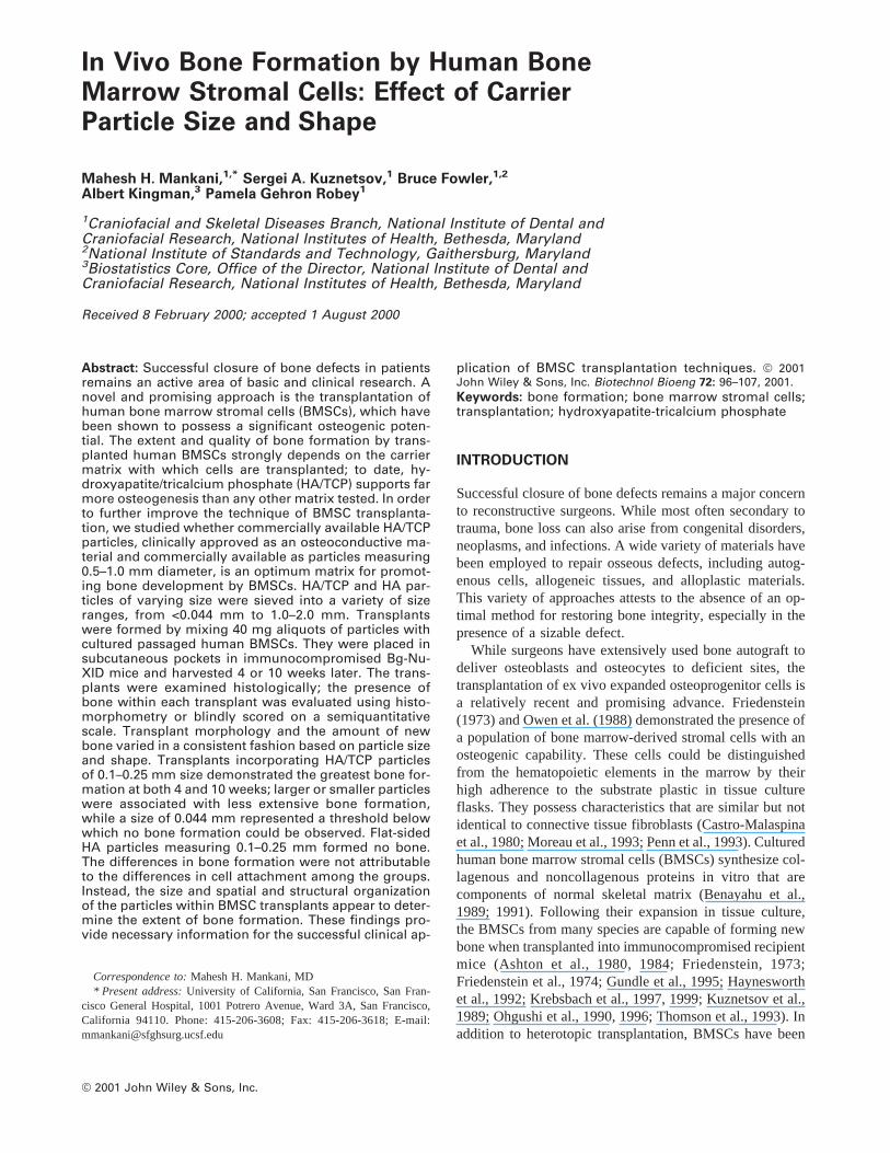
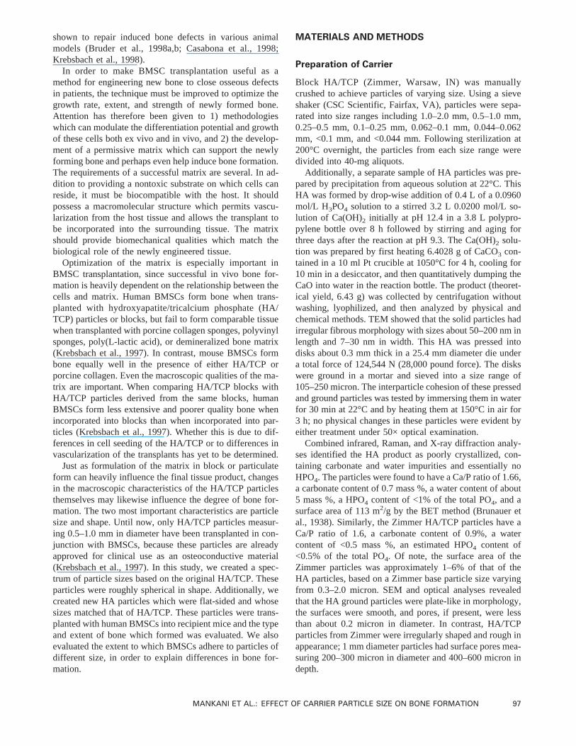
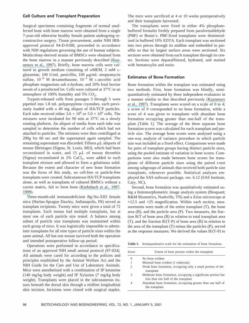
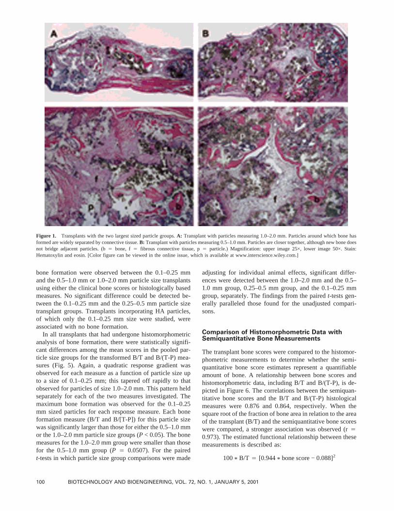
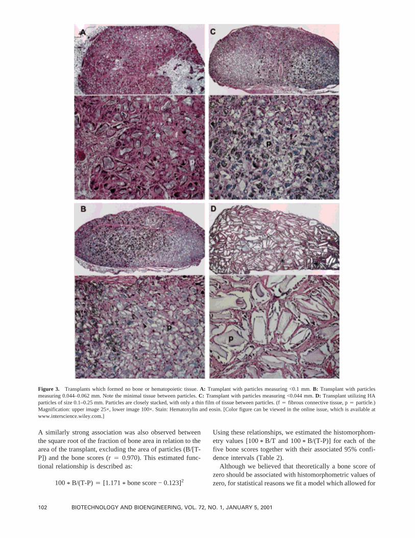
ploying the largest HA/TCP particles (1.0–2.0 mm) werecharacterized by particles widely separated by nonmineral-ized tissue (Fig. 1A). Bone was sporadic and discrete andassociated with individual particles. Where it occurred, thebone was lamellar in character. Hematopoietic tissue wassparse. This was true as well for transplants with the second-largest HA/TCP particles, 0.5–1.0 mm (Fig. 1B). In con-trast, transplants with HA/TCP particles measuring 0.25–0.5 mm and 0.1–0.25 mm contained abundant bone andassociated hematopoietic tissue and occasional adipocytes(Fig. 2A,B). Particles were closely spaced, with bone occu-pying much of the interparticle space. In many areas, boneassociated with individual particles appeared to coalesce,forming a rim of bone along the exterior surface of thetransplant and a latticework of bone within the transplant.Transplants with slightly smaller HA/TCP particles, mea-suring 0.062–0.1 mm, demonstrated well-developed rimand lattice-like bone which was limited to a few discreteareas, while the remainder of the transplants were occupiedby fibrous tissue (Fig. 2C). Transplants with the smallestHA/TCP particles, those measuring either <0.1 mm, 0.044–0.062 mm, and <0.044 mm formed no bone (Fig. 3A–C). Ofgreatest interest, transplants utilizing HA particles of size0.1–0.25 mm formed no bone and exhibited limited vascu-larization. In contrast to similarly sized particles of HA/TCP, the 0.1–0.25 mm HA particles were flat-sided; theyformed arrangements resembling closely packed stacks ofplates (Fig. 3D). All transplants and peritransplant tissueswere characterized by the absence of an inflammatory re-action.
Timing and Extent of Bone Formation
Significant amounts of new bone was observed as early as4 weeks in transplants with particles as large as 2.0 mm andas small as 0.062 mm (P < 0.001, Fig. 4). The extent of boneformation observed as a function of transplant particle sizewas fairly stable, but with some variability for the 4-weekevaluation (P > 0.05 for all comparisons) above an apparentparticle size threshold of roughly 0.05 mm (Fig. 4).
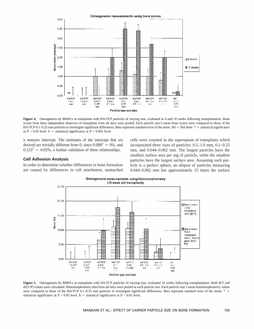
Bone formation was increased significantly in all groupsexcept the 0.044–0.062 group at 10 weeks (Fig. 4). Theextent of bone formation varied as a quadratic function ofparticle size after 10 weeks, using either semiquantitativebone scores (Fig. 4) or histomorphometric evaluation (Fig.5). Transplants incorporating HA/TCP particles of 0.1–0.25mm size demonstrated the greatest bone formation, closelyfollowed by transplants incorporating particles ranging insize from 0.25–0.5 mm. Further increasing particle size re-sulted in diminishing bone formation, although the largestparticle size was still associated with moderate amounts ofbone. Particles ranging in size from 0.044–0.1 mm wereassociated with weak to moderate bone formation, whileparticles smaller than 0.044 were associated with no boneformation. Among HA/TCP particles, a size of 0.044 mmrepresented a threshold below which no bone formationcould be observed. Significant differences in the extent of
MANKANI ET AL.: EFFECT OF CARRIER PARTICLE SIZE ON BONE FORMATION 99
bone formation were observed between the 0.1–0.25 mmand the 0.5–1.0 mm or 1.0–2.0 mm particle size transplantsusing either the clinical bone scores or histologically basedmeasures. No significant difference could be detected be-tween the 0.1–0.25 mm and the 0.25–0.5 mm particle sizetransplant groups. Transplants incorporating HA particles,of which only the 0.1–0.25 mm size were studied, wereassociated with no bone formation.
In all transplants that had undergone histomorphometricanalysis of bone formation, there were statistically signifi-cant differences among the mean scores in the pooled par-ticle size groups for the transformed B/T and B/(T-P) mea-sures (Fig. 5). Again, a quadratic response gradient wasobserved for each measure as a function of particle size upto a size of 0.1–0.25 mm; this tapered off rapidly to thatobserved for particles of size 1.0–2.0 mm. This pattern heldseparately for each of the two measures investigated. Themaximum bone formation was observed for the 0.1–0.25mm sized particles for each response measure. Each boneformation measure (B/T and B/[T-P]) for this particle sizewas significantly larger than those for either the 0.5–1.0 mmor the 1.0–2.0 mm particle size groups (P < 0.05). The bonemeasures for the 1.0–2.0 mm group were smaller than thosefor the 0.5–1.0 mm group (P 4 0.0507). For the pairedt-tests in which particle size group comparisons were made
adjusting for individual animal effects, significant differ-ences were detected between the 1.0–2.0 mm and the 0.5–1.0 mm group, 0.25–0.5 mm group, and the 0.1–0.25 mmgroup, separately. The findings from the pairedt-tests gen-erally paralleled those found for the unadjusted compari-sons.
Comparison of Histomorphometric Data withSemiquantitative Bone Measurements
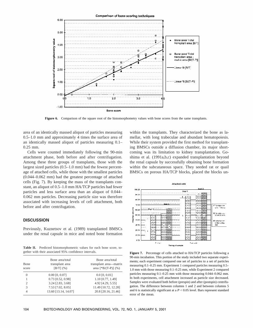
The transplant bone scores were compared to the histomor-phometric measurements to determine whether the semi-quantitative bone score estimates represent a quantifiableamount of bone. A relationship between bone scores andhistomorphometric data, including B/T and B/(T-P), is de-picted in Figure 6. The correlations between the semiquan-titative bone scores and the B/T and B/(T-P) histologicalmeasures were 0.876 and 0.864, respectively. When thesquare root of the fraction of bone area in relation to the areaof the transplant (B/T) and the semiquantitative bone scoreswere compared, a stronger association was observed (r40.973). The estimated functional relationship between thesemeasurements is described as:
100* B/T 4 [0.944* bone score − 0.088]2
Figure 1. Transplants with the two largest sized particle groups.A: Transplant with particles measuring 1.0–2.0 mm. Particles around which bone hasformed are widely separated by connective tissue.B: Transplant with particles measuring 0.5–1.0 mm. Particles are closer together, although new bone doesnot bridge adjacent particles. (b4 bone, f 4 fibrous connective tissue, p4 particle.) Magnification: upper image 25×, lower image 50×. Stain:Hematoxylin and eosin. [Color figure can be viewed in the online issue, which is available at www.interscience.wiley.com.]
100 BIOTECHNOLOGY AND BIOENGINEERING, VOL. 72, NO. 1, JANUARY 5, 2001
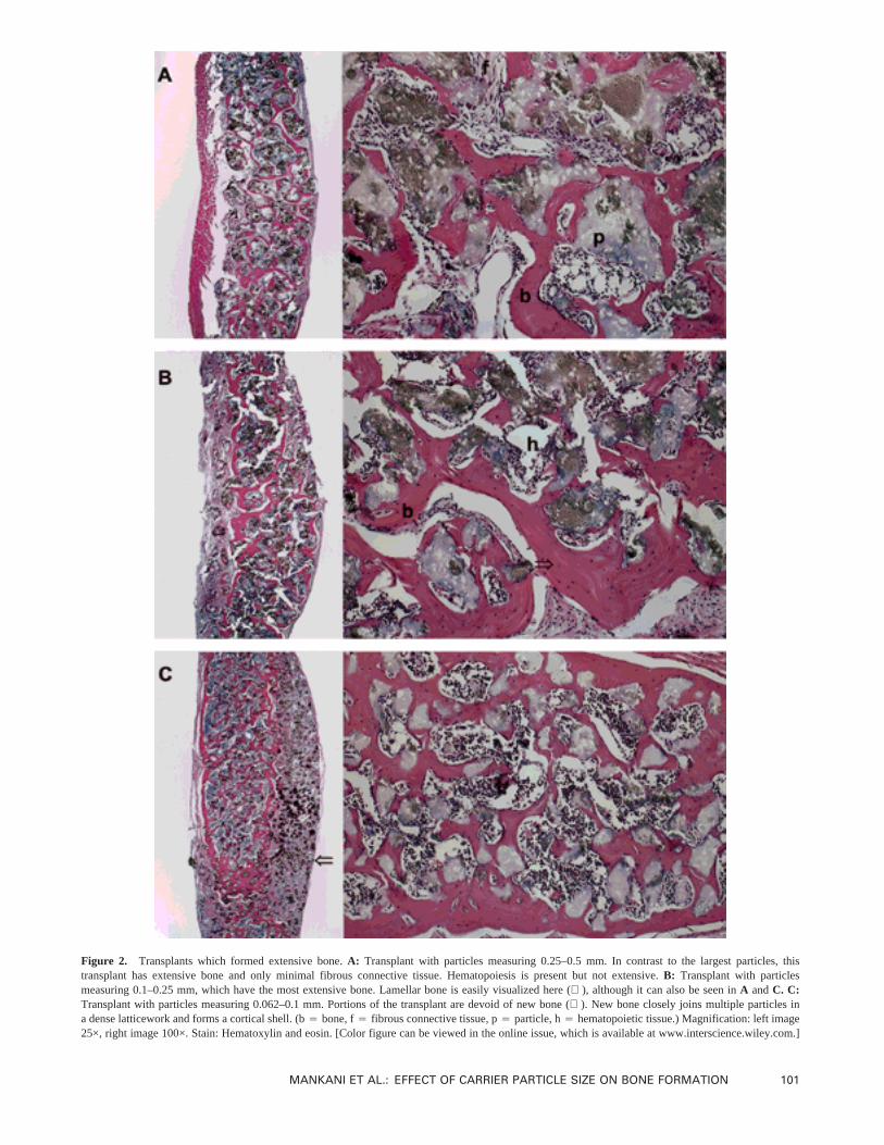
Figure 2. Transplants which formed extensive bone.A: Transplant with particles measuring 0.25–0.5 mm. In contrast to the largest particles, thistransplant has extensive bone and only minimal fibrous connective tissue. Hematopoiesis is present but not extensive.B: Transplant with particlesmeasuring 0.1–0.25 mm, which have the most extensive bone. Lamellar bone is easily visualized here (⇒), although it can also be seen inA andC. C:Transplant with particles measuring 0.062–0.1 mm. Portions of the transplant are devoid of new bone (⇐). New bone closely joins multiple particles ina dense latticework and forms a cortical shell. (b4 bone, f4 fibrous connective tissue, p4 particle, h4 hematopoietic tissue.) Magnification: left image25×, right image 100×. Stain: Hematoxylin and eosin. [Color figure can be viewed in the online issue, which is available at www.interscience.wiley.com.]
MANKANI ET AL.: EFFECT OF CARRIER PARTICLE SIZE ON BONE FORMATION 101
A similarly strong association was also observed betweenthe square root of the fraction of bone area in relation to thearea of the transplant, excluding the area of particles (B/[T-P]) and the bone scores (r4 0.970). This estimated func-tional relationship is described as:
100* B/(T-P)4 [1.171* bone score − 0.123]2
Using these relationships, we estimated the histomorphom-etry values [100* B/T and 100* B/(T-P)] for each of thefive bone scores together with their associated 95% confi-dence intervals (Table 2).
Although we believed that theoretically a bone score ofzero should be associated with histomorphometric values ofzero, for statistical reasons we fit a model which allowed for
Figure 3. Transplants which formed no bone or hematopoietic tissue.A: Transplant with particles measuring <0.1 mm.B: Transplant with particlesmeasuring 0.044–0.062 mm. Note the minimal tissue between particles.C: Transplant with particles measuring <0.044 mm.D: Transplant utilizing HAparticles of size 0.1–0.25 mm. Particles are closely stacked, with only a thin film of tissue between particles. (f4 fibrous connective tissue, p4 particle.)Magnification: upper image 25×, lower image 100×. Stain: Hematoxylin and eosin. [Color figure can be viewed in the online issue, which is available atwww.interscience.wiley.com.]
102 BIOTECHNOLOGY AND BIOENGINEERING, VOL. 72, NO. 1, JANUARY 5, 2001
a nonzero intercept. The estimates of the intercept that wederived are trivially different from 0, since 0.0882 4 0%, and0.1232 4 0.02%, a further validation of these relationships.
Cell Adhesion Analysis
In order to determine whether differences in bone formationare caused by differences in cell attachment, unattached
cells were counted in the supernatant of transplants whichincorporated three sizes of particles: 0.5–1.0 mm, 0.1–0.25mm, and 0.044–0.062 mm. The largest particles have thesmallest surface area per mg of particle, while the smallestparticles have the largest surface area. Assuming each par-ticle is a perfect sphere, an aliquot of particles measuring0.044–0.062 mm has approximately 15 times the surface
Figure 4. Osteogenesis by BMSCs in transplants with HA/TCP particles of varying size, evaluated at 4 and 10 weeks following transplantation. Bonescores from three independent observers of transplants from all mice were pooled. Each particle size’s mean bone scores were compared to those of theHA/TCP 0.1–0.25 mm particles to investigate significant differences. Bars represent standard error of the mean. Nd4 Not done. *4 statistical significanceat P < 0.05 level. #4 statistical significance atP < 0.001 level.
Figure 5. Osteogenesis by BMSCs in transplants with HA/TCP particles of varying size, evaluated 10 weeks following transplantation. Both B/T andB/(T-P) values were calculated. Histomorphometry data from all mice were pooled at each particle size. Each particle size’s mean histomorphometry valueswere compared to those of the HA/TCP 0.1–0.25 mm particles to investigate significant differences. Bars represent standard error of the mean. *4
statistical significance atP < 0.05 level. $4 statistical significance atP < 0.01 level.
MANKANI ET AL.: EFFECT OF CARRIER PARTICLE SIZE ON BONE FORMATION 103
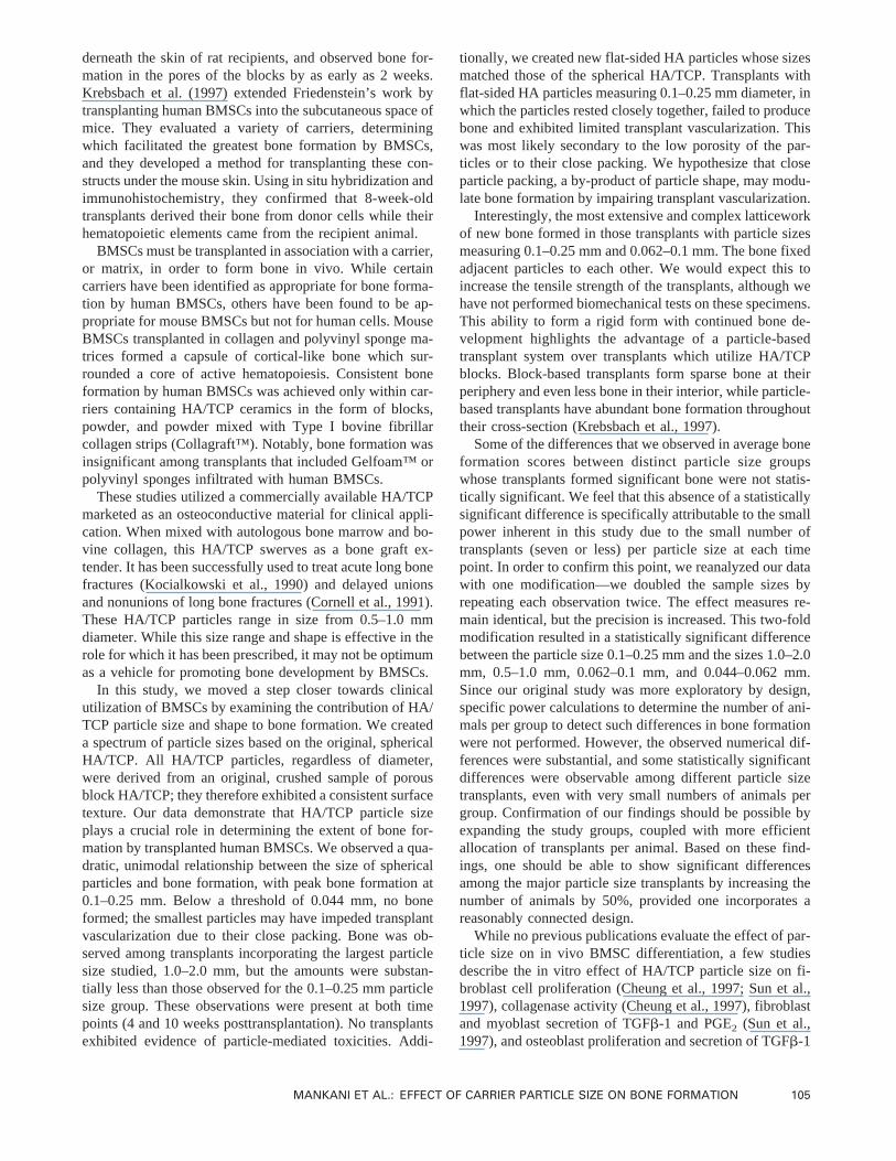
area of an identically massed aliquot of particles measuring0.5–1.0 mm and approximately 4 times the surface area ofan identically massed aliquot of particles measuring 0.1–0.25 mm.
Cells were counted immediately following the 90-minattachment phase, both before and after centrifugation.Among these three groups of transplants, those with thelargest sized particles (0.5–1.0 mm) had the fewest percent-age of attached cells, while those with the smallest particles(0.044–0.062 mm) had the greatest percentage of attachedcells (Fig. 7). By keeping the mass of the transplants con-stant, an aliquot of 0.5–1.0 mm HA/TCP particles had fewerparticles and less surface area than an aliquot of 0.044–0.062 mm particles. Decreasing particle size was thereforeassociated with increasing levels of cell attachment, bothbefore and after centrifugation.
DISCUSSION
Previously, Kuznetsov et al. (1989) transplanted BMSCsunder the renal capsule in mice and noted bone formation
within the transplants. They characterized the bone as la-mellar, with long trabeculae and abundant hematopoiesis.While their system provided the first method for transplant-ing BMSCs outside a diffusion chamber, its major short-coming was its limitation to kidney transplantation. Go-shima et al. (1991a,b,c) expanded transplantation beyondthe renal capsule by successfully obtaining bone formationwithin the subcutaneous space. They seeded rat or quailBMSCs on porous HA/TCP blocks, placed the blocks un-
Table II. Predicted histomorphometric values for each bone score, to-gether with their associated 95% confidence intervals.
Bonescore
Bone area/totaltransplant area
[B/T] (%)
Bone area/totaltransplant area—matrix
area [*B/(T-P)] (%)
0 0.00 [0, 0.07] 0.0 [0, 0.01]1 0.73 [0.52, 0.98] 1.10 [0.77, 1.49]2 3.24 [2.83, 3.68] 4.92 [4.29, 5.55]3 7.53 [7.02, 8.05] 11.49 [10.72, 12.28]4 13.60 [13.14, 14.07] 20.8 [20.16, 21.46]
Figure 7. Percentage of cells attached to HA/TCP particles following a90-min incubation. This portion of the study included two separate experi-ments; each experiment compared one set of particles to a set of particlesmeasuring 0.1–0.25 mm. Experiment 1 compared particles measuring 0.5–1.0 mm with those measuring 0.1–0.25 mm, while Experiment 2 comparedparticles measuring 0.1–0.25 mm with those measuring 0.044–0.062 mm.In both experiments, cell attachment increased as particle size decreased.Samples were evaluated both before (prespin) and after (postspin) centrifu-gation. The difference between columns 1 and 2 and between columns 5and 6 is statistically significant at aP < 0.05 level. Bars represent standarderror of the mean.
Figure 6. Comparison of the square root of the histomorphometry values with bone scores from the same transplants.
104 BIOTECHNOLOGY AND BIOENGINEERING, VOL. 72, NO. 1, JANUARY 5, 2001
derneath the skin of rat recipients, and observed bone for-mation in the pores of the blocks by as early as 2 weeks.Krebsbach et al. (1997) extended Friedenstein’s work bytransplanting human BMSCs into the subcutaneous space ofmice. They evaluated a variety of carriers, determiningwhich facilitated the greatest bone formation by BMSCs,and they developed a method for transplanting these con-structs under the mouse skin. Using in situ hybridization andimmunohistochemistry, they confirmed that 8-week-oldtransplants derived their bone from donor cells while theirhematopoietic elements came from the recipient animal.
BMSCs must be transplanted in association with a carrier,or matrix, in order to form bone in vivo. While certaincarriers have been identified as appropriate for bone forma-tion by human BMSCs, others have been found to be ap-propriate for mouse BMSCs but not for human cells. MouseBMSCs transplanted in collagen and polyvinyl sponge ma-trices formed a capsule of cortical-like bone which sur-rounded a core of active hematopoiesis. Consistent boneformation by human BMSCs was achieved only within car-riers containing HA/TCP ceramics in the form of blocks,powder, and powder mixed with Type I bovine fibrillarcollagen strips (Collagraft™). Notably, bone formation wasinsignificant among transplants that included Gelfoam™ orpolyvinyl sponges infiltrated with human BMSCs.
These studies utilized a commercially available HA/TCPmarketed as an osteoconductive material for clinical appli-cation. When mixed with autologous bone marrow and bo-vine collagen, this HA/TCP swerves as a bone graft ex-tender. It has been successfully used to treat acute long bonefractures (Kocialkowski et al., 1990) and delayed unionsand nonunions of long bone fractures (Cornell et al., 1991).These HA/TCP particles range in size from 0.5–1.0 mmdiameter. While this size range and shape is effective in therole for which it has been prescribed, it may not be optimumas a vehicle for promoting bone development by BMSCs.
In this study, we moved a step closer towards clinicalutilization of BMSCs by examining the contribution of HA/TCP particle size and shape to bone formation. We createda spectrum of particle sizes based on the original, sphericalHA/TCP. All HA/TCP particles, regardless of diameter,were derived from an original, crushed sample of porousblock HA/TCP; they therefore exhibited a consistent surfacetexture. Our data demonstrate that HA/TCP particle sizeplays a crucial role in determining the extent of bone for-mation by transplanted human BMSCs. We observed a qua-dratic, unimodal relationship between the size of sphericalparticles and bone formation, with peak bone formation at0.1–0.25 mm. Below a threshold of 0.044 mm, no boneformed; the smallest particles may have impeded transplantvascularization due to their close packing. Bone was ob-served among transplants incorporating the largest particlesize studied, 1.0–2.0 mm, but the amounts were substan-tially less than those observed for the 0.1–0.25 mm particlesize group. These observations were present at both timepoints (4 and 10 weeks posttransplantation). No transplantsexhibited evidence of particle-mediated toxicities. Addi-
tionally, we created new flat-sided HA particles whose sizesmatched those of the spherical HA/TCP. Transplants withflat-sided HA particles measuring 0.1–0.25 mm diameter, inwhich the particles rested closely together, failed to producebone and exhibited limited transplant vascularization. Thiswas most likely secondary to the low porosity of the par-ticles or to their close packing. We hypothesize that closeparticle packing, a by-product of particle shape, may modu-late bone formation by impairing transplant vascularization.
Interestingly, the most extensive and complex latticeworkof new bone formed in those transplants with particle sizesmeasuring 0.1–0.25 mm and 0.062–0.1 mm. The bone fixedadjacent particles to each other. We would expect this toincrease the tensile strength of the transplants, although wehave not performed biomechanical tests on these specimens.This ability to form a rigid form with continued bone de-velopment highlights the advantage of a particle-basedtransplant system over transplants which utilize HA/TCPblocks. Block-based transplants form sparse bone at theirperiphery and even less bone in their interior, while particle-based transplants have abundant bone formation throughouttheir cross-section (Krebsbach et al., 1997).
Some of the differences that we observed in average boneformation scores between distinct particle size groupswhose transplants formed significant bone were not statis-tically significant. We feel that this absence of a statisticallysignificant difference is specifically attributable to the smallpower inherent in this study due to the small number oftransplants (seven or less) per particle size at each timepoint. In order to confirm this point, we reanalyzed our datawith one modification—we doubled the sample sizes byrepeating each observation twice. The effect measures re-main identical, but the precision is increased. This two-foldmodification resulted in a statistically significant differencebetween the particle size 0.1–0.25 mm and the sizes 1.0–2.0mm, 0.5–1.0 mm, 0.062–0.1 mm, and 0.044–0.062 mm.Since our original study was more exploratory by design,specific power calculations to determine the number of ani-mals per group to detect such differences in bone formationwere not performed. However, the observed numerical dif-ferences were substantial, and some statistically significantdifferences were observable among different particle sizetransplants, even with very small numbers of animals pergroup. Confirmation of our findings should be possible byexpanding the study groups, coupled with more efficientallocation of transplants per animal. Based on these find-ings, one should be able to show significant differencesamong the major particle size transplants by increasing thenumber of animals by 50%, provided one incorporates areasonably connected design.
While no previous publications evaluate the effect of par-ticle size on in vivo BMSC differentiation, a few studiesdescribe the in vitro effect of HA/TCP particle size on fi-broblast cell proliferation (Cheung et al., 1997; Sun et al.,1997), collagenase activity (Cheung et al., 1997), fibroblastand myoblast secretion of TGFb-1 and PGE2 (Sun et al.,1997), and osteoblast proliferation and secretion of TGFb-1
MANKANI ET AL.: EFFECT OF CARRIER PARTICLE SIZE ON BONE FORMATION 105
and PGE2. These studies suggest that particles measuring<0.1 mm are poorly tolerated by cells. Transplantation ofHA/TCP particles alone into hard tissue defects in rabbitsand dogs was associated with superior bone formationaround particles measuring 0.3–0.6 mm, while larger andsmaller particles were associated with less bone formation(Higashi et al., 1996; Kuroda, 1995). These results are inagreement with our findings.
Relationship of Cell Attachment to Particle Sizeand Bone Formation
In an attempt to describe the mechanism by which particlesize influences bone formation, we studied the attachmentof BMSCs to particles of different sizes. The number ofcells attaching to particles is not directly responsible for thedifferences in bone formation between particles of differentsizes. In these studies, an increase in particle size was as-sociated with a decrease in cell attachment. Aliquots of thelargest particles, 0.5–1.0 mm diameter, captured only 40%of cells during the incubation period, while the particlesmeasuring 0.044–0.062 mm captured 62% of cells. Yet thelarger particles were associated with a reasonable degree ofbone formation, while the smaller particles were associatedwith essentially no bone formation. The greater number ofcells captured by the smaller particles apparently offered noadvantage for bone formation. The larger particles capturedfewer cells than the smaller particles because they offeredless surface area per aliquot. Thus, it is not the number ofcells attached to the particles that determines the fate of thetransplants. Rather, osteogenic differentiation of trans-planted BMSCs is influenced by the relative arrangement ofceramic and cells within the transplants; some empty spaceis necessary to allow capillary ingrowth, while excessiveempty space favors development of nonmineralized fibroustissue. A more precise mechanism by which particle sizeinfluences bone formation is yet to be described.
Histomorphometry vs. SemiquantitativeBone Measurements
Finally, the previous use of semiquantitative bone measure-ments was here validated by their comparison to histomor-phometric results. As bone score increased, the average areaof the transplant occupied by new bone increased accord-ingly. Bone scores of 0 represented no bone formation byhistomorphometry, while a bone score of 4 indicated thatbone occupied 13.6% and 21.2% of the cross-sectional areaof the total transplant and the nonmatrix area of the trans-plant, respectively. It is our conclusion that semiquantitativebone scores are representative of total bone formation. His-tomorphometric analysis, while providing more completeinformation, is much more labor-intensive; in cases wheredozens of transplants are to be analyzed, semiquantitativescoring may be acceptable. Comparisons between histologicbone scores and histomorphometric measures of bone-containing ceramic cubes have also demonstrated the utility
of subjective measures (Dennis et al., 1998). However, ourbone scoring system differs from Dennis’ in several ways.His method examines the percentage of bone-containingpores, whereas ours measures the absolute amount of bonepresent within the transplant. Additionally, our system de-scribes an exponential relationship with a high r2.
This study demonstrates that HA/TCP particle size andshape play a critical role in bone formation by transplantedhuman BMSCs, and it describes parameters which can beoptimized to improve the clinical utility of the technique.
The authors thank Zimmer (Warsaw, IN) for its gift of HA/TCP.
References
Ashton BA, Allen TD, Howlett CR, Eaglesom CC, Hattori A, Owen M.1980. Formation of bone and cartilage by marrow stromal cells indiffusion chambers in vivo. Clin Orthop 294–307.
Ashton BA, Eaglesom CC, Bab I, Owen ME. 1984. Distribution of fibro-blastic colony-forming cells in rabbit bone marrow and assay of theirosteogenic potential by an in vivo diffusion chamber method. CalcifTissue Int 36:83–86.
Benayahu D, Kletter Y, Zipori D, Wientroub S. 1989. Bone marrow-derived stromal cell line expressing osteoblastic phenotype in vitro andosteogenic capacity in vivo. J Cell Physiol 140:1–7.
Benayahu D, Fried A, Zipori D, Wientroub S. 1991. Subpopulations ofmarrow stromal cells share a variety of osteoblastic markers. CalcifTissue Int 49:202–207.
Bruder SP, Kraus KH, Goldberg VM, Kadiyala S. 1998a. Critical-sizedcanine segmental femoral defects are healed by autologous mesenchy-mal stem cell therapy. 44th Annual Meeting, Osteopaedic ResearchSociety, vol. 23. New Orleans: Orthopaedic Research Society. p 147.
Bruder SP, Kurth AA, Shea M, Hayes WC, Jaiswal N, Kadiyala S. 1998b.Bone regeneration by implantation of purified, culture-expanded hu-man mesenchymal stem cells. J Orthop Res 16:155–162.
Brunauer S, Emmett PH, Teller E. 1938. Adsorption of gases in multimo-lecular layers. J Am Chem Soc 60:309–319.
Casabona F, Martin I, Muraglia A, Berrino P, Santi P, Cancedda R, QuartoR. 1998. Prefabricated engineered bone flaps: an experimental modelof tissue reconstruction in plastic surgery. Plast Reconstr Surg 101:577–581.
Castro-Malaspina H, Gay RE, Resnick G, Kapoor N, Meyers P, ChiarieriD, McKenzie S, Broxmeyer HE, Moore MA. 1980. Characterization ofhuman bone marrow fibroblast colony-forming cells (CFU-F) and theirprogeny. Blood 56:289–301.
Cheung HS, Devine TR, Hubbard W. 1997. Calcium phosphate particleinduction of metalloproteinase and mitogenesis: effect of particlesizes. Osteoarthr Cartil 5:145–151.
Cornell CN, Lane JM, Chapman M, Merkow R, Seligson D, Henry S,Gustilo R, Vincent K. 1991. Multicenter trial of collagraft as bonegraft substitute. J Orthop Trauma 5:1–8.
Dennis JE, Konstantakos EK, Arm D, Caplan AI. 1998. In vivo osteogen-esis assay: a rapid method for quantitative analysis. Biomaterials 19:1323–1328.
Friedenstein AJ. 1973. Determined and inducible osteogenic precursorcells. In: Hard tissue growth, repair and remineralization, vol. 11.Amsterdam: Elsevier. p 169–185.
Friedenstein AJ, Chailakhyan RK, Latsinik NV, Panasyuk AF, Keiliss-Borok IV. 1974. Stromal cells responsible for transferring the micro-environment of the hematopoietic tissues. Cloning in vitro and retrans-plantation in vivo. Transplantation 17:331–340.
Goshima J, Goldberg VM, Caplan AI. 1991a. The origin of bone formed incomposite grafts of porous calcium phosphate ceramic loaded withmarrow cells. Clin Orthop 274–283.
Goshima J, Goldberg VM, Caplan AI. 1991b. Osteogenic potential of
106 BIOTECHNOLOGY AND BIOENGINEERING, VOL. 72, NO. 1, JANUARY 5, 2001
culture-expanded rat marrow cells as assayed in vivo with porouscalcium phosphate ceramic. Biomaterials 12:253–258.
Goshima J, Goldberg VM, Caplan AI. 1991c. The osteogenic potential ofculture-expanded rat marrow mesenchymal cells assayed in vivo incalcium phosphate ceramic blocks. Clin Orthop 298–311.
Gundle R, Joyner CJ, Triffitt JT. 1995. Human bone tissue formation indiffusion chamber culture in vivo by bone-derived cells and marrowstromal fibroblastic cells. Bone 16:597–601.
Haynesworth SE, Baber MA, Caplan AI. 1992. Cell surface antigens onhuman marrow-derived mesenchymal cells are detected by monoclo-nal antibodies. Bone 13:69–80.
Higashi T, Okamoto H. 1996. Influence of particle size of calcium phos-phate ceramics as a capping agent on the formation of a hard tissuebarrier in amputated dental pulp. J Endod 22:281–283.
Kocialkowski A, Wallace WA, Prince HG. 1990. Clinical experience witha new artificial bone graft: preliminary results of a prospective study.Injury 21:142–144.
Krebsbach PH, Kuznetsov SA, Satomura K, Emmons RV, Rowe DW,Robey PG. 1997. Bone formation in vivo: comparison of osteogenesisby transplanted mouse and human marrow stromal fibroblasts. Trans-plantation 63:1059–1069.
Krebsbach PH, Mankani MH, Satomura K, Kuznetsov SA, Robey PG.1998. Repair of craniotomy defects using bone marrow stromal cells.Transplantation 66:1272–1278.
Krebsbach PA, Kuznetsov SA, Bianco P, Gehron Robey P. 1999. Bonemarrow stromal cells: characterization and clinical application. CritRev Oral Biol Med 10:165–181.
Kuroda T. 1995. [Bone formation and mechanical properties of the can-cellous bone defect site filled with hydroxyapatite granules]. NipponSeikeigeka Gakkai Zasshi 69:1037–1049.
Kuznetsov SA, Grosheva AG, Friedenstein A. 1989. [Osteogenic properties
of adhesive cells in Dexter culture of the mouse bone marrow]. BiullEksp Biol Med 108:236–238.
Kuznetsov SA, Krebsbach PH, Satomura K, Kerr J, Riminucci M,Benayahu D, Robey PG. 1997. Single-colony derived strains of humanmarrow stromal fibroblasts form bone after transplantation in vivo. JBone Miner Res 12:1335–1347.
Moreau I, Duvert V, Caux C, Galmiche MC, Charbord P, Banchereau J,Saeland S. 1993. Myofibroblastic stromal cells isolated from humanbone marrow induce the proliferation of both early myeloid and B-lymphoid cells. Blood 82:2396–2405.
Ohgushi H, Okumura M. 1990. Osteogenic capacity of rat and humanmarrow cells in porous ceramics. Experiments in athymic (nude) mice.Acta Orthop Scand 61:431–434.
Ohgushi H, Dohi Y, Yoshikawa T, Tamai S, Tabata S, Okunaga K,Shibuya T. 1996. Osteogenic differentiation of cultured marrow stro-mal stem cells on the surface of bioactive glass ceramics. J BiomedMater Res 32:341–348.
Owen M, Friedenstein AJ. 1988. Stromal stem cells: marrow-derived os-teogenic precursors. Ciba Found Symp 136:42–60.
Penn PE, Jiang DZ, Fei RG, Sitnicka E, Wolf NS. 1993. Dissecting thehematopoietic microenvironment. IX. Further characterization of mu-rine bone marrow stromal cells. Blood 81:1205–1213.
Sun JS, Tsuang YH, Chang WH, Li J, Liu HC, Lin FH. 1997. Effect ofhydroxyapatite particle size on myoblasts and fibroblasts. Biomaterials18:683–690.
Thomson BM, Bennett J, Dean V, Triffitt J, Meikle MC, Loveridge N.1993. Preliminary characterization of porcine bone marrow stromalcells: skeletogenic potential, colony-forming activity, and response todexamethasone, transforming growth factor beta, and basic fibroblastgrowth factor. J Bone Miner Res 8:1173–1183.
MANKANI ET AL.: EFFECT OF CARRIER PARTICLE SIZE ON BONE FORMATION 107
Related Documents


![GENETICALLY ENGINEERED BONE MARROW STROMAL CELLS … · 2020. 9. 23. · References: 83., [2] Byers et al. (in press) J of Bone and Mi Runx2-expressing stromal cell cultures demonstrated](https://static.cupdf.com/doc/110x72/60a836022ca0396c8a6080c2/genetically-engineered-bone-marrow-stromal-cells-2020-9-23-references-83.jpg)
