Int.J.Curr.Microbiol.App.Sci (2017) 6(7): 1010-1022 1010 Review Article https://doi.org/10.20546/ijcmas.2017.607.122 In vitro Propagation of Stevia rebaudiana (Bertoni): An Overview Manvender Singh * , Vinod Saharan, Jyotsna Dayma, Deepak Rajpurohit, Yadunandan Sen and Ajay Sharma Department of Molecular Biology and Biotechnology, Rajasthan Collage of Agriculture, Maharana Pratap University of Agriculture and Technology, Udaipur, Rajasthan, India *Corresponding author ABSTRACT Introduction Stevia rebaudiana (Bertoni) is a medicinal plant belongs to Asteraceae family and known as stevia, sweet leaf, honey leaf, and candy leaf. Stevia is native to Paraguay (South America) and also called sweet herb of Paraguay. The leaves of this plant has a pleasantly sweet and refreshing taste which is educe by diterpene glycosides (stevioside and rebaudiosides), a high-potency sweeteners and substitute to sugar, being 300 times sweeter than sucrose (Madan et al., 2010; Megeji et al., 2005; Singh and Rao, 2005; Soejarto et al., 1983; Soejarto DD, Kinghorn AD, 1982; Yadav et al., 2011). Stevia is a well-known therapeutic agent serve as an efficient medication for diabetes, hypertension, myocardial and antimicrobial infections, dental troubles, and tumors (Chan et al., 1998; Gregersen et al., 2004; Jayaraman et al., 2008; Jeppesen et al., 2003, 2002; Kujur et al., 2010; Marcinek and Krejpcio, 2016; Muanda et al., 2011; Philippe et al., 2014; Planas and Kucacute, 1968; Shivanna et al., 2013; Šic Žlabur et al., 2013; Singh et al., 2015). In present time people are very calorie conscious which increases use of stevia in preparation of non-calorie food stuffs and become a major sweetening agent in food products in South-east Asia (Ashwell, 2015; Durán A. et al., 2013; Marcinek and Krejpcio, 2016; Panpatil and Polasa, 2008; Salunkhe and Bhise, 2010; Savita et al., 2010). Recently FDA of United States of America issued GRAS status to stevia product. The market grows at a rate of 4% per annum and has a business of around 1.3 International Journal of Current Microbiology and Applied Sciences ISSN: 2319-7706 Volume 6 Number 7 (2017) pp. 1010-1022 Journal homepage: http://www.ijcmas.com Stevia rebaudiana is herbaceous perennial plant of Asteraceae family and an excellent substitute of sugar with medicinal importance. The potential uses of Stevia which produces stevioside, a non-caloric sweetener that does not metabolize in the human body, hence control blood sugar level. Conventional propagation methods are not produce adequate planting material. Plant tissue culture techniques are only technique to produce quality planting material. In present review, micro propagation methods and protocols are compile and generate the information to researchers’ for further exploration for improvement of this valuable medicinal plant. Keywords In-vitro culture, Micro propagation, Stevia, Rebaudioside. Accepted: 17 June 2017 Available Online: 10 July 2017 Article Info

Welcome message from author
This document is posted to help you gain knowledge. Please leave a comment to let me know what you think about it! Share it to your friends and learn new things together.
Transcript
Int.J.Curr.Microbiol.App.Sci (2017) 6(7): 1010-1022
1010
Review Article https://doi.org/10.20546/ijcmas.2017.607.122
In vitro Propagation of Stevia rebaudiana (Bertoni): An Overview
Manvender Singh*, Vinod Saharan, Jyotsna Dayma, Deepak Rajpurohit,
Yadunandan Sen and Ajay Sharma
Department of Molecular Biology and Biotechnology, Rajasthan Collage of Agriculture,
Maharana Pratap University of Agriculture and Technology, Udaipur, Rajasthan, India *Corresponding author
A B S T R A C T
Introduction
Stevia rebaudiana (Bertoni) is a medicinal
plant belongs to Asteraceae family and known
as stevia, sweet leaf, honey leaf, and candy
leaf. Stevia is native to Paraguay (South
America) and also called sweet herb of
Paraguay. The leaves of this plant has a
pleasantly sweet and refreshing taste which is
educe by diterpene glycosides (stevioside and
rebaudiosides), a high-potency sweeteners
and substitute to sugar, being 300 times
sweeter than sucrose (Madan et al., 2010;
Megeji et al., 2005; Singh and Rao, 2005;
Soejarto et al., 1983; Soejarto DD, Kinghorn
AD, 1982; Yadav et al., 2011). Stevia is a
well-known therapeutic agent serve as an
efficient medication for diabetes,
hypertension, myocardial and antimicrobial
infections, dental troubles, and tumors (Chan
et al., 1998; Gregersen et al., 2004;
Jayaraman et al., 2008; Jeppesen et al., 2003,
2002; Kujur et al., 2010; Marcinek and
Krejpcio, 2016; Muanda et al., 2011; Philippe
et al., 2014; Planas and Kucacute, 1968;
Shivanna et al., 2013; Šic Žlabur et al., 2013;
Singh et al., 2015). In present time people are
very calorie conscious which increases use of
stevia in preparation of non-calorie food
stuffs and become a major sweetening agent
in food products in South-east Asia (Ashwell,
2015; Durán A. et al., 2013; Marcinek and
Krejpcio, 2016; Panpatil and Polasa, 2008;
Salunkhe and Bhise, 2010; Savita et al.,
2010). Recently FDA of United States of
America issued GRAS status to stevia
product. The market grows at a rate of 4% per
annum and has a business of around 1.3
International Journal of Current Microbiology and Applied Sciences ISSN: 2319-7706 Volume 6 Number 7 (2017) pp. 1010-1022 Journal homepage: http://www.ijcmas.com
Stevia rebaudiana is herbaceous perennial plant of Asteraceae family and
an excellent substitute of sugar with medicinal importance. The potential
uses of Stevia which produces stevioside, a non-caloric sweetener that does
not metabolize in the human body, hence control blood sugar level.
Conventional propagation methods are not produce adequate planting
material. Plant tissue culture techniques are only technique to produce
quality planting material. In present review, micro propagation methods
and protocols are compile and generate the information to researchers’ for
further exploration for improvement of this valuable medicinal plant.
K e y w o r d s
In-vitro culture,
Micro
propagation,
Stevia,
Rebaudioside.
Accepted:
17 June 2017
Available Online: 10 July 2017
Article Info
Int.J.Curr.Microbiol.App.Sci (2017) 6(7): 1010-1022
1011
billion US dollar. Two international business
groups, Wilmar and Olam, have started a joint
venture to invest 106.2 million US dollar to
globally enhance the production and
consumption of Stevia. India is supposed to
have suitable conditions for the cultivation of
stevia. It has been found that Indian stevia
plant gives a higher stevioside yield of 10–18
percent in comparison to the reported 8–12
percent from other countries (Yadav et al.,
2011).
An International stevia supplier called GLG
Life Tech has taken the initiative of
introducing stevia to the Indian market and to
facilitate its production and extraction in
India. India itself is also stepping forward to
compete in the stevia sweetener international
market (Savita et al., 2010; Yadav et al.,
2011). This will paved the path for
commercial cultivation of stevia in developed
as well as in developing countries, ahead to
replace the cane sugar with a global
production estimated to be around 40,000
million tons and for India it may be around
600 tons (Ahmed B., Hossain M., Islam R.,
Kumar Saha A., 2011; Gantait et al., 2015;
Yadav et al., 2011). But still the cultivation
and commercialization of stevia has not
achieved the expected heights as the
conventional propagation methods lack in
providing rapid quality planting material to
the farmers.
Today demand will need to be supported by
high biomass yield varieties with improved
agronomical traits as well as higher quantities
and quality of diterpene glycosides
production. This generates the need of stevia
development in vitro.
The purpose of this overview is to summarize
the existing literature for the in vitro culture
of stevia that may help to recognize from
beginning to end tissue culture technology in
stevia and provide baseline for further
improvement.
Conventional propagation methods in
stevia
In nature, seed germination in stevia is poor
and unsuccessful commonly due to infertile
seed (Goettemoeller and Ching, 1999; Kumar,
2013) and small endosperm (Yadav et al.,
2011). Even some plant selections produce
virtually no viable seed due to their self-
incompatibility (Raina et al., 2013; Ramesh et
al., 2006; Yadav et al., 2011). Plant raised
from seed does not allow the production of
homogenous plant population resulting in
great variability in important features like
sweetening levels and compositions(Brandle
and Telmer, 2007; Kovylyaeva et al., 2007).
Numbers of reports are available on
successful propagation of Stevia via stem
cuttings (Ramesh et al., 2006; Shock, 1982;
Smitha and Umesha, 2011). But the direct
planting of stem cuttings in the field has
limited success due to poor rooting (Khalil et
al., 2014; Pande and Gupta, 2013; Smitha and
Umesha, 2011). Further, location from where
cuttings are taken, the pair of leaves, length of
the cuttings as well as the season effect
rooting percentage and growth (Brandle et al.,
1998; Carneiro et al., 1997; Ceunen and
Geuns, 2013; Khalil et al., 2014; Rajasekaran
et al., 2007). Published work indicated the
necessity of higher quantities of starch,
carbohydrates, sugar and phenolic compounds
for root initiation (Brandle et al., 1998;
Ibrahim et al., 2008).
Rooting of cuttings can sometimes be
stimulated by the use of growth regulators
(Brandle et al., 1998; Ibrahim et al., 2008).
Moreover, a huge number of stem cuttings are
required for mass propagation of plants which
is an obstacle as number of mother plant need
to be vanish. Although, vegetative
propagation has been found next considerable
option for the mass propagation of stevia. But
ample supply of planting material is still not
adequate for farmers because vegetative

Int.J.Curr.Microbiol.App.Sci (2017) 6(7): 1010-1022
1012
propagation is limited by production of less
number of individuals multiply from a single
explant (Gantait et al., 2015; Pande and
Gupta, 2013; Philippe et al., 2014; Yadav et
al., 2011).
In vitro tissue culture of stevia
Plant regeneration from in vitro culture can be
achieved by either organogenesis or
embryogenesis. Administration of different
phytohormone enhances and accelerates the
production of in vitro plants with good
agronomical traits and steviosides content in
leaves (Brandle et al., 1998; Durán A. et al.,
2013; Jain et al., 2014). However, there are
some fundamental factors like growth
condition of the source material, medium
composition, culture conditions and
genotypes of donor plants whose role cannot
be avoided for the successful in vitro culture
plants (Gantait et al., 2015; Ibrahim et al.,
2008; Pande and Gupta, 2013; Philippe et al.,
2014).
Establishment of in vitro culture of stevia
through nodal explant
Nodal part is the choicest explant for in vitro
culture of stevia for researchers. Various
reports on in vitro use of nodal explant had
been well documented (Ahmed et al., 2007;
Atalay et al., 2011; Debnath, 2008; Laribi et
al., 2012; Mitra and Pal, 2007; Nepovim and
Vanek, 1998; Sivaram and Mukundan, 2003;
Sung, 2006; Tamura et al., 1984;
Thiyagarajan and Venkatachalam, 2012).
Initial work on nodal explant was stated by
Yang and his co-workers with the
achievement of highest axillary shoot
proliferation. They concluded that the type of
cytokinin was the most important factor
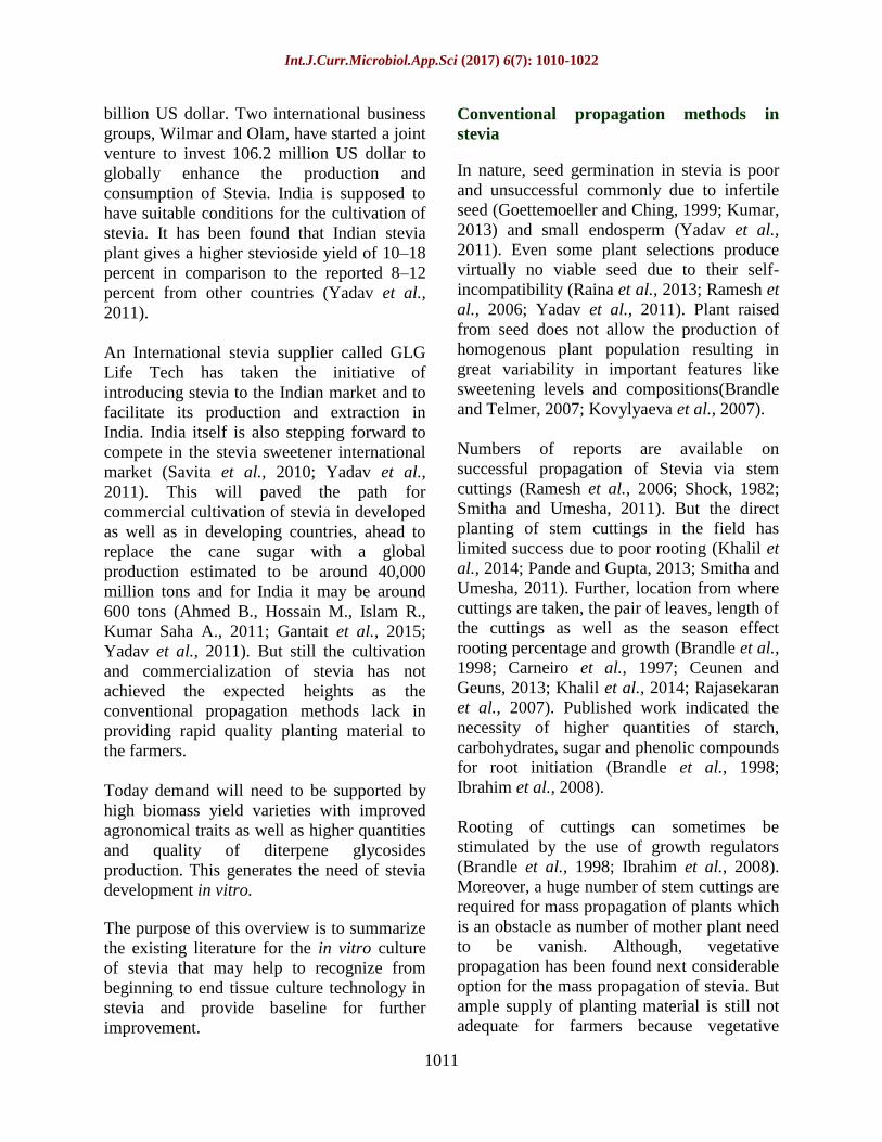
affecting shoot multiplication (Table 1). Also
in our lab we have obtain similar results,
using TDZ for shoot induction and kin for
shoot multiplication (Fig 1). Increasing BA
concentration promoted shoot multiplication.
Similar results were obtained by Sivram and
Mukundan with shoot apex and leaf explants
(Sivaram and Mukundan, 2003). However,
medium supplemented with kin resulted in
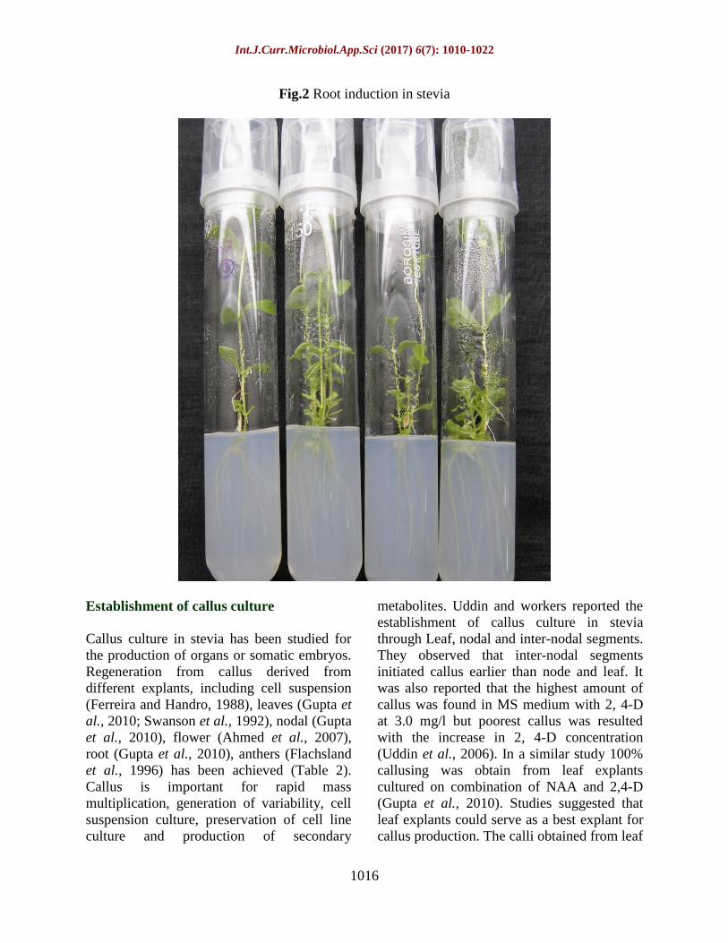
elongated shoots. For root induction, different
concentrations of IBA and NAA were
assayed. IBA showed to be more significant
and effective for rooting than NAA in all
concentrations used. The maximum root
induction (100%) was observed on medium
supplemented with 1.0 or 2.0 IBA (mg/l) and
in our lab we observed root induction on MS
basal medium without PGR/low concentration
of IBA i.e. 0.5 mg/l (Fig 2). Similar result was
obtained by medium fortified with low
concentration of IAA (0.1 mg/l) and it was
found that root induction gradually decreased
with increasing concentration of auxin
(Ahmed et al., 2007; Atalay et al., 2011).
Venkatachalam also studied the effect of
different concentrations of BAP in
combinations with various auxins on multiple
shoot bud regeneration. Of the three auxins
combinations tested (IAA/IBA/NAA), BAP
with IAA combination was found to be
superior for induction of highest percent
(92%) of multiple shoot bud development,
followed by BAP and NAA (83%) and BAP
along with IBA (75%) combinations.
However, both last combinations (BAP with
NAA and BAP with IBA) produced more
callus with low percent (50%) of multiple
shoot bud regenerations (Thiyagarajan and
Venkatachalam, 2012). The lower percent of
multiple shoot bud regenerations may be due
to the profuse callusing at the basal part of
differentiated shoot buds.
Establishment of in vitro culture of stevia
through stem tip explant
In vitro clonal propagation by stem-tips
culture with a few leaf primordia was reported
by Tamura and his group (Tamura et al.,
1984). They found that neither roots nor

Int.J.Curr.Microbiol.App.Sci (2017) 6(7): 1010-1022
1013
callus developed when stem-tips were grown
on a medium supplemented with cytokinin
only whereas, the similar dose of kinetin
proved effective in vegetative propagation,
yielding 50-100 shoots from a single stem-tip
in 80 days. Addition of auxin (NAA) along
with kincould not enhances shoot formation,
but did induce callus formation. The study
suggested that the ability to form multiple
shoots is dependent on the size of excised
stem tip and / or the number of leaf primordia.
Similar studies on shoot proliferation had
been performed (Akita et al., 1994; Ferreira
and Handro, 1988; Sivaram and Mukundan,
2003) (Table 1).
They also reported that plant hormone is
necessary for shooting, elongation and
rooting. Recently similar results with MS
medium in place of previously used LS
medium nourished with kin were obtained by
Das and coworkers (Das et al., 2011). For
root induction, they found that MS media
without growth regulators worked
dynamically whereas when it was
supplemented with auxins (IAA and BA), it
had an adverse effect on root induction. They
also performed peroxidase assay along with
Inter-simple sequence repeat (ISSR)
fingerprinting to confirm the genetic fidelity
of in vitro generated propagules.
Establishment of in vitro culture of stevia
through leaf explant
It is well established that a precise level of
cytokinin would play a critical role in shoot
organogenesis in various plants. Sreedhar and
co-workers had tested various combinations
of growth regulators and found that BA and
kin could successfully induced adventitious
bud from the midrib of leaf explant.
It was the first report of direct shoot induction
from leaf explant. Initially, the shoot buds
appeared as white knob like structures which
later turned green leading to the formation of
a pair of green leaves (Sreedhar et al., 2008).
In an earlier study, kin in combination with
NAA or IAA failed to induce shoot formation
on leaf explants of stevia whereas BA was
found to be more effective (Sivaram and
Mukundan, 2003). Contrarily, Tamura et al.,
showed the formation of shoots only from the
margins of stevia leaves in shoots cultured on
a very high concentration of kin for 40 days
(Tamura et al., 1984).
This was probably because of the
accumulation of high amount of cytokinin
that is known to induce adventitious shoot
formation. Effect of copper on in vitro culture
of stevia leaf explants was also studied. It was
found that an optimum level of copper helps
to achieve maximum shoot bud induction and
elongation along with BAP and IAA PGRs.
This has also gave a positive effect on
chlorophyll and biomass production(Jain et
al., 2009). A promising method of micro
propagation of stevia has been developed with
an aim to increase the biomass, survivability
of the plantlets and stevioside production,
using chlorocholine chloride, a plant growth
retardant. The application of chlorocholine
chloride along with IBA on in vitro generated
microshoots from cotyledonary leaves
explants found to be effective for inducing
certain beneficial changes like desirable
reduction in stem, elongation, profuse rooting,
bigger leaf size, increase fresh weight of the
plantlets, longer chlorophyll retaining
capacity and higher stevioside production
(Dey et al., 2013).
Recently, Ramírez-Mosqueda and Iglesias-
Andreu, 2016 made a recipe for in vitro
plantlet regeneration through thin cell layer
method. Different concentration and
combination of exogenous growth regulators
(BA and 2, 4-D) were tested with transverse
thin cell layers and 2, 4-D showed best
organogenesis.

Int.J.Curr.Microbiol.App.Sci (2017) 6(7): 1010-1022
1014
Table.1 In vitro culture of stevia
Type of explants Shooting media Rooting media References
Leaflets 2-10 mg/1 BA Chen and Li, 1993
Shoot primordial 1.0 mg/1 BA 0.1 mg/l NAA Akita et al., 1994
Shoot apex,
nodal, and leaf explants
2.0 mg/l BA and 1.0
mg/l IAA
1.0 mg/l IBA Sivaram and Mukundan,
2003
Nodal 2.0 mg/l IAA and 0.5
mg/ l kin
2.0 mg/l IBA Hwang, 2006
Nodal 1.5 mg/l BA + 0.5 mg/l
kin
0.1 mg /l IAA Ahmed et al., 2007
Nodal 2.0 mg/l BA and 1.13
mg/l IAA
2.0 mg/l IBA Debnath, 2007
Midrib 2.0 mg/l BA and 1.0
mg/l kin
1.0 mg/l IBA Sreedhar, 2008
Leaf and Nodal 0.5mg/l BA and 0.5
mg/l IAA
Jain et al., 2009
Nodal 0.5 mg/l BA + 0.5 mg/l
kin
1.0 or 2.0 mg/l IBA Alhady, 2011
Nodal 1.0 mg/l BA 0.4 mg/l NAA Venkatachalam, and
Thiyagarajan, 2012
Nodal 1.0 mg/l BA 0.5 mg/l IBA or IAA Labiri, 2012
Stem-tips with a few leaf
primordial
10 mg/l kin 0.1 mg/l NAA Labiri, 2012
Nodal segment MS + 0.5 BAP mg/l
+2.0 Kin mg/l
MS +1.0 IBA mg/l Mehta et al., 2012
Nodal segment MS + 0.5 BAP mg/l +
2.0 Kin mg/l
MS +0.1 IBA mg/l +100
ppm Charcoal
Modi et al., 2012
Shoot tip, nodal segment MS + 1 BAP mg/l + 2
Kin mg/l
MS + 0.5 IBA mg/l El-Motaleb et al., 2013
Shoot tip MS + 1.5 BAP mg/l +
10 Spermine mg/l
MS + 1.5 IAA mg/l Guruchandran and
Sasikumar, 2013
Shoot tip, nodal segment MS + 2.0 BAP mg/l MS+0.5 IBA mg/l Hassanen and Khalil,
2013
Shoot tip MS + 1.0 BAP mg/l MS + 0.4 IBAmg/l Javad et al., 2013
Nodal segment MultSht
MS + 1.0 lM TDZ ½ MS Lata et al., 2013
Nodal explant
½ MS + 0.01 TDZ mg/l ½ MS + 1.0 IBA mg/l Singh and Dwivedi, 2013
Nodal segment MS + 1.0 BAP mg/l +
0.05 NAA mg/l
MS+0.5 IAA mg/l Soliman et al., 2013
Shoot tip MS + 1.0 IAA mg/l +1.0
BA mg/l
Taleie et al., 2013
Nodal segment MS + 1.0 BA mg/l MS + 0.2 IAA mg/l Nower, 2014
Nodal segment MultSht MS + 0.5 Kin + 1.0 IBA Singh et al., (2014)
Thin cell layer 6.78 µM 2, 4-D Without PGR Ramírez-Mosqueda and
Iglesias-Andreu, 2016
Int.J.Curr.Microbiol.App.Sci (2017) 6(7): 1010-1022
1015
Table.2 Somatic embryogenesis studies in stevia plants
Explants media References
Cell suspensions BA (0.5 mg/l)+2,4D (0.5 mg/l) Ferreira and Handro, 1988
Leaf NAA, 0.5 mg/l+ BAP, 0.5 mg/l Swanson et al., 1992
Anther BAP, 0.1 to 1.0 mg/l Flachsland et al., 1996
Floret 4.0 mg/l 2,4-D
0.5 mg/l kin
Bespalhok-Filho and Hattori,
1997
Leaf 2,4-D (2.0 mg/l)+ kin (0.2 mg/l) Das et al., 2006
nodal, leaf and root NAA (1.0 mg/l)
NAA (2.0 mg/l)
IBA (0.5 mg/l)
Gupta et al., 2010
Leaf disc MS + 1.5 NAA mg/l or
5.0 Cyanobacterial media
Banerjee and Sarkar, 2008
Nodal and leaf segment MS + 1.0 2,4-D mg/l + 0.2 BAP
mg/l + 0.2 TDZ mg/l
Banerjee and Sarkar, 2009
Fig.1 Shoot multiplication through nodal explants
Int.J.Curr.Microbiol.App.Sci (2017) 6(7): 1010-1022
1016
Fig.2 Root induction in stevia
Establishment of callus culture
Callus culture in stevia has been studied for
the production of organs or somatic embryos.
Regeneration from callus derived from
different explants, including cell suspension
(Ferreira and Handro, 1988), leaves (Gupta et
al., 2010; Swanson et al., 1992), nodal (Gupta
et al., 2010), flower (Ahmed et al., 2007),
root (Gupta et al., 2010), anthers (Flachsland
et al., 1996) has been achieved (Table 2).
Callus is important for rapid mass
multiplication, generation of variability, cell
suspension culture, preservation of cell line
culture and production of secondary
metabolites. Uddin and workers reported the
establishment of callus culture in stevia
through Leaf, nodal and inter-nodal segments.
They observed that inter-nodal segments
initiated callus earlier than node and leaf. It
was also reported that the highest amount of
callus was found in MS medium with 2, 4-D
at 3.0 mg/l but poorest callus was resulted
with the increase in 2, 4-D concentration
(Uddin et al., 2006). In a similar study 100%
callusing was obtain from leaf explants
cultured on combination of NAA and 2,4-D
(Gupta et al., 2010). Studies suggested that
leaf explants could serve as a best explant for
callus production. The calli obtained from leaf

Int.J.Curr.Microbiol.App.Sci (2017) 6(7): 1010-1022
1017
and root explants were shiny green while with
nodal explants it was hard and brown. Though
the role of 2, 4-D was well established in
callus production but the study of Das and
group reported that 2, 4-D in combination
with kin is best for callus induction whereas,
NAA and BAP are superior for callus
maintenance (Das et al., 2014). Recently, it is
found that leaf explants of stevia when
subjected to varying concentrations of sodium
azide and colchicine (0- 0.250%) solution for
varying period (12- 24 h), this influence the
callus induction and growth but same will be
delay when the concentration of mutagen
increases (Pande and Khetmalas, 2012).
Somatic embryogenesis in stevia
Bespalhok-Filho and coworkers reported the
somatic embryogenesis in stevia from leaf
explant to investigate the influence of growth
regulators on the induction of somatic
embryogenesis. They concluded that,
combination of 10 or 25 mM 2, 4-D and 1.0
mM BA were found to be effective for
somatic embryogenesis (Bespalhok-Filho et
al., 1993). In another experiment they used
floret as explants and employed 2, 4-D and
kin and observed a light green or light yellow
color embryogenic callus, which was
characterized by compact structure and
presence of globular somatic embryos on its
surface (Bespalhok-Filho and Hattori, 1997).
In conclusion, Stevia rebaudiana is a new
emerging alternative source of calorie free
sweetener gaining popularity worldwide.
Lack of quality planting material is a bottle
neck in large-scale cultivation of stevia.
Tissue culture technique proved to be boon
for the production of high quantity and quality
planting material for farmers. At present,
direct regeneration of plantlets via
adventitious shoot bud induction from nodal
explants is considered as preferred method for
stevia plant regeneration. Future research
emphasised on the development of protocols
for direct regeneration of shoot buds from leaf
explants, protocols for regeneration through
somatic embryo-genesis need to be developed
as it can help in producing true to type and
homozygous plants with improved quality and
development of improved genotypes with a
high content of rebaudioside-A, higher
biomass production, wider adaptability, better
germination, viable seed production.
Presently, the research is going on isolation,
selection and multiplication of variants with
high stevioside content to sustainably meet
the worldwide demand of Stevia to the food
processing and pharmaceutical industry.
Acknowledgement
The study was supported by the Department
of Science and Technology, Government of
Rajasthan and Rajiv Gandhi National
Fellowship by University Grant Commission,
Government of India.
References
Ahmed, M.B., Salahin, M., Karim, R., Razvy,
M. A., Hannan, M.M., Sultana, R.,
Hossain, M., and Islam, R. 2007. An
Efficient Method for in vitro Clonal
Propagation of a Newly Introduced
Sweetener Plant (Stevia rebaudiana
Bertoni.) in Bangladesh. American-
Eurasian Journal of Scientific Research.
2 (2): 121–125.
Ahmed, B., Hossain, M., Islam, R., Kumar,
S.A., and Mandal, A. 2011. A review on
natural sweetener plant - stevia having
medicinal and commercial importance.
Agronomski Glansnik. 1(2): 75–92.
Akita, M., Shigeoka, T., Koizumi, Y., and
Kawamura, M. 1994. Mass propagation
of shoots of Stevia rebaudiana using a
large scale bioreactor. Plant Cell
Reports. 13: 180–183.
Ashwell, M. 2015. Stevia, Nature’s Zero-

Int.J.Curr.Microbiol.App.Sci (2017) 6(7): 1010-1022
1018
Calorie Sustainable Sweetener: A New
Player in the Fight Against Obesity.
Nutrition today.50: 129–134.
Atalay, E., Erisen, S., Yorgancilar, M., and
Tanur, M. 2011. Micropropagation of
Stevia rebaudiana Bertoni. Current
Opinion in Biotechnology. 22S: S15–
S152.
Banerjee, M., and Sarkar, P. 2008. In vitro
callusing in Stevia rebaudiana Bertoni
using cyanobacterial media- a novel
approach to tissue culture. International
Journal of Integrative Biology. 3: 163–
168.
Banerjee, M., and Sarkar, P. 2009. Somatic
embryogenesis in Stevia rebaudiana
Bertoni using different concentration of
growth hormones. International Journal
of Plant Science. 5: 284–289.
Bespalhok-Filho, J. C. and Hattori, K. 1997.
Embryogenic callus formation and
histological studies from Stevia
rebaudiana (Bert.) Bertoni floret
explants. Revista Brasileira de
Fisiologia Vegetal. 9: 185-188.
Bespalhok-Filho, J. C., Hashimoto, J. M. and
Vieira, L. G. E. 1993. Induction of
somatic embryogenesis from leaf
explants of Stevia rebaudiana. Revista
Brasileira de Fisiologia Vegetal. 5: 51-
53.
Brandle, J.E., Starratt, A.N., and Gijzen, M.
1998. Stevia rebaudiana : Its
agricultural, biological, and chemical
properties. Canadian Journal of Plant
Science. 78: 527–536.
Brandle, J.E., Telmer, P.G., 2007. Steviol
glycoside biosynthesis. Phytochemistry.
68: 1855–1863.
Carneiro, J.W.P., Muniz, A.S., and Guedes,
T.A. 1997. Greenhouse bedding plant
production of Stevia rebaudiana (Bert)
Bertoni.Canadian Journal of Plant
Science. 77: 473-474.
Ceunen, S., and Geuns, J.M.C. 2013.
Influence of photoperiodism on the
spatio-temporal accumulation of steviol
glycosides in Stevia rebaudiana
(Bertoni). Plant Science. 198:72–82.
Chan, P., Xu, D.Y., Liu, J.C., Chen, Y.J.,
Tomlinson, B., Huang, W.P., and
Cheng, J.T. 1998. The effect of
stevioside on blood pressure and plasma
catecholamines in spontaneously
hypertensive rats. Life sciences. 63:
1679–84.
Chen, S.Y. and Li, Q.R. 1993. Effect of
growth substances on the stevioside
content of Stevia rebaudiana. Plant
Physiol. 29: 265-267.
Das, A., Gantait, S., and Mandal, N. 2011.
Micropropagation of an elite medicinal
plant: Stevia rebaudiana bert.
Internation Journal of Agricultural
Research. 6(1): 40-48.
Das, K., Dang, R., and Rajasekharan, P.E.
2014. Establishment and maintenance
of callus of Stevia rebaudiana bertoni
under aseptic environment. Indian
Journal of Natural Products and
Resources. 5: 373–376.
Debnath, M. 2008. Clonal propagation and
antimicrobial activity of an endemic
medicinal plant Stevia rebaudiana.
Journal of Medicinal Plants Research.
2(2): 045-051.
Dey, A., Kundu, S., Bandyopadhyay, A., and
Bhattacharjee, A. 2013. Efficient
micropropagation and chlorocholine
chloride induced stevioside production
of Stevia rebaudiana Bertoni. Comptes
Rendus-Biologies. 336: 17–28.
Durán A.S., Rodríguez, N.M.P., Cordón,
A.K., and Record, C.J. 2013. Stevia
(Stevia rebaudiana), non-caloric natural
sweetener. Estevia (Stevia rebaudiana),
edulcorante natural y no calórico. 39:
203–206.
El-Motaleb, M.A., El-Hady, M.A.S., El-
Kholy, M.A., and Badr, A. 2013. In
vitro propagation of Stevia rebaudiana
Bertoni in Egypt. Journal of Applied
Int.J.Curr.Microbiol.App.Sci (2017) 6(7): 1010-1022
1019
Science Research. 9: 4597–4605.
Ferreira, C.M., and Handro, W. 1988.
Production, maintenance and plant
regeneration from cell suspension
cultures of Stevia rebaudiana (Bert.)
Bertoni. Plant Cell Repotrs. 7: 123–126.
Flachsland, E., Mroginski, L., and Daviña, J.
1996. Regeneration of plants from
anthers of Stevia rebaudiana Bertoni
(Compositae) cultivated in vitro.
Biocell. 20: 87–90.
Gantait, S., Das, A., and Mandal, N. 2015.
Stevia: A Comprehensive Review on
Ethnopharmacological Properties and In
Vitro Regeneration. Sugar Tech. 17:
95–106.
Goettemoeller, J., and Ching, A. 1999. Seed
Germination in Stevia rebaudiana.
Perspects New Crops and New Uses.
510–511.
Gregersen, S., Jeppesen, P.B., Holst, J.J., and
Hermansen, K. 2004.
Antihyperglycemic effects of stevioside
in type 2 diabetic subjects. Metabolism:
Clinical and Experimental. 53: 73–76.
Gupta, P., Sharma, S., and Saxena, S. 2010.
Micropropagation of Stevia rebaudiana
(natural sweetener) using kinetin for
Steviol glycoside production. Research
Journal of Biotechnology. 5: 63–67.
Guruchandran, V., and Sasikumar, C. 2013.
Effect of polyamines on in vitro
organogenesis using shoot tip explants
of Stevia rebaudiana Bert. International
Journal Current Biotechnology. 1: 16–
18.
Hassanen, S.A., and Khalil, R.M.A. 2013.
Biotechnological studies for improving
of Stevia (Stevia rebaudiana Bertoni) in
vitro plantlets. Middle-East Journal of
Scientific Research. 14: 93–106.
Ibrahim, I.A., Nasr, M.I., Mohammed, B.R.,
and El-Zefzafi, M.M. 2008. Plant
growth regulators affecting in vitro
cultivation of Stevia rebaudiana. Sugar
Tech. 10: 254–259.
Jain, P., Kachhwaha, S., and Kothari, S.L.
2014. Biotehcnology and metabolic
engineering of Stevia rebaudiana
(Bert.) Bertoni: Perspective and
Possibilities. International Journal of
Life Sciences Biotechnology and
Pharma Research. 3: 25.
Jain, P., Kachhwaha, S., and Kothari, S.L.
2009. Improved micropropagation
protocol and enhancement in biomass
and chlorophyll content in Stevia
rebaudiana (Bert.) Bertoni by using
high copper levels in the culture
medium. Scientia Horticulturae.119:
315–319.
Javad, S., Naz, S., Ilyas, S., and Mateen, B.
2013. Establishment of the honey crop
(Stevia rebaudiana) in hot semiarid
climate. The Journal of Animal and
Plant Sciences. 23: 108–113.
Jayaraman, S., Manoharan, M.S., and
Illanchezian, S. 2008. In-vitro
Antimicrobial and Antitumor Activities
of Stevia rebaudiana (Asteraceae) Leaf
Extracts. Tropical Journal of
Pharmaceutical Research. 7: 1143–
1149.
Jeppesen, P.B., Gregersen, S., Alstrup, K.K.,
and Hermansen, K. 2002. Stevioside
induces antihyperglycaemic,
insulinotropic and glucagonostatic
effects in vivo: studies in the diabetic
Goto-Kakizaki (GK) rats.
Phytomedicine : international journal of
phytotherapy and phytopharmacology.
9: 9–14.
Jeppesen, P.B., Gregersen, S., Rolfsen,
S.E.D., Jepsen, M., Colombo, M.,
Agger, A., Xiao, J., Kruhøffer, M.,
Ørntoft, T., and Hermansen, K. 2003.
Antihyperglycemic and blood pressure-
reducing effects of stevioside in the
diabetic Goto-Kakizaki rat. Metabolism:
Clinical and Experimental. 52: 372–
378.
Khalil, S.A., Zamir, R., and Ahmad, N. 2014.

Int.J.Curr.Microbiol.App.Sci (2017) 6(7): 1010-1022
1020
Selection of suitable propagation
method for consistent plantlets
production in Stevia rebaudiana
(Bertoni). Saudi Journal of Biological
Sciences. 21: 566–573.
Kovylyaeva, G.I., Bakaleinik, G.A.,
Strobykina, I.Y., Gubskaya, V.I.,
Sharipova, R.R., Al’Fonsov, V.A.,
Kataev, V.E., and Tolstikov, A.G. 2007.
Glycosides from Stevia rebaudiana.
Chemistry of Natural Compounds. 43:
81–85.
Kujur, R.S., Singh, V., Ram, M., Yadava,
H.N., Singh, K.K., Kumari, S., and Roy,
B.K. 2010. Antidiabetic activity and
phytochemical screening of crude
extract of Stevia rebaudiana in alloxan-
induced diabetic rats. Pharmacognosy
research. 2: 258–263.
Kumar, R. 2013. Seed Germination of Stevia
rebaudiana Influenced by Various
Potting Media. Octa Journal
Biosciences. 1: 143–146.
Lata, H., Chandra, S., Wang, Y.H., Raman,
V., and Khan, I.A. 2013. TDZ-induced
high frequency plant regeneration
through direct shoot organogenesis in
Stevia rebaudiana Bertoni: an important
medicinal plant and a natural sweetener.
American Journal of Plant Sciences. 4:
117–128.
Laribi, B., Rouatbi, N., Kouki, K., and
Bettaieb, T. 2012. In vitro propagation
of Stevia rebaudiana (Bert.) -A non
caloric sweetener and antidiabetic
medicinal plant. International Journal of
Medicinal and Aromatic Plants. 2:
2249–4340.
Madan, S., Ahmad, S., Singh, G.N., Kohli,
K., Kumar, Y., Singh, R., and Garg, M.
2010. Stevia rebaudiana (Bert.) Bertoni
- A Review. Indian Journal of Natural
Products and Resources.1: 267–286.
Marcinek, K., and Krejpcio, Z. 2016. Stevia
rebaudiana Bertoni: health promoting
properties and therapeutic applications.
Journal für Verbraucherschutz und
Lebensmittelsicherheit. 11: 3–8.
Megeji, N.W., Kumar, J.K., Singh, V., Kaul,
V.K., and Ahuja, P.S. 2005. Introducing
Stevia rebaudiana, a natural zero-
calorie sweetener. Current Science. 88:
801–804.
Mehta, J., Sain, M., Sharma, D.R., Gehlot, P.,
Sharma, P., and Dhaker, J.K. 2012.
Micro propagation of an antidiabetic
plant-Stevia rebaudiana Bertoni,
(Natural Sweetener) in Hadoti region of
south-east Rajasthan, India. ISCA
Journal of Biological Science. 1: 37–42.
Mitra, A., andPal, A. 2007. In vitro
regeneration of Stevia rebaudiana
(Bert) from the nodal explant. Journal
of Plant Biochemistry and
Biotechnology. 16: 59–62.
Modi, A.R., Patil, G., Kumar, N., Singh, A.S.,
and Subhash, N. 2012. A simple and
efficient in vitro mass multiplication
procedure for Stevia rebaudiana Bertoni
and analysis of genetic fidelity of in
vitro raised plants through RAPD.
Sugar Tech. 14: 391–397.
Muanda, F.N., Soulimani, R., Diop, B., and
Dicko, A. 2011. Study on chemical
composition and biological activities of
essential oil and extracts from Stevia
rebaudiana Bertoni leaves. LWT - Food
Science and Technology. 44: 1865–
1872.
Nepovim, A., and Vanek, T. 1998. In vitro
propagation of Stevia rebaudina plants
using multiple shoot culture. Planta
medica. 64: 775-776.
Nower, A.A. 2014. In vitro propagation and
synthetic seeds production: An efficient
method for Stevia rebaudiana Bertoni.
Sugar Tech. 16: 100–108.
Pande, S., and Khetmalas, M. 2012.
Biological Effect of Sodium Azide and
Colchicine on Seed Germination and
Callus Induction in Stevia rebaudiana.
Asian Journal of Experimental
Int.J.Curr.Microbiol.App.Sci (2017) 6(7): 1010-1022
1021
Biological sciences. 3: 93–98.
Pande, S.S., and Gupta, P. 2013. Plant tissue
culture of Stevia rebaudiana (Bertoni):
A review. Journal of Pharmacognosy
and Phytotherapy. 5: 26–33.
Panpatil, V.V., and Polasa, K. 2008.
Assessment of stevia (Stevia
rebaudiana)-natural sweetener: A
review. Journal of Food Science and
Technology. 6: 467-473.
Philippe, R.N., De Mey, M., Anderson, J., and
Ajikumar, P.K. 2014. Biotechnological
production of natural zero-calorie
sweeteners. Current Opinion in
Biotechnology.26: 155-161.
Planas, G., and Kucacute, J. 1968.
Contraceptive Properties of Stevia
rebaudiana. Science. 162: 1007–1009.
Raina, R., Bhandari, S.K., Chand, R., and
Sharma, Y. 2013. Strategies to improve
poor seed germination in Stevia
rebaudiana, a low calorie sweetener.
Journal of Medicinal Plants Research. 7:
1793–1799.
Rajasekaran, T., Giridhar, P., and Gokare, R.
2007. Production of stevioside in ex
vitro and in vivo grown Stevia
rebaudiana Bertoni. Journal of the
Science of Food and Agriculture. 87:
420–424.
Ramesh, K., Singh, V., and Megeji, N.W.
2006. Cultivation of Stevia [Stevia
rebaudiana (Bert.) Bertoni]: A
Comprehensive Review. Advances in
Agronomy. 89: 137-177.
Ramírez-Mosqueda, M.A., and Iglesias-
Andreu, L.G. 2016. Direct
Organogenesis of Stevia rebaudiana
Bertoni Using Thin Cell Layer (TCL)
Method. Sugar Tech. 18: 424–428.
Salunkhe, V.R., and Bhise, S.B. 2010. Stevia
rebaudiana: An alternative to synthetic
sweeteners. Indian Drugs. 47: 5-13.
Savita, S.M., Sheela, K., Sunanda, S.,
Shankar, A.G., and Ramakrishna, P.
2010. Stevia rebaudiana - A functional
component for food industry.
J.Hum.Ecol. 15: 261–264.
Shivanna, N., Naika, M., Khanum, F., and
Kaul, V.K. 2013. Antioxidant, anti-
diabetic and renal protective properties
of Stevia rebaudiana. Journal of
Diabetes and its Complications. 27:
103–113.
Shock, C.C., 1982. Rebaudi’s stevia : natural
noncaloric. Shock 4–5.
Šic Žlabur, J., Voća, S., Dobričević, N.,
Ježek, D., Bosiljkov, T., and Brnčić, M.
2013. Stevia rebaudiana Bertoni- A
review of nutritional and biochemical
properties of natural sweetener.
Agriculturae Conspectus Scientificus.
78: 25–30.
Singh, A., Singh, K., Singh, P., and Singh,
M.P. 2015. Medicinal prospective and
floral biology of candy leaf (Stevia
rebaudiana Bertoni). International
Journal of Advanced Research. 3: 628–
636.
Singh, P., and Dwivedi, P. 2013. Two-stage
culture procedure using thidiazuron for
efficient micropropagation of Stevia
rebaudiana, an anti-diabetic medicinal
herb. 3 Biotech 4: 431–437.
Singh, P., Dwivedi, P., and Atri, N. 2014. In
vitro shoot multiplication of Stevia and
assessment of stevioside content and
genetic fidelity of the regenerants.
Sugar Tech 16: 430–439.
Singh, S.D., and Rao, G.P. 2005. Stevia: The
herbal sugar of 21st century. Sugar
Tech. 7: 17–24.
Sivaram, L., and Mukundan, U. 2003. In vitro
culture studies on Stevia rebaudiana. In
Vitro Cellular and Developmental
Biology - Plant. 39: 520–523.
Smitha, G.R., and Umesha, K. 2011.
Vegetative propagation of stevia [Stevia
rebaudiana (Bertoni) Hemsl.] through
stem cuttings.Journal of Tropical
Agriculture. 50: 72–75.
Soejarto, D.D., Compadre, C.M., Medon, P.J.,

Int.J.Curr.Microbiol.App.Sci (2017) 6(7): 1010-1022
1022
Kamath, S.K., and Kinghorn, A.D.
1983. Potential sweetening agents of
plant origin. II. field search for sweet-
tasting Stevia species. Economic
Botany. 37: 71–79.
Soejarto, D.D., Kinghorn, A.D.,
andFarnsworth, N.R. 1982. Potential
sweetening agents of plant origin. III.
Organoleptic evaluation of Stevia leaf
herbarium samples for sweetness.
Journal of Natural Products. 45: 590–
99.
Soliman, H.I.A., Metwali, E.M.R., and
Almaghrabi, O.A. 2013.
Micropropagation of Stevia rebaudiana
Betroni and assessment of genetic
stability of in vitro regenerated plants
using inter simple sequence repeat
(ISSR) marker. Archives Des Sciences.
66: 343–359.
Sreedhar, R. V., Venkatachalam, L.,
Thimmaraju, R., Bhagyalakshmi, N.,
Narayan, M.S., and Ravishankar, G.A.
2008. Direct organogenesis from leaf
explants of Stevia rebaudiana and
cultivation in bioreactor. Biologia
Plantarum. 52: 355–360.
Sung, J.H. 2006. Rapid in vitro propagation
and enhanced stevioside accumulation
in Stevia rebaudiana bert. Journal of
Plant Biology. 49: 267–270.
Swanson, S.M., Mahady, G.B., and Beecher,
C.W.W. 1992. Stevioside biosynthesis
by callus, root, shoot and rooted-shoot
cultures in vitro. Plant Cell, Tissue and
Organ Culture. 28: 151-157.
Taleie, N., Hamidoghli, S., and Hamidoghli,
Y. 2012. In vitro plantlet propagation of
Stevia rebaudiana Bertoni. South West
Journal Horticulture Biology and
Environment 3: 99–108.
Tamura, Y., Nakamura, S., Fukui, H. and
Tabata, M. 1984. Comparison of Stevia
plants grown from seeds, cuttings and
stem-tip cultures for growth and sweet
diterpene glucosides. Plant Cell
Reports. 3: 180–182.
Thiyagarajan, M., and Venkatachalam, P.
2012. Large scale in vitro propagation
of Stevia rebaudiana (bert) for
commercial application:
Pharmaceutically important and
antidiabetic medicinal herb. Industrial
Crops and Products. 37: 111–117.
Uddin, M.S., Chowdhury, M.S.H., Khan,
M.M.M.H., Uddin, M.B., Ahmed, R.,
and Baten, M.A. 2006. In vitro
propagation of Stevia rebaudiana Bert
in Bangladesh. African Journal of
Biotechnology. 5: 1238–1240.
Yadav, A.K., Singh, S., Dhyani, D., and
Ahuja, P.S. 2011. A review on the
improvement of stevia [Stevia
rebaudiana (Bertoni)]. Canadian
Journal of Plant Science. 91(1): 1–27.
How to cite this article:
Manvender Singh, Vinod Saharan, Jyotsna Dayma, Deepak Rajpurohit, Yadunandan Sen and
Ajay Sharma. 2017. In vitro Propagation of Stevia rebaudiana (Bertoni): An overview.
Int.J.Curr.Microbiol.App.Sci. 6(7): 1010-1022. doi: https://doi.org/10.20546/ijcmas.2017.607.122
Related Documents


![CULTIVATION AND USES OF STEVIA (Stevia rebaudiana Bertoni ... · Stevia [Stevia rebaudiana Bertoni; Family Asteraceae] is a natural sweetener plant that is grown commercially in many](https://static.cupdf.com/doc/110x72/5e72492d6311fa6493415583/cultivation-and-uses-of-stevia-stevia-rebaudiana-bertoni-stevia-stevia-rebaudiana.jpg)