REGULAR ARTICLE In vitro multipotent differentiation and barrier function of a human mammary epithelium Aaron M. Marshall & Vaibhav P. Pai & Maureen A. Sartor & Nelson D. Horseman Received: 2 April 2008 / Accepted: 6 October 2008 # Springer-Verlag 2008 Abstract As demonstrated by a variety of animal studies, barrier function in the mammary epithelium is essential for a fully functioning and differentiated gland. However, there is a paucity of information on barrier function in human mammary epithelium. Here, we have established character- istics of a polarizing differentiating model of human mammary epithelial cells capable of forming a high- resistance/low-conductance barrier in a predictable manner, viz., by using MCF10A cells on permeable membranes. Inulin flux decreased and transepithelial electrical resistance (TEER) increased over the course of several days after seeding MCF10A cells on permeable membranes. MCF10A cells exhibited multipotent phenotypic differenti- ation into layers expressing basal and lumenal markers when placed on permeable membranes, with at least two distinct cell phenotypes. A clonal subline of MCF10A, generated by culturing stem-like cells under non-adherent conditions, also generated a barrier-forming epithelial membrane with cells expressing markers of both basal and lumenal differentiation (CD10 and MUC1, respectively). Progressive changes associated with differentiation, includ- ing wholesale inhibition of cell-cycle genes and stimulation of cell and tissue morphogenic genes, were observed by gene expression profiling. Clustering and gene ontology categorization of significantly altered genes revealed a pattern of lumenal epithelial-cell-specific differentiation. Keywords Breast . Transepithelial electrical resistance . Tight junctions . Milk protein . MCF10A . Gene expression profile . Human Introduction Postnatal mammary gland development is controlled by interactions between endocrine hormones and factors that act locally to drive mammary development and maintain homeostasis (Srivastava et al. 2003; Wood et al. 2000; Stull et al. 2007; Neville et al. 2002). Rodent models, i.e., rats and mice, have been the predominant experimental context in which to study mammary physiology and development. These animal models share many features with the human breast and are experimentally tractable, but rodent models are not ideal for studying the influence of local factors on human mammary gland development. Obvious differences between rodent mammary gland and human breast are structural. The rodent mammary epithelium is associated with an extensive adipose stroma, whereas the human mammary epithelium is surrounded by fibrous connective tissue, and the human breast adipose tissue is largely remote from the epithelium. These morphologic features manifest themselves as differences in the physiological Cell Tissue Res DOI 10.1007/s00441-008-0719-0 A.M.M. and V.P.P. contributed equally to this work and should be considered primary coauthors. This work was supported in part by grants from the National Institutes of Health (DK52134) and Department of the Army (BC052576) to N.D.H. and a predoctoral fellowship (HD007463) to A.M.M. This project was also supported by National Research Initiative Competitive Grant no. 2007-35206-17898 from the USDA Cooperative State Research, Education, and Extension Service. A. M. Marshall : V. P. Pai : N. D. Horseman (*) Systems Biology and Physiology Program, Department of Molecular and Cellular Physiology, University of Cincinnati, Cincinnati OH 45267-0576, USA e-mail: [email protected] M. A. Sartor Center for Environmental Genetics, Department of Environmental Health, University of Cincinnati, Cincinnati OH 45267-0576, USA

Welcome message from author
This document is posted to help you gain knowledge. Please leave a comment to let me know what you think about it! Share it to your friends and learn new things together.
Transcript
REGULAR ARTICLE
In vitro multipotent differentiation and barrier functionof a human mammary epithelium
Aaron M. Marshall & Vaibhav P. Pai &Maureen A. Sartor & Nelson D. Horseman
Received: 2 April 2008 /Accepted: 6 October 2008# Springer-Verlag 2008
Abstract As demonstrated by a variety of animal studies,barrier function in the mammary epithelium is essential fora fully functioning and differentiated gland. However, thereis a paucity of information on barrier function in humanmammary epithelium. Here, we have established character-istics of a polarizing differentiating model of humanmammary epithelial cells capable of forming a high-resistance/low-conductance barrier in a predictable manner,viz., by using MCF10A cells on permeable membranes.Inulin flux decreased and transepithelial electrical resistance(TEER) increased over the course of several days afterseeding MCF10A cells on permeable membranes.MCF10A cells exhibited multipotent phenotypic differenti-ation into layers expressing basal and lumenal markerswhen placed on permeable membranes, with at least twodistinct cell phenotypes. A clonal subline of MCF10A,generated by culturing stem-like cells under non-adherent
conditions, also generated a barrier-forming epithelialmembrane with cells expressing markers of both basal andlumenal differentiation (CD10 and MUC1, respectively).Progressive changes associated with differentiation, includ-ing wholesale inhibition of cell-cycle genes and stimulationof cell and tissue morphogenic genes, were observed bygene expression profiling. Clustering and gene ontologycategorization of significantly altered genes revealed apattern of lumenal epithelial-cell-specific differentiation.
Keywords Breast . Transepithelial electrical resistance .
Tight junctions .Milk protein .MCF10A .
Gene expression profile . Human
Introduction
Postnatal mammary gland development is controlled byinteractions between endocrine hormones and factors thatact locally to drive mammary development and maintainhomeostasis (Srivastava et al. 2003; Wood et al. 2000; Stullet al. 2007; Neville et al. 2002). Rodent models, i.e., ratsand mice, have been the predominant experimental contextin which to study mammary physiology and development.These animal models share many features with the humanbreast and are experimentally tractable, but rodent modelsare not ideal for studying the influence of local factors onhuman mammary gland development. Obvious differencesbetween rodent mammary gland and human breast arestructural. The rodent mammary epithelium is associatedwith an extensive adipose stroma, whereas the humanmammary epithelium is surrounded by fibrous connectivetissue, and the human breast adipose tissue is largelyremote from the epithelium. These morphologic featuresmanifest themselves as differences in the physiological
Cell Tissue ResDOI 10.1007/s00441-008-0719-0
A.M.M. and V.P.P. contributed equally to this work and should beconsidered primary coauthors.
This work was supported in part by grants from the National Institutesof Health (DK52134) and Department of the Army (BC052576) toN.D.H. and a predoctoral fellowship (HD007463) to A.M.M. Thisproject was also supported by National Research InitiativeCompetitive Grant no. 2007-35206-17898 from the USDACooperative State Research, Education, and Extension Service.
A. M. Marshall :V. P. Pai :N. D. Horseman (*)Systems Biology and Physiology Program,Department of Molecular and Cellular Physiology,University of Cincinnati,Cincinnati OH 45267-0576, USAe-mail: [email protected]
M. A. SartorCenter for Environmental Genetics,Department of Environmental Health, University of Cincinnati,Cincinnati OH 45267-0576, USA

interactions between stroma and epithelium in the humanand mouse (Parmar and Cunha 2004). On a practical level,it has not been possible to grow and differentiate normalhuman mammary epithelial cells in the mouse mammary fatpad (Sheffield 1988). Recent studies have attempted tocircumvent this problem by co-transplanting human stromaand epithelium in mice (Kuperwasser et al. 2004; Proia andKuperwasser 2006). This human-in-mouse model is usefulbut has some practical limitations as a platform for studyingmany aspects of development, homeostasis, and tumori-genesis. Important hormonal differences also exist betweenhuman and rodent, including the lack of a functional lutealphase in rodents (Cardiff and Wellings 1999) and poorbinding of rodent prolactin to human prolactin receptors(Utama et al. 2006). Overall, there continues to be a need forstudies of differentiation and signaling in well-characterizedhuman mammary epithelial systems, including investigationsof local signaling events, epithelial transport ion fluxes, andcell-cell interactions.
Tight junctions in the mammary epithelium havebeen studied primarily in the context of pregnancy,parturition, lactation, and involution because of theirintegral role in these events. Although the resting gland(non-pregnant, non-lactating) has not been extensivelystudied, tight junction proteins are expressed in thehuman ductal network at this stage; however, thepermeability of the membrane is not known (Tokes etal. 2005). At the time of parturition, high-resistance tightjunctions are established in the human mammary epithe-lium (Nguyen et al. 2001). Compared with true milk, thefirst-milk (colostrum) contains higher concentrations ofserum proteins, sodium, and chloride, and lower concen-trations of potassium and lactose. The changes thataccompany the transition to true milk are attributable tobarrier formation, which results from the closure of thetight junctions (Nguyen and Neville 1998). Conversely,involution is accompanied by the opening of the tightjunctions and the breaching of the epithelial barrierresulting in leakage of milk components into the inter-stitium and plasma (Nguyen and Neville 1998; Shamay etal. 2003). In the context of breast cancer, changes in theexpression of tight junction proteins correlate with tumorprogression, and tight junction dysregulation is necessaryfor tumor cell migration and invasion (Kominsky et al.2003; Martin et al. 2004).
Given the central role of barrier formation in mammaryepithelial development and pathogenesis, any usefulmodel for studying local regulation in the mammaryepithelium must be able to establish a patent tight junctionbarrier in a predictable manner. Advances in understand-ing the microenvironment of the human mammary glandare currently restricted because few studies have involvedhuman cells that display the characteristics of a differen-
tiated mammary epithelium (Rudland and Barraclough1990; DiRenzo et al. 2002).
Here, we report physiological changes and gene expres-sion profiles of an in vitro model by using a multipotenthuman mammary epithelial cell line, MCF10A (Soule et al.1990). This model has enabled our laboratory to character-ize a novel signaling system involved in the homeostaticcontrol of milk synthesis and secretion (Stull et al. 2007).
Materials and methods
Cell culture and fluorescein-isothiocyanate-inulin flux Animmortalized human mammary epithelial cell line,MCF10A, was used. The normal growth medium forMCF10As was DMEM:F12 (1:1; Cellgro) with 2 mMglutamine, containing 5% horse serum, insulin (10 μg ml−1;Gibco), hydrocortisone (0.5 μg ml−1; Sigma), epithelialgrowth factor (EGF; 20 ng ml−1; Upstate), 1 IU ml−1
penicillin, 0.1 μg ml−1 streptomycin, 0.25 μg ml−1
amphotericin B (Cellgro). Cells were grown in monolayerto 90%–95% confluency, trypsinized, and counted forseeding onto permeable supports (Transwell, Corning;0.4 μm pores, polyester) in normal growth medium.MCF10A cells were seeded on 12-well Transwells at 105
cells/cm2. Both chambers of media were changed strictly ona 24-h schedule, unless otherwise noted. Where indicated,9-cis-retinoic acid (9-cis RA; Sigma) was administered inboth chambers. Transepithelial electrical resistance (TEER)was measured daily with an Epithelial Volt-Ohm Meter(EVOM; World Precision Instruments), prior to mediachange. For inulin flux experiments, 0.5 mg/ml offluorescein-isothiocyanate-inulin (FITC-inulin) was includ-ed in the bottom chamber medium and allowed toequilibrate for 20–22 h. After incubation, 100 μl of boththe top and bottom chamber media was transferred to a 96-well plate for fluorescence quantification on a plate reader(Labsystems Fluoroskan II).
Unlike anchorage-dependent cells that express lineage-specific phenotypes, cells with progenitor/stem cell proper-ties survive and proliferate when cultured in the presence ofa low-adherence substrate. The resulting colonies arecapable of generating self-renewing lineage-restricteddaughter cell populations (Liu et al. 2005). In the case ofmammary epithelium, these stem cell colonies have beenreferred to as non-adherent mammospheres (Dontu et al.2003a). A trypsinized suspension of MCF10As was filteredthrough a 40-μm sieve and then counted. The single cellsuspension was transferred to a 6-well ultra-low-adherenceplate (Corning) at a density of 2,500 cells/well. Non-adherent culture medium was composed of the following:MEBM (Cambrex), B-27 supplement (Invitrogen), humanEGF (20 ng/ml), insulin (5 μg/ml), and hydrocortisone
Cell Tissue Res
(0.5 μg/ml). After 5 days in culture, the survivingnon-adherent mammosphere colonies were serially dilutedonto normal tissue culture plastic multiwell dishes. Awell containing a single colony was expanded, passagedonto T75 culture flasks (designated MCF10A-UC1), andthen cultured on Transwell permeable membranes foranalyses.
Immunofluorescence and imaging Cells were grown onpermeable supports to peak TEER, fixed by a briefincubation in 4% paraformaldehyde (in phosphate-bufferedsaline), and immunostained as floating sections of mem-branes. The floating sections were permeabilized in 0.1%Triton X-100 and incubated in borate buffer pH 8.5(80 mM boric acid, 20 mM sodium borate) overnight at75°C for antigen retrieval. The following antibodies andfluorescent stains were used: rabbit polyclonal anti-occludin(5 μg ml−1; Zymed), mouse monoclonal anti-E-cadherin(0.5 μg ml−1; BD Bioscience), mouse anti-mucin 1(0.8 μg ml−1; Santa Cruz), rabbit anti-CD10 (0.8 μg ml−1;Santa Cruz), rabbit anti-human milk proteins serum (1:500;Nordic Immunology), goat anti-rabbit Alexa Fluor 488(2 μg ml−1; Molecular Probes), goat anti-rabbit Alexa Fluor546 (2 μg ml−1; Molecular Probes), goat anti-mouse AlexaFluor 488 (2 μg ml−1; Molecular Probes), goat anti-mouseAlexa Fluor 546 (2 μg ml−1; Molecular Probes), LipidTOX(1:500; Invitrogen), and TO-PRO-3 iodide dye (1 μM; 642/661; Molecular Probes). Images were collected by using aZeiss LSM510 Confocal Microscope. The excitation sour-ces were argon lasers at 488 nm and 543 nm and a helium/neon laser at 633 nm, with emission collection at 505–530 nm, 560–615 nm, and 650–720 nm, respectively.
Matrigel culture MCF10A cells were cultured in Matrigel(BD Bioscience) as previously described with some modifi-cations (Debnath et al. 2003). The medium supplied duringdifferentiation in Matrigel contained 2% horse serum, insulin(10 μg ml−1; Gibco), hydrocortisone (0.5 μg ml−1; Sigma),EGF (5 ng ml−1; Upstate), 1 IU ml−1 penicillin, 0.1 μg ml−1
streptomycin, 0.25 μg ml−1 amphotericin B (Cellgro) but didnot contain cholera toxin; 9-cis-RA (Sigma) was added at thefirst medium change (day 3) at 10 nM.
RNA extraction Total RNA from cells grown in monolayerand Transwell cultures was extracted by using Tri reagent(MRC, Cincinnati, Ohio) and cleaned by using DNaseI(Promega, Madison, WI) followed by standard phenol/chloroform precipitation and extraction. The RNA extractedrepresenting Monolayer MCF10A was extracted 5 daysafter the cells reached 100% confluence. Total RNA fromTranswell cultures was extracted at the following trans-epithelial resistances: 200–300 Ω*cm2 (Base), 1400–1600 Ω*cm2 (Midpoint), and 3000–3200 Ω*cm2 (Plateau).
Microarray hybridization and analysis Gene expressionexperiments were carried out on the Affymetrix HumanGenome U133 Plus 2.0 Array (Affymetrix, Santa Clara,Calif.). The quality of total RNA was first analyzed by anAgilent Bioanalyzer 2100, and RNA samples with an RNAintegrity number greater than 7.0 were considered accept-able for microarray analysis. Total RNA samples wereamplified for one round, and the amplified RNA (aRNA)was biotinylated by using the Ambion Biotin-EnhancedMessage Amp II kit (#1791, Ambion, Austin, Texas),following the manufacturer’s protocols. Hybridization,staining, and washing were carried out with the AffymetrixGeneChip Hybridization Wash and Stain Kit following themanufacturer’s protocols, and the arrays were hybridizedfor 16 h at 45°C. The data were analyzed to identifydifferentially expressed genes among the Monolayer, Base,Midpoint, and Plateau conditions, as defined as above.Analysis was performed by using R statistical software andthe limma Bioconductor package (Smyth 2004). All stepsof data preprocessing, including background correction,normalization, and expression set summaries were per-formed by using RMA (Robust Multi-chip Average).Estimated fold changes at each time point were calculatedby an analysis of variance (ANOVA), and resultingt-statistics from each comparison were modified by usingan intensity-based empirical Bayes method (Sartor et al.2006) to obtain accurate significance levels. P-values wereadjusted by using the False Discovery Rate (FDR) method(Benjamini and Hochberg 1995), and genes with FDR<0.05and a fold change of >50% were considered significant fordifferential expression and were used in the creation of theVenn diagram and expression profile patterns, and for geneontology (GO) enrichment analysis. The latter was per-formed with David 2007 (Dennis et al. 2003) and used totest separately genes up- and down-regulated through thedifferentiation process. For assignment of genes to specificexpression profile patterns, a fold change of >50% alone wasemployed. Microarray data for the three independentreplicates of Monolayer, Base, Midpoint, and Plateau areavailable from the Gene Expression Omnibus (GEO)database (http://www.ncbi.nlm.nih.gov/geo/) with accessionnumber GSE10070.
Results
Polarization and barrier establishment of MCF10ATranswell model
Formations of adherens junctions and tight junctions arehallmarks of the polarization and barrier function of anyepithelium. The cells in this model formed a stratified
Cell Tissue Res
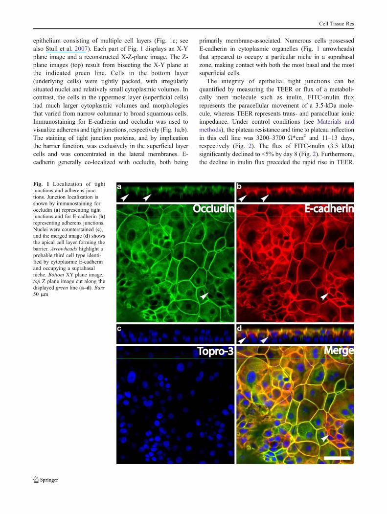
epithelium consisting of multiple cell layers (Fig. 1c; seealso Stull et al. 2007). Each part of Fig. 1 displays an X-Yplane image and a reconstructed X-Z-plane image. The Z-plane images (top) result from bisecting the X-Y plane atthe indicated green line. Cells in the bottom layer(underlying cells) were tightly packed, with irregularlysituated nuclei and relatively small cytoplasmic volumes. Incontrast, the cells in the uppermost layer (superficial cells)had much larger cytoplasmic volumes and morphologiesthat varied from narrow columnar to broad squamous cells.Immunostaining for E-cadherin and occludin was used tovisualize adherens and tight junctions, respectively (Fig. 1a,b).The staining of tight junction proteins, and by implicationthe barrier function, was exclusively in the superficial layercells and was concentrated in the lateral membranes. E-cadherin generally co-localized with occludin, both being
primarily membrane-associated. Numerous cells possessedE-cadherin in cytoplasmic organelles (Fig. 1 arrowheads)that appeared to occupy a particular niche in a suprabasalzone, making contact with both the most basal and the mostsuperficial cells.
The integrity of epithelial tight junctions can bequantified by measuring the TEER or flux of a metaboli-cally inert molecule such as inulin. FITC-inulin fluxrepresents the paracellular movement of a 3.5-kDa mole-cule, whereas TEER represents trans- and paracelluar ionicimpedance. Under control conditions (see Materials andmethods), the plateau resistance and time to plateau inflectionin this cell line was 3200–3700 Ω*cm2 and 11–13 days,respectively (Fig. 2). The flux of FITC-inulin (3.5 kDa)significantly declined to <5% by day 8 (Fig. 2). Furthermore,the decline in inulin flux preceded the rapid rise in TEER.
Fig. 1 Localization of tightjunctions and adherens junc-tions. Junction localization isshown by immunostaining foroccludin (a) representing tightjunctions and for E-cadherin (b)representing adherens junctions.Nuclei were counterstained (c),and the merged image (d) showsthe apical cell layer forming thebarrier. Arrowheads highlight aprobable third cell type identi-fied by cytoplasmic E-cadherinand occupying a suprabasalniche. Bottom XY plane image,top Z plane image cut along thedisplayed green line (a–d). Bars50 μm
Cell Tissue Res
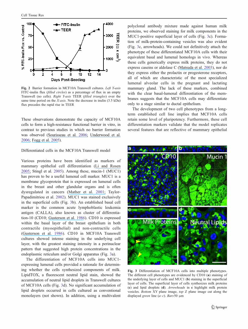
These observations demonstrate the capacity of MCF10Acells to form a high-resistance functional barrier in vitro, incontrast to previous studies in which no barrier formationwas observed (Sourisseau et al. 2006; Underwood et al.2006; Fogg et al. 2005).
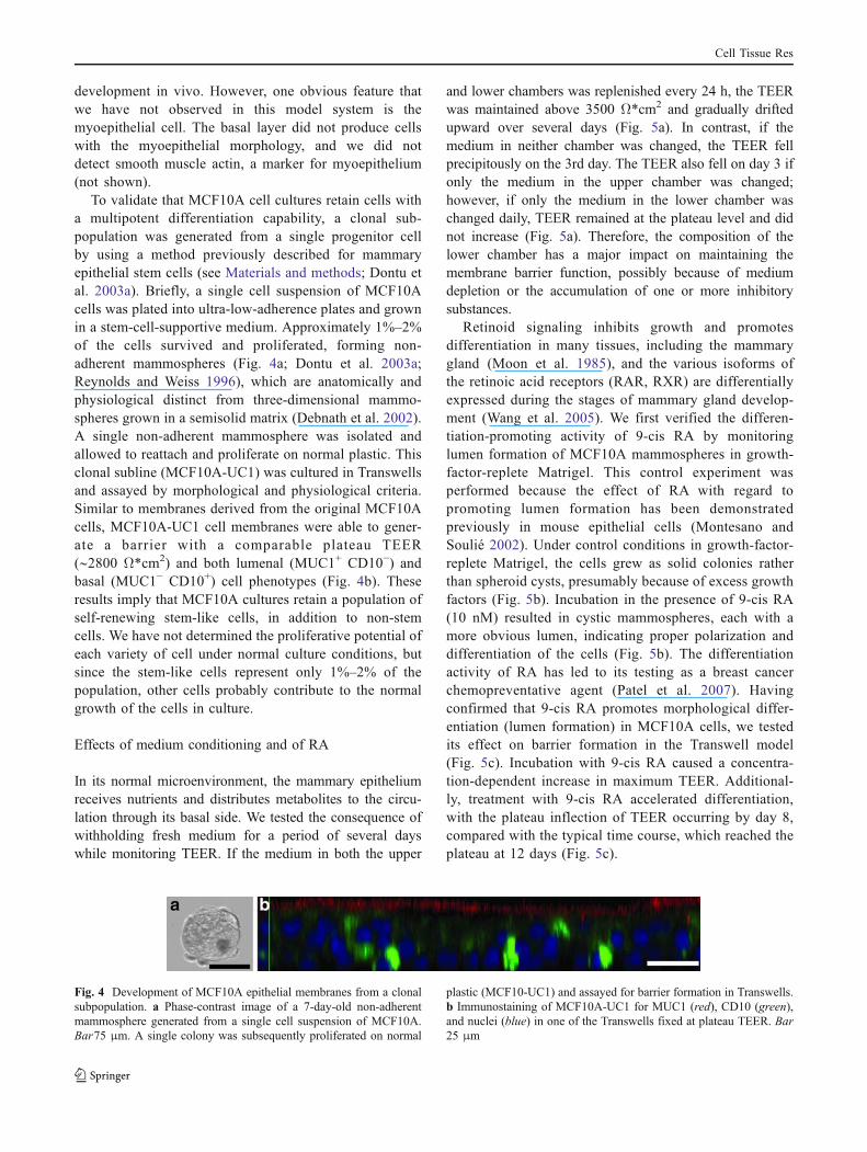
Differentiated cells in the MCF10A Transwell model
Various proteins have been identified as markers ofmammary epithelial cell differentiation (Li and Rosen2005; Stingl et al. 2005). Among these, mucin-1 (MUC1)has proven to be a useful lumenal cell marker. MUC1 is amembrane glycoprotein that is expressed on lumenal cellsin the breast and other glandular organs and is oftendysregulated in cancers (Mather et al. 2001; Taylor-Papadimitriou et al. 2002). MUC1 was stained exclusivelyin the superficial cells (Fig. 3b). An established basal cellmarker is the common acute lymphoblastic leukemiaantigen (CALLA), also known as cluster of differentia-tion-10 (CD10; Gusterson et al. 1986). CD10 is expressedwithin the basal layer of the breast epithelium in bothcontractile (myoepithelial) and non-contractile cells(Gusterson et al. 1986). CD10 in MCF10A Transwellcultures showed intense staining in the underlying celllayer, with the greatest staining intensity in a perinuclearpattern that suggested high protein concentrations in theendoplasmic reticulum and/or Golgi apparatus (Fig. 3a).
The differentiation of MCF10A cells into MUC1-expressing lumenal cells provided a rationale for determin-ing whether the cells synthesized components of milk.LipidTOX, a fluorescent neutral lipid stain, showed theaccumulation of neutral lipid droplets in Transwell culturesof MCF10A cells (Fig. 3d). No significant accumulation oflipid droplets occurred in cells cultured as conventionalmonolayers (not shown). In addition, using a multivalent
polyclonal antibody mixture made against human milkproteins, we observed staining for milk components in theMUC1-positive superficial layer of cells (Fig. 3c). Forma-tion of milk-protein-containing vesicles was also evident(Fig. 3c, arrowheads). We could not definitively attach thephenotype of these differentiated MCF10A cells with theirequivalent basal and lumenal homologs in vivo. Whereasthese cells generically express milk proteins, they do notexpress caseins or aldolase C (Matsuda et al. 2003), nor dothey express either the prolactin or progesterone receptors,all of which are characteristic of the most specializedlumenal alveolar cells in the pregnant and lactatingmammary gland. The lack of these markers, combinedwith the clear basal-lumenal differentiation of the mem-branes suggests that the MCF10A cells may differentiateonly to a stage similar to ductal epithelium.
The development of two cell phenotypes from a long-term established cell line implies that MCF10A cellsretain some level of pluripotency. Furthermore, these celldifferentiation markers validate that the model replicatesseveral features that are reflective of mammary epithelial
Fig. 2 Barrier formation in MCF10A Transwell cultures. Left Y-axisFITC-inulin flux (filled circles) as a percentage of flux in an emptyTranswell (no cells). Right Y-axis TEER (filled triangles) over thesame time period on the X-axis. Note the decrease in inulin (3.5 kDa)flux precedes the rapid rise in TEER
Fig. 3 Differentiation of MCF10A cells into multiple phenotypes.The different cell phenotypes are evidenced by CD10 (a) staining ofthe underlying layer of cells and MUC1 (b) staining in the superficiallayer of cells. The superficial layer of cells synthesizes milk proteins(c) and lipid droplets (d). Arrowheads in c highlight milk proteinvesicles. Bottom XY plane image, top Z plane image cut along thedisplayed green line (a–c). Bars50 μm
Cell Tissue Res
development in vivo. However, one obvious feature thatwe have not observed in this model system is themyoepithelial cell. The basal layer did not produce cellswith the myoepithelial morphology, and we did notdetect smooth muscle actin, a marker for myoepithelium(not shown).
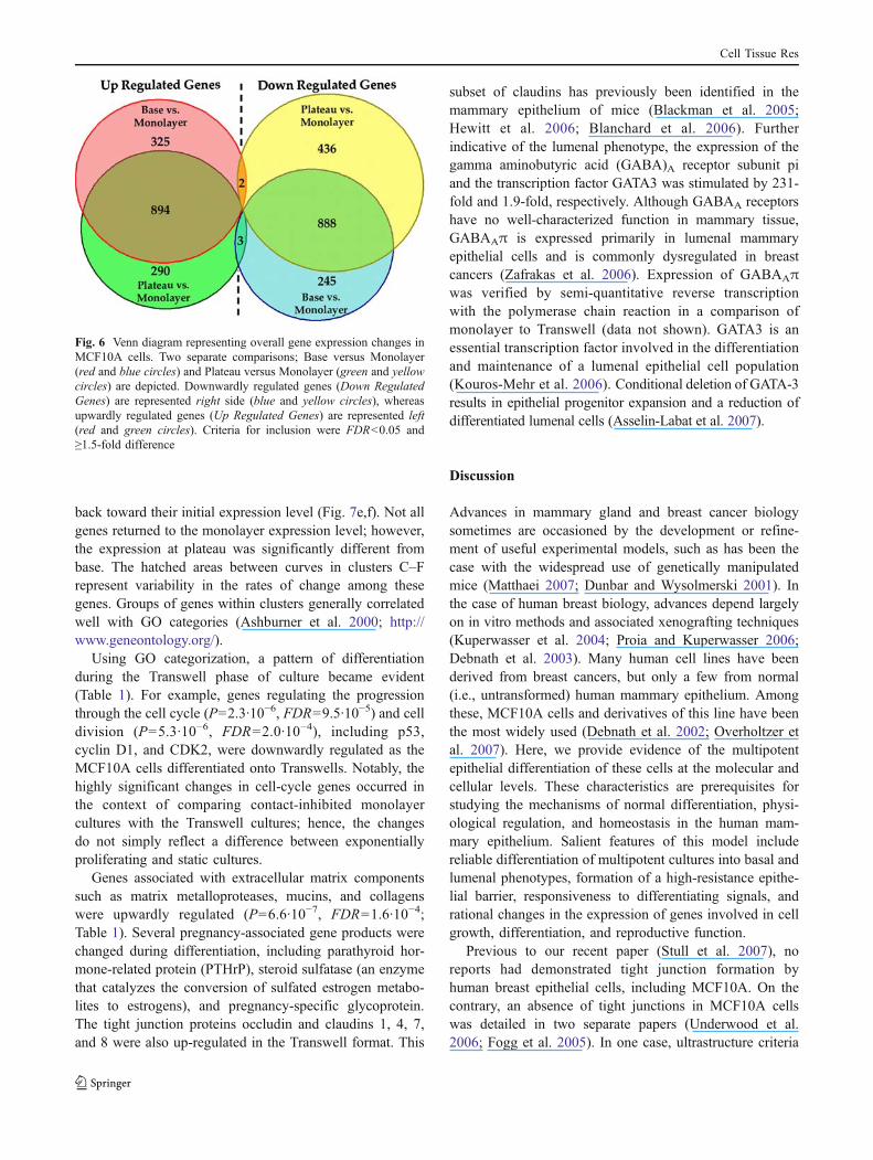
To validate that MCF10A cell cultures retain cells witha multipotent differentiation capability, a clonal sub-population was generated from a single progenitor cellby using a method previously described for mammaryepithelial stem cells (see Materials and methods; Dontu etal. 2003a). Briefly, a single cell suspension of MCF10Acells was plated into ultra-low-adherence plates and grownin a stem-cell-supportive medium. Approximately 1%–2%of the cells survived and proliferated, forming non-adherent mammospheres (Fig. 4a; Dontu et al. 2003a;Reynolds and Weiss 1996), which are anatomically andphysiological distinct from three-dimensional mammo-spheres grown in a semisolid matrix (Debnath et al. 2002).A single non-adherent mammosphere was isolated andallowed to reattach and proliferate on normal plastic. Thisclonal subline (MCF10A-UC1) was cultured in Transwellsand assayed by morphological and physiological criteria.Similar to membranes derived from the original MCF10Acells, MCF10A-UC1 cell membranes were able to gener-ate a barrier with a comparable plateau TEER(∼2800 Ω*cm2) and both lumenal (MUC1+ CD10−) andbasal (MUC1− CD10+) cell phenotypes (Fig. 4b). Theseresults imply that MCF10A cultures retain a population ofself-renewing stem-like cells, in addition to non-stemcells. We have not determined the proliferative potential ofeach variety of cell under normal culture conditions, butsince the stem-like cells represent only 1%–2% of thepopulation, other cells probably contribute to the normalgrowth of the cells in culture.
Effects of medium conditioning and of RA
In its normal microenvironment, the mammary epitheliumreceives nutrients and distributes metabolites to the circu-lation through its basal side. We tested the consequence ofwithholding fresh medium for a period of several dayswhile monitoring TEER. If the medium in both the upper
and lower chambers was replenished every 24 h, the TEERwas maintained above 3500 Ω*cm2 and gradually driftedupward over several days (Fig. 5a). In contrast, if themedium in neither chamber was changed, the TEER fellprecipitously on the 3rd day. The TEER also fell on day 3 ifonly the medium in the upper chamber was changed;however, if only the medium in the lower chamber waschanged daily, TEER remained at the plateau level and didnot increase (Fig. 5a). Therefore, the composition of thelower chamber has a major impact on maintaining themembrane barrier function, possibly because of mediumdepletion or the accumulation of one or more inhibitorysubstances.
Retinoid signaling inhibits growth and promotesdifferentiation in many tissues, including the mammarygland (Moon et al. 1985), and the various isoforms ofthe retinoic acid receptors (RAR, RXR) are differentiallyexpressed during the stages of mammary gland develop-ment (Wang et al. 2005). We first verified the differen-tiation-promoting activity of 9-cis RA by monitoringlumen formation of MCF10A mammospheres in growth-factor-replete Matrigel. This control experiment wasperformed because the effect of RA with regard topromoting lumen formation has been demonstratedpreviously in mouse epithelial cells (Montesano andSoulié 2002). Under control conditions in growth-factor-replete Matrigel, the cells grew as solid colonies ratherthan spheroid cysts, presumably because of excess growthfactors (Fig. 5b). Incubation in the presence of 9-cis RA(10 nM) resulted in cystic mammospheres, each with amore obvious lumen, indicating proper polarization anddifferentiation of the cells (Fig. 5b). The differentiationactivity of RA has led to its testing as a breast cancerchemopreventative agent (Patel et al. 2007). Havingconfirmed that 9-cis RA promotes morphological differ-entiation (lumen formation) in MCF10A cells, we testedits effect on barrier formation in the Transwell model(Fig. 5c). Incubation with 9-cis RA caused a concentra-tion-dependent increase in maximum TEER. Additional-ly, treatment with 9-cis RA accelerated differentiation,with the plateau inflection of TEER occurring by day 8,compared with the typical time course, which reached theplateau at 12 days (Fig. 5c).
Fig. 4 Development of MCF10A epithelial membranes from a clonalsubpopulation. a Phase-contrast image of a 7-day-old non-adherentmammosphere generated from a single cell suspension of MCF10A.Bar75 μm. A single colony was subsequently proliferated on normal
plastic (MCF10-UC1) and assayed for barrier formation in Transwells.b Immunostaining of MCF10A-UC1 for MUC1 (red), CD10 (green),and nuclei (blue) in one of the Transwells fixed at plateau TEER. Bar25 μm
Cell Tissue Res

Gene expression analysis of differentiating MCF10A cellson Transwells
To improve our understanding of the differences occurringin MCF10A cells as they polarized and differentiated in theTranswell model, we performed gene expression profilingwith Affymetrix Human Genome U133 Plus 2.0 Arrays.Four experimental time points, each performed in triplicate,were sampled, viz., conventional cultures of MCF10A cellscultured 5 days after reaching confluence on plastic(Monolayer), and MCF10A cells plated on Transwellssampled at three TEER values: 200–300 Ω*cm2 (Base),1400–1600 Ω*cm2 (Midpoint), and 3000–3200 Ω*cm2
(Plateau). Consistent with the immunostaining results inFig. 3, the microarray assay showed MUC1 and CD10 to besignificantly up-regulated in the Transwell cultures (byapproximately 13-fold and five-fold, respectively). Themajority of gene expression changes observed werebetween confluent monolayer MCF10A cells and any ofthe Transwell time points assayed. Figure 6 depicts a Venndiagram for two of those comparisons (Base versus
Monolayer and Plateau versus Monolayer). For example,of the 1133 genes that were significantly down-regulated inBase versus Monolayer, 888 (78.4%) were also down-regulated in the Plateau versus Monolayer comparison(Fig. 6). Of the 1219 genes that were up-regulated in Baseversus Monolayer, 894 (73.3%) were also up-regulated inPlateau versus Monolayer comparison. Five genes wereoppositely regulated on comparing Base versus Plateau;three were up-regulated in Plateau and down-regulated inBase, while the other two were vice versa.
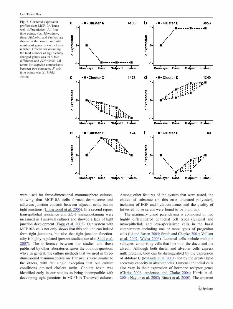
Gene clustering organizes gene expression patterns intocoordinate regulatory profiles. Figure 7 depicts six func-tionally informative clustering patterns. Large numbers ofgenes were downwardly or upwardly regulated comparingmonolayer cultures with the Transwell format and did notchange significantly thereafter (Fig. 7a,b). Genes thatgradually changed as the TEER increased, ultimatelyreaching significant differences, are illustrated by clustersC and D, respectively (Fig. 7c,d). Clusters E and Frepresent genes that changed significantly in the transitionfrom monolayer to base and again changed significantly
Fig. 5 Effect of medium condi-tioning and retinoic acid (RA) onMCF10A polarization. a Main-tenance of the barrier only whenthe basal chamber is replen-ished. After peak TEER wasachieved, only the listed cham-bers of medium were replen-ished daily. The polarizedepithelium was dependent onthe basal chamber being replen-ished. b, b’ RA, a powerfuldifferentiation agent, caused lu-men formation in MCF10Amammospheres. c In the contextof the MCF10A Transwell mod-el, RA caused a dose-dependentincrease in peak TEER achievedand shortened the time to pla-teau inflection. Means on thefinal day of the experiment wereanalyzed by one-way ANOVA(n=6) followed by the Holm-Sidak post-hoc test (GraphPadInStat software). Data are pre-sented as means±SEM and areconsidered significant when thechance of type I error was <0.05
Cell Tissue Res
back toward their initial expression level (Fig. 7e,f). Not allgenes returned to the monolayer expression level; however,the expression at plateau was significantly different frombase. The hatched areas between curves in clusters C–Frepresent variability in the rates of change among thesegenes. Groups of genes within clusters generally correlatedwell with GO categories (Ashburner et al. 2000; http://www.geneontology.org/).
Using GO categorization, a pattern of differentiationduring the Transwell phase of culture became evident(Table 1). For example, genes regulating the progressionthrough the cell cycle (P=2.3∙10−6, FDR=9.5∙10−5) and celldivision (P=5.3∙10−6, FDR=2.0∙10−4), including p53,cyclin D1, and CDK2, were downwardly regulated as theMCF10A cells differentiated onto Transwells. Notably, thehighly significant changes in cell-cycle genes occurred inthe context of comparing contact-inhibited monolayercultures with the Transwell cultures; hence, the changesdo not simply reflect a difference between exponentiallyproliferating and static cultures.
Genes associated with extracellular matrix componentssuch as matrix metalloproteases, mucins, and collagenswere upwardly regulated (P=6.6∙10−7, FDR=1.6∙10−4;Table 1). Several pregnancy-associated gene products werechanged during differentiation, including parathyroid hor-mone-related protein (PTHrP), steroid sulfatase (an enzymethat catalyzes the conversion of sulfated estrogen metabo-lites to estrogens), and pregnancy-specific glycoprotein.The tight junction proteins occludin and claudins 1, 4, 7,and 8 were also up-regulated in the Transwell format. This
subset of claudins has previously been identified in themammary epithelium of mice (Blackman et al. 2005;Hewitt et al. 2006; Blanchard et al. 2006). Furtherindicative of the lumenal phenotype, the expression of thegamma aminobutyric acid (GABA)A receptor subunit piand the transcription factor GATA3 was stimulated by 231-fold and 1.9-fold, respectively. Although GABAA receptorshave no well-characterized function in mammary tissue,GABAAπ is expressed primarily in lumenal mammaryepithelial cells and is commonly dysregulated in breastcancers (Zafrakas et al. 2006). Expression of GABAAπwas verified by semi-quantitative reverse transcriptionwith the polymerase chain reaction in a comparison ofmonolayer to Transwell (data not shown). GATA3 is anessential transcription factor involved in the differentiationand maintenance of a lumenal epithelial cell population(Kouros-Mehr et al. 2006). Conditional deletion of GATA-3results in epithelial progenitor expansion and a reduction ofdifferentiated lumenal cells (Asselin-Labat et al. 2007).
Discussion
Advances in mammary gland and breast cancer biologysometimes are occasioned by the development or refine-ment of useful experimental models, such as has been thecase with the widespread use of genetically manipulatedmice (Matthaei 2007; Dunbar and Wysolmerski 2001). Inthe case of human breast biology, advances depend largelyon in vitro methods and associated xenografting techniques(Kuperwasser et al. 2004; Proia and Kuperwasser 2006;Debnath et al. 2003). Many human cell lines have beenderived from breast cancers, but only a few from normal(i.e., untransformed) human mammary epithelium. Amongthese, MCF10A cells and derivatives of this line have beenthe most widely used (Debnath et al. 2002; Overholtzer etal. 2007). Here, we provide evidence of the multipotentepithelial differentiation of these cells at the molecular andcellular levels. These characteristics are prerequisites forstudying the mechanisms of normal differentiation, physi-ological regulation, and homeostasis in the human mam-mary epithelium. Salient features of this model includereliable differentiation of multipotent cultures into basal andlumenal phenotypes, formation of a high-resistance epithe-lial barrier, responsiveness to differentiating signals, andrational changes in the expression of genes involved in cellgrowth, differentiation, and reproductive function.
Previous to our recent paper (Stull et al. 2007), noreports had demonstrated tight junction formation byhuman breast epithelial cells, including MCF10A. On thecontrary, an absence of tight junctions in MCF10A cellswas detailed in two separate papers (Underwood et al.2006; Fogg et al. 2005). In one case, ultrastructure criteria
Fig. 6 Venn diagram representing overall gene expression changes inMCF10A cells. Two separate comparisons; Base versus Monolayer(red and blue circles) and Plateau versus Monolayer (green and yellowcircles) are depicted. Downwardly regulated genes (Down RegulatedGenes) are represented right side (blue and yellow circles), whereasupwardly regulated genes (Up Regulated Genes) are represented left(red and green circles). Criteria for inclusion were FDR<0.05 and≥1.5-fold difference
Cell Tissue Res
were used for three-dimensional mammosphere cultures,showing that MCF10A cells formed desmosome andadherens junction contacts between adjacent cells, but notight junctions (Underwood et al. 2006). In a second report,transepithelial resistance and ZO-1 immunostaining weremeasured in Transwell cultures and showed a lack of tightjunction development (Fogg et al. 2005). Our system withMCF10A cells not only shows that this cell line can indeedform tight junctions, but also that tight junction function-ality is highly regulated (present studies; see also Stull et al.2007). The difference between our studies and thosepublished by other laboratories raises the obvious question:why? In general, the culture methods that we used in three-dimensional mammospheres on Transwells were similar tothe others, with the single exception that our cultureconditions omitted cholera toxin. Cholera toxin wasidentified early in our studies as being incompatible withdeveloping tight junctions in MCF10A Transwell cultures.
Among other features of the system that were tested, thechoice of substrate (in this case uncoated polyester),inclusion of EGF and hydrocortisone, and the quality oflot-tested horse serum were found to be important.
The mammary gland parenchyma is composed of twohighly differentiated epithelial cell types (lumenal andmyoepithelial) and less-specialized cells in the basalcompartment including one or more types of progenitorcells (Li and Rosen 2005; Smith and Chepko 2001; Vaillantet al. 2007; Wicha 2006). Lumenal cells include multiplesubtypes, comprising cells that line both the ducts and thealveoli. Although both ductal and alveolar cells expressmilk proteins, they can be distinguished by the expressionof aldolase C (Matsuda et al. 2003) and by the greater lipidsecretory capacity in alveolar cells. Lumenal epithelial cellsalso vary in their expression of hormone receptor genes(Clarke 2006; Anderson and Clarke 2004; Harris et al.2004; Naylor et al. 2003; Binart et al. 2000). The apparent
Fig. 7 Clustered expressionprofiles over MCF10A Trans-well differentiation. All fourtime points, viz., Monolayer,Base, Midpoint, and Plateau areshown on the X-axis, and totalnumber of genes in each clusteris listed. Criteria for obtainingthe total number of significantlychanged genes was ≥1.5-folddifference and FDR<0.05. Cri-terion for stepwise comparisonsbetween two connected X-axistime points was ≥1.5-foldchange
Cell Tissue Res

ductal phenotype of the lumenal cells in this model isevidenced by their lack of prolactin receptor expression andthe down-regulation of keratin 18 (unpublished results;Table 1; Rudland and Hughes 1989). Myoepithelial cellsare specialized contractile cells found between the lumenalcells and the stroma. They express smooth muscle α-actinand CD10 and are stimulated to contract by oxytocin.Within the basal compartment, stem and/progenitor cellscan be found, which various authors have proposed tonumber from <1% to nearly 10%. Many studies haveattempted to characterize the mammary gland stem cellseither by functional assays (self-renewal, long-term labelretention) or by the presence and absence of particular cellmarkers such as MUC1, CD10, keratins, smooth muscleα-actin, and epithelial-specific antigen (Liu et al. 2005;Dontu et al. 2003a; Smith and Chepko 2001; Gudjonsson etal. 2005; Dontu et al. 2003b). As multifaceted and vast asthis body of literature is, the nature and complexity of theapparent hierarchy of mammary stem/progenitor cells isstill unresolved. Our model shows that the MCF10A cellspossess properties of multipotent progenitor cells, asevidenced by differentiation into basal (CD10 + , MUC1−)and lumenal (CD10−, MUC1+) phenotypes. Consequently,this model should be a useful tool for testing pathwaysinvolved in the proliferation and differentiation of thesepopulations.
Broad-based gene profiling by microarray hybridizationhas been highly valuable as a means of validating the kind ofchanges that one might expect during the differentiation ofMCF10A cultures. One issue that this method resolves is thenature of themultiple cell layering that occurs on the Transwellsubstrate. Highly proliferative, substrate-independent cancercells avoid “contact inhibition” and pile up in multiplelayers based on unrestrained proliferation. The suppressionof cell-cycle genes and the induction of epithelialdevelopment and differentiation genes (Table 1), togetherwith the morphological evidence (Fig. 1), demonstrate thatthe multiple cell layers in Transwell-cultured MCF10Arepresent an organized developmental process that issimilar to the multiple cell layers observed in vivo, asopposed to a disorganized pathology. One source ofambiguity in gene profiling is the presence of multiple celltypes. Whereas this is less of a problem when using anestablished cell line, clearly the differentiated MCF10A cellsamples that we have profiled include at least two(superficial and underlying) cell types, and probably a third(suprabasal cells with cytoplamic E-cadherin; Fig. 1darrowheads). Although this limits the information that wecould extract from this initial profiling study, the relativesimplicity of producing differentiated MCF10A cell pop-ulations, compared with purifying single cell populationfrom human specimens, should make it possible to performextensive gene profiling on well-defined cell populations
Table 1 Gene ontology (GO) categories of significantly changedgenes. Criteria for inclusion were FDR<0.05 and ≥1.5-fold difference.Individual genes listed are not a complete list of all genes within eachGO category but are representative. Cluster denotations refer to Fig. 7.Complete data series of microarray chips are available at the GeneExpression Omnibus (GEO) database (http://www.ncbi.nlm.nih.gov/geo/) with accession number GSE10070
GO category/gene name Cluster Maximum fold differencefrom monolayer
Cell cyclep53 C −2.0CDK2 C −2.3cyclin E2 A −3.1CDC25A A −4.2cyclin D1 A −1.6Extracellular matrixMucin 4 B 3.5MMP1,3,9,10 D 60.2, 2.6, 8.0, 78.5Structural constituentof the cytoskeletonKeratins 4, 6A, 6B, 13, 14,16, 17, 23
B 72.4, 2.9, 33.7, 76.9, 12.0,45.0, 8.8, 12.7
Keratin 18 A −3.2CytoskeletonMUC1 B 13.3Involucrin B 11.8Integrin β8 B 2.0Adducin3 B 1.7Tissue developmentCollagen Type 1α1 B 10.3Collagen Type 7α1 B 6.4Cellular retinoic acidbinding protein 2
B 8.1
Desmoplakin B 3.0Reproductive physiologicalprocessInhibin, beta A F −1.8Parathyroid hormone-related protein
D 7.8
Pappalysin 1 A −2.3Steroid sulfatase D 2.3Pregnancy-specific beta-1-glycoprotein 4
D 1.6
MorphogenesisWnt5A F −2.2Transforming growthfactor-β2
F −4.1
Insulin-like growth factorbinding protein-3
D 25.2
Gap junction protein, alpha1, (connexin 43)
D 114.4
MiscellaneousGATA-3 D 1.9Forkhead Box Q1 F −2.5Forkhead box O1A B 3.3Gamma aminobutyric acidA receptor π
B 231.0
Crystallin, alpha B D 27.8CD10 (MME, CALLA) B 4.8E-cadherin B 2.3Occludin B 3.5Claudin 1, 4, 7, 8 B 2.3, 3.8, 2.9, 20.5
Cell Tissue Res
that can be sorted from the MCF10A cultures based on themarkers that we have identified here. One interestingquestion to address would be the relationship of theunderlying population of MCF10A cells to basal cells invivo, particularly the myoepithelium.
Among the gene expression changes in the Transwelldifferentiation process, particularly interesting genes wereidentified, in addition to the general suppression of the cellcycle and shifts in the expression of cytoskeleton andextracellular matrix genes. For example, PTHrP expressionwas induced approximately 10-fold during differentiation.This hormone is a master signal for the development andmaintenance of the mammary epithelial phenotype (Dunbarand Wysolmerski 1999; Foley et al. 2001). PTHrP receptorswere also expressed in the MCF10A cultures, but thereceptor levels did not change under the various conditions.The presence of the receptors together with the differentialexpression of the ligand leads one to speculate that PTHrPfunctions as an autocrine/paracrine factor in the MCF10Acultures. Among the small number of genes that fell intoCluster F (declined significantly after plating on Transwells,then reinduced as TEER increased), insulin-like growth factorbinding protein-3, transforming growth factor-β, and FOXQ1are associated in a growth inhibitory pathway downstream ofAkt/protein kinase B (Roarty and Serra 2007; Samatar et al.2002). The induction of these genes implies that a secondAkt-dependent growth inhibitory pathway is induced duringthe final differentiation of the MCF10A cultures.
In summary, we have demonstrated the multipotentdifferentiation of human mammary epithelial cells thatpredictably assemble tight junctions and thus develop alow-conductance barrier. Tight junction patency is depen-dent on the composition of the bottom chamber medium,and RA accelerates and amplifies the TEER of MCF10Amembranes. Overall, the MCF10A Transwell model reca-pitulates several important characteristics of the in vivoenvironment of the mammary gland (barrier formation,lumenal vs basal cell phenotypes with appropriate expres-sion of markers for each). This model is highly useful forstudying endocrine and autocrine-paracrine developmentalregulation and physiological changes of human mammaryepithelium. This system can probably be further refined.
Acknowledgements The authors thank Danielle Halbleib andMelissa Orr for technical contributions.
References
Anderson E, Clarke RB (2004) Steroid receptors and cell cycle in normalmammary epithelium. J Mammary Gland Biol Neoplasia 9:3–13
Ashburner M, Ball CA, Blake JA, Botstein D, Butler H, Cherry JM,Davis AP, Dolinski K, Dwight SS, Eppig JT, Harris MA, Hill DP,
Issel-Tarver L, Kasarskis A, Lewis S, Matese JC, Richardson JE,Ringwald M, Rubin GM, Sherlock G (2000) Gene ontology: toolfor the unification of biology. The Gene Ontology Consortium.Nat Genet 25:25–29
Asselin-Labat ML, Sutherland KD, Barker H, Thomas R, Shackleton M,Forrest NC, Hartley L, Robb L, Grosveld FG, van der Wees J,Lindeman GJ, Visvader JE (2007) Gata-3 is an essential regulator ofmammary-gland morphogenesis and luminal-cell differentiation.Nat Cell Biol 9:201–209
Benjamini Y, Hochberg Y (1995) Controlling the false discovery rate:a practical and powerful approach to multiple testing. J R StatSoc [B] 57:289–300
Binart N, Ormandy CJ, Kelly PA (2000) Mammary gland develop-ment and the prolactin receptor. Adv Exp Med Biol 480:85–92
Blackman B, Russell T, Nordeen SK, Medina D, Neville MC (2005)Claudin 7 expression and localization in the normal murinemammary gland and murine mammary tumors. Breast CancerRes 7:R248–R255
Blanchard AA, Watson PH, Shiu RP, Leygue E, Nistor A, Wong P,Myal Y (2006) Differential expression of claudin 1, 3, and 4during normal mammary gland development in the mouse. DNACell Biol 25:79–86
Cardiff RD, Wellings SR (1999) The comparative pathology of humanand mouse mammary glands. J Mammary Gland Biol Neoplasia4:105–122
Clarke RB (2006) Ovarian steroids and the human breast: regulationof stem cells and cell proliferation. Maturitas 54:327–334
Debnath J, Mills KR, Collins NL, Reginato MJ, Muthuswamy SK,Brugge JS (2002) The role of apoptosis in creating and maintain-ing luminal space within normal and oncogene-expressingmammary acini. Cell 111:29–40
Debnath J, Muthuswamy SK, Brugge JS (2003) Morphogenesis andoncogenesis of MCF-10A mammary epithelial acini grown inthree-dimensional basement membrane cultures. Methods30:256–268
Dennis G Jr, Sherman BT, Hosack DA, Yang J, Gao W, Lane HC,Lempicki RA (2003) DAVID: Database for Annotation, Visual-ization, and Integrated Discovery. Genome Biol 4:P3
DiRenzo J, Signoretti S, Nakamura N, Rivera-Gonzalez R, Sellers W,Loda M, Brown M (2002) Growth factor requirements and basalphenotype of an immortalized mammary epithelial cell line.Cancer Res 62:89–98
Dontu G, Abdallah WM, Foley JM, Jackson KW, Clarke MF,Kawamura MJ, Wicha MS (2003a) In vitro propagation andtranscriptional profiling of human mammary stem/progenitorcells. Genes Dev 17:1253–1270
Dontu G, Al-Hajj M, Abdallah WM, Clarke MF, Wicha MS (2003b)Stem cells in normal breast development and breast cancer. CellProlif 36 (Suppl 1):59–72
Dunbar ME, Wysolmerski JJ (1999) Parathyroid hormone-relatedprotein: a developmental regulatory molecule necessary formammary gland development. J Mammary Gland Biol Neoplasia4:21–34
Dunbar ME, Wysolmerski JJ (2001) Mammary ductal and alveolardevelopment: lesson learned from genetically manipulated mice.Microsc Res Tech 52:163–170
Fogg VC, Liu CJ, Margolis B (2005) Multiple regions of Crumbs3 arerequired for tight junction formation in MCF10A cells. J Cell Sci118:2859–2869
Foley J, Dann P, Hong J, Cosgrove J, Dreyer B, Rimm D, Dunbar M,Philbrick W, Wysolmerski J (2001) Parathyroid hormone-relatedprotein maintains mammary epithelial fate and triggers nippleskin differentiation during embryonic breast development. De-velopment 128:513–525
Gudjonsson T, Adriance MC, Sternlicht MD, Petersen OW, Bissell MJ(2005) Myoepithelial cells: their origin and function in breast
Cell Tissue Res
morphogenesis and neoplasia. J Mammary Gland Biol Neoplasia10:261–272
Gusterson BA, Monaghan P, Mahendran R (1986) Identification ofmyoepithelial cells in human and rat breasts by anti-commonacute lymphoblastic leukemia antigen antibody A12. J NatlCancer Inst 77:343–349
Harris J, Stanford PM, Oakes SR, Ormandy CJ (2004) Prolactin andthe prolactin receptor: new targets of an old hormone. Ann Med36:414–425
Hewitt KJ, Agarwal R, Morin PJ (2006) The claudin gene family:expression in normal and neoplastic tissues. BMC Cancer 6:186
Kominsky SL, Argani P, Korz D, Evron E, Raman V, Garrett E, ReinA, Sauter G, Kallioniemi O-, Sukumar S (2003) Loss of the tightjunction protein claudin-7 correlates with histological grade inboth ductal carcinoma in situ and invasive ductal carcinoma ofthe breast. Oncogene 22:2021–2033
Kouros-Mehr H, Slorach EM, Sternlicht MD, Werb Z (2006) GATA-3maintains the differentiation of the luminal cell fate in themammary gland. Cell 127:1041–1055
Kuperwasser C, Chavarria T, Wu M, Magrane G, Gray JW, Carey L,Richardson A, Weinberg RA (2004) Reconstruction of function-ally normal and malignant human breast tissues in mice. ProcNatl Acad Sci USA 101:4966–4971
Li Y, Rosen JM (2005) Stem/progenitor cells in mouse mammarygland development and breast cancer. J Mammary Gland BiolNeoplasia 10:17–24
Liu S, Dontu G, Wicha MS (2005) Mammary stem cells, self-renewalpathways, and carcinogenesis. Breast Cancer Res 7:86–95
Martin TA, Watkins G, Mansel RE, Jiang WG (2004) Loss of tightjunction plaque molecules in breast cancer tissues is associatedwith a poor prognosis in patients with breast cancer. Eur J Cancer40:2717–2725
Mather IH, Jack LJ, Madara PJ, Johnson VG (2001) The distributionof MUC1, an apical membrane glycoprotein, in mammaryepithelial cells at the resolution of the electron microscope:implications for the mechanism of milk secretion. Cell TissueRes 304:91–101
Matsuda M, Lockefeer JA, Horseman ND (2003) Aldolase C/zebringene regulation by prolactin during pregnancy and lactation.Endocrine 20:91–100
Matthaei KI (2007) Genetically manipulated mice: a powerful toolwith unsuspected caveats. J Physiol (Lond) 582:481–488
Montesano R, Soulié P (2002) Retinoids induce lumen morphogenesisin mammary epithelial cells. J Cell Sci 115:4419–4431
Moon RC, Mehta RG, McCormick DL (1985) Retinoids andmammary gland differentiation. Ciba Found Symp 113:156–167
Naylor MJ, Lockefeer JA, Horseman ND, Ormandy CJ (2003)Prolactin regulates mammary epithelial cell proliferation viaautocrine/paracrine mechanism. Endocrine 20:111–114
Neville MC, McFadden TB, Forsyth I (2002) Hormonal regulation ofmammary differentiation and milk secretion. J Mammary GlandBiol Neoplasia 7:49–66
Nguyen DA, Neville MC (1998) Tight junction regulation in themammary gland. J Mammary Gland Biol Neoplasia 3:233–246
Nguyen DA, Parlow AF, Neville MC (2001) Hormonal regulation oftight junction closure in the mouse mammary epithelium during thetransition from pregnancy to lactation. J Endocrinol 170:347–356
Overholtzer M, Mailleux AA, Mouneimne G, Normand G, Schnitt SJ,King RW, Cibas ES, Brugge JS (2007) A nonapoptotic cell deathprocess, entosis, that occurs by cell-in-cell invasion. Cell131:966–979
Parmar H, Cunha GR (2004) Epithelial-stromal interactions in themouse and human mammary gland in vivo. Endocr Relat Cancer11:437–458
Patel JB, Mehta J, Belosay A, Sabnis G, Khandelwal A, Brodie AMH,Soprano DR, Njar VCO (2007) Novel retinoic acid metabolism
blocking agents have potent inhibitory activities on human breastcancer cells and tumour growth. Br J Cancer 96:1204–1215
Proia DA, Kuperwasser C (2006) Reconstruction of human mammarytissues in a mouse model. Nat Protoc 1:206–214
Reynolds BA, Weiss S (1996) Clonal and population analysesdemonstrate that an EGF-responsive mammalian embryonicCNS precursor is a stem cell. Dev Biol 175:1–13
Roarty K, Serra R (2007) Wnt5a is required for proper mammarygland development and TGF-beta-mediated inhibition of ductalgrowth. Development 134:3929–3939
Rudland PS, Barraclough R (1990) Differentiation of simian virus 40transformed human mammary epithelial stem cell lines tomyoepithelial-like cells is associated with increased expressionof viral large T antigen. J Cell Physiol 142:657–665
Rudland PS, Hughes CM (1989) Immunocytochemical identificationof cell types in human mammary gland: variations in cellularmarkers are dependent on glandular topography and differentia-tion. J Histochem Cytochem 37:1087–1100
Samatar AA, Wang L, Mirza A, Koseoglu S, Liu S, Kumar CC (2002)Transforming growth factor-beta 2 is a transcriptional target forAkt/protein kinase B via forkhead transcription factor. J BiolChem 277:28118–28126
Sartor MA, Tomlinson CR, Wesselkamper SC, Sivaganesan S,Leikauf GD, Medvedovic M (2006) Intensity-based hierarchicalBayes method improves testing for differentially expressed genesin microarray experiments. BMC Bioinformatics 7:538
Shamay A, Shapiro F, Leitner G, Silanikove N (2003) Infusions of caseinhydrolyzates into the mammary gland disrupt tight junction integrityand induce involution in cows. J Dairy Sci 86:1250–1258
Sheffield LG (1988) Organization and growth of mammary epitheliain the mammary gland fat pad. J Dairy Sci 71:2855–2874
Smith GH, Chepko G (2001) Mammary epithelial stem cells. MicroscRes Tech 52:190–203
Smyth GK (2004) Linear models and empirical bayes methods forassessing differential expression in microarray experiments. StatAppl Genet Mol Biol 3:article 3
Soule HD, Maloney TM, Wolman SR, Peterson WD Jr, Brenz R,McGrath CM, Russo J, Pauley RJ, Jones RF, Brooks SC (1990)Isolation and characterization of a spontaneously immortalizedhuman breast epithelial cell line, MCF-10. Cancer Res 50:6075–6086
Sourisseau T, Georgiadis A, Tsapara A, Ali RR, Pestell R, Matter K,Balda MS (2006) Regulation of PCNA and cyclin D1 expressionand epithelial morphogenesis by the ZO-1-regulated transcriptionfactor ZONAB/DbpA. Mol Cell Biol 26:2387–2398
Srivastava S, Matsuda M, Hou Z, Bailey JP, Kitazawa R, Herbst MP,Horseman ND (2003) Receptor activator of NF-kappaB ligandinduction via Jak2 and Stat5a in mammary epithelial cells. J BiolChem 278:46171–46178
Stingl J, Raouf A, Emerman JT, Eaves CJ (2005) Epithelialprogenitors in the normal human mammary gland. J MammaryGland Biol Neoplasia 10:49–59
Stull MA, Pai V, Vomachka AJ, Marshall AM, Jacob GA, Horseman ND(2007) Mammary gland homeostasis employs serotonergic regula-tion of epithelial tight junctions. Proc Natl Acad Sci USA104:16708–16713
Taylor-Papadimitriou J, Burchell JM, Plunkett T, Graham R, Correa I,Miles D, Smith M (2002) MUC1 and the immunobiology ofcancer. J Mammary Gland Biol Neoplasia 7:209–221
Tokes AM, Kulka J, Paku S, Szik A, Paska C, Novak PK, Szilak L,Kiss A, Bogi K, Schaff Z (2005) Claudin-1, -3 and -4 proteinsand mRNA expression in benign and malignant breast lesions: aresearch study. Breast Cancer Res 7:R296–R305
Underwood JM, ImbalzanoKM,Weaver VM, Fischer AH, ImbalzanoAN,Nickerson JA (2006) The ultrastructure of MCF-10A acini. J CellPhysiol 208:141–148
Cell Tissue Res

Utama FE, LeBaron MJ, Neilson LM, Sultan AS, Parlow AF,Wagner KU, Rui H (2006) Human prolactin receptors areinsensitive to mouse prolactin: implications for xenotransplantmodeling of human breast cancer in mice. J Endocrinol 188:589–601
Vaillant F, Asselin-Labat ML, Shackleton M, Lindeman GJ, Visvader JE(2007) The emerging picture of the mouse mammary stem cell.Stem Cell Rev 3:114–123
Wang YA, Shen K, Wang Y, Brooks SC (2005) Retinoic acidsignaling is required for proper morphogenesis of mammarygland. Dev Dyn 234:892–899
Wicha MS (2006) Identification of murine mammary stem cells:implications for studies of mammary development and carcino-genesis. Breast Cancer Res 8:109
Wood TL, Richert MM, Stull MA, Allar MA (2000) The insulin-likegrowth factors (IGFs) and IGF binding proteins in postnataldevelopment of murine mammary glands. J Mammary GlandBiol Neoplasia 5:31–42
Zafrakas M, Chorovicer M, Klaman I, Kristiansen G, Wild PJ,Heindrichs U, Knuchel R, Dahl E (2006) Systematic character-isation of GABRP expression in sporadic breast cancer andnormal breast tissue. Int J Cancer 118:1453–1459
Cell Tissue Res
Related Documents