Development/Plasticity/Repair Wnt-Responsive Lgr5 Globose Basal Cells Function as Multipotent Olfactory Epithelium Progenitor Cells Mengfei Chen, 1 Shenghe Tian, 1,2 Xiaoling Yang, 2 Andrew P. Lane, 3 Randall R. Reed, 4 and Hongjun Liu 1,2 1 Department of Microbiology and Molecular Genetics and 2 Department of Ophthalmology, University of Pittsburgh School of Medicine, Pittsburgh, Pennsylvania 15213, and 3 Department of Otolaryngology–Head and Neck Surgery and 4 Center for Sensory Biology, Johns Hopkins University School of Medicine, Baltimore, Maryland 21205 Persistent neurogenesis in the olfactory epithelium provides a unique model to study neural stem cell self-renewal and fate determina- tion. In the olfactory neuroepithelium, globose basal cells (GBCs) are considered to be the direct progenitors of olfactory neurons. However, the study of neurogenesis from GBCs has been impeded by the paucity of GBC-specific markers. Here we report that Lgr5, a recently discovered adult stem cell marker, is exclusively expressed in GBCs in neonatal and adult mice. Lgr5 cells display character- istics of cycling stem cells, including Ki67 expression and EdU incorporation. Lineage tracing analysis demonstrates that Lgr5 GBCs regenerate multiple cell types under normal turnover condition or after olfactory lesion. Furthermore, upregulation or downregulation of Wnt signaling in vivo indicates a key role of Wnt signaling not only in maintaining Lgr5 cell proliferation and promoting neurore- generation, but also in delaying sensory neuron maturation. Together, our observations provided new insights into the dynamics of neurogenesis in the olfactory epithelium. Key words: globose basal cells; Lgr5; neural stem cells; olfactory epithelium; regeneration; Wnt Introduction The olfactory epithelium is organized as a pseudostratified epi- thelial structure mainly composed of olfactory sensory neurons, apical sustentacular cells, Bowman’s gland/ducts, microvillous cells, and neural stem cells (Schwob, 2002). Horizontal basal cells (HBCs) and globose basal cells (GBCs) are considered candidate stem cells in the basal compartment of the olfactory epithelium (Carter et al., 2004; Chen et al., 2004). In defined conditions, HBCs can generate multiple differentiated neuronal and glial progeny in vitro (Carter et al., 2004). Fate-mapping studies using an HBC-specific Krt5-Cre strongly support the concept that HBCs are multipotent stem cells that can regenerate GBCs and subsequently give rise to both neuronal and non-neuronal lin- eages in the olfactory epithelium (Leung et al., 2007). In this scenario, HBCs are relatively quiescent and serve as a reserve stem cell pool, which can be stimulated after severe injury. GBCs are situated in the basal olfactory epithelium between HBCs and immature olfactory sensory neurons. Among the GBC population, there are committed neuronal precursors, whereas others are mitotically activated, representing transit-amplifying progenitor cells (Caggiano et al., 1994; Huard et al., 1998). Dur- ing olfactory epithelial neurogenesis, the Ascl1 (Mash1), Ngn1, and NeuroD1 transcription factors sequentially express in dis- tinct but overlapping stages of GBC differentiation (Cau et al., 2002; Manglapus et al., 2004; Packard et al., 2011a). A subset of GBCs are Ascl1 transit-amplifying cells that primarily give rise to Ngn1 and NeuroD1 immediate neuronal precursors, sup- porting the notion that the GBCs are a fate-restricted cell popu- lation with a neuron-specific differentiation potency (Cau et al., 2002; Guo et al., 2010). A previous study using retroviral lineage tracing suggests that a multipotent progenitor cell population resides in the basal layer of the olfactory epithelium that contains both HBCs and GBCs (Huard et al., 1998). In addition, sorted GBCs also give rise to the majority of olfactory epithelium cell types after transplantation (Chen et al., 2004). However, definitive evidence that supports the idea that GBCs function as multipotent olfactory epithelium progenitor cells in vivo remains missing (Jang et al., 2003; Mur- doch and Roskams, 2007). Here we report that Lgr5, a G-protein-coupled receptor fam- ily protein that has been identified as a marker of adult stem cells in multiple tissues and organs (Barker et al., 2007; Jaks et al., 2008; Chai et al., 2012; Shi et al., 2012; Yee et al., 2013), is expressed in the GBCs of the olfactory epithelium. Using a genetic lineage tracing approach, we further demonstrated that Lgr5 GBCs are capable of regenerating multiple olfactory epithelium cell lin- eages, except HBCs, under normal turnover or after epithelial lesion. In addition, we observed that the generation of new olfac- tory epithelial cells from Lgr5 GBCs is tightly regulated by the canonical Wnt signaling pathway, which controls both the pro- Received Jan. 11, 2014; revised April 17, 2014; accepted April 21, 2014. Author contributions: M.C. and H.L. designed research; M.C., S.T., X.Y., and H.L. performed research; M.C. and H.L. contributed unpublished reagents/analytic tools; M.C., S.T., X.Y., A.P.L., R.R.R., and H.L. analyzed data; M.C., A.P.L., R.R.R., and H.L. wrote the paper. This work was supported by National Institutes of Health Grant R00AG032356 (H.L.). M.C. is supported by the National Natural Science Foundation of China (81200684). We are thankful to K.D. Ophir for the gift of pGEM-Lgr5 plasmid and L. Guzik for cell sorting. The authors declare no competing financial interests. Correspondence should be addressed to Hongjun Liu, Department of Ophthalmology, University of Pittsburgh School of Medicine, BST S721, 203 Lothrop Street, Pittsburgh, PA 15213. E-mail: [email protected]. DOI:10.1523/JNEUROSCI.0240-14.2014 Copyright © 2014 the authors 0270-6474/14/348268-09$15.00/0 8268 • The Journal of Neuroscience, June 11, 2014 • 34(24):8268 – 8276

Welcome message from author
This document is posted to help you gain knowledge. Please leave a comment to let me know what you think about it! Share it to your friends and learn new things together.
Transcript
Development/Plasticity/Repair
Wnt-Responsive Lgr5� Globose Basal Cells Function asMultipotent Olfactory Epithelium Progenitor Cells
Mengfei Chen,1 Shenghe Tian,1,2 Xiaoling Yang,2 Andrew P. Lane,3 Randall R. Reed,4 and Hongjun Liu1,2
1Department of Microbiology and Molecular Genetics and 2Department of Ophthalmology, University of Pittsburgh School of Medicine, Pittsburgh,Pennsylvania 15213, and 3Department of Otolaryngology–Head and Neck Surgery and 4Center for Sensory Biology, Johns Hopkins University School ofMedicine, Baltimore, Maryland 21205
Persistent neurogenesis in the olfactory epithelium provides a unique model to study neural stem cell self-renewal and fate determina-tion. In the olfactory neuroepithelium, globose basal cells (GBCs) are considered to be the direct progenitors of olfactory neurons.However, the study of neurogenesis from GBCs has been impeded by the paucity of GBC-specific markers. Here we report that Lgr5, arecently discovered adult stem cell marker, is exclusively expressed in GBCs in neonatal and adult mice. Lgr5 � cells display character-istics of cycling stem cells, including Ki67 expression and EdU incorporation. Lineage tracing analysis demonstrates that Lgr5 � GBCsregenerate multiple cell types under normal turnover condition or after olfactory lesion. Furthermore, upregulation or downregulationof Wnt signaling in vivo indicates a key role of Wnt signaling not only in maintaining Lgr5 � cell proliferation and promoting neurore-generation, but also in delaying sensory neuron maturation. Together, our observations provided new insights into the dynamics ofneurogenesis in the olfactory epithelium.
Key words: globose basal cells; Lgr5; neural stem cells; olfactory epithelium; regeneration; Wnt
IntroductionThe olfactory epithelium is organized as a pseudostratified epi-thelial structure mainly composed of olfactory sensory neurons,apical sustentacular cells, Bowman’s gland/ducts, microvillouscells, and neural stem cells (Schwob, 2002). Horizontal basal cells(HBCs) and globose basal cells (GBCs) are considered candidatestem cells in the basal compartment of the olfactory epithelium(Carter et al., 2004; Chen et al., 2004). In defined conditions,HBCs can generate multiple differentiated neuronal and glialprogeny in vitro (Carter et al., 2004). Fate-mapping studies usingan HBC-specific Krt5-Cre strongly support the concept thatHBCs are multipotent stem cells that can regenerate GBCs andsubsequently give rise to both neuronal and non-neuronal lin-eages in the olfactory epithelium (Leung et al., 2007). In thisscenario, HBCs are relatively quiescent and serve as a reserve stemcell pool, which can be stimulated after severe injury.
GBCs are situated in the basal olfactory epithelium betweenHBCs and immature olfactory sensory neurons. Among the GBCpopulation, there are committed neuronal precursors, whereas
others are mitotically activated, representing transit-amplifyingprogenitor cells (Caggiano et al., 1994; Huard et al., 1998). Dur-ing olfactory epithelial neurogenesis, the Ascl1 (Mash1), Ngn1,and NeuroD1 transcription factors sequentially express in dis-tinct but overlapping stages of GBC differentiation (Cau et al.,2002; Manglapus et al., 2004; Packard et al., 2011a). A subset ofGBCs are Ascl1� transit-amplifying cells that primarily give riseto Ngn1� and NeuroD1� immediate neuronal precursors, sup-porting the notion that the GBCs are a fate-restricted cell popu-lation with a neuron-specific differentiation potency (Cau et al.,2002; Guo et al., 2010).
A previous study using retroviral lineage tracing suggests thata multipotent progenitor cell population resides in the basal layerof the olfactory epithelium that contains both HBCs and GBCs(Huard et al., 1998). In addition, sorted GBCs also give rise to themajority of olfactory epithelium cell types after transplantation(Chen et al., 2004). However, definitive evidence that supportsthe idea that GBCs function as multipotent olfactory epitheliumprogenitor cells in vivo remains missing (Jang et al., 2003; Mur-doch and Roskams, 2007).
Here we report that Lgr5, a G-protein-coupled receptor fam-ily protein that has been identified as a marker of adult stem cellsin multiple tissues and organs (Barker et al., 2007; Jaks et al., 2008;Chai et al., 2012; Shi et al., 2012; Yee et al., 2013), is expressed inthe GBCs of the olfactory epithelium. Using a genetic lineagetracing approach, we further demonstrated that Lgr5� GBCs arecapable of regenerating multiple olfactory epithelium cell lin-eages, except HBCs, under normal turnover or after epitheliallesion. In addition, we observed that the generation of new olfac-tory epithelial cells from Lgr5� GBCs is tightly regulated by thecanonical Wnt signaling pathway, which controls both the pro-
Received Jan. 11, 2014; revised April 17, 2014; accepted April 21, 2014.Author contributions: M.C. and H.L. designed research; M.C., S.T., X.Y., and H.L. performed research; M.C. and H.L.
contributed unpublished reagents/analytic tools; M.C., S.T., X.Y., A.P.L., R.R.R., and H.L. analyzed data; M.C., A.P.L.,R.R.R., and H.L. wrote the paper.
This work was supported by National Institutes of Health Grant R00AG032356 (H.L.). M.C. is supported by theNational Natural Science Foundation of China (81200684). We are thankful to K.D. Ophir for the gift of pGEM-Lgr5plasmid and L. Guzik for cell sorting.
The authors declare no competing financial interests.Correspondence should be addressed to Hongjun Liu, Department of Ophthalmology, University of Pittsburgh
School of Medicine, BST S721, 203 Lothrop Street, Pittsburgh, PA 15213. E-mail: [email protected]:10.1523/JNEUROSCI.0240-14.2014
Copyright © 2014 the authors 0270-6474/14/348268-09$15.00/0
8268 • The Journal of Neuroscience, June 11, 2014 • 34(24):8268 – 8276

liferation of Lgr5� GBCs and the differentiation of their imme-diate progeny. Together, these findings provide novel insightsinto the mechanisms underlying olfactory neuroepitheliumregeneration.
Materials and MethodsMice and tamoxifen induction. The Lgr5EGFP-Ires-CreERT2 knock-in mice(Barker et al., 2007) and Rosa26-LacZ reporter mice were obtainedfrom The Jackson Laboratory. To induce Cre recombinase inLgr5EGFP-Ires-CreERT2; Rosa26-LacZ mice, tamoxifen (Sigma) was dissolvedin corn oil and injected intraperitoneally from postnatal day 3 to 4 (P3–P4; 0.3 mg/g body weight). Animals older than 3 weeks were treated withtamoxifen (0.4 mg/g body weight) for 3–5 consecutive days. Animalexperiments were conducted with both male and female mice, and wereapproved by the Animal Care and Use Committee of the University ofPittsburgh.
RT-PCR. Total RNA was extracted from freshly isolated tissue usingNucleoSpin RNA II Kit (Macherey-Nagel). SuperScript III SuperMix(Invitrogen) was used to synthesize first-strand cDNA. cDNA was am-plified with Power SYBR Green PCR Master Mix (Applied Biosystems)on a StepOne Plus Real-Time-PCR System (Applied Biosystems). Ascl1and Lgr5 mRNA expression was amplified by specific primers (Barker etal., 2010; Fletcher et al., 2011). Two most stable reference genes wereselected using geNorm software (Vandesompele et al., 2002). Values ofmRNA expression were presented as relative expression normalized tothe geometrical mean of selected reference genes.
Olfactory epithelium lesion. Methimazole induces widespread olfactoryneuroepithelium damage in a dose-dependent manner (Genter et al.,1995). For injury-induced regeneration experiments, 3- to 4-week-oldanimals were intraperitoneally injected with methimazole (50 �g/g bodyweight) as previously reported (Leung et al., 2007).
5-Ethynyl-2�-deoxyuridine incorporation. Adult mice or pups at P2were injected with 5-ethynyl-2�-deoxyuridine (EdU; Invitrogen) twotimes (50 �g/g) at 6 h intervals. EdU incorporation was detected with aClick-iT Alexa Fluor 555 Imaging Kit (Invitrogen).
Immunohistology and immunofluorescence. Anesthetized animals weretranscardially perfused with PBS and 4% PFA before the olfactory epi-thelium tissue was taken. Olfactory epithelium tissues were then post-fixed in 4% PFA in PBS overnight at 4°C. After being washed in PBS,tissues were equilibrated sequentially in 5, 15, and 30% sucrose (oldermice were decalcified in 250 mM EDTA) and embedded in OCT with thenose pointing up. Sectioning was initiated from the olfactory bulb side(Leung et al., 2007).
Immunohistological staining of tissue sections or fixed cell sampleswas performed according to standard protocol. Primary antibodies andtheir dilutions used in the experiments were rabbit anti-GFP (1:1000;Invitrogen), chicken anti-GFP (1:2000; Abcam), goat anti-ICAM-1 (1:500; R&D), rabbit anti-K14 (1:500; Thermo Fisher), mouse anti-K14(1:500; Abcam; post fixation 1–2 h on ice for this anti-CK14 staining,overnight fixation will mask the epitope), mouse anti-Ascl1 (1: 100; BD),goat anti-NeuroD1 (1:500; Santa Cruz Biotechnology), mouse anti-Ki67(1:100; BD), rabbit anti-�-galactosidase (1:4000; Cappel MP Biomedi-cal), mouse anti-GAP43 (1:200; Millipore), goat anti-OMP (1:1000;Wako), mouse anti-CK18 (1:200; Abcam; post fixation 1–2 h on ice forthis anti-CK18 staining), rabbit anti-CK18 (1:200; Abcam, ab32118), andmouse anti-Tuj1 (1:100; Millipore). For goat anti-OMP, mouse anti-GAP43, and rabbit anti-CK18 staining, antigen retrieval was performedby microwaving (5 min in 10 mM NaCitrate/0.05% Tween 20, pH 6.0) aspreviously described (Milho et al., 2012).
Imaging and quantitative analysis. Confocal images were collected witha FV1000 Olympus confocal laser scanning microscope (Olympus). Foreach antibody, cells in olfactory epithelium sections were counted(Fletcher et al., 2011). LacZ � clusters were counted according to thecriterion as previously described (Huard et al., 1998). Cell counts werecorrected using Abercrombie’s formula: Corrected number � Count �[section thickness/(section thickness � mean nuclei size)] (Abercrom-bie, 1946). Data were presented as mean � SD from three independentexperiments.
�-gal (LacZ) staining. Olfactory epithelium tissues were processed asdescribed above, but were fixed in 4% PFA for only 1 h for LacZ staining.Cryosections of olfactory epithelium were stained in staining solutioncontaining 1 mg/ml X-gal, 5 mM K3Fe (CN)6, 5 mM K4Fe(CN)6 � 3H2O,0.02% Nonidet P40, and 2 mM MgCl2 in PBS overnight at 35°C (Barker etal., 2007).
Flow cytometry cell sorting and differentiation. Olfactory neuroepitheliumwas dissected from Lgr5EGFP-Ires-CreERT2 pups at P5. Tissues were dissociatedinto single cell suspension using the Papain dissociation kit (Worthington).After being incubated with an Alexa Fluor 488-conjugated anti-GFP anti-body for 1 h (Invitrogen), cells were washed and filtered through a 70 �m cellstrainer (BD Biosciences). Lgr5-positive cells were sorted using the BD FAC-SAria II machine (BD Biosciences) and cultured in NSC medium (Stem CellTechnology) supplemented with 20 ng/ml EGF, 10 ng/ml bFGF, and 2�g/ml heparin. To induce differentiation, Neurobasal medium supple-mented with N2 and B27 was added from day 3.
Statistical analysis. Quantified results were expressed as the mean �SEM. Two-tailed Student’s t test was used to analyze data. A value of p �0.05 was considered statistically significant.
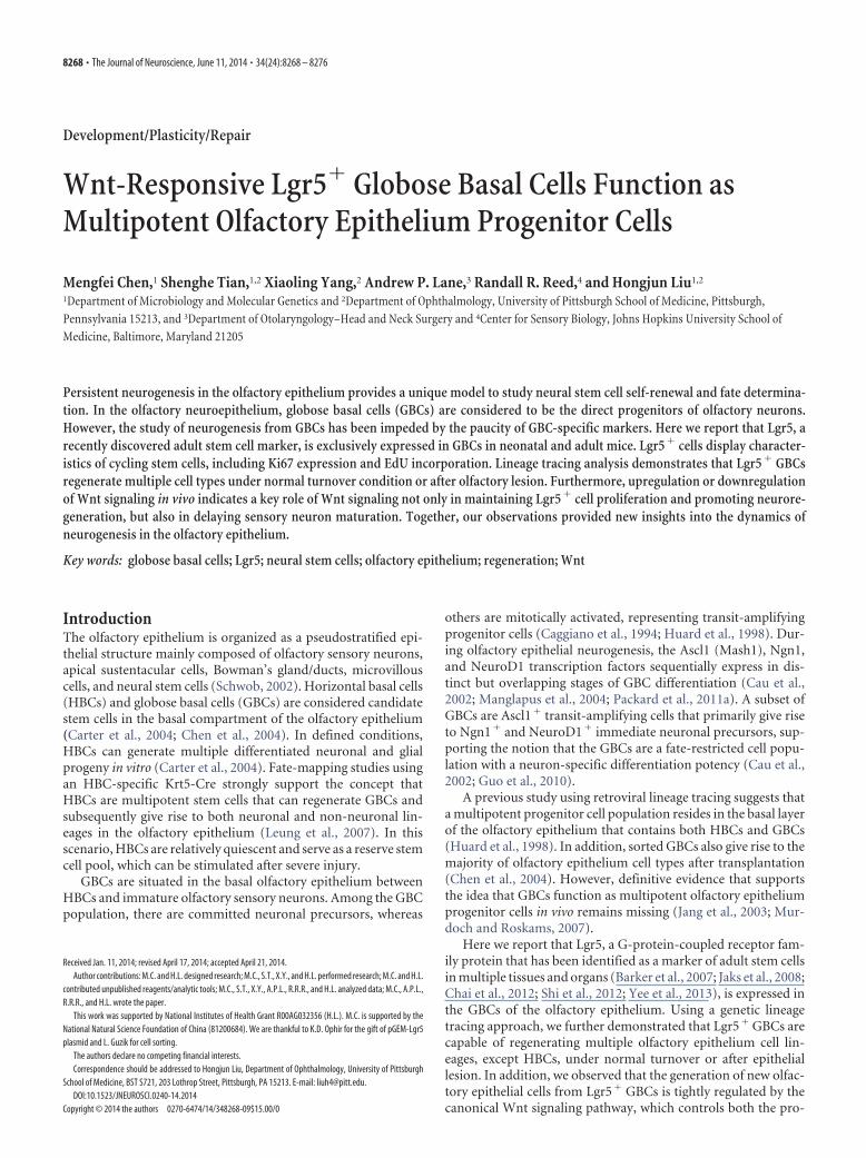
ResultsLgr5 marks GBCs in the olfactory epitheliumWe performed quantitative RT-PCR to investigate the spatial andtemporal expression patterns of Lgr5 mRNA in neonatal andadult olfactory epithelium (Fig. 1A). Expression of Lgr5 was de-tected in neonatal olfactory epithelium and at a reduced level inadulthood. Interestingly, the pattern of Lgr5 expression paral-leled the expression of Ascl1 (a neural progenitor cell marker) ateach time point. This expression pattern suggests a potential roleof Lgr5 in olfactory epithelium development and proliferation.
To characterize Lgr5 expression in more detail, we took ad-vantage of the Lgr5EGFP-Ires-CreERT2 knock-in mouse. In this strain,the endogenous Lgr5 promoter controls expression of EGFPand the modified Cre recombinase CreERT2 to faithfully repre-sent the endogenous Lgr5 expression (Barker et al., 2007). Con-focal images showed that the Lgr5-driven EGFP signal (referredto as Lgr5-EGFP hereafter) was detectable at E12 (data notshown). Around P2, the majority of Lgr5-EGFP� cells were re-stricted to the basal compartment (Fig. 1B), and the number ofLgr5-EGFP� cells reached a peak. Notably, the expression ofLgr5-EGFP was absent in the olfactory sensory neuron layer. Al-though the number of Lgr5-EGFP � cells decreased graduallyover time, Lgr5-EGFP signal remained in the basal layer inadulthood (Fig. 1C). The abundance of Lgr5-EGFP � cells inneonatal olfactory epithelium and their location in the basallayer are consistent with the properties observed for recog-nized progenitor cells in olfactory epithelium.
Two main cell populations, HBCs and GBCs, reside in thebasal layer. HBCs have a flattened morphology and are locateddirectly above the basal lamina. They express markers includingKrt5, Krt14, ICAM-1, and P63 (Holbrook et al., 1995; Carter etal., 2004; Fletcher et al., 2011; Packard et al., 2011b). We thereforeperformed immunostaining to determine which population ex-presses Lgr5-EGFP in the Lgr5EGFP-Ires-CreERT2 knock-in mice. Theresults showed that Lgr5-EGFP� cells did not express the HBCmarkers, K14 and ICAM-1 (Fig. 1D,E). Instead, Lgr5-EGFP�
cells were localized immediately above the ICAM-1 or K14-positive HBCs, and displayed a distinct round morphology thatwas more characteristic of the GBC population. Consistentwith this, at P2, Lgr5-EGFP � cells frequently coexpress Ascl1or NeuroD1, markers of GBCs and their immediate progenydifferentiated toward the neuronal lineage (Fig. 1F,H). Amongthe Lgr5-EGFP� population, 39.8% of the cells are Ascl1� and54.3% of the cells are NeuroD1� (Fig. 1J). In adults, the numberof Ascl1� cells in the Lgr5-EGFP� population was significantly
Chen et al. • Lgr5� GBCs Regenerate Olfactory Epithelium J. Neurosci., June 11, 2014 • 34(24):8268 – 8276 • 8269
reduced in the olfactory epithelium of3-month-old mice (Fig. 1G,J). Likewise,NeuroD1 is rarely observed in GBCs ofadult mice (Fig. 1I,J). These data suggestthat Lgr5 is expressed in GBCs in adult mice.
Lgr5 � cells are cycling and maintainolfactory epithelium renewalBased on the established neural progeni-tor cell characteristics of GBCs, we hy-pothesized that some Lgr5-EGFP� GBCswould be cycling progenitor cells thatmaintain olfactory epithelium renewal,similar to its role in the small intestine andskin epithelium (Barker et al., 2007; Jaks etal., 2008). We observed that at P2, nearlyall of the Lgr5-EGFP� cells (91.2% of2520 cells, n � 3) expressed Ki67, a prolif-eration marker (Fig. 2A,F). Similarly,Lgr5-EGFP� cells also maintained prolif-eration (81.9% of 1180 cells, n � 3) inadult mice (Fig. 2B,F).
We further validated the activity ofLgr5-EGFP� cell proliferation by EdU (athymidine analog that labels cycling cellsduring S-phase) incorporation experi-ment. As expected, injection of EdU la-beled 85.6% (2180 cells, n � 3) and 72.6%(2320 cells, n � 3) of Lgr5-EGFP� cells inP2 (Fig. 2C,F) and adult (Fig. 2D,F) mice,respectively. We also examined the fate ofLgr5-EGFP� cell progeny in adult micewith an EdU pulse and chase experiment.A substantial fraction of the EdU-labeledcells migrated apically and lost the Lgr5-EGFP signal after a 2 week chase (Fig. 2E),suggesting that progeny of Lgr5-EGFP�
GBCs have differentiated and migratedinto the neuronal zone of the olfactory ep-ithelium. Together, these observationsconfirmed that Lgr5-EGFP� GBCs repre-sented actively cycling progenitor cells innormal olfactory neuroepithelium.
Previous studies have demonstratedthat GBCs as neural progenitor cells di-rectly contribute to olfactory sensory neu-ron regeneration (Caggiano et al., 1994;Jang et al., 2003). Transplantation ofGBC-2-positive FACS-sorted GBCs into alesioned, recipient olfactory epitheliumsuggested a multilineage differentiationpotential including all epithelial celltypes (Chen et al., 2004). However, theregenerative capacity of GBC in unle-sioned olfactory epithelium remains tobe further investigated. To track theLgr5-EGFP � cell fate in vivo, we crossedthe Lgr5EGFP-Ires-CreERT2 mouse to theRosa26-LacZ reporter strain, which allowsus to mark permanently Lgr5-EGFP� cells and their progenywith the LacZ reporter transgene by tamoxifen injection.
We injected double-heterozygous mice with tamoxifen atP3–P4 and then analyzed them 3– 4 weeks later. X-gal staining
showed that LacZ� cells formed clusters and contained the majorcell types of the olfactory neuroepithelium (Fig. 2G–I). In mostcases, LacZ� clusters contained two to six cells and individualcells appeared separate and discrete (Fig. 2G). However, compact
Figure 1. Lgr5 marks GBCs in the olfactory epithelium. A, Quantitative analysis of Lgr5 expression in the olfactory epithelium byreal-time PCR. B, C, Confocal images of Lgr5-EGFP �cells in the olfactory epithelium of mice at P2 (B) and 3 months (3M) of age (C). Imagespresented are from olfactory epithelium coronal sections stained with an anti-EGFP antibody to amplify the endogenous EGFP signal.Lgr5-EGFP �cells inthebasal layeraremarkedbyarrowheads. D, E,CoimmunohistologicalstainingofEGFPwithHBCmarkers,K14(D)andICAM-1(E).Lgr5-EGFP �cellsalwaysrestdirectlyontheICAM-1 �orK14 �HBCs. F–I,CoexpressionofGBCmarkersAscl1orNeuroD1withLgr5-EGFP in P2 (F, H ) and adult olfactory epithelium (G, I ). Many Lgr5-EGFP-positive cells lost Ascl1 expression (G), and NeuroD1 couldhardly be detected in 3-month-old animals (I ). Boxed areas are highlighted on the right. Lgr5-EGFP �Ascl1 � cells and Lgr5-EGFP �NeuroD1 � cells are marked by arrowheads. J, The percentage of Ascl1 � and NeuroD1 � cells in Lgr5-EGFP � population. Themean � SD of three independent experiments is presented. Scale bars: B–I, 50 �m.
Figure 2. Lgr5 � cells proliferate and maintain olfactory epithelium renewal. A, B, Coexpression of Lgr5-EGFP and the proliferationmarker Ki67 in P2 (A) and adult (B) olfactory epithelium. Lgr5-EGFP � Ki67 � cells are marked by arrowheads. C, EdU labeling demon-strates that the Lgr5 � GBCs are proliferating cells in neonatal pups. D, In adult olfactory epithelium, EdU � cells are restricted to the basallayer and mostly are Lgr5-EGFP �GBCs. Pups (P2) or adults (2 months old) were injected with EdU two times and analyzed directly 6 h afterthe last injection. Arrowheads mark representative Lgr5-EGFP �EdU � cells. E, In normal olfactory epithelium, EdU � cells migratedapically after a 2 week chase. F, The percentage of Ki67 � and EdU � cells in Lgr5-EGFP � population. Data are given as mean�SD. G–I,LacZ � cells exhibit morphology of olfactory sensory neurons (G, H ), supportive sustentacular cells (H ), and Bowman’s gland and duct cells(I ). Olfactory epithelium cryosections from Lgr5 EGFP-Ires-CreERT2; Rosa26-LacZ mice were stained with X-gal 1 month after tamoxifen induc-tion. The arrowhead in H marks a sustentacular cell. Typical morphology of Bowman’s gland and duct cells was indicated by arrows andarrowheads (I ). J, K, Confocal images show that �-gal-positive cells coexpress OMP and CK18. Scale bars: A–E, G–K, 50 �m.
8270 • J. Neurosci., June 11, 2014 • 34(24):8268 – 8276 Chen et al. • Lgr5� GBCs Regenerate Olfactory Epithelium
clusters containing �6 cells could be detected occasionally. Fromthe apical to the basal layers, LacZ� descendants of Lgr5� cellspresented morphological characteristics of sustentacular cells, ol-factory sensory neurons, and GBCs (Fig. 2H). In addition, LacZ-labeled cells with unique morphology of Bowman’s glands andducts were also detected (Fig. 2I). However, no LacZ� cells wereobserved in the most basal layer of the olfactory epithelium. Onlya small fraction of Lgr5� cells (�1%) was labeled with LacZ inour experiments. The frequency of LacZ� cells dramatically de-creased when tamoxifen was injected at adulthood, and we didnot detect the formation of LacZ� cluster. The limited labelingefficiency can be explained by the weak expression of Lgr5 inneonatal and the cell proliferation further slowdown in adult. Weobserved no recombination in control littermates without ta-moxifen treatment as previously reported (Barker et al., 2010).
The multipotent regenerative capacity of Lgr5� cells was fur-ther confirmed by immunocolocalization with olfactory epithe-lium cell lineage-specific markers. As evidenced by confocalimaging, LacZ� cells were frequently colabeled with the olfactorymarker protein OMP, suggesting that Lgr5� cells gave rise to
mature olfactory sensory neurons (Fig. 2J). In addition, LacZ�
cells also coexpressed cytokeratin 18 (CK18), a marker of susten-tacular cells (Fig. 2K). The apical location and distinct morphol-ogy further supported the sustentacular cell identification.However, we did not detect LacZ-positive cells in the HBC layer(Fig. 2G–K). Together, these results suggest that a subset ofLgr5� GBCs function as multipotent progenitors that can gener-ate the major cell types of the olfactory epithelium under normalphysiological conditions.
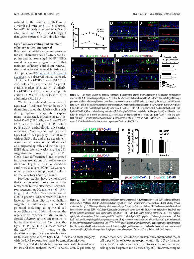
Lgr5 � cells regenerate olfactory neuroepithelial lineages aftermethimazole injuryThe remarkable regenerative capacity of olfactory epithelium hasbeen well documented. Damage of the olfactory epithelium withthe antithyroid drug methimazole has been identified as a reliableolfactory lesion model (Genter et al., 1995; Leung et al., 2007). Toidentify the characteristics of Lgr5� cells after severe lesion, weinjected 3- to 4-week-old Lgr5 EGFP-Ires-CreERT2 mice with methim-azole and analyzed Lgr5-EGFP� cells and their progeny at earlystages of regeneration. In striking contrast to nonlesioned con-
Figure 3. Lgr5 � cells regenerate multiple olfactory epithelium lineages after methimazole injury. A–C, Robust proliferation of Lgr5-EGFP � cells was activated in the early stage of regeneration.Two-month-old Lgr5EGFP-Ires-CreERT2 mice were injected with methimazole to induce olfactory epithelium damage. At day 2, Lgr5-EGFP � cells were activated to proliferate and usually coexpressedKi67. At postlesion days 5–7, the number of Lgr5-EGFP � cells reached a peak, and abundant proliferation continued to day 14 (A). B, Lgr5-EGFP � cells colabeled with GBC marker Ascl1 in the veryearly stage of regeneration, but a significant reduction of Ascl1 � cells was observed at postlesion day 14 (B). C, Lgr5-EGFP is expressed exclusively by the GBCs that are located above the K14 � HBCs.Arrowheads mark Lgr5-EGFP �Ki67 � cells in A and Lgr5-EGFP �Ascl1 � cells in B. D, Quantitative analysis of Lgr5-EGFP � cells during regeneration. E, The percentage of Ki67 � and Ascl1 � cellsin the Lgr5-EGFP � population. The data are shown as mean � SD from three experiments. F, Genetic lineage tracing using 1-month-old Lgr5EGFP-Ires-CreERT2; Rosa26-LacZ mice. G–I, Lgr5 � cellsregenerate multiple cell types in the olfactory epithelium. LacZ � cells are present in basal layer, olfactory sensory neurons, sustentacular cells (G), Bowman’s gland and ducts (H ), and microvillouscells (I ). J–L, Confocal images of olfactory epithelium sections double stained with �-gal and OMP (J ) or CK18 (K, L). �-gal signal colocalizes with both OMP and CK18. M, No colocalization wasobserved between �-gal and CK14 in HBCs. N, EdU labeling (arrow) remained within Lgr5-EGFP � GBCs after methimazole-induced lesion followed by a 2 week EdU chase. The arrowhead marks anLgr5-EGFP �EdU � cell. O, LacZ � cells are retained in basal layer 6 months after tamoxifen induction. Scale bars: A–C, G, H, J–O, 50 �m; I, 20 �m.
Chen et al. • Lgr5� GBCs Regenerate Olfactory Epithelium J. Neurosci., June 11, 2014 • 34(24):8268 – 8276 • 8271
trols, robust proliferation of Lgr5-EGFP�
cells was activated throughout the neuro-epithelium at postinjury day 2 (Fig.3A,D). During the dynamic process of re-generation, almost all Lgr5-EGFP� cellscoexpressed the proliferation markerKi67. The proliferation of Lgr5-EGFP�
cells peaked at days 5–7 and extended today 14 (Fig. 3A,D,E). As evidenced bydouble staining, most Lgr5-EGFP� cells(63.5% of 820 cells at day 2; 56.5% of 1050cells at day 5) coexpressed the GBCmarker Ascl1 (Fig. 3B,E). At day 14, thefrequency of Ascl1-labeled Lgr5-EGFP�
cells was clearly diminished. In addition,Lgr5-EGFP� cells were always restrictedto the GBC layer immediately above theK14-positive HBCs without overlap (Fig.3C). These results demonstrate that theLgr5-EGFP� GBC population is readilyactivated to proliferate after olfactory ep-ithelium lesion.
To more directly determine the contri-bution of Lgr5-EGFP� GBCs to olfactoryepithelium lineages, we traced the Lgr5-EGFP� cells after methimazole-induced le-sion in Lgr5EGFP-Ires-CreERT2; Rosa26-LacZmice (Fig. 3F). At 30 d after tamoxifeninjection, LacZ� clusters are distributedwidely in the septum and turbinate neu-roepithelium (Fig. 3G). Compared with nonlesioned mice, boththe number of LacZ� clusters and the number of cells in eachcluster were dramatically increased, suggesting the progeny ofLgr5� cells had undergone extensive proliferation and some ofthem had divided multiple rounds during regeneration.
As judged by their position and morphology, LacZ� clustersfrequently contained GBCs, olfactory sensory neurons, susten-tacular cells, and cells of the Bowman’s glands and ducts (Fig.3G–I). Double staining demonstrated that the LacZ-labeled cellsgenerated OMP� mature olfactory sensory neurons (Fig. 3J),CK18� Bowman’s glands and ducts (Fig. 3K), and sustentacularcells (Fig. 3L). In the regenerated olfactory epithelium, we did notdetect LacZ� cells in the basal layer that displayed HBC morphol-ogy or colocalized with K14 (Fig. 3M). These observationsstrongly suggest that Lgr5� cells are multipotent progenitor cellsthat contribute to most olfactory neuroepithelial lineages (exceptHBCs) after extensive depletion.
We observed that Lgr5-EGFP� cells retained EdU labeling forat least 2 weeks after injection (Fig. 3N). These results are consis-tent with a recent finding that GBCs contain BrdU or EdU label-retaining cells in neonatal or lesioned-recovering olfactoryepithelium (Jang et al., 2014). Furthermore, despite the decreasein the number of LacZ� clusters as the neuroepithelium turnedover, sporadic LacZ� cells in the GBC layer could be found 6months after tamoxifen exposure (Fig. 3O), suggesting the char-acter of slow cycling progenitor cells. Together, these observa-tions confirmed that Lgr5-marked GBCs contain not onlyactively cycling but also relatively quiescent progenitor cells, thelatter of which can only be activated by lesion.
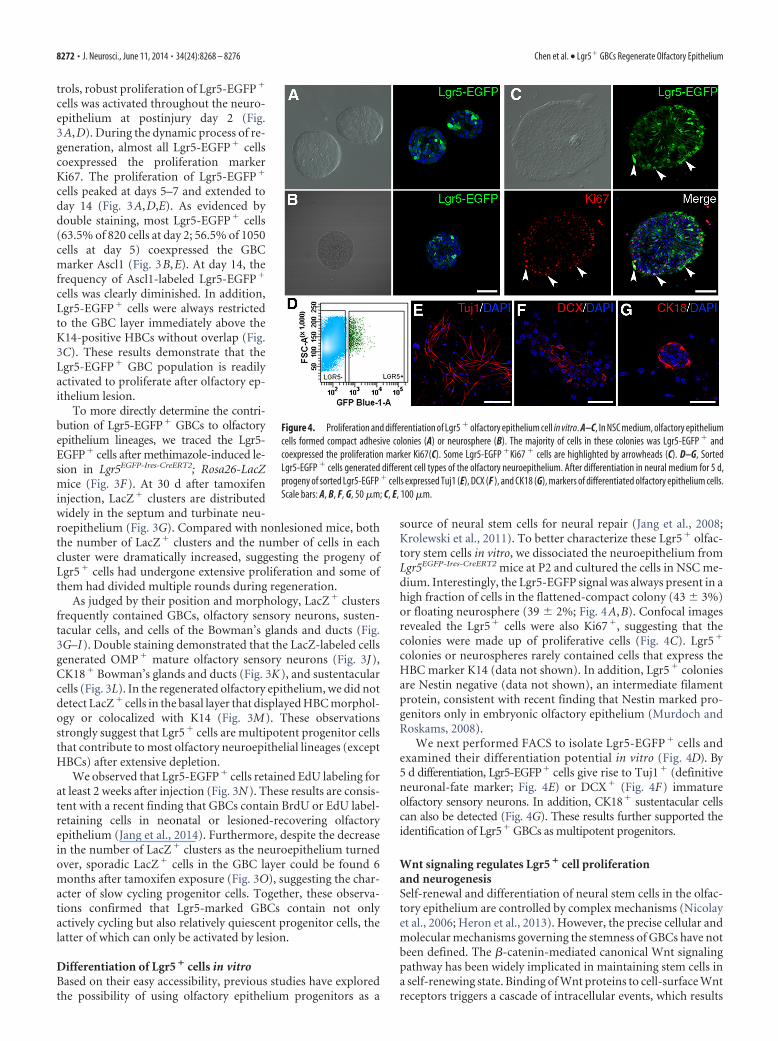
Differentiation of Lgr5 � cells in vitroBased on their easy accessibility, previous studies have exploredthe possibility of using olfactory epithelium progenitors as a
source of neural stem cells for neural repair (Jang et al., 2008;Krolewski et al., 2011). To better characterize these Lgr5� olfac-tory stem cells in vitro, we dissociated the neuroepithelium fromLgr5EGFP-Ires-CreERT2 mice at P2 and cultured the cells in NSC me-dium. Interestingly, the Lgr5-EGFP signal was always present in ahigh fraction of cells in the flattened-compact colony (43 � 3%)or floating neurosphere (39 � 2%; Fig. 4A,B). Confocal imagesrevealed the Lgr5� cells were also Ki67�, suggesting that thecolonies were made up of proliferative cells (Fig. 4C). Lgr5�
colonies or neurospheres rarely contained cells that express theHBC marker K14 (data not shown). In addition, Lgr5� coloniesare Nestin negative (data not shown), an intermediate filamentprotein, consistent with recent finding that Nestin marked pro-genitors only in embryonic olfactory epithelium (Murdoch andRoskams, 2008).
We next performed FACS to isolate Lgr5-EGFP � cells andexamined their differentiation potential in vitro (Fig. 4D). By5 d differentiation, Lgr5-EGFP� cells give rise to Tuj1� (definitiveneuronal-fate marker; Fig. 4E) or DCX� (Fig. 4F) immatureolfactory sensory neurons. In addition, CK18� sustentacular cellscan also be detected (Fig. 4G). These results further supported theidentification of Lgr5� GBCs as multipotent progenitors.
Wnt signaling regulates Lgr5 � cell proliferationand neurogenesisSelf-renewal and differentiation of neural stem cells in the olfac-tory epithelium are controlled by complex mechanisms (Nicolayet al., 2006; Heron et al., 2013). However, the precise cellular andmolecular mechanisms governing the stemness of GBCs have notbeen defined. The �-catenin-mediated canonical Wnt signalingpathway has been widely implicated in maintaining stem cells ina self-renewing state. Binding of Wnt proteins to cell-surface Wntreceptors triggers a cascade of intracellular events, which results
Figure 4. Proliferation and differentiation of Lgr5 � olfactory epithelium cell in vitro. A–C, In NSC medium, olfactory epitheliumcells formed compact adhesive colonies (A) or neurosphere (B). The majority of cells in these colonies was Lgr5-EGFP � andcoexpressed the proliferation marker Ki67(C). Some Lgr5-EGFP �Ki67 � cells are highlighted by arrowheads (C). D–G, SortedLgr5-EGFP � cells generated different cell types of the olfactory neuroepithelium. After differentiation in neural medium for 5 d,progeny of sorted Lgr5-EGFP � cells expressed Tuj1 (E), DCX (F ), and CK18 (G), markers of differentiated olfactory epithelium cells.Scale bars: A, B, F, G, 50 �m; C, E, 100 �m.
8272 • J. Neurosci., June 11, 2014 • 34(24):8268 – 8276 Chen et al. • Lgr5� GBCs Regenerate Olfactory Epithelium
in �-catenin accumulation in the cytoplasm and subsequent nu-clear translocation (Clevers and Nusse, 2012). �-Catenin inter-acts with the Tcf/Lef-family transcription factors to activate Wnttarget genes.
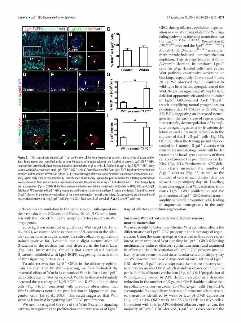
Since Lgr5 was identified originally as a Wnt target (Barker etal., 2007), we examined the expression of �-catenin in the olfac-tory epithelium. In adults, almost the entire olfactory epitheliumstained positive for �-catenin, but a slight accumulation of�-catenin in the nucleus was only detected in the basal layer(Fig. 5A). Interestingly, the cells that accumulated nuclear�-catenin colabeled with Lgr5-EGFP, suggesting the activationof Wnt signaling in these cells.
To address whether Lgr5 � GBCs in the olfactory epithe-lium are regulated by Wnt signaling, we first evaluated thepotential effect of Wnt3a (a canonical Wnt isoform) on Lgr5�
cell proliferation in vitro. As expected, Wnt3a treatment substantiallyincreased the percentage of Lgr5-EGFP and Ki67 double positivecells (Fig. 5B,C), consistent with previous observation thatWnt3a enhances neuroblast proliferation in hippocampal pro-genitor cells (Lie et al., 2005). This result suggested that Wntsignaling is involved in regulating Lgr5� GBC proliferation.
We next investigated the role of the Wnt/�-catenin signalingpathway in regulating the proliferation and neurogenesis of Lgr5�
GBCs during olfactory epithelium regener-ation in vivo. We manipulated the Wnt sig-naling pathway by injecting tamoxifen intothe Lgr5EGFP-Ires-CreERT2; Rosa26-LacZ;APCflox/flox mice and the Lgr5EGFP-Ires-CreERT2;Rosa26-LacZ; �-catenin flox/flox mice aftermethimazole-induced neuroepitheliumdepletion. This strategy leads to APC or�-catenin deletion in newborn Lgr5�
cells (or �-gal-labeled cells) and causesWnt pathway constitutive activation orblocking, respectively (Clevers and Nusse,2012). We observed that in contrast towild-type littermates, upregulation of theWnt/�-catenin signaling pathway by APCdeletion apparently elevated the numberof Lgr5� GBC-derived Ascl1�/�-gal�
transit-amplifying neural progenitors onpostinjury day 14 (74.2% vs 21.9%; Fig.5D,F,G), suggesting an increased neuro-genesis in the early stage of regeneration.Interestingly, downregulation of Wnt/�-catenin signaling activity by �-catenin de-letion caused a dramatic reduction in thenumber of Ascl1�/�-gal� cells (Fig. 5E).Of note, when the tracing period was ex-tended to 1 month, �-gal� clusters withneuroblast morphology could still be de-tected in the basal layer and many of thesecells coexpressed the proliferation markerKi67 (Fig. 5H). Furthermore, APC dele-tion clearly increased the number of�-gal� clusters (Fig. 5I) as well as thenumber of cells in each cluster (data notshown) on postinjury day 30. Together,these data suggest that Wnt activation stim-ulates Lgr5� GBC proliferation and theproliferation of Lgr5� GBC-derived transit-amplifying neural progenitor cells, leadingto augmented neurogenesis in the early
stage of olfactory epithelium regeneration.
Sustained Wnt activation delays olfactory sensoryneuron maturationWe next sought to determine whether Wnt activation affects thedifferentiation of Lgr5� GBC progeny in the latter stage of regen-eration. Using the same strategy as described in the above exper-iment, we manipulated Wnt signaling in Lgr5� GBCs followingmethimazole-induced olfactory epithelium injury and examinedits effects on the differentiation of Lgr5� GBC progeny into ol-factory sensory neurons and sustentacular cells at postinjury day30. We observed that in wild-type control mice, 69.9% of Lgr5�
GBC-derived �-gal� cells coexpressed the mature olfactory sen-sory neuron marker OMP, which mainly is expressed in the up-per half of the olfactory epithelium (Fig. 6A,D). Upregulation ofWnt signaling caused by APC deletion resulted in a dramaticreduction in the number of �-gal and OMP double positive ma-ture olfactory sensory neurons (20.6% in �-gal� cells; Fig. 6C,D),accompanied by a significant increase of immature olfactory sen-sory neurons identified by weak or lack of OMP expression(Fig. 6C; 42.4% OMP weak and 35.7% OMP-negative cells).Consistent with this, in APC-deleted olfactory epithelium, themajority of Lgr5 � GBC-derived �-gal � cells coexpressed the
Figure 5. Wnt signaling maintains Lgr5 � cell proliferation. A, Confocal images of �-catenin staining in the olfactory epithe-lium. Boxed region was magnified at the bottom. Compared with upper adjacent cells (marked by arrows), Lgr5-EGFP � GBCs(marked with arrowheads) have increased nuclear accumulation of �-catenin. B, Confocal images of Lgr5-EGFP � GBC colonystained with Ki67. Arrowheads mark Lgr5-EGFP �Ki67 � cells. C, Quantification of Ki67 and Lgr5-EGFP double positive cells in thepresence and/or absence of Wnt3a in culture. D–F, Confocal images of the olfactory epithelium stained with antibodies for Ascl1and �-gal in early stage of regeneration. G, Quantification of Ascl1 and �-gal double positive cells in the olfactory epithelium ofmice as shown in D–F. Wnt activation significantly increased the percentage of Lgr5 � GBC-derived Ascl1 � transit-amplifyingneural progenitors (*p � 0.000). H, Confocal images of olfactory epithelium stained with antibodies for OMP, Ki67, and �-gal.Deletion of APC maintained Lgr5 � GBC progeny in a proliferative state in the basal layer 1 month after lesion. I, Quantification of�-gal � clusters in the olfactory epithelium of the three mice strains 1 month after injury. Data presented are the numbers ofclusters that contains 6 or �6 �-gal � cells (*p � 0.003). Scale bars: A, 25 �m; B, D–F, H, 50 �m. WT, wild-type.
Chen et al. • Lgr5� GBCs Regenerate Olfactory Epithelium J. Neurosci., June 11, 2014 • 34(24):8268 – 8276 • 8273

immature olfactory sensory neuronmarker GAP43 (Fig. 6E–H ), suggestingthat sustained Wnt activation delayed ter-minal maturation.
However, downregulation of Wnt sig-naling caused by �-catenin deletion sig-nificantly reduced the number of �-gal�
clusters and none of the clusters observedcontained �6 �-gal� cells (Fig. 6B,F,J),suggesting that neuroregeneration was al-most extinguished by downregulation ofWnt activity. These data are consistentwith recent observations that downregu-lation of Wnt signaling inhibits neuronalproduction in the embryonic cortex (Mu-nji et al., 2011). On the other hand, APCdeletion significantly decreased sustentac-ular differentiation, with only 1.9 � 2.3%of �-gal-labeled cells colabeled with CK18(Fig. 6K,L), compared with15.9 � 6.7%of �-gal� cells differentiated into CK18�
sustentacular cells in wide-type mice (Fig.6 I,L). Together, these observations suggestthat the Wnt/�-catenin signaling pathwayplays an important role in regulating Lgr5�
GBC progeny terminal differentiation.We also examined the effect of Wnt
activation on Lgr5� GBC differentiationin vitro. In differentiation media, sortedLgr5� cells readily differentiated intoTuj1� neurons (Fig. 6M). However,Wnt3a treatment significantly decreasedthe number of Tuj1� cells while main-taining Lgr5� cells in an undifferentiatedstate (Fig. 6M,N), further suggesting thatWnt activation prevents Lgr5� cells fromdifferentiating into olfactory sensory neu-rons. Together, these results support thenotion that Wnt signaling regulates olfac-tory neuroepithelium regeneration notonly by controlling the proliferation/self-renewal of Lgr5 � GBCs, but alsoby affecting their subsequent terminaldifferentiation.
DiscussionIn the peripheral nervous system, the olfactory epithelium hasthe unique property of continuously regenerating new neu-rons throughout life, replacing cells lost due to environmentalinsult or disease (Bermingham-McDonogh and Reh, 2011). Therobust neurogenesis and relative anatomic simplicity of the olfac-tory epithelium provides a useful model to understand adultneurogenesis.
GBCs are the major proliferating population of the olfactoryepithelium, replenishing olfactory sensory neurons throughoutlife (Caggiano et al., 1994; Huard and Schwob, 1995; Weiler andFarbman, 1997; Leung et al., 2007). In the current study, we havedemonstrated that Lgr5 marks neural progenitor cells in the GBClayer of the olfactory epithelium. Lgr5� cells represent an activecycling population that is retained in adult olfactory epithelium,reminiscent of observations in gastric, intestinal, and skin epithe-lium (Barker et al., 2007, 2010; Jaks et al., 2008). In these lattersystems, Lgr5 marks a long-term, self-renewing population that
maintains epithelial homeostasis. Although most of the Lgr5�
GBCs in the olfactory epithelium are transit amplifying undernormal physiological conditions, a small population of long-term renewal cells can also be found after lesioning. These uniquedynamic processes of Lgr5� cells closely match previous descrip-tions of the heterogeneous nature of GBCs (Guo et al., 2010).
Many studies implicate GBCs as a multipotent progenitor. Inthis study, our in vivo lineage tracing and in vitro culture datastrongly suggest that Lgr5� GBCs serve as multipotent progeni-tor cells and contribute to the regeneration of multiple olfactoryepithelial cell lineages. However, Lgr5� GBC-derived �-gal�
HBCs were not detected in our lineage-tracing studies under nor-mal physiological conditions or following olfactory epitheliuminjury. These observations reinforce the concept that GBCs aregenerated from HBCs after severe epithelial damage (Leung et al.,2007). Beyond revealing an actively proliferating and multipo-tent GBC population using the Lgr5 marker, the current experi-
Figure 6. Sustained Wnt activation delays olfactory sensory neuron maturation. A–C, Confocal images of olfactory epitheliumstained with antibodies specific for OMP and �-gal. Compared with wild-type control (A), downregulation of Wnt activity by�-catenin deletion obviously reduced the number and size of �-gal � clusters (B). In APC-deleted olfactory epithelium, �-gal �
clusters were located in the immature or basal layer, and the majority of these cells was OMP negative or had weak OMP expressionas highlighted by arrowheads in C. D, Quantitative analysis of OMP � olfactory sensory neurons in �-gal � clusters (*p � 0.001).Due to the low frequency of labeling, we did not calculate the �-gal � cells after �-catenin deletion. E–G, Confocal images of theolfactory epithelium stained with GAP43 and �-gal. �-gal �/GAP43 � cells were highlighted by arrowheads in G. H, Quantifica-tion of GAP43 � immature neuron in �-gal � population (*p � 0.003). I–K, Confocal images of the olfactory epithelium stainedwith antibodies specific for CK18 and �-gal. Compared with wild-type control mice (I ), Wnt activation significantly reduced thenumber of �-gal � sustentacular cells located on the surface layer (K ). L, Quantification of CK18 � sustentacular cells that express�-gal (*p � 0.008). M, Confocal images of sorted Lgr5-EGFP � GBCs in the presence or absence of Wnt3a. N, Quantification of thepercentage of Tuj1-positive and EGFP-positive cells in culture (*p � 0.000; **p � 0.002). Scale bars: A–C, E–G, I–K, 25 �m; M,50 �m.
8274 • J. Neurosci., June 11, 2014 • 34(24):8268 – 8276 Chen et al. • Lgr5� GBCs Regenerate Olfactory Epithelium
ments do not resolve the origin of these cells, which is worthy offuture follow-up studies.
Despite the remarkable life-long regenerative capacity of theolfactory epithelium, severe injury often leads to olfactory sen-sory decline and loss (Schwob et al., 1995). The identification ofLgr5� GBCs as the major regenerative source within the olfac-tory epithelium provides us novel therapeutic opportunities. In-deed, recent studies have shown that olfactory progenitorsisolated from neonatal epithelium can be efficiently expanded invitro and incorporate into the host epithelium after transplanta-tion (Krolewski et al., 2011). It is therefore of particular interest toinvestigate the in vivo behavior of isolated Lgr5� GBCs afterbeing transplanted into lesioned olfactory epithelium in animalmodels.
Lgr5 was originally identified as a Wnt target, and it also af-fects Wnt signaling by interacting with Wnt receptor complex(Barker et al., 2007; Carmon et al., 2011; de Lau et al., 2011). Wntsignaling plays an important role in regulating progenitor prolif-eration and differentiation in many adult tissues and organs, in-cluding the central and the peripheral nervous systems (Lie et al.,2005; Kalani et al., 2008; Munji et al., 2011; Chai et al., 2012 ). Inthe neuronal tissues and/or organs, Wnt signaling stimulatesneural stem/progenitor proliferation and neurogenesis (Wang etal., 2011; Jang et al., 2013; Seib et al., 2013; Shi et al., 2013).Consistent with these previous studies, we observed that Wntactivation augments Lgr5� GBC proliferation and subsequentolfactory epithelium neurogenesis. Interestingly, by taking ad-vantage of the pseudostratified structure of the olfactory epithe-lium, we also observed that sustained Wnt activity preventstransit-amplifying neuronal progenitor cells from further differ-entiating into mature olfactory sensory neurons. These resultssuggest that delicate balance of progenitor cell proliferation anddifferentiation during olfactory epithelium neurogenesis is tem-porally and spatially fine-tuned by Wnt signaling activity. A mul-tiple zinc finger transcription factor, Zfp423/OAZ, has beensuggested to participate in regulating the transition from differ-entiation to maturation in olfactory sensory neurons (Cheng andReed, 2007). It would be interesting to determine the correlationbetween Wnt and Zfp423/OAZ.
In summary, we demonstrated that the adult stem cell markerLgr5 marks a population of GBCs. These Lgr5� GBCs possessmultipotent regenerative capacity. The generation of other olfac-tory epithelium lineages from Lgr5� GBCs is delicately regulatedby the Wnt signaling pathway, which not only controls the pro-liferation of Lgr5� GBCs and their immediate progeny, but alsoaffects their subsequent terminal differentiation. Together, ourobservations provided new insights into the understanding of themechanisms underlying olfactory epithelium regeneration.
ReferencesAbercrombie M (1946) Estimation of nuclear population from microtome
sections. Anat Rec 94:239 –247. CrossRef MedlineBarker N, van Es JH, Kuipers J, Kujala P, van den Born M, Cozijnsen M,
Haegebarth A, Korving J, Begthel H, Peters PJ, Clevers H (2007) Identi-fication of stem cells in small intestine and colon by marker gene Lgr5.Nature 449:1003–1007. CrossRef Medline
Barker N, Huch M, Kujala P, van de Wetering M, Snippert HJ, van Es JH, SatoT, Stange DE, Begthel H, van den Born M, Danenberg E, van den Brink S,Korving J, Abo A, Peters PJ, Wright N, Poulsom R, Clevers H (2010)Lgr5(�ve) stem cells drive self-renewal in the stomach and build long-lived gastric units in vitro. Cell Stem Cell 6:25–36. CrossRef Medline
Bermingham-McDonogh O, Reh TA (2011) Regulated reprogramming inthe regeneration of sensory receptor cells. Neuron 71:389 – 405. CrossRefMedline
Caggiano M, Kauer JS, Hunter DD (1994) Globose basal cells are neuronal
progenitors in the olfactory epithelium: a lineage analysis using areplication-incompetent retrovirus. Neuron 13:339 –352. CrossRefMedline
Carmon KS, Gong X, Lin Q, Thomas A, Liu Q (2011) R-spondins functionas ligands of the orphan receptors LGR4 and LGR5 to regulate Wnt/beta-catenin signaling. Proc Natl Acad Sci U S A 108:11452–11457. CrossRefMedline
Carter LA, MacDonald JL, Roskams AJ (2004) Olfactory horizontal basalcells demonstrate a conserved multipotent progenitor phenotype. J Neu-rosci 24:5670 –5683. CrossRef Medline
Cau E, Casarosa S, Guillemot F (2002) Mash1 and Ngn1 control distinctsteps of determination and differentiation in the olfactory sensory neuronlineage. Development 129:1871–1880. Medline
Chai R, Kuo B, Wang T, Liaw EJ, Xia A, Jan TA, Liu Z, Taketo MM, Oghalai JS,Nusse R, Zuo J, Cheng AG (2012) Wnt signaling induces proliferation ofsensory precursors in the postnatal mouse cochlea. Proc Natl Acad SciU S A 109:8167– 8172. CrossRef Medline
Chen X, Fang H, Schwob JE (2004) Multipotency of purified, transplantedglobose basal cells in olfactory epithelium. J Comp Neurol 469:457– 474.CrossRef Medline
Cheng LE, Reed RR (2007) Zfp423/OAZ participates in a developmentalswitch during olfactory neurogenesis. Neuron 54:547–557. CrossRefMedline
Clevers H, Nusse R (2012) Wnt/beta-catenin signaling and disease. Cell 149:1192–1205. CrossRef Medline
de Lau W, Barker N, Low TY, Koo BK, Li VS, Teunissen H, Kujala P, Haege-barth A, Peters PJ, van de Wetering M, Stange DE, van Es JE, Guardavac-caro D, Schasfoort RB, Mohri Y, Nishimori K, Mohammed S, Heck AJ,Clevers H (2011) Lgr5 homologues associate with Wnt receptors andmediate R-spondin signalling. Nature 476:293–297. CrossRef Medline
Fletcher RB, Prasol MS, Estrada J, Baudhuin A, Vranizan K, Choi YG, Ngai J(2011) p63 regulates olfactory stem cell self-renewal and differentiation.Neuron 72:748 –759. CrossRef Medline
Genter MB, Deamer NJ, Blake BL, Wesley DS, Levi PE (1995) Olfactorytoxicity of methimazole: dose-response and structure-activity studies andcharacterization of flavin-containing monooxygenase activity in theLong–Evans rat olfactory mucosa. Toxicol Pathol 23:477– 486. CrossRefMedline
Guo Z, Packard A, Krolewski RC, Harris MT, Manglapus GL, Schwob JE(2010) Expression of pax6 and sox2 in adult olfactory epithelium.J Comp Neurol 518:4395– 4418. CrossRef Medline
Heron PM, Stromberg AJ, Breheny P, McClintock TS (2013) Molecularevents in the cell types of the olfactory epithelium during adult neurogen-esis. Mol Brain 6:49. CrossRef Medline
Holbrook EH, Szumowski KE, Schwob JE (1995) An immunochemical, ul-trastructural, and developmental characterization of the horizontal basalcells of rat olfactory epithelium. J Comp Neurol 363:129 –146. CrossRefMedline
Huard JM, Schwob JE (1995) Cell cycle of globose basal cells in rat olfactoryepithelium. Dev Dyn 203:17–26. CrossRef Medline
Huard JM, Youngentob SL, Goldstein BJ, Luskin MB, Schwob JE (1998)Adult olfactory epithelium contains multipotent progenitors that give riseto neurons and non-neural cells. J Comp Neurol 400:469 – 486. CrossRefMedline
Jaks V, Barker N, Kasper M, van Es JH, Snippert HJ, Clevers H, Toftgård R(2008) Lgr5 marks cycling, yet long-lived, hair follicle stem cells. NatGenet 40:1291–1299. CrossRef Medline
Jang MH, Bonaguidi MA, Kitabatake Y, Sun J, Song J, Kang E, Jun H, ZhongC, Su Y, Guo JU, Wang MX, Sailor KA, Kim JY, Gao Y, Christian KM,Ming GL, Song H (2013) Secreted frizzled-related protein 3 regulatesactivity-dependent adult hippocampal neurogenesis. Cell Stem Cell 12:215–223. CrossRef Medline
Jang W, Youngentob SL, Schwob JE (2003) Globose basal cells are requiredfor reconstitution of olfactory epithelium after methyl bromide lesion.J Comp Neurol 460:123–140. CrossRef Medline
Jang W, Lambropoulos J, Woo JK, Peluso CE, Schwob JE (2008) Maintain-ing epitheliopoietic potency when culturing olfactory progenitors. ExpNeurol 214:25–36. CrossRef Medline
Jang W, Chen X, Flis D, Harris M, Schwob JE (2014) Label-retaining, qui-escent globose basal cells are found in the olfactory epithelium. J CompNeurol 522:731–749. CrossRef Medline
Kalani MY, Cheshier SH, Cord BJ, Bababeygy SR, Vogel H, Weissman IL, Palmer
Chen et al. • Lgr5� GBCs Regenerate Olfactory Epithelium J. Neurosci., June 11, 2014 • 34(24):8268 – 8276 • 8275

TD, Nusse R (2008) Wnt-mediated self-renewal of neural stem/progenitorcells. Proc Natl Acad Sci U S A 105:16970–16975. CrossRef Medline
Krolewski RC, Jang W, Schwob JE (2011) The generation of olfactory epi-thelial neurospheres in vitro predicts engraftment capacity followingtransplantation in vivo. Exp Neurol 229:308 –323. CrossRef Medline
Leung CT, Coulombe PA, Reed RR (2007) Contribution of olfactory neuralstem cells to tissue maintenance and regeneration. Nat Neurosci 10:720 –726. CrossRef Medline
Lie DC, Colamarino SA, Song HJ, Desire L, Mira H, Consiglio A, Lein ES,Jessberger S, Lansford H, Dearie AR, Gage FH (2005) Wnt signallingregulates adult hippocampal neurogenesis. Nature 437:1370 –1375.CrossRef Medline
Manglapus GL, Youngentob SL, Schwob JE (2004) Expression patterns ofbasic helix-loop-helix transcription factors define subsets of olfactoryprogenitor cells. J Comp Neurol 479:216 –233. CrossRef Medline
Milho R, Frederico B, Efstathiou S, Stevenson PG (2012) A heparan-dependent herpesvirus targets the olfactory neuroepithelium for host en-try. PLoS Pathog 8:e1002986. CrossRef Medline
Munji RN, Choe Y, Li G, Siegenthaler JA, Pleasure SJ (2011) Wnt signalingregulates neuronal differentiation of cortical intermediate progenitors.J Neurosci 31:1676 –1687. CrossRef Medline
Murdoch B, Roskams AJ (2007) Olfactory epithelium progenitors: insightsfrom transgenic mice and in vitro biology. J Mol Histol 38:581–599.CrossRef Medline
Murdoch B, Roskams AJ (2008) A novel embryonic nestin-expressing radialglia-like progenitor gives rise to zonally restricted olfactory and vomero-nasal neurons. J Neurosci 28:4271– 4282. CrossRef Medline
Nicolay DJ, Doucette JR, Nazarali AJ (2006) Transcriptional regulation ofneurogenesis in the olfactory epithelium. Cell Mol Neurobiol 26:803–821. Medline
Packard A, Giel-Moloney M, Leiter A, Schwob JE (2011a) Progenitor cellcapacity of NeuroD1-expressing globose basal cells in the mouse olfactoryepithelium. J Comp Neurol 519:3580 –3596. CrossRef Medline
Packard A, Schnittke N, Romano RA, Sinha S, Schwob JE (2011b) Delt-aNp63 regulates stem cell dynamics in the mammalian olfactory epithe-lium. J Neurosci 31:8748 – 8759. CrossRef Medline
Schwob JE (2002) Neural regeneration and the peripheral olfactory system.Anat Rec 269:33– 49. CrossRef Medline
Schwob JE, Youngentob SL, Mezza RC (1995) Reconstitution of the rat ol-factory epithelium after methyl bromide-induced lesion. J Comp Neurol359:15–37. CrossRef Medline
Seib DR, Corsini NS, Ellwanger K, Plaas C, Mateos A, Pitzer C, Niehrs C,Celikel T, Martin-Villalba A (2013) Loss of Dickkopf-1 restores neuro-genesis in old age and counteracts cognitive decline. Cell Stem Cell 12:204 –214. CrossRef Medline
Shi F, Kempfle JS, Edge AS (2012) Wnt-responsive Lgr5-expressing stemcells are hair cell progenitors in the cochlea. J Neurosci 32:9639 –9648.CrossRef Medline
Shi F, Hu L, Edge AS (2013) Generation of hair cells in neonatal mice bybeta-catenin overexpression in Lgr5-positive cochlear progenitors. ProcNatl Acad Sci U S A 110:13851–13856. CrossRef Medline
Vandesompele J, De Preter K, Pattyn F, Poppe B, Van Roy N, De Paepe A,Speleman F (2002) Accurate normalization of real-time quantitativeRT-PCR data by geometric averaging of multiple internal control genes.Genome Biol 3:RESEARCH0034. Medline
Wang YZ, Yamagami T, Gan Q, Wang Y, Zhao T, Hamad S, Lott P, SchnittkeN, Schwob JE, Zhou CJ (2011) Canonical Wnt signaling promotes theproliferation and neurogenesis of peripheral olfactory stem cells duringpostnatal development and adult regeneration. J Cell Sci 124:1553–1563.CrossRef Medline
Weiler E, Farbman AI (1997) Proliferation in the rat olfactory epithelium:age-dependent changes. J Neurosci 17:3610 –3622. Medline
Yee KK, Li Y, Redding KM, Iwatsuki K, Margolskee RF, Jiang P (2013) Lgr5-EGFP marks taste bud stem/progenitor cells in posterior tongue. StemCells 31:992–1000. CrossRef Medline
8276 • J. Neurosci., June 11, 2014 • 34(24):8268 – 8276 Chen et al. • Lgr5� GBCs Regenerate Olfactory Epithelium
Related Documents