Periodico di Otorinolaringologia, Patologia Cervico Facciale, Audiologia e Foniatria Periodic of otolaryngology, cervico-facial pathology, audiology and speech therapy ISSN 2038-4793 www.frontieraorl.it Distribuzione gratuita - Anno V, N. 1 - gennaio/marzo 2014 Free press - Year V, N. 1 - january/march 2014

Welcome message from author
This document is posted to help you gain knowledge. Please leave a comment to let me know what you think about it! Share it to your friends and learn new things together.
Transcript

Periodico di Otorinolaringologia, Patologia Cervico Facciale, Audiologia e FoniatriaPeriodic of otolaryngology, cervico-facial pathology, audiology and speech therapy
ISSN 2038-4793
www.frontieraorl.it
Distr
ibuzio
ne gr
atuita
- Ann
o V, N
. 1 -
genn
aio/m
arzo
2014
Free
pres
s - Ye
ar V,
N. 1
- jan
uary/
march
2014

2
Frontiera ORL – Anno V, N. 1 – gennaio/marzo 2014
www.frontieraorl.it
Impianti cocleari ed all’orecchio medio:la nostra esperienza con la tecnica “the down-up bone bridge approach”Cochlear and middle ear implants: our experience with the down-up bone bridge approachF. Achena 3
Screening neonatale universale dell’udito. Esperienza in un ospedale italiano: metodi e aspetti epidemiologiciNewborn Hearing screening.Experience in Italian Hospital: methods and epidemiological aspectsE. Milazzo, F. Candido, N. Rondina, E. Di Crescenzo, E. Battisti,C. Cingolani, D. Giardini, G. Latini, E. Gentile, N. Vitelli, G. Migliori 10
Laringofissura anteriore con innesto di cartilagine tiroidea in pazienti adultiLaryngeal anterior split with thyroid cartilage graft in adult patientsG. Stok, M. A. Caldez García, M. L. Rospide 14
Metastasi mandibolare di un carcinoma renale: case reportMandibular metastasis from renal cell carcinoma: a case reportA. Mardassi, N. Mathlouthi, S. Nefzaoui, S. Mezri,C. Zgolli, G. Chebbi, R. Ben Mhamed, K. Akkari, S. Benzarti 19
Case report: Laringocele gigante misto Case report: giant mixed laryngoceleO. Marotta, F. Catapano, M. Cocchiarella, C. Crisci, C. Di Meo 23
In questo numero...
... nel precedente
Periodico trimestralerivolto alla classe medica
Quarterly magazine for medical class
Direttore editoriale/EditorOrtensio Marotta
Coordinatore di redazioneEditorial CoordinatorVito Marotta
Direttore responsabile/Editor chiefLoredana Guida
Hanno partecipato a questo numeroF. Achena, E. Milazzo, F. Candido,N. Rondina, E. Di Crescenzo, E. Battisti, C. Cingolani, D. Giardini, G. Latini,E. Gentile, N. Vitelli, G. Migliori, G. Stok, M. A. Caldez García, M. L. Rospide,A. Mardassi, N. Mathlouthi, S. Nefzaoui,S. Mezri, C. Zgolli, G. Chebbi, R.Ben Mhamed, K. Akkari, S. Benzarti,O. Marotta, F. Catapano, M. Cocchiarella, C. Crisci, C. Di Meo
Redazione/Editorial office Via Fuga, 64 - Caserta
Dialogo con i lettori/Dialogue with readersOrtensio Marotta [email protected] UOC diOtorinolaringoiatria,AORN “S. Anna e S. Sebastiano”,Caserta
Realizzazione editoriale/Production
Anno V, N. 1gennaio/marzo 2014Registrazione/RegistratedTribunale di S. Maria Capua Vetere n°765 del 22/06/2010
Info: [email protected]
... in the previous number
In this number...
è inserita:• inDOAJ(Directory of Open Access Journals,
http://www.doaj.org )
•neldatabasemondialedellerivisteon-line
•nell’elencodellerivistescientificheitaliane
• neldatabaselocaledellabibliotecadigitaleSFX(Sistema Bibliotecario di Ateneo) a disposizione dell’Università di Napoli “Federico II”
• iltitolo“Frontiera ORL” è inserito, da tempo, nell’autorevoleACNP(Archivio Collettivo Nazionale dei Periodici)
• inDOAJ(Directory of Open Access Journals, http://www.doaj.org)
• intheglobaldatabaseof Italianscientificjournal on-line
• intheitalianscientificjournals
• inthedigitallibrarySFX(university library system)fullyaccessibletotheUniversityof Naples “Federico II”
• thetitle“Frontiera ORL” is included in the ACNP(Periodicals Archive National Collective)
is included:
Il ruolo della chirurgia del sacco endolinfaticonel trattamento della malattia di MénièreThe surgery’s role in the treatment of endolymphatic sac in meniere’s diseaseG. N. Frau
Effetti dell’adenotonsillectomia sulla funzione vocaleEffect of adenotonsillectomyon vocal functionB. Iorio, T. Abate, M. Landi, G. Del Santo, M. Iengo, M. Mesolella
Presenza di HPV ad alto rischio in 10 casi di carcinoma laringeoHigh risk HPV presence in ten cases of laringeal carcinomaP. Fierro, G. Barba, F. Maiello, M.C. D’Ambrosio, A. Menna, I. Diomaiuto, G. Tortoriello
Vertigine Parossistica Posizionale Benigna: una panoramicaBenign Paroxysmal Positional Vertigo: an overviewL. Califano, M. G. Melillo, F. Salafia, S. Mazzone
Case report di decorso anomalo del nervo facciale con obliterazione totale della finestra ovaleUnexpected course of facial nerve with obliteration of the oval window: case reportO. Marotta, M. Cocchiarella, C. Di Meo, C. Crisci,F. Catapano, A. Montalbano A. Bernardo
3
FrontieraORL–YearV,N.1–january/march2014
www.frontieraorl.it
AbstractL’autore descrive la nuova procedura chirurgica “the
down-up bone bridge approach”, da lui adottata per il fis-saggiosiadegliimpianticocleari(IC)chedialcunepro-tesiacusticheimpiantabiliall’orecchiomedio(VSB),sen-za che sia necessario l’utilizzo di punti di sutura. Il nome della suddetta tecnica deriva dalla necessità di creare un ponte osseo tra la cavità mastoidea da un lato ed il letto osseo creato per l’alloggiamento dell’impianto dall’altro. Il ponte osseo viene inizialmente interrotto nella sua por-zionesuperiore(bonebridgedown)equindiricostruitonellefasifinalidell’intervento(bonebridgeup)conpol-vere d’osso. Si descrivono i tempi della tecnica chirurgica. La casistica consta di un campione di 53 pazienti, 3 casi bilaterali (56orecchie) tutti sottoposti alla tecnica “thedown-up bone bridge approach” sia per l’applicazione di IC che di VSB presso la U.O.C. di otorinolaringoiatria del P.O. CTO di Iglesias.
Tutti i pazienti sono stati impiantati con successo, non si sono verificate complicanze a carico della ferita chirur-gica ne estrusioni dell’impianto. Il follow-up era compre-so tra 6 mesi e 4,5 anni, media 2,1 anni. Attualmente tutti i pazienti così impiantati utilizzano il presidio protesico a tempo pieno. In conclusione la nostra tecnica “the down-up bone bridge approach” ha dimostrato una minore morbidità rispetto ad altre tecniche chirurgiche conven-zionali, una grande sicurezza nel tempo oltre che sempli-cità di esecuzione.
Introduzione A partire dagli anni ’80, con l’avvento dell’impianto co-
clearemulticanale(1,2),gliotologihannoavutoadispo-sizione una ottima opzione terapeutica per la cura della sordità severa/profonda. Gli apparecchi acustici digitali di ultima generazione, d’altro canto, hanno rappresentato sinora il “gold standard” per la riabilitazione delle ipoa-cusie neurosensoriali di media-grave entità, ciononostan-tealcuniapparecchiacusticiimpiantabiliqualiilVibrantSoundBridge®(Med-El),sisonritagliatineltempo,un
Impianti cocleari ed all’orecchio medio: la nostra esperienza con la tecnica “the down-up bone bridge approach”
Cochlear and middle ear implants: our experience with the down-up bone bridge approach
F. Achena
U.O.C. di Otorinolaringoiatria, P.O. CTO, Iglesias, Italia
■ Indirizzo per corrispondenza / Address for corrispondence: F. Achena - U.O.C. di Otorinolaringoiatria, P.O. CTO, Iglesias, Italia
E-mail: [email protected]
AbstractThe author describes a new method, the bone bridge down-up
approach, to secure the cochlear and middle ear implants with-out a tie-down ligature. The surgical procedure’s name “the down\up bone bridge approach” is a consequence of the neces-sity to create between the mastoid cavity and the bony well for the implant insertion, a bone bridge. This is initially break-down superiorly (down bone bridge) and then rebuilt with bone pate (up bone bridge). Study Design: technical description with cases reports and surgical outcomes. Materials: 53 patients (56 ears – 3 cases bilateral) submitted at the surgical approach for cochlear and middle ear implantation by the author at the ENT Department of Iglesias were studied. Results: all pa-tients were successfully implanted by the down\up bone bridge approach, wound healing was uneventful in all cases. At fol-low-up (mean 2,1 years, range 6 months – 4,5 years ) no mi-gration of the receiver-stimulator were observed. At the mo-ment all recipients are full-time users of the device. Conclusion: with the down\up bone bridge approach it’s possible to achieve reduced surgical morbidity and moreover it shows to be a safe and easily reproducible surgical procedure.
IntroductionThe advent of multichannel cochlear implant in the 1980s
(1, 2) gave to the otologysts in the last 30 years, an excellent option in the treatment of patients with bilateral profound to severe sensorineural hearing loss. On the other hand today, al-though hearing aids are the devices of choice for patients with a sensorineural hearing loss, however new alternatives such as implantable middle ear hearing devices, have been developed and are now available for these patients. In particular in those patients with a moderate to severe sensorineural hearing loss and lack of benefit from a traditional hearing aid, the Vibrant Sound Bridge® (Med-El) cut out an important role.
Concerning the surgical approach, after the firsts more inva-sive procedures (3), during the past years, several minimal in-vasive approaches have been proposed for the fashioning of the bony recess and fixation of cochlear and middle ear implants by different authors in various countries (4, 8). We recently
4
Frontiera ORL – Anno V, N. 1 – gennaio/marzo 2014
www.frontieraorl.it
ruolo sempre più importante come alternativa terapeuti-ca ai suddetti apparecchi acustici convenzionali.Perquantoriguardaletecnichechirurgicheadottateper
l’applicazione degli impianti cocleari o all’orecchio medio, dopo le iniziali tecniche abbastanza invasive, incisione a C di tipo otoneurochirurgica, incisione endoaurale allar-gata(3),nelrecentepassato,diversiapproccichirurgiciditipo mini-invasivo sono stati proposti per l’applicazione e l’alloggiamentodegli impianti cocleari (4, 8).Noi re-centemente abbiamo messo a punto una nuova metodica chirurgica per l’alloggiamento in sicurezza sia degli im-pianticocleari(IC)chedelVibrantSoundBridge(VSB)senza l’applicazione di punti di sutura di ancoraggio. Ab-biamo chiamato la procedura “the down-up bone bridge approach”(9),chesièrivelataefficacenellenostremanie riteniamo possa essere utile anche per altri otochirur-ghi che volessero utilizzarla. Lo scopo del nostro lavoro èstatoquellodidibatterecircaleindicazioni,lariabilita-zione, il trattamento chirurgico in pazienti operati col IC multicanale o con il VSB presentando i risultati ottenuti presso la UOC di otorinolaringoiatria di Iglesias.
Materiali e metodiLanostracasisticaconstadi53pazienti (56orecchie,
3 casi bilaterali) operati presso la UOC di Otorinolarin-goiatria di Iglesias dal gennaio 2008 al maggio 2013. Si trattava di 29 maschi e 24 femmine di età compresa tra i 6 ed i 79 anni, età media 40,5 anni, tutti sottoposti ad intervento con la procedura del “down-up bone bridge approach”.Fraquesti,30pazientisonostatioperatiperICconilNucleusFreedom®dellaCochlear.Perquantoconcerne l’etiologia, nella maggior parte dei casi si trat-tava di ipoacusia idiopatica ad andamento progressivo, in 1 caso di Sindrome di Turner, in 4 pazienti di otosclero-si coclearizzata, in un caso di sordità postraumatica, in 2 casi di sordità congenita. La sordità era prelinguale in 6casi,postlingualenei rimanenti.L’arrayelettrodicoèstato introdotto completamente all’interno della coclea in tutti i pazienti eccetto che nella paziente affetta da sin-dromediTurner.Inquest’ultimocasoperunproblemadi ipoplasia cocleare è stato possibile introdurre con suc-cesso 15 elettrodi.Perquanto concernegli altri23pazienti (tutti adulti
- 1 caso bilaterale) sono stati impiantati con l’impianto all’orecchiomedioVSB. In 18 di questi l’FMT è statoagganciato al processo lungo dell’incudine, in 4 casi alla finestra rotonda e nei rimanenti 2 tra il capitello e la crus posterioredellastaffa.Perquantoconcerneleindicazio-ni audiologiche nei pazienti trattati con IC, si trattava in tuttiicasidiipoacusieneurosensorialisevere(sogliaudi-tiva≥75dBper leFz500-1000-2000Hz) oprofonde(sogliauditiva≥90dBperleFz500-1000-2000Hz),ladiscriminazionevocaleera≤al50%a65dBSPLinpa-zienti con apparecchio acustico inserito binauralmente. Riguardo i pazienti trattati con VSB, in 18 casi si tratta-
developed a new method, to secure the cochlear and middle ear implants without a tie-down ligature, we called the down\up bone bridge approach (9) that works well for us and that we feel could be useful to others. The purpose of our article is to debate about indications, rehabilitation, surgical manage-ment in patients treated with multichannel CI and VSB and to present our experience about these patients at the ENT depart-ment of CTO hospital in Iglesias.
Materials and methodsFrom January 2008 to May 2013 53 patients, 3 cases bi-
lateral (56 ears) 29 males and 24 females. were operated on at the ENT Department of Iglesias with the down-up bone bridge approach, age ranged between 6 and 79 years, mean age at implantation 40,5 years. Among them, 30 patients (21 adults and 9 children, 2 case bilateral) underwent cochlear im-plantation with the Nucleus®Freedom cochlear implant with Contour Advance™ Electrode. In most of them the aetiology was progressive idiopathic hearing loss (HL), in 1 case it was a Turner’s syndrome, in 4 cases a progressive otosclerosis, in 1 case post-traumatic HL; there were 2 cases congenitally. In 6 cases the HL was pre-lingual in all the other cases post-lin-gual. The array was fully introduced in the cochlea in all cases except in the patients with the Turner’s syndrome in whom, consequently to cochlear hypoplasia, just 15 electrodes were in-troduced successfully
The other 23 patients (all were adults, 1 case bilateral) re-ceived The Vibrant® Soundbridge™ (Vibrant Med-El) mid-dle ear implant, in 18 of wich the Fmt was clipped onto the long process of the incus, in 4 patients the Fmt was placed in the round window niche and in two cases the Fmt was clipped between the stapes posterior branch and capitolum.
The audiological indication for cochlear implant patients was the sensorineural hearing loss from severe (pure tone thresholds for frequency 500-1000-200 Hz ≥75 dB) to profound (pure tone thresholds for frequency 500-1000-200 Hz ≥90 dB), speech score should be ≤ 50% at 65 db SPL in the best aided condition. In the vibrant sound bridge (VSB) patients the au-diological indication was the presence of moderate to severe sensorineural hearing loss in 8 cases and the presence of mixed hearing loss in the other 4 cases.
The follow-up period ranged from 6 months to 4,5 years, mean 2,1 years, after implantation.
Surgical techniqueThe down-up bone bridge approach was adopted in all cases. Under general anesthesia, shaving only extend around the
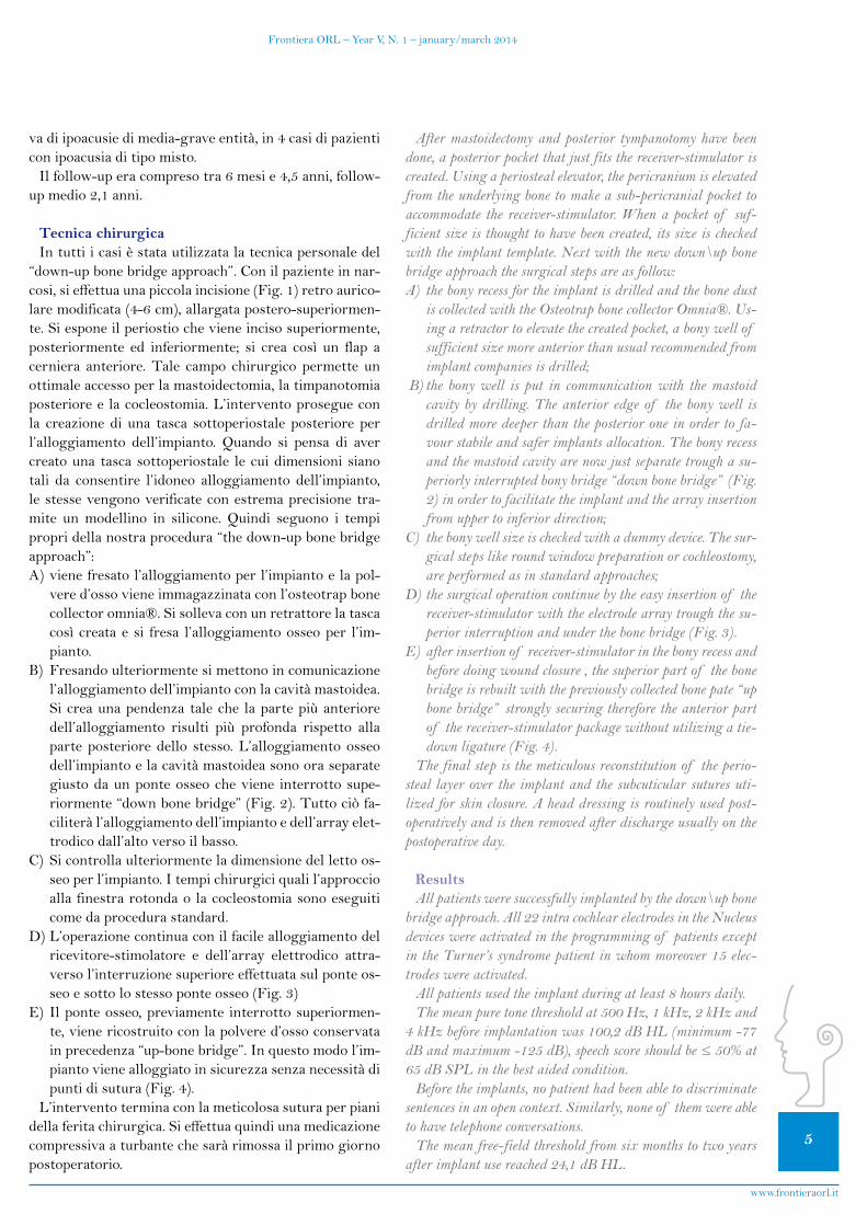
hairline on the retro-auricular aria, a small modified retro-auricular incision (4-6 cm) is made (Fig. 1). The incision is enlarged postero-superiorly rather than a conventional retro-auricular incision. The periosteum is then exposed and incised superiorly, posteriorly and inferiorly, carefully elevating an an-terior based flap. This surgical exposure provides an excellent access for cortical mastoidectomy, posterior tympanotomy and cochleostomy.
5
FrontieraORL–YearV,N.1–january/march2014
www.frontieraorl.it
va di ipoacusie di media-grave entità, in 4 casi di pazienti con ipoacusia di tipo misto.
Il follow-up era compreso tra 6 mesi e 4,5 anni, follow-up medio 2,1 anni.
Tecnica chirurgicaIn tutti i casi è stata utilizzata la tecnica personale del
“down-up bone bridge approach”. Con il paziente in nar-cosi,sieffettuaunapiccolaincisione(Fig.1)retroaurico-laremodificata(4-6cm),allargatapostero-superiormen-te. Si espone il periostio che viene inciso superiormente, posteriormente ed inferiormente; si crea così un flap a cerniera anteriore. Tale campo chirurgico permette un ottimale accesso per la mastoidectomia, la timpanotomia posteriore e la cocleostomia. L’intervento prosegue con la creazione di una tasca sottoperiostale posteriore per l’alloggiamento dell’impianto. Quando si pensa di aver creato una tasca sottoperiostale le cui dimensioni siano tali da consentire l’idoneo alloggiamento dell’impianto, le stesse vengono verificate con estrema precisione tra-mite un modellino in silicone. Quindi seguono i tempi propri della nostra procedura “the down-up bone bridge approach”:A) viene fresato l’alloggiamento per l’impianto e la pol-
vere d’osso viene immagazzinata con l’osteotrap bone collector omnia®. Si solleva con un retrattore la tasca così creata e si fresa l’alloggiamento osseo per l’im-pianto.
B) Fresando ulteriormente si mettono in comunicazione l’alloggiamento dell’impianto con la cavità mastoidea. Si crea una pendenza tale che la parte più anteriore dell’alloggiamento risulti più profonda rispetto alla parte posteriore dello stesso. L’alloggiamento osseo dell’impianto e la cavità mastoidea sono ora separate giusto da un ponte osseo che viene interrotto supe-riormente“downbonebridge”(Fig.2).Tuttociòfa-ciliteràl’alloggiamentodell’impiantoedell’arrayelet-trodico dall’alto verso il basso.
C) Si controlla ulteriormente la dimensione del letto os-seoperl’impianto.Itempichirurgiciqualil’approccioalla finestra rotonda o la cocleostomia sono eseguiti come da procedura standard.
D) L’operazione continua con il facile alloggiamento del ricevitore-stimolatore e dell’array elettrodico attra-verso l’interruzione superiore effettuata sul ponte os-seoesottolostessoponteosseo(Fig.3)
E) Il ponte osseo, previamente interrotto superiormen-te, viene ricostruito con la polvere d’osso conservata inprecedenza“up-bonebridge”.Inquestomodol’im-pianto viene alloggiato in sicurezza senza necessità di puntidisutura(Fig.4).
L’intervento termina con la meticolosa sutura per piani dellaferitachirurgica.Sieffettuaquindiunamedicazionecompressiva a turbante che sarà rimossa il primo giorno postoperatorio.
After mastoidectomy and posterior tympanotomy have been done, a posterior pocket that just fits the receiver-stimulator is created. Using a periosteal elevator, the pericranium is elevated from the underlying bone to make a sub-pericranial pocket to accommodate the receiver-stimulator. When a pocket of suf-ficient size is thought to have been created, its size is checked with the implant template. Next with the new down\up bone bridge approach the surgical steps are as follow:A) the bony recess for the implant is drilled and the bone dust
is collected with the Osteotrap bone collector Omnia®. Us-ing a retractor to elevate the created pocket, a bony well of sufficient size more anterior than usual recommended from implant companies is drilled;
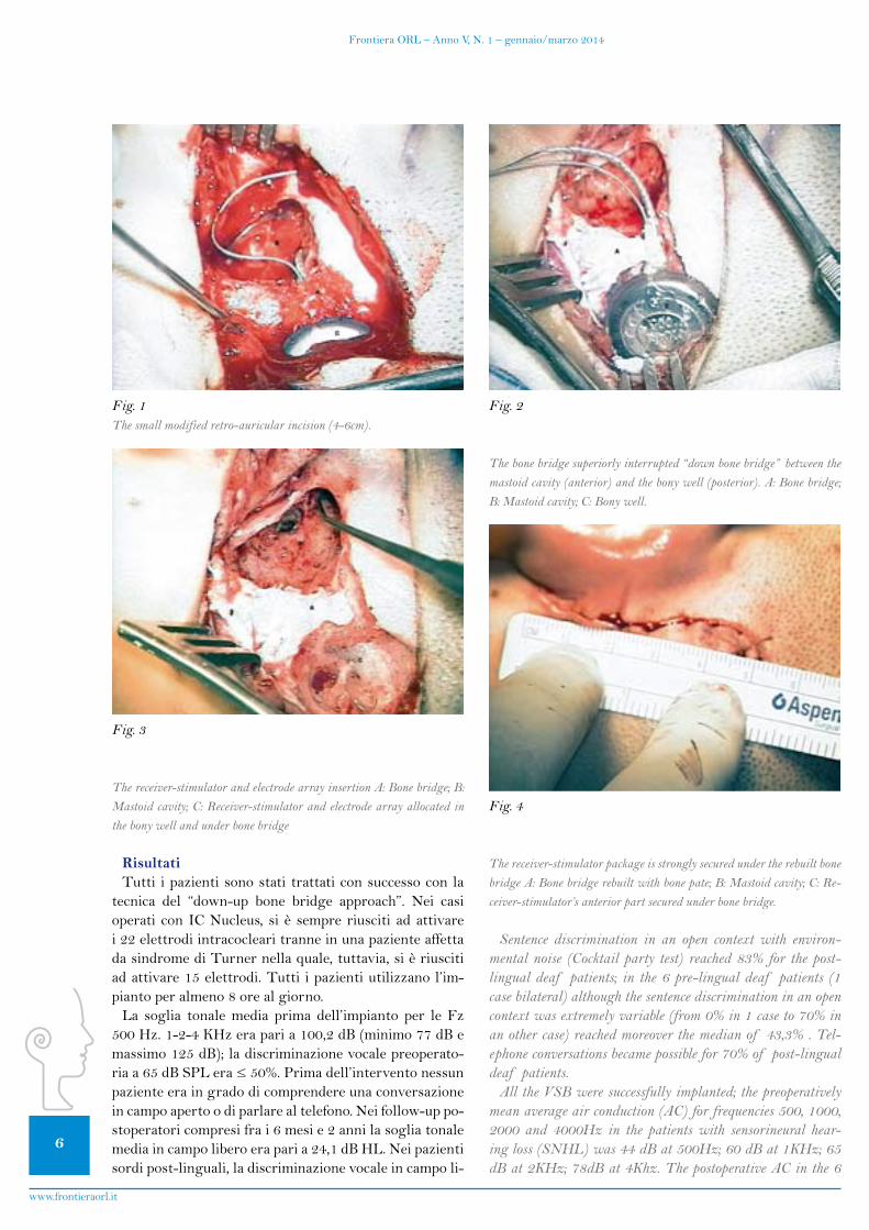
B) the bony well is put in communication with the mastoid cavity by drilling. The anterior edge of the bony well is drilled more deeper than the posterior one in order to fa-vour stabile and safer implants allocation. The bony recess and the mastoid cavity are now just separate trough a su-periorly interrupted bony bridge “down bone bridge” (Fig. 2) in order to facilitate the implant and the array insertion from upper to inferior direction;
C) the bony well size is checked with a dummy device. The sur-gical steps like round window preparation or cochleostomy, are performed as in standard approaches;
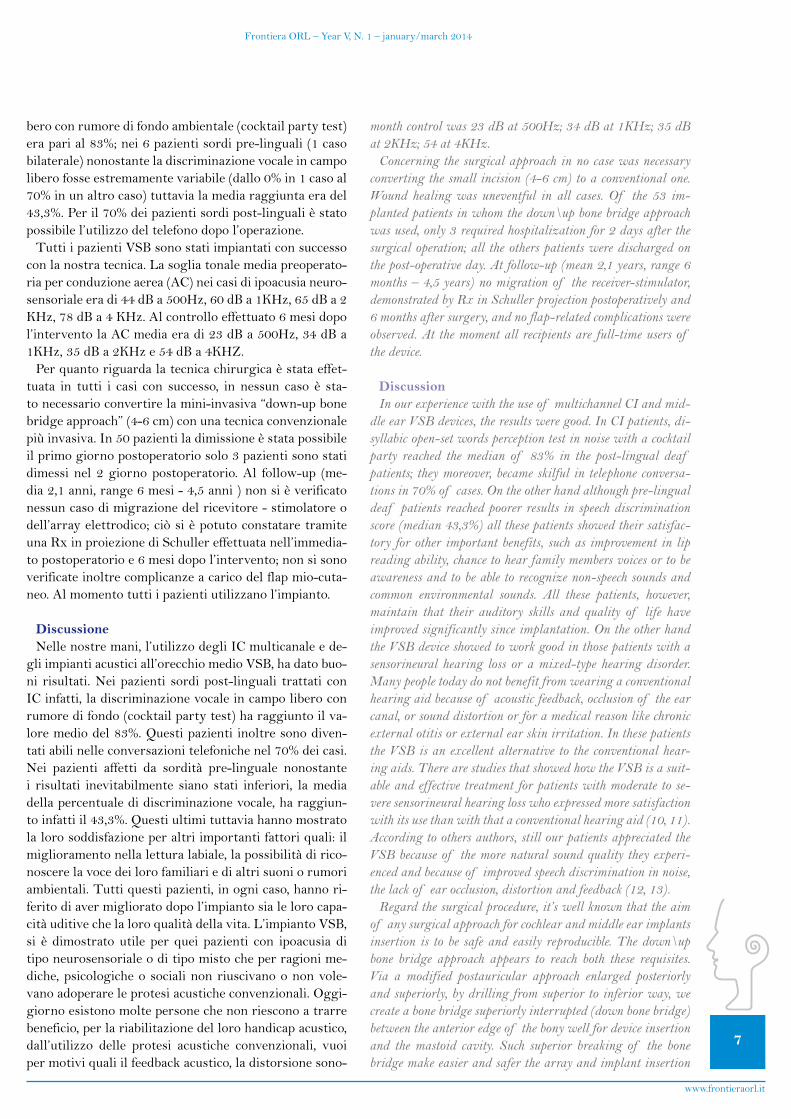
D) the surgical operation continue by the easy insertion of the receiver-stimulator with the electrode array trough the su-perior interruption and under the bone bridge (Fig. 3).
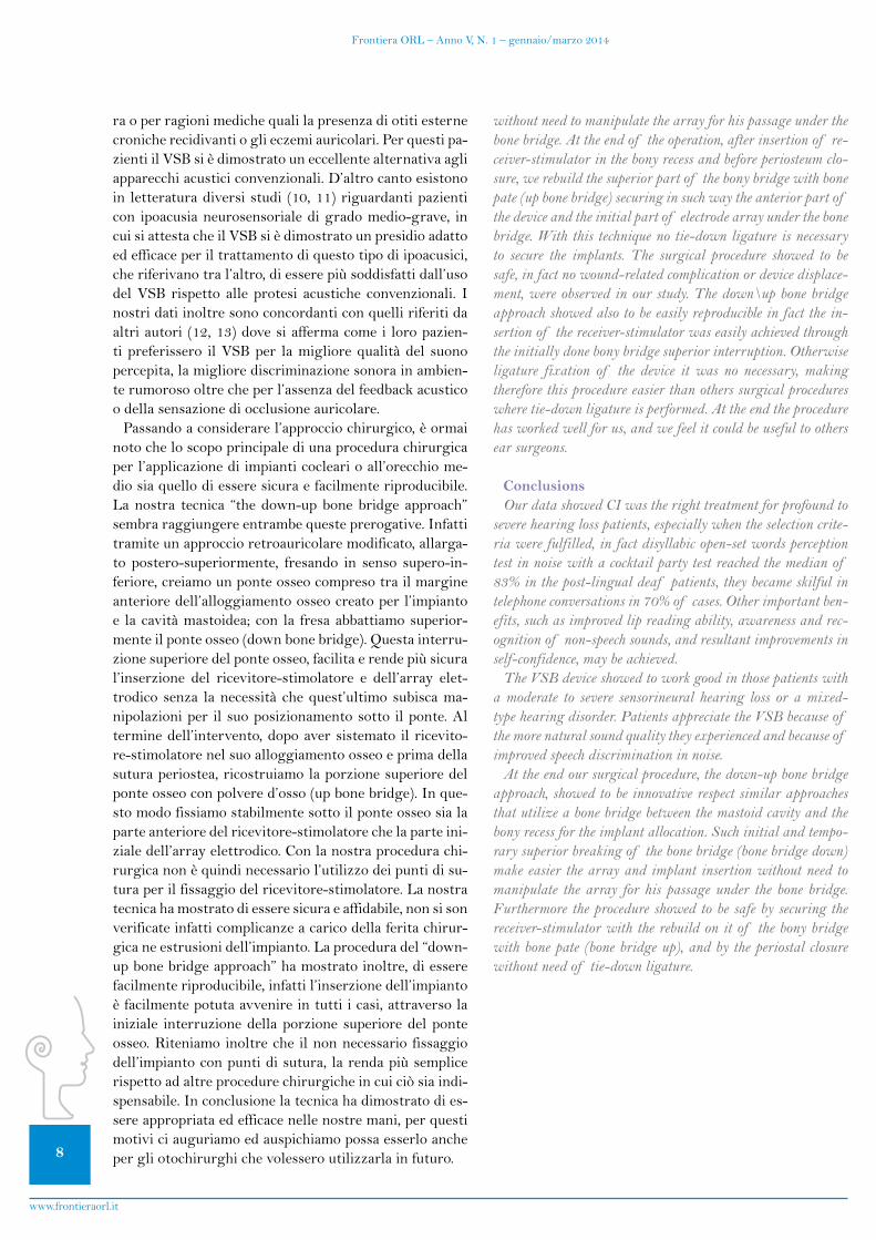
E) after insertion of receiver-stimulator in the bony recess and before doing wound closure , the superior part of the bone bridge is rebuilt with the previously collected bone pate “up bone bridge” strongly securing therefore the anterior part of the receiver-stimulator package without utilizing a tie-down ligature (Fig. 4).
The final step is the meticulous reconstitution of the perio-steal layer over the implant and the subcuticular sutures uti-lized for skin closure. A head dressing is routinely used post-operatively and is then removed after discharge usually on the postoperative day.
ResultsAll patients were successfully implanted by the down\up bone
bridge approach. All 22 intra cochlear electrodes in the Nucleus devices were activated in the programming of patients except in the Turner’s syndrome patient in whom moreover 15 elec-trodes were activated.
All patients used the implant during at least 8 hours daily.The mean pure tone threshold at 500 Hz, 1 kHz, 2 kHz and
4 kHz before implantation was 100,2 dB HL (minimum -77 dB and maximum -125 dB), speech score should be ≤ 50% at 65 dB SPL in the best aided condition.
Before the implants, no patient had been able to discriminate sentences in an open context. Similarly, none of them were able to have telephone conversations.
The mean free-field threshold from six months to two years after implant use reached 24,1 dB HL.
6
Frontiera ORL – Anno V, N. 1 – gennaio/marzo 2014
www.frontieraorl.it
RisultatiTutti i pazienti sono stati trattati con successo con la
tecnica del “down-up bone bridge approach”. Nei casi operati con IC Nucleus, si è sempre riusciti ad attivare i 22 elettrodi intracocleari tranne in una paziente affetta dasindromediTurnernellaquale,tuttavia,sièriuscitiad attivare 15 elettrodi. Tutti i pazienti utilizzano l’im-pianto per almeno 8 ore al giorno.
La soglia tonale media prima dell’impianto per le Fz 500Hz.1-2-4KHzeraparia100,2dB(minimo77dBemassimo 125 dB); la discriminazione vocale preoperato-riaa65dBSPLera≤50%.Primadell’interventonessunpaziente era in grado di comprendere una conversazione in campo aperto o di parlare al telefono. Nei follow-up po-stoperatori compresi fra i 6 mesi e 2 anni la soglia tonale media in campo libero era pari a 24,1 dB HL. Nei pazienti sordi post-linguali, la discriminazione vocale in campo li-
Sentence discrimination in an open context with environ-mental noise (Cocktail party test) reached 83% for the post-lingual deaf patients; in the 6 pre-lingual deaf patients (1 case bilateral) although the sentence discrimination in an open context was extremely variable (from 0% in 1 case to 70% in an other case) reached moreover the median of 43,3% . Tel-ephone conversations became possible for 70% of post-lingual deaf patients.
All the VSB were successfully implanted; the preoperatively mean average air conduction (AC) for frequencies 500, 1000, 2000 and 4000Hz in the patients with sensorineural hear-ing loss (SNHL) was 44 dB at 500Hz; 60 dB at 1KHz; 65 dB at 2KHz; 78dB at 4Khz. The postoperative AC in the 6
Fig. 1The small modified retro-auricular incision (4-6cm).
Fig. 2
The bone bridge superiorly interrupted “down bone bridge” between the mastoid cavity (anterior) and the bony well (posterior). A: Bone bridge; B: Mastoid cavity; C: Bony well.
Fig. 3
The receiver-stimulator and electrode array insertion A: Bone bridge; B: Mastoid cavity; C: Receiver-stimulator and electrode array allocated in the bony well and under bone bridge
Fig. 4
The receiver-stimulator package is strongly secured under the rebuilt bone bridge A: Bone bridge rebuilt with bone pate; B: Mastoid cavity; C: Re-ceiver-stimulator’s anterior part secured under bone bridge.
7
FrontieraORL–YearV,N.1–january/march2014
www.frontieraorl.it
beroconrumoredifondoambientale(cocktailpartytest)eraparial83%;nei6pazientisordipre-linguali(1casobilaterale) nonostante la discriminazione vocale in campo liberofosseestremamentevariabile(dallo0%in1casoal70%inunaltrocaso)tuttavialamediaraggiuntaeradel43,3%.Peril70%deipazientisordipost-lingualièstatopossibile l’utilizzo del telefono dopo l’operazione.
Tutti i pazienti VSB sono stati impiantati con successo con la nostra tecnica. La soglia tonale media preoperato-riaperconduzioneaerea(AC)neicasidiipoacusianeuro-sensoriale era di 44 dB a 500Hz, 60 dB a 1KHz, 65 dB a 2 KHz, 78 dB a 4 KHz. Al controllo effettuato 6 mesi dopo l’intervento la AC media era di 23 dB a 500Hz, 34 dB a 1KHz, 35 dB a 2KHz e 54 dB a 4KHZ.Perquantoriguardalatecnicachirurgicaèstataeffet-
tuata in tutti i casi con successo, in nessun caso è sta-to necessario convertire la mini-invasiva “down-up bone bridgeapproach”(4-6cm)conunatecnicaconvenzionalepiù invasiva. In 50 pazienti la dimissione è stata possibile il primo giorno postoperatorio solo 3 pazienti sono stati dimessinel 2giornopostoperatorio.Al follow-up (me-dia 2,1 anni, range 6 mesi - 4,5 anni ) non si è verificato nessun caso di migrazione del ricevitore - stimolatore o dell’arrayelettrodico;ciòsièpotutoconstataretramiteuna Rx in proiezione di Schuller effettuata nell’immedia-to postoperatorio e 6 mesi dopo l’intervento; non si sono verificate inoltre complicanze a carico del flap mio-cuta-neo. Al momento tutti i pazienti utilizzano l’impianto.
DiscussioneNelle nostre mani, l’utilizzo degli IC multicanale e de-
gli impianti acustici all’orecchio medio VSB, ha dato buo-ni risultati. Nei pazienti sordi post-linguali trattati con IC infatti, la discriminazione vocale in campo libero con rumoredifondo(cocktailpartytest)haraggiuntoilva-loremediodel83%.Questipazientiinoltresonodiven-tatiabilinelleconversazionitelefonichenel70%deicasi.Nei pazienti affetti da sordità pre-linguale nonostante i risultati inevitabilmente siano stati inferiori, la media della percentuale di discriminazione vocale, ha raggiun-toinfattiil43,3%.Questiultimituttaviahannomostratolalorosoddisfazioneperaltriimportantifattoriquali:ilmiglioramento nella lettura labiale, la possibilità di rico-noscere la voce dei loro familiari e di altri suoni o rumori ambientali.Tuttiquestipazienti,inognicaso,hannori-ferito di aver migliorato dopo l’impianto sia le loro capa-citàuditivechelaloroqualitàdellavita.L’impiantoVSB,si è dimostrato utile per quei pazienti con ipoacusia ditipo neurosensoriale o di tipo misto che per ragioni me-diche, psicologiche o sociali non riuscivano o non vole-vano adoperare le protesi acustiche convenzionali. Oggi-giorno esistono molte persone che non riescono a trarre beneficio, per la riabilitazione del loro handicap acustico, dall’utilizzo delle protesi acustiche convenzionali, vuoi permotiviqualiilfeedbackacustico,ladistorsionesono-
month control was 23 dB at 500Hz; 34 dB at 1KHz; 35 dB at 2KHz; 54 at 4KHz.
Concerning the surgical approach in no case was necessary converting the small incision (4-6 cm) to a conventional one. Wound healing was uneventful in all cases. Of the 53 im-planted patients in whom the down\up bone bridge approach was used, only 3 required hospitalization for 2 days after the surgical operation; all the others patients were discharged on the post-operative day. At follow-up (mean 2,1 years, range 6 months – 4,5 years) no migration of the receiver-stimulator, demonstrated by Rx in Schuller projection postoperatively and 6 months after surgery, and no flap-related complications were observed. At the moment all recipients are full-time users of the device.
DiscussionIn our experience with the use of multichannel CI and mid-
dle ear VSB devices, the results were good. In CI patients, di-syllabic open-set words perception test in noise with a cocktail party reached the median of 83% in the post-lingual deaf patients; they moreover, became skilful in telephone conversa-tions in 70% of cases. On the other hand although pre-lingual deaf patients reached poorer results in speech discrimination score (median 43,3%) all these patients showed their satisfac-tory for other important benefits, such as improvement in lip reading ability, chance to hear family members voices or to be awareness and to be able to recognize non-speech sounds and common environmental sounds. All these patients, however, maintain that their auditory skills and quality of life have improved significantly since implantation. On the other hand the VSB device showed to work good in those patients with a sensorineural hearing loss or a mixed-type hearing disorder. Many people today do not benefit from wearing a conventional hearing aid because of acoustic feedback, occlusion of the ear canal, or sound distortion or for a medical reason like chronic external otitis or external ear skin irritation. In these patients the VSB is an excellent alternative to the conventional hear-ing aids. There are studies that showed how the VSB is a suit-able and effective treatment for patients with moderate to se-vere sensorineural hearing loss who expressed more satisfaction with its use than with that a conventional hearing aid (10, 11). According to others authors, still our patients appreciated the VSB because of the more natural sound quality they experi-enced and because of improved speech discrimination in noise, the lack of ear occlusion, distortion and feedback (12, 13).
Regard the surgical procedure, it’s well known that the aim of any surgical approach for cochlear and middle ear implants insertion is to be safe and easily reproducible. The down\up bone bridge approach appears to reach both these requisites. Via a modified postauricular approach enlarged posteriorly and superiorly, by drilling from superior to inferior way, we create a bone bridge superiorly interrupted (down bone bridge) between the anterior edge of the bony well for device insertion and the mastoid cavity. Such superior breaking of the bone bridge make easier and safer the array and implant insertion
8
Frontiera ORL – Anno V, N. 1 – gennaio/marzo 2014
www.frontieraorl.it
raoperragionimedichequalilapresenzadiotitiesternecronicherecidivantioglieczemiauricolari.Perquestipa-zienti il VSB si è dimostrato un eccellente alternativa agli apparecchi acustici convenzionali. D’altro canto esistono in letteraturadiversistudi (10,11)riguardantipazienticon ipoacusia neurosensoriale di grado medio-grave, in cui si attesta che il VSB si è dimostrato un presidio adatto edefficaceperiltrattamentodiquestotipodiipoacusici,che riferivano tra l’altro, di essere più soddisfatti dall’uso del VSB rispetto alle protesi acustiche convenzionali. I nostridatiinoltresonoconcordanticonquelliriferitidaaltriautori(12,13)dovesiaffermacomei loropazien-ti preferissero ilVSBper lamigliorequalitàdel suonopercepita, la migliore discriminazione sonora in ambien-te rumoroso oltre che per l’assenza del feedback acustico o della sensazione di occlusione auricolare.
Passando a considerare l’approccio chirurgico, è ormai noto che lo scopo principale di una procedura chirurgica per l’applicazione di impianti cocleari o all’orecchio me-diosiaquellodiesseresicuraefacilmenteriproducibile.La nostra tecnica “the down-up bone bridge approach” sembraraggiungereentrambequesteprerogative.Infattitramite un approccio retroauricolare modificato, allarga-to postero-superiormente, fresando in senso supero-in-feriore, creiamo un ponte osseo compreso tra il margine anteriore dell’alloggiamento osseo creato per l’impianto e la cavità mastoidea; con la fresa abbattiamo superior-menteilponteosseo(downbonebridge).Questainterru-zione superiore del ponte osseo, facilita e rende più sicura l’inserzione del ricevitore-stimolatore e dell’array elet-trodico senza lanecessità chequest’ultimosubiscama-nipolazioni per il suo posizionamento sotto il ponte. Al termine dell’intervento, dopo aver sistemato il ricevito-re-stimolatore nel suo alloggiamento osseo e prima della sutura periostea, ricostruiamo la porzione superiore del ponteosseoconpolvered’osso(upbonebridge).Inque-sto modo fissiamo stabilmente sotto il ponte osseo sia la parte anteriore del ricevitore-stimolatore che la parte ini-zialedell’arrayelettrodico.Conlanostraprocedurachi-rurgicanonèquindinecessariol’utilizzodeipuntidisu-tura per il fissaggio del ricevitore-stimolatore. La nostra tecnica ha mostrato di essere sicura e affidabile, non si son verificate infatti complicanze a carico della ferita chirur-gica ne estrusioni dell’impianto. La procedura del “down-up bone bridge approach” ha mostrato inoltre, di essere facilmente riproducibile, infatti l’inserzione dell’impianto è facilmente potuta avvenire in tutti i casi, attraverso la iniziale interruzione della porzione superiore del ponte osseo. Riteniamo inoltre che il non necessario fissaggio dell’impianto con punti di sutura, la renda più semplice rispettoadaltreprocedurechirurgicheincuiciòsiaindi-spensabile. In conclusione la tecnica ha dimostrato di es-sereappropriataedefficacenellenostremani,perquestimotivi ci auguriamo ed auspichiamo possa esserlo anche per gli otochirurghi che volessero utilizzarla in futuro.
without need to manipulate the array for his passage under the bone bridge. At the end of the operation, after insertion of re-ceiver-stimulator in the bony recess and before periosteum clo-sure, we rebuild the superior part of the bony bridge with bone pate (up bone bridge) securing in such way the anterior part of the device and the initial part of electrode array under the bone bridge. With this technique no tie-down ligature is necessary to secure the implants. The surgical procedure showed to be safe, in fact no wound-related complication or device displace-ment, were observed in our study. The down\up bone bridge approach showed also to be easily reproducible in fact the in-sertion of the receiver-stimulator was easily achieved through the initially done bony bridge superior interruption. Otherwise ligature fixation of the device it was no necessary, making therefore this procedure easier than others surgical procedures where tie-down ligature is performed. At the end the procedure has worked well for us, and we feel it could be useful to others ear surgeons.
Conclusions Our data showed CI was the right treatment for profound to
severe hearing loss patients, especially when the selection crite-ria were fulfilled, in fact disyllabic open-set words perception test in noise with a cocktail party test reached the median of 83% in the post-lingual deaf patients, they became skilful in telephone conversations in 70% of cases. Other important ben-efits, such as improved lip reading ability, awareness and rec-ognition of non-speech sounds, and resultant improvements in self-confidence, may be achieved.
The VSB device showed to work good in those patients with a moderate to severe sensorineural hearing loss or a mixed- type hearing disorder. Patients appreciate the VSB because of the more natural sound quality they experienced and because of improved speech discrimination in noise.
At the end our surgical procedure, the down-up bone bridge approach, showed to be innovative respect similar approaches that utilize a bone bridge between the mastoid cavity and the bony recess for the implant allocation. Such initial and tempo-rary superior breaking of the bone bridge (bone bridge down) make easier the array and implant insertion without need to manipulate the array for his passage under the bone bridge. Furthermore the procedure showed to be safe by securing the receiver-stimulator with the rebuild on it of the bony bridge with bone pate (bone bridge up), and by the periostal closure without need of tie-down ligature.
9
FrontieraORL–YearV,N.1–january/march2014
www.frontieraorl.it
Bibliografia - References1. ClarkG.M.,FrancisD.M.TheUniversityof Melbourne/
Cochlear Corporation (Nucleus) program. Otolaryngol ClinNorth Am 1986; 19:329-54.2. Gantz B.J., Tye-Murray N., Tyler R.Word recognition
performance with single channel and multichannel cochlear im-plantation.AmJOtol1989;10(2):91-4.
NUMERO GRATUITO
800 91 08 08www.amplifon.it
frontiera orl vers2 def.pdf 1 14/12/2011 17.21.08
ConclusioniI risultati del nostro lavoro dimostrano come l’IC sia
il trattamento più efficace nella cura dei pazienti affetti da sordità di grado severo-profondo, sopratutto laddove siano state poste le giuste indicazioni all’intervento. La discriminazione vocale in ambiente rumoroso (cocktailpartytest)neipazientiaffettidasorditàpost-linguale,haraggiuntoinfattilamediadel83%.Glistessipazientiri-uscivano ad effettuare normali conversazioni telefoniche nel70%deicasi.Intuttiicasiipazientihannomanife-stato inoltre, il miglioramento nella lettura labiale, nel riconoscimento dei rumori ambientali oltre che una mag-giore sicurezza nei propri mezzi.
L’impianto acustico VSB, d’altronde, ha mostrato di es-sereutileedefficacenelriabilitarequeipazienticonipo-acusia neurosensoriale di media-grave entità o con ipo-acusia di tipo misto. Nel nostro studio i pazienti hanno apprezzatoilVSBperlamigliorequalitàdelsuonoper-cepita oltre che per l’accresciuta discriminazione vocale in ambiente rumoroso.
Riguardo la procedura chirurgica da noi proposta “the down-up bone bridge approach”, ha dimostrato di essere innovativa rispetto ad approcci simili in cui si utilizza il ponte osseo tra la cavità mastoidea e l’alloggiamento os-seo per l’impianto. La iniziale e parziale interruzione su-perioredelponteosseo(bonebridgedown)infatti,facilital’inserzionedell’arrayelettrodicoedell’impiantoaldisottodel ponte osseo stesso, senza la necessità di particolari ma-nipolazioni. La procedura infine si è dimostrata sicura nel fissare l’impianto, tramite la ricostruzione della porzione superioredelponteosseo(bonebridgeup)adoperadellapolvere d’osso accumulata durante l’intervento oltre che con la sutura periostale al disopra dello stesso.
3. WebbR.L.,LehnhardtE.,ClarkG.M.,LaszigR.,PymanB.C., Franz B.K. Surgical complications with the cochlear mul-tiple-channel intracochlear implant: experience at Hannover andMelbourne.AnnOtolRhinolLaryngol1991;100:131-6.4. JamesA.L., PapsinB.C.Cochlear implant surgery at 12
monthsof ageoryounger.Laryngoscope2004;114:2191-5.5. JamesA.L.,PapsinB.C.Devicefixationandsmallincision
access forpediatric cochlears implants. Int JPedOtorhinola-ryngol2004;68:1017-22.6. JiangD.,BibasA.,FitzgeraldO’ConnorA.Minimallyin-
vasive approach and fixation of cochlear and middle ear im-plants.ClinOtolaryngol2004;29:618-20.
7. O’ Donoghue G.M., Nikolopoulos T.P. Minimal access sur-geryforpediatriccochlearimplantation.OtolNeurotol2002,6:891-4.
8. D. Cuda. Cochlear implantation with Pulsar Med. El: a no-velsmallincisiontechnique.ActaOtorhinolaringologicaItalica2009; 29: 76-78
9. 9.Achena F., Montaldo C., Nucaro A.L. The down-up bone bridge approach for cochlear and middle ear implants: Our ex-periencein34patients.ClinOtolaryngol2012;37:148-61.
10. Todt I., Seidl R.O., Gross M., Ernst A. Comparison of dif-ferent vibrant soundbridge audioprocessors with conventional hearing aids. Otol Neurotol 2002; 23:669-73
11. Uziel A., Mondain M., Hagen P., Dejean F., Doucet G. Re-habilitation for high frequency sensorineural hearing impai-rmentinadultswiththesymphonixvibrantsoundbridge.A12.SterkersO.,boucarraD.,LabassiS.,BebearJ.P.,DubreilC.,
FrachetB.etal.Amiddleearimplantthesymphonixvibrantsoundbridge:retrospectivestudyof thefirst125patientsim-planted in France. Otol Neurotol 2003; 24:427-3613.Schmuziger N., Scimmann F.,Wengen D., Patscheke J.,
probst R. Long-term assessment after implantation of the vi-brant soundbridge device. Otol Neurotol 2006; 27:183-8
Acknowledgements and Conflicts of InterestThe author dedicate this article to Dr’s Antonio De la Cruz
memoryandwishestothankhim,“Masterof otologyandof life”, responsible to made a positive difference in his life and me-dical career. The authors report no conflicts of interest.
10
Frontiera ORL – Anno V, N. 1 – gennaio/marzo 2014
www.frontieraorl.it
■ Indirizzo per corrispondenza / Address for corrispondence: T.
E-mail:
AbstractL’obiettivodiquestostudioèdivalutarel’efficaciadiun
protocollo di screening alla luce delle linee guida inter-nazionali presso l’Azienda Marche Nord Pesaro-Fano.
Lo screening universale neonatale dell’udito è la mi-gliore metodica di diagnosi precoce dell’ipoacusia nei bambini.
IntroduzioneLa diagnosi precoce della sordità pre-linguale è fonda-
mentaleperuncorrettoinquadramentoterapeuticoafinidell’apprendimento del linguaggio e delle future abilita comunicative del bambino.
Le ipoacusie neurosensoriali o percettive sono suddi-viseinretrocleariecocleari,maesistonoanchequelledinatura trasmissiva dovute ad anomalie dell’orecchio me-dio o esterno.
All’interno dell’ipoacusia neurosensoriale vi è la neu-ropatia uditiva caratterizzata da otoemissioni acustiche presenti e tracciato ABR destrutturato.
L’identificazione dell’ipoacusia nei neonati viene realiz-zato attraverso un programma di screening universale, l’assenza dello screening universale determina una perdi-ta del 30 per cento di bambini affetti da ipoacusia grave.LaposizionedelJOINTCOMMITTEhafornitoleli-
nee guida riguardano i fattori di rischi pre e post natali, le metodiche elettrofisiologiche consigliate durante il 1° anno di vita.
Con DGR N 467 del 2011 il governo regionale delle Marche ha reso obbligatorio l’esecuzione del programma di screening in tutti i punti nascita della regione.Loscopodiquestolavoroedipresentarel’attivitàdiun
centro di II° livello.
Screening neonatale universale dell’udito.Esperienza in un ospedale italiano:
metodi e aspetti epidemiologiciNewborn Hearing screening.
Experience in Italian Hospital. Methods and epidemiological aspects
E. Milazzo^, F. Candido^, N. Rondina*, E. Di Crescenzo°, E. Battisti^,C. Cingolani^, D. Giardini^, G. Latini^, E. Gentile^, N. Vitelli^, G. Migliori^
^UOC Otorinolaringoiatria, Azienda Marche Nord Pesaro Fano*UOC Ingegneria Clinica, Azienda Marche Nord Pesaro Fano
°UOC Controllo di gestione, Azienda Marche Nord Pesaro Fano
AbstractThe aim of this study is to investigate the prevalence of
hearing loss among newborns delivered at the hospital Azien-da Marche Nord Pesaro-Fano and to evalutate the usefulness of our screening protocol.
Introduction Universal newborn hearing screening is the best way of ear-
ly detection of hearing loss in children. The identification and early diagnosis of pre lingual deafness necessary to prevent or minimize the serious consequences of hearing impairment on language development and on the future communication skill of a baby (1, 8). More than 80% of hearing losses in children are congenital or acquired in the neonatal period Permanent hearing loss can be conductive, due to congenital anomalies. Of the external and/or middle ear, or sensorineural: sensory (cochlear 80%) and neural which may include Auditory neu-ropaty (2, 3, 18). Auditory neuropaty/auditotory dissincrony is a hearing disorders showing. Normal otoacustic in contrast to the lack of Auditory brain system response. The first step for early detection of Hearing loss in newborns in any country is universal newborn hearing screening program. If the newborn hearing screening is not universal, more than 30% of cases of permanent hearing loss remain undiscovered (4). Currently, hearing screening in newborns is performed via TEOAE and ABR test. The Joint Committee on Infant Hearing in the 2007 position statement has identified the issue of late onset hearing loss and has defined the risks factors that requires an audio-logical follow up during the first year of life.
With DGR n647 of 2011 the Local Regional Government of Marche has made compulsory the execution of the neonatal screening audiological screening program in all the birth cen-tres of the region (1, 8, 11).
11
FrontieraORL–YearV,N.1–january/march2014
www.frontieraorl.it
Materiali e metodiL’esecuzione dello screening neonatale è stato eseguito
su tutti i neonati entro le prime 24 ore di vita in ambien-te insonorizzato, dopo l’eventuale rimozione di colostro all’interno del condotto uditivo esterno con posiziona-mento di una sonda e registrazione delle TEOAE.
L’esecuzione del test veniva trascritta in un apposito registro e consegnato il risultato del test ai genitori, in caso di fail il test veniva eseguito una seconda volta, e se il risultato era ancora fail, i genitori venivano invitati ad eseguire un nuovo test a distanza di 8 giorni dalla dimis-sione, con esecuzione di TEOAE E DPOAE.L’eventualefailinquestostepcomportaval’esecuzione
di ABR; a distanza di un mese, due mesi e tre mesi dalla nascita.
DiscussioneLoscopodelloscreeningèquellodisepararelapopola-
zione in 2 gruppi: popolazione sana da popolazione ma-lata.Untestidealesarebbequellocheraggiungeil100per
cento di sensibilità e specificità, pur tuttavia bisogna rag-giungere un compromesso.
In letteratura l’incidenza dell’ipoacusia è mag-giore rispetto ai tassi per galattosemia 2/100000 e fenilchetonuria10/100000 e 25/100000 per ipotiroidi-smo.
Le otoemissioni vengono registrate con una sonda inserita nel meato acustico esterno e nella quale sonocontenuti un trasduttore e un microfono, per ottenerle vengono impiegati toni clicks o toni burst attraverso il trasduttore vengono inviati degli impulsi della durata di 80 msec con una ripetività di 50 stimoli al sec e con un intensità di 80-83 db SPL.
L’ABR rappresenta la metodica più sensibile nell’indi-viduazione delle ipoacusie nei bambini al di sotto dei due anni di vita.
Le componenti principali sono costituite dalla I, III, V onda, e analizzano la risposta compresa fra il nervo acu-stico e la regione sotto-talamica.
L’onda I corrisponde al potenziale globale della porzio-ne distale dell’8 paio, mentre la II onda è generata dal-la porzione prossimale del nervo al suo punto d’ingres-so nella giunzione bulbo pontina. L’onda III nei nuclei cocleari ipsilaterali. L’onda V è da mettersi in relazione all’attività del lenisco laterale all’ingresso del collicolo inferiore controlaterale all’orecchio stimolato.
Pertanto i parametri interonda ed i parametri interau-rali la presenza/assenza delle onde, ed in misura minore le loro ampiezze sono validi indicatori della funzionalità del nervo uditivo e della via uditiva troncoencefalica. Nel bambino si hanno valori di latenza dei singoli picchi, e specialmentequellitardiviIII-Vsuperiorichenell’adul-to e i valori normali vengono raggiunti tra 8 e il 12 mese di vita.
The aim of this study is to present and discuss the results au-diological centre of II level.
Material e methods During the study period TEOAE test were carried out on
all inborn infants within 24 hours of life at their bedside or inside the nursery room in the post natal ward in the neonatal intensive care unit, the test was conducted in an isolation room ore newborns were discharged.
After ear inspection and removal of any vernix or fluid in the external ear canal, the probe was inserted into the EAC and adjusted. The TEOAE was the performed and the result of pass or fail recorded, in newborns with a fail result, a sec-ond test was immediately performed after appropriate adjust-ment of the positions of the probe.
When a fail was obtained on the second attempt the new-born was considered as having failed the screening test. Par-ents were notified of the screening result immediately.
Newborns who failed the screening test were given appoint-ment a one months.
A repeat TEOAE and DPOAE was performed. Those who failed at this stage were again tested at two
months and three months of age with TEOAE-DPOAE and auditory brain system response (ABR).
Discussion The purpose of a screening test is to separate a population
into two groups: those who are normal and whose who are af-fected by target targeted disorder. How wekll a hearing test achieves this purpose will be influenced by the performance characterstics of the test chosen, the pass/fail criterion, and targeted hearing loss (10, 15).
A perfect screening test would pass all individuals who are normal ( i.e 100% specifity) and fail all individuals who have target disorder (i.e 100% sensitivity).
No screening test is perfect, however, and we must accept a compromise between sensitivity and specifity.
In the literature, the incidence of permanent childhood hear-ing impairment is reported at about 100-300 children for eve-ry 100.000 births as compared to the incidence of commonly screened newborn disorders which is 2/100000 for galactos-emia, 10/100000 for phenilchetonuria, 25/100000 for hy-pothyroidism. Therefore, bilateral congenital permanent HI is sufficiently common to justify screening all children and just those at high risk (5) (6) (20).
Teoae are produced in response to very brief stimuli, such as clicks. The Teoae is also known as the click evoked otoa-custic emission, Kemp eco, or cochlear echo. Evoked otoacustic emissions occur either during long-term stimulation or after a brief stimulus. Responses to brief stimuli are known as tran-sient evoked otoacustic emissions (TEOAE).
The probe apparatus includes a miniature loudspeaker and microfone. The signal in the ear canal is sampled for – 20 msec after the stimulus is presented to the ear. Although im-
12
Frontiera ORL – Anno V, N. 1 – gennaio/marzo 2014
www.frontieraorl.it
Anche la morfologia nel neonato è differente rispetto aquelladell’adultopotendosiosservareabitualmente3delle consuete 5 onde. Talvolta è necessario un numero minoridistimoliperrenderepiùchiaral’onda,quindiènecessario crearsi una propria tabella normativa sia negli adulti che nei bambini.
RisultatiDal 1 gennaio 2010 al 31 Dicembre 2011, 2126 neo-
nati sono stati sottoposti a screening Universale presso il servizio di Audiologia nell’Azienda Marche Nord the Pesaro–Fano.72neonaticonfattoridirischio(93,5%)nell’anno2010
e 70 nell’anno 2011 hanno eseguito le il test mediante le otoemissioni acustiche da transienti che sono risultate positive. Nel2010sonostatisottoposti77(29.6%)neonatie82
(29,4%)nel2011alIIlivellomedianteregistrazionedeipotenziali evocati uditivi per ricerca di soglia.
Neonati con anamnesi familiare positiva per ipoacusia 6 nel 2010 e 6 nel 2011, 7 neonati hanno avuto una dia-gnosi di ipoacusia: 3 di ipoacusia media, 1 monolaterale profonda, 3 di ipoacusia profonda bilaterale. L’incidenza diipoacusiaèstatapariall’0,33%.L’incidenzadelCMVnel20105,6%e4,3%nel2011.Toxoplasmosihaavutounincidenzadel6,9%nel2010
e7,1%nel2011.Laprematuritàha incisoper19,4%nel2010e21,4%
nel 2011.AnomalieCraniofaciali(S.Kleefstra)1in2011
ConclusioniL’obbiettivodiquestostudioèstatodiverificarelare-
ale incidenza dell’ipoacusia e di verificare l’efficacia del protocollo.
portant exceptions exist most of the published data regarding TEOAE have been obtained through the use hardware and software supplied by otodynamics and known as ILO -88.The typical TEOAE is a bief, broad-spectrum click, but one bursts also may be used to elicit the response.
The response follows the stimulus with a latency that is deter-mined by cochlear travelling wave dynamics. As illustrated in high frequency components appear with a shorter latency than lower frequency components. This time delay reflects cochlear dynamics and anatomy: the travelling wave response is much more rapid for high-frequency stimulus components than for low frequency.
The TEOAE response is recorded though the use of tech-niques that are identical to those used to record the short latency auditory evoked potentials, or auditory brain system response.
Approximately 1000 stimuli are presented to improve the SNR of the response in each of two (A and B) Average wave-forms.
As is the case for electrofisiological recordings, the repeat-ed sampling reduces the random variance in the signal by an amount predicted by the square root of the sample size.
The effect of this is to reduce the noise 30 db, allowing an emission to be extracted from the ambient noise in the ear canal. The typical clinical recording involves stimulation at a level of 80 dB peak equivalent SPL or 40 Db spectrum leveland response amplitudes are typically at level 60 to 70 dB blow the level of the stimulus.
The ABR has been the gold standard physiologic test of in-fant hearing since is introduction in the early 1970s.
Studies have demonstrated that the sensitivity, specificity, and reliability of the ABR test in newborn screening are high; thus, refer rates will be low and ,and missing a true hearing loss will be low, and missing a true hearing loss will be rare (7).
The waveform in neonates consists primarily of three com-ponent peaks, corresponding to waves I, III, V of the adult ABR.
During the first 18 months of life, the other component peaks of the ABR emerge, until the until the waveform assumes an adult morphology.
Wave I amplitude in newborns is larger than adults, possi-bly because the recording electrode is closer to the cochlea due to the smaller head size of infants. On the other hand, wave V amplitude is smaller in infants than adults, therefore the wave V/I amplitude ratio will be reduced for infants, often having a value less than 1.0
The latencies of the ABR waveform components are longer in neonates compared with adults and decrease progressively throughout the neonatal period due to maturation of the coch-lea and brainsistem. Wave I matures most rapidly, assuming an adult latency value by about 2 to 3 months of age.
Various reports show wave V assuming an adult value either by 12 to 18 months, by slightly over 2 years or as late as 2.5 years of age.
This differential maturation of I and V means that the wave I-V IPL progressively shortens as the infant grows. The Wave I.V IPL decreases from 5.0 msec at the term to the adult value of 4.0 msec by 12 to 18 months of age.
The time course of wave III maturation follows that of wave V.
The infant ABR is more vulnerable to the effects of increasing stimulation rate, consequently, slower rates of stimulus presen-tation may need to be used to maximixe waveform clarity.
When testing infants from birth 18 to 24 months of age, it is important to use normative data appropriate for age, rather than adults values (16, 17).
Results Statistical analysis was performed using SPPS program. From the 1th of January year 2010 to the 31 December
2011, 2126 newborn in the Pesaro–Fano Azienda Marche Nord have undergone newborn hearing screening.
TEOAE pass in 72 patient with risk factors (93,5%) in 2010 year and 70 in 2011 year (85,4%).
13
FrontieraORL–YearV,N.1–january/march2014
www.frontieraorl.it
The II level (ABR) in 2010 are 77 (29,6%) and 82 (29,4%) in 2011.
Family history of hearing loss 6 in 2010 and 6 in 2011 7 patient had a final diagnosis of hearing impairment: 3
mild, 1 unilateral hearing loss, 3 severe /profound. The incidence of hearing impairment is 0,33%. The incidence of CMV in 2010 5,6% and 4,3% in 2011. Toxo 6,9% in 2010 and 7,1% in 2011. Prematurity 19,4% in 2010 and 21,4% in 2011. Family history of congenital HI 8,3% and 8,6% in 2011.
Craniofacial anomalies (S. Kleefstra) 1 in 2011
Bibliografia – References1. Joint Committee on Infant Hearing Year 2007 Position
Statement:PrinciplesandGuidelinesforEarlyHearingdetec-tion and Intervention Programs. 2. J.Graham,G.K.Scadding,P.Bull.PediatricENT,Springer
Verlag, Berlin 2007 pag 487 pp. 3. W.PGibson,J.Graham,Editorial:“Auditoryneuropathy”
and cochlear implantation miths and facts, Cochlear implants international9(1)2008www.interscience.wiley.-com/cgi-bin/fulltext/117905585/PDFSTART.4. J.L.Johnson,K.RWhite,J.EWiden,J.S.Gravel,M.James,
T.KennaleyetalAmulticenterevalutationof howmanyinfantswith permanent hearing loss a two stage otacustic emissions/automaticauditorybrainsistemresponsenewbornhearingscre-eninigprotocol,Pediatrics116(3)2005pagg.663-672.5. KennedyC.R.,Neonatalscreeningforhearingimpairment
Arch Dis Child 2000, 83:377-382. 6. KennedyC.R.,McCannD.C.,Universalneonatalhearing
screening moving from evidence to practice Arch Dis Child 2004; 89 F378-383. 7. R.J.Roser,M.Valente,H.Hosford-DunnAudiologyDia-
gnosis: Thieme ed. 8. T. Puig, A. Municio, C. Medà Cochrane Collaboration
2008 Universal Neonatal Hearing Screening versus selective screening as part of the management of childhood deafness (Review).
9. M.G. Calevo, P. Mezzano E. Zullino, P. Padovani, G. Ser-ra, and Stern group Ligurian experience on neonatal hearing screening: Clinical and epidemiological aspect Acta Paediatrica 2007 pagg. 1592-1599. 10.JankaJakubikova,ZuzanaKabatova,GabrielaPavlovcino-
va,MilanProfantNewbornhearingscreeninigandstrategyfoeearlydetectionof hearinglossininfantsInternationalJournalof Otorhinolaringolgy2009pagg.607-612.
11. P. Ghirri, A. Liumbruno, S. Lunardi, F. Forli, A. Boldri-ni, A. Baggiani, S. Berrettini Universal Neonatal Audiologi-
calScreeninig:Experienceof theUniversityHospitalof PisaJournalof Pediatricspagg.1-8(2011).12.StanleyAGelfandEssentialof Audiologysecondedition,
Thieme ed.13. C.P.B. van der Ploeg, N.N. Uilenburg, M.A.Kauffman-de
Boer, A.M.Oudesluys-Murphy, P.H.VerkekNewborn hearingscreeninginyouthhealtcareintheNederlands:Nationalresultof implementationandfollow-up.InternationalJournalof Au-diology(2012)pagg.584-590.14.W. D. Eiserman, D. Hartel, L. Shisler, J. Buhrmann, K.
White,T.FoustUsingotoacusticemission toscreen forhea-ringlossinealychildhoodcaresettingsInternationalPaedia-tricOtorhinolaryngology(2008)pagg.475-482.15.P.Watkin,j.etalNeonatalhearingScreening:havewetaken
therightRoad?Resultsfroma10yeartargetedscreenlongi-tudinallyfollowedupinasingleDistrictAhmedAudiologicalMedicine(2005)pagg.175-184.16.JacbsonJetal.Newbornandinfantauditorybrainsistem
response applications pp 313-344 Boston Allin e Bacon. 17.GORGAM.P.Somecomparisonbetweenauditorybrain-
sistem response thresholds, latencies and the pure tone audio-gramEarandHearing,6(2)105-112.
18. G. Kirkim, Bulent Serbetcioglu, Taner Kemal Erdag, Ke-rimCeryanThefrequencyof auditoryneuropathydetectedbyuniversal newborn hearing screening program International Journalof PediatricOtorhinolaryngology(2008)pagg.1461-1469. 19.WendyMcCraken,AlysYoung,HelenTattersallUniver-
sal newborn hearing screening: Parental Reflections on veryearlyAudiologicalManagementEarandHearingpagg54-64(2008).
20. P. Mezzano, G. Serra, M. Grazia Calevo, for the Stern groupCostof analysisof anItalianneonatalhearingscree-ningprogrammeTheJournalof MaternalFetalandneonatalmedicine, September 2009; pagg. 806-811.
Conclusion The goal of newborn auditory screening is to identify infants
with significant hearing impairment in the most rapid and cost effective way allowing the opportunity to begin early interven-tion services for family with infants. the principles of JCHI have been achieved: the infants underwent hearing screening within 1 month of; all the referred children receveid an appropriate audi-ologic and medical evalutations no later than 3 months of age; all infants with confirmed permanent hearing loss received early intervention service before the age of 4 months.
Financial disclosure and declaration The authors report no conflict of interest.
14
Frontiera ORL – Anno V, N. 1 – gennaio/marzo 2014
www.frontieraorl.it
AbstractMetodi: 17 pazienti con stenosi sottoglottica sono stati
operati utilizzando innesto di interposizione cartilagineo. Essi sono stati divisi in due gruppi
- Gruppo A: 7 pazienti con laringoplastica anteriore utilizzandocartilaginedell’alatiroidea(TAC)
- Gruppo B: gruppo di controllo di 10 pazienti con la-ringoplastica e interposizione di cartilagine costale.
Risultati: Gruppo A (TAC), la complicanza più fre-quenteèladispneacheèapparsoinunafaseinizialenel42%deipazienticonostruzionedellostentcontappodimuco,odispneaapparsainunafasesuccessivanel57%dei casi a causa dello sviluppo di tessuto di granulazio-ne.GruppoB(cartilaginecostale)complicazioni:dispneaprecoceapparsonel10%deipazienti,mentreladispneatardivaacausadigranulomiapparsonel20%deicasi.
Conclusione: L’uso di innesto di cartilagine dell’ala ti-roidea nella stenosi laringea permette una espansione si-cura del diametro sottoglottideo come è stato verificato con l’uso di cartilagine costale.
IntroduzioneLa stenosi delle vie aeree di adulti è causata, in molti
casi, da ventilazione meccanica prolungata. Per risolvere questapatologia,vengonoutilizzatediversetecnichechi-rurgiche, a seconda della posizione e lunghezza della ste-nosi.
Nel caso di stenosi sottoglottica, la resezione cricotra-chealeèlaproceduraprincipale,maquandolastenosièameno di 5 mm sotto le corde vocali o le avvolge, la proce-duracitatanonpuòessereeseguita;lametodicachirurgi-ca preferita è l’interposizione di cartilagine per espandere lospaziosottoglottico/glottico(1).
La laringoplastica con l’innesto di cartilagine è stata descrittanel1955daRethi(9)epubblicizzatodaFearon1972Cotton(10)edèdiventataunatecnicaampiamenteaccettata per riparare la stenosi sottoglottica con glotti-
Laringofissura anteriore con innestodi cartilagine tiroidea in pazienti adulti
Laryngeal anterior split with thyroid cartilage graft in adult patients
G. Stok^, M. A. Caldez García*, M. L. Rospide*
^Jefe del Servicio de Otorrinolaringología, Hospital Centro de Salud “Zenón J. Santillán”, San Miguel de Tucumán, Tucumán, Argentina
*Staff del Servicio de Otorrinolaringología, Hospital Centro de Salud “Zenón J. Santillán”, San Miguel de Tucumán, Tucumán, Argentina
AbstractMethod: 17 patients with subglottic stenosis were operated
using cartilage interposition graft. They were divided into two groups: group A: 7 patients with anterior laryngoplasty using thyroid ala cartilage -- group B: control group of 10 patients with laryngoplasty and interposition of costal cartilage.
Results: Group A (TAC) the most frequent complication was dyspnea which appeared at an early stage in 42% of patients with mucus plug stent obstruction, or dyspnea either appeared at a later stage in 57% of the cases due to the de-velopment of granulation tissue. Group B (costal cartilage) complications: early dyspnea appeared in 10% of patients, while dyspnea at a later stage due to granulomas appeared in 20% of cases.
Conclusion: The use of thyroid ala cartilage graft in la-ryngeal stenosis allows an expansion of subglottic diameter as safe as it has been verified with the use of costal cartilage.
IntroductionThe adults’s airway stenosis is caused, in most cases, due to
prolonged mechanical ventilation. To solve this pathology, dif-ferent surgical techniques are used, depending of the location and length of the stenosis.
In the case of subglottic stenosis, cricotracheal resection is the main procedure, but when the stenosis is less than 5 mm below the vocal cords or encompasses them, and the mentioned proce-dure cannot be performed, cartilage interposing for expanding subglotis/glottis is the preferred surgical method (1).
Laryngoplasty with the cartilage graft was described in 1955 by Rethi (9) and popularized by Fearon 1972 Cotton (10) and has become a widely accepted technique to repair sub-glottic stenosis with compromised glottis or the nearest area. This surgery divides the stenotic laryngotracheal segment in the sagital plane and introduces the graft cartilage, anterior and/or posterior, to achieve the expansion of the subglottic di-ameter. Different grafts were used from 1970 onwards such as the costal cartilage, thyroid, hyoid bone, ear cartilage (2, 3).
■ Indirizzo per corrispondenza / Address for corrispondence: G. Stok - Jefe del Servicio de Otorrinolaringología, Hospital Centro de Salud “Zenón J. Santillán”, San
Miguel de Tucumán, Argentina - Email: [email protected]
15
FrontieraORL–YearV,N.1–january/march2014
www.frontieraorl.it
deo l’areapiùvicinacompromessa.Conquestatecnicasi divide il segmento laringotrachealestenotico sul piano sagittale e si introduce l’innesto di cartilagine, anterio-re e/o posteriore, per ottenere l’espansione del diametro sottoglottideo. Diversi innesti sono stati utilizzati a par-tire dal 1970, come la cartilagine costale, tiroidea, osso ioideecartilaginedell’orecchio(2,3).
Materialie MetodiLo studio è stato condotto tra gli anni 2003 - 2010
presso il Santillán Zenón Health Center Hospital di San Miguel de Tucumán, Tucumán, Argentina. Sono stati in-clusi 17 pazienti adulti con stenosi subglottica causata da ventilazione meccanica prolungata, diagnosticata me-diante endoscopia rigida e TAC. Al momento della valu-tazione pre-chirurgica, 10 pazienti erano stati sottoposti a tracheotomia, e solo 2 pazienti hanno mostrato un im-pegno della glottide posteriore.
La Stenosi laringea è stata classificata in base alla scala diCotton-Meyers(Tab.1)
I 17 pazienti avevano i seguenti gradi di stenosi, secon-do la classificazione di cui sopra: 7 pazienti con grado IV, 7 pazienti con grado III, e 3 pazienti con grado II.
Tutti i pazienti sono stati operati con cervicotomia an-teriore, laringofissura, ed interposizione di cartilagine anteriore. 7 pazienti hanno ricevuto uninnesto di ala di cartilagine tiroidea, 10 pazienti sono stati innestati con cartilagine costale. Nell’ultimo gruppo, 2 pazienti è sta-to utilizzati anche innesto per espandere la commissura posteriore.
Tutti i pazienti avevano uno stentposizionato per soste-nere e mantenere la posizione della cartilagine innestata. Il tempo di permanenza dello stent era tra 4 e 8 mesi. Il monitoraggio è stato eseguito con endoscopia flessibile, prima e dopo la rimozione dello stent entro un periodo di 1 anno circa.
Il criterio di successo della procedura è stata la decan-nulazione dei pazienti senza dispnea.
Tecnica chirurgicaLa tecnica chirurgica consiste nella cervicotomia, si di-
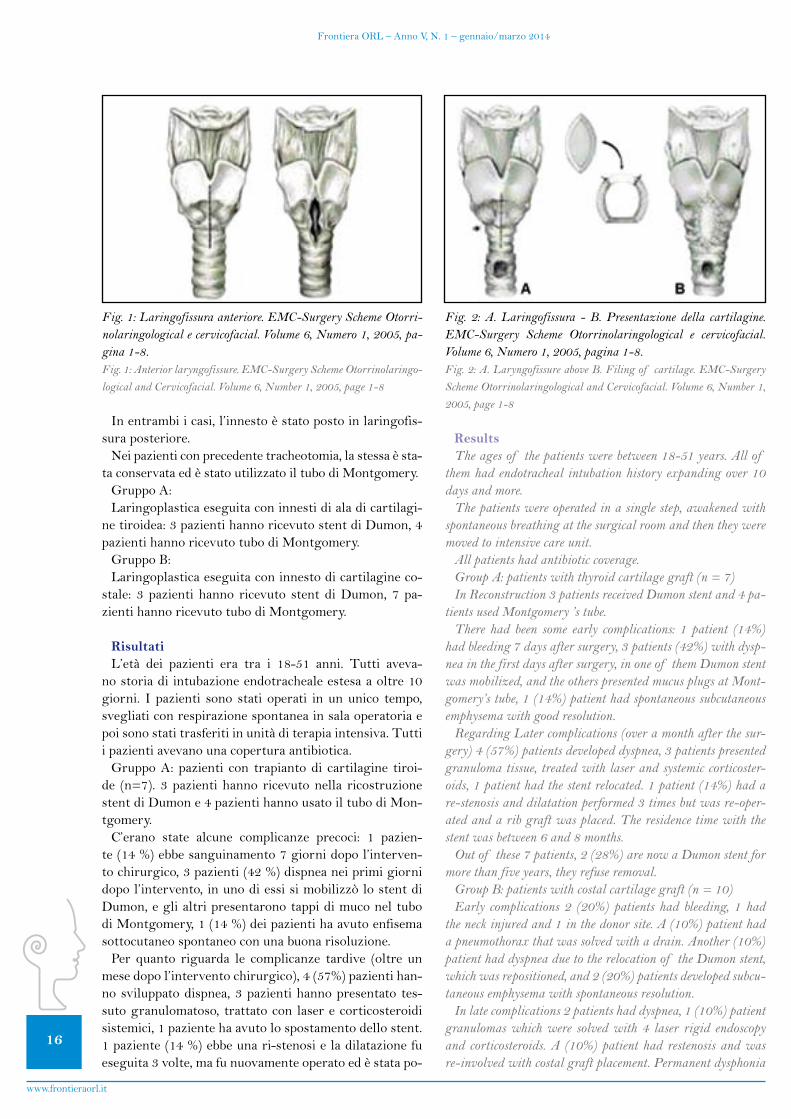
varicano i muscoli prelaringei, si effettua la laringofissu-ra anteriore dal terzo superiore della cartilagine tiroide al primo o secondo anello tracheale; viene preparato l’in-nesto cartilagineo che è interposto tra i bordi della larin-gofissura,perespanderelevieaeree(Fig.1eFig.2).
Materials and MethodsThe study was performed between the years 2003 – 2010 at
the Santillán Zenón Health Center Hospital in San Miguel de Tucumán, Tucumán, Argentina. 17 adult patients were in-cluded with subglottic stenosis due to prolonged mechanical ventilation, which were diagnosed by rigid endoscopy and CT scan. At the time of presurgical evaluation, 10 patients had undergone tracheotomy, and just 2 patients showed a posterior glottic commitment.
Laryngeal stenosis were classified according to the degree of Cotton – Meyers. Table 1
The 17 patients had the following grades stenosed, according to the classification mentioned above: 7 patients with grade IV, 7 patients with grade III, and 3 patients with grade II.
All patients were operated with anterior cervicotomy, laryn-gofissure, and anterior cartilage interposition. 7 patients re-ceived a thyroid ala cartilage graft, and 10 patients were grafted with costal cartilage. In the last group, 2 patients also used graft to expand the posterior commissure.
All patients had a stent placed for supporting and maintain-ing the position of the grafted cartilage. The residence time of the stent was between 4 and 8 months. Monitoring was per-formed with flexible endoscopy, before and after removal of the stent within a period of 1 year approximately.
The procedure success criterion was decannulation of pa-tients without dyspnea.
Surgical TechniqueThe technique described above is cervicotomy, prelaryngeal
muscle divulsion and anterior laryngofissure from the upper third of the thyroid cartilage to the first or second tracheal rings. Cartilage graft is obtained and is interposed between the edges of the laryngofissure, to expand the airway (Fig. 1 and Fig. 2).
In both cases, graft was placed in the posterior larigofisure by synechiae.
In patients with previous tracheotomies, the same were re-spected and used for Montgomery’s tube.
Group A:Laryngoplasty performed with thyroid ala cartilage grafts:
3 patients received Dumon stent, 4 patients received Mont-gomery’s tube.
Group B: Laryngoplasty performed with costal cartilage graft: 3 pa-
tients received Dumon stent, 7 patients received Montgomery’s tube.
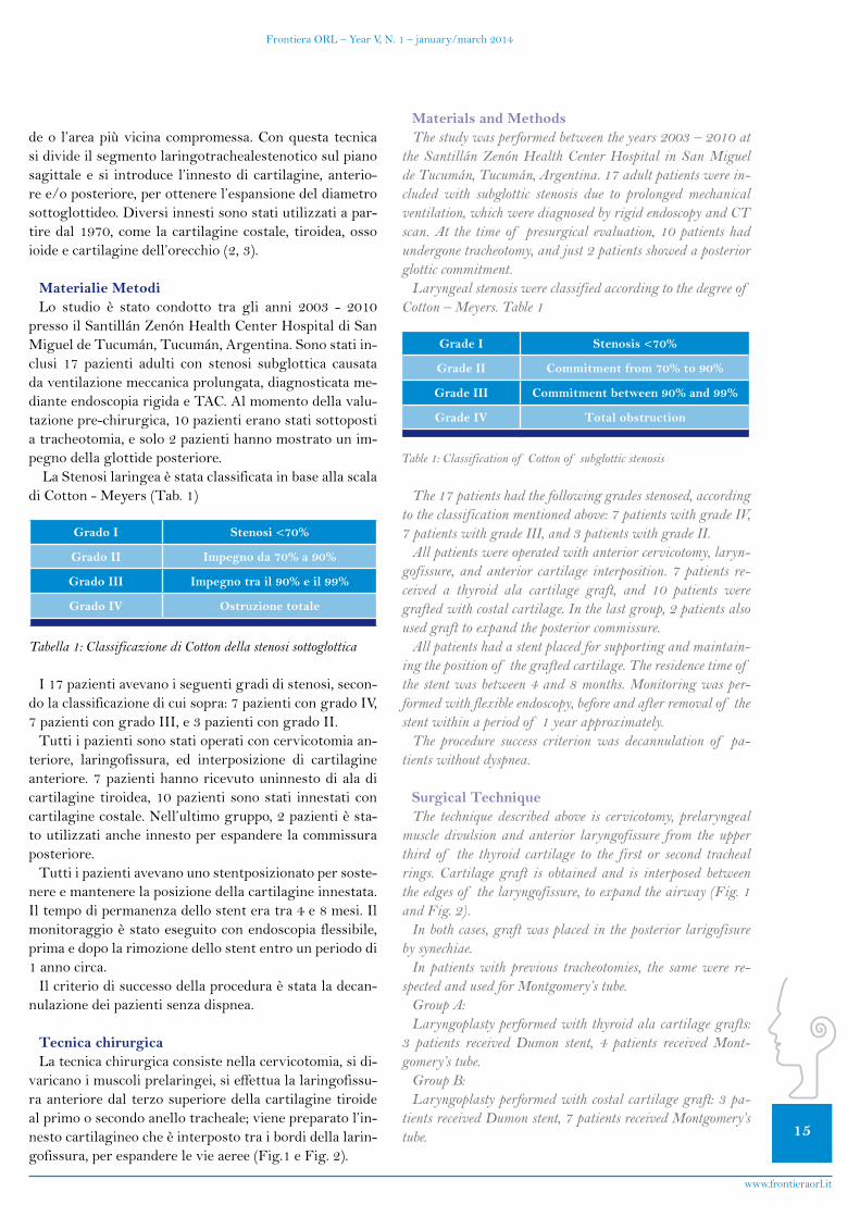
Grado I Stenosi <70%
Grado II Impegno da 70% a 90%
Grado III Impegno tra il 90% e il 99%
Grado IV Ostruzione totale
Tabella 1: Classificazione di Cotton della stenosi sottoglottica
Grade I Stenosis <70%
Grade II Commitment from 70% to 90%
Grade III Commitment between 90% and 99%
Grade IV Total obstruction
Table 1: Classification of Cotton of subglottic stenosis
16
Frontiera ORL – Anno V, N. 1 – gennaio/marzo 2014
www.frontieraorl.it
In entrambi i casi, l’innesto è stato posto in laringofis-sura posteriore.
Nei pazienti con precedente tracheotomia, la stessa è sta-taconservataedèstatoutilizzatoiltubodiMontgomery.
Gruppo A:Laringoplastica eseguita con innesti di ala di cartilagi-
ne tiroidea: 3 pazienti hanno ricevuto stent di Dumon, 4 pazientihannoricevutotubodiMontgomery.
Gruppo B:Laringoplastica eseguita con innesto di cartilagine co-
stale: 3 pazienti hanno ricevuto stent di Dumon, 7 pa-zientihannoricevutotubodiMontgomery.
RisultatiL’età dei pazienti era tra i 18-51 anni. Tutti aveva-
no storia di intubazione endotracheale estesa a oltre 10 giorni. I pazienti sono stati operati in un unico tempo, svegliati con respirazione spontanea in sala operatoria e poi sono stati trasferiti in unità di terapia intensiva. Tutti i pazienti avevano una copertura antibiotica.
Gruppo A: pazienti con trapianto di cartilagine tiroi-de (n=7). 3pazientihanno ricevutonella ricostruzionestent di Dumon e 4 pazienti hanno usato il tubo di Mon-tgomery.
C’erano state alcune complicanze precoci: 1 pazien-te(14%)ebbesanguinamento7giornidopol’interven-tochirurgico,3pazienti(42%)dispneaneiprimigiornidopol’intervento,inunodiessisimobilizzòlostentdiDumon, e gli altri presentarono tappi di muco nel tubo diMontgomery,1(14%)deipazientihaavutoenfisemasottocutaneo spontaneo con una buona risoluzione.Per quanto riguarda le complicanze tardive (oltre un
mesedopol’interventochirurgico),4(57%)pazientihan-no sviluppato dispnea, 3 pazienti hanno presentato tes-suto granulomatoso, trattato con laser e corticosteroidi sistemici, 1 paziente ha avuto lo spostamento dello stent. 1paziente(14%)ebbeunari-stenosieladilatazionefueseguita 3 volte, ma fu nuovamente operato ed è stata po-
ResultsThe ages of the patients were between 18-51 years. All of
them had endotracheal intubation history expanding over 10 days and more.
The patients were operated in a single step, awakened with spontaneous breathing at the surgical room and then they were moved to intensive care unit.
All patients had antibiotic coverage.Group A: patients with thyroid cartilage graft (n = 7)In Reconstruction 3 patients received Dumon stent and 4 pa-
tients used Montgomery ’s tube.There had been some early complications: 1 patient (14%)
had bleeding 7 days after surgery, 3 patients (42%) with dysp-nea in the first days after surgery, in one of them Dumon stent was mobilized, and the others presented mucus plugs at Mont-gomery’s tube, 1 (14%) patient had spontaneous subcutaneous emphysema with good resolution.
Regarding Later complications (over a month after the sur-gery) 4 (57%) patients developed dyspnea, 3 patients presented granuloma tissue, treated with laser and systemic corticoster-oids, 1 patient had the stent relocated. 1 patient (14%) had a re-stenosis and dilatation performed 3 times but was re-oper-ated and a rib graft was placed. The residence time with the stent was between 6 and 8 months.
Out of these 7 patients, 2 (28%) are now a Dumon stent for more than five years, they refuse removal.
Group B: patients with costal cartilage graft (n = 10)Early complications 2 (20%) patients had bleeding, 1 had
the neck injured and 1 in the donor site. A (10%) patient had a pneumothorax that was solved with a drain. Another (10%) patient had dyspnea due to the relocation of the Dumon stent, which was repositioned, and 2 (20%) patients developed subcu-taneous emphysema with spontaneous resolution.
In late complications 2 patients had dyspnea, 1 (10%) patient granulomas which were solved with 4 laser rigid endoscopy and corticosteroids. A (10%) patient had restenosis and was re-involved with costal graft placement. Permanent dysphonia
Fig. 1: Laringofissura anteriore. EMC-Surgery Scheme Otorri-nolaringological e cervicofacial. Volume 6, Numero 1, 2005, pa-gina 1-8.Fig. 1: Anterior laryngofissure. EMC-Surgery Scheme Otorrinolaringo-logical and Cervicofacial. Volume 6, Number 1, 2005, page 1-8
Fig. 2: A. Laringofissura - B. Presentazione della cartilagine. EMC-Surgery Scheme Otorrinolaringological e cervicofacial. Volume 6, Numero 1, 2005, pagina 1-8.Fig. 2: A. Laryngofissure above B. Filing of cartilage. EMC-Surgery Scheme Otorrinolaringological and Cervicofacial. Volume 6, Number 1, 2005, page 1-8
17
FrontieraORL–YearV,N.1–january/march2014
www.frontieraorl.it
sto un innesto di costola. Il tempo di permanenza dello stent era tra 6 e 8 mesi.Diquesti7pazienti,2 (28%)hannooraunostentdi
Dumondapiùdicinqueannierifiutanolarimozione.Gruppo B: pazienti con trapianto di cartilagine costale
(n=10).Lecomplicanzeprecoci:2(20%)pazientiavevanosan-
guinamento, 1 alla ferita al collo e uno nel sito donatore. 1 (10%)pazienteavevaunpneumotoracecheèstatorisol-toconundrenaggio.Altri(10%)pazientiavevanoavutodispnea a causa della delocalizzazione dello stent di Du-mon,cheèstatoriposizionato,e2(20%)hannosviluppa-to enfisema sottocutaneo, con risoluzione spontanea.Complicanzetardive:2pazientiavevanodispnea,1(10
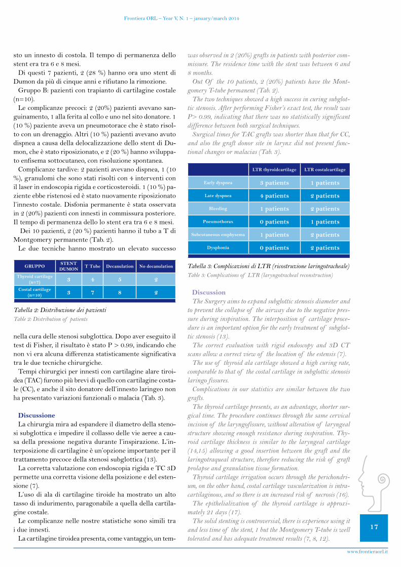
%),granulomichesonostatirisolticon4interventiconillaserinendoscopiarigidaecorticosteroidi.1(10%)pa-ziente ebbe ristenosi ed è stato nuovamente riposizionato l’innesto costale. Disfonia permanente è stata osservata in2(20%)pazienticoninnestiincommissuraposteriore.Il tempo di permanenza dello lo stent era tra 6 e 8 mesi.Dei10pazienti,2(20%)pazientihannoiltuboaTdi
Montgomerypermanente(Tab.2).Le due tecniche hanno mostrato un elevato successo
nella cura delle stenosi subglottica. Dopo aver eseguito il test di Fisher, il risultato è stato P > 0.99, indicando che non vi era alcuna differenza statisticamente significativa tra le due tecniche chirurgiche.
Tempi chirurgici per innesti con cartilagine alare tiroi-dea(TAC)furonopiùbrevidiquelloconcartilaginecosta-le(CC),eancheilsitodonatoredell’innestolaringeononhapresentatovariazionifunzionaliomalacia(Tab.3).
DiscussioneLa chirurgia mira ad espandere il diametro della steno-
si subglottica e impedire il collasso delle vie aeree a cau-sa della pressione negativa durante l’inspirazione. L’in-terposizione di cartilagine è un’opzione importante per il trattamentoprecocedellastenosisubglottica(13).
La corretta valutazione con endoscopia rigida e TC 3D permette una corretta visione della posizione e del esten-sione(7).
L’uso di ala di cartilagine tiroide ha mostrato un alto tassodiindurimento,paragonabileaquelladellacartila-gine costale.
Le complicanze nelle nostre statistiche sono simili tra i due innesti.
La cartilagine tiroidea presenta, come vantaggio, un tem-
was observed in 2 (20%) grafts in patients with posterior com-missure. The residence time with the stent was between 6 and 8 months.
Out Of the 10 patients, 2 (20%) patients have the Mont-gomery T-tube permanent (Tab. 2).
The two techniques showed a high success in curing subglot-tic stenosis. After performing Fisher’s exact test, the result was P> 0.99, indicating that there was no statistically significant difference between both surgical techniques.
Surgical times for TAC grafts was shorter than that for CC, and also the graft donor site in larynx did not present func-tional changes or malacias (Tab. 3).
DiscussionThe Surgery aims to expand subglottic stenosis diameter and
to prevent the collapse of the airway due to the negative pres-sure during inspiration. The interposition of cartilage proce-dure is an important option for the early treatment of subglot-tic stenosis (13).
The correct evaluation with rigid endoscopy and 3D CT scans allow a correct view of the location of the estensis (7).
The use of thyroid ala cartilage showed a high curing rate, comparable to that of the costal cartilage in subglottic stenosis laringo fissures.
Complications in our statistics are similar between the two grafts.
The thyroid cartilage presents, as an advantage, shorter sur-gical time. The procedure continues through the same cervical incision of the laryngofissure, without alteration of laryngeal structure showing enough resistance during inspiration. Thy-roid cartilage thickness is similar to the laryngeal cartilage (14,15) allowing a good insertion between the graft and the laringotraqueal structure, therefore reducing the risk of graft prolapse and granulation tissue formation.
Thyroid cartilage irrigation occurs through the perichondri-um, on the other hand, costal cartilage vascularization is intra-cartilaginous, and so there is an increased risk of necrosis (16).
The epithelialization of the thyroid cartilage is approxi-mately 21 days (17).
The solid stenting is controversial, there is experience using it and less time of the stent, 1 but the Montgomery T-tube is well tolerated and has adequate treatment results (7, 8, 12).
GRUPPOSTENT
DUMONT Tube Decanulation No decanulation
Thyroid cartilage(n=7) 3 4 5 2
Costal cartilage(n=10) 3 7 8 2
Tabella 2: Distribuzione dei pazientiTable 2: Distribution of patients
LTR thyroidcartilage LTR costalcartilage
Early dyspnea 3 patients 1 patients
Late dyspnea 4 patients 2 patients
Bleeding 1 patients 2 patients
Pneumothorax 0 patients 1 patients
Subcutaneous emphysema 1 patients 2 patients
Dysphonia 0 patients 2 patients
Tabella 3: Complicazioni di LTR (ricostruzione laringotracheale)Table 3: Complications of LTR (laryngotracheal reconstruction)
18
Frontiera ORL – Anno V, N. 1 – gennaio/marzo 2014
www.frontieraorl.it
po chirurgico più breve. La procedura prosegue attraver-so la stessa incisione cervicale della laringofissura, senza alterazione della struttura della laringe mostrando abba-stanza resistenza durante l’inspirazione. Lo spessore della cartilaginetiroideaèsimileallacartilagelaringea(14,15)consentendo una buon inserimento tra l’innesto e la strut-turalaringotracheale,riducendoquindiilrischiodiprolas-so dell’innesto e formazione di tessuto di granulazione.
L’irrorazione della cartilagine tiroidea avviene attra-verso il pericondrio, invece, la vascolarizzazione della cartilaginecostaleè intra-cartilaginea,equindivièunaumentatorischiodinecrosi(16).
La riepitelizzazione della cartilagine tiroidea è di circa 21giorni(17).
L’uso di stent solido è controverso, vi è esperienza con essoedamenotempo,mailtuboaTdiMontgomeryèbentolleratoeharisultatiadeguatialtrattamento(7,8,12).
Noi preferiamo stent Dumon o tubi a T di Montgome-ryallostentsolido,permettendolafonazionealpazientee la respirazione per bocca.
La disfonia nella nostra casistica era presente solo come un sintomo che infastidiva il paziente in innesti messi in commissura posteriore. Ci sono studi che mostrano di-sfonianel44-55%deicasi(4).
L’interposizione di cartilagine in una laringofissura successivamente è stato raccomandato da Maddaus et al. (11)quandolastenosiprovocaunacicatricemucosain-tra - aritenoidea.
L’uso di osso ioide e cartilagine auricolare presenta un alto tassodi successoperquanto riguarda l’espansionesottoglottidea,quindièun’alternativaalgrafting(5,6).
La decannulazione fu simile in entrambi i gruppi, solo 2 pazienti, uno per ciascun gruppo furono lasciati con una protesi definitiva.
In conclusione, l’uso di ala di cartilagine tiroidea è sicuro come l’innesto costale. Presenta ottima risposta alla pres-sione negativa in ispirazione, mostra un tempo chirurgi-co più breve dell’ innesto costale. Nessuna deformazione è stata osservata nella laringe, e nessuna differenza signifi-cativa tra innesto costale e innesto di cartilagine tiroidea.
We prefer Dumon stent or Montgomery T tubes for solid stent, allowing the patient phonation and mouth respiration.
Dysphonia in our series was only present as a symptom that bothered the patient in those grafts placed in the posterior com-missure. There are studies that show dysphonia in 44-55% (4).
The interposition of cartilage at a later laryngofissure was recommended by Maddaus et al.(11) when the stenosis causes a mucosa intra-arytenoids scar.
The use of Hyoid bone and auricular cartilage shows a high success rate regarding subglottis expansion therefore it is an al-ternative to grafting (5, 6).
The decannulation was similar in both groups, only 2 pa-tients, one from each group left with a definitive prosthesis.
In conclusion, the use of thyroid ala cartilage is as safe as the costal graft. It presents excellent response to inspiration negative pressure, showing a shorter surgical time than that of costal graft. No deformation was observed in the larynx, and no significant differences between costal graft and thyroid cartilage graft.
Bibliografia - References1- Ricardo Mingarini Terra, MD, He´lio Minamoto, MD, Felipe
Carneiro, MD, Paulo Manuel Pego–Fernandes, MD, and Fa´bio Bi-scegliJatene,MD.Laryngealsplitandribcartilageinterpositionalgrafting: Treatment option for glottic/subglottic stenosis in adults. TheJournalof ThoracicandCardiovascularSurgerycVolume137,Number 4; 2009; 137:818-23.2- NguyenCV, Bent JP, ShahMB, Parikh SR.Pediatric prima-
ry anterior laryngotracheoplasty: Thyroid ala vs costal cartilagegrafts.ArchOtolaryngolHeadNeckSurg.2010Feb;136(2):171-4.2009.224.3- P.Fayoux*,F.Vachin,O.Merrot,N.Bernheim.Thyroidalar
cartilagegraftinpaediatriclaryngotrachealreconstruction.IntJPe-diatrOtorhinolaryngol.2006Apr;70(4):717-24.4- Younis RT, Lazar RH. Laryngotracheal reconstruction wi-
thoutstenting.Otolaryngol.HeadNeckSurg.1997;116:358-62.
5- McGuirtWFJr,LittleJP,HealyGB.Anteriorcricoidsplit.Use of hyoid as autologous graftingmaterial.ArchOtolaryngolHeadNeckSurg.1997Dec;123(12):1277-80.
6- Lusk RP, Kang DR, Muntz HR. Auricular cartilage grafts in laryngotracheal reconstruction.AnnOtolRhinolLaryngol. 1993Apr;102(4Pt1):247-54.7- GaissertHA,LofgrenRH,GrilloH.Upperairwaycompromi-
seafterinha¬lationinjury.AnnSurg1993;218:672–678.8- EliacharI,MosconaR,JoachimHZ,HirschowitzB,ShiloR.
Themanage¬mentof laryngotrachealstenosisinburnedpatients.Plastic Reconstr Surg 1981; 68:11–16.9- RethiA.Unenouvelle techniquechirurgicalepour le traite-
mentdelafixationbilatéraledescordesvocales.LaryngolRhinolOtol(Stuttg)1955;34:464–472.10-CottonR.Managementof subglotticstenosisininfancyand
childhood:reviewof aconsecutiveseriesof casesmanagedbysurgi-calreconstruc¬tion.AnnOtolRhinolLaryngol1978;87:649–657.11-MaddausMA,TothJL,GullanePJ,PearsonFG.Subglottic
tracheal resec¬tion and synchronous laryngeal reconstruction. JThorac Cardiovasc Surg 1992; 104:1443–1450.12-ZaimaA,BitohY,MoritaK,TsugawaJ,IshiiT,SatohS,Nishijima
E.Long-termT-tubestentingasdefinitivetreatmentof severeacquiredsubglotticstenosisinchildren.JPediatrSurg.2010May;45(5):996-913-ChoJK,KoMH,RyuJ,JeongHS.Anteriorcricoidsplitpro-
cedurewithtranspositionof cricoidcartilagesegment.JLaryngolOtol.2009Sep;123(9):1045-8.14-A.L.DeJong,A.H.Park,E.Raveh,M.R.Schwartz,V.Forte,
Comparisonof thyroid,auricular,andcostaldonorsitesforlaryn-gotrachealreconstructioninananimalmodel,Arch.Otolaryngol.HeadNeckSurg.126(2000)49—53.15-A.H.Park,V.Forte,Effectof harvestingautogenouslarynge-
alcartilageforlaryngotrachealreconstructiononlaryngealgrowthandsupport,Laryngoscope109(1999)307—311.16-P.R.Wheater,H.G.Burkitt,V.G.Daniels,Wheater’sFunctio-
nalHistology,thirded.,ChurchillLivingstone,London,1993.17-I.N.Jacobs,P.Podrebarac,S.D.Boden,M.Chen,Grafthealing
in laryngotracheal reconstruction: an experimental rabbitmodel,Ann.Otol.Rhinol.Laryngol.108(1999)599—608.
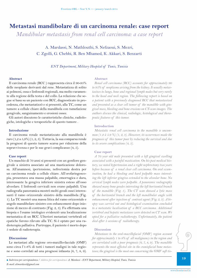
19
FrontieraORL–YearV,N.1–january/march2014
www.frontieraorl.it
AbstractIlcarcinomarenale(RCC)rappresentacircail90-95%
delle neoplasie derivanti dal rene. Metastatizza di solito ai polmoni, ossa e linfonodi regionali, ma molto raramen-te alla regione della testa e del collo. La relazione che se-gue si basa su un paziente con RCC, diagnosticato in pre-cedenza,chemetastatizzòesipresentò,allaTC,comeuntumore a cellule chiare della mandibola con tumefazione gengivale, sanguinamento e erosioni ossee.
Gli autori discutono le caratteristiche cliniche, radiolo-giche,istologicheeterapeutichediquestotumore.
IntroduzioneIl carcinoma renale metastatizzato alla mandibola è
raro(1,2a5,9%)(1,2,3).Tuttavia,lasuacomparsarendelaprognosidiquestotumorescarsaperriduzionedellasopravvivenzaeperlesuegravicomplicanze(4,5).
Case reportUnmaschiodi70annisipresentòconungonfioregen-
givale a sinistra associato ad una masticazione doloro-sa. All’anamnesi, ipertensione, nefrectomia destra per un carcinoma renale a cellule chiare. All’orofaringosco-pia, presentava una massa palpabile, emorragica e dura, interessante la gengiva inferiore sinistra esteso all’osso alveolare. I linfonodi cervicali non erano palpabili. Una radiografiapanoramicamostròmoltigeodiosseiinteres-santi il ramoorizzontale sinistrodellamandibola (Fig.1).LaTCmostròunamassaliticadelramoorizzontaleeangolo mandibolare sinistro con enhancement dopo inie-zionedimezzodicontrasto(Fig.2,3).Fueffettuataunabiopsiael’esameistologicoevidenziòunalocalizzazionemetastatica di un RCC. Ulteriori metastasi vertebrali ed epatiche furono rilevate alla TC. Si è optato per una ra-dioterapia palliativa. Purtroppo, il paziente è morto dopo 5 sedute di radioterapia.
Discussione Lemetastasi alla regione oro-maxillo-facciale (OMF)
sonocircal’1-8%dituttiitumorimaligniintaleregio-neesonocorrelatiadunaprognosiinfausta(6,7,8,9).
Metastasi mandibolare di un carcinoma renale: case reportMandibular metastasis from renal cell carcinoma: a case report
A. Mardassi, N. Mathlouthi, S. Nefzaoui, S. Mezri,C. Zgolli, G. Chebbi, R. Ben Mhamed, K. Akkari, S. Benzarti
ENT Department, Military Hospital of Tunis, Tunisia
AbstractRenal cell carcinoma (RCC) accounts for approximately 90
to 95% of neoplasms arising from the kidney. It usually metas-tasizes to lungs, bone and regional lymph nodes but very rarely to the head and neck region. The following report is based on a patient with a previously diagnosed RCC that metastasized and presented as a clear cell tumor of the mandible with gin-gival mass, bleeding and bone erosions on CT-scan images. The authors discuss the clinical, radiologic, histological and thera-peutic features of this tumor.
IntroductionMetastatic renal cell carcinoma to the mandible is uncom-
mon (1.2 à 5.9 %) (1, 2, 3). However, its occurrences made the prognosis of this tumor poor by reducing the survival and due to its severe complications (4, 5).
Case reportA 70 year old male presented with a left gingival swelling
associated with a painful mastication. On his past medical his-tory, we noted hypertension and a right nephrectomy two years before because of a renal clear cell carcinoma. On oral exam-ination, he had a bleeding and hard palpable mass interest-ing the left inferior gingiva extended to the alveolar bone. No cervical lymph nodes were palpable. A panoramic radiography showed many bone geodes interesting the left horizontal branch of the mandible (Fig. 1). The CT scan showed a lytic mass of the horizontal branch and the left mandibular angle with enhancement after injection of contrast agent (Fig. 2, 3). A bi-opsy was carried out and histological examination concluded to a metastatic localisation of a RCC carcinoma. Additional vertebral and hepatic metastases were detected on CT scan. We opted for a palliative radiotherapy. Unfortunately, the patient died after 5 sessions of radiotherapy.
DiscussionMetastases to the oral-maxillofacial (OMF) region account
for approximately 1 to 8% of all malignancy in the region and are correlated with a poor prognosis (6, 7, 8, 9). The mandible represents the most affected site in the craniofacial bone metas-tases; the gingival is the most one concerning the OMF soft tis-
■ Indirizzo per corrispondenza / Address for corrispondence: A. Mardassi - ENT Department, Military Hospital, Tunis, Tunisia
E-mail: [email protected]
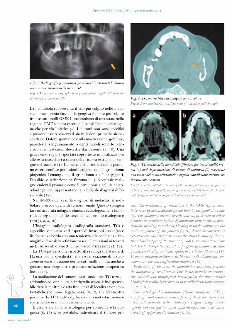
20
Frontiera ORL – Anno V, N. 1 – gennaio/marzo 2014
www.frontieraorl.it
La mandibola rappresenta il sito più colpito nelle meta-stasi ossee cranio facciali; la gengiva è il sito più colpito fra i tessuti molli OMF. Il meccanismo di metastasi nella regione OMF sembra essere più per diffusione ematoge-nachepervia linfatica(5).Isintominonsonospecificie possono essere osservati sia in lesioni primarie sia se-condarie. Dolore spontaneo o alla masticazione, gonfiore, parestesia, sanguinamento o denti mobili sono le prin-cipalimanifestazioni descritte dai pazienti (3, 10).Unagrave emorragia è riportata soprattutto in localizzazioni alle ossa mascellari a causa della riserva estrema di san-guedeltumore(1).Lemetastasiaitessutimolliposso-no essere confuse per lesioni benigne come il granuloma piogenico, l’emangioma, il granuloma a cellule giganti, l’epulide, o irritazioneda fibroma (11).Neoplasiemali-gne endorali primarie come il carcinoma a cellule chiare odontogenico rappresentano la principale diagnosi diffe-renziale(12).Nel 20-33% dei casi, la diagnosi dimetastasimandi-
bolareprecedequelladitumorerenale.Questospingeafare un’accurata indagine clinica e radiologica per i tumo-ri della regione maxillo-facciale il cui profilo istologico è raro(1,3,5,10).L’indagine radiologica (radiografia standard, TC) è
aspecifica emostravari aspetti di invasioni ossee (areelitiche senza bordo con una tendenza alla confluenza, im-magini diffuse di rarefazione ossea ...), invasioni ai tessuti molliadiacentioaspettidiipervascolarizzazione(1,13).
La TC è più sensibile rispetto alla radiografia standard. Ha una buona specificità nella visualizzazione di distru-zione ossea e invasione dei tessuti molli e aiuta anche a guidare una biopsia o a praticare un’azione terapeutica locale(14).
La stadiazione del tumore, praticando una TC toraco-addomino-pelvica e una scintigrafia ossea, è indispensa-biledatalamultiplaealtafrequenzadilocalizzazionime-tastatiche(polmone,fegato,ossa)(8,12,15).Nelnostropaziente,laTCtotal-bodyharivelatometastasiosseeeepatiche che erano clinicamente latenti.
È essenziale l’analisi istologica per confermare la dia-gnosi (8, 16) e, se possibile, individuare il tumore pri-
sues. The mechanism of metastases to the OMF region seems to be more by hematogenous spread than by the lymphatic route (5). The symptoms are not specific and might be seen in either primary or secondary lesions. Spontaneous pain or due to mas-tication, swelling, paresthesia, bleeding or tooth mobility are the main complaints of the patients (3, 10). Severe hemorrhage is reported especially in jaw bones localizations because of the ex-treme blood supply of the tumor (1). Soft tissues metastases may be taken for benign lesions such as pyogenic granuloma, heman-gioma, giant cell granuloma, epulis, or irritation fibroma (11). Primary intraoral malignancies like clear cell odontogenic car-cinoma are the main differential diagnosis (12).
In 20-33% of the cases, the mandibular metastasis precedes the diagnosis of renal tumor. This incites to make an exhaus-tive clinical and radiological investigation for tumor whose histological profile is uncommon to maxillofacial tumor region (1, 3, 5, 10).
The radiological examination (X-ray standards, CT) is nonspecific and shows various aspects of bone invasions (lytic areas without border with a tendency to confluence, diffuse im-ages of bone rarefaction ...) or adjacent soft tissue invasions or aspects of hypervascularization (1, 13).
Fig. 1: Radiografia panoramica: geodi ossei interessanti la branca orizzontale sinistra della mandibolaFig. 1: Panoramic radiography: bone geodes interesting the left horizon-tal branch of the mandible Fig. 2: TC, massa litica dell’angolo mandibolare
Fig. 2: Bone window Ct-scan, lytic mass of the left mandible angle
Fig. 3: TC assiale della mandibola (finestra per tessuti molli) pri-ma (a) and dopo iniezione di mezzo di contrasto (b) mostrante una massa del ramo orizzontale e angolo mandibolare sinistro con intenso enhancementFig. 3: Axial mandibular CT scan (soft window) before (a) and after in-jection of contrast agent (b) showing a mass of the left horizontal branch and the left mandibular angle with intensive enhancement
21
FrontieraORL–YearV,N.1–january/march2014
www.frontieraorl.it
marioperguidarelastrategiaterapeutica(14).Iltipicoaspettodeitumoriacellulechiareèquellodiunostratodi cellule rotonde o poligonali con abbondante citopla-sma ricco di colesterolo, esteri di colesterolo, fosfolipi-di e glicogeno. Lo studio immunoistochimico rivela un altotassodicheratinaevimentina(2,3,7).Generalmen-te non esiste espressione di proteine di CK7 e CK20. Tut-tavia,l’80%deitumoriesprimonol’antigenemembranaEMA (antigene epiteliale dimembrana) (7).Quando iltumore primario non è ancora noto, la difficoltà consiste nelfattochequestecellulechiarepossonocaratterizzarealtritipiditumori(ghiandolesalivari,tiroide,mammella,polmoneecolon)(2).La gravità della prognosi di queste neoplasie, nono-
stante tutte le possibili terapie, spinge, spesso, gli auto-ri a favore di un trattamento palliativo per migliorare la qualitàdellavitadeipazienti(3,4,5,7,10).LaChirurgiaradicale è proposta per metastasi isolate della mandibola odelcavooralealfinedimigliorarelaqualitàdivitaeottimizzareiltassodisopravvivenza(2,4,5).
L’embolizzazione del peduncolo vascolare, prima della escissione chirurgica, appare come una tecnica interes-santeseètecnicamentepossibile(13).Altreterapiesonostate proposte, come i bifosfonati o, recentemente, farma-ci antiangiogenici ma il risultato non sembra essere mi-gliore(15,16).
La chemioterapia non è molto efficace nel cancro del rene perché è un tumore chemioresistente a causa dell’esi-stenza di un glicoproteina Pcodificata dal gene MDR 1. Le uniche molecole che sembrano essere parzialmente efficaci sono vinblastina e floxuridina (7). L’impatto diquestachemioterapiainterminidisopravvivenzarima-ne modesta.
È chiaro che l’obiettivo principale delle attuali terapie rimaneilcontrollolocaledeisintomi(dolore,sanguina-mento,disturbidellamasticazione)permigliorarelaqua-litàdivitadeipazienti(17).
Le metastasi renali hanno una prognosi scarsa con un tassodisopravvivenzaa5annicheraggiungeil7-13%.Ilsettanta per cento dei pazienti muore entro un anno dalla diagnosi suggerendo che la metastasi mandibolare si ac-compagna a significativa diffusione del tumore primario conmetastasimultiple(2.3,10).Diversifattoriprogno-stici influenzano la sopravvivenza del paziente: età, salute generale, perdita di peso, il numero di siti metastatici e la scarsatolleranzaaltrattamento(7).
ConclusioneQuesto caso clinico illustra l’importanza di sospettare
una lesione metastatica in un rigonfiamento mandibolare ogengivale.Ilcarcinomaacellulerenalipuòraramenteessere il tumore originale. La sua diagnosi richiede una stadiazione accurata e una terapia adeguata per miglio-rarelaqualitàdivitadiquestipazienti,ancheselapro-gnosi rimane scadente, soprattutto con metastasi sincro-ne multiple.
CT-scan is more sensitive than standard X-ray. It has a good specificity in the visualization of bone destruction and soft tis-sue invasion and helps also to guide a biopsy or to undergo a local therapeutic action (14).
The staging of the tumor including a thoraco-abdominal-pelvic CT-scan and a bone scintigraphy is imperative given the multiple and high frequency of metastatic localizations (lung, liver, bone) (8, 12, 15). In our patient, the body scan revealed bone and liver metastases that were clinically latent.
Histological analysis is essential to confirm the diagnosis (8, 16) and if possible, to identify the primary tumor in order to guide the therapeutic strategy (14). The typical appearance of clear cell tumors is that of a sheet of round or polygonal cells with abundant cytoplasm rich in cholesterol, cholesterol esters, phospholipids and glycogen. Immunohistochemical study reveal a high rate of keratin and vimentin (2, 3, 7). There is general-ly no protein expression of CK7 and CK20. However, 80% of the tumors express membrane antigen EMA (epithelial mem-brane antigen) (7). When the primary tumor is not yet known, the difficulty lies in the fact that these clear cells can character-ize other types of tumors (salivary glands, thyroid, breast, lung and colon) (2).
The poverty of the prognosis of these neoplasms despite all possible therapies pushes, often, the authors to advocate a pal-liative treatment to improve the quality of life of patients (3, 4, 5, 7, 10). Radical surgery is proposed for isolated metastases of the mandible or of the oral cavity in order to improve the quality of life and optimize the survival rate (2, 4, 5).
The embolization of vascular pedicle, before surgical exci-sion, appears as an attractive technique if it is technically pos-sible (13). Other therapies have been proposed, such as bisphos-phonates or, recently, antiangiogenic drugs but the result does not seem to be better (15, 16).
Chemotherapy is not very effective in cancer kidney because it is a chemoresistant tumor due to the existence of a P-glycoprotein encoded by the MDR 1 gene. The only molecules that seem to be partially effective are vinblastine and floxuridine (7). The im-pact of this chemotherapy in terms of survival remains modest.
It is clear that the main objective of actual therapeutics remain the local control of the symptoms (pain, bleeding, disorders of mastication) to improve the quality of life of the patients (17).
Metastatic kidney has a poor prognosis with a 5-year surviv-al rate reaching 7 to 13%. Seventy percent of patients die with-in a year of diagnosis suggesting that mandibular metastasis involves significant dissemination of the primary tumor with multiple metastases (2, 3, 10). Several prognostic factors influ-ence patient survival: age, general health, weight loss, number of metastatic sites and poor tolerance to treatment (7).
ConclusionThis clinical case illustrates the importance to suspect a meta-
static lesion of a mandibular or gingival swelling. Renal cell carcinoma may rarely be the original tumor. Its diagnosis re-quires an exhaustive staging and an adequate therapy to im-prove the quality of life of these patients even if the prognosis remains poor especially with synchronous multiple metastasis.
22
Frontiera ORL – Anno V, N. 1 – gennaio/marzo 2014
www.frontieraorl.it
Bibliografia - References1) ShenM.L.,KangJ.,WenY.L.,YingW.M.,YiJ.,HuaC.G.
et al. Metastatic Tumors to the Oral and Maxillofacial Region: aRetrospectiveStudyof 19CasesinWestChinaandReviewof theChineseandEnglishLiterature.JOralMaxillofacSurg2009; 67:718-37.
2) Makos C., Psomaderis K. A Literature Review in Renal Carcinoma Metastasis to the Oral Mucosa and a New Report of anEpulis-LikeMetastasis.JOralMaxillofacSurg2009;67:653-60. 3) SastreJ.,NavalL.,MunozM.,GamalloM.,DiazF.J.Me-
tastaticrenalcellcarcinomatothemandible.OtolaryngolHeadNeckSurg 2005; 132:663-4.4) Siriwardenaa B.S., Tilakaratnea W.M., Rajapaksa R.M.
Metastatictransitionalcellcarcinomaof theurinarybladdertothemandible.OralOncologyEXTRA2005;41:22-4.
5) Hirshberg A., Shnaiderman-Shapiro A., Kaplan I., Ber-gerR.Metastatictumourstotheoralcavity:pathogenesisandanalysisof 673cases.OralOncology2008;44:743-52.6) MéjeanA.,LebretT.Sitesmétastatiquesatypiquesducan-
cer du rein. Progrès en Urologie 2008; 7:320-6. 7) Vidart A., Fehri K., Pfister C. Métastasesinhabituelles du
cancer du rein. Annales d’urologie 2006; 40:211-9.8) Déquanter D., Lothaire P., Andry G., Gil T. Métastase
d’un carcinome de prostate mimant une tumeurparotidienne : à propos d’un cas. Rev StomatolChirMaxillofac 2004; 105:182-4. 9) LebretT.,MéjeanA.Lessitesmétastatiquesatypiquesdes
cancers de la prostate. Progrès en Urologie2008 ; 7:357-64.
10)JayasooriyaaP.R.,GunarathnaaI.A.,AttygallabA.M.,Ti-lakaratneW.M.Metastaticrenalcellcarcinomapresentingasaclearcelltumourintheheadandneckregion.OralOncologyEXTRA2004;40:50-3.11)KnottenbeltD.C.,KellyD.F.Oralanddentaltumors.Equi-
neDentistry(thirdedition)2011:149-181.12)CheraB.S.,VillaretD.B.,OrlandoC.A.,MendenhallW.M.
Clear cell odontogenic carcinoma of the maxilla: a case report and literature review. American Journal of Otolaryngology-HeadandNeckMedicineandSurgery2008;29:284-90.13)Guyot L., Sauvant J., Menasse F., Garcia S., Portier F.,
GolaR.Métastasemandibulairehémorragiqued’origineréna-le:intérêtdel’embolisationthérapeutique.LaPresseMédicale1999; 20:1066. 14)Tubiana-HulinM.,DeMaulmontC.,Guinebretière J.M.
Stratégie de prise en charge des métastasesosseusesrévélatrices. Revue du Rhumatisme 2008; 75:332-8.
15) Brunello A., Saia G., Bedogni A., Scaglione D., Basso U. Worseningof osteonecrosisof thejawduringtreatmentwithsunitinib in a patient with metastatic renal cell carcinoma. Bone 2009; 44:173-5. 16)BocciG.,LoupakisF.Thepossibleroleof chemotherapy
in antiangiogenic drug resistance.MedicalHypotheses 2012;78:646-8. 17)VédrineL.,ChargariC.,LeMoulecS.,FayolleM.,Ceccal-
di B., Bauduceau O. Chimiothérapie des cancers des voiesaéro-digestivessupérieures. CancerRadiothérapie 2008; 12 :110-9.
Advanced Bionics
The world’s first and only swimmable sound processor
AdvancedBionics.com*Pending regulatory approval MK_NEPT2_EN_Ad_11_A
23
FrontieraORL–YearV,N.1–january/march2014
www.frontieraorl.it
Advanced Bionics
The world’s first and only swimmable sound processor
AdvancedBionics.com*Pending regulatory approval MK_NEPT2_EN_Ad_11_A
AbstractIl laringocele è una rara dilatazione benigna, ripiena
d’aria, del sacculo laringeo. Simanifesta più frequente-mente quando il laringe è esposto a continuo aumentodellapressioneinterna:èpresente,piùfrequentemente,insoffiatori del vetro, suonatori di strumenti a fiato, soggetti affetti da tosse cronica. Gli Autori riportano il caso di un paziente affetto da laringocele misto formatosi in assenza di fattori predisponenti e ne discutono la presentazione clinica, le indagini praticate e la gestione chirurgica.
IntroduzioneIl ventricolo laringeo di Morgagni è una piccola rien-
tranza che si trova tra le corde vocali vere e false. Il tratto antero-superiore della rientranza termina in un sacchetto cieco, che si chiama appendice del ventricolo di Morgagni o sacculo. Un laringocele è una anomala dilatazione sac-ciforme dell’appendice del ventricolo laringeo di Morga-gni. Ha contenuto aereo ed è ricoperto da epitelio ciliato colonnare pseudo-stratificato. Comunica con il ventricolo medianteunostrettopeduncoloepuòessereditipoin-terno, esterno o misto, a seconda del tipo di rapporto che contraeconlamembranetiroioidea(1).Accrescendosisiestende in direzione mediale e superiore alla cartilagine tiroidea fino a raggiungere la membrana tiroioidea. Se non fora la membrana e rimane all’interno della laringe si definisce interno. Quando la perfora e sporge attraverso la membrana tiroioidea nel collo diventa un laringocele esterno. Un laringocele, sia mediale sia laterale alla mem-brana tiroioidea, si chiama misto. È solitamente unilate-rale, ma è stato riportato in letteratura sia il laringocele internobilateralesiaillaringoceleesternobilaterale(2).La suaeziopatogenesi è ignota.Sipresenta,però,più
frequentemente,insoggettiespostiauncontinuoaumen-to della pressione endolaringea. Infatti le categorie più colpite sono soffiatori del vetro, suonatori di strumenti a fiato,soggettiaffettidatossecronica.Avolte,puòsvilup-parsiinassenzadifattoripredisponenti(3).
L’esatta incidenza del laringocele non è nota; colpisce più frequentemente il sesso maschile (5-7:1). Nell’85%deicasièunilaterale;il30%èesterno,il20%internoeil50%misto(4).
Case report: Laringocele gigante mistoCase report: giant mixed laryngocele
O. Marotta, F. Catapano, M. Cocchiarella, C. Crisci, C. Di Meo
U.O.C. di Otorinolaringoiatria, Azienda Ospedaliera di Rilievo Nazionale e di Alta Specializzazione
S. Anna e S. Sebastiano”, Caserta
AbstractThe laryngocele is a rare benign dilatation of the laryngeal
saccule. It occurs when the larynx is exposed to a continuous increase of the internal pressure; for this reason it is mostly present in glassblowers, players of wind instruments, patients with chronic cough. The Authors report the case of a patient with mixed laringocele formed in the absence of predisposing factors and discuss the clinical presentation, investigations and surgical management.
Introduction The laryngeal ventricle of Morgagni is a small recess locat-
ed between the true and false vocal cords. The anterior superior portion of the recess ends in a blind pouch, which is called the appendix of the ventricle of Morgagni or saccule. A laryn-gocele is an abnormal saccular dilatation of the appendix of the laryngeal ventricle of Morgagni. It contains air and is lined with pseudo- stratified ciliated columnar epithelium. It communicates with the ventricle by a narrow stalk and can be of three type: internal, external or mixed, according to the type of relationship with the thyrohyoid membrane (1). When the laryngocele increases in size, it extends medial and superior to the thyroid cartilage till it reaches the thyrohyoid membrane. If it does not pierce the membrane and remains inside the lar-ynx is called internal. When it pierces the membrane and pro-trudes in the neck becomes an external laryngocele (2).
Its etiology is unknown. However, it appears more frequently in subjects exposed to a continuous increase in intralaryngeal pressure, such as that caused by chronic cough, glass blowing, playing wind instruments. It sometimes can develops in the ab-sence of predisposing factors (3).
The exact incidence is unknown; males are more affected than women (5-7:1). Laryngocele is unilateral in 85% of cases, ex-ternal in 30%, internal in 20% and mixed in 50% (4).
It may be asymptomatic or, when increases in size, present with cough, dysphonia (hoarseness, stridor), dyspnea, sore throat, and swelling of the neck in external type (5).
Case reportWe report the case of a patient, a 51 year old male, M.U.,
hospitalized for significant dyspnea and dysphagia, who un-derwent an urgent tracheostomy.
■ Indirizzo per corrispondenza / Address for corrispondence: O. Marotta - Azienda Ospedaliera “Sant’Anna e San Sebastiano”, Caserta - U.O.C. di Otorinolaringoiatria -
E-mail: [email protected]
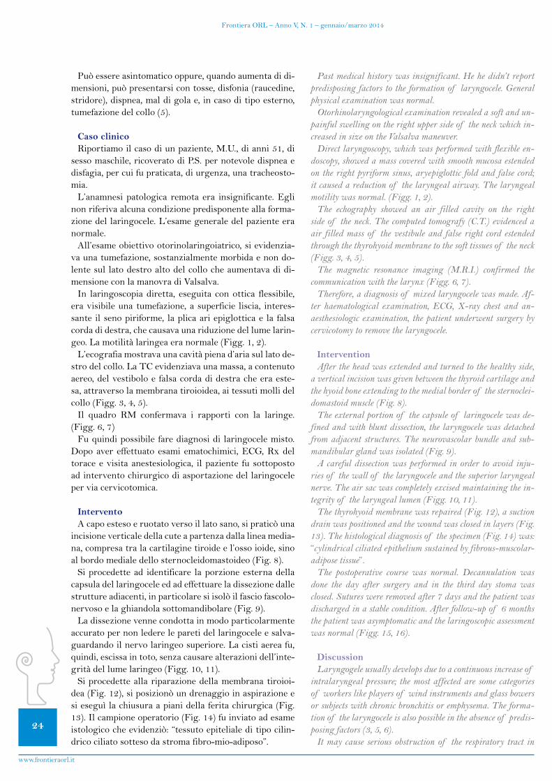
24
Frontiera ORL – Anno V, N. 1 – gennaio/marzo 2014
www.frontieraorl.it
Puòessereasintomaticooppure,quandoaumentadidi-mensioni,puòpresentarsicontosse,disfonia(raucedine,stridore), dispnea, mal di gola e, in caso di tipo esterno, tumefazionedelcollo(5).
Caso clinicoRiportiamo il caso di un paziente, M.U., di anni 51, di
sesso maschile, ricoverato di P.S. per notevole dispnea e disfagia, per cui fu praticata, di urgenza, una tracheosto-mia.
L’anamnesi patologica remota era insignificante. Egli non riferiva alcuna condizione predisponente alla forma-zione del laringocele. L’esame generale del paziente era normale.
All’esame obiettivo otorinolaringoiatrico, si evidenzia-va una tumefazione, sostanzialmente morbida e non do-lente sul lato destro alto del collo che aumentava di di-mensione con la manovra di Valsalva.
In laringoscopia diretta, eseguita con ottica flessibile, era visibile una tumefazione, a superficie liscia, interes-sante il seno piriforme, la plica ari epiglottica e la falsa corda di destra, che causava una riduzione del lume larin-geo.Lamotilitàlaringeaeranormale(Figg.1,2).
L’ecografia mostrava una cavità piena d’aria sul lato de-stro del collo. La TC evidenziava una massa, a contenuto aereo, del vestibolo e falsa corda di destra che era este-sa, attraverso la membrana tiroioidea, ai tessuti molli del collo(Figg.3,4,5).Il quadro RM confermava i rapporti con la laringe.
(Figg.6,7)Fuquindipossibile farediagnosidi laringocelemisto.
Dopo aver effettuato esami ematochimici, ECG, Rx del torace e visita anestesiologica, il paziente fu sottoposto ad intervento chirurgico di asportazione del laringocele per via cervicotomica.
InterventoAcapoestesoeruotatoversoillatosano,sipraticòuna
incisione verticale della cute a partenza dalla linea media-na, compresa tra la cartilagine tiroide e l’osso ioide, sino albordomedialedellosternocleidomastoideo(Fig.8).
Si procedette ad identificare la porzione esterna della capsula del laringocele ed ad effettuare la dissezione dalle struttureadiacenti,inparticolaresiisolòilfasciofascolo-nervosoelaghiandolasottomandibolare(Fig.9).
La dissezione venne condotta in modo particolarmente accurato per non ledere le pareti del laringocele e salva-guardando il nervo laringeo superiore. La cisti aerea fu, quindi,escissaintoto,senzacausarealterazionidell’inte-gritàdellumelaringeo(Figg.10,11).
Si procedette alla riparazione della membrana tiroioi-dea(Fig.12),siposizionòundrenaggioinaspirazioneesieseguìlachiusuraapianidellaferitachirurgica(Fig.13).Ilcampioneoperatorio(Fig.14)fuinviatoadesameistologicocheevidenziò:“tessutoepitelialeditipocilin-drico ciliato sotteso da stroma fibro-mio-adiposo”.
Past medical history was insignificant. He he didn’t report predisposing factors to the formation of laryngocele. General physical examination was normal.
Otorhinolaryngological examination revealed a soft and un-painful swelling on the right upper side of the neck which in-creased in size on the Valsalva maneuver.
Direct laryngoscopy, which was performed with flexible en-doscopy, showed a mass covered with smooth mucosa estended on the right pyriform sinus, aryepiglottic fold and false cord; it caused a reduction of the laryngeal airway. The laryngeal motility was normal. (Figg. 1, 2).
The echography showed an air filled cavity on the right side of the neck. The computed tomografy (C.T.) evidenced a air filled mass of the vestibule and false right cord estended through the thyrohyoid membrane to the soft tissues of the neck (Figg. 3, 4, 5).
The magnetic resonance imaging (M.R.I.) confirmed the communication with the larynx (Figg. 6, 7).
Therefore, a diagnosis of mixed laryngocele was made. Af-ter haematological examination, ECG, X-ray chest and an-aesthesiologic examination, the patient underwent surgery by cervicotomy to remove the laryngocele.
InterventionAfter the head was extended and turned to the healthy side,
a vertical incision was given between the thyroid cartilage and the hyoid bone extending to the medial border of the sternoclei-domastoid muscle (Fig. 8).
The external portion of the capsule of laringocele was de-fined and with blunt dissection, the laryngocele was detached from adjacent structures. The neurovascolar bundle and sub-mandibular gland was isolated (Fig. 9).
A careful dissection was performed in order to avoid inju-ries of the wall of the laryngocele and the superior laryngeal nerve. The air sac was completely excised maintaining the in-tegrity of the laryngeal lumen (Figg. 10, 11).
The thyrohyoid membrane was repaired (Fig. 12), a suction drain was positioned and the wound was closed in layers (Fig. 13). The histological diagnosis of the specimen (Fig. 14) was: “cylindrical ciliated epithelium sustained by fibrous-muscolar-adipose tissue”.
The postoperative course was normal. Decannulation was done the day after surgery and in the third day stoma was closed. Sutures were removed after 7 days and the patient was discharged in a stable condition. After follow-up of 6 months the patient was asymptomatic and the laringoscopic assessment was normal (Figg. 15, 16).
DiscussionLaryngogele usually develops due to a continuous increase of
intralaryngeal pressure; the most affected are some categories of workers like players of wind instruments and glass bowers or subjects with chronic bronchitis or emphysema. The forma-tion of the laryngocele is also possible in the absence of predis-posing factors (3, 5, 6).
It may cause serious obstruction of the respiratory tract in
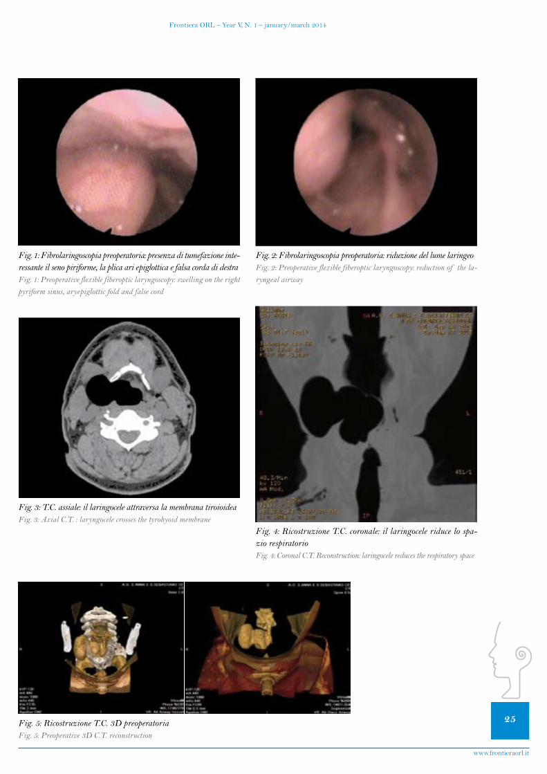
25
FrontieraORL–YearV,N.1–january/march2014
www.frontieraorl.it
Fig. 1: Fibrolaringoscopia preoperatoria: presenza di tumefazione inte-ressante il seno piriforme, la plica ari epiglottica e falsa corda di destraFig. 1: Preoperative flexible fiberoptic laryngoscopy: swelling on the right pyriform sinus, aryepiglottic fold and false cord
Fig. 2: Fibrolaringoscopia preoperatoria: riduzione del lume laringeoFig. 2: Preoperative flexible fiberoptic laryngoscopy: reduction of the la-ryngeal airway
Fig. 3: T.C. assiale: il laringocele attraversa la membrana tiroioideaFig. 3: Axial C.T. : laryngocele crosses the tyrohyoid membrane
Fig. 4: Ricostruzione T.C. coronale: il laringocele riduce lo spa-zio respiratorioFig. 4: Coronal C.T. Reconstruction: laringocele reduces the respiratory space
Fig. 5: Ricostruzione T.C. 3D preoperatoriaFig. 5: Preoperative 3D C.T. reconstruction
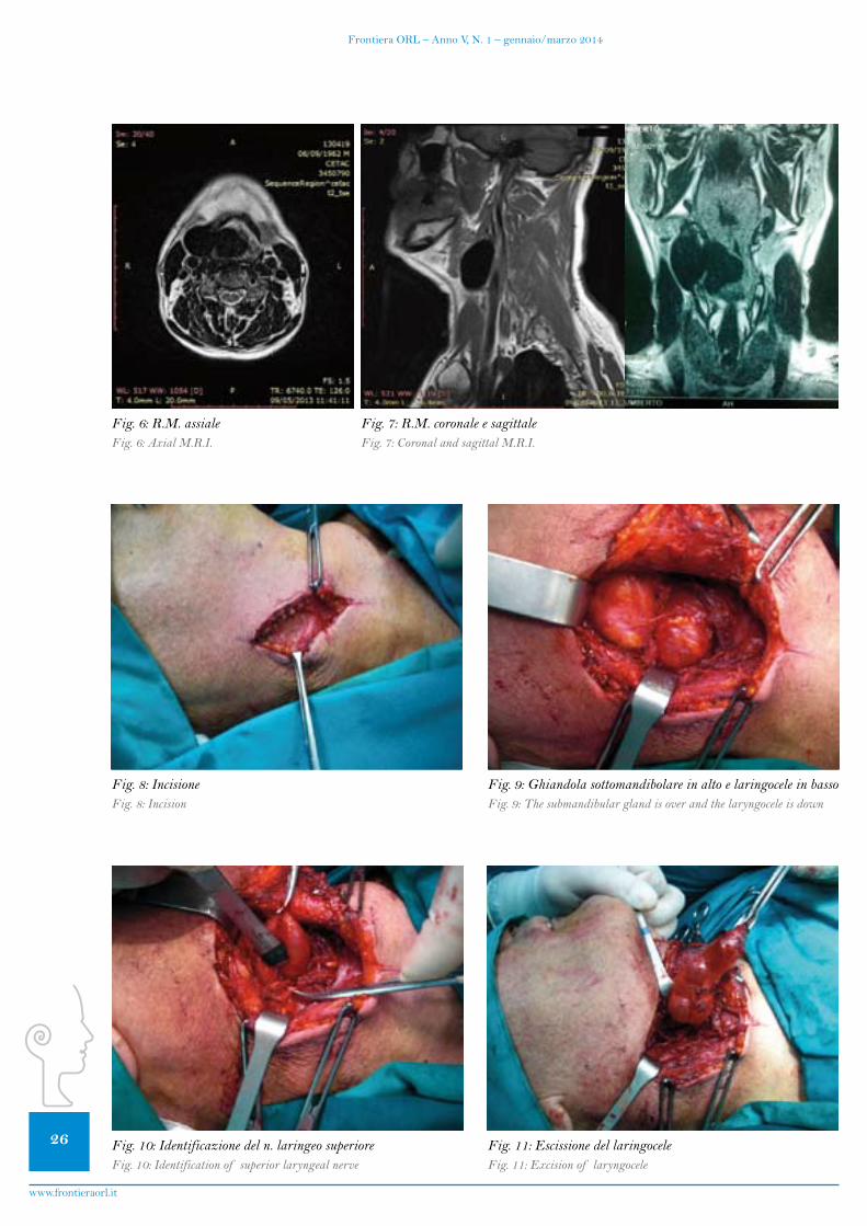
26
Frontiera ORL – Anno V, N. 1 – gennaio/marzo 2014
www.frontieraorl.it
Fig. 6: R.M. assialeFig. 6: Axial M.R.I.
Fig. 7: R.M. coronale e sagittaleFig. 7: Coronal and sagittal M.R.I.
Fig. 8: IncisioneFig. 8: Incision
Fig. 9: Ghiandola sottomandibolare in alto e laringocele in bassoFig. 9: The submandibular gland is over and the laryngocele is down
Fig. 10: Identificazione del n. laringeo superioreFig. 10: Identification of superior laryngeal nerve
Fig. 11: Escissione del laringoceleFig. 11: Excision of laryngocele
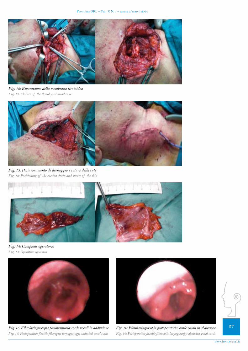
27
FrontieraORL–YearV,N.1–january/march2014
www.frontieraorl.it
Fig. 12: Riparazione della membrana tiroioideaFig. 12: Closure of the thyrohyoid membrane
Fig. 13: Posizionamento di drenaggio e sutura della cuteFig. 13: Positioning of the suction drain and suture of the skin
Fig. 14: Campione operatorioFig. 14: Operative specimen
Fig. 15: Fibrolaringoscopia postoperatoria: corde vocali in adduzioneFig. 15: Postoperative flexible fiberoptic laryngoscopy: adducted vocal cords
Fig. 16: Fibrolaringoscopia postoperatoria: corde vocali in abduzioneFig. 16: Postoperative flexible fiberoptic laryngoscopy: abducted vocal cords
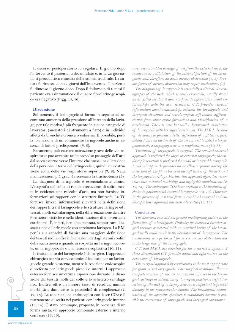
28
Frontiera ORL – Anno V, N. 1 – gennaio/marzo 2014
www.frontieraorl.it
Il decorso postoperatorio fu regolare. Il giorno dopo l’intervento il paziente fu decannulato e, in terza giorna-ta, si procedette a chiusura della stomia tracheale. La su-tura fu rimossa dopo 7 giorni dall’intervento e il paziente fu dimesso il giorno dopo. Dopo il follow-up di 6 mesi il pazienteeraasintomaticoeilquadrofibrolarinogoscopi-coeranegativo(Figg.15,16).
DiscussioneSolitamente, il laringogele si forma in seguito ad un
continuo aumento della pressione all’interno della larin-ge;pertalemotivo,èpiùfrequenteinalcunecategoriedilavoratori(suonatoridistrumentiafiato)oinindividuiaffettidabronchitecronicaoenfisema.Èpossibile,però,la formazione di un voluminoso laringocele anche in as-senzadifattoripredisponenti(5,6).Raramente,puòcausareostruzionegravedelleviere-
spiratorie:puòavvenireunimprovvisopassaggiodell’ariadal sacco esterno verso l’interno che causa una dilatazione dellaporzioneinternadellaringocelee,quindi,unaostru-zione acuta delle vie respiratorie superiori (7, 8).Nellemanifestazionipiùgraviènecessarialatracheotomia(9).
La diagnosi di laringocele è essenzialmente clinica. L’ecografia del collo, di rapida esecuzione, di solito met-te in evidenza una raccolta d’aria, ma non fornisce in-formazioni sui rapporti con le strutture limitrofe. La TC fornisce, invece, informazioni rilevanti nella definizione dei rapporti tra il laringocele e le strutture laringee ed i tessuti molli extralaringei, nella differenziazione da altre formazioni cistiche e nella identificazione di un eventuale carcinoma. È, infatti, ben documentata, seppur rara, l’as-sociazione di laringocele con carcinoma laringeo. La RM, per la sua capacità di fornire una maggiore definizione dei tessuti molli, offre informazioni dettagliate sui confini dellasaccaaereaequandosisospettaunlaringomucoce-le,unlaringopioceleounalesioneneoplastica(10,11).
Il trattamento del laringocele è chirurgico. L’approccio chirurgico per via cervicotomica è indicato per un larion-gocele grande o esterno, mentre la resezione endoscopica è preferita per laringoceli piccoli o interni. L’approccio esterno fornisce un’ottima esposizione durante la disse-zione dai tessuti molli del collo e lo scheletro cartilagi-neo. Inoltre, offre un minore tasso di recidiva, minima morbilità e diminuisce la possibilità di complicanze (2,12, 13). La asportazione endoscopica con laser CO2 è il trattamento di scelta nei pazienti con laringocele interno (13,14).Èstato,comunque,proposto,inpresenzadiunforma mista, un approccio combinato esterno e interno conlaser(13,15).
rare cases: a sudden passage of air from the external sac to the inside causes a dilatation of the internal portion of the larin-gocele and, therefore, an acute airway obstruction (7, 8). Seri-ous forms of airway obstruction may requir tracheotomy (9).
The diagnosis of laryngocele is essentially a clinical. An ech-ography of the neck, which is easily executable, usually shows an air filled sac, but it does not provide information about re-lationships with the near structures. C.T. provides relevant information about relationships between the laryngocele and laryngeal structures and extralaryngeal soft tissues, differen-tiation from other cystic formations and identification of a carcimoma. There is rare, but well - documented, association of laryngocele with laryngeal carcinoma. The M.R.I., because of its ability to provide a better definition of soft tissue, gives detailed data on the limits of the air sac and to detect a laryn-gomucocele, a laryngopyocele or a neoplastic mass (10, 11).
Treatment of laryngocele is surgical. The cervical external approach is preferred for large or external laryngocele; the en-doscopic resection is preferred for small or internal laryngocele. External approach provides an excellent exposure during the dissection of the plane between the soft tissues of the neck and the laryngeal cartilage. Further this approach offers less recur-rence rate, minimal morbidity, and negligible complications (2, 12, 13). The endoscopic CO2 laser excision is the treatment of choice in patients with internal laryngocele (13, 14). However, in the presence of a mixed form, a combined external and en-doscopic laser approach has been advocated (13, 15).
ConclusionThe described case did not present predisposing factors to the
formation of a laringocele. Probably the increased intralaryn-geal pressure associated with an acquired laxity of the laryn-geal walls could result in the development of laryngocele. The tracheotomy was performed for severe airway obstruction due to the large size of the laryngogele.
C.T. and M.R.I. are essential for the a correct diagnosis. A three-dimensional C.T. provides additional information on the expansion of laryngocele.
The surgical approach by cervicotomy is the most appropriate for giant mixed laryngocele. This surgical technique allows a complete excision of the air sac without injuries to the laryn-geal cartilage or alterations of laryngeal function; careful dis-section of the neck of a laryngocele sac is important to prevent damage to the neurovascular bundle. The histological exami-nation of the operative specimen is mandatory because is pos-sible the coexistence of laryngocele and laryngeal carcinoma.
29
FrontieraORL–YearV,N.1–january/march2014
www.frontieraorl.it
ConclusioniNel caso descritto non erano presenti fattori predispo-
nenti alla formazione di un laringocele. È verosimile, quindi,chel’aumentodellapressioneendolaringeaasso-ciatoadunaprobabileacquisitalassitàdelleparetilarin-geepuòavercausatolosviluppodellaringocele.Latra-cheotomia fu necessaria per le notevoli dimensioni della sacca interna, che causava una grave insufficienza respi-ratoria.
Ai fini di una corretta diagnosi, sono fondamentali TC edRM.UnaTC con ricostruzioni tridimensionali puòfornire ulteriori informazioni sulla estensione del larin-gocele negli spazi paraglottici.
L’approccio chirurgico per via cervicotomica risulta es-sere il più indicato in presenza di un voluminoso larin-gocele misto. È stato possibile, infatti, con tale tecnica chirurgica, praticare una escissione completa della cisti aerea senza causare lesioni dello scheletro cartilagineo o alterazioni della funzionalità laringea. È sempre necessa-rio praticare l’esame istologico del tessuto asportato in quantoèpossibilelacoesistenzadilaringoceleecarcino-ma laringeo.
Bibliografia - References1. De Santo L.W. Laryngocele, laryngeal mucocele, large
saccules and large saccular cysts: a developmantal spectrum.Laryngoscope1974;84:1291-6.2. BatemanG.H.Caseof bilateralexternallaryngocele.JLa-
ryngolOtol1957;71:769-72.3. Erdogmus B., Yazici B., Ozturk O., Ataoglu S., Yazici S.
Laryngocele in assocition with ankylosing spondylitis.WienKlinWochenschr2005;117:718-20.4. IngramsD.,HeinD.,MarksN.BatemanGH.Laryngoce-
le:ananatomicalvariant.JLaryngolOtol1999;113:675-6775. LancellaA.,AbbateG.,DosdeganiR.Mixedlaryngocele:
acasereportandreviewof theliterature.ActaOtorhinolaryn-golocica italica 2007; 27:255-57.
6. Mannella V.K., Freni F., Santoro R., Galletti B. Laringoce-le gigante in un paziente con storia di stipsi ed emorroidi. Argo-mentidiActaOtorhinolaryngolocicaitalica2013;7(1):5-7.7. AminM.,MaranA.G.:Theaetiologyof laryngocoele.Cli-
nOtolaryngolAlliedSci1988;13:267-728. Stell P.M., Maran A.G.: Laryngocoele. J LaryngolOtol
1975; 89:915-24.9. PenningsR.J., Van denHoogenF.J.,MarresH.A.Giant
laryngocoeles:acauseof upperairwayobstruction.EurArchOtorhinolaryngol2001;16:258-66.
10. Mitroi M., Capitanescu A., Popescu F.C., Popescu C., Mo-goantaC.A.,MitroiG.,SurlinC.Laryngoceleassociatedwithlaryngealcarcinoma.RomJMorpholEmbryol2011;52:183-4
11. Uguz M.Z., Onal K., Karagoz S., Gokce A.H., Firat U. Co-existenceof laryngealcancerandlaryngocele:aradiologicandpathologic evaluation. Kulak Burun Bogaz Ithis Derg 2002; 9: 46-52
12. Thome R., Thome D.C., De La Cortina RAC. Lateral thyrotomy approach on the paraglottic space for laryngoceleresection.Laryngoscope2000;110:447–50.13.DursunG.,OzgursoyO.B.,BetonS.,BatikhanH.Current
diagnosisandtreatmentof laryngoceleinadults.OtolaryngolHead Neck Surg 2007;136: 211-5.14.MatinoSolerE.,MartinezVecinaV.,LeonVintroX.,Que-
rAgustiM.,BurguesVila J.,de JuanM.Laryngocele: clinicaland therapeutic study of 60 cases.ActaOtorrinolaringolEsp1995; 46: 279-86.15.EttemaS.L.,CarothersD.G.,HofmannH.T.Laryngocele
resectionbycombinedexternalandendoscopiclaserapproach.AnnOtolRhinolLaryngol2003;112:361–4.
Indigo™
High-Speed Otologic Drill
IPC™ SystemINTEGRATED POWER CONSOLE
Innovating for life.
Leica M525 F20La visione tridimensionale che ogni chirurgo da molto tempa sogna con una manegevolezza e un comfort „best-in-class“!
www.leica-microsystems.com
Living up to Life

Frontiera ORL pubblica le migliori esperienze nel campo delle scienze mediche e chirurgiche concernenti soprattutto la patologia testa-collo.Promuove l’interazione tra professio-nisti sul territorio nazionale e inter-nazionale, oltre che l’organizzazione di corsi e convegni.
Si ringraziano gli sponsor che hanno dimostrato interesse e fiducia nel progetto,offrendo il proprio prezioso contributo.
Il materiale pubblicato è visionabile on line all’indirizzo www.frontieraorl.it
Frontier ORL publishes the best ex-periences in the field of medical sciences and surgical pathology on the head and neck.Promoting interaction between pro-fessionists on national and interna-tional territory.
Thanks to the sponsors who proved their interest and trust in this project,offering their own contribuition.
Materials can be consulted at www.frontieraorl.it
Li senti tutti...? Se così non è ascolta il tuo otorino
Un impianto cocleare ti permette di superare il muro
del silenzio
Tutto ciò che serve sta nel pugno di un
bambino e in una grande tecnologia
Un impianto cocleare per tornare a sentire la vita attorno a teDistributore in Italia: Tecsan Srl - Padova - tel. +39 049 720760 – fax +39 049 720900 – [email protected]
Impianti Coclerari
Related Documents