Implication of snoRNA U50 in human breast cancer Xue-Yuan Dong a , Peng Guo a , Jeff Boyd b , Xiaodong Sun a , Qunna Li a , Wei Zhou a , and Jin- Tang Dong a,* a Department of Hematology and Medical Oncology and Winship Cancer Institute, Emory University School of Medicine, 1365 Clifton Road, Atlanta, Georgia 30322, USA b Fox Chase Cancer Center, 333 Cottman Avenue, Philadelphia, PA 19111, USA Abstract Deletion of chromosome 6q is frequent in breast cancer, and the deletion often involves a region in 6q14-q16. At present, however, the underlying tumor suppressor gene has not been established. Based on a recent study identifying snoRNA U50 as a candidate for the 6q14-16 tumor suppressor gene in prostate cancer, we investigated whether U50 is also involved in breast cancer. PCR-based approaches showed that U50 underwent frequent genomic deletion and transcriptional downregulation in cell lines derived from breast cancer. Mutation screening identified the same 2- bp deletion of U50 as in prostate cancer in both cell lines and primary tumors from breast cancer, and the deletion was both somatic and in germline. Genotyping of a cohort of breast cancer cases and controls for the mutation demonstrated that, while homozygous genotype of the mutation was rare, its heterozygous genotype occurred more frequently in women with breast cancer. Functionally, re-expression of U50 resulted in the inhibition of colony formation in breast cancer cell lines. These results suggest that noncoding snoRNA U50 plays a role in the development and/or progression of breast cancer. Keywords snoRNA; U50; tumor suppressor gene; breast cancer Introduction Breast cancer is a common malignancy and a leading cause of cancer deaths in women (Smith et al., 2007). It results from the accumulation of genetic alterations including activation of oncogenes and inactivation of tumor suppressor genes (Callahan and Campbell, 1989). Identification and characterization of these genes improve our understanding of breast cancer and management of patients. Chromosomal deletion is a hallmark of tumor suppressor genes, because it can reveal recessive mutations, cause haploinsufficiency, or truncate/abolish a gene through loss of heterozygosity (LOH), heterozygous deletion or homozygous deletion (Dong, 2001). Deletion of part or all of chromosome 6, often involving 6q14-q27, is the second most frequent genetic alteration in different types of cancers including breast cancer (Orphanos et al., 1995; Dong, 2001). Deletion at 6q in breast cancer has been detected by different approaches including cytogenetic analysis (Dutrillaux et al., 1990), fluorescence in situ hybridization (FISH) (Zhang et al., 1998), comparative genomic hybridization (CGH) (Knuutila et al., 1999), and LOH study (Devilee et al., 1991; Rodriguez et al., 2000). Functionally, introduction of chromosome 6 into tumor cells by micro-cell-mediated chromosome transfer inhibited tumorigenicity in MDA-MB-231 breast cancer cells (Theile et * Corresponding author. Tel: +1-404-712 2568; Fax: +1-404-712 2571. [email protected]. NIH Public Access Author Manuscript J Genet Genomics. Author manuscript; available in PMC 2010 April 14. Published in final edited form as: J Genet Genomics. 2009 August ; 36(8): 447–454. doi:10.1016/S1673-8527(08)60134-4. NIH-PA Author Manuscript NIH-PA Author Manuscript NIH-PA Author Manuscript

Welcome message from author
This document is posted to help you gain knowledge. Please leave a comment to let me know what you think about it! Share it to your friends and learn new things together.
Transcript
Implication of snoRNA U50 in human breast cancer
Xue-Yuan Donga, Peng Guoa, Jeff Boydb, Xiaodong Suna, Qunna Lia, Wei Zhoua, and Jin-Tang Donga,*a Department of Hematology and Medical Oncology and Winship Cancer Institute, Emory UniversitySchool of Medicine, 1365 Clifton Road, Atlanta, Georgia 30322, USAb Fox Chase Cancer Center, 333 Cottman Avenue, Philadelphia, PA 19111, USA
AbstractDeletion of chromosome 6q is frequent in breast cancer, and the deletion often involves a region in6q14-q16. At present, however, the underlying tumor suppressor gene has not been established.Based on a recent study identifying snoRNA U50 as a candidate for the 6q14-16 tumor suppressorgene in prostate cancer, we investigated whether U50 is also involved in breast cancer. PCR-basedapproaches showed that U50 underwent frequent genomic deletion and transcriptionaldownregulation in cell lines derived from breast cancer. Mutation screening identified the same 2-bp deletion of U50 as in prostate cancer in both cell lines and primary tumors from breast cancer,and the deletion was both somatic and in germline. Genotyping of a cohort of breast cancer casesand controls for the mutation demonstrated that, while homozygous genotype of the mutation wasrare, its heterozygous genotype occurred more frequently in women with breast cancer. Functionally,re-expression of U50 resulted in the inhibition of colony formation in breast cancer cell lines. Theseresults suggest that noncoding snoRNA U50 plays a role in the development and/or progression ofbreast cancer.
KeywordssnoRNA; U50; tumor suppressor gene; breast cancer
IntroductionBreast cancer is a common malignancy and a leading cause of cancer deaths in women (Smithet al., 2007). It results from the accumulation of genetic alterations including activation ofoncogenes and inactivation of tumor suppressor genes (Callahan and Campbell, 1989).Identification and characterization of these genes improve our understanding of breast cancerand management of patients. Chromosomal deletion is a hallmark of tumor suppressor genes,because it can reveal recessive mutations, cause haploinsufficiency, or truncate/abolish a genethrough loss of heterozygosity (LOH), heterozygous deletion or homozygous deletion (Dong,2001). Deletion of part or all of chromosome 6, often involving 6q14-q27, is the second mostfrequent genetic alteration in different types of cancers including breast cancer (Orphanos etal., 1995; Dong, 2001). Deletion at 6q in breast cancer has been detected by differentapproaches including cytogenetic analysis (Dutrillaux et al., 1990), fluorescence in situhybridization (FISH) (Zhang et al., 1998), comparative genomic hybridization (CGH)(Knuutila et al., 1999), and LOH study (Devilee et al., 1991; Rodriguez et al., 2000).Functionally, introduction of chromosome 6 into tumor cells by micro-cell-mediatedchromosome transfer inhibited tumorigenicity in MDA-MB-231 breast cancer cells (Theile et
* Corresponding author. Tel: +1-404-712 2568; Fax: +1-404-712 2571. [email protected].
NIH Public AccessAuthor ManuscriptJ Genet Genomics. Author manuscript; available in PMC 2010 April 14.
Published in final edited form as:J Genet Genomics. 2009 August ; 36(8): 447–454. doi:10.1016/S1673-8527(08)60134-4.
NIH
-PA Author Manuscript
NIH
-PA Author Manuscript
NIH
-PA Author Manuscript
al., 1996). These studies suggest that chromosome 6 harbors one or more putative tumorsuppressor genes, although the underlying genes have not been finally established.
A recent study identified the snoRNA U50 gene as a reasonable candidate for the 6q tumorsuppressor gene in human prostate cancer (Dong et al., 2008). The U50 gene, located in thecommon region of deletion at 6q14.3-15, is transcriptionally downregulated in cancer cells,and has a somatic deletion of 2 basepairs (bps) in cancer cells. Furthermore, deletion of thesame 2-bp also occurs in germline, and homozygous genotype of the deletion is significantlyassociated with clinically significant prostate cancer in a prospectively analyzed cohort ofprostate cancer cases and controls (Dong et al., 2008). Functionally, U50 significantly inhibitsU50-deficient cancer cell growth.
In this study, we examined whether U50 is also involved in breast cancer and analyzed itsdeletion, expression and function in breast cancer cells. By examining germline DNA fromboth breast cancer patients and control women, we found that heterozygous genotype of thesame deletion occurred more frequently in women with breast cancer.
Materials and methodsCell lines and clinical specimens from breast cancer
Immortalized non-neoplastic breast epithelial cell line BRF-97T and breast cancer cell lineBRF-71T1 were purchased from Biological Research Faculty & Facility (BRFF, Ijamsville,MD, USA). All other cell lines were purchased from the American Type Culture Collection(ATCC) (Manassas, VA, USA) including three immortalized non-neoplastic breast epithelialcell lines (184A1, 184B4 and MCF10A) and 31 breast cancer cell lines (BT-20, BT-474,BT-483, BT-549, CAMA-1, DU4475, HCC38, HCC70, HCC202, HCC1143, HCC1937,HCC1395, HCC1500, HCC1599, HCC1806, HCC2218, Hs 578T, MDA-MB-134, MDA-MB-157, MDA-MB-175, MDA-MB-231, MDA-MB-361, MDA-MB-415, MDA-MB-453,MDA-MB-468, SK-BR-3, SW527, T-47D, UACC893, ZR-75-1 and ZR-75-30). Fourperipheral blood cell lines, HCC38BL, HCC1143BL, HCC1937BL and Hs 578Bst, also fromATCC, were obtained from the same breast cancer patients from whom breast cancer cell lines,HCC38, HCC1143, HCC1937 and Hs 578T were established, respectively. Cells werepropagated following standard protocols from ATCC. Primary culture of human mammaryepithelial cells (HMEC) was purchased from Cambrex (East Rutherford, NJ, USA).
Cancer cells and matched non-cancer cells from 49 clinically primary breast cancer sampleswere manually dissected following the procedures described in our previous studies (Dong etal., 2006). Genomic DNA for all the samples and RNA for all the cell lines were extractedfollowing standard procedures (Dong et al., 2006). Genomic DNA from a normal humanplacenta and total RNA from a pool of two normal human mammary tissues were purchasedfrom Clontech (Palo Alto, CA, USA) as controls in specific experiments as stated.
Detection of chromosome deletion at the U50 locusChromosome deletion at the U50 locus was first analyzed by SYBR Green based-real timePCR using the ABI SYBR Green Kit and the ABI Prism 7000 Sequence Detection System(Applied Biosystems, Foster City, CA, USA), and then confirmed by duplex PCR using STSmarker SHGC-85726 as described previously (Dong et al., 2006). A heterozygous deletionwas considered to be present when the ratio of signal intensity for SHGC-85726 marker to thatfor the control marker in a tumor sample was less than that in the normal human placenta DNA(Clontech). The control marker was from exon 5 of the KAI1 gene, which is rarely altered atthe genomic level in human cancers (Dong et al., 2000).
Dong et al. Page 2
J Genet Genomics. Author manuscript; available in PMC 2010 April 14.
NIH
-PA Author Manuscript
NIH
-PA Author Manuscript
NIH
-PA Author Manuscript
Expression analysisExpression levels of U50 snoRNA in immortalized non-neoplastic breast epithelial and breastcancer cell lines were determined by SYBR Green based-real time PCR using U50-specificPCR amplification as described in our previous study (Dong et al., 2008). Briefly, a primerwith a linker sequence attached to a U50-specific sequence (5′-TCGAGCGGCCGCCCGGGCAGGTATCTCAGAAGCCAGATCCG-3′, linker sequence isunderlined) was used to direct cDNA synthesis using the Iscript cDNA Synthesis Kit (Bio-RadLaboratories, CA, USA). The following pairs of primers, 5′-TCGAGCGGCCGCCCGGGC-3′(complementary to the linker sequence) and 5′-TATCTGTGATGATCTTATCCCGAACCTGAAC-3′ for U50, and 5′-GTGG TCCAGGGGTCTTACTC-3′ and5′-TTCAACAGCGAC ACCCACTC-3′ for GAPDH, were used to detect U50 expression.Total RNA from a pool of two normal human mammary tissues was used as normal control.Expression of U50 in each sample was indicated by the ratio of U50 expression reading to thereading of GAPDH, which was normalized by the normal control.
U50 mutation assayFor the detection of mutation in U50, we performed PCR combined with denaturingpolyacrylamide gel electrophoresis in all samples, as previously described (Sun et al., 2005).For a shifted band in a sample, which indicated a sequence alteration, genomic DNA from thatsample was amplified and PCR products were purified using the QIAquick PCR PurificationKit (Qiagen, Germany) and sequenced to reveal the sequence alteration.
Colony formation assayExpression plasmids for snoRNA U50 (pSIREN-RetroQ-U50) and vector control (pSIREN-RetroQ), as previously described (Dong et al., 2008), were transfected into breast cancer celllines MDA-MB-231 and Hs 578T in which U50 was homozygously mutated using theLipofectamine 2000 (Invitrogen, Carlsbad, CA, USA). Briefly, cells were seeded in 12-wellplates at a density of 1 × 10⁵ cells per well. On the following day, 1.2 μg of pSIREN-RetroQ-U50 or pSIREN-RetroQ plasmids was transfected in triplicate. Forty hours later, one set ofcells were harvested to determine U50 expression by real time PCR, while another set of cellswere grown in selection medium containing 2 μg/mL puromycin for 12 days for colonyformation assay. As a positive control, pcDNA3-FLAG-FOXO1A, which expresses knowntumor suppressor gene FOXO1A, was transfected into MDA-MB-231 and Hs 578T cells incolony formation assay. At the end of selection, cells were fixed with 10% trichloroacetic acidand stained with sulforhodamine B (SRB). Optical densities, which indicated cell numbers,were measured following the protocol in a previous study (Sun et al., 2005).
Association study of U50 mutation in breast cancerDNA samples from the peripheral blood of 395 women with incident invasive breastcarcinomas, collected by Jeff Boyd when he was at the Memorial Sloan-Kettering CancerCenter (MSKCC, USA), were used as cases. All 396 controls were from healthy women withno personal history of cancer, collected in the same institution (MSKCC) and matched for ageand ethnicity. Genotyping for U50 deletion was conducted following the same procedurepreviously described (Dong et al., 2008). Statistical differences in frequencies of differentgenotypes between cases and controls were calculated by using the Chi-square test.
ResultsU50 is frequently deleted in breast cancer
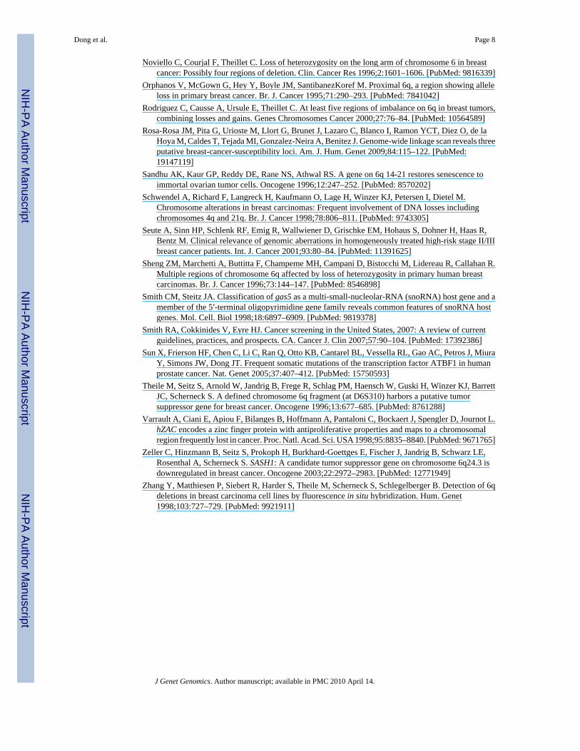
We first performed real time PCR to determine the deletion status of U50 in 31 breast cancercell lines. While no homozygous deletion was detected, 9 of 31 (29%) breast cancer cell lines
Dong et al. Page 3
J Genet Genomics. Author manuscript; available in PMC 2010 April 14.
NIH
-PA Author Manuscript
NIH
-PA Author Manuscript
NIH
-PA Author Manuscript
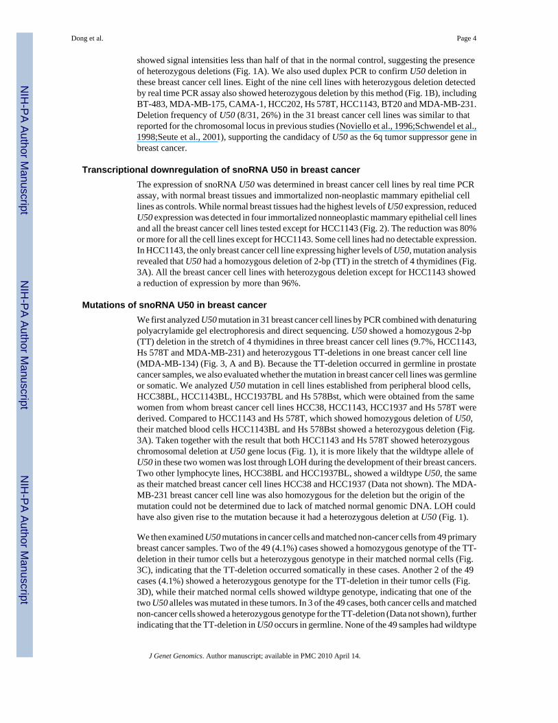
showed signal intensities less than half of that in the normal control, suggesting the presenceof heterozygous deletions (Fig. 1A). We also used duplex PCR to confirm U50 deletion inthese breast cancer cell lines. Eight of the nine cell lines with heterozygous deletion detectedby real time PCR assay also showed heterozygous deletion by this method (Fig. 1B), includingBT-483, MDA-MB-175, CAMA-1, HCC202, Hs 578T, HCC1143, BT20 and MDA-MB-231.Deletion frequency of U50 (8/31, 26%) in the 31 breast cancer cell lines was similar to thatreported for the chromosomal locus in previous studies (Noviello et al., 1996;Schwendel et al.,1998;Seute et al., 2001), supporting the candidacy of U50 as the 6q tumor suppressor gene inbreast cancer.
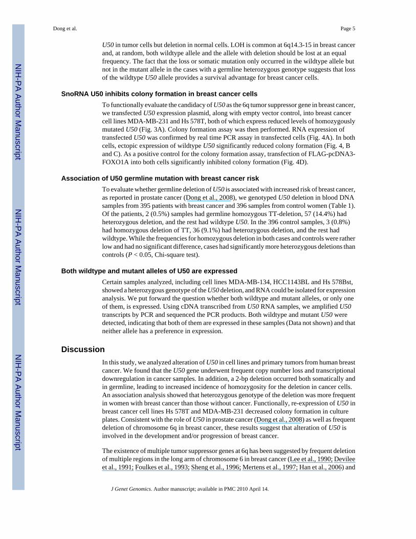
Transcriptional downregulation of snoRNA U50 in breast cancerThe expression of snoRNA U50 was determined in breast cancer cell lines by real time PCRassay, with normal breast tissues and immortalized non-neoplastic mammary epithelial celllines as controls. While normal breast tissues had the highest levels of U50 expression, reducedU50 expression was detected in four immortalized nonneoplastic mammary epithelial cell linesand all the breast cancer cell lines tested except for HCC1143 (Fig. 2). The reduction was 80%or more for all the cell lines except for HCC1143. Some cell lines had no detectable expression.In HCC1143, the only breast cancer cell line expressing higher levels of U50, mutation analysisrevealed that U50 had a homozygous deletion of 2-bp (TT) in the stretch of 4 thymidines (Fig.3A). All the breast cancer cell lines with heterozygous deletion except for HCC1143 showeda reduction of expression by more than 96%.
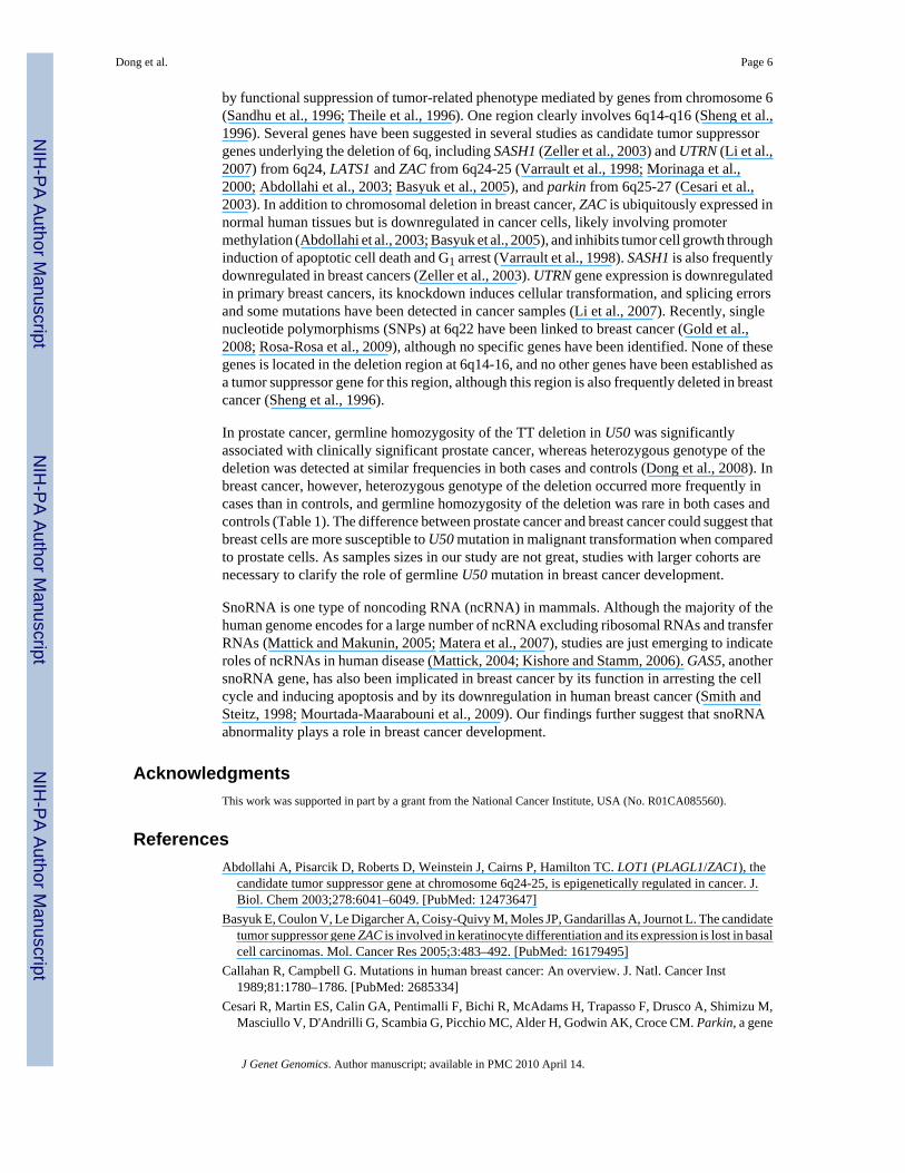
Mutations of snoRNA U50 in breast cancerWe first analyzed U50 mutation in 31 breast cancer cell lines by PCR combined with denaturingpolyacrylamide gel electrophoresis and direct sequencing. U50 showed a homozygous 2-bp(TT) deletion in the stretch of 4 thymidines in three breast cancer cell lines (9.7%, HCC1143,Hs 578T and MDA-MB-231) and heterozygous TT-deletions in one breast cancer cell line(MDA-MB-134) (Fig. 3, A and B). Because the TT-deletion occurred in germline in prostatecancer samples, we also evaluated whether the mutation in breast cancer cell lines was germlineor somatic. We analyzed U50 mutation in cell lines established from peripheral blood cells,HCC38BL, HCC1143BL, HCC1937BL and Hs 578Bst, which were obtained from the samewomen from whom breast cancer cell lines HCC38, HCC1143, HCC1937 and Hs 578T werederived. Compared to HCC1143 and Hs 578T, which showed homozygous deletion of U50,their matched blood cells HCC1143BL and Hs 578Bst showed a heterozygous deletion (Fig.3A). Taken together with the result that both HCC1143 and Hs 578T showed heterozygouschromosomal deletion at U50 gene locus (Fig. 1), it is more likely that the wildtype allele ofU50 in these two women was lost through LOH during the development of their breast cancers.Two other lymphocyte lines, HCC38BL and HCC1937BL, showed a wildtype U50, the sameas their matched breast cancer cell lines HCC38 and HCC1937 (Data not shown). The MDA-MB-231 breast cancer cell line was also homozygous for the deletion but the origin of themutation could not be determined due to lack of matched normal genomic DNA. LOH couldhave also given rise to the mutation because it had a heterozygous deletion at U50 (Fig. 1).
We then examined U50 mutations in cancer cells and matched non-cancer cells from 49 primarybreast cancer samples. Two of the 49 (4.1%) cases showed a homozygous genotype of the TT-deletion in their tumor cells but a heterozygous genotype in their matched normal cells (Fig.3C), indicating that the TT-deletion occurred somatically in these cases. Another 2 of the 49cases (4.1%) showed a heterozygous genotype for the TT-deletion in their tumor cells (Fig.3D), while their matched normal cells showed wildtype genotype, indicating that one of thetwo U50 alleles was mutated in these tumors. In 3 of the 49 cases, both cancer cells and matchednon-cancer cells showed a heterozygous genotype for the TT-deletion (Data not shown), furtherindicating that the TT-deletion in U50 occurs in germline. None of the 49 samples had wildtype
Dong et al. Page 4
J Genet Genomics. Author manuscript; available in PMC 2010 April 14.
NIH
-PA Author Manuscript
NIH
-PA Author Manuscript
NIH
-PA Author Manuscript
U50 in tumor cells but deletion in normal cells. LOH is common at 6q14.3-15 in breast cancerand, at random, both wildtype allele and the allele with deletion should be lost at an equalfrequency. The fact that the loss or somatic mutation only occurred in the wildtype allele butnot in the mutant allele in the cases with a germline heterozygous genotype suggests that lossof the wildtype U50 allele provides a survival advantage for breast cancer cells.
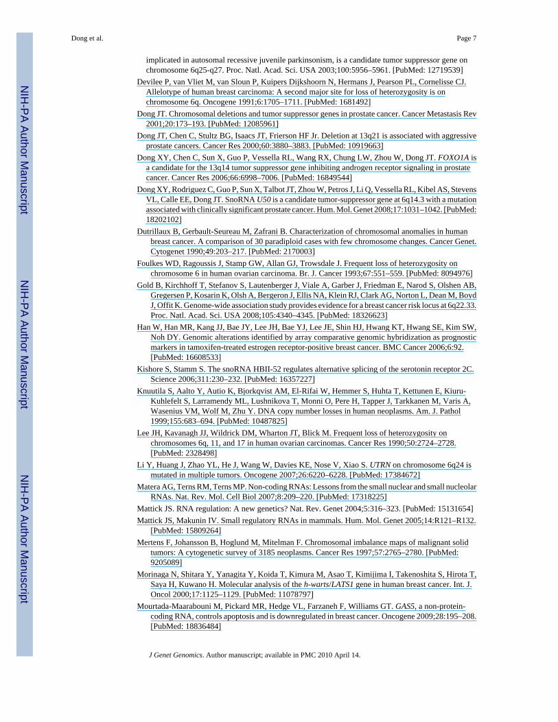
SnoRNA U50 inhibits colony formation in breast cancer cellsTo functionally evaluate the candidacy of U50 as the 6q tumor suppressor gene in breast cancer,we transfected U50 expression plasmid, along with empty vector control, into breast cancercell lines MDA-MB-231 and Hs 578T, both of which express reduced levels of homozygouslymutated U50 (Fig. 3A). Colony formation assay was then performed. RNA expression oftransfected U50 was confirmed by real time PCR assay in transfected cells (Fig. 4A). In bothcells, ectopic expression of wildtype U50 significantly reduced colony formation (Fig. 4, Band C). As a positive control for the colony formation assay, transfection of FLAG-pcDNA3-FOXO1A into both cells significantly inhibited colony formation (Fig. 4D).
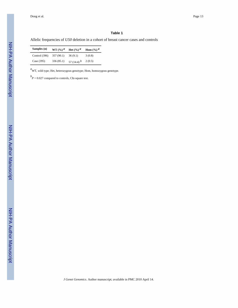
Association of U50 germline mutation with breast cancer riskTo evaluate whether germline deletion of U50 is associated with increased risk of breast cancer,as reported in prostate cancer (Dong et al., 2008), we genotyped U50 deletion in blood DNAsamples from 395 patients with breast cancer and 396 samples from control women (Table 1).Of the patients, 2 (0.5%) samples had germline homozygous TT-deletion, 57 (14.4%) hadheterozygous deletion, and the rest had wildtype U50. In the 396 control samples, 3 (0.8%)had homozygous deletion of TT, 36 (9.1%) had heterozygous deletion, and the rest hadwildtype. While the frequencies for homozygous deletion in both cases and controls were ratherlow and had no significant difference, cases had significantly more heterozygous deletions thancontrols (P < 0.05, Chi-square test).
Both wildtype and mutant alleles of U50 are expressedCertain samples analyzed, including cell lines MDA-MB-134, HCC1143BL and Hs 578Bst,showed a heterozygous genotype of the U50 deletion, and RNA could be isolated for expressionanalysis. We put forward the question whether both wildtype and mutant alleles, or only oneof them, is expressed. Using cDNA transcribed from U50 RNA samples, we amplified U50transcripts by PCR and sequenced the PCR products. Both wildtype and mutant U50 weredetected, indicating that both of them are expressed in these samples (Data not shown) and thatneither allele has a preference in expression.
DiscussionIn this study, we analyzed alteration of U50 in cell lines and primary tumors from human breastcancer. We found that the U50 gene underwent frequent copy number loss and transcriptionaldownregulation in cancer samples. In addition, a 2-bp deletion occurred both somatically andin germline, leading to increased incidence of homozygosity for the deletion in cancer cells.An association analysis showed that heterozygous genotype of the deletion was more frequentin women with breast cancer than those without cancer. Functionally, re-expression of U50 inbreast cancer cell lines Hs 578T and MDA-MB-231 decreased colony formation in cultureplates. Consistent with the role of U50 in prostate cancer (Dong et al., 2008) as well as frequentdeletion of chromosome 6q in breast cancer, these results suggest that alteration of U50 isinvolved in the development and/or progression of breast cancer.
The existence of multiple tumor suppressor genes at 6q has been suggested by frequent deletionof multiple regions in the long arm of chromosome 6 in breast cancer (Lee et al., 1990; Devileeet al., 1991; Foulkes et al., 1993; Sheng et al., 1996; Mertens et al., 1997; Han et al., 2006) and
Dong et al. Page 5
J Genet Genomics. Author manuscript; available in PMC 2010 April 14.
NIH
-PA Author Manuscript
NIH
-PA Author Manuscript
NIH
-PA Author Manuscript
by functional suppression of tumor-related phenotype mediated by genes from chromosome 6(Sandhu et al., 1996; Theile et al., 1996). One region clearly involves 6q14-q16 (Sheng et al.,1996). Several genes have been suggested in several studies as candidate tumor suppressorgenes underlying the deletion of 6q, including SASH1 (Zeller et al., 2003) and UTRN (Li et al.,2007) from 6q24, LATS1 and ZAC from 6q24-25 (Varrault et al., 1998; Morinaga et al.,2000; Abdollahi et al., 2003; Basyuk et al., 2005), and parkin from 6q25-27 (Cesari et al.,2003). In addition to chromosomal deletion in breast cancer, ZAC is ubiquitously expressed innormal human tissues but is downregulated in cancer cells, likely involving promotermethylation (Abdollahi et al., 2003; Basyuk et al., 2005), and inhibits tumor cell growth throughinduction of apoptotic cell death and G1 arrest (Varrault et al., 1998). SASH1 is also frequentlydownregulated in breast cancers (Zeller et al., 2003). UTRN gene expression is downregulatedin primary breast cancers, its knockdown induces cellular transformation, and splicing errorsand some mutations have been detected in cancer samples (Li et al., 2007). Recently, singlenucleotide polymorphisms (SNPs) at 6q22 have been linked to breast cancer (Gold et al.,2008; Rosa-Rosa et al., 2009), although no specific genes have been identified. None of thesegenes is located in the deletion region at 6q14-16, and no other genes have been established asa tumor suppressor gene for this region, although this region is also frequently deleted in breastcancer (Sheng et al., 1996).
In prostate cancer, germline homozygosity of the TT deletion in U50 was significantlyassociated with clinically significant prostate cancer, whereas heterozygous genotype of thedeletion was detected at similar frequencies in both cases and controls (Dong et al., 2008). Inbreast cancer, however, heterozygous genotype of the deletion occurred more frequently incases than in controls, and germline homozygosity of the deletion was rare in both cases andcontrols (Table 1). The difference between prostate cancer and breast cancer could suggest thatbreast cells are more susceptible to U50 mutation in malignant transformation when comparedto prostate cells. As samples sizes in our study are not great, studies with larger cohorts arenecessary to clarify the role of germline U50 mutation in breast cancer development.
SnoRNA is one type of noncoding RNA (ncRNA) in mammals. Although the majority of thehuman genome encodes for a large number of ncRNA excluding ribosomal RNAs and transferRNAs (Mattick and Makunin, 2005; Matera et al., 2007), studies are just emerging to indicateroles of ncRNAs in human disease (Mattick, 2004; Kishore and Stamm, 2006). GAS5, anothersnoRNA gene, has also been implicated in breast cancer by its function in arresting the cellcycle and inducing apoptosis and by its downregulation in human breast cancer (Smith andSteitz, 1998; Mourtada-Maarabouni et al., 2009). Our findings further suggest that snoRNAabnormality plays a role in breast cancer development.
AcknowledgmentsThis work was supported in part by a grant from the National Cancer Institute, USA (No. R01CA085560).
ReferencesAbdollahi A, Pisarcik D, Roberts D, Weinstein J, Cairns P, Hamilton TC. LOT1 (PLAGL1/ZAC1), the
candidate tumor suppressor gene at chromosome 6q24-25, is epigenetically regulated in cancer. J.Biol. Chem 2003;278:6041–6049. [PubMed: 12473647]
Basyuk E, Coulon V, Le Digarcher A, Coisy-Quivy M, Moles JP, Gandarillas A, Journot L. The candidatetumor suppressor gene ZAC is involved in keratinocyte differentiation and its expression is lost in basalcell carcinomas. Mol. Cancer Res 2005;3:483–492. [PubMed: 16179495]
Callahan R, Campbell G. Mutations in human breast cancer: An overview. J. Natl. Cancer Inst1989;81:1780–1786. [PubMed: 2685334]
Cesari R, Martin ES, Calin GA, Pentimalli F, Bichi R, McAdams H, Trapasso F, Drusco A, Shimizu M,Masciullo V, D'Andrilli G, Scambia G, Picchio MC, Alder H, Godwin AK, Croce CM. Parkin, a gene
Dong et al. Page 6
J Genet Genomics. Author manuscript; available in PMC 2010 April 14.
NIH
-PA Author Manuscript
NIH
-PA Author Manuscript
NIH
-PA Author Manuscript
implicated in autosomal recessive juvenile parkinsonism, is a candidate tumor suppressor gene onchromosome 6q25-q27. Proc. Natl. Acad. Sci. USA 2003;100:5956–5961. [PubMed: 12719539]
Devilee P, van Vliet M, van Sloun P, Kuipers Dijkshoorn N, Hermans J, Pearson PL, Cornelisse CJ.Allelotype of human breast carcinoma: A second major site for loss of heterozygosity is onchromosome 6q. Oncogene 1991;6:1705–1711. [PubMed: 1681492]
Dong JT. Chromosomal deletions and tumor suppressor genes in prostate cancer. Cancer Metastasis Rev2001;20:173–193. [PubMed: 12085961]
Dong JT, Chen C, Stultz BG, Isaacs JT, Frierson HF Jr. Deletion at 13q21 is associated with aggressiveprostate cancers. Cancer Res 2000;60:3880–3883. [PubMed: 10919663]
Dong XY, Chen C, Sun X, Guo P, Vessella RL, Wang RX, Chung LW, Zhou W, Dong JT. FOXO1A isa candidate for the 13q14 tumor suppressor gene inhibiting androgen receptor signaling in prostatecancer. Cancer Res 2006;66:6998–7006. [PubMed: 16849544]
Dong XY, Rodriguez C, Guo P, Sun X, Talbot JT, Zhou W, Petros J, Li Q, Vessella RL, Kibel AS, StevensVL, Calle EE, Dong JT. SnoRNA U50 is a candidate tumor-suppressor gene at 6q14.3 with a mutationassociated with clinically significant prostate cancer. Hum. Mol. Genet 2008;17:1031–1042. [PubMed:18202102]
Dutrillaux B, Gerbault-Seureau M, Zafrani B. Characterization of chromosomal anomalies in humanbreast cancer. A comparison of 30 paradiploid cases with few chromosome changes. Cancer Genet.Cytogenet 1990;49:203–217. [PubMed: 2170003]
Foulkes WD, Ragoussis J, Stamp GW, Allan GJ, Trowsdale J. Frequent loss of heterozygosity onchromosome 6 in human ovarian carcinoma. Br. J. Cancer 1993;67:551–559. [PubMed: 8094976]
Gold B, Kirchhoff T, Stefanov S, Lautenberger J, Viale A, Garber J, Friedman E, Narod S, Olshen AB,Gregersen P, Kosarin K, Olsh A, Bergeron J, Ellis NA, Klein RJ, Clark AG, Norton L, Dean M, BoydJ, Offit K. Genome-wide association study provides evidence for a breast cancer risk locus at 6q22.33.Proc. Natl. Acad. Sci. USA 2008;105:4340–4345. [PubMed: 18326623]
Han W, Han MR, Kang JJ, Bae JY, Lee JH, Bae YJ, Lee JE, Shin HJ, Hwang KT, Hwang SE, Kim SW,Noh DY. Genomic alterations identified by array comparative genomic hybridization as prognosticmarkers in tamoxifen-treated estrogen receptor-positive breast cancer. BMC Cancer 2006;6:92.[PubMed: 16608533]
Kishore S, Stamm S. The snoRNA HBII-52 regulates alternative splicing of the serotonin receptor 2C.Science 2006;311:230–232. [PubMed: 16357227]
Knuutila S, Aalto Y, Autio K, Bjorkqvist AM, El-Rifai W, Hemmer S, Huhta T, Kettunen E, Kiuru-Kuhlefelt S, Larramendy ML, Lushnikova T, Monni O, Pere H, Tapper J, Tarkkanen M, Varis A,Wasenius VM, Wolf M, Zhu Y. DNA copy number losses in human neoplasms. Am. J. Pathol1999;155:683–694. [PubMed: 10487825]
Lee JH, Kavanagh JJ, Wildrick DM, Wharton JT, Blick M. Frequent loss of heterozygosity onchromosomes 6q, 11, and 17 in human ovarian carcinomas. Cancer Res 1990;50:2724–2728.[PubMed: 2328498]
Li Y, Huang J, Zhao YL, He J, Wang W, Davies KE, Nose V, Xiao S. UTRN on chromosome 6q24 ismutated in multiple tumors. Oncogene 2007;26:6220–6228. [PubMed: 17384672]
Matera AG, Terns RM, Terns MP. Non-coding RNAs: Lessons from the small nuclear and small nucleolarRNAs. Nat. Rev. Mol. Cell Biol 2007;8:209–220. [PubMed: 17318225]
Mattick JS. RNA regulation: A new genetics? Nat. Rev. Genet 2004;5:316–323. [PubMed: 15131654]Mattick JS, Makunin IV. Small regulatory RNAs in mammals. Hum. Mol. Genet 2005;14:R121–R132.
[PubMed: 15809264]Mertens F, Johansson B, Hoglund M, Mitelman F. Chromosomal imbalance maps of malignant solid
tumors: A cytogenetic survey of 3185 neoplasms. Cancer Res 1997;57:2765–2780. [PubMed:9205089]
Morinaga N, Shitara Y, Yanagita Y, Koida T, Kimura M, Asao T, Kimijima I, Takenoshita S, Hirota T,Saya H, Kuwano H. Molecular analysis of the h-warts/LATS1 gene in human breast cancer. Int. J.Oncol 2000;17:1125–1129. [PubMed: 11078797]
Mourtada-Maarabouni M, Pickard MR, Hedge VL, Farzaneh F, Williams GT. GAS5, a non-protein-coding RNA, controls apoptosis and is downregulated in breast cancer. Oncogene 2009;28:195–208.[PubMed: 18836484]
Dong et al. Page 7
J Genet Genomics. Author manuscript; available in PMC 2010 April 14.
NIH
-PA Author Manuscript
NIH
-PA Author Manuscript
NIH
-PA Author Manuscript
Noviello C, Courjal F, Theillet C. Loss of heterozygosity on the long arm of chromosome 6 in breastcancer: Possibly four regions of deletion. Clin. Cancer Res 1996;2:1601–1606. [PubMed: 9816339]
Orphanos V, McGown G, Hey Y, Boyle JM, SantibanezKoref M. Proximal 6q, a region showing alleleloss in primary breast cancer. Br. J. Cancer 1995;71:290–293. [PubMed: 7841042]
Rodriguez C, Causse A, Ursule E, Theillet C. At least five regions of imbalance on 6q in breast tumors,combining losses and gains. Genes Chromosomes Cancer 2000;27:76–84. [PubMed: 10564589]
Rosa-Rosa JM, Pita G, Urioste M, Llort G, Brunet J, Lazaro C, Blanco I, Ramon YCT, Diez O, de laHoya M, Caldes T, Tejada MI, Gonzalez-Neira A, Benitez J. Genome-wide linkage scan reveals threeputative breast-cancer-susceptibility loci. Am. J. Hum. Genet 2009;84:115–122. [PubMed:19147119]
Sandhu AK, Kaur GP, Reddy DE, Rane NS, Athwal RS. A gene on 6q 14-21 restores senescence toimmortal ovarian tumor cells. Oncogene 1996;12:247–252. [PubMed: 8570202]
Schwendel A, Richard F, Langreck H, Kaufmann O, Lage H, Winzer KJ, Petersen I, Dietel M.Chromosome alterations in breast carcinomas: Frequent involvement of DNA losses includingchromosomes 4q and 21q. Br. J. Cancer 1998;78:806–811. [PubMed: 9743305]
Seute A, Sinn HP, Schlenk RF, Emig R, Wallwiener D, Grischke EM, Hohaus S, Dohner H, Haas R,Bentz M. Clinical relevance of genomic aberrations in homogeneously treated high-risk stage II/IIIbreast cancer patients. Int. J. Cancer 2001;93:80–84. [PubMed: 11391625]
Sheng ZM, Marchetti A, Buttitta F, Champeme MH, Campani D, Bistocchi M, Lidereau R, Callahan R.Multiple regions of chromosome 6q affected by loss of heterozygosity in primary human breastcarcinomas. Br. J. Cancer 1996;73:144–147. [PubMed: 8546898]
Smith CM, Steitz JA. Classification of gas5 as a multi-small-nucleolar-RNA (snoRNA) host gene and amember of the 5′-terminal oligopyrimidine gene family reveals common features of snoRNA hostgenes. Mol. Cell. Biol 1998;18:6897–6909. [PubMed: 9819378]
Smith RA, Cokkinides V, Eyre HJ. Cancer screening in the United States, 2007: A review of currentguidelines, practices, and prospects. CA. Cancer J. Clin 2007;57:90–104. [PubMed: 17392386]
Sun X, Frierson HF, Chen C, Li C, Ran Q, Otto KB, Cantarel BL, Vessella RL, Gao AC, Petros J, MiuraY, Simons JW, Dong JT. Frequent somatic mutations of the transcription factor ATBF1 in humanprostate cancer. Nat. Genet 2005;37:407–412. [PubMed: 15750593]
Theile M, Seitz S, Arnold W, Jandrig B, Frege R, Schlag PM, Haensch W, Guski H, Winzer KJ, BarrettJC, Scherneck S. A defined chromosome 6q fragment (at D6S310) harbors a putative tumorsuppressor gene for breast cancer. Oncogene 1996;13:677–685. [PubMed: 8761288]
Varrault A, Ciani E, Apiou F, Bilanges B, Hoffmann A, Pantaloni C, Bockaert J, Spengler D, Journot L.hZAC encodes a zinc finger protein with antiproliferative properties and maps to a chromosomalregion frequently lost in cancer. Proc. Natl. Acad. Sci. USA 1998;95:8835–8840. [PubMed: 9671765]
Zeller C, Hinzmann B, Seitz S, Prokoph H, Burkhard-Goettges E, Fischer J, Jandrig B, Schwarz LE,Rosenthal A, Scherneck S. SASH1: A candidate tumor suppressor gene on chromosome 6q24.3 isdownregulated in breast cancer. Oncogene 2003;22:2972–2983. [PubMed: 12771949]
Zhang Y, Matthiesen P, Siebert R, Harder S, Theile M, Scherneck S, Schlegelberger B. Detection of 6qdeletions in breast carcinoma cell lines by fluorescence in situ hybridization. Hum. Genet1998;103:727–729. [PubMed: 9921911]
Dong et al. Page 8
J Genet Genomics. Author manuscript; available in PMC 2010 April 14.
NIH
-PA Author Manuscript
NIH
-PA Author Manuscript
NIH
-PA Author Manuscript
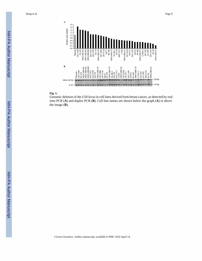
Fig. 1.Genomic deletion of the U50 locus in cell lines derived from breast cancer, as detected by realtime PCR (A) and duplex PCR (B). Cell line names are shown below the graph (A) or abovethe image (B).
Dong et al. Page 9
J Genet Genomics. Author manuscript; available in PMC 2010 April 14.
NIH
-PA Author Manuscript
NIH
-PA Author Manuscript
NIH
-PA Author Manuscript
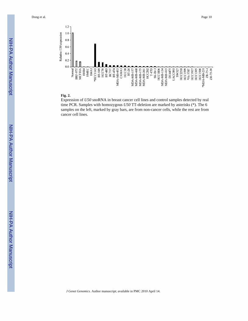
Fig. 2.Expression of U50 snoRNA in breast cancer cell lines and control samples detected by realtime PCR. Samples with homozygous U50 TT-deletion are marked by asterisks (*). The 6samples on the left, marked by gray bars, are from non-cancer cells, while the rest are fromcancer cell lines.
Dong et al. Page 10
J Genet Genomics. Author manuscript; available in PMC 2010 April 14.
NIH
-PA Author Manuscript
NIH
-PA Author Manuscript
NIH
-PA Author Manuscript
Fig. 3.Detection of U50 mutations in breast cancer by gel electrophoresis (A, C and D) and DNAsequencing (B). A: homozygosity of U50 mutation in breast cancer cell lines Hs 578T, MDA-MB-231 and HCC1143 and heterozygous deletion in breast cancer cell line MDA-MB-134and peripheral blood cell lines HCC1143BL and Hs 578Bst. B: DNA sequencing resultsshowing wildtype, heterozygous mutant, and homozygous mutant of U50 in a normal sampleand breast cancer cell lines MDA-MB-134 and MDA-MB-231, respectively. C: homozygousmutation of U50 in two primary breast cancer samples compared to their matched normal cellswith a heterozygous mutation. D: heterozygous mutation of U50 in two primary breast cancersamples compared to their matched normal cells with wildtype U50. In C and D, T, tumorcells; N, matched non-cancer cells.
Dong et al. Page 11
J Genet Genomics. Author manuscript; available in PMC 2010 April 14.
NIH
-PA Author Manuscript
NIH
-PA Author Manuscript
NIH
-PA Author Manuscript
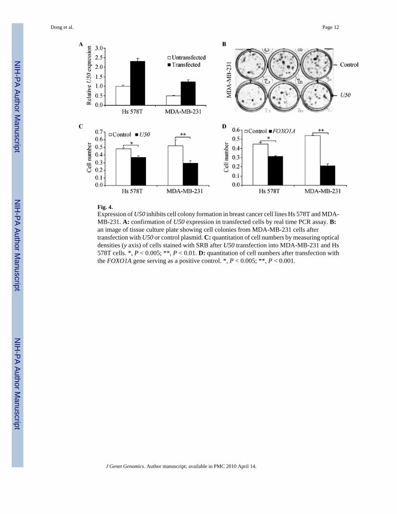
Fig. 4.Expression of U50 inhibits cell colony formation in breast cancer cell lines Hs 578T and MDA-MB-231. A: confirmation of U50 expression in transfected cells by real time PCR assay. B:an image of tissue culture plate showing cell colonies from MDA-MB-231 cells aftertransfection with U50 or control plasmid. C: quantitation of cell numbers by measuring opticaldensities (y axis) of cells stained with SRB after U50 transfection into MDA-MB-231 and Hs578T cells. *, P < 0.005; **, P < 0.01. D: quantitation of cell numbers after transfection withthe FOXO1A gene serving as a positive control. *, P < 0.005; **, P < 0.001.
Dong et al. Page 12
J Genet Genomics. Author manuscript; available in PMC 2010 April 14.
NIH
-PA Author Manuscript
NIH
-PA Author Manuscript
NIH
-PA Author Manuscript
NIH
-PA Author Manuscript
NIH
-PA Author Manuscript
NIH
-PA Author Manuscript
Dong et al. Page 13
Table 1
Allelic frequencies of U50 deletion in a cohort of breast cancer cases and controls
Samples (n) WT (%) a Het (%) a Hom (%) a
Control (396) 357 (90.1) 36 (9.1) 3 (0.8)
Case (395) 336 (85.1) 57 (14.4) b 2 (0.5)
aWT, wild type; Het, heterozygous genotype; Hom, homozygous genotype.
bP = 0.027 compared to controls, Chi-square test.
J Genet Genomics. Author manuscript; available in PMC 2010 April 14.
Related Documents