Human miRNA Precursors with Box H/ACA snoRNA Features Michelle S. Scott 1 *, Fabio Avolio 2 , Motoharu Ono 2 , Angus I. Lamond 2 , Geoffrey J. Barton 1 1 Division of Biological Chemistry and Drug Discovery, College of Life Sciences, University of Dundee, Dundee, United Kingdom, 2 Wellcome Trust Centre for Gene Regulation and Expression, College of Life Sciences, University of Dundee, Dundee, United Kingdom Abstract MicroRNAs (miRNAs) and small nucleolar RNAs (snoRNAs) are two classes of small non-coding regulatory RNAs, which have been much investigated in recent years. While their respective functions in the cell are distinct, they share interesting genomic similarities, and recent sequencing projects have identified processed forms of snoRNAs that resemble miRNAs. Here, we investigate a possible evolutionary relationship between miRNAs and box H/ACA snoRNAs. A comparison of the genomic locations of reported miRNAs and snoRNAs reveals an overlap of specific members of these classes. To test the hypothesis that some miRNAs might have evolved from snoRNA encoding genomic regions, reported miRNA-encoding regions were scanned for the presence of box H/ACA snoRNA features. Twenty miRNA precursors show significant similarity to H/ACA snoRNAs as predicted by snoGPS. These include molecules predicted to target known ribosomal RNA pseudouridylation sites in vivo for which no guide snoRNA has yet been reported. The predicted folded structures of these twenty H/ACA snoRNA-like miRNA precursors reveal molecules which resemble the structures of known box H/ACA snoRNAs. The genomic regions surrounding these predicted snoRNA-like miRNAs are often similar to regions around snoRNA retroposons, including the presence of transposable elements, target site duplications and poly (A) tails. We further show that the precursors of five H/ACA snoRNA-like miRNAs (miR-151, miR-605, mir-664, miR-215 and miR-140) bind to dyskerin, a specific protein component of functional box H/ ACA small nucleolar ribonucleoprotein complexes suggesting that these molecules have retained some H/ACA snoRNA functionality. The detection of small RNA molecules that share features of miRNAs and snoRNAs suggest that these classes of RNA may have an evolutionary relationship. Citation: Scott MS, Avolio F, Ono M, Lamond AI, Barton GJ (2009) Human miRNA Precursors with Box H/ACA snoRNA Features. PLoS Comput Biol 5(9): e1000507. doi:10.1371/journal.pcbi.1000507 Editor: Ron Unger, Bar-Ilan University, Israel Received May 12, 2009; Accepted August 14, 2009; Published September 18, 2009 Copyright: ß 2009 Scott et al. This is an open-access article distributed under the terms of the Creative Commons Attribution License, which permits unrestricted use, distribution, and reproduction in any medium, provided the original author and source are credited. Funding: MSS is a recipient of post-doctoral fellowships from the Canadian Institutes of Health Research (CIHR) as well as the Caledonian Research Foundation. Funding for this research was provided by a Wellcome Trust Programme to AIL (Ref: 073980/Z/03/Z). The funders had no role in study design, data collection and analysis, decision to publish, or preparation of the manuscript. Competing Interests: The authors have declared that no competing interests exist. * E-mail: [email protected] Introduction Small nucleolar RNAs (snoRNAs) and microRNAs (miRNAs) are two classes of abundant non-coding regulatory RNAs that carry out fundamental cellular activities but that have only been comprehen- sively investigated in recent years. SnoRNAs are small RNA molecules of approximately 60–300 nucleotides in length which generally serve as guides for the catalytic modification of selected ribosomal RNA nucleotides [1,2]. SnoRNAs associate with specific proteins, which are conserved amongst all eukaryotes, to form small nucleolar ribonucleoparticles (snoRNPs). Two main groups of snoRNAs have been described. The box C/D snoRNAs, which bind the four conserved core box C/D snoRNP proteins fibrillarin, NOP56, NOP5/NOP58 and NHP2L1, are involved in 29-O-ribose methylation. The box H/ACA snoRNAs, which bind the four conserved core box H/ACA snoRNP proteins DKC1 (dyskerin), GAR1, NHP2 and NOP10, catalyse pseudouridylation. In vertebrates, most snoRNAs have been shown to reside in introns of protein coding host genes and are processed out of the excised introns [3]. However, two box C/D snoRNAs have recently been found to be transcribed from independent RNA pol II units [4]. MiRNAs are ,18–24 nucleotide-long RNAs that are processed out of ,70 nucleotide-long hairpin structures (called pre-miRNAs) [5]. In mammals, miRNAs have been shown to be involved mainly in mRNA translation inhibition [6] although recently, they have also been reported to activate translation [7]. A large class of miRNAs are encoded in introns of protein-coding genes and are co-expressed with these host genes [8–10]. The remaining miRNAs are encoded in independent transcription units. Some of these miRNAs have been shown to be under the control of the RNA polymerase II [11] while others are transcribed by the RNA polymerase III [12]. Many members of the snoRNA and miRNA classes are well conserved throughout evolution [1,2,13]. Correspondence be- tween several yeast and human snoRNAs and their target sites have been established and many snoRNAs have a very high sequence identity within mammals as shown in the snoRNAbase database [14]. In the case of miRNAs, several families have been found to be well conserved in metazoans [13,15]. However, recent reports also suggest the existence of species- and lineage- specific snoRNAs and miRNAs [13,16,17]. These and other reports on their origin and evolution are providing clues about the emergence of large groups of these recently evolved molecules. Through bioinformatic searches, Weber [17] and Luo and Li [16] identified hundreds of human snoRNAs and snoRNA-related molecules that are derived from transposable PLoS Computational Biology | www.ploscompbiol.org 1 September 2009 | Volume 5 | Issue 9 | e1000507

Welcome message from author
This document is posted to help you gain knowledge. Please leave a comment to let me know what you think about it! Share it to your friends and learn new things together.
Transcript
Human miRNA Precursors with Box H/ACA snoRNAFeaturesMichelle S. Scott1*, Fabio Avolio2, Motoharu Ono2, Angus I. Lamond2, Geoffrey J. Barton1
1 Division of Biological Chemistry and Drug Discovery, College of Life Sciences, University of Dundee, Dundee, United Kingdom, 2 Wellcome Trust Centre for Gene
Regulation and Expression, College of Life Sciences, University of Dundee, Dundee, United Kingdom
Abstract
MicroRNAs (miRNAs) and small nucleolar RNAs (snoRNAs) are two classes of small non-coding regulatory RNAs, which have beenmuch investigated in recent years. While their respective functions in the cell are distinct, they share interesting genomicsimilarities, and recent sequencing projects have identified processed forms of snoRNAs that resemble miRNAs. Here, weinvestigate a possible evolutionary relationship between miRNAs and box H/ACA snoRNAs. A comparison of the genomiclocations of reported miRNAs and snoRNAs reveals an overlap of specific members of these classes. To test the hypothesis thatsome miRNAs might have evolved from snoRNA encoding genomic regions, reported miRNA-encoding regions were scanned forthe presence of box H/ACA snoRNA features. Twenty miRNA precursors show significant similarity to H/ACA snoRNAs aspredicted by snoGPS. These include molecules predicted to target known ribosomal RNA pseudouridylation sites in vivo forwhich no guide snoRNA has yet been reported. The predicted folded structures of these twenty H/ACA snoRNA-like miRNAprecursors reveal molecules which resemble the structures of known box H/ACA snoRNAs. The genomic regions surroundingthese predicted snoRNA-like miRNAs are often similar to regions around snoRNA retroposons, including the presence oftransposable elements, target site duplications and poly (A) tails. We further show that the precursors of five H/ACA snoRNA-likemiRNAs (miR-151, miR-605, mir-664, miR-215 and miR-140) bind to dyskerin, a specific protein component of functional box H/ACA small nucleolar ribonucleoprotein complexes suggesting that these molecules have retained some H/ACA snoRNAfunctionality. The detection of small RNA molecules that share features of miRNAs and snoRNAs suggest that these classes of RNAmay have an evolutionary relationship.
Citation: Scott MS, Avolio F, Ono M, Lamond AI, Barton GJ (2009) Human miRNA Precursors with Box H/ACA snoRNA Features. PLoS Comput Biol 5(9): e1000507.doi:10.1371/journal.pcbi.1000507
Editor: Ron Unger, Bar-Ilan University, Israel
Received May 12, 2009; Accepted August 14, 2009; Published September 18, 2009
Copyright: � 2009 Scott et al. This is an open-access article distributed under the terms of the Creative Commons Attribution License, which permitsunrestricted use, distribution, and reproduction in any medium, provided the original author and source are credited.
Funding: MSS is a recipient of post-doctoral fellowships from the Canadian Institutes of Health Research (CIHR) as well as the Caledonian Research Foundation.Funding for this research was provided by a Wellcome Trust Programme to AIL (Ref: 073980/Z/03/Z). The funders had no role in study design, data collection andanalysis, decision to publish, or preparation of the manuscript.
Competing Interests: The authors have declared that no competing interests exist.
* E-mail: [email protected]
Introduction
Small nucleolar RNAs (snoRNAs) and microRNAs (miRNAs) are
two classes of abundant non-coding regulatory RNAs that carry out
fundamental cellular activities but that have only been comprehen-
sively investigated in recent years. SnoRNAs are small RNA
molecules of approximately 60–300 nucleotides in length which
generally serve as guides for the catalytic modification of selected
ribosomal RNA nucleotides [1,2]. SnoRNAs associate with specific
proteins, which are conserved amongst all eukaryotes, to form small
nucleolar ribonucleoparticles (snoRNPs). Two main groups of
snoRNAs have been described. The box C/D snoRNAs, which
bind the four conserved core box C/D snoRNP proteins fibrillarin,
NOP56, NOP5/NOP58 and NHP2L1, are involved in 29-O-ribose
methylation. The box H/ACA snoRNAs, which bind the four
conserved core box H/ACA snoRNP proteins DKC1 (dyskerin),
GAR1, NHP2 and NOP10, catalyse pseudouridylation. In
vertebrates, most snoRNAs have been shown to reside in introns
of protein coding host genes and are processed out of the excised
introns [3]. However, two box C/D snoRNAs have recently been
found to be transcribed from independent RNA pol II units [4].
MiRNAs are ,18–24 nucleotide-long RNAs that are processed
out of ,70 nucleotide-long hairpin structures (called pre-miRNAs)
[5]. In mammals, miRNAs have been shown to be involved mainly
in mRNA translation inhibition [6] although recently, they have
also been reported to activate translation [7]. A large class of
miRNAs are encoded in introns of protein-coding genes and are
co-expressed with these host genes [8–10]. The remaining
miRNAs are encoded in independent transcription units. Some
of these miRNAs have been shown to be under the control of the
RNA polymerase II [11] while others are transcribed by the RNA
polymerase III [12].
Many members of the snoRNA and miRNA classes are well
conserved throughout evolution [1,2,13]. Correspondence be-
tween several yeast and human snoRNAs and their target sites
have been established and many snoRNAs have a very high
sequence identity within mammals as shown in the snoRNAbase
database [14]. In the case of miRNAs, several families have been
found to be well conserved in metazoans [13,15]. However,
recent reports also suggest the existence of species- and lineage-
specific snoRNAs and miRNAs [13,16,17]. These and other
reports on their origin and evolution are providing clues about
the emergence of large groups of these recently evolved
molecules. Through bioinformatic searches, Weber [17] and
Luo and Li [16] identified hundreds of human snoRNAs and
snoRNA-related molecules that are derived from transposable
PLoS Computational Biology | www.ploscompbiol.org 1 September 2009 | Volume 5 | Issue 9 | e1000507
elements (TEs), thus confirming the widespread nature of this
phenomenon, initially described for a small number of snoRNAs
[2,18]. These analyses suggest that many snoRNAs result from
the retroposition of existing snoRNAs that used long interspersed
nuclear element (LINE) machinery to transpose themselves to
new genomic locations. Many of these snoRNA-related mole-
cules are surrounded by the presence of sequence features typical
of retrogenes such as target site duplications (TSDs) and poly (A)
tails at their 39 end. These snoRNA retroposition events
generated hundreds of sno-related molecules, termed snoRTs
(snoRNA retroposons) by Weber [17], many of which had never
been previously identified, but some of which were previously
described as functional snoRNAs [16]. SnoRNA retroposition
thus not only permits maintenance of a pool of intact snoRNA
copies to safeguard against the effects of deleterious mutations
but could possibly also allow for the creation of regulatory RNA
molecules that might bind new targets [17]. Given the stringent
thresholds used to search for snoRNA copies in both studies, it is
likely that many more such molecules exist in the human genome
but might have diverged further from their parental copies and
are yet to be discovered.
Recent reports have also described some miRNAs as being
derived from TEs, suggesting a possible mechanism for the rapid
generation of miRNAs and their corresponding target sites. In the
first such report, Smalheiser and Torvik identified six miRNAs
that are derived from TEs [19]. Two subsequent studies identified
a further 95 [12] and 55 [20,21] known miRNAs that might be
derived from TEs as well as an additional 85 predicted novel TE-
derived miRNA genes [20]. The TEs that are most frequently
found in association with miRNAs are the L2 and MIR families
[20]. As TEs are the most non-conserved sequence elements in
eukaryotic genomes [22], the generation of miRNAs through TEs
represents a mechanism that could be a driving force in speciation
events and evolution by rapidly creating new regulatory elements
in the control of protein production [19,20].
A recent report investigating the small RNAs present in human
cells has demonstrated the existence of specific small RNA
fragments derived from larger known non-coding RNA mole-
cules [23]. In particular, distinct small fragments of sizes between
23 and 25 nucleotides were found to map to four box H/ACA
snoRNAs [23] (listed in Table 1). In addition to this, Ender and
colleagues have recently reported eight box H/ACA snoRNA-
derived miRNA-like molecules that can be immunoprecipitated
with Ago proteins [24]. While these short H/ACA snoRNA-
derived fragments might be discounted merely as non-functional
degradation products, several unrelated observations suggest
otherwise. Firstly, only specific fragments derived from one
Author Summary
The major functions known for RNA were long believed tobe either messenger RNAs, which function as intermedi-ates between genes and proteins, or ribosomal RNAs andtransfer RNAs which carry out the translation process. Inrecent years, however, newly discovered classes of smallRNAs have been shown to play important cellular roles.These include microRNAs (miRNAs), which can regulate theproduction of specific proteins, and small nucleolar RNAs(snoRNAs), which recognise and chemically modify specificsequences in ribosomal RNA. Although miRNAs andsnoRNAs are currently believed to be generated bydifferent cellular pathways and to function in differentcellular compartments, members of these two types ofsmall RNAs display numerous genomic similarities, and asmall number of snoRNAs have been shown to encodemiRNAs in several organisms. Here we systematicallyinvestigate a possible evolutionary relationship betweensnoRNAs and miRNAs. Using computational analysis, weidentify twenty genomic regions encoding miRNAs withhighly significant similarity to snoRNAs, both on the levelof their surrounding genomic context as well as theirpredicted folded structure. A subset of these miRNAsdisplay functional snoRNA characteristics, strengtheningthe possibility that these miRNA molecules might haveevolved from snoRNAs.
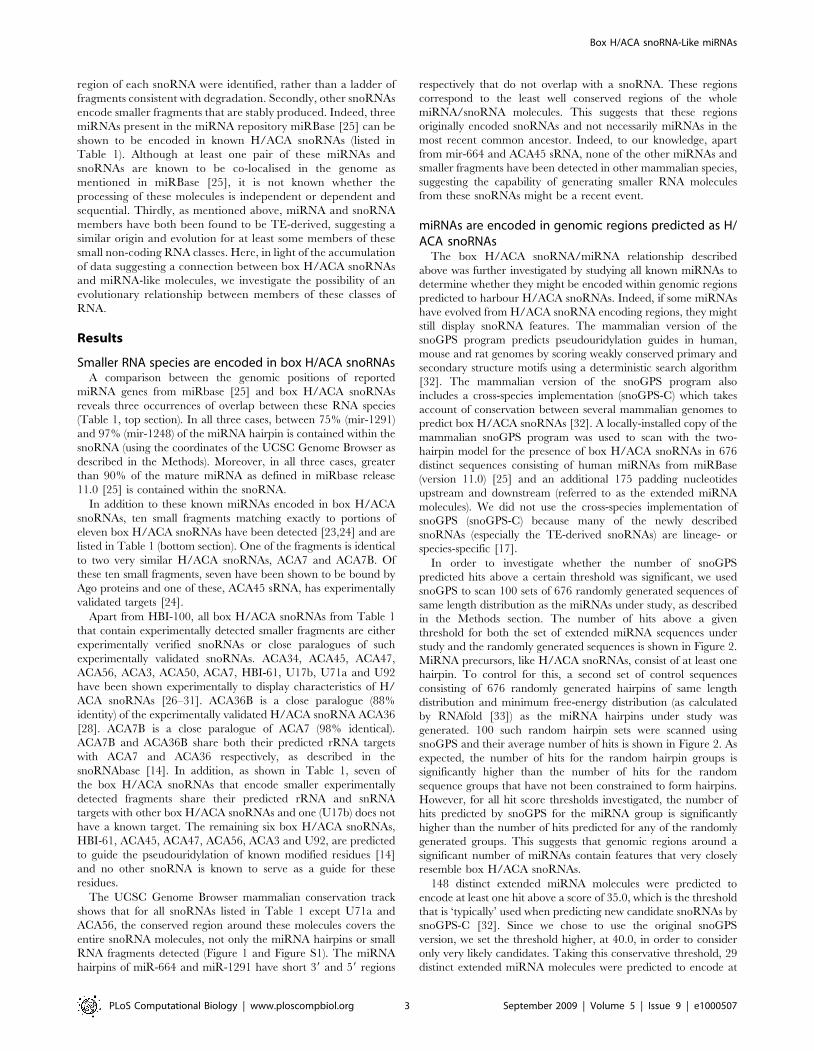
Table 1. Small fragments generated from snoRNAs.
Box H/ACAsnoRNA Chromosome
GenomicCoordinatesof snoRNA
Other H/ACA snoRNAswith same predictedrRNA target site
Known miRNAencoded withinsnoRNA
GenomicCoordinates ofmiRNA hairpin
Smallerfragmentdetected
Lengthof smallfragment
ACA36B 1 218440511–218440641 ACA36, ACA8, ACA50,ACA62, SNORA36C
mir-664 218440503–218440584 mir-664 [24,58] 23
HBI-61 3 187987158–187987335 - mir-1248 187987155–187987260 mir-1248 [58] 27
ACA34 12 47334432–47334568 ACA2A, ACA2B mir-1291 47334494–47334580 mir-1291 [58] 24
ACA7 3 12856811–12856949 ACA7B - N/A [23] 25
ACA7B 3 130598743–130598881 ACA7 - N/A [23] 25
U17b 1 28707657–28707861 unknown target - N/A [23] 23
U71a 20 36489363–36489500 U71b, U71c, U71d - N/A [23] 24
ACA45 15 81221751–81221877 - ACA45 sRNA N/A ACA45 sRNA [24] 23
ACA47 17 72596984–72597170 - - N/A ACA47 sRNA [24] 22
HBI-100 1 174204156–174204299 ACA12 - N/A HBI-100 sRNA [24] 22
ACA56 X 153656467–153656595 - - N/A ACA56 sRNA [24] 23
ACA3 11 8662350–8662479 - - N/A ACA3 sRNA [24] 23
ACA50 16 57151201–57151336 ACA62, ACA8,ACA36, ACA36B
- N/A ACA50 sRNA [24] 24
U92 9 19053654–19053784 - - N/A U92 sRNA [24] 24
doi:10.1371/journal.pcbi.1000507.t001
Box H/ACA snoRNA-Like miRNAs
PLoS Computational Biology | www.ploscompbiol.org 2 September 2009 | Volume 5 | Issue 9 | e1000507
region of each snoRNA were identified, rather than a ladder of
fragments consistent with degradation. Secondly, other snoRNAs
encode smaller fragments that are stably produced. Indeed, three
miRNAs present in the miRNA repository miRBase [25] can be
shown to be encoded in known H/ACA snoRNAs (listed in
Table 1). Although at least one pair of these miRNAs and
snoRNAs are known to be co-localised in the genome as
mentioned in miRBase [25], it is not known whether the
processing of these molecules is independent or dependent and
sequential. Thirdly, as mentioned above, miRNA and snoRNA
members have both been found to be TE-derived, suggesting a
similar origin and evolution for at least some members of these
small non-coding RNA classes. Here, in light of the accumulation
of data suggesting a connection between box H/ACA snoRNAs
and miRNA-like molecules, we investigate the possibility of an
evolutionary relationship between members of these classes of
RNA.
Results
Smaller RNA species are encoded in box H/ACA snoRNAsA comparison between the genomic positions of reported
miRNA genes from miRbase [25] and box H/ACA snoRNAs
reveals three occurrences of overlap between these RNA species
(Table 1, top section). In all three cases, between 75% (mir-1291)
and 97% (mir-1248) of the miRNA hairpin is contained within the
snoRNA (using the coordinates of the UCSC Genome Browser as
described in the Methods). Moreover, in all three cases, greater
than 90% of the mature miRNA as defined in miRbase release
11.0 [25] is contained within the snoRNA.
In addition to these known miRNAs encoded in box H/ACA
snoRNAs, ten small fragments matching exactly to portions of
eleven box H/ACA snoRNAs have been detected [23,24] and are
listed in Table 1 (bottom section). One of the fragments is identical
to two very similar H/ACA snoRNAs, ACA7 and ACA7B. Of
these ten small fragments, seven have been shown to be bound by
Ago proteins and one of these, ACA45 sRNA, has experimentally
validated targets [24].
Apart from HBI-100, all box H/ACA snoRNAs from Table 1
that contain experimentally detected smaller fragments are either
experimentally verified snoRNAs or close paralogues of such
experimentally validated snoRNAs. ACA34, ACA45, ACA47,
ACA56, ACA3, ACA50, ACA7, HBI-61, U17b, U71a and U92
have been shown experimentally to display characteristics of H/
ACA snoRNAs [26–31]. ACA36B is a close paralogue (88%
identity) of the experimentally validated H/ACA snoRNA ACA36
[28]. ACA7B is a close paralogue of ACA7 (98% identical).
ACA7B and ACA36B share both their predicted rRNA targets
with ACA7 and ACA36 respectively, as described in the
snoRNAbase [14]. In addition, as shown in Table 1, seven of
the box H/ACA snoRNAs that encode smaller experimentally
detected fragments share their predicted rRNA and snRNA
targets with other box H/ACA snoRNAs and one (U17b) does not
have a known target. The remaining six box H/ACA snoRNAs,
HBI-61, ACA45, ACA47, ACA56, ACA3 and U92, are predicted
to guide the pseudouridylation of known modified residues [14]
and no other snoRNA is known to serve as a guide for these
residues.
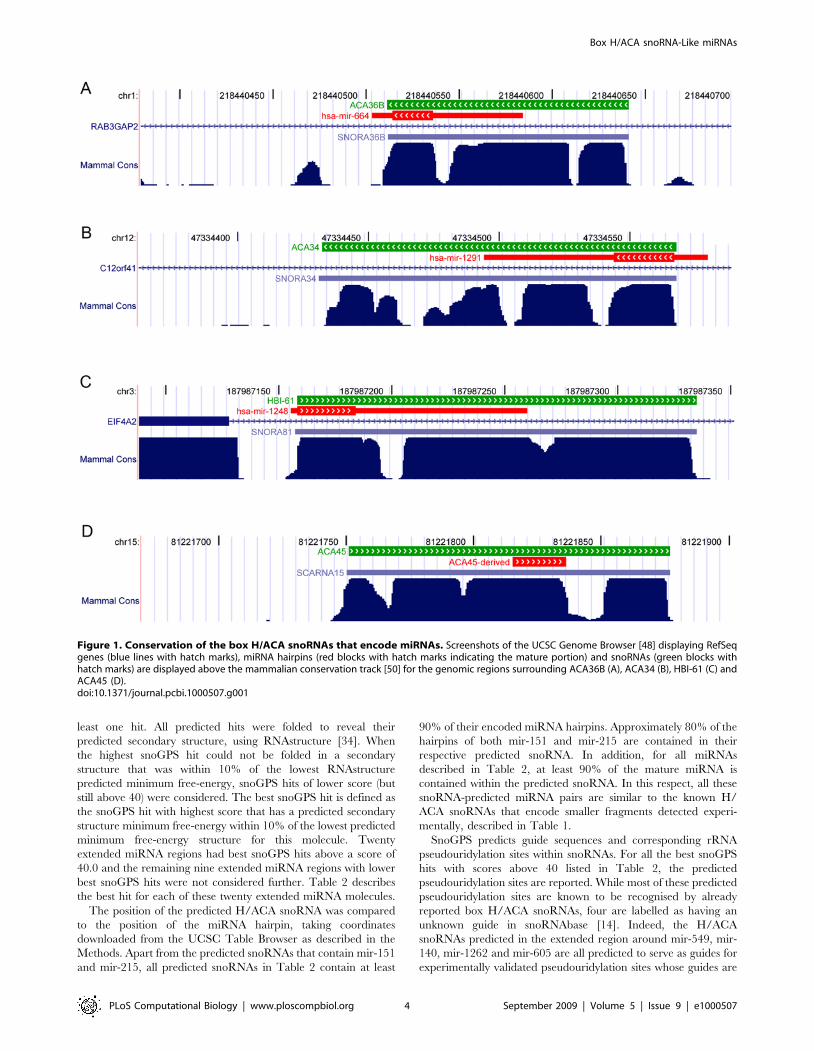
The UCSC Genome Browser mammalian conservation track
shows that for all snoRNAs listed in Table 1 except U71a and
ACA56, the conserved region around these molecules covers the
entire snoRNA molecules, not only the miRNA hairpins or small
RNA fragments detected (Figure 1 and Figure S1). The miRNA
hairpins of miR-664 and miR-1291 have short 39 and 59 regions
respectively that do not overlap with a snoRNA. These regions
correspond to the least well conserved regions of the whole
miRNA/snoRNA molecules. This suggests that these regions
originally encoded snoRNAs and not necessarily miRNAs in the
most recent common ancestor. Indeed, to our knowledge, apart
from mir-664 and ACA45 sRNA, none of the other miRNAs and
smaller fragments have been detected in other mammalian species,
suggesting the capability of generating smaller RNA molecules
from these snoRNAs might be a recent event.
miRNAs are encoded in genomic regions predicted as H/ACA snoRNAs
The box H/ACA snoRNA/miRNA relationship described
above was further investigated by studying all known miRNAs to
determine whether they might be encoded within genomic regions
predicted to harbour H/ACA snoRNAs. Indeed, if some miRNAs
have evolved from H/ACA snoRNA encoding regions, they might
still display snoRNA features. The mammalian version of the
snoGPS program predicts pseudouridylation guides in human,
mouse and rat genomes by scoring weakly conserved primary and
secondary structure motifs using a deterministic search algorithm
[32]. The mammalian version of the snoGPS program also
includes a cross-species implementation (snoGPS-C) which takes
account of conservation between several mammalian genomes to
predict box H/ACA snoRNAs [32]. A locally-installed copy of the
mammalian snoGPS program was used to scan with the two-
hairpin model for the presence of box H/ACA snoRNAs in 676
distinct sequences consisting of human miRNAs from miRBase
(version 11.0) [25] and an additional 175 padding nucleotides
upstream and downstream (referred to as the extended miRNA
molecules). We did not use the cross-species implementation of
snoGPS (snoGPS-C) because many of the newly described
snoRNAs (especially the TE-derived snoRNAs) are lineage- or
species-specific [17].
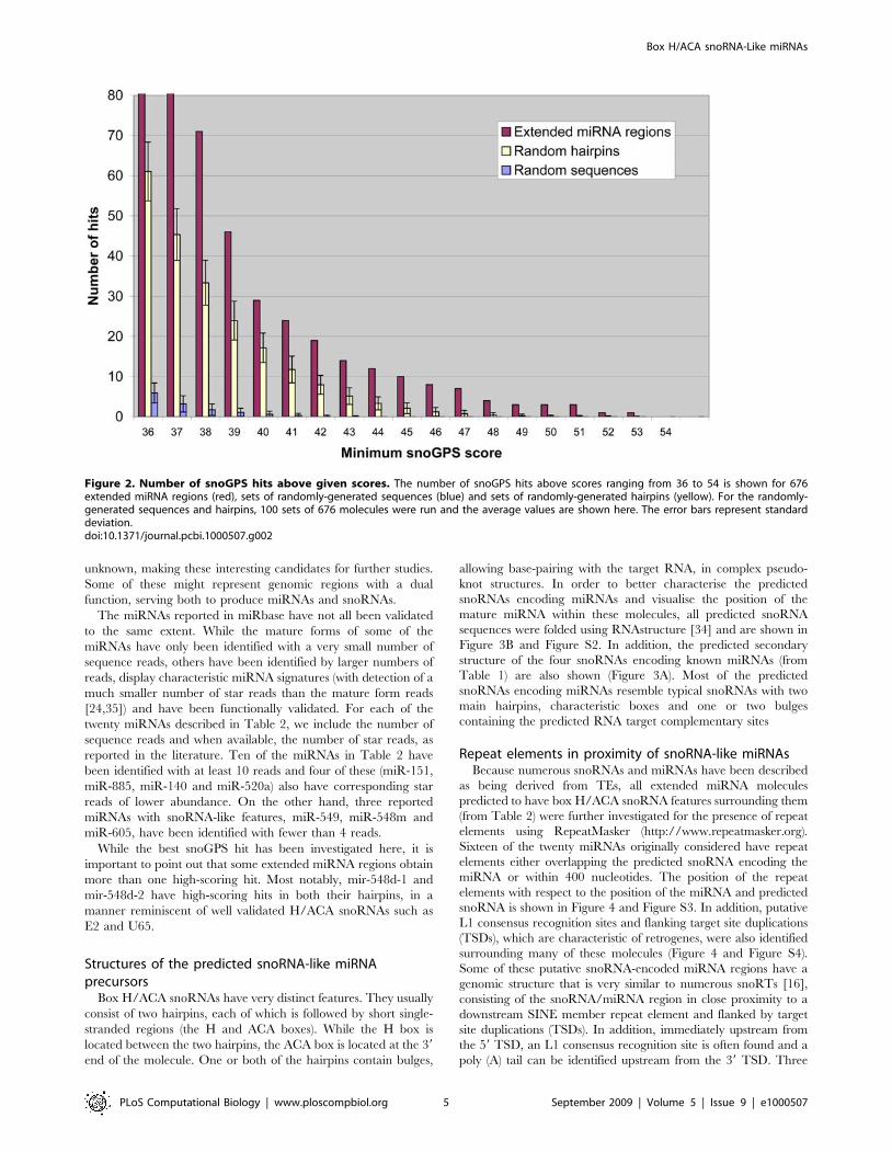
In order to investigate whether the number of snoGPS
predicted hits above a certain threshold was significant, we used
snoGPS to scan 100 sets of 676 randomly generated sequences of
same length distribution as the miRNAs under study, as described
in the Methods section. The number of hits above a given
threshold for both the set of extended miRNA sequences under
study and the randomly generated sequences is shown in Figure 2.
MiRNA precursors, like H/ACA snoRNAs, consist of at least one
hairpin. To control for this, a second set of control sequences
consisting of 676 randomly generated hairpins of same length
distribution and minimum free-energy distribution (as calculated
by RNAfold [33]) as the miRNA hairpins under study was
generated. 100 such random hairpin sets were scanned using
snoGPS and their average number of hits is shown in Figure 2. As
expected, the number of hits for the random hairpin groups is
significantly higher than the number of hits for the random
sequence groups that have not been constrained to form hairpins.
However, for all hit score thresholds investigated, the number of
hits predicted by snoGPS for the miRNA group is significantly
higher than the number of hits predicted for any of the randomly
generated groups. This suggests that genomic regions around a
significant number of miRNAs contain features that very closely
resemble box H/ACA snoRNAs.
148 distinct extended miRNA molecules were predicted to
encode at least one hit above a score of 35.0, which is the threshold
that is ‘typically’ used when predicting new candidate snoRNAs by
snoGPS-C [32]. Since we chose to use the original snoGPS
version, we set the threshold higher, at 40.0, in order to consider
only very likely candidates. Taking this conservative threshold, 29
distinct extended miRNA molecules were predicted to encode at
Box H/ACA snoRNA-Like miRNAs
PLoS Computational Biology | www.ploscompbiol.org 3 September 2009 | Volume 5 | Issue 9 | e1000507
least one hit. All predicted hits were folded to reveal their
predicted secondary structure, using RNAstructure [34]. When
the highest snoGPS hit could not be folded in a secondary
structure that was within 10% of the lowest RNAstructure
predicted minimum free-energy, snoGPS hits of lower score (but
still above 40) were considered. The best snoGPS hit is defined as
the snoGPS hit with highest score that has a predicted secondary
structure minimum free-energy within 10% of the lowest predicted
minimum free-energy structure for this molecule. Twenty
extended miRNA regions had best snoGPS hits above a score of
40.0 and the remaining nine extended miRNA regions with lower
best snoGPS hits were not considered further. Table 2 describes
the best hit for each of these twenty extended miRNA molecules.
The position of the predicted H/ACA snoRNA was compared
to the position of the miRNA hairpin, taking coordinates
downloaded from the UCSC Table Browser as described in the
Methods. Apart from the predicted snoRNAs that contain mir-151
and mir-215, all predicted snoRNAs in Table 2 contain at least
90% of their encoded miRNA hairpins. Approximately 80% of the
hairpins of both mir-151 and mir-215 are contained in their
respective predicted snoRNA. In addition, for all miRNAs
described in Table 2, at least 90% of the mature miRNA is
contained within the predicted snoRNA. In this respect, all these
snoRNA-predicted miRNA pairs are similar to the known H/
ACA snoRNAs that encode smaller fragments detected experi-
mentally, described in Table 1.
SnoGPS predicts guide sequences and corresponding rRNA
pseudouridylation sites within snoRNAs. For all the best snoGPS
hits with scores above 40 listed in Table 2, the predicted
pseudouridylation sites are reported. While most of these predicted
pseudouridylation sites are known to be recognised by already
reported box H/ACA snoRNAs, four are labelled as having an
unknown guide in snoRNAbase [14]. Indeed, the H/ACA
snoRNAs predicted in the extended region around mir-549, mir-
140, mir-1262 and mir-605 are all predicted to serve as guides for
experimentally validated pseudouridylation sites whose guides are
Figure 1. Conservation of the box H/ACA snoRNAs that encode miRNAs. Screenshots of the UCSC Genome Browser [48] displaying RefSeqgenes (blue lines with hatch marks), miRNA hairpins (red blocks with hatch marks indicating the mature portion) and snoRNAs (green blocks withhatch marks) are displayed above the mammalian conservation track [50] for the genomic regions surrounding ACA36B (A), ACA34 (B), HBI-61 (C) andACA45 (D).doi:10.1371/journal.pcbi.1000507.g001
Box H/ACA snoRNA-Like miRNAs
PLoS Computational Biology | www.ploscompbiol.org 4 September 2009 | Volume 5 | Issue 9 | e1000507
unknown, making these interesting candidates for further studies.
Some of these might represent genomic regions with a dual
function, serving both to produce miRNAs and snoRNAs.
The miRNAs reported in miRbase have not all been validated
to the same extent. While the mature forms of some of the
miRNAs have only been identified with a very small number of
sequence reads, others have been identified by larger numbers of
reads, display characteristic miRNA signatures (with detection of a
much smaller number of star reads than the mature form reads
[24,35]) and have been functionally validated. For each of the
twenty miRNAs described in Table 2, we include the number of
sequence reads and when available, the number of star reads, as
reported in the literature. Ten of the miRNAs in Table 2 have
been identified with at least 10 reads and four of these (miR-151,
miR-885, miR-140 and miR-520a) also have corresponding star
reads of lower abundance. On the other hand, three reported
miRNAs with snoRNA-like features, miR-549, miR-548m and
miR-605, have been identified with fewer than 4 reads.
While the best snoGPS hit has been investigated here, it is
important to point out that some extended miRNA regions obtain
more than one high-scoring hit. Most notably, mir-548d-1 and
mir-548d-2 have high-scoring hits in both their hairpins, in a
manner reminiscent of well validated H/ACA snoRNAs such as
E2 and U65.
Structures of the predicted snoRNA-like miRNAprecursors
Box H/ACA snoRNAs have very distinct features. They usually
consist of two hairpins, each of which is followed by short single-
stranded regions (the H and ACA boxes). While the H box is
located between the two hairpins, the ACA box is located at the 39
end of the molecule. One or both of the hairpins contain bulges,
allowing base-pairing with the target RNA, in complex pseudo-
knot structures. In order to better characterise the predicted
snoRNAs encoding miRNAs and visualise the position of the
mature miRNA within these molecules, all predicted snoRNA
sequences were folded using RNAstructure [34] and are shown in
Figure 3B and Figure S2. In addition, the predicted secondary
structure of the four snoRNAs encoding known miRNAs (from
Table 1) are also shown (Figure 3A). Most of the predicted
snoRNAs encoding miRNAs resemble typical snoRNAs with two
main hairpins, characteristic boxes and one or two bulges
containing the predicted RNA target complementary sites
Repeat elements in proximity of snoRNA-like miRNAsBecause numerous snoRNAs and miRNAs have been described
as being derived from TEs, all extended miRNA molecules
predicted to have box H/ACA snoRNA features surrounding them
(from Table 2) were further investigated for the presence of repeat
elements using RepeatMasker (http://www.repeatmasker.org).
Sixteen of the twenty miRNAs originally considered have repeat
elements either overlapping the predicted snoRNA encoding the
miRNA or within 400 nucleotides. The position of the repeat
elements with respect to the position of the miRNA and predicted
snoRNA is shown in Figure 4 and Figure S3. In addition, putative
L1 consensus recognition sites and flanking target site duplications
(TSDs), which are characteristic of retrogenes, were also identified
surrounding many of these molecules (Figure 4 and Figure S4).
Some of these putative snoRNA-encoded miRNA regions have a
genomic structure that is very similar to numerous snoRTs [16],
consisting of the snoRNA/miRNA region in close proximity to a
downstream SINE member repeat element and flanked by target
site duplications (TSDs). In addition, immediately upstream from
the 59 TSD, an L1 consensus recognition site is often found and a
poly (A) tail can be identified upstream from the 39 TSD. Three
Figure 2. Number of snoGPS hits above given scores. The number of snoGPS hits above scores ranging from 36 to 54 is shown for 676extended miRNA regions (red), sets of randomly-generated sequences (blue) and sets of randomly-generated hairpins (yellow). For the randomly-generated sequences and hairpins, 100 sets of 676 molecules were run and the average values are shown here. The error bars represent standarddeviation.doi:10.1371/journal.pcbi.1000507.g002
Box H/ACA snoRNA-Like miRNAs
PLoS Computational Biology | www.ploscompbiol.org 5 September 2009 | Volume 5 | Issue 9 | e1000507
such examples resembling the HBI-61c snoRT from [16] are shown
in Figure 4A–C. Shown in Figure 4D is the genomic region
surrounding mir-605, which consists of two pairs of TSDs, one of
which flanks a SINE repeat element and the other of which flanks
the whole snoRNA/miRNA/SINE region. This structure resem-
bles the ACA18e snoRT example from [16]. The examples shown
in Figure 4 suggest that it is the predicted snoRNA and not the
miRNA hairpin that was captured in the retrogene construct and
initially transposed, in a manner similar to snoRT events previously
described [16,17].
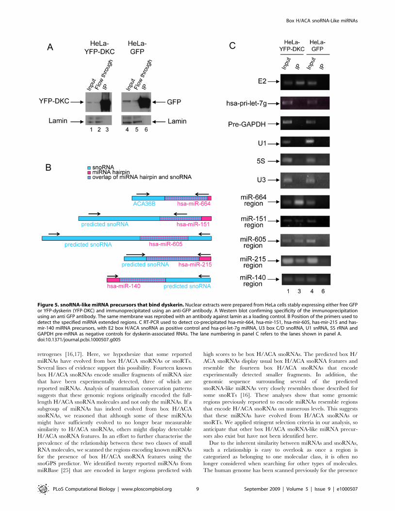
Predicted snoRNA-like miRNA precursors that binddyskerin and localise in the nucleolus
To explore further potential snoRNA-like features of these miRNA
precursors and to investigate whether they have retained some H/
ACA snoRNA functionality, we tested whether they can bind
dyskerin, a protein component of the functional box H/ACA
snoRNPs. Dyskerin serves as the pseudouridine synthase [36] and is
proposed to bind the ACA box [37]. Five of the twenty snoRNA-like
miRNAs, mir-664, mir-151, mir-605, mir-215 and mir-140 were
selected for this analysis because they are expressed in HeLa cells.
Purified nuclei from HeLa cells expressing YFP-dyskerin or GFP as a
control were immunoprecipitated using an antibody against the
fluorescent proteins as described in the Methods. The RNA isolated
from these samples was analysed by RT-PCR for the presence of the
molecules of interest. As shown in Figure 5C, in addition to E2 (a
well-characterised box H/ACA snoRNA), five snoRNA-like miRNA
precursors (the extended regions of mir-664, mir-151, mir-605, mir-
215 and mir-140) are bound by dyskerin. In contrast, the precursor of
hsa-let-7g, a miRNA precursor with no similarity to box H/ACA
snoRNAs is not pulled down by dyskerin. And as expected, other
abundant nuclear RNAs including GAPDH pre-mRNA, the small
nuclear RNA U1, the box C/D snoRNA U3 and 5S rRNA are not
immunoprecipitated by dyskerin. These binding experiments confirm
the in silico predictions that some miRNA precursors sufficiently
resemble box H/ACA snoRNAs to be bound by dyskerin.
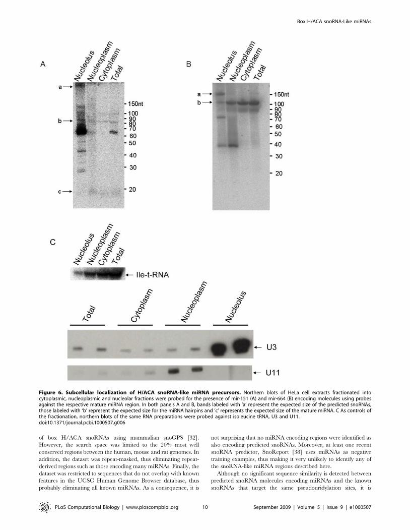
Two of the dyskerin-bound miRNA precursors were further
characterised by fractionated northern analysis to investigate
where the predicted snoRNAs and smaller fragments localise in
the cell. As shown in Figure 6, bands of the size of the predicted
H/ACA snoRNA full-length molecules encoding mir-151 and
mir-664 localise to the nucleolus (bands labelled with ‘a’ in panels
6A and 6B). Bands of the size of the miRNA hairpins are detected
in all three fractions although the putative mir-151 hairpin is
mainly nucleolar whereas the putative mir-664 hairpin accumu-
lates predominantly in the nucleoplasm and cytoplasm (bands
labelled with ‘b’ in panels 6A and 6B). The mir-151 mature form is
also detected and mainly found in the nucleoplasm and cytoplasm
(bands labelled with ‘c’ in panel 6A. For a longer exposure, please
see Figure S5). And a band slightly larger than the mir-664 mature
form localises mainly in the nucleoplasm.
Discussion
Numerous miRNAs have been previously shown to be repeat-
derived [12,19–21] and many snoRNAs have been described as
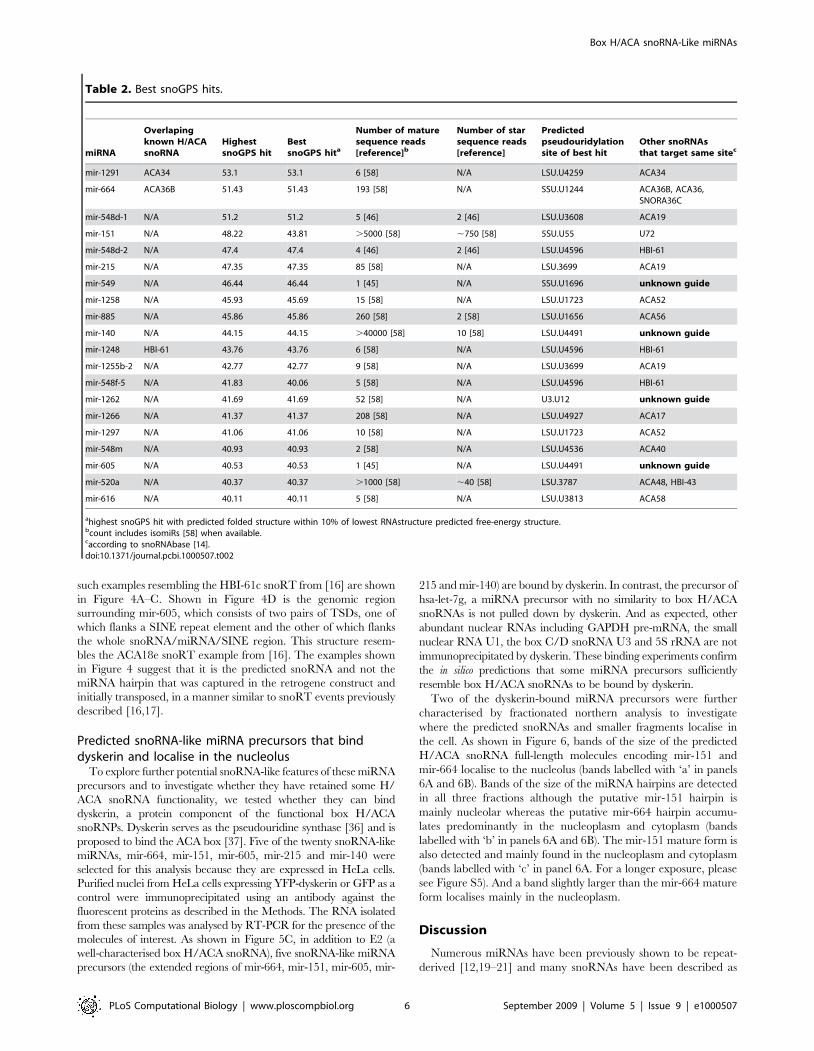
Table 2. Best snoGPS hits.
miRNA
Overlapingknown H/ACAsnoRNA
HighestsnoGPS hit
BestsnoGPS hita
Number of maturesequence reads[reference]b
Number of starsequence reads[reference]
Predictedpseudouridylationsite of best hit
Other snoRNAsthat target same sitec
mir-1291 ACA34 53.1 53.1 6 [58] N/A LSU.U4259 ACA34
mir-664 ACA36B 51.43 51.43 193 [58] N/A SSU.U1244 ACA36B, ACA36,SNORA36C
mir-548d-1 N/A 51.2 51.2 5 [46] 2 [46] LSU.U3608 ACA19
mir-151 N/A 48.22 43.81 .5000 [58] ,750 [58] 5SU.U55 U72
mir-548d-2 N/A 47.4 47.4 4 [46] 2 [46] LSU.U4596 HBI-61
mir-215 N/A 47.35 47.35 85 [58] N/A LSU.3699 ACA19
mir-549 N/A 46.44 46.44 1 [45] N/A SSU.U1696 unknown guide
mir-1258 N/A 45.93 45.69 15 [58] N/A LSU.U1723 ACA52
mir-885 N/A 45.86 45.86 260 [58] 2 [58] LSU.U1656 ACA56
mir-140 N/A 44.15 44.15 .40000 [58] 10 [58] LSU.U4491 unknown guide
mir-1248 HBI-61 43.76 43.76 6 [58] N/A LSU.U4596 HBI-61
mir-1255b-2 N/A 42.77 42.77 9 [58] N/A LSU.U3699 ACA19
mir-548f-5 N/A 41.83 40.06 5 [58] N/A LSU.U4596 HBI-61
mir-1262 N/A 41.69 41.69 52 [58] N/A U3.U12 unknown guide
mir-1266 N/A 41.37 41.37 208 [58] N/A LSU.U4927 ACA17
mir-1297 N/A 41.06 41.06 10 [58] N/A LSU.U1723 ACA52
mir-548m N/A 40.93 40.93 2 [58] N/A LSU.U4536 ACA40
mir-605 N/A 40.53 40.53 1 [45] N/A LSU.U4491 unknown guide
mir-520a N/A 40.37 40.37 .1000 [58] ,40 [58] LSU.3787 ACA48, HBI-43
mir-616 N/A 40.11 40.11 5 [58] N/A LSU.U3813 ACA58
ahighest snoGPS hit with predicted folded structure within 10% of lowest RNAstructure predicted free-energy structure.bcount includes isomiRs [58] when available.caccording to snoRNAbase [14].doi:10.1371/journal.pcbi.1000507.t002
Box H/ACA snoRNA-Like miRNAs
PLoS Computational Biology | www.ploscompbiol.org 6 September 2009 | Volume 5 | Issue 9 | e1000507
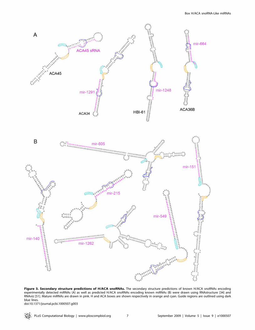
Figure 3. Secondary structure predictions of H/ACA snoRNAs. The secondary structure predictions of known H/ACA snoRNAs encodingexperimentally detected miRNAs (A) as well as predicted H/ACA snoRNAs encoding known miRNAs (B) were drawn using RNAstructure [34] andRNAviz [51]. Mature miRNAs are drawn in pink. H and ACA boxes are shown respectively in orange and cyan. Guide regions are outlined using darkblue lines.doi:10.1371/journal.pcbi.1000507.g003
Box H/ACA snoRNA-Like miRNAs
PLoS Computational Biology | www.ploscompbiol.org 7 September 2009 | Volume 5 | Issue 9 | e1000507
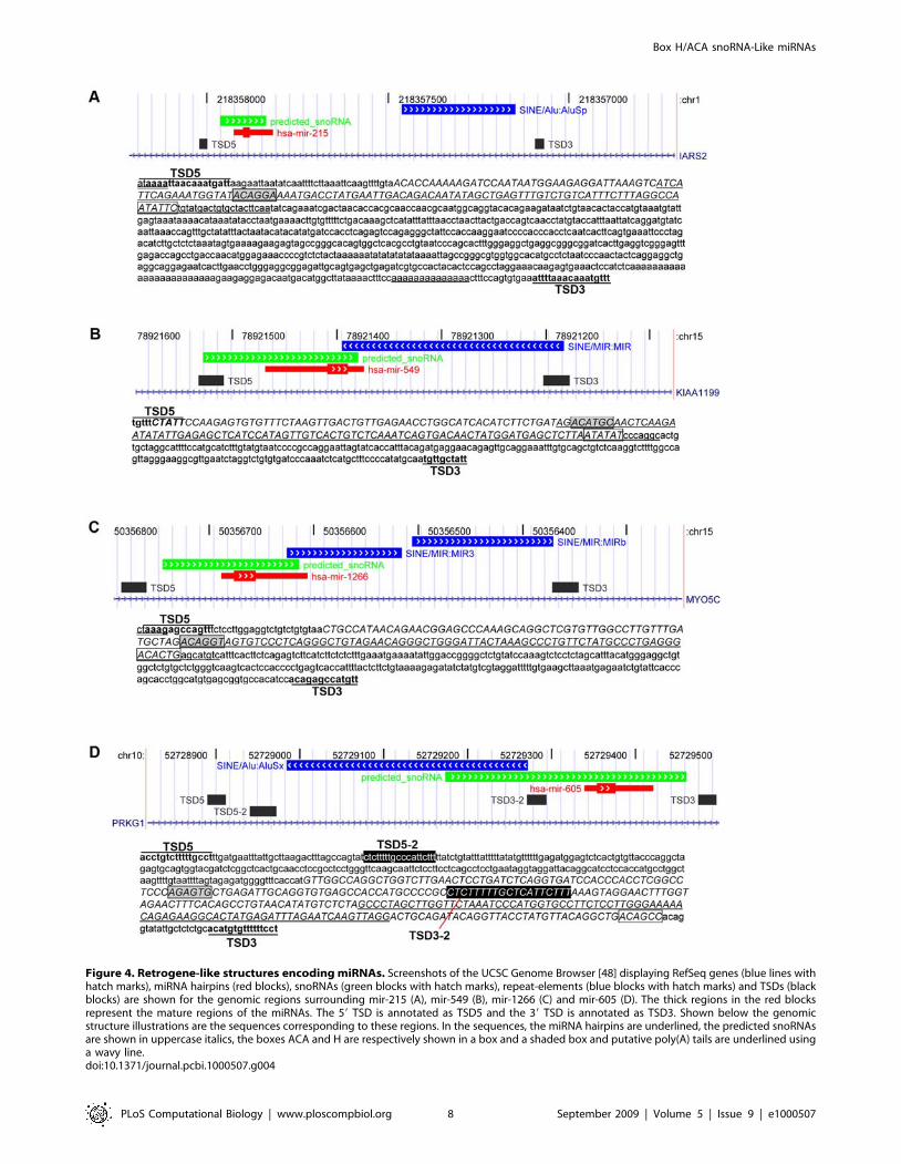
Figure 4. Retrogene-like structures encoding miRNAs. Screenshots of the UCSC Genome Browser [48] displaying RefSeq genes (blue lines withhatch marks), miRNA hairpins (red blocks), snoRNAs (green blocks with hatch marks), repeat-elements (blue blocks with hatch marks) and TSDs (blackblocks) are shown for the genomic regions surrounding mir-215 (A), mir-549 (B), mir-1266 (C) and mir-605 (D). The thick regions in the red blocksrepresent the mature regions of the miRNAs. The 59 TSD is annotated as TSD5 and the 39 TSD is annotated as TSD3. Shown below the genomicstructure illustrations are the sequences corresponding to these regions. In the sequences, the miRNA hairpins are underlined, the predicted snoRNAsare shown in uppercase italics, the boxes ACA and H are respectively shown in a box and a shaded box and putative poly(A) tails are underlined usinga wavy line.doi:10.1371/journal.pcbi.1000507.g004
Box H/ACA snoRNA-Like miRNAs
PLoS Computational Biology | www.ploscompbiol.org 8 September 2009 | Volume 5 | Issue 9 | e1000507
retrogenes [16,17]. Here, we hypothesize that some reported
miRNAs have evolved from box H/ACA snoRNAs or snoRTs.
Several lines of evidence support this possibility. Fourteen known
box H/ACA snoRNAs encode smaller fragments of miRNA size
that have been experimentally detected, three of which are
reported miRNAs. Analysis of mammalian conservation patterns
suggests that these genomic regions originally encoded the full-
length H/ACA snoRNA molecules and not only the miRNAs. If a
subgroup of miRNAs has indeed evolved from box H/ACA
snoRNAs, we reasoned that although some of these miRNAs
might have sufficiently evolved to no longer bear measurable
similarity to H/ACA snoRNAs, others might display detectable
H/ACA snoRNA features. In an effort to further characterise the
prevalence of the relationship between these two classes of small
RNA molecules, we scanned the regions encoding known miRNAs
for the presence of box H/ACA snoRNA features using the
snoGPS predictor. We identified twenty reported miRNAs from
miRBase [25] that are encoded in larger regions predicted with
high scores to be box H/ACA snoRNAs. The predicted box H/
ACA snoRNAs display usual box H/ACA snoRNA features and
resemble the fourteen box H/ACA snoRNAs that encode
experimentally detected smaller fragments. In addition, the
genomic sequence surrounding several of the predicted
snoRNA-like miRNAs very closely resembles those described for
some snoRTs [16]. These analyses show that some genomic
regions previously reported to encode miRNAs resemble regions
that encode H/ACA snoRNAs on numerous levels. This suggests
that these miRNAs have evolved from H/ACA snoRNAs or
snoRTs. We applied stringent selection criteria in our analysis, so
anticipate that other box H/ACA snoRNA-like miRNA precur-
sors also exist but have not been identified here.
Due to the inherent similarity between miRNAs and snoRNAs,
such a relationship is easy to overlook as once a region is
categorized as belonging to one molecular class, it is often no
longer considered when searching for other types of molecules.
The human genome has been scanned previously for the presence
Figure 5. snoRNA-like miRNA precursors that bind dyskerin. Nuclear extracts were prepared from HeLa cells stably expressing either free GFPor YFP-dyskerin (YFP-DKC) and immunoprecipitated using an anti-GFP antibody. A Western blot confirming specificity of the immunoprecipitationusing an anti GFP antibody. The same membrane was reprobed with an antibody against lamin as a loading control. B Position of the primers used todetect the specified miRNA extended regions. C RT-PCR used to detect co-precipitated hsa-mir-664, hsa-mir-151, hsa-mir-605, has-mir-215 and has-mir-140 miRNA precursors, with E2 box H/ACA snoRNA as positive control and hsa-pri-let-7g miRNA, U3 box C/D snoRNA, U1 snRNA, 5S rRNA andGAPDH pre-mRNA as negative controls for dyskerin-associated RNAs. The lane numbering in panel C refers to the lanes shown in panel A.doi:10.1371/journal.pcbi.1000507.g005
Box H/ACA snoRNA-Like miRNAs
PLoS Computational Biology | www.ploscompbiol.org 9 September 2009 | Volume 5 | Issue 9 | e1000507
of box H/ACA snoRNAs using mammalian snoGPS [32].
However, the search space was limited to the 20% most well
conserved regions between the human, mouse and rat genomes. In
addition, the dataset was repeat-masked, thus eliminating repeat-
derived regions such as those encoding many miRNAs. Finally, the
dataset was restricted to sequences that do not overlap with known
features in the UCSC Human Genome Browser database, thus
probably eliminating all known miRNAs. As a consequence, it is
not surprising that no miRNA encoding regions were identified as
also encoding predicted snoRNAs. Moreover, at least one recent
snoRNA predictor, SnoReport [38] uses miRNAs as negative
training examples, thus making it very unlikely to identify any of
the snoRNA-like miRNA regions described here.
Although no significant sequence similarity is detected between
predicted snoRNA molecules encoding miRNAs and the known
snoRNAs that target the same pseudouridylation sites, it is
Figure 6. Subcellular localization of H/ACA snoRNA-like miRNA precursors. Northern blots of HeLa cell extracts fractionated intocytoplasmic, nucleoplasmic and nucleolar fractions were probed for the presence of mir-151 (A) and mir-664 (B) encoding molecules using probesagainst the respective mature miRNA region. In both panels A and B, bands labeled with ‘a’ represent the expected size of the predicted snoRNAs,those labeled with ‘b’ represent the expected size for the miRNA hairpins and ‘c’ represents the expected size of the mature miRNA. C As controls ofthe fractionation, northern blots of the same RNA preparations were probed against isoleucine tRNA, U3 and U11.doi:10.1371/journal.pcbi.1000507.g006
Box H/ACA snoRNA-Like miRNAs
PLoS Computational Biology | www.ploscompbiol.org 10 September 2009 | Volume 5 | Issue 9 | e1000507
interesting to note that three snoRNAs (ACA52, HBI-61 and
ACA19) share their target pseudouridylation sites with eight of the
predicted snoRNAs encoding miRNAs (Table 2). This situation
also exists amongst known snoRNAs, some of which share the
same target site without displaying significant sequence similarity.
In particular, examples exist of a snoRNA harbouring two guide
regions, each of which is shared with a different snoRNA. For
example, ACA22 which shares one of its targets with ACA33 and
the other with U64, has no significant sequence similarity with
either molecule. Similarly, ACA50 shares target sites with ACA36,
ACA8 and ACA62 but while it has high sequence identity with
ACA62, it has no significant sequence similarity with ACA8 or
ACA36.
This redundancy in rRNA complementarity may suggest some
box H/ACA snoRNAs and snoRTs are not under as much
selective pressure to avoid mutations. We hypothesize that some of
these snoRNA encoding regions might be in the process of
evolving from functional snoRNAs to miRNA-like precursors.
This process might be facilitated by the fact that box H/ACA
snoRNAs have a structure (2 hairpins) that is probably favourable
to the formation of miRNAs. The in silico data presented here
support these ideas. We tested the predictions by experimentally
showing that the precursors of five of the predicted snoRNA-like
miRNAs, mir-664, mir-151, mir-605, mir-215 and mir-140,
interact with dyskerin, a protein component of functional H/
ACA snoRNPs. While a lack of interaction to dyskerin could not
rule out an evolutionary relationship between these miRNAs and
snoRNAs as the miRNAs might have evolved sufficiently to no
longer interact with functional protein components of snoRNPs,
the detection of such an interaction considerably reinforces such
claims. These results show that the snoRNA-like miRNA
precursors sufficiently resemble box H/ACA snoRNAs to bind
dyskerin, strengthening the possibility of an evolutionary relation-
ship between these molecules. Further experiments will be
necessary to investigate whether these molecules also retain the
capability of targeting rRNA in vivo. It will also be necessary to
experimentally test whether the remaining fifteen predicted
snoRNA-like miRNA precursors also display aspects of H/ACA
snoRNA functionality. The fact that three of these molecules (mir-
548d-1, mir-1297 and mir-616) display the sequence AGA instead
of the canonical ACA box could indicate that they have evolved
sufficiently to no longer retain H/ACA snoRNA functionality. We
do note that while identifying molecules that display both miRNA
and snoRNA functionality supports our evolutionary hypothesis,
we also expect to find a larger number of molecules that display
features of both molecules but do not represent completely
prototypical examples.
It is interesting to note that Saccharomyces cerevisiae has snoRNAs
but no reported miRNAs, consistent with a relationship where
primordial snoRNAs may have given rise to certain classes of
miRNAs. This idea is supported by a recent article by Saraiya and
Wang reporting that the primitive parasitic protozoan Giardia
lamblia, which does not have RNA interference capabilities but has
miRNA processing machinery, uses box C/D snoRNAs as
miRNA precursors [39]. In addition to this, Taft and colleagues
have recently reported that most snoRNAs in animals, Arabidopsis
and Schizosaccharomyces pombe generate small RNAs (of ,20–24
nucleotides in length for animal box H/ACA snoRNAs), which
are associated with argonaute proteins [40]. Current data such as
these dual function molecules with both miRNA and snoRNA
capabilities which exist in both human [24] and Giardia [39] and
likely many other organisms [40] suggest this process of evolving
from a snoRNA encoding genomic region to a miRNA-like
encoding region could be ongoing.
In addition to investigating whether some of the H/ACA
snoRNA-like miRNA precursors display functional H/ACA
snoRNA capability by binding to dyskerin (Figure 5), we have
also characterised the cellular localisation of two of these
molecules: the precursors of mir-151 and mir-664 (Figure 6).
While bands of the size of the predicted full-length H/ACA
snoRNA molecules localise to the nucleolus, consistent with their
binding to dyskerin, bands of the size of the predicted hairpin form
of these miRNAs can be found in all three fractions considered but
accumulate mainly in the nucleolus (in the case of mir-151) and in
the nucleoplasm and cytoplasm (in the case of mir-664). The
mature form of mir-151 accumulates mainly in the nucleoplasm
which is unusual for a miRNA but might be a consequence of the
snoRNA features displayed by its precursor. These results are
consistent with a recent study showing that the precursors and/or
mature form of a number of rat miRNAs accumulate in the
nucleolus [41]. Further studies will be required to investigate the
exact nature and role of each of these molecules in these cellular
compartments as well as how they are processed.
While all the miRNAs characterised in Table 2 are classified as
miRNAs in miRBase [25], they have not all been extensively
analysed. Of the five extended miRNA regions that we
experimentally found to be bound by dyskerin, three (mir-151,
mir-140 and mir-215) have been further characterized and
functionally validated, either by studies of their processing into
their mature form or validation of their targets and effects. Indeed,
mir-151 has been shown to be processed into its mature form by
usual miRNA processing machinery [42] while functional targets
of mir-140 have been experimentally validated [43]. And mir-215,
which has been shown to have reduced expression in cancer tissues
compared to normal cells, is capable of inducing cell-cycle arrest,
colony suppression and cell detachment from a solid support when
transfected into cells [44]. Mir-664 and mir-605 have not, to our
knowledge, been further functionally validated and will require
additional experimental evidence to confirm they are true
miRNAs. In particular, mir-605 has only been identified
previously with one sequence read [45]. Given that the extended
region of mir-605 is predicted to serve as a guide for an
experimentally validated pseudouridylation site whose guide is
unknown, we postulate that this region encodes an H/ACA
snoRNA rather than a miRNA. This type of analysis can thus be
used to filter out unlikely miRNA candidates from the large
miRNA repositories which contain many poorly characterized
molecules.
A recent large-scale study defining an expression atlas for
mammalian miRNAs, by Landgraf and colleagues, classifies
known miRNAs into four different groups: prototypical, repeat-
derived, repeat-clustered and unclassified [46]. A lack of
repetitiveness, evolutionary conservation and 59 end processing
were considered to classify miRNAs as prototypical. Only two
(mir-215 and mir-140) of our twenty miRNAs encoded in
predicted snoRNAs are classified as protypical in this study. The
remaining eighteen miRNAs were either classified as repeat-
derived (5 miRNAs), repeat-clustered (1 miRNA), unclassified (2
miRNAs) or were not considered in the study (10 miRNAs). The
results of a recent deep-sequencing study [45] analysed in the
context of the Landgraf study reveals that only 17% of all
miRNAs identified in this manner are prototypical [46]. The
remaining miRNAs displayed irregularities in their processing or
unusual sequence conservation patterns [46]. The authors
further went on to investigate possible functional implications,
determining that unlike non-prototypical miRNAs, prototypical
miRNAs showed enrichment of putative target sites in 39UTRs
[46]. In light of the relationship uncovered here between box H/
Box H/ACA snoRNA-Like miRNAs
PLoS Computational Biology | www.ploscompbiol.org 11 September 2009 | Volume 5 | Issue 9 | e1000507

ACA snoRNAs and miRNAs, it is reasonable to speculate that
snoRNA-encoded miRNAs might be in the process of evolving
into functional miRNAs but still retain some non-miRNA-like
features which could preclude them from being classified as
prototypical. Alternatively, perhaps these molecules should be
classified as a separate small RNA class. Further analysis will be
necessary to clarify the role and exact relationship of these
molecules.
Methods
Sequence analysis of genomic regions surroundingmiRNA genes
The genomic positions, sequences and flanking regions of
human miRNA genes and box H/ACA snoRNA genes were
downloaded from the UCSC Table Browser [47], wgRNA table
[14,25] using the March 2006 assembly of the human genome.
Repeat elements surrounding these genomic regions were
identified with the RepeatMasker program (http://www.
repeatmasker.org). MiRNA and box H/ACA snoRNA genes
as well as repeat-elements and sequence features were visualised
with the UCSC Genome Browser [48], using information from
the wgRNA table [14,25], the Vertebrate Multiz Alignment and
PhastCons Conservation utilities [49,50] as well as custom
tracks.
Prediction of H/ACA snoRNAs676 distinct human miRNA encoding regions (including the
miRNA hairpins and 175 flanking nucleotides on either side) were
scanned for the presence of box H/ACA snoRNAs using a locally-
installed copy of the snoGPS program [32]. In addition to known
miRNAs, two sets of control sequences were also scanned using
snoGPS. The first sets of control sequences consisted of 676
randomly generated sequences having the same nucleotide
composition as human intronic sequences and the same length
distribution as the miRNA encoding regions. The second sets of
control sequences consisted of randomly generated hairpins having
the same length distribution and minimum free-energy distribution
as the miRNA hairpin set. RNAfold [33] was used to predict the
minimum free-energy of the randomly-generated hairpin sequenc-
es. The snoGPS parameters used throughout this analysis were as
follow. The target sites for pseudouridylation were defined in the file
‘snAndRrna.targ’ provided with the snoGPS download. The
descriptor and scoretable files used were respectively ‘Mam-
GUs2.v3.desc’ and ‘human.v3.scoretables’, provided with the
snoGPS download.
Secondary structure visualisationRNA secondary structures were predicted using RNAstructure
4.5 [34] and annotated using RnaViz 2.0 [51].
Immunoprecipitation and quantitative RT-PCRImmunoprecipitations were prepared as previously described [52].
Nuclear lysates were prepared from HeLaYFP-Dyskerin and HeLaGFP
stable cell lines. Purified nuclei were resuspended in RIPA buffer to
solubilise proteins. Fluorescence proteins were immunoprecipitated
using GFP binder (ChromoTek) covalently coupled to NHS-
activated Sepharose 4 Fast Flow beads (GE Healthcare at 1 mg/ml
as previously described [53]). Samples were divided in two and for
Input samples RNA was isolated from one half of each nuclear lysate.
RNA was isolated by the TRIzol method with DNase I treatment,
according to manufacturer’s instructions (Invitrogen). RT-PCR was
performed to detect immunoprecipitated RNAs. Reverse transcrip-
tion and PCR were performed with the following gene-specific
primers (hsa-pri-let-7g: 59-CGCTCCGTTTCCTTTTGCCTG-39
and 59 TACAGTTATCTCCTGTACCGG-39, U3: 59-AGAGG-
TAGCGTTTTCTCCTGAGCG-39 and 59 ACCACTCAGACC-
GCGTTCTC-39, pre-GAPDH: 59-CGCATCTTCTTTTGCGT-
CGCCAG-39 and 59-GGTCAATGAAGGGGTCATTGATGGC-
39, U1: 59-TACCTGGCAGGGGAGATACCATGATC-39 and 59-
GCAGTCGAGTTTCCCACATTTGGGG-39, 5S: 59-ACGCGC-
CCGATCTCGTCTGAT-39 and 59-GCCTACAGCACCCGGT-
ATTCCC-39, miR-664: 59-GTGTTAAGTTCAGTTCAGGGTA-
G-39 and 59-CATTTTGTAGGCTGGGGATAAATG-39, miR-
151: 59-GGCTTACCCTATGCTGCTATA-39 and 59-GTAGGG-
GATGAGACATACTAGAC-39, miR-605: 59-CTGGTCTTGAA-
CTCCTGATCTC-39 and 59- GCTGTCAGCCTGTAACATA-
GG-39, miR-215: 59-CCAAAAAGATCCAATAATGGAAGAG-
GATTAAAG-39 and 59-TTGAAGTAGCACAGTCATACAG-39,
miR-140: 59-GTGTGTCTCTCTCTGTGTCC-39 and 59-GGA-
TGTCCCAAGGGGGCCAG-39) using the SuperScript one-step
RT-PCR kit (Invitrogen). To decide linearity of cycles, we
performed real time PCR using the Superscript III Platinum
SYBR Green one-step qRT-PCR Kit (Invitrogen) and Rotor-
Gene RG-3000 system (Corbett Research). The same amount of
RNA for input and immunoprecipitated RNA (IP) was used as
templates for RT-PCR reactions. Each experiment was repeated
three times independently.
Northern and high sensitivity RNA blot analysis.HeLa cell extracts were fractionated using sucrose gradients, as
previously described [54–56]. Total HeLa cell RNA and RNA
from separate cytoplasmic, nucleoplasmic and nucleolar fractions
was isolated using the TRIzol method, with Dnase I treatment,
according to manufacturer’s instructions (Invitrogen). Equal
amounts of RNA from each sample were separated by 8M Urea
polyacrylamide denaturing gel electrophoresis in 16MOPS buffer
and the RNA transferred onto nylon membrane (Hybond-N;
Amersham) by electro blotting. After chemical cross linking, the
membrane was hybridized with 32P 59 end-labelled oligoribonu-
cleotide probes specific for the following RNA species; (mir-664:
59- UGUAGGCUGGGGAUAAAUGAAUA-39, mir-151: 59-CC-
UCAAGGAGCUUCAGUCUAG-39, tRNA-Ile 59-UGGUGGC-
CCGUACGGGGAUCGA-39, U11: 59-TCTTGATGTCGATT-
CCGCACGCAGAGCAATCGAGTTGCCC-39 and U3: 59-CA-
CTCAGACCGCGTTCTCTCCCTCTCACTCCCCAATACG-
G-39). High sensitivity RNA blots were prepared as previously
described [57].
Supporting Information
Figure S1 Mammalian conservation of box H/ACA snoRNAs
that encode experimentally detected smaller fragments
Found at: doi:10.1371/journal.pcbi.1000507.s001 (1.63 MB PDF)
Figure S2 Predicted secondary structures of H/ACA snoRNA-
like miRNA precursors
Found at: doi:10.1371/journal.pcbi.1000507.s002 (0.07 MB PDF)
Figure S3 Repeat elements in proximity of H/ACA snoRNA-
like miRNA genomic regions
Found at: doi:10.1371/journal.pcbi.1000507.s003 (3.58 MB PDF)
Figure S4 Sequence elements surrounding selected miRNAs
Found at: doi:10.1371/journal.pcbi.1000507.s004 (0.03 MB PDF)
Figure S5 Northern blot showing the subcellular localization of
H/ACA snoRNA-like miRNA precursors (same as Figure 6AB but
longer exposure)
Found at: doi:10.1371/journal.pcbi.1000507.s005 (0.31 MB PDF)
Box H/ACA snoRNA-Like miRNAs
PLoS Computational Biology | www.ploscompbiol.org 12 September 2009 | Volume 5 | Issue 9 | e1000507

Acknowledgments
We would like to thank Dr Tom Walsh for technical support as well as Drs
Chris Cole and Gyorgy Hutvagner for helpful discussions.
Author Contributions
Conceived and designed the experiments: MSS MO AIL. Performed the
experiments: MSS FA. Analyzed the data: MSS MO GJB. Wrote the
paper: MSS FA AIL GJB.
References
1. Kiss T (2002) Small nucleolar RNAs: an abundant group of noncoding RNAswith diverse cellular functions. Cell 109: 145–148.
2. Bachellerie JP, Cavaille J, Huttenhofer A (2002) The expanding snoRNA world.
Biochimie 84: 775–790.3. Filipowicz W, Pogacic V (2002) Biogenesis of small nucleolar ribonucleoproteins.
Curr Opin Cell Biol 14: 319–327.4. Tycowski KT, Aab A, Steitz JA (2004) Guide RNAs with 59 caps and novel box
C/D snoRNA-like domains for modification of snRNAs in metazoa. Curr Biol
14: 1985–1995.5. Lai EC (2005) miRNAs: whys and wherefores of miRNA-mediated regulation.
Curr Biol 15: R458–460.6. Lai EC (2003) microRNAs: runts of the genome assert themselves. Curr Biol 13:
R925–936.7. Vasudevan S, Tong Y, Steitz JA (2007) Switching from repression to activation:
microRNAs can up-regulate translation. Science 318: 1931–1934.
8. Baskerville S, Bartel DP (2005) Microarray profiling of microRNAs revealsfrequent coexpression with neighboring miRNAs and host genes. Rna 11:
241–247.9. Kim YK, Kim VN (2007) Processing of intronic microRNAs. Embo J 26:
775–783.
10. Rodriguez A, Griffiths-Jones S, Ashurst JL, Bradley A (2004) Identification ofmammalian microRNA host genes and transcription units. Genome Res 14:
1902–1910.11. Lee Y, Kim M, Han J, Yeom KH, Lee S, et al. (2004) MicroRNA genes are
transcribed by RNA polymerase II. Embo J 23: 4051–4060.
12. Borchert GM, Lanier W, Davidson BL (2006) RNA polymerase III transcribeshuman microRNAs. Nat Struct Mol Biol 13: 1097–1101.
13. Niwa R, Slack FJ (2007) The evolution of animal microRNA function. CurrOpin Genet Dev 17: 145–150.
14. Lestrade L, Weber MJ (2006) snoRNA-LBME-db, a comprehensive database ofhuman H/ACA and C/D box snoRNAs. Nucleic Acids Res 34: D158–162.
15. Hertel J, Lindemeyer M, Missal K, Fried C, Tanzer A, et al. (2006) The
expansion of the metazoan microRNA repertoire. BMC Genomics 7: 25.16. Luo Y, Li S (2007) Genome-wide analyses of retrogenes derived from the human
box H/ACA snoRNAs. Nucleic Acids Res 35: 559–571.17. Weber MJ (2006) Mammalian Small Nucleolar RNAs Are Mobile Genetic
Elements. PLoS Genet 2: e205.
18. Vitali P, Royo H, Seitz H, Bachellerie JP, Huttenhofer A, et al. (2003)Identification of 13 novel human modification guide RNAs. Nucleic Acids Res
31: 6543–6551.19. Smalheiser NR, Torvik VI (2005) Mammalian microRNAs derived from
genomic repeats. Trends Genet 21: 322–326.20. Piriyapongsa J, Marino-Ramirez L, Jordan IK (2007) Origin and evolution of
human microRNAs from transposable elements. Genetics 176: 1323–1337.
21. Piriyapongsa J, Jordan IK (2007) A family of human microRNA genes fromminiature inverted-repeat transposable elements. PLoS ONE 2: e203.
22. Lander ES, Linton LM, Birren B, Nusbaum C, Zody MC, et al. (2001) Initialsequencing and analysis of the human genome. Nature 409: 860–921.
23. Kawaji H, Nakamura M, Takahashi Y, Sandelin A, Katayama S, et al. (2008)
Hidden layers of human small RNAs. BMC Genomics 9: 157.24. Ender C, Krek A, Friedlander MR, Beitzinger M, Weinmann L, et al. (2008) A
Human snoRNA with MicroRNA-Like Functions. Mol Cell 32: 519–528.25. Griffiths-Jones S, Grocock RJ, van Dongen S, Bateman A, Enright AJ (2006)
miRBase: microRNA sequences, targets and gene nomenclature. Nucleic AcidsRes 34: D140–144.
26. Ganot P, Caizergues-Ferrer M, Kiss T (1997) The family of box ACA small
nucleolar RNAs is defined by an evolutionarily conserved secondary structureand ubiquitous sequence elements essential for RNA accumulation. Genes Dev
11: 941–956.27. Gu AD, Zhou H, Yu CH, Qu LH (2005) A novel experimental approach for
systematic identification of box H/ACA snoRNAs from eukaryotes. Nucleic
Acids Res 33: e194.28. Kiss AM, Jady BE, Bertrand E, Kiss T (2004) Human box H/ACA
pseudouridylation guide RNA machinery. Mol Cell Biol 24: 5797–5807.29. Kiss T, Filipowicz W (1993) Small nucleolar RNAs encoded by introns of the
human cell cycle regulatory gene RCC1. Embo J 12: 2913–2920.
30. Ruff EA, Rimoldi OJ, Raghu B, Eliceiri GL (1993) Three small nucleolar RNAsof unique nucleotide sequences. Proc Natl Acad Sci U S A 90: 635–638.
31. Darzacq X, Jady BE, Verheggen C, Kiss AM, Bertrand E, et al. (2002) Cajalbody-specific small nuclear RNAs: a novel class of 29-O-methylation and
pseudouridylation guide RNAs. Embo J 21: 2746–2756.
32. Schattner P, Barberan-Soler S, Lowe TM (2006) A computational screen formammalian pseudouridylation guide H/ACA RNAs. Rna 12: 15–25.
33. Hofacker IL, Fontana W, Stadler PF, Bonhoeffer LS, Tacker M, et al. (1994)
Fast folding and comparison of RNA secondary structures. Monatshefte furChemie 125: 167–188.
34. Mathews DH, Disney MD, Childs JL, Schroeder SJ, Zuker M, et al. (2004)
Incorporating chemical modification constraints into a dynamic programmingalgorithm for prediction of RNA secondary structure. Proc Natl Acad Sci U S A
101: 7287–7292.
35. Bartel DP (2004) MicroRNAs: genomics, biogenesis, mechanism, and function.Cell 116: 281–297.
36. Hoang C, Ferre-D’Amare AR (2001) Cocrystal structure of a tRNA Psi55
pseudouridine synthase: nucleotide flipping by an RNA-modifying enzyme. Cell107: 929–939.
37. Li L, Ye K (2006) Crystal structure of an H/ACA box ribonucleoprotein
particle. Nature 443: 302–307.
38. Hertel J, Hofacker IL, Stadler PF (2008) SnoReport: computational identifica-tion of snoRNAs with unknown targets. Bioinformatics 24: 158–164.
39. Saraiya AA, Wang CC (2008) snoRNA, a Novel Precursor of microRNA in
Giardia lamblia. PLoS Pathog 4: e1000224.
40. Taft RJ, Glazov EA, Lassmann T, Hayashizaki Y, Carninci P, et al. (2009) SmallRNAs derived from snoRNAs. Rna 15: 1233–1240.
41. Politz JC, Hogan EM, Pederson T (2009) MicroRNAs with a nucleolar location.
Rna.
42. Kawahara Y, Zinshteyn B, Chendrimada TP, Shiekhattar R, Nishikura K(2007) RNA editing of the microRNA-151 precursor blocks cleavage by the
Dicer-TRBP complex. EMBO Rep 8: 763–769.
43. Nicolas FE, Pais H, Schwach F, Lindow M, Kauppinen S, et al. (2008)Experimental identification of microRNA-140 targets by silencing and
overexpressing miR-140. Rna 14: 2513–2520.
44. Braun CJ, Zhang X, Savelyeva I, Wolff S, Moll UM, et al. (2008) p53-Responsive micrornas 192 and 215 are capable of inducing cell cycle arrest.
Cancer Res 68: 10094–10104.
45. Cummins JM, He Y, Leary RJ, Pagliarini R, Diaz LA Jr, et al. (2006) Thecolorectal microRNAome. Proc Natl Acad Sci U S A 103: 3687–3692.
46. Landgraf P, Rusu M, Sheridan R, Sewer A, Iovino N, et al. (2007) A
mammalian microRNA expression atlas based on small RNA librarysequencing. Cell 129: 1401–1414.
47. Karolchik D, Hinrichs AS, Furey TS, Roskin KM, Sugnet CW, et al. (2004) The
UCSC Table Browser data retrieval tool. Nucleic Acids Res 32: D493–496.
48. Kent WJ, Sugnet CW, Furey TS, Roskin KM, Pringle TH, et al. (2002) Thehuman genome browser at UCSC. Genome Res 12: 996–1006.
49. Blanchette M, Kent WJ, Riemer C, Elnitski L, Smit AF, et al. (2004) Aligning
multiple genomic sequences with the threaded blockset aligner. Genome Res 14:708–715.
50. Siepel A, Bejerano G, Pedersen JS, Hinrichs AS, Hou M, et al. (2005)
Evolutionarily conserved elements in vertebrate, insect, worm, and yeastgenomes. Genome Res 15: 1034–1050.
51. De Rijk P, Wuyts J, De Wachter R (2003) RnaViz 2: an improved representation
of RNA secondary structure. Bioinformatics 19: 299–300.
52. Trinkle-Mulcahy L, Andersen J, Lam YW, Moorhead G, Mann M, et al. (2006)Repo-Man recruits PP1 gamma to chromatin and is essential for cell viability.
J Cell Biol 172: 679–692.
53. Rothbauer U, Zolghadr K, Muyldermans S, Schepers A, Cardoso MC, et al.(2008) A versatile nanotrap for biochemical and functional studies with
fluorescent fusion proteins. Mol Cell Proteomics 7: 282–289.
54. Andersen JS, Lam YW, Leung AK, Ong SE, Lyon CE, et al. (2005) Nucleolarproteome dynamics. Nature 433: 77–83.
55. Andersen JS, Lyon CE, Fox AH, Leung AK, Lam YW, et al. (2002) Directed
proteomic analysis of the human nucleolus. Curr Biol 12: 1–11.
56. Lam YW, Lamond AI, Mann M, Andersen JS (2007) Analysis of nucleolarprotein dynamics reveals the nuclear degradation of ribosomal proteins. Curr
Biol 17: 749–760.
57. Pall GS, Codony-Servat C, Byrne J, Ritchie L, Hamilton A (2007)Carbodiimide-mediated cross-linking of RNA to nylon membranes improves
the detection of siRNA, miRNA and piRNA by northern blot. Nucleic Acids
Res 35: e60.
58. Morin RD, O’Connor MD, Griffith M, Kuchenbauer F, Delaney A, et al. (2008)Application of massively parallel sequencing to microRNA profiling and
discovery in human embryonic stem cells. Genome Res 18: 610–621.
Box H/ACA snoRNA-Like miRNAs
PLoS Computational Biology | www.ploscompbiol.org 13 September 2009 | Volume 5 | Issue 9 | e1000507
Related Documents