DEPARTAMENTO DE MEDICINA Y CIRUGÍA ANIMAL FACULTAD DE VETERINARIA UNIVERSIDAD DE CORDOBA (ESPAÑA) IMPLEMENTACIÓN DE LA VITRIFICACIÓN EMBRIONARIA EN RAZAS EQUINAS COMO HERRAMIENTA PARA SU CONSERVACIÓN Y PROGRESO Córdoba, mayo de 2016 Trabajo presentado por D. Guillermo Vizuete Calero para optar al título de doctor Esta tesis doctoral ha sido parcialmente subvencionada por el programa predoctoral de Formación de Profesorado Universitario del Ministerio de Educación, Cultura y Deporte (FPU, convocatoria publicada en el BOE nº 302 de 13 de diciembre de 2012, Orden EDU/3204/2010) y por fondos del Instituto Nacional de Investigación y Tecnología Agraria y Alimentaria, en el marco del Subprograma Nacional de “Conservación de recursos genéticos de interés agroalimentario” del Plan Nacional de Investigación Científica, Desarrollo e Innovación Tecnológica (I+D+I) y fondos FEDER (Proyecto RZ2008-00025-00-00).

Welcome message from author
This document is posted to help you gain knowledge. Please leave a comment to let me know what you think about it! Share it to your friends and learn new things together.
Transcript

DEPARTAMENTO DE MEDICINA Y CIRUGÍA ANIMAL
FACULTAD DE VETERINARIA
UNIVERSIDAD DE CORDOBA (ESPAÑA)
IMPLEMENTACIÓN DE LA VITRIFICACIÓN EMBRIONARIA
EN RAZAS EQUINAS COMO HERRAMIENTA PARA SU
CONSERVACIÓN Y PROGRESO
Córdoba, mayo de 2016
Trabajo presentado por D. Guillermo Vizuete Calero para optar al
título de doctor
Esta tesis doctoral ha sido parcialmente subvencionada por el programa predoctoral de
Formación de Profesorado Universitario del Ministerio de Educación, Cultura y Deporte (FPU,
convocatoria publicada en el BOE nº 302 de 13 de diciembre de 2012, Orden EDU/3204/2010)
y por fondos del Instituto Nacional de Investigación y Tecnología Agraria y Alimentaria, en el
marco del Subprograma Nacional de “Conservación de recursos genéticos de interés
agroalimentario” del Plan Nacional de Investigación Científica, Desarrollo e Innovación
Tecnológica (I+D+I) y fondos FEDER (Proyecto RZ2008-00025-00-00).

TITULO: Implementación de la vitrificación embrionaria en razas equinas comoherramienta para su conservación y progreso
AUTOR: Guillermo Vizuete Calero
© Edita: UCOPress. 2016 Campus de RabanalesCtra. Nacional IV, Km. 396 A14071 Córdoba
www.uco.es/[email protected]


UNIVERSIDAD DE CÓRDOBA
DEPARTAMENTO DE MEDICINA Y CIRUGÍA ANIMAL
UNIVERSIDAD DE CÓRDOBA
IMPLEMENTACIÓN DE LA VITRIFICACIÓN EMBRIONARIA
EN RAZAS EQUINAS COMO HERRAMIENTA PARA SU
CONSERVACIÓN Y PROGRESO
Memoria de Tesis presentada por Guillermo Vizuete Calero, licenciado en
Veterinaria, para optar al grado de DOCTOR EN VETERINARIA
El director:
Dr. Carlos C. Pérez Marín


CARLOS C. PEREZ MARIN, DOCTOR EN VETERINARIA POR LA UNIVERSIDAD DE
CORDOBA, PROFESOR TITULAR DEL DEPARTAMENTO DE MEDICINA Y CIRUGÍA
ANIMAL DE LA UNIVERSIDAD DE CORDOBA,
INFORMA:
Que D. Guillermo Vizuete Calero, licenciado en Veterinaria, ha realizado en el
Departamento de Medicina y Cirugía Animal y bajo mi dirección, el trabajo titulado
"Implementación de la vitrificación embrionaria en razas equinas como herramienta
para su conservación y progreso", y que reúne los méritos científicos necesarios para
optar al grado de Doctor en Veterinaria.
Y para que así conste, firmo el presente informe en Córdoba a 28 de marzo de 2016.
Fdo. Carlos Carmelo Pérez Marín


IMPLEMENTACIÓN DE LA VITRIFICACIÓN EMBRIONARIA
EN RAZAS EQUINAS COMO HERRAMIENTA PARA SU
CONSERVACIÓN Y PROGRESO
Memoria de Tesis Doctoral presentada por Guillermo Vizuete Calero, Licenciado en
Veterinaria, para optar al grado de DOCTOR EN VETERINARIA
Aportaciones científicas derivadas del presente trabajo de Tesis Doctoral:
1. Publicaciones en revistas incluidas en el Journal Citation Report (JCR)
-Comparison of different treatments for oestrous induction in seasonally anovulatory mares. Vizuete, G., Diez, E., Galisteo, J., Agüera, E., Aguilera-Tejero, E., Pérez-Marín, C. (2013) Reproduction in Domestic Animals 48 (3) PP. 463 - 469. doi: 10.1111/rda.12098
2. Comunicaciones en congresos nacionales e internacionales
-V Jornadas de Innovación en Docencia Universitaria, comunicación oral titulada:
“Desarrollo de un simulador de partos en grandes animales” Córdoba 2 de abril
de 2014.
-Factors affecting the embryo recovery in a vitrification program in Hispano -Arabe equine breed. 11º Congreso Internacional de la Asociación Española de Reproducción Animal. Córdoba del 13 al 16 de junio de 2012. -Bacterial species present in the uterus of embryo donor mares and isolated in
flushing filters. 11º Congreso Internacional de la Asociación Española de Reproducción Animal. Córdoba del 13 al 16 de junio de 2012.
-Leptin levels in mares under treatment for estrus induction. 11º Congreso
Internacional de la Asociación Española de Reproducción Animal. Córdoba del 13 al
16 de junio de 2012.


MENCIÓN DE DOCTORADO INTERNACIONAL
La presente tesis cumple los criterios para la obtención de la mención Doctorado
Internacional, concedido por la Universidad de Córdoba, y regulado por el RD99/2011.
Para ello, se presentan los siguientes requisitos:
1. Estancias predoctoral becada por el Ministerio de Educación, Cultura y Deporte
(Beca FPU) para obtención de doctorado internacional en la Facultad de
Veterinaria de la Universidad Hannover (Alemania) bajo la supervisión de la
Dra. Profa. Christine Wrenzycki, desde 30-1-12 al 30-4-12.
Estancias predoctoral becada por el Ministerio de Educación, Cultura y Deporte
(Beca FPU) para obtención de doctorado internacional en la Facultad de
Veterinaria de la Universidad Utrecht (Holanda) bajo la supervisión de la Dr.
Prof. Tom AE Stout, desde 10-4-14 al 10-6-14.
2. Esta tesis está avalada por los siguientes informes de idoneidad realizados por 2
doctores de otros centros de investigación internacionales :
- Dra. Carolina Bianchi, Universidad Nacional del Centro de la Provincia de
Buenos Aires, Argentina.
- Dr. Dariusz Skarzynski, Institute of Animal Reproduction and Food Research of
Polish Academy of Sciences, Olsztyn, Polonia.
3. La defensa de la tesis y el texto se han realizado parcialmente en dos idiomas
europeos, español e inglés.
4. Entre los miembros del tribunal se encuentra la Dra. Graça Ferreira Dias,
doctora procedente de un centro de educación superior europeo (Universidad
de Lisboa, Portugal).
Córdoba a 30 de marzo de 2016
El doctorando: El director:
Fdo: Guillermo Vizuete Calero Fdo: Carlos C. Pérez Marín


“La única manera de ser un maestro de algo,
es amar sinceramente lo que haces.”
Anónimo
“Hay una fuerza motriz más poderosa que el vapor,
la electricidad y la energía atómica: la voluntad.”
Albert Einstein


1. RESUMEN ...................................................................................................................... 1
2. INTRODUCCIÓN............................................................................................................. 9
3. OBJETIVOS................................................................................................................... 17
4.REVISION BIBLIOGRAFICA ............................................................................................ 19
4.1. CICLO ESTRAL DE LA YEGUA Y LA BURRA ............................................................ 19
4.2. REGULACIÓN ENDOCRINA Y ACTIVIDAD FOLICULAR .......................................... 22
4.3. EMBRIOGENESIS .................................................................................................. 26
4.4. CAÍDA DEL EMBRIÓN AL ÚTERO .......................................................................... 30
4.5. TRATAMIENTOS DE SUPEROVULACIÓN............................................................... 31
4.6. RECOGIDA DE EMBRIONES .................................................................................. 39
4.7. EVALUACIÓN DE EMBRIONES .............................................................................. 40
4.7.1. VALORACIÓN MORFOLÓGICA....................................................................... 41
4.7.2. TINCIÓN DE EMBRIONES .............................................................................. 42
4.7.3. CULTIVO IN VITRO......................................................................................... 49
4.7.4. VALORACIÓN METABÓLICA .......................................................................... 49
4.7.5. TRANSFERENCIA AL ÚTERO DE UNA RECEPTORA ........................................ 49
4.8. PRESERVACIÓN DE EMBRIONES EQUINOS .......................................................... 50
4.8.1. CONGELACIÓN CONVENCIONAL................................................................... 50
4.8.2. VITRIFICACIÓN .............................................................................................. 57
4.8.3. CRIOPROTECTORES ....................................................................................... 65
5. MATERIALES Y MÉTODOS ........................................................................................... 71
ANIMALES ................................................................................................................... 71
ESTUDIO ECOGRÁFICO................................................................................................ 71
OBTENCIÓN DEL PLASMA SANGUÍNEO ...................................................................... 71
ANÁLISIS HORMONALES ............................................................................................. 72
TOMA DE MUESTRAS UTERINAS ................................................................................ 72

CULTIVO UTERINO E IDENTIFICACIÓN DE MICROORGANISMOS ............................... 72
LAVADO UTERINO ....................................................................................................... 72
EVALUACIÓN MORFOLÓGICA ..................................................................................... 73
CONGELACIÓN Y VITRIFICACIÓN ................................................................................ 73
DESCONGELACIÓN O CALENTAMIENTO ..................................................................... 74
EVALUACIÓN DE LA VIABILIDAD EMBRIONARIA ........................................................ 74
TRANSFERENCIA DE EMBRIONES................................................................................ 75
ANÁLISIS ESTADÍSTICO ................................................................................................ 75
6. RESULTADOS ............................................................................................................... 79
TRABAJO INVESTIGACION 1 ........................................................................................ 81
TRABAJO INVESTIGACION 2 ........................................................................................ 91
TRABAJO INVESTIGACION 3 ...................................................................................... 107
TRABAJO INVESTIGACION 4 ...................................................................................... 121
7. DISCUSIÓN GENERAL ................................................................................................ 143
8. CONCLUSIONES ......................................................................................................... 157
9. BIBLIOGRAFIA............................................................................................................ 161
10. AGRADECIMIENTOS ................................................................................................ 181

1
1. RESUMEN
La industria equina es un sector que posee una gran importancia dentro la economía y
la sociedad a nivel mundial, y donde ciertas razas de nuestro entorno gozan de un
especial significado a nivel socio-cultural. Esta es una de las razones por la que
debemos aumentar su conservación, producción y desarrollo. Y para conseguirlo,
tradicionalmente se han tomado una serie de medidas que pasaban por la gestión de
sementales y/o compra-venta de yeguas a ganaderos. En la actualidad, y gracias a los
avances en el campo de investigación, contamos con multitud de técnicas relacionadas
con la reproducción asistida como la sincronización de celos, obtención de semen,
inseminación artificial, transferencia de embriones o criopreservación de esperma,
óvulos y embriones, entre otras. Todas ellas poseen una gran importancia hoy día
aunque, en el caso de los équidos, su eficiencia se encuentra muy por detrás de otras
especies de producción. En las últimas décadas, la transferencia de embriones ha
adquirido una gran notoriedad en el sector, permitiendo que la genética materna
pueda comercializarse de manera parecida a como se hace con el semen en los
sementales más importantes. Sin embargo, los resultados obtenidos en la
criopreservación de embriones equinos no son del todo satisfactorios, debido a las
características anatómicas y fisiológicas propias de esta especie. El avance en esta
técnica y, por tanto, la mejora de los resultados supondría una serie de ventajas en el
sector; por un lado, se produciría un mayor número de crías al año de la yegua y/o
semental deseados; por otro, sería posible el nacimiento de potros cuyas madres
poseen problemas que le imposibilitan el parto o yeguas que se encuentren
compitiendo, sin la necesidad de tener que dejar de ampliar su palmarés; además, se
facilitaría la comercialización internacional de material genético, sin tener que
transportar animales, solo embriones; y otras muchas ventajas de tipo sanitario,
asociadas a la mejora genética o incluso favorecedoras de la investigación.
La finalidad de la presente Tesis Doctoral ha sido proponer alternativas a la
conservación y desarrollo de ciertas razas equinas en peligro de extinción, como el
caballo Hispano–árabe (H-a) y el burro Andaluz y Zamorano-leones, implementando la
vitrificación como método de criopreservación que puede simplificar y abaratar los
costes de dicho proceso. El trabajo llevado a cabo consta de cuatro estudios
experimentales en un total de 125 animales, como se resume a continuación.
En el primer estudio se utilizaron 41 yeguas H-a, que fueron sometidas a diferentes
protocolos de inducción y sincronización de celo. Todas ellas se encontraban en
anestro estacional, entendiéndose como tal cuando no existían signos de celo en al
menos 45 días previos, los folículos presentaban diámetro inferior a 20mm, había
ausencia de cuerpo lúteo y los niveles de progesterona eran inferiores a 1ng/ml. El
grupo control contaba con 15 animales, mientras que el resto de yeguas se distribuyó
en 3 tratamientos: T1, administración oral de altrenogest (0.044mg/kg; Regumate®,
Esteve) durante 11 días; T2, aplicación de un dispositivo intravaginal de progesterona

2
(1.38g; CIDR®) durante 11 días; y T3, aplicación del mismo dispositivo intravaginal de
progesterona (1.38g; CIDR®) durante solo 8 días. Todas las yeguas incluidas en este
estudio recibieron 1 ml intramuscular de DL-Cloprostenol un día después de terminar
el tratamiento. Los animales fueron sometidos a seguimiento ecográfico y se tomaron
muestras de sangre para determinar los niveles de progesterona y leptina.
Observamos que en ninguno de los grupos estudiados aparecieron signos clínicos de
endometritis que pudieran afectar a la fertilidad, y además ningún dispositivo se
perdió durante el estudio, por lo que, desde el punto de vista práctico, los dispositivos
intravaginales podían considerarse como una alternativa terapéutica eficaz a la
progesterona oral, debido a su fácil aplicación y a la homogeneidad en su dosificación.
Como resultado, un total de 88.5% de las yeguas recuperaron la ciclicidad tras el
tratamiento, ninguna ovuló durante el mismo y, tanto el porcentaje de yeguas en celo
como la tasa de ovulación, fueron similares en los grupos comparados. La tasa de
gestación tras la primera ovulación en los grupos de tratamiento con progestágenos
(47.4%) fue similar a la obtenida en el grupo control (46.7%). Al estudiar los niveles de
leptina, observamos una mayor concentración en las hembras que sí respondieron al
tratamiento con progesterona que en las que fracasaron, por lo que sugerimos que la
leptina podría utilizarse como indicador antes de aplicar estos tratamientos.
En el segundo de los trabajos realizados se llevó a cabo un estudio retrospectivo del
programa de conservación de embriones en equinos H-a y asnos españoles,
concretamente Andaluces y Zamorano-leoneses. Se emplearon 61 yeguas H-a
donantes y 17 sementales, así como 13 burras donantes y 5 garañones. El estudio
analizó la tasa de recogida embrionaria por lavado y por ovulación, y evaluó la
influencia de diferentes factores en el programa. Se observaron diferencias
significativas en la tasa de ovulación entre las especies, siendo mucho mayor en burra
que en yegua. La tasa de recogida mostró diferencias significativas en función de la
estacionalidad, siendo mayor durante época reproductiva que fuera de ella; esta tasa
también aumentó en función del número ovulaciones. Sin embargo, otras variables
como la edad de la donante, el día de lavado o la calidad del lavado no afectaron sobre
la tasa de recogida de embriones. En cuanto a la tasa de ovulación, no estuvo
influenciada por la edad de las hembras donantes ni por la estación reproductiva. El
90% de los embriones recogidos fueron de calidad excelente o buena, y la gran
variabilidad observada en los diámetros y estados embrionarios encontrados se puede
explicar por la asincronía entre ovulaciones cuando existían ovulaciones múltiples.
El tercer estudio fue planteado para conocer cómo podía afectar el ambiente uterino
de la donante en el momento del flushing sobre la recogida y calidad embrionaria. En
el grupo control (n=8) se incluyeron yeguas que presentaban endometritis clínica, y a
las que se realizó una citología y cultivo uterino mediante hisopo, que fueron
comparados con el líquido retenido en el filtro de embriones tras realizar un lavado (tal
y como se hace para la recogida de embriones), y al que también se sometió a un

3
estudio citológico y bacteriológico. En otro grupo se incluyeron yeguas donantes
(n=20) pertenecientes a un programa de criopreservación embrionaria, a las que se
realizó un lavado uterino para recolección de embriones. El líquido retenido en el filtro
fue sometido a análisis citológico y bacteriológico. La tasa de recogida de embriones
fue del 30%, y se apreció que en cuatro de los efluentes donde se recogió un embrión
hubo crecimiento bacteriano. Se observó crecimiento bacteriano en 12 de las
muestras, aunque no se observaron células inflamatorias en ninguna de ellas. El
análisis del efluente retenido en el filtro de embriones se mostró como un método
eficiente para la evaluación bacteriológica y citológica del estado del útero de las
yeguas donantes.
El cuarto estudio comparó diferentes protocolos de criopreservación de embriones
equinos. Para ello, se utilizaron 14 embriones de yeguas H-a y 12 embriones de burra
(de raza Andaluz y Zamorano-leones) que fueron distribuidos en 3 grupos. En los
embriones de caballo se compararon 3 métodos: grupo 1 (n=5), congelación
convencional en etilenglicol 1.5M y pajuela de 0.25 ml; grupo 2 (n=5), vitrificación en
glicerol y etilenglicol en Fibreplug y superficie "superfría" (CMV, CryoLogic); y grupo 3
(n=4), vitrificación en glicerol y etilenglicol en pajuela de 0.25 ml. En el caso de la
burra, se compararon 3 grupos de embriones criopreservados de la siguiente manera:
grupo 1 (n=4), congelación convencional en etilenglicol 1.5M y pajuela de 0.25 ml;
grupo 2 (n=4), vitrificación en glicerol y etilenglicol, envasados en Fibreplug y CMV; y
grupo 3 (n=4), vitrificación en etilenglicol, utilizando Fibreplug y CMV. Para evaluar el
daño sufrido tras los diferentes protocolos de criopreservación utilizados, los
embriones fueron teñidos con DAPI, TUNEL y faloidina mediante microscopia confocal.
Se apreció una pérdida de calidad morfológica entre los diferentes grupos tras el
proceso de criopreservación. Los porcentajes de muerte celular y apoptosis fueron
mayores en los embriones de caballo vitrificados que en los congelados. Sin embargo,
los embriones de asno vitrificados mostraron un índice más bajo de muerte celular y
apoptosis. En cuanto al citoesqueleto, no se observaron diferencias entre los grupos
evaluados y todos los embriones mostraron un grado I o II de citoesqueleto. La
vitrificación empleando Fibreplug y superficie sólida alcanzó mayores tasas de
viabilidad celular en los embriones de caballo. Los embriones equinos fueron más
susceptibles a la vitrificación que a la congelación convencional. En burro, la
congelación embrionaria indujo un mayor daño celular que la vitrificación, aunque las
diferencias no fueron significativas. Los resultados in vitro obtenidos sugieren que la
vitrificación es el mejor método para criopreservar embriones de burro, aunque es
necesario continuar los estudios para corroborar estos hallazgos.

4

5
ABSTRACT
The equine industry is a sector that has a great importance in the society and economy
of the world, where some breeds of our region have a special socio-culture relevance.
For that reason, it is a sector in which we must improve the production, conservation
and development of this specie. Traditionally, equine farmers have been interested in
the stallion management and also in the sale of mares, in order to improve their farms.
Nowadays, the research has allow to implement numerous assisted reproduction
techniques as oestrus synchronization, semen collection, artificial insemination, or
germplasm cryopreservation. These tools are relevant for the improve of the e quine
industry, but they are less efficient than in other animal species. In the last decades,
the embryo transfer has acquired a large relevance in the equine sector, but the
results of embryo cryopreservation are not satisfactory due to the physiological and
anatomical characteristics of equids. The improvement of this technology will offer
advantages to the equine sector: more foals could be produced from distinguised
mares and/or stallions; subfertile and/or sport mares could produce foals; also, the
exportation of genetic material would be easier, avoiding the transport of animals.
The present Doctoral Thesis evaluates different methods for embryo cryopreservation,
in order to improve the development and conservation of endangered equine breeds,
as occurrs in Hispano-arabian horse and Andalusian and Zamorano-leones donkey
breeds. Four experimental studies were carried out, involving a total of 125 animals.
In the first study, 41 mares in anoestrus season (no heat signs for at least 45 days,
follicles <20mm, no corpus luteo and level of progesterone <1ng/ml) were used.
Animals were distributed in control (n = 15) and three treated groups. In group T1
(n=11), mares received altrenogest (0.044mg/kg) for 11 days; in group T2 (n=7), device
of progesterone (1.38g; CIDR®) were intravaginally inserted for 11 days; and in group
T3 (n=8), intravaginal progesterone device was inserted for 8 days. All treated mares
received 1 ml im DL-Cloprostenol one day after finishing the treatment. Animals were
daily sonographycally monitores and blood samples were taken to determine the
progesterona and leptine levels. No endometritis signs (which might affect the fertility)
were observed after intravaginal treatments, and any devices were loosed, which
suggest that this treatment is an efficient choice instead of oral progesterone, due to
their easy use. A total of 88.5% of mares recovered the ovarian cyclicity after the
treatment, none ovulated during the treatment, and the percentage of estrous mares
and the ovulation rate were similar between different treatments. Results showed that
pregnancy rate was not negatively affected by progesterone treatment. Leptin levels
were higher in mares showing oestrus after treatment, and it could be suggested that
leptin levels could inform which mares have a higher probability to response to the
synchronization treatment.

6
Secondly, a retrospective study was carried out to analyze the factors affecting the
embryo cryopreservation program in Hispano-arabian horse and in Spanish donkey
(Andalusian and Zamorano-leones breeds). A total of 61 mares and 17 stallion, and 13
jennies and 5 jackasses were involved. During this study was analyzed the embryo
recovery rate per flushing and per ovulation, and the main factors implicated. The
ovulation rate in jennies were significantly higher than in mares. The embryo recovery
rate was significantly higher during breeding season, and also was associated with high
ovulation rate. However, donor age, day of flush, and efflux quality did not affect the
embryo recovery rate. The ovulation rate was not affected by donor age or
seasonality. A total of 90% of embryos showed excellent or good quality, and the wide
variability in embryo diameter and status could be explained by the asynchroneous
ovulations in poliovular cycles.
In the third study was tested the hypothesis that donor mares with positive cytology or
bacteriology, or both together, have a reduced success in the embryo recovery and
quality, and that the flushing fluid could be a useful sample to carry out these analyses
in donor mares. Primarily, a group of mares (n=8) displaying evident clinical signs of
endometritis was selected to evaluate the cytological and bacteriological findings in
filters after uterine flushing and uterine cotton swabs. Two uterine samples (for
cytological and bacterial evaluation) were taken with cotton swabs, and the uterus was
flushed and the efflux was also subjected to bacteriological and cytological analysis.
Later, a group of donors (n=20) were also involved to evaluate the presence of bacteria
and PMN. After embryo flushing and collection, the efflux retained in the filter was
assessed for cytology and bacteriology. The embryo recovery rate was 30%; bacteria
were isolated in 4 efflux samples collected from embryo-productive flushings, while
the other 2 yielded negative cultures. Bacterial growth was observed in a total of 12
samples, although no inflammatory cells were detected. Analysis of the efflux retained
in the filter after embryo collection in mares is an efficient method of evaluating the
bacteriological and cytological status of the donor uterus.
Finally, the effect of different cryopreservation methods on the embryo quality were
analyzed in horses and donkeys. Fourteen horse embryos and twelve donkey embryos,
recovered on day 6.5 and 7.5 (in morulae, early blastocyst or blastocyst status) were
assigned to one of three treatments. The horse embryos were preserved by 1.5M
ethylene glycol by slow-freezing in a 0.25 ml straw (group 1; n=5), by vitrification in
glycerol plus ethylene glycol, using Fibreplug and solid surface (CMV, CryoLogic) (group
2; n=5); and similar than group 2, but packaged into 0.25 ml straw (group 3, n=4). The
donkey embryos were preserved as following: In group 1 (n=4) were cryopreserved by
slow-freezing (1.5M ethylene glycol in 0.25ml straws; in group 2 (n=4), embryos were
vitrified in ethylene glycol and glycerol on Fibreplug and using CMV block surface; and
in group 3 (n=4), were vitrified in 7M ethylene glycol and placed on the Fibreplug
device, using CMV block.

7
Embryos were thawed/warmed, and stained for assessing the number of dead nuclei,
percentage of apoptotic and fragmented nuclei, and actin cytoskeleton quality by
confocal microscopy. There was a reduction of embryo quality after cryopreservation.
The percentages of death and apoptotic cells were higher in vitrified than frozen horse
embryos. However, vitrified donkey embryos showed a low percentage of death and
apoptotic nuclei. In reference to the cytoskeleton, there was not any difference
between groups, and there were not embryos with grade III after cryopreservation.
Vitrification process using the Fibreplug and solid surface showed higher cellular
embryo viability in horse. Equine embryos are more susceptible to vitrification than to
conventional freezing methods. In donkey, slow freezing induced higher cellular
damage than vitrification, although differences were not significant. In vitro results
suggest that vitrification is better for the cryopreservation of donkey embryos, but
further studies should be continued to elucidate it.

8
9
2. INTRODUCCIÓN
El caballo es un animal de mucha tradición en nuestro país, y una especie que con
frecuencia se ha utilizado para el trabajo, transporte y ocio. Desde su domesticación es
la especie animal que más ha condicionado la civilización humana y la historia del
mundo, tal y como lo conocemos. Es a partir de la revolución industrial, con la
incorporación de la maquinaria al trabajo agrícola y el descubrimiento del automóvil,
cuando el caballo deja de ser una herramienta fundamental en el mundo tal y como se
conocía, y pasa a ocupar un lugar secundario como animal de compañía o
entretenimiento. Pero no por ello hay que pensar que se trata de un sector en
decadencia, ya que se estima que la industria equina alcanza un valor de 100 billones
de euros al año según la European Horse Network, con un impacto económico de más
de 102 billones de dólares para la economía americana. Del mismo modo, si tenemos
en cuenta el impacto en la economía de España observamos que la industria equina
supone el 0.51% del producto interior bruto nacional del año 2012, lo que se traduce
en más de 5.000 millones de euros, con más de 61.200 puestos de trabajo directos,
con un total aproximado de 175.429 explotaciones equinas y 723.496 ejemplares en
España, de los cuales el 30% son caballos de raza. Por todo lo expuesto, es este un
importante sector a tener en cuenta en nuestro país, según la Real Federación Hípica
Española.
A pesar de la importancia y auge del ganado equino, se ha observado una caída
abismal en sus censos (FAO 2007), que hace necesario tomar medidas para la
conservación y la protección de ciertas razas. Según los datos de la FAO (2007), el
71.5% de las razas equinas europeas se encuentra en peligro de extinción y existen
múltiples razones de tipo cultural, ecológico, biológico, socioeconómico y científicas
para su conservación.
El caballo Hispano-árabe (H-a) y los asnos Andaluz y Zamorano-Leonés pertenecen a
unas razas autóctonas españolas, calificadas como "en peligro de extinción" según los
criterios nacionales e internacionales, debido al bajo número de animales censados,
por lo que es fundamental conservarlas y promocionarlas por parte de los organismos
públicos, al ser un importante recurso genético, zootécnico y socio-cultural para
nuestro país (MAGRAMA, 2016). Las razas autóctonas, son consideradas un pilar básico
para un desarrollo ganadero futuro, junto con la mayor consciencia de nuestra
sociedad con el medio ambiente, siempre que se respete la riqueza cultural, histórica y
social de la variabilidad genética animal y vegetal. Al ser considerada una raza en
peligro de extinción deberíamos tomar conciencia y adoptar las medidas adecuadas de
conservación para poder hacer una buena gestión y uso de dichas razas.
Tradicionalmente las formas de conservación de especies se clasifican como “ in situ” o
“ex situ” (Braverman, 2014). La conservación "in situ" se ocupa de los ecosistemas y
hábitats naturales, así como el mantenimiento y recuperación de poblaciones viables

10
de especies en su entorno natural, mientras que la conservación "ex situ" se centra en
aquellos componentes biológicos que se encuentran fuera de su hábitats. En la
actualidad, no se considera una clasificación tan estricta y se recomienda que ambas
medidas se fusionen para un bien común (Braverman, 2014).
La raza equina Hispano-árabe cuenta con una población censada de 8567 ejemplares,
encontrándose distribuidos por diferentes zonas geográficas de España (55% en
Andalucía, 13% Castilla y León, 6% en Extremadura, Cataluña y Castilla La Mancha y el
porcentaje restante distribuido por otras Comunidades). Respecto a la situación de las
poblaciones de las diferentes razas asnales en España, todas ellas se encuentran en
una situación crítica y según la FAO se clasifican como "en peligro de extinción". La
consanguinidad es uno de los riesgos más importantes en estas poblaciones,
habiéndose censado un total de 443 hembras y 100 machos reproductores en la raza
asnal Andaluz, y 564 hembras y 95 machos reproductores en la raza asnal Zamorano-
Leones (MAGRAMA, 2016) Las tablas 1-6 muestran un resumen de los datos censales
de las diferentes razas equinas estudiadas en la presente Tesis Doctoral.
Dentro de las medidas de que disponemos para la conservación de especies
amenazadas, cabe destacar la aplicación de algunas técnicas de reproducción asistida
que permitan extraer el material genético de los progenitores para conservarlo y
poderlo utilizar posteriormente mediante la inseminación artificial, transferencia de
embriones o producción in vitro. La creación de bancos de embriones o de
germoplasma permitirá obtener una mayor descendencia y una disminución del
intervalo generacional, opciones que resultan fundamentales para la conservación de
especies en peligro de extinción (Andrabi & Maxwell, 2007).

11
Tabla 1. Características productivas de la raza equina caballar Hispano-árabe (fuente:
MAGRAMA).
Tabla 2. Datos censales de caballos de raza Hispano-árabe distribuidos por Comunidades
Autónomas (fuente: MAGRAMA).

12
Tabla 3. Características productivas de la raza asnal Andaluza (fuente: MAGRAMA).
Tabla 4. Datos censales de asnos de raza Andaluza distribuidos por Comunidad Autónoma
(fuente: MAGRAMA).

13
Tabla 5. Características productivas de la raza asnal Zamorano-leones (fuente: MAGRAMA).
Tabla 6. Datos censales de asnos de raza Zamorano-leones distribuidos por Comunidad
Autónoma (fuente: MAGRAMA).

14
Los métodos de criopreservación de gametos en la especie equina están bastante
avanzados, especialmente en lo relativo a espermatozoides, en los que se obtienen
resultados aceptables. Sin embargo, la criopreservación de embriones equinos sigue
siendo, a día de hoy, un gran reto para los especialistas en reproducción equina por
diferentes motivos. Hasta la fecha son pocos los nacimientos a nivel mundial de
productos obtenidos de criopreservación de forma comercial (IETS, 2014). De los
registros mundiales del año 2013, de los casi 20.200 embriones que se recolectaron,
tan solo se transfieren 9 embriones criopreservados, lo que nos da una clara idea de la
situación en la cual se encuentra esta técnica (Figura 1). Aunque uno de los motivos de
estos bajos índices puede ser la falta de notificación, parece claro que son pocos los
equipos que realizan preservación de embriones en equinos (IETS, 2014).
Figura 1. Número de embriones de diferentes especies animales (excepto vacuno) transferidos en
Europa.
A pesar de todo ello, la preservación embrionaria es una herramienta que ofrece
muchas ventajas ya que permite optimizar la produc tividad de los programas de cría y
abre una puerta a la exportación internacional, además de permitir el almacenamiento
durante un tiempo ilimitado de un material genético importantísimo. Existen multitud
de protocolos diferentes, con resultados cada vez más esperanzadores, aunque no
llegan a ser del todo satisfactorios para los ganaderos que tienen que pagar unas
amplias sumas de dinero, con resultados no siempre todo lo bueno que desearíamos,
más aún si se comparan con los de otras especies como el vacuno (Scherzer y col.,

15
2008). De ahí los datos tan bajos de preservación de embriones equinos que
encontramos a nivel mundial.
Actualmente se están desarrollando otras técnicas como el diagnóstico genético
preimplantacional, a través del cual se puede determinar el sexo (importante obtener
hembras para deportes como el polo) o diagnosticar diferentes enfermedades
genéticas antes de que el potro nazca, o incluso decidir si transferimos o no el
embrión. La biopsia y/o punción embrionaria ofrecen resultados muy es peranzadores
en la criopreservación de embriones de gran tamaño, ya que permite perforar la
cápsula, eliminar contenido de la cavidad blastocélica y facilita la entrada del
crioprotector (Choi y col., 2010; Seidel y col., 2010; Choi y col., 2011; Scherzer y col.,
2011; Hinrich & Choi, 2012; Diaz y col., 2016). Sin embargo, estas siguen siendo
técnicas muy costosas y al alcance de muy pocos, ya sean especialistas veterinarios, o
clientes que puedan rentabilizar dicha inversión. A pesar de las ventajas que nos
propone, la criopreservación presenta algunos inconvenientes como lesiones celulares
que van a variar dependiendo de muchos factores como el tamaño y la edad del
embrión, la permeabilidad de la membrana, la calidad y sensibilidad del crioprotector
o el método de criopreservación empleado, entre otros (Vajta & Kuwayama, 2006).
El objetivo principal de la criopreservación lo entenderemos mejor si pensamos en
llegar a criopreservar órganos vitales para mantenerlos y utilizarlos en el momento que
se necesiten. Para ello, se debe disminuir la temperatura hasta alcanzar un estado en
que el metabolismo celular permanezca en reposo, en el que virtualmente los
procesos degenerativos se detienen, permitiendo que la célula puede preservarse por
un periodo teóricamente indefinido (Fowler y col., 2006). Casi todas las estrategias de
criopreservación se basan en dos pilares principales: el crioprotector utilizado y la
velocidad de enfriamiento y calentamiento. Según esto, disponemos de 2 formas
diferentes de criopreservar embriones: la congelación convencional y la vitrificación
(Vajta & Kuwayama, 2006).
La congelación convencional emplea una rampa de descenso de temperatura lenta
hasta alcanzar la congelación, que trata de disminuir al máximo las lesiones celulares
asociadas a la congelación, como son la formación de cristales de hielo, los choques
osmóticos, el efecto tóxico de los crioprotectores, la elevada concentración de
electrolitos intracelulares, los daños por enfriamiento, las fracturas en la zona pelúcida
y las alteraciones que se inducen en las organelas intracelulares, el citoesqueleto o el
contacto intercelular (Massip y col., 1995, Dobrinsky 1996, Martino y col., 1996; Vajta
& Kuwayama, 2006; Ruiz y col., 2010).
La vitrificación es la solidificación del líquido sin formación de cristales de hielo debido
a un incremento de la viscosidad del medio, que alcanza un estado denominado vítreo.
(Rall & Fahy, 1985; Vajta & Kuwayama, 2006; Isachenko y col., 2008; Ruiz y col., 2010).
Para conseguir que un medio se vitrifique, es necesario que la concentración del

16
crioprotector sea más elevada, que lleva asociado un aumento de su toxicidad. Para
paliar este inconveniente, es necesario disminuir el tiempo de contacto con dicho
medio, es decir, necesitamos que la velocidad de congelación sea lo más rápida
posible. En conjunto, se trata de un método más rápido, más sencillo y más barato que
la congelación convencional o lenta, ya que no necesita de equipamiento sofisticado
para su realización (Vajta & Kuwayama, 2006).

17
3. OBJETIVOS
El objetivo principal de esta Tesis Doctoral es la estandarización y optimización de la
técnica de vitrificación embrionaria para la conservación de la raza equina Hispano-
árabe y razas asnales españolas (como la raza Andaluza y Zamorano-Leonés), como
paso inicial para la futura creación de bancos de embriones de calidad.
Y para alcanzar dicho objetivo, se definieron cuatro objetivos específicos:
Objetivo 1. Evaluación de diferentes protocolos de inducción y sincronización de celo
en yeguas durante el periodo de anestro y transición, que puedan ser utilizados en
yeguas donantes para incrementar su periodo de producción de embriones.
Objetivo 2. Tras la estandarización de la técnica de recogida embrionaria, se analizó la
influencia que podían tener diferentes factores sobre la calidad embrionaria y la tasa
de recogida, de manera que podamos hacer más eficaces los programa de recogida y
conservación de embriones equinos en razas en peligro de extinción.
Objetivo 3. Evaluar si la presencia de cultivos y citología uterina positivas en yeguas
donantes podía afectar a la recogida y calidad embrionaria, y si el uso del fluido
recogido en el filtro tras el lavado uterino podía ser utilizado como muestra para
realizar los cultivos y citologías.
Objetivo 4. Evaluación del efecto de diferentes protocolos de congelación y
vitrificación sobre la viabilidad, apoptosis y fragmentación del ADN embrionario, y
sobre su citoesqueleto de actina en embriones equinos y asnales.

18

19
4.REVISION BIBLIOGRAFICA
4.1. CICLO ESTRAL DE LA YEGUA Y LA BURRA
La yegua es considerada una especie poliestrica estacional de días largos, por lo que va
a presentar su época más favorable para la reproducción en los días en los que el
fotoperiodo es creciente, es decir, cuando los días tienen una mayor duración de luz
solar. A pesar de que están descritas entre un 5% y 30% de ovulaciones dobles en
función de la raza y los sistemas de cría (Davies Morel y col., 2005; Stout, 2006), la
yegua se considera una especie monovulatoria (Beg & Ginter, 2006). La estacionalidad
de la yegua nos permite establecer la llamada época reproductiva, la etapa de
transición de otoño, el anestro profundo y la transición de primavera. Las fechas en las
que se desarrollan cada una de estas etapas varían en función de la latitud y la
proximidad al Ecuador. En el hemisferio norte, la época reproductiva va desde abril a
octubre, extendiéndose la transición de otoño desde octubre a noviembre, el anestro
profundo desde noviembre a febrero, y la etapa de transición de primavera desde
febrero a marzo (Williams y col., 2012). Sin embargo, hay que tener en cuenta que hay
animales que ciclan durante todo el año y que las épocas transicionales pueden variar
en tiempo y duración, dependiendo de factores como la edad, estado reproductivo,
nutrición, condición corporal y temperatura ambiente, entre otros (Aurich y col., 2011;
Williams y col., 2012). Al igual que sucede en otras especies, el principal factor de
control ambiental de la reproducción en los equinos es el fotoperiodo. La duración del
día sirve de marcapasos para sincronizar la actividad del ovario y conseguir la
concordancia con las condiciones ambientales adecuadas, favorables para la
supervivencia de la especie (Williams y col., 2012). Este mecanismo asociado al
fotoperiodo ha sido descrito en profundidad (Goldman y col., 2001; Hazlerigg y col.,
2001; Williams y col., 2012). Así, basándose en las señales lumínicas de la duración de
luz del día, se genera una cantidad de melatonina durante la noche que es recogida
por los órganos diana en el cerebro y la glándula pineal, que interpretan estas señales
para activar el ciclo reproductivo o permanecer en reposo (Figura 2), dependiendo de
si la época es favorable o no para la reproducción y cría (Hazlerigg y col., 2001; Lincol n
y col., 2003; Williams y col., 2012).
En el caso de la burra esta estacionalidad es menos marcada y parece que
simplemente se manifiesta como una disminución de la sintomatología de celo y un
periodo con las ovulaciones irregulares, sin llegar a ser un anestro tan marcado como
se produce en la yegua (Ginther y col., 1987), de hecho los nuevos estudios realizados
en burra demuestran que el eje hipotalámico-hipofisario-gonadal no entra en reposo, y
que la burra esta activa y ciclando durante todo el año (Contri y col., 2014).

20
Figura 2. Esquema simple de la regulación hormonal del ciclo de la yegua y la función de la glándula
pineal (en: Manual of Equine Reproduction, Brinsko S., 2011).
El ciclo estral de la yegua tiene una duración media de 22 días, de los cuales, entre 5 y
7 días constituyen la fase de estro, y de 14 a 16 días, la fase de diestro. La duración del
ciclo puede variar en función de diferentes factores, como p.e. la época reproductiva o
la lactación. Así, en yeguas lactantes, el ciclo dura 21.2 1.8 días, frente a los 22.8 1.4
días de las no lactantes (Heidler y col., 2004; Aurich, 2011). La edad es otro factor que
influye en la duración del ciclo, observándose que en yeguas viejas el intervalo
interovulatorio es mas largo que en las yeguas jóvenes debido al menor crecimiento
folicular (Ginther y col., 2008; Aurich, 2011). El ciclo de la burra se caracteriza por una
duración media de unos 23-24 días con un estro de 7 días y un diestro de 16 a 17 días
(Contri y col., 2014; Quaresma & Payan Carreira, 2015), describiéndose el efecto de la
edad sobre la duración del periodo interovulatorio (cuanto más viejas, más largo es el
intervalo entre celos) (Quaresma & Payan Carreira, 2015).
Durante la etapa de transición, el ciclo es muy irregular en lo que respecta a
sintomatología de celo y actividad ovárica, y la duración de esta etapa es variable,
como se refirió anteriormente. Por otro lado, en la época de anestro, la yegua entra en
un estado de reposo sexual y no presenta actividad ovárica ni sintomatología de celo,
viendo modificada su duración en función de la latitud. Esta sintomatología va
acompañada de unas evidencias hormonales como una disminución en la
concentración de LH circulante (Collins y col., 2007; Williams y col., 2012) y menor
presencia de células productoras y secretoras de gonadotropina en las regiones

21
especificas de la pituitaria y del hipotálamo (Tortonese y col., 2001; Williams y col.,
2012).
Conocer el estado en el que se encuentra la yegua es fundamental para poder
gestionar la actividad reproductiva del animal y, para ello, disponemos de diferentes
herramientas. La sintomatología de celo en la yegua se evidencia mejor en los
animales salvajes o que se crían en un régimen de tipo extensivo, donde un semental
dispone de su harén de yeguas junto con sus crías, lo que les permite establecer
estrechas relaciones sociales. Cuando la yegua entra en celo muestra un mayor interés
por el semental; es ella la que se acerca al semental mostrando los signos más
característicos de celo como son la separación de los cuartos traseros, adopción de
postura de micción y retirada de la cola para exponer el clítoris de forma rítmica, lo
que se conoce como “centelleo del clítoris” (Crowell-Davis, 2007; Aurich, 2011). La
yegua, durante los días de estro, aumenta el número de micciones al día, facilitando la
secreción de feromonas y posibilitando que el semental realice el acto de “flemen”
durante el cortejo, para finalmente realizar la monta durante los días en los que la
yegua permanezca en celo. Signos menos evidentes son una expresión facial única,
caracterizada por la relajación de la musculatura facial, bajada de la cabeza y orejas
orientadas hacia los lados (Crowell-Davis, 2007). La sintomatología de celo de la burra
frente a un semental es muy característica. Aunque es muy similar al de la yegua,
presenta signos propios de la burra, como la apertura y cierre de la boca como si
estuviera masticando, el aumento de la salivación y la orientación hacia atrás de las
orejas (Meira y col., 1995; Contri y col., 2014; Quaresma & Payan Carreira, 2015).
En la actualidad, los sistemas de producción y cría no permiten estas relaciones
sociales ya que los animales están alojados en boxes o en cercas, donde las yeguas
están separadas de los sementales, impidiendo a los animales desarrollar un
comportamiento de sociabilización. Esto propicia que, en algunas ocasiones, la yegua a
pesar de estar en celo no muestre esta sintomatología típica, lo que se conoce como
“celo silente”. Por tanto, se hace indispensable el uso de otras herramientas para
controlar y optimizar el celo en équidos, entre las que destaca la ecografía. Mediante
el empleo de la ecografía transrectal podemos ver las diferentes estructuras del
aparato reproductor, evidenciando los cambios que se producen dependiendo del
estado del ciclo en el que se encuentre la yegua. Dichos cambios se explican de forma
resumida como un aumento de estrógenos y una disminución de la progesterona
durante el celo, lo que conlleva la aparición de edema de las paredes uterinas y
apertura del cérvix (Figura 3). Además, los estrógenos proceden del líquido contenido
en los folículos de gran tamaño, estructuras fácilmente reconocibles por ecografía,
siempre en ausencia de cuerpo lúteo funcional. Zonas anecoicas representan edema
uterino, mientras que zonas ecoicas indican dónde se encuentra la pared del útero.
Cuando la yegua está en diestro el útero es de contractibilidad suave y con el cérvix
cerrado debido a la mayor influencia de la progesterona procedente del cuerpo lúteo.

22
Figura 3. Esquema de los 5 tipos de edema uterino de la yegua observado mediante ecografía (en:
Equine Breeding Management and Artificial Insemination. Samper J., 2009).
4.2. REGULACIÓN ENDOCRINA Y ACTIVIDAD FOLICULAR
En la yegua, al igual que en el resto de las hembras domesticas, existe un control
hormonal dirigido por las relaciones que existen entre el eje hipotálamo-hipófisis-
ovario. La liberación de GnRH hipotalámica está regulada mediante mecanismos de
retroalimentación o feed-back. Sin embargo, la existencia de receptores esteroideos
de GnRH no se han evidenciado todavía en el caballo, por lo que se supone que los
mecanismos feed-back tienen que estar regulados por las zonas superiores del
cerebro. En los caballos, los sistemas opiodérgicos endógenos son activados por la
progesterona y el estradiol. Durante la fase lútea, los sistemas opiodérgicos inhiben la
GnRH hipotalámica y la liberación de LH hipofisaria, mientras que en la fase folicular
este sistema se inactiva y permite el aumento de la secreción de LH (Aurich y col.,
1995; Aurich, 2011). Las células gonadotropas, localizadas en el “pars distalis” y en el
“pars tuberalis” de la hipófisis equina, producen y almacenan gonadotropinas. En el
“pars distalis” se han identificado 3 tipos celulares (denominadas mono-hormonales
FSH, mono-hormonales LH y bi-hormonales) pero, sin embargo, en la “pars tuberalis”
no está muy clara su existencia y se estima que solo presenta unas pocas células
mono-hormonales FSH. Esta heterogeneidad en el patrón de almacenamiento de LH y
FSH dentro de la población celular gonadotropa se considera la base morfológica para
la regulación diferencial de la secreción de FSH y LH durante el ciclo reproductivo de la
yegua (Tortonese y col., 2001; Aurich, 2011; Williams y col., 2012).
Por todo ello, parece que el patrón de liberación de FSH y LH en la yegua muestra una
gran divergencia. A diferencia de otras especies domesticas, no existe un pico de LH
antes de la ovulación, y la yegua alcanza la concentración máxima de LH, como muy
23
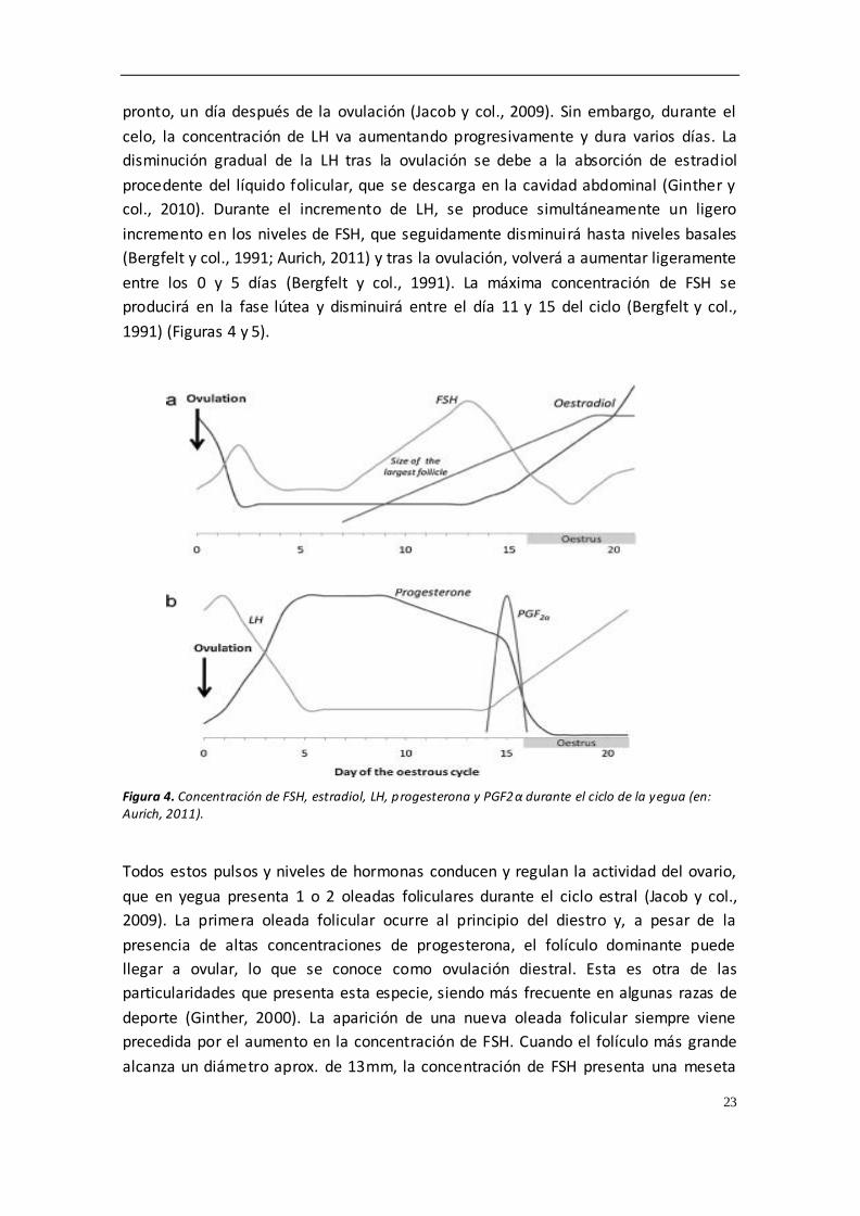
pronto, un día después de la ovulación (Jacob y col., 2009). Sin embargo, durante el
celo, la concentración de LH va aumentando progresivamente y dura varios días. La
disminución gradual de la LH tras la ovulación se debe a la absorción de estradiol
procedente del líquido folicular, que se descarga en la cavidad abdominal (Ginther y
col., 2010). Durante el incremento de LH, se produce simultáneamente un ligero
incremento en los niveles de FSH, que seguidamente disminuirá hasta niveles basales
(Bergfelt y col., 1991; Aurich, 2011) y tras la ovulación, volverá a aumentar ligeramente
entre los 0 y 5 días (Bergfelt y col., 1991). La máxima concentración de FSH se
producirá en la fase lútea y disminuirá entre el día 11 y 15 del ciclo (Bergfelt y col.,
1991) (Figuras 4 y 5).
Figura 4. Concentración de FSH, estradiol, LH, progesterona y PGF2α durante el ciclo de la yegua (en: Aurich, 2011).
Todos estos pulsos y niveles de hormonas conducen y regulan la actividad del ovario,
que en yegua presenta 1 o 2 oleadas foliculares durante el ciclo estral (Jacob y col.,
2009). La primera oleada folicular ocurre al principio del diestro y, a pesar de la
presencia de altas concentraciones de progesterona, el folículo dominante puede
llegar a ovular, lo que se conoce como ovulación diestral. Esta es otra de las
particularidades que presenta esta especie, siendo más frecuente en algunas razas de
deporte (Ginther, 2000). La aparición de una nueva oleada folicular siempre viene
precedida por el aumento en la concentración de FSH. Cuando el folículo más grande
alcanza un diámetro aprox. de 13mm, la concentración de FSH presenta una meseta
24
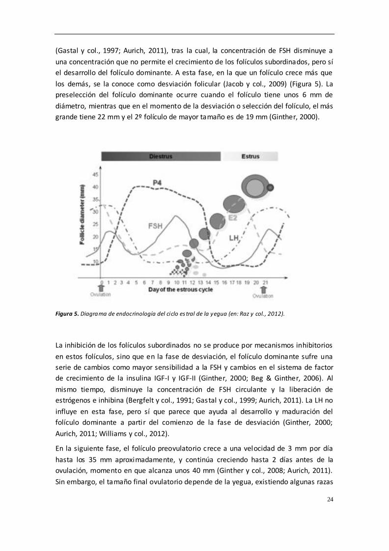
(Gastal y col., 1997; Aurich, 2011), tras la cual, la concentración de FSH disminuye a
una concentración que no permite el crecimiento de los folículos subordinados, pero sí
el desarrollo del folículo dominante. A esta fase, en la que un folículo crece más que
los demás, se la conoce como desviación folicular (Jacob y col., 2009) (Figura 5). La
preselección del folículo dominante ocurre cuando el folículo tiene unos 6 mm de
diámetro, mientras que en el momento de la desviación o selección del folículo, el más
grande tiene 22 mm y el 2º folículo de mayor tamaño es de 19 mm (Ginther, 2000).
Figura 5. Diagrama de endocrinología del ciclo es tral de la yegua (en: Raz y col., 2012).
La inhibición de los folículos subordinados no se produce por mecanismos inhibitorios
en estos folículos, sino que en la fase de desviación, el folículo dominante sufre una
serie de cambios como mayor sensibilidad a la FSH y cambios en el sistema de factor
de crecimiento de la insulina IGF-I y IGF-II (Ginther, 2000; Beg & Ginther, 2006). Al
mismo tiempo, disminuye la concentración de FSH circulante y la liberación de
estrógenos e inhibina (Bergfelt y col., 1991; Gastal y col., 1999; Aurich, 2011). La LH no
influye en esta fase, pero sí que parece que ayuda al desarrollo y maduración del
folículo dominante a partir del comienzo de la fase de desviación (Ginther, 2000;
Aurich, 2011; Williams y col., 2012).
En la siguiente fase, el folículo preovulatorio crece a una velocidad de 3 mm por día
hasta los 35 mm aproximadamente, y continúa creciendo hasta 2 días antes de la
ovulación, momento en que alcanza unos 40 mm (Ginther y col., 2008; Aurich, 2011).
Sin embargo, el tamaño final ovulatorio depende de la yegua, existiendo algunas razas
25
que ovulan por encima de 55 mm de diámetro, algo que suele repetirse en ciclos
posteriores (Cuervo-Arango & Newcombe, 2008; Aurich, 2011). Durante la maduración
del folículo preovulatorio, se produce una amplia expansión de la capa de células de la
granulosa (Sayasith y col., 2007; Aurich, 2011). Cuando se produce la ovulación, el
ovocito rodeado por la corona radiata cae desde la fosa de la ovulación a las trompas
uterinas, mientras que la mayor parte del líquido folicular se queda en el peritoneo.
Las hormonas de este líquido se absorben rápidamente y esto se traduce en un
aumento de la concentración de inhibina (Bergfelt y col., 1991, Ginther y col., 2010).
En el caso de la burra, los perfiles hormonales son perfectamente comparables a los
que conocemos en la yegua (Contri y col., 2014). En cuanto al seguimiento ecográfico,
el desarrollo del folículo dominante va a variar dependiendo de la raza, pero las razas
europeas ovulan entre 35 a 45 mm de diámetro y normalmente un día antes del final
de celo (Contri y col., 2014; Payan Carreira & Quaresma, 2014). La ovulación múltiple
es frecuente y, en un porcentaje muy alto, se produce ovulación asincrónica
(Quaresma & Payan Carreira, 2015).
Las ovulaciones dobles dependen de muchos factores (raza, estado reproductivo,
edad), pero pueden llegar a presentarse hasta en un 30 % de los ciclos (Davies Morel y
col., 2005; Stout, 2006; Aurich, 2011). La ovulación doble puede ser sincrónica (<12
horas entre ovulaciones) o asincrónica (se han descrito casos de más de 2 días de
desfase desde la primera ovulación). En estos casos, las yeguas presentan un
crecimiento folicular más lento de los folículos dominantes, probablemente debido a la
menor concentración de FSH, asociada a la mayor concentración de estradiol cuando
están presentes dos folículos preovulatorios (Ginther y col., 2008; Aurich, 2011).
Tras la ovulación se forma el cuerpo lúteo que presenta células lúteas y no lúteas, y
dentro de las últimas se subdividen en células grandes y pequeñas. Las células no
lúteas son principalmente fibroblastos, células musculares lisas, macrófagos y células
endoteliales. Al comienzo de la fase lútea existe producción de progesterona y alta
actividad mitótica (Aguilar y col., 2006; Aurich, 2011), junto a una intensa angiogénesis
regulada por factores producidos desde las células lúteas (Watson y col., 2003; Aurich,
2011). Justo después de la ovulación, empieza a aumentar la concentración de
progesterona (Roberto da Costa y col., 2005; Aurich, 2011). Los niveles de
progesterona se correlacionan con el volumen de las células productoras de hormona
luteínica desde el inicio de la fase lútea hasta mediados de la fase lútea, momento en
el que estas células alcanzan el mayor volumen (Aguilar y col., 2006; Aurich, 2011). El
cuerpo lúteo esta bajo la influencia de LH y progesterona (al igual que en otras
especies domesticas), pero la síntesis de progesterona sólo ocurre en las células lúteas
grandes (a diferencia de otras especies en las que también se produce en las
pequeñas) (Roberto da Costa y col., 2005; Ferreira-Dias y col., 2007; Aurich, 2011).

26
La disminución de la concentración de progesterona en sangre entre el día 15 y 17,
junto con la regresión del cuerpo lúteo, es lo que caracteriza a la fase de luteolisis, que
también coincide con una disminución de la expresión del citocromo P450 (Watson y
col., 2005; Aurich, 2011). En este tiempo, la mayoría de las células productoras de
hormona lútea aumentan sus marcadores de apoptosis y muerte celular. En la yegua
no preñada, la señal de iniciación de la luteolisis es la secreción endometrial de PGF2,
a la vez que se observa un incremento en las células endometriales de la COX-2, por lo
que la falta de reconocimiento materno embrionario induce dichas secreciones
(Boerboom y col., 2004; Aurich, 2011). Por último, la oxitocina junto con la PGF2a
aceleran la luteolisis en la yegua no preñada mediante un sistema autocrino-paracrino;
parece que la secreción inicial de oxitocina proviene de la hipófisis pero es en la yegua
en la única especie doméstica donde se ha evidenciado oxitocina a nivel del
endometrio (Bae & Watson, 2003; Aurich, 2011).
4.3. EMBRIOGENESIS
El desarrollo embrionario en équidos muestra una serie de particularidades, en
comparación con otras especies. Como en todos los mamíferos, el ovocito equino
presente en el ovario se detiene durante su desarrollo fetal en la profase de la primera
división meiótica, y solo unos cuantos son seleccionados para seguir desarrollándose
hasta la segunda metafase, estado que alcanza justo antes de la ovulación, cuando es
un ovocito maduro (King y col., 1987)(Figura 6). En el momento de la ovulación, el
ovocito maduro viaja desde la fosa de ovulación al oviducto, donde se encuentran los
espermatozoides (si la yegua ha sido cubierta o inseminada), que esperan para
fecundarlo (Aguilar y col., 2012).
Figura 6. Esquema y fotografía de ovocito maduro (en: Equine Breeding Management and Artificial Insemination. Samper J, 2009).

27
Una de las particularidades que presenta el embrión equino es que la relación entre la
madre y el embrión se pone de manifiesto desde las etapas más tempranas del
desarrollo embrionario, ya que si el ovocito no se fecunda o el embrión degenera antes
del estadio de 16 células, este permanece retenido en las paredes del oviducto (Aguilar
y col., 2012). Esta retención del embrión en las trompas se sugiere que puede deberse
a que los embriones viables secretan prostaglandina E2, sustancias con acción
uterotónica que facilita el transporte del embrión a través del oviducto, debido a la
relajación que ejerce sobre la musculatura de las trompas uterinas, y que permite su
caída al útero (Weber y col., 1991; Weber y col., 1995; Sharp, 2000; Smits y col., 2012).
Se estima que el embrión equino alcanza el útero hacia el día 6 post ovulación, es
decir, entre las 144 y 156 horas tras la dehiscencia folicular (Freeman y col., 1991;
Smits y col., 2011).
Otra de las peculiaridades de la reproducción equina reside en el proceso de
fecundación, cuyos mecanismos no han sido aún completamente descifrados. Esto
explica la gran diferencia observada entre la fecundación in vitro mediante cocultivo,
que no ofrece resultados exitosos, y la fecundación natural, en la que se obtiene hasta
un 90% de fertilidad (Betteridge, 2007; Galli y col., 2007). La respuesta es multifactorial
y no se conoce, entre otras razones, por la dificultad de obtener embriones de edades
comprendidas entre 0 y 6 días, que es la etapa en la que se desarrollan en el oviducto.
Además, es necesario el acceso quirúrgico para su obtención, lo que dificulta dicho
proceso de estudio (Betteridge, 2007; Galli y col., 2007). Por ello, se hace necesario el
uso de la producción in vitro para el estudio y la compresión de los procesos que se
llevan a cabo durante el desarrollo de estos estadios tempranos del embrión.
No son muchos los estudios con embriones in vivo en sus fases tempranas, pero se
evidencia la existencia de polaridad en sus células, tanto en ovocitos, como en cigotos
y embriones de 4 células, en los que se aprecian blastómeros con distinta densidad.
Parece haber una estrecha relación entre las células foliculares y el ovocito, que
desaparece cuando se realiza maduración y cultivo embrionario in vitro (Betteridge,
2007). Esta polaridad no se sabe si continúa en estadios posteriores a los antes
mencionados, por lo que siguen existiendo muchas incógnitas al respecto, pero su
conocimiento puede permitir el desarrollo de sistemas de producción embrionaria
eficientes.
En los últimos años, algunos grupos están trabajando en el desarrollo de la producción
in vitro, aunque no está tan desarrollada como en otras especies, debido a que cuenta
con una serie de hándicaps. La producción in vitro de embriones a partir de ovarios de
matadero cuenta con una menor disposición de ovarios que en otras especies, debido
al bajo número de mataderos, las muestras y los especímenes no están disponibles
todo el año debido a la marcada estacionalidad, la menor tasa de recogida de ovocitos
por ovario, la ausencia de respuesta a tratamientos de superovulación y las

28
dificultades que encontramos durante la punción y aspiración ovárica (Squires y col.,
2003; Carnevale y col., 2012). A todo lo anterior hay que sumarle también el menor
interés de los ganaderos por la producción de embriones in vitro y de la escasa
credulidad de las asociaciones en estas técnicas (Galli y col., 2007).
Los diferentes métodos establecidos para recolectar un ovocito ideal son complicados.
El “ovocito ideal” es aquel obtenido inmediatamente después de la ovulación. Si la
aspiración se hace 30-35 h. tras la administración de sustancias inductoras de la
ovulación obtendremos un ovocito maduro (en metafase II), pero si lo recogemos 24 h.
tras la administración necesitará cocultivo en medio de maduración durante 12 a 18 h.
(Hinrichs y col., 1999; Coutinho da Silva y col., 2002; Carnevale y col., 2012; Galli y col.,
2014). También se pueden realizar aspiraciones de folículos pequeños y someter los
complejos cúmulo-oocito (COC) a medio de maduración con hormonas, lo que ofrece
resultados de maduración oocitaria del 50% al 70% (Galli y col., 2002; Hinrichs y col.,
2002; Preis y col., 2004; Carnevale y col., 2012). Para obtener los ovocitos contenidos
en los folículos se realiza aspiración folicular transvaginal, aunque también puede
realizarse por el flanco (Galli y col., 2014).
Una alternativa diferente a la aspiración de folículos in vivo es la recogida de ovocitos
procedentes de ovarios de matadero, aunque presenta una modificación con respecto
a la aspiración de folículos en otras especies. La punción del folículo lleva asociado el
raspado de las paredes foliculares, necesario para que la maduración de ovocitos in
vitro sea efectiva, ya que existen estrechas conexiones entre las células del cúmulo y la
membrana de la granulosa, y entre estas últimas y la pared folicular (Galli y col., 2007;
Hinrichs, 2010; Carnevale y col., 2012). De ahí la importancia de realizar este raspado
para la obtención de los ovocitos, a pesar de requerir más tiempo. De esta forma se
consigue desde un 20 hasta un 85% de maduración de ovocitos usando diferentes
medios, aunque sin éxito en la fecundación in vitro con cocultivo (Galli y col., 2007).
El éxito del proceso de maduración de los ovocitos se evidencia por la ob tención de
buenas tasas de fertilidad tras la transferencia al oviducto de yeguas receptoras
previamente inseminadas (Hindrichs y col., 2002; Carnevale y col., 2004; McPartlin y
col., 2009; Carnevale y col., 2012), pero también los cambios morfológicos pueden
utilizarse como indicadores de éxito. En este sentido, las células de la granulosa
adquieren una apariencia mucoide y dentro del cúmulo encontramos una extensa
matriz, como describen otros (Carnevale & Maclelan, 2006; Carnevale y col., 2012). Sin
embargo, con mayor o menor éxito, la maduración de ovocitos, tanto in vivo como in
vitro, hoy no es un problema. La verdadera dificultad reside en la fase de fecundación,
que sigue siendo un misterio, y parece estar relacionado con la falta de capacitación
del esperma y con cambios producidos en la zona pelúcida (Dell’Aquila y col., 1999;
Alm y col., 2001; Galli y col., 2007).

29
El éxito de la fecundación in vitro en caballo ha sido marginal, obteniéndose solo un
potro nacido vivo mediante el cocultivo de ovocitos y espermatozoides (Palmer y col.,
1991), a pesar de los enormes esfuerzos invertidos en la investigación y desarrollo de
dicho proceso durante varias década. Al parecer, la principal barrera es la falta de
capacitación de espermatozoides. Los cambios que se observan en un espermatozoide
capacitado son un aumento de la fosforilación en tirosina, reacción acrosómica y
exocitosis acrosomal (McPartlin y col., 2009). Además, para conseguir la fecundación in
vitro, los espermatozoides deben adquirir hiperactivación, por la que aumenta el
desplazamiento lateral de la cabeza y la amplitud y asimetría flagelar, necesarios para
penetrar la zona pelúcida (McPartlin y col., 2009). Hasta la fecha, los protocolos de
fecundación in vitro en cocultivo se extrapolaban de otras especies, sin tener en
cuenta la especificidad y los tiempos requeridos para capacitar al esperma de caballo,
y el principal problema radicaba en la incapacidad del espermatozoide para atravesar
la zona pelúcida. La adición de procaína al medio ha permitido salvar esta barrera,
pero debido al bajo número de ovocitos que se obtienen, tanto por aspiración
transvaginal como por aspiración de ovarios de matadero, el método estandarizado y
de elección para la fecundación in vitro de ovocitos es la inyección intracitoplasmática
de espermatozoide (ICSI) (McPartlin y col., 2009) que, a pesar de no desenmascarar el
mecanismo de fecundación, solventa los problemas relacionados con el cocultivo de
ovocitos y espermatozoides, permitiendo tasas aceptables de fecundación y desarrollo
embrionario.
La técnica de ICSI ha sido una herramienta fundamental para la producción in vitro en
equino, como se comentó con anterioridad. Está técnica permitió obtener la primera
gestación en la década de los 90 (Squires y col., 1996), pero no se estandarizó hasta el
descubrimiento del piezo taladro, instrumento que produce vibraciones controladas en
la pipeta de microinyección, facilitando la penetración en el oolema a través de la zona
pelúcida (Carnevale y col., 2012). El método es sencillo y requiere la selección de un
espermatozoide, que se procesa y se inmoviliza mediante la sección de la pieza
intermedia. El ovocito madurado se fija por aspiración con la posición del corpúsculo
polar a las 12:00 o a las 6:00, y se perfora la zona pelúcida con el taladro en la posición
de las 3:00. A continuación, se carga el espermatozoide en la pipeta y se introduce en
el citoplasma. Para comprobar que estamos en el interior del ovocito, se aspira un
poco de oolema y se deposita el espermatozoide en el centro. Hecho esto, el ovocito
se deja en medio de cultivo o se transfiere al oviducto de la receptora (Galli y col.,
2002; Choi y col., 2002, Carnevale y col., 2012).
Multitud de medios se han empleado para el cultivo in vitro de embriones, el más
empleado es el DMEM/F12 con 10% de suero fetal bovino en atmosfera de 5% O 2, 5%
CO2 y 90% N2, a una temperatura de 38.2 a 38.5ºC (Carnevale y col., 2012). Este medio
resulta más exitoso porque tiene una mayor concentración de glucosa, alcanzando

30
tasas de desarrollo de blastocistos más que aceptables, de aprox. el 30% (Galli y col.,
2014).
A pesar del éxito en la producción in vitro de los últimos años, existen diferencias entre
los embriones producidos in vitro y los recogidos en lavados in vivo, que pueden influir
tanto a la hora del estudio del embrión como en el éxito en el desarrollo del mismo.
Los embriones in vitro tienen 7 veces menos células que embriones in vivo de la misma
edad y un diámetro 2.5 veces menor, además de sufrir daños en la membrana,
presentar un mayor porcentaje de núcleos fragmentados y signos de apoptosis
(Tremolada y col., 2003; Pomar y col., 2005; Carnevale y col., 2012). Una característica
diferente y aún más importante es la ausencia de la cápsula glicoproteica en los
embriones producidos in vitro, la cuál es necesaria para la supervivencia del embrión
en el útero hasta el día 21 de gestación (Stout, 2005; Betteridge, 2007; Carnevale y
col., 2012; Stout, 2012). Por último, se han encontrado grandes diferencias en la
expresión de genes que están encargados de sintetizar proteínas y producción de
energía (Smits y col., 2011; Carnevale y col., 2012; Smits y col., 2012).
4.4. CAÍDA DEL EMBRIÓN AL ÚTERO
En torno al día 6 se produce la caída del embrión al útero. El momento exacto se
estima que es entre las 144 y 156 horas post-ovulación (Freeman y col., 1991; Smits y
col., 2011), aunque varía en función de diferentes factores como la edad de la yegua, la
estacionalidad o el tipo de semen (Stout, 2006). En este periodo, la cavidad
blastocélica queda demarcada por el endodermo y, a diferencia de los embriones de
rumiantes y cerdos, esas células del endodermo forman colonias que se distribuyen
esporádicamente en el interior del trofoblasto y acaban uniéndose con otras
(Betteridge, 2007). En el embrión equino es más difícil diferenciar los estadios
embrionarios de mórula y blastocisto temprano, al estar las células de la masa celular
interna más dispersas, en comparación con otras especies donde son claramente
diferenciables (Betteridge, 2007) (Figura 7).
A las pocas horas de entrar en el útero se desarrolla la cápsula del embrión equino. Se
trata de una estructura única entre las especies de abasto, que se desarrolla entre la
cara interna de la zona pelúcida y el trofoblasto. La cápsula es una capa acelular
glicoproteica que envuelve al embrión desde el día 6 al día 21 aproximadamente
(Betteridge, 2007), y se considera una estructura única y vital para el desarrollo de la
gestación (Flood y col., 1981; Stout, 2005; Carnevale y col., 2012). Entre las funciones
que se le presuponen a la cápsula se describe que permite la comunicación con el
medio uterino y el intercambio de señales moleculares y nutrientes, da soporte para
mantener la forma esférica y así protege al embrión de las contracciones miometriales
durante su etapa migratoria por el útero, y parece tener importancia en la producción
de ácido siálico que impide la adherencia al miometrio durante dicha fase, mientras

31
envía señales de su presencia a la madre, evitando la luteolisis y pérdida de la preñez
temprana. Además, la cápsula tiene importancia en el proceso de fijación y orientación
del embrión en el útero de la yegua (Oriol y col., 1993; Herrler & Beier, 2000; Allen &
Stewart, 2001; Stout & Allen 2001; Stout y col., 2005; Smits y col., 2012; Diaz y col.,
2016).
Figura 7. Esquema de las partes de un blastocisto temprano y su imagen.
La cápsula solo se desarrolla en el embrión en condiciones in vivo, no in vitro, lo que
apoya la tendencia de que necesitamos mucha más información para poder desarrollar
embriones en condiciones in vitro. A pesar de identificarse las glicoproteínas en
condiciones in vitro, estas no se aglutinan ni se estructuran para formar la cápsula
(Smits y col., 2012). Entre las principales causas de dicho fracaso parece encontrarse
que las glicoproteínas se dispersan en el medio de cultivo impidiendo su organización,
y que existe falta de componentes uterinos conocidos que posibilitarían la unión de
estas glicoproteinas (Tremoleda y col., 2003; Smits y col., 2012).
4.5. TRATAMIENTOS DE SUPEROVULACIÓN
Uno de los principales problemas que presenta la transferencia de embriones equina
es la baja tasa de recogida de embriones mediante flushing si lo comparamos con otras
especies. Tan solo se obtienen embriones en un 50-80% de los lavados realizados en
yeguas, y esto depende de multitud de factores, por lo que una de las claves del éxito
o del fracaso podría residir en los tratamientos de superovulación.
La superovulación o inducción de ovulaciones múltiples en la yegua es una técnica que,
mediante el empleo de tratamientos hormonales, controla el desarrollo de mas
folículos dominantes o la inhibición de la regresión de los folículos subordinados
(Squire & McCue, 2007). El número de folículos que ovulan en respuesta a los
tratamientos exógenos no se puede comparar con el obtenido en otras especies como
la vaca o la oveja (Hunter y col., 2004; Cullingfor y col., 2010). En los intentos para
conseguir un mayor número de folículos que ovulen por ciclo se han empleado
diferentes hormonas o productos, como la gonadotropina coriónica equina (eCG)

32
(Dinger y col., 1982), GnRH (Becker & Johnson, 1992; Dippert y col., 1992), FSH porcina
(Cullingford y col., 2010; Raz y col., 2010) y la inmunización contra la inhibina (McCue y
col., 1992; Derar y col., 2004; Roser & Meyers-Brown, 2012), cuyos resultados han sido
muy variables. La propia anatomía y fisiología del ovario dificulta la superovulación en
este especie. Por un lado, la fosa de ovulación presenta un área limitada para la
ovulación de los folículos preovulatorios, los cuales tienen un gran tamaño (mayor de
3.5 cm). Además, el tejido gonadal equino presenta una baja concentración de
receptores de gonadotropina coriónica equina (eCG), por lo que la respuesta a esta
hormona es muy limitada. Entre los beneficios potenciales de la superovulación se
encuentran el incremento de la tasa de recogida embrionaria o de ovocitos, la
disponibilidad de embriones para congelación o vitrificación, la mejora de la fertilidad
en yeguas subfértiles y la posibilidad de acelerar la primera ovulación.
Actualmente, no hay ningún método práctico y eficiente para inducir la superovulación
en las yeguas y esto limita la eficiencia de la transferencia embrionaria. Tenemos
diversos tratamientos hormonales empleados para producir superovulación en la
yegua, y que vamos a describir brevemente.
- Gonadotropinas extraídas de glándulas pituitarias equinas (EPE)
EPE o extracto de pituitaria equina es una preparación de gonadotropina equina
purificada con un 6 a 10% de LH y un 2 a 4% de FSH. Con este tratamiento se
alcanzan tasas elevadas de ovulación y recogida de blastocistos. Diversos estudios
indican que dosis elevadas de EPE (50 mg, una vez al día vía intravenosa) generan
una mejor respuesta ovárica, con un mayor número de folículos preovulatorios,
mientras que si incrementamos la frecuencia del tratamiento sin incrementar
necesariamente la dosis (25 mg, dos veces al día vía intravenosa) obtenemos un
aumento en la recogida de blastocistos de la yegua donante. De 6 a 11 días después
de la ovulación se administra PGF2α a las yeguas donantes, y se comienza el
tratamiento con 25 mg EPE dos veces al día hasta que se produce la ovulación. Con
esta dosis y frecuencia de administración obtendremos una mayor tasa de
ovulación (2.4-7.1) y de recogida de blastocistos (1.6-3.5) (Alvarenga y col., 2001).
Scoggin y col. (2002) obtuvieron 4.7 ovulaciones con dosis de 25 mg de EPE dos
veces al día, mientras que cuando la dosis administrada era la mitad (12.5 mg de
EPE dos veces al día), la tasa de recogida fue mayor, alcanzando 2.6 embriones por
flushing.
Se conoce que la respuesta del ovario a la EPE es muy similar a la obtenida con FSH
equina, lo que indica que la LH presente en EPE no afecta a la superovulación en la
yegua. Los factores que afectan a la respuesta a EPE y FSH equina son el día de
inicio del tratamiento, el tamaño del folículo al inicio del tratamiento y la frecuencia
de administración. Los óvulos procedentes de ovulaciones simples sin tratamientos

33
hormonales y aquellos procedentes de tratamientos con EPE o FSH equina dan lugar
a tasas de gestación similares. En este sentido, Squires y col. (2007) demostraron
que las tasas de gestación de embriones recogidos de yeguas donantes sometidas a
tratamientos de superovulación y transferidos a yeguas receptoras fueron similares
a las tasas de gestación de embriones obtenidos de yeguas con ovulación
espontánea. Además, se demostró que la administración de EPE durante un ciclo e
incluso hasta 6 ciclos consecutivos no alteraba los valores de ovulación en ciclos
sucesivos.
- Hormona Foliculoestimulante Equina (FSHe)
La FSHe es un preparado con proporciones conocidas del ratio FSH/LH, que ronda
10:1 o 5:1, mientras que en el EPE no se sabe la pureza ni la proporción de dichas
hormonas (Welch y col., 2006). El tratamiento estándar con FSHe comienza unos 5-
7 días después de la ovulación, cuando los folículos presentan un diámetro de 22 a
25 mm, y finaliza cuando éstos alcanzan ≥32-35 mm. El segundo día después de
iniciar el tratamiento con FSHe, se administra PGF2α para producir luteolisis. El
tratamiento de inducción de la ovulación con hCG o GnRH se realiza unas 36 a 42 h.
después de finalizar el tratamiento de superovulación con el objetivo de evitar una
hiperestimulación ovárica. La inseminación se realiza el día siguiente a la
administración de hCG o GnRH. La recogida embrionaria tiene lugar desde el día 6,
si queremos obtener embriones de menor tamaño para realizar congelación o
vitrificación, hasta el día 8, si queremos hacer transferencia directa o refrigeración
de los embriones.
Niswender y col. (2003) emplearon un protocolo de superovulación a base de 12mg
de FSHe dos veces al día, a partir del 5 día postovulacion. Este se mantenía hasta
que los folículos preovulatorios alcanzaban 35 mm de diámetro, en cuyo momento
se administraban 2500 UI de hCG para inducir la ovulación. Con este tratamiento la
tasa de ovulación fue 3.6±0.5 y la tasa de recogida por yegua de 1.9±0.3. Otros
autores han descrito que modificando las dosis de FSHe y el protocolo de
administración se podían mantener o aumentar los resultados ovulatorios y la tasa
de recogida de embriones antes descrita (Welch y col., 2006; Logan y col., 2006;
Logan y col., 2007; Peres y col., 2007).
- FSH recombinante (FSHre) y LH recombinante (LHre)
Estos productos surgieron de la dificultad de obtener hipófisis equinas y además
eran productos más puros y con proporciones más controladas. Meyer-Brown y col.
(2010) emplearon estas sustancias con resultados no todo lo bueno que se
esperaban ya que, como se ha descrito en los tratamientos anteriores, mejoraban la

34
tasa de folículos preovulatorios pero muchos de estos no llegaban a ovular y, por lo
tanto, la tasa de recogida seguía siendo igual o inferior a la del grupo control. En
trabajos posteriores se consiguió tener el mismo número de folículos
preovulatorios, pero disminuyó el número de anovulatorios por lo que también
aumentó la tasa de recogida de embriones por ovulación. Pero, para ello, se
necesitaba un mayor control del tamaño folicular y un mayor número de
inyecciones al día, lo que convertía este tratamiento en tedioso y difícil de aplicar
(Meyer-Brown y col., 2011).
En resumen, podríamos decir que todos estos protocolos no tendrá n ningún
significado si no se mejoran los conocimientos referentes a las fases de oleadas
foliculares, fases de crecimiento, desviación y dominancia, periodo periovulatorio y
todos los factores que influyen en cada una de ellos, y que pueden modificar la
respuesta de un mismo protocolo (Roser & Meyer-Brown, 2012). Además, la baja tasa
de recogida en yeguas superovuladas parece deberse a una combinación de la pobre
maduración de los ovocitos y la dificultad para que el ovocito alcance la fosa de
ovulación (Carmo y col., 2006; Meyer-Brown y col., 2011).
A continuación se detallan los principales protocolos de superovulación descritos en
yegua (tabla 7).

35
Autores Tratamiento Tasa de
ovulación
Tasa de recogida
por lavado
Tasa de recogida
por ovulación (%)
Tasa de
gestación
Alvarenga y col., 2001 Grupo 1: 25 mg EPE sid Grupo 2: 25 mg EPE bid
2.4 ± 1.8 7.1 ± 5.1
1.6 ± 1 3.5 ± 1.8
68.0% 49.0%
Scoggin y col., 2002
Grupo 1: 25 mg EPE sid Grupo 2: 50 mg EPE sid
Grupo 3: 12.5 mg EPE bid Grupo 4: 25 mg bid Grupo 5: control
3.4±0.6 4.4±0.7
3.4±0.8 4.7±0.6 1.2±0.1
1.2±0.4 1.5±0.3
2.6±0.6 2.1±0.6 0.4±0.2
35.3% 36.4%
67.6% 43.2% 36.4%
Niswender y col., 2003 ESTUDIO 1:
Grupo 1: control Grupo 2: 25 mg FSHe bid.+ Des
Grupo 3: 12 mg FSHe bid + HCG Grupo 4: 12 mg FSHe bid + Des
ESTUDIO 2:
Grupo 1: control
Grupo 2: 12 mg FSHe bid + HCG
1.1±0.1
3.3±0.9 3.4±0.7 1.8±0.7
1.0±0.0 3.6±0.5
0.5±0.1 1.9±0.3
50.0% 52.8%
0.6±0.1
0.6±0.3 1.8±0.8 0.8±0.5
Carmo y col., 2006 Grupo 1: control Grupo 2: 25mg de EPE bid + HCG
1.1±0.33 4.4 ±2.9
1.0±0.5 2.6±1.6
Logan y col., 2006 ESTUDIO 1:
Grupo 1: control. Grupo 2: 12.5 mg FSH bid Grupo 3: 12.5 mg FSH bid+ periodo de
espera
ESTUDIO 2:
Grupo 1: 12.5 mg FSH bid+ periodo de espera
Grupo 2: 6.25 mg FSH bid+ periodo de espera
ESTUDIO 3:
Grupo 1: 12.5 mg FSH bid 3 días+ periodo
de espera
1.2±0.1 3.3±0.4
4.1±0.4 4.9±0.6
2.1±0.2
3.8±0.8
1.0±0.1 1.9±0.3
2.6±0.3 1.8±0.4
1.8±0.3
0.8±0.3

36
Autores Tratamiento Tasa de
ovulación
Tasa de recogida
por lavado
Tasa de recogida
por ovulación (%)
Tasa de
gestación
Welch y col., 2006 Grupo 1: 12.5 mg FSHe bid+ HCG
Grupo 2: 12.5 mg FSHe bid+ HCG
Grupo 3: 12.5 mg FSHe bid+ FSH:LH bid +
HCG Grupo 4: 25 mg FSHe sid + HCG
3.4±1.2 3.8±1.2 2.5±1.4
2.5±1.2
1.6±0.8 1.7±1.4 1.5±1.1
1.3±1.3
47.0% 42.0% 68.0%
55.0%
Logan y col., 2007 ESTUDIO 1:
Grupo 1: control. Grupo 2: 12,5 mg FSHe bid Grupo 3: P+E 10 días diestro + FSH
Grupo 4: P+E 10 días estro + FSH ESTUDIO 2:
Grupo 1: 12.5 mg FSH bid + HCG Grupo 2: 6.25 mg FSH bid + HCG Grupo 3: 12.5 mg FSH bid + Des
Grupo 4: 6.25 mg FSH bid + Des
1.2±0.5 5.5±0.4
2.5±0.6 3.3±0.7 5.2±0.8
2.1±0.8 2.4±0.8 2.1±0.8
0.8±0.6 2.6±0.5
1.5±0.7 0.7±0.7 1.6±0.4
1.9±0.4 1.1±0.4 1.7±0.4
66.7% 47.0%
60.0% 21.7% 30.8%
90.4% 45.8% 80.9%
Peres y col., 2007 Grupo 1: control Grupo 2: 12.5 mg FSHe bid
1.0±0.0 5.6±4.5
0.7±0.5 2.0±1.8
69.0% 36.0%
Cullingford y col., 2010 ESTUDIO 1:
Grupo 1: control Grupo 2: 25mg FSHp bid + HCG Grupo 3: 50 mg FSHp bid + HCG
Grupo 4: 100 mg FSHp bid + HCG ESTUDIO 2:
Grupo 1: 150 mg FSHp bid + HCG Grupo 2: 0.5 mg FSHre bid + HCG
1.11 1
1.08 1.31 0.98
0.85
0.66 0.68
1.08 0.66 0.7
0.69

37
Autores Tratamiento Tasa de
ovulación
Tasa de recogida
por lavado
Tasa de recogida
por ovulación (%)
Tasa de
gestación
Meyer Brown y col., 2010 ESTUDIO 1:
Grupo 1: control.
Grupo 1: 0.85 mg FSHre bid + 0.75mg LHe
+HCG ESTUDIO 2:
Grupo 1: control
Grupo 2: 12.5 mg FSHe bid + HCG
Grupo 3:0.35 mg FSHre bid + HCG
Grupo 4: 0.5 mg FSHre bid + HCG
Grupo 5: 0.65 mg FSHre bid + HCG
ESTUDIO 3: Grupo 1: 0.85 mg FSHre bid + 1.5 mg LHre
Grupo 2: 0.85 mg FSHre sid +1.5 mg LHre Grupo 3: 0.50 mg FSHre bid + 1.5 mg LHre Grupo 4: 12.5 mg FSHe bid + 1.5 mg LHre
1.25±0.2 3.75±0.7
1.3±0.5 3.0±0.6
2.1±0.6 4.6±0.6 3.3±0.6
3.4±0.7 5.4±0.4 3.2±0.5
3.8± 1.1
0.5±0.2 1.75±0.4
0.8±0.4 2.4±0.6
1.4±0.5 1.5±0.5 2.7±0.5
44.0% 49.0%
60.0% 60.0%
80.0% 30.0% 50.0%
Raz y col., 2010 Grupo 1: 12.5 mg FSHe bid + HCG
Grupo 2: 25 mg FSHp bid + HCG
2.6±0.4 1.2±0.3
1.8±0.3 1.0±0.0
Meyer Brown y col., 2011 Grupo 1: 0.65 mg FSHre bid/sid + HCG
Grupo 2: 0.65 mg FSHre +1.5 mg LHre bid/tid + HCG
3.05±0.52 4.62 ±0.88
2.0 ±0.53 3.87 ±0.87
66.0% 83.0%
Ignácio y col., 2013 ESTUDIO 1:
Grupo 1: Control
Grupo 2: 25 mg FSHp bid + HCG
ESTUDIO 2:
Grupo 1: Control
Grupo 2: 50 mg FSHp bid + HCG ESTUDIO 3:
Grupo 1: Control Grupo 2: 25/50 mg FSHp bid HCG Grupo 3; 12.5 mg EPE bid + HCG
1.1±0.0.1
1.1 ±0.1
1.2±0.1 1.3±0.2 1.3±0.2
1.0±0.0 2.1±0.4
0.6±0.2
0.7±0.3
0.5±0.2 0.4±0.2 1.2±0.2
1.0±0.0 1.6±0.5
50.0%
62.0%
42.0% 33.0% 75.0%
100 % 73.0%
Tabla 7. Principales tratamientos de superovulación y los resultados de cada tratamiento según el autor (Bid: 2 dosis al día; Sid: 1 dosis al día; Tid: 3 dosis al día; Des:
Deslorelina).

38

39
4.6. RECOGIDA DE EMBRIONES
El lavado uterino es el primer paso para realizar una transferencia de embriones
(Squires y col., 2003) y la mayoría de los embriones recogidos para llevar a cabo la
transferencia en fresco se obtienen en el día 7, 8 ó 9 post-ovulación. La técnica de
recolección de embriones es similar, a grandes rasgos, para todos los equipos de
transferencia de embriones y ha sufrido pocas modificaciones en las últimas décadas.
Se emplea una sonda tipo Foley de un calibre de 14 a 16 g, que se introduce vía
transcervical y, tras atravesar el cérvix, se fija al interior del útero mediante la
insuflación del balón de aire (con 40-60 cm de aire o líquido). Tras comprobar que está
correctamente fijado, comenzaremos a infundir 1-2 litros de medio de lavado en el
útero de la yegua. Seguidamente este líquido se recoge a través de un filtro, donde el
embrión quedará retenido. El procedimiento es repetido varias veces para tratar de
incrementar las probabilidades de obtener un embrión (Figura 8).
Figura 8. Ilustración del esquema de flushing para recogida de embriones (en: Manual of Equine Reproduction. Brinsko S., 2011).
Los embriones recogidos deben ser identificados, medidos y clasificados. La mayoría
de los embriones miden entre 0.1 a 1 mm de diámetro, pudiendo clasificarse como
mórulas, blastocistos temprano, blastocistos o blastocistos expandidos. Antes de la
transferencia, los embriones son pasados varias veces por medio de lavado y
mantenimiento con el objetivo de evitar su posible contaminación y eliminar los restos
celulares. La inmensa mayoría de los embriones se transfieren sin cirugía a receptoras
sincronizadas.
Se describen diferencias en lo relativo al tipo de medio empleado para realizar el
flushing (con/sin BSA o FCS, con/sin antibióticos), el volumen final empleado en cada

40
lavado, el modelo de filtro para la recogida, o la aplicación o no de sustancias ecbólicas
acompañado de masaje de los cuernos uterinos, entre otras, y que dependerán de las
preferencias de cada equipo de trabajo (Figura 9).
La técnica de transferencia no quirúrgica consiste en cargar el embrión en una pajuela,
que se adapta a un catéter de transferencia y, a continuación, el técnico cateteriza el
cérvix de la hembra receptora y deposita el embrión en el cuerpo uterino (Squires y
col., 2003; Moussa y col., 2005; Jacob y col., 2012).
La tasa de recogida embrionaria se encuentra en torno al 50%, siendo los principales
factores que afectan a dicha tasa el día del lavado, el número de ovulaciones, la edad
de la yegua y la calidad del semen (Squires y col., 2003; Stout, 2006; Jacob y col., 2012;
Camargo y col., 2013). Otros factores que cobran importancia son la época del año en
la que se realiza el lavado (etapa reproductiva o transición) (Koblischke y col., 2008), la
tasa de ovulación (ya que las ovulaciones dobles aumentan la tasa de recogida por
lavado) (Davies Morel y col., 2005; Stout, 2006; Panarace y col., 2014; Panzani y col.,
2014), la calidad del flushing (expresado como debris celular que dificulta la búsqueda
y posible pérdida de embriones) (McCue y col., 2010) y el masaje uterino junto a la
administración de oxitocina (que pueden aumentar hasta un 10% la tasa de recogida)
(Hudson & McCue, 2004; Scherzer y col., 2008; McCue y col., 2010). También se ha
informado de que el empleo de animales de deporte o el aumento de temperatura
podrían afectar al desarrollo folicular y ocasionar problemas en el intervalo
interovulatorio y la foliculogénesis (Mortesen y col., 2009; Pessosa y col., 2011).
Figura 9. Fotografía del material necesario para realizar un flushing.
4.7. EVALUACIÓN DE EMBRIONES
Existen diferentes métodos para el estudio y valoración de la viabilidad de un embrión,
entre los que destacan la valoración morfológica, tinción de embriones, cultivo in vitro,
estudio metabólico y la transferencia de embriones a una receptora, analizando su
capacidad para que la gestación se instaure y siga adelante.

41
4.7.1. Valoración morfológica
El método más empleado para evaluar un embrión es el que se basa en su morfología.
Se utiliza una clasificación de 1 a 5, calificando a los embriones como excelentes,
buenos, regulares, malos y no fertilizados (UFOs) o degenerados. Para ello, la
descripción morfológica se basa en su esfericidad, uniformidad del tamaño celular,
color y textura de los blastómeros (Slade y col., 1985). Pero esta técnica tiene el
inconveniente de que es un método subjetivo, y la categorización varía de unos
técnicos a otros (Abe y col., 2003) ya que no todos percibimos y entendemos
exactamente lo mismo. Es un método insuficiente para decidir si un embrión es viable,
ya que existen algunos parámetros de viabilidad que no se pueden identificar
mediante la valoración morfológica clásica (Moussa y col., 2004). Una de las
clasificaciones más empleada es la que describieran Slade y col. en 1985:
Grado 1. Excelente o bueno: embrión simétrico y esférico, con uniformidad en el
tamaño de las células, color y densidad. No existen evidencias de fragmentación
celular o perdida de citoplasma y granulación en el espacio perivitelino.
Grado 2. Bueno: moderadas irregularidades en la forma del embrión, pequeños signos
de fragmentación en el citoplasma, leves signos de granulación y perdida de espacio
perivitelino.
Grado 3. Regular: Mayores irregularidades en la forma de la masa celular y en el
diámetro, mayor grado de fragmentación y degeneración, variabilidad del tamaño de
los blastómeros, con forma irregular y un gran espacio perivitelino.
Grado 4. Pobre: Muy alto grado de fragmentación citoplasmática, irregularidad en el
tamaño y color de los blastómeros, ausencia de compactación celular, numerosos
blastómeros extrudidos y degenerados.
Grado 5. UFO o embrión muerto: Ovocito no fecundado o embrión completamente
degenerado.
A pesar de todo ello, existen evidencias de que la calidad del embrión descrita a través
de su morfología presenta diferencias significativas de los embriones de grado 1 y 2,
frente a los embriones de menor calidad, con respecto al porcentaje de preñez en
transferencias realizadas con embriones en fresco (Squires y col., 2003; Camargo y col.,
2013). Por ello, la clasificación morfológica embrionaria se considera un método no
invasivo, rápido y sencillo, que permite dar una ligera predicción del resultado.

42
4.7.2. Tinción de embriones
En respuesta a la búsqueda de otros métodos más objetivos para clasificar a los
embriones en función a su viabilidad, en otras especies se han empleado tinciones
vitales. Existen multitud de sondas, fluoróforas o no, que tienen afinidad por
diferentes estructuras del embrión y que nos dan información sobre el estado en que
se encuentra dicha estructura (Tabla 8). Unos de los primeros colorantes empleados
fueron el rojo neutro, para valorar ovocitos fertilizados de oveja (Kardymowicz, 1972;
Moussa y col., 2004), o el tripán azul (Hutz y col., 1985; Majumdar, 1990; Moussa y
col., 2004). Otro tipo de sustancias empleadas son las tinciones fluorescentes como el
diacetato de fluoresceína empleado en embriones de bovino y ratón (Schilling &
Doepke, 1978; Schilling y col., 1979; Mohr & Trounson, 1980) y en embriones de
caballo (Pruitt y col., 1988; Barry y col., 1989), que combinado con medidores de
potencial de membrana son capaces de identificar la actividad de enzimas
citoplasmáticas y sirven como marcadores de la viabilidad embrionaria al excitarse con
luz ultravioleta (Overström, 1996). Otro marcador es la rodamina 123, que describe la
distribución de las mitocondrias en el citoplasma, estudiando la integridad metabólica
de cigotos y ovocitos de mamíferos (Petter & Lucy, 1987). Ese compuesto penetra a
través de la membrana plasmática y se queda adherido en el interior de la membrana
interna de las mitocondrias (Royall & Ischiropoulos, 1993).
En este mismo ámbito de la fluorescencia hay una vertiente que describe la integridad
de la membrana, como son los fluorocromos con afinidad por el ADN. El más empleado
es el 4-6-diamino-2-fenilindol o DAPI, una tinción que puede diferenciar si las células
están vivas o muertas ya que es capaz de penetrar en ellas cuando se encuentran
dañadas, siendo un método rápido, simple y fiable para estudiar la viabilidad del
embrión (Overström, 1996; Moussa y col., 2004). En este mismo sentido, se utilizan
tinciones como el Hoechst combinadas con yoduro de propidio (IP) (Oberstein y col.,
2001; Lagares y col., 2009), las cuáles también se unen al ADN y ARN, y que emiten a
diferente longitud de onda. En el caso del yoduro de propidio, este no atraviesa la
membrana de células que están en perfecto estado (o vivas), por lo que es una forma
rápida de contar todas las células del embrión, diferenciando las células vivas (Hoechst
+) de las células con la membrana rota (IP +) (Moreno y col., 2000; Pomar y col., 2005;
O’Donovan y col., 2001; Oberstein y col., 2001; Lagares y col., 2009). Del mismo modo
actúa el diacetato de fluoresceína (FDA) con DAPI o Hoechst (Chaves y col., 1997).

43
Figura 10. Sala de microscopia confocal.
Un punto interesante a la hora de la evaluación embrionaria es la diferenciación de las
células muertas de forma brusca o completa, de las que lo hacen de forma programada
(mediante apoptosis). La apoptosis es un mecanismo fisiológico de eliminación de
células degeneradas, y se puede evaluar mediante el examen morfológico de las
células embrionarias que presentan signos de condensación o fragmentación nuclear.
Estos cambios morfológicos se pueden evidenciar con colorantes de ADN (DAPI o
Hoechst). La identificación de cadenas de ADN rotas o simples también es un signo de
apoptosis e indica degradación del ADN, pudiendo determinarse mediante dTUP
(TUNEL) (Moreno y col., 2000; Pomar y col., 2005, Baranska y col., 2012; Baranska y
col., 2014).
Figura 11. Esquema de imagen confocal de embrión de caballo (A: DAPI, B: TUNEL, C: Faloidina,
D: 3 canales superpuestos).

44
La determinación del porcentaje de células que están en división en el embrión es un
indicador de viabilidad y puede llevarse a cabo mediante el empleo de
bromodeoxyuridin (BrDU), un nucleótido sintético que permite identificar las células
que se encuentran en la fase S o fase de síntesis del ciclo de división celular (Moussa y
col., 2005). También los colorantes vitales que tiñen las células embrionarias han sido
utilizados para diagnosticar la calidad de los embriones antes de la transferencia.
Existe fluorocromos que se unen a otras partes del embrión, como por ejemplo al
citoesqueleto, permitiendo el análisis del daño estructural que sufre el embrión a nivel
de la cápsula y el citoesqueleto. Tharasanit y col. (2005) emplearon faloidina (unida al
fluoróforo Alexa flúor), que se une a los filamentos de actina. Estos son parte
fundamental en la estructura del citoesqueleto ya que dan consistencia y sostén a la
célula, y se consideran fundamentales para el crecimiento y desarrollo del embrión.
Esta evaluación, por tanto, es otro indicador de viabilidad embrionaria y su
clasificación ha sido establecida entre 1 y 3, en base al daño embrionario (Fig ura 12). El
grado 1 lo utilizamos para clasificar a aquellos embriones de mejor calidad, en los que
se observa que los filamentos de actina recubren el borde de las células presentes en
el embrión. En embriones de grado 2 se aprecia que no todas las células presentan
filamentos de actina rodeándolas y pequeños agregados de actina pueden verse en el
interior de citoplasma. Y por último, en los de grado 3 no existe recubrimiento de
actina en el borde de las células, formándose grandes agregados en el interior del
citoplasma (Tharasanit y col., 2005; Hendriks y col., 2015).
Figura 12. Clasificación del grado de calidad del citoesqueleto, según Tharasanit y col. (2005) (A: grado I; B:grado II; C:grado III) .
Mitotracker son una gama de fluorocromos que se fijan a las mitocondrias, y se
pueden combinar con otros para estudiar la viabilidad embrionaria. Los patrones de
distribución mitocondrial se han descrito en ovocitos y embriones de ratón y humanos,
estableciéndose que aquellas células con baja energía citoplasmática presentaban una
distribución homogénea con pequeños agregados en el citoplasma, mientras que en
células con citoplasma metabólicamente activo, la distribución de agregados seguía un
patrón heterogéneo. En particular, la agregación de mitocondrias activas alrededor de
la periferia del citoplasma y alrededor del núcleo se considera como indicativo de

45
citoplasma en condiciones saludables (Martino y col., 2013). A pesar de las diferentes
clasificaciones, existe una enorme controversia en relación a la distribución
mitocondrial y su relacionan con la integridad y la viabilidad celular. Se asocia la mayor
actividad mitocondrial con la capacidad de reparación de células dañadas, más que con
su salud o integridad (Hendricks & Stout, 2010; Hendriks y col., 2015).

46

47
Autores Método de criopreservación Método de valoración de viabilidad % de células o de preñez
Chaves y col., 1997 10% Gly + 20% EG 25% Gly + 25% EG
DAPI, FDA
2M EG 8M EG
Oberstein y col., 2001
Grupo 1: SF 1.8M EG Morfología. Hoechts, IP tras cultivo 20 horas
<300
Grupo 1: 74% células vivas
Grupo 2: Vitri OPS 16.5% EG + 16.5% DMSO +0.5M Suc
Grupo 2: 51% células vivas
Grupo 3: Vitri Cryoloop 17.5% EG + 17.5% DMSO + 1M Suc + 0.25µM Ficoll
Grupo 3: 48% células vivas
O’Donovan y col., 2001 Grupo 1:0.3M Gal 2M EG + 0.3M Gal
Morfología. Hoechts, IP Actividad metabólica
53% células vivas
Grupo 2: 0.3M Gal 2M EG + 0.3M Gal 4.5M EG + 0.3M Gal
21% células vivas
Grupo 3: 0.3M Gal 2M EG + 0.3M Gal 4.5M EG + 0.3M Gal 2M EG + 0.8M Gal
6% células vivas
Moussa y col., 2004 Fresco TUNEL, DAPI 0.16% y 0% respectivamente
6h 0.18% y 0.08% respectivamente
EM CARE24 h 1.36% y 0.47% respectivamente
HAMS 24h 1.44% y 0.44% respectivamente
Vigro 24h 1.54% y 0.36% respectivamente
Moussa y col., 2005 Grupo 1: S-F 10% Gly Straw Morfologia , DAPI
Fase S , Hoechts
42.6 ±6% células muertas
Grupo 2: Vitri OPS 7.5% EG+ 7.5% DMSO 18% EG + 18% DMSO
46.9 ±9% células muertas
Pomar y col., 2005 Fresco Ethidio, TUNEL, Hoechts 0%-0%-0%
In vitro 0.5%-2.2%-4.2%
Tharasanit y col., 2005 SF 10% Gly Straw DAPI, phalloidine
14.6 ± 2.4 % células muertas
Pre con tryp 9.6 ± 2.8% células muertas
Mas Cito-B 9.7% ± 2.4% células muertas
Lagares y col., 2009 Grupo 1: Vitri Straw <300 40% EG
Hoechts, IP Grupo1: 0.8% células intactas
Grupo 2: Vitri Straw <300 40% EG + 18% Ficoll + 0.3M Suc
Grupo2: 26.7% células intactas
Grupo 3: Vitri Straw <300 40% EG+ 18% Ficoll + 0.3M Threal
Grupo3: 19.2% células intactas.

48
Autores Método de criopreservación Método de valoración de viabilidad % de células o de preñez
Hendriks y col., 2010 SF 10% Gly Straw <300
DAPI <300 SF 21% células muertas
Vitri <300 Straw 1.4M Gly 1.4M Gly + 3.6M EG 3.4M Gly + 4.6M EG
<300 Vitri 30% células muertas
Hendriks y col., 2010 SF 10% Gly Straw
>300
DAPI >300 S-F 57% células muertas
Vitri >300 Straw 1.4M Gly 1.4M Gly + 3.6M EG 3.4M Gly + 4.6M EG
>300 Vitri 75% células muertas
Baranska y col., 2012 Fresco TUNEL, DAPI Índice de apoptosis en fresco es de 0.8% 1.4% para 6 días 1.1% para 7 días 0.2% para 8 días
Baranska y col., 2014 Fresco TUNEL, DAPI 0.3%
Vitri Straw 2.3%
Vitri rapid i 0.3%
Hendriks y col., 2015 SF 10% Gly Straw <300
DAPI EXPUESTO 1.5% Células muerta
SF 3.3% células muertas
Vitri <300 Straw 1.4M Gly 1.4MGly + 3.6M EG 3.4M Gly + 4.6M EG
EXPUESTO 1.2% Células muertas
Vitri 19.9% células muertas
SF 10% Gly Straw >300
DAPI Expuesto 0.3% Células muertas
SF 26.3% células muertas
Vitri >300 Straw 1.4MGly 1.4MGly + 3.6M EG 3.4M Gly + 4.6M EG
DAPI Expuesto 6.8% Células muertas
Vitri 53.3% células muertas
Tabla 8. Esquema de las principales tinciones y valoraciones de embriones equinos.
Gly: Glicerol; EG: Etilenglicol; DMSO: Dimetilsulfóxido; Suc: Sacarosa; Gal: Galactosa; Threal: Trealosa; Tryp: Tripsina; Cito-B: Citocalasina-B; 6h: tras 6 horas en refrigeración; 24h: tras 24 horas en refrigeración; SF: Congelación convencional; Vitri: Vitrificación; Straw: Pajuela; OPS: Dispositivo de pajuela abierta; EM
CARE: medio de mantenimiento; HAMS: medio de mantenimiento; Pre: previamente tratado; Expues to: solo se expone a soluciones de criopreservación.

49
4.7.3. Cultivo in vitro
Otro método empleado y desarrollado para el estudio de la viabilidad de los embriones
es la observación de su crecimiento en condiciones in vitro después de someterlos a
diferentes procesos o tratamientos (pruebas de toxicidad, biopsias, criopreservación
etc.). Para ello, los embriones se cultivan entre 24 y 72 h. en un medio adecuado (p.e.
50:50 DMEM/Ham’s F12 + 10% FCS; o TCM199 + 10% FCS; o SOFaa) a una temperatura
de 38.5ºC y una atmosfera de 5% CO2, 5% O2 y 90% N2. Para valorar la viabilidad se
observa el desarrollo a lo largo del tiempo, midiendo el diámetro antes del cultivo y a
las 24, 48 o 72 h. en medio de cultivo. En este tiempo se comprueba si el embrión
continúa su desarrollo (aumentando su diámetro y diferenciándose las estructuras ) o
se queda estancado y degenera, perdiendo calidad y muriendo (Oberstein y col., 2001;
Barfield y col., 2009; Choi y col., 2011; Carnevale & Dawn, 2012; Diaz y col., 2016).
4.7.4. Valoración metabólica
Este tipo de valoración es un método no invasivo y que correlaciona el uso de sustratos
con la viabilidad y actividad del embrión. Para ello, se usan sustratos radiomarcados y
ensayos de fluorescencia ligados a enzimas que detectan la aparición o desaparición de
los sustratos estudiados, a partir de un medio de cultivo conocido (Lewis & Sturmey,
2015). Se ha demostrado la existencia de correlación entre el consumo de oxigeno o el
aumento del CO2, con el incremento de la tasa media de respiración, la calidad
morfológica y el mejor desarrollo en los embriones humanos. También existen
biomarcadores que se liberan al medio de cultivo (ATP, Glucosa, aminoácidos, etc)
(O’Donovan y col., 2001) y mediante espectroscopía se establecen correlaciones con el
metabolismo embrionario, lo que nos aporta conocimiento sobre la viabilidad del
embrión tras ser sometido a los diferentes procesos. Esta técnica debe ser combinada
con la que evalúa el desarrollo tras la incubación en condiciones in vitro.
4.7.5. Transferencia al útero de una receptora
Consiste en la transferencia de los embriones a las hembras receptoras para analizar la
viabilidad in vivo. Para ello, se deposita el embrión en el útero de la receptora, que
debe encontrarse en un ambiente hormonal similar al que tenía la donante cuando se
obtuvo el embrión, pudiendo existir una asincronía de +1 o -3. Esto quiere decir que las
receptoras pueden haber ovulado 1 día antes o hasta 3 días después del día en que
ovuló la donante. Tras realizar la transferencia, se comprueba el desarrollo
embrionario mediante ecografía 5 a 7 días después, repitiéndolas cuando el embrión
alcanza los 25-30 días, momento en que detectaremos la presencia del latido cardiaco
(Barfiel y col., 2009; Araujo y col., 2010; Choi y col., 2011; Hinrichs, 2012). Este método
tiene el inconveniente de que es dicotómico, es decir, o se queda la yegua preñada (y

50
se considera que el embrión era de buena calidad) o se queda vacía (en cuyo caso
hablamos de mala calidad embrionaria). Esto significa que no se tiene en cuenta el
enorme número de variables (receptora, fertilidad, edad del embrión, época del año,
experiencia del técnico, etc.) que influyen en la transferencia de embriones.
4.8. PRESERVACIÓN DE EMBRIONES EQUINOS
El fundamento de la criopreservación es alcanzar un estado de "latencia", es decir, de
reposo en lo que a actividad biológica y metabólica se refiere, para que el tejido pueda
mantenerse de forma indefinida, y todo ello mediante la disminución de la
temperatura (Dobrinsky & Nagashima, 2003 ; Fuller & Paynter, 2004). En este caso,
todas las reacciones biológicas se reducen al mínimo, lo que permite el
almacenamiento prolongado de las células (Stolzing y col., 2012). Los principales
métodos de criopreservación de gametos y embriones son la congelación lenta o
convencional, en la que se emplean bajas concentraciones de crioprotector y una
velocidad de descenso de temperatura controlado, y la vitrificación que es un método
que combina el rápido descenso de la temperatura con una mayor concentración de
crioprotector, provocando el aumento de la viscosidad del medio, la rápida velocidad
de enfriamiento y la disminución de la formación de cristales de hielo intracelulares
(Arav y col., 2002; Vajta y col., 2006).
A pesar de todos los avances conseguidos en criopreservación de embriones equinos,
estos no son suficientes en comparación con los logros en otras especies. Por ello, es
necesario investigar en el desarrollo de nuevos protocolos y dispositivos que permitan
una mayor tasa de preñez tras el proceso de criopreservación (Cabrera y col., 2006).
4.8.1. Congelación convencional
La congelación convencional está asociada a bajas concentraciones de crioprotector y
una rampa de descenso de temperatura programada (Tabla 9). Estos procesos
muestran una baja toxicidad celular, aunque van asociados a equipos más costosos
para desarrollar la criopreservación (Moussa y col., 2005; Stout, 2012). Con este
control de velocidad se busca establecer un delicado equilibrio entre los principales
factores que producen el daño celular en la criopreservación, permitiendo un
intercambio entre los fluidos intracelular y extracelular, y reduciendo los efectos
tóxicos, osmóticos y estructurales sobre la célula (Vajta & Kuwayama, 2006). Tras
añadir los crioprotectores, se inicia una fase de equilibrado y se procede a descender
lentamente la temperatura hasta alcanzar los -35ºC, a una velocidad de entre 0.3 a 0.5
ºC por minuto, para favorecer la deshidratación embrionaria (Fuller & Painter, 2007;
Leibo, 2008). Durante este proceso tienen lugar cambios osmóticos que promueven la
eliminación de agua a través de la membrana plasmática celular, evitando la formación

51
de cristales de hielo en el interior de la misma, lo que se considera la principal causa de
lesión celular durante la congelación (Mazur, 2004; Leibo 2008). Tras esto, el embrión
alcanzará un estado de inactividad metabólica, momento en el que será introducido
directamente en nitrógeno líquido a -196ºC (Leibo, 2008).
De forma resumida, la congelación convencional pretende enfriar lentamente al
embrión, permitiendo que el agua salga fuera de la célula y se alcance un porcentaje
de deshidratación del 90%, mientras que el fluido residual no podrá congelarse debido
a su alta concentración de crioprotectores (Leibo, 2008). De esta forma, se previene la
formación de grandes cristales de hielo, que provocarían un daño letal en el embrión, y
se aumentan sus posibilidades de supervivencia (Mazur, 1977; Risco y col., 2007;
Stolzing y col., 2012).

52

53
Autores Preñez Diámetro Edad embrión
Crioprotector y concentración
Dispositivo Pasos
Allen, 1982 0/9 - 7-8 1.5M DMSO Tubo de cristal 6 Yamamoto y col., 1982 1/11 - 6 0.5M y 1M Gly Pirex tube 2 0/5 - 8 0.5M y 1M Gly Pirex tube 2
0/12 - 6 0.5; 1 y 1.5M DMSO Pirex tube 3 0/4 - 8 0.5; 1 y 1.5M DMSO Pirex tube 3 Yamamoto y Hachinohe, 1984 1/16 - 6 0.5M y 1M Gly Pirex tube 2 3/15 - 7 0.5M y 1M Gly Pirex tube 2 0/6 - 6 0.33; 0.66 y 1M Gly Pirex tube 3 0/4 - 7 0.33; 0.66 y 1M Gly Pirex tube 3
Takeda y col., 1984 2/4 - 6 0.44; 0.88 y 1.32M Gly 1 ml cristal 3
Slade y col., 1985 9/17 <220 6 5% y 10% Gly 0.5ml plástico straw 3 Czlonowska y col., 1985 0/1 - 6.5 2.5%; 5%; 7.5% y 10% Gly 0.25ml glass tube 4
Boyle y col., 1985 1/1 - 6 2.5%; 5%; 7.5% y 10% Gly 0.25ml glass tube 4 2/10 - 7 2.5%; 5%; 7.5% y 10% Gly 0.25ml glass tube 4 1/2 - 8 2.5%; 5%; 7.5% y 10% Gly 0.25ml glass tube 4 0/1 - 6 2.5%; 5%; 7.5% y 10% Gly Small glass tube 4 2/8 - 7 2.5%; 5%; 7.5% y 10% Gly Small glass tube 4
Wilson y col., 1986 3/10 <250 6 10% Gly 0.5ml plastic straw 1 Farinasso y col., 1989 5/14 - 6 5% y 10% Gly Plastic straw 2
Squires y col., 1989 4/8 <200 6.5-7 5% y 10% Gly Plastic straw 2 3/25 >200 6.5-7 10% Gly y 10% Glic + 15% Suc Plastic straw 2
Gaubert 1990 1/6 <160 6 2.5%; 5%; 7.5% y 10% Gly 0.25ml plastic straw 4 1/8 >160 7 2.5%; 5%; 7.5% y 10% Gly 0.25ml plastic straw 4 1/9 <160 6 2.5%; 5%; 7.5% y 10% 1,2 prop 0.25ml plastic straw 4 0/8 >200 7 2.5%; 5%; 7.5% y 10% 1,2 prop 0.25ml plastic straw 4
Lagneaux y Palmer, 1991 8/26 <160 6 2.5%; 5%; 7.5% y 10% Gly 0.25ml plastic straw 4 0/22 >160 6 2.5%; 5%; 7.5% y 10% Gly 0.25ml plastic straw 4 Skidmore y col.,1991 4/10 <230 6 5% y 10% Gly 0.5ml plastic straw 2 6/11 <230 6 2.5%; 5%; 7.5% y 10% Gly 0.5ml plastic straw 4
Day y col, 1993 0/5 - 6 1.5M EG 0.25ml plastic straw 1 1/5 - 6 1.37M EG 0.25ml plastic straw 1
Meira y Alvarenga, 1993 3/6 - 6 0.75M y 1.5M Gly 0.5ml plastic straw 2 Allen y col.,1993 7/19 - 6 Ns
Meira y col 1993 5/27 - 6-7 5.5% y 11% Gly 0.5ml plastic straw 2 0/15 - 6-7 5.5% y 11% 1,2 prop 0.5ml plastic straw 2
Landim-Alvarenga y col., 1993 6/15 - 6 5.5% y 11% Gly 0.5ml plastic straw 2 0/15 - 6-7 5.5% y 11% 1,2 prop 0.5ml plastic straw 2 0/15 - 6-7 5.5%; 11% Gly y 20% Gly + 0.5M Suc 0.5ml plastic straw 3
Pfaff, 1993 6/12 - 7 1.5M Gly - - 1/5 - 7 1.5M EG - -

54
Autores Preñez Diámetro Edad embrión
Crioprotector y concentración
Dispositivo Pasos
Hochi y col., 1996 5/16 - 6 5% y 10% Gly 0.25ml plastic straw 2 7/11 - 6 10% EG + 0.25M Suc 0.25ml plastic straw 1 Ferreira y col., 1997 2/15 - 6 0.75M Gly y 1.5M Gly + 0.25M Suc 0.25ml plastic straw 2
0/7 - 6 0.35M Gly + 0.35M 1,2 prop 0.75M Gly + 0.75M 1,2 prop
0.25ml plastic straw 2
Huhtinen y col., 1997 9/20 <220 6 2.5%; 5%; 7.5%; 10% Gly 0.25ml plastic straw 4
Young y col., 1997 1/6 >350 7-8 2M Gly 4M Gly
2M Gly + 0.3M Gal
0.25ml plastic straw 3
Lagneaux y col., 1999 3/10 <290 6.5 2.5%; 5%; 7.5%; 10% Gly 0.25ml plastic straw 4 1/10 <200 6.5 2.5% Gly + 20mM Glut
5% Gly + 20mM Glut 7.5% Gly + 20mM Glut 10% Gly + 20mM Glut
0.25ml plastic straw 4
6/10 <240 6.5 2.5% Gly + 100mM Glut 5% Gly + 100mM Glut
7.5% Gly + 100mM Glut 10% Gly + 100mM Glut
0.25ml plastic straw 4
Huhtinen y Paranko, 2000 4/9 <190 6 2.5% Gly + 50mM Glut
5% Gly + 50mM Glut 7.5% Gly + 50mM Glut 10% Gly + 50mM Glut
0.25ml plastic straw 4
3/10 <190 6 1.5M EG 0.25ml plastic straw 1 Lascombes y Pashen, 2000 14/43 <200 6.5 5% y 10% Gly 0.25ml plastic straw 2
Legrand y col., 2002 0/1 700 Pre Tryp 0.2%: 0.5M; 1M y 1.5M Gly
0.25ml plastic straw 4
2/3 >1000 Pre Tryp 0.2%: 0.5M; 1M y 1.5M Gly
0.25ml plastic straw 4
Galli y col., 2002 4/16 - IVP 10% Gly 0.25ml plastic straw - Maclellan y col., 2002 12/17 <300 7.5-8.5 5% y 10% Gly 0.25ml plastic straw 2 4/7 >300 7.5-8.5 Gly 5% y 10% 0.25ml plastic straw 3 0/7 >300 7.5-8.5 Pre Tryp 0.2%
5% y 10% Gly 0.25ml plastic straw 3
3/7 >300 7.5-8.5 Pre CytoB 7.5µg/ml 5% y 10% Gly
0.25ml plastic straw 3
0/7 >300 7.5-8.5 Pre Tryp + CytoB Gly 5% y 10%
0.25ml plastic straw 3
Legrand y col., 2002 0/8 >200 7-8 0.5M; 1M; 1.5M Gly 0.25ml plastic straw 3 2/11 >200 7-8 Pre Tryp 0.2%
0.5M; 1M; 1.5M Gly 0.25ml plastic straw 4

55
Autores Preñez Diámetro Edad embrión
Crioprotector y concentración
Dispositivo Pasos
Maclellan y col., 2003 4/5 <300 6.5-7.5 5% y 10% Gly 0.25ml plastic straw 2 2/5 >300 6.5-7.5 5% y 10% Gly 0.25ml plastic straw 2 7/20 <300 6.5-7.5 Pre refrigerado
5% y 10% Gly 0.25ml plastic straw 3
6/18 >300 6.5-7.5 Pre refrigerado 5% y 10% Gly
0.25ml plastic straw 3
Ulrich y Nowshari, 2002 1/1 - 7 1.5M EG + 0.25M Suc 0.25ml plastic straw 1 Bass y col., 2004 8/21 300-1000 7-8 5% y 10% Gly 0.25ml plastic straw 2 5/22 300-1000 7-8 8% y 4.8% Methanol 0.25ml plastic straw 2
Duchamp y col., 2006 5/19 <300 6.75 2.5% Gly + 100mM Glut 5% Gly + 100mM Glut
7.5% Gly + 100mM Glut 10% Gly + 100mM Glut
0.25ml plastic straw 4
4/19 <250 6.75 10% Gly + 100mM Glut 0.25ml plastic straw 1
Lelong 2007 6/19 <300 6.75 2.5% Gly + 100mM Glut 5% Gly + 100mM Glut
7.5% Gly + 100mM Glut 10% Gly + 100mM Glut
0.25ml plastic straw 4
Galli y col., 2007 9/13 - - 10% Gly - -
Barfield e col., 2009 2/26 >300 7 y 8 1.5M Gly + 0.6M Gal 0.25ml plastic straw 2
Tabla 9. Tabla resumen de los tratamientos de congelación lenta empleados en equino y su eficacia.
DMOSO: Dimetilsulfóxido; Gly: Glicerol; EG: Etilenglicol; 1,2prop: 1.2.propanodiol; Suc: Sacarosa; Gal: Galactosa; Glut: Glutamina; Tryp: Tri psina; CitoB: Citocalasina B; Pre: Previamente tratado.

56

57
4.8.2. Vitrificación
La vitrificación es un proceso físico de crioconservación donde una solución líquida es
transformada en un sólido particular amorfo y estable (llamado vítreo) cuando se
congela a bajas temperaturas y se utilizan altas concentraciones de crioprotectores. En
este proceso participan 3 factores: la velocidad de enfriamiento (se consigue que sea
más alta por inmersión en nitrógeno líquido o en nieve de nitrógeno líquido), la
viscosidad del medio (depende de la concentración de crioprotector y el punto de
transición vítrea) y el volumen del contenedor (volúmenes más pequeños permiten
una mayor transferencia del calor) (Saragusti & Arav, 2011). Como resultado, los
fluidos intra y extracelulares se tornan más viscosos a medida que el medio se enfría,
evitando la unión de moléculas de agua y consecuentemente la formación de cristales
de hielo. Para lograr esto, el enfriamiento debe ser muy rápido obteniendo el
denominado estado vítreo de manera casi inmediata. Este estado tiene la distribución
iónica y molecular de un líquido, por eso se evitan los efectos nocivos (mecánicos y
químicos) de los cristales de hielo que se forman durante la criocongelación
convencional.
La estrategia de la vitrificación es básicamente diferente a la congelación lenta, ya que
esta última intenta mantener un balance delicado entre diversos factores tales como la
formación de hielo, el daño osmótico, la alteración del citoesqueleto, el efecto tóxico
de los crioprotectores, la concentraciones intracelulares de electrolitos, los daños
causados por el frío, la fractura de la zona pelúcida y la alteración de orgánulos
intracelulares. Aunque la vitrificación puede dañar también al embrión por efecto de
las altas concentraciones de crioprotectores necesarias, principalmente debido a su
toxicidad y por el efecto osmótico que provoca la salida de líquido del interior de la
célula y el arrugamiento del embrión (Figura 13); aunque la vitrificación palía también
los aspectos negativos con otros muy positivos como que elimina totalmente los daños
mecánicos causados por la formación de cristales de hielo, tanto intra como
extracelular, que son los máximos responsables de malos resultados tras la
congelación. Con la vitrificación se pasa de congelar el embrión a una velocidad de
0.5ºC/min (en congelación lenta) a hacerlo a 8.000 o hasta 30.000 ºC/min. Con ello se
disminuye el daño al embrión (en su citoesqueleto y contenido lipídico de la
membrana) y no sólo mejoramos en calidad embrionaria, sino que reducimos el
tiempo de trabajo (ya que se requieren unos 3-4 minutos, frente a más de una hora en
congelación lenta) y la inversión (ya que no es necesario equipamiento complejo).

58
Figura 13. Cambio en la estructura del embrión. En la imagen A se muestra la estructura antes del
proceso de vitrificación. En B, el embrión es sometido a sustancias crioprotectoras que lo deshidratan.
Los diferentes procesos de vitrificación han ido evolucionando desde que se obtuvieron los primeros resultados con éxito en condiciones de laboratorio (Riha y col. ,
1991; Vajta & Kuwayama, 2006), lo que impulsó el desarrollo de forma comercial, partiendo de la simple idea de dejar caer el embrión, previamente en contacto con un pequeño volumen de solución, en el nitrógeno líquido.
A partir de esa idea y para disminuir los principales daños de la criopreservación (fractura por frío, estrés osmótico, toxicidad por el crioprotector y formación de
cristales de hielo), se busca reducir el volumen de vitrificación, que a su vez consigue un aumento significativo del rango de enfriamiento y posterior calentamiento
(Saragusty & Arav, 2011). Las diferentes estrategias para conseguir dicho fin consisten en el diseño de diferentes soportes y herramientas para criopreservar el embrión,
entre los que se encuentran:
- Micro-rejillas de cobre, que permiten introducir directamente al embrión en
nitrógeno líquido (Martino y col., 1996; Vajta & Kuwayama, 2006)
- Open Pulled Straw o pajuela de 0.25 ml que, tras ser calentadas, se estiran hasta conseguir un diámetro interno 0.8 mm y un espesor de la pared 0.075 mm, permitiendo una disminución de la concentración del crioprotector y una mayor velocidad de enfriamiento (Vajta y col., 1997; Vajta & Kuwayama, 2006; Risco y col., 2007)
- Cryoloop, es un bucle de nylon donde se suspende la célula con el medio que lo contiene y seguidamente en el nitrógeno líquido (Lane y col., 1999; Vajta & Kuwayama, 2006; Risco y col., 2007)
- Hemipajuelas, son dispositivos sobre los que se deja el embrión en 1 µl de crioprotector y este se introduce en el interior de una pajuela normal (Vanderzwalmen y col., 2000; Saragusty & Arav, 2011)
- Cryotop, es una fibra extrafina de polipropileno unido a un mango de plástico. Sobre
la punta de la fina tira se pone el embrión con un volumen mínimo de crioprotector y este se introduce directamente en nitrógeno líquido (Kuwayama & Kato, 2000;
Saragusty & Arav, 2011)

59
- Plastic blade es una lamina de plástico en forma de T que permite dejar los embriones en el centro de la T y se sumergen directamente en nitrógeno líquido
para luego introducirse en la funda previamente enfriada (Almodin y col., 2010; Saragusty & Arav, 2011)
- Ultravit son unos microcapilares de cuarzo con un diámetro interno de 0.3mm que permite depositar el material que se desea vitrificar y se deja caer directamente en nitrógeno líquido tras lo cual se mete en una funda de plástico protectora, permitiendo tener al ovocito o embrión en un volumen de 0.2 µl (Sholz, 2012)
Además de estos soportes para los embriones, se desarrollan otras técnicas que pretenden enfriar el nitrógeno líquido por debajo de su punto de ebullición,
alcanzando una temperatura de -205ºC a -210ºC. Para ello se utilizan cámaras de vacío, basándonos en que la temperatura es función de la presión. En este caso, el
nitrógeno pasa a una fase de agua nieve que no produce vapores y aumenta la velocidad de enfriamiento (Arav y col., 2002; Vajta & Kuwayama, 2006; Risco y col.,
2007).
Otra estrategia diferente para evitar los vapores y mejorar la transferencia de calor es utilizar la vitrificación en superficie o bloque sólido. Para ello, se introduce un bloque metálico en nitrógeno líquido y cuando se enfría, la muestra se pone directamente en contacto con la superficie, que favorece la transferencia de frío hasta la muestra
(Dinnyes y col., 2000; Vajta & Kuwayama, 2006; Risco y col., 2007). Este bloque reduce el contacto directo del embrión con el nitrógeno líquido, evitando así posibles
contaminaciones.
A raíz del problema de la contaminación debido al contacto directo de los embriones
con el nitrógeno líquido y por compartir contene dores con muestras con posibilidad de contaminación, empezaron a desarrollarse dispositivos cerrados que evitan ese
contacto (Vajta & Kuwayama, 2006). La desventaja que presenta este tipo de dispositivos es que la velocidad de enfriamiento no es tan alta como en el contacto directo.
Otros dispositivos cerrados diseñados en la actualidad son:
- Cryoleaf, muy similar al cryotop pero más seguro con una doble protección y ser sistema cerrado (Chian y col., 2005, Saragusty & Arav, 2011)
- Vitri Inga, es un dispositivo muy parecido al cryotop pero en la punta tiene un pequeño círculo para sujetar al embrión en un pequeño volumen y este se introduce
en una funda que es una pajuela de 0.5 ml previamente enfriada (Almodin y col., 2010)
- Fibreplug, es un dispositivo que combina la congelación en superficie de bloque con
el sistema cerrado. Para ello se pone el embrión en el extremo del dispositivo con un pequeño volumen de vitrificación. El extremo tiene forma de gancho y tras el tiempo
de equilibrado se pone en contacto con el bloque, que está en contacto con nitrógeno líquido y, por lo tanto, a la misma temperatura, pero no contaminado. Tras
el enfriamiento, se guarda en una funda de plástico enfriada por el bloque (Muthukumar y col., 2008; Saragusty & Arav, 2011)

60
- Rapid-i está basado en el principio de cryoloop. El embrión se pone en el mínimo volumen en el centro del dispositivo, pero en lugar de poner directamente en
nitrógeno líquido se introduce en una Rapid Straw enfriada previamente en un contenedor de nitrógeno líquido (Larman & Gardner, 2010; Saragusty & Arav, 2011)
- Vitrificación en pajuela altamente segura es una pajuela de resina de ionóforo que resiste bajas temperaturas. El embrión se carga con una pipeta en el fondo del canal interno del dispositivo con un volumen inferior a 0.5 µl y se encaja en el interior de la pajuela. Por último, se sella el extremo abierto y se introduce en nitrógeno líquido (Camus y col., 2006; Saragusty & Arav, 2011)
- CryoTip es un dispositivo que consiste en una pajuela estirada por calor que tiene una
punta muy fina donde se carga el embrión y lleva una cubierta protectora de metal. Se le acopla una micro jeringuilla para cargar el embrión en el dispositivo y se sella.
Tras ello se cubre con la cubierta de metal y se mete en nitrógeno líquido (Kuwayama y col., 2005; Saragusty & Arav, 2011)
- Cryopette son dispositivos de los capilares de denudación de ovocitos a los que se le acopla un perilla pequeña para cargar el embrión. El volumen máximo es de 1.2 µl y, por lo tanto, se consigue un altísima velocidad de enfriamiento (Portmann y col., 2010; Saragusty & Arav, 2011)
Tanto unos métodos como otros tienen grandes diferencias en cuanto a la tasa de enfriamiento, con velocidades que oscilan entre los 2.500 ºC/min cuando se emplean pajuelas de 0.25 ml, los 30.000 ºC/min en pequeños volúmenes (He y col., 2008), o los 250.000 ºC/min cuando se vitrifica con ultravit en volúmenes de 0.2 microlitros.

61
Autores Preñez Diámetro Edad del embrión
Crioprotector y concentración
Dispositivo Pasos
Hochi y col., 1994 2/5 - 5-7 20% EG 40% EG + 18% Ficoll + 0.3M Suc
0.25ml straw 2
Eldridge-Panuska y col., 2005 0/3 >300 7-8 1.4M Gly
1.4M Gly + 3.6M EG 3.4M Gly + 4.6M EG
0.25ml straw 3
Eldridge-Panuska y col., 2005 4/6 <300 6 1.4M Gly 1.4M Gly + 3.6M EG 3.4M Gly + 4.6M EG
0.25ml straw 3
Eldridge-Panuska y col., 2005 10/22 <300 1.4M Gly 1.4M Gly + 3.6M EG 3.4M Gly + 4.6M EG
0.25ml straw 3
Eldridge-Panuska y col., 2005 16/26 <300 1.4M Gly 1.4M Gly + 3.6M EG 3.4M Gly + 4.6M EG
0.25ml straw 3
Eldridge-Panuska y col., 2005 0/10 >300 1.4M Gly 1.4M Gly + 3.6M EG 3.4M Gly + 4.6M EG
0.25ml straw 3
Eldridge-Panuska y col., 2005 0/9 >300 1. 4M Gly 1.4M Gly + 3.6M EG 3.4M Gly + 4.6M EG
0.25ml straw 3
Jason Hudson y col., 2006 13/20 <300 6.5 Pre refrigerado 1.4M Gly 1.4M Gly + 3.6M EG 3.4M Gly + 4.6M EG
0.25ml straw 4
Jason Hudson y col., 2006 15/20 <300 6.5 1.4M Gly 1.4M Gly + 3.6M EG 3.4M Gly + 4.6M EG
0.25ml straw 3
Campos-Chillon y col., 2006 0/27 >300
5-7 1.4M Gly 1.4M Gly + 3.6M EG 3.4M Gly + 4.6M EG
0.25ml straw 3
Campos-Chillon y col., 2006 6/11 300-400 5-7 1.5M EG 3M EG 5M EG 7M EG
0.25ml straw 4
Campos-Chillon y col., 2009 0/6 >400 5-7 1.5M EG 3M EG 5M EG 7M EG
0.25ml straw 4

62
Autores Preñez Diámetro Edad del embrión
Crioprotector y concentración
ispositivo Pasos
Seidel y col., 2010 4/15 - 6.5-7 1.4M Gly 1.4M Gly + 3.6M EG 3.4M Gly + 4.6M EG
0.25ml straw 3
Seidel y col., 2010 1/15 - 6.5-7 Pre biopsia 1.4M Gly 1.4M Gly + 3.6M EG 3.4M Gly + 4.6M EG
0.25ml straw 4
Seidel y col., 2010 9/15 <300 6.5-7 Pre biopsia 1.4M Gly 1.4M Gly + 3.6M EG 3.4M Gly + 4.6M EG
0.25ml straw 4
Seidel y col., 2010 0/3 >300 6.5-7 Pre biopsia 1.4M Gly 1.4M Gly + 3.6M EG 3.4M Gly + 4.6M EG
0.25ml straw 4
Scherzer y col., 2008 0/3 >300 8 1. 4M Gly 1.4M Gly + 3.6M EG 3.4M Gly + 4.6M EG
0.25ml straw 3
Scherzer y col., 2008 1/2 >300 8 Pre biopsia 1.4M Gly 1.4M Gly + 3.6M EG 3.4M Gly + 4.6M EG
0.25ml straw 4
Scherzer y col., 2011 3/4 <300 6-7 7,5% EG + 7,5% DMSO 15% EG + 15% DMSO
Cryoleaf 2
Scherzer y col., 2011 0/1 >300 6-7 7,5% EG + 7,5% DMSO 15% EG + 15% DMSO
Cryoleaf 2
Scherzer y col., 2011 0/1 <300 6-7 Pre Biopsia 7,5% EG + 7,5% DMSO 15% EG + 15% DMSO
Cryoleaf 3
Scherzer y col., 2011 4/9 >300 6-7 Pre Biopsia 7,5% EG + 7,5% DMSO 15% EG + 15% DMSO
Cryoleaf 3

63
Autores Preñez Diámetro Edad del embrión
Crioprotector y concentración
Dispositivo Pasos
Choi y col., 2011 3/8 <250 IVP 1.34M EG + 1.05M DMSO 2.95M EG + 2.32M DMSO+0.9M Suc
microloaders 2
Choi y col., 2011 0/4 <250 normal 1.34M EG + 1.05M DMSO 2.95M EG + 2.32M DMSO+0.9M Suc
microloaders 2
Choi y col., 2011 3/6 <300 1.4M Gly 1.4M Gly + 3.6M EG 3.4M Gly + 4.6M EG
microloaders 3
Choi y col., 2011 8/16 >300 Pre Biopsia 1.34M EG + 1.05M DMSO 2.95M EG + 2.32M DMSO+0.9M Suc
microloaders 3
Choi y col., 2011 6/13 >300 Pre biopsia 1.4M Gly 1.4M Gly + 3.4M EG 3.4M Gly + 4.6M EG
microloaders 4
Choi y col., 2011 5/15 >300 1.4M Gly 1.4M Gly + 3.4M EG 3.4M Gly + 4.6M EG
microloaders 3
Choi y col., 2011 0/1 700 Pre biopsia 1.4M Gly 1.4M Gly + 3.6M EG 3.4M Gly + 4.6M EG
microloaders 4
Choi y col., 2011 6/7 >400 Pre biopsia 1.4M Gly
1.4M Gly + 3.6M EG 3.4M Gly + 4.6M EG
microloaders 4
Diaz y col., 2016 5/6 400-1200 8 Pre biopsia 1.4M Gly 1.4M Gly + 3.6M EG 3.4M Gly + 4.6M EG
Cryolock 4
Tabla 10. Tabla resumen de los tratamientos de vitrificación empleados en equino y su eficacia.
EG: Etilenglicol; Gly: Glicerol; DMSO: Dimetilsulfóxido; Suc: Sacarosa; Pre: Previamente refrigerado o previamente biopsiado antes de la criopreserv ación.

64

65
4.8.3. Crioprotectores
Los crioprotectores son sustancias que ayudan a proteger a la célula de los daños que
se producen durante el proceso de congelación, mejorando su supervivencia tras el
proceso de congelación y descongelación (o calentamiento, en el caso de la
vitrificación). Mediante su adición, se consigue bloquear la formación de núcleos de
hielo intracelular (Stolzing y col., 2012). Existen multitud de diferentes compuestos
como alcoholes, aminas, azucares, sales orgánicas y algunas macromoléculas (Karows,
1997) que se utilizan con este fin, pero a pesar de los efectos beneficiosos de los
crioprotectores, también pueden provocar daños debidos a su toxicidad (según el
producto y la concentración) o al daño osmótico (Karows, 1997) .
Se han descrito multitud de protocolos de criopreservación en función de la
combinación del tipo de crioprotector, la concentración, el tiempo de equilibrado y la
velocidad de la temperatura de congelación. Pero debido a las diferencias en el
tamaño del embrión, composición de la membrana, presencia o ausencia de cápsula y
otras muchas propiedades propias de cada especie, no se pueden extrapolar
protocolos de criopreservación de una especie a otra, ni incluso dentro de la misma
especie, ya que, como se ha comentado, existirán importantes diferencias asociadas al
estadio embrionario en el que se encuentre (Leibo, 2008). Los crioprotectores pueden
clasificarse en agentes penetrantes y no penetrantes, basándose en su capacidad para
atravesar la membrana celular:
CRIOPROTECTORES PENETRANTES: Los crioprotectores penetrantes son sustancias
de bajo peso molecular y que atraviesan perfectamente la membrana plasmática
de la célula (Leibo, 2008; Stolzing y col., 2012). Estas sustancias aumentan la
concentración de soluto intracelular y disminuye el punto de congelación del
fluido intracelular (Leibo & Pool, 2011). Entre los crioprotectores penetrantes más
utilizados en embriones destacan el dimetilsulfóxido, glicerol, etilenglicol,
propilenglicol y metanol (Leibo, 2008; Stolzing y col., 2012).
CRIOPROTECTORES NO PENETRANTES: Los crioprotectores no penetrantes son
sustancias de alto peso molecular, entre los que destacan la sacarosa, lactosa,
glucosa, distintas proteínas, así como lípidos, como la yema de huevo. Estas
sustancias aportan efectos protectores durante el enfriamiento e interaccionan
directamente con la membrana plasmática al formar puentes de hidrogeno en el
lugar donde se elimina el agua (Stolzing y col., 2012). Además, debido a sus
propiedades físicas, estos crioprotectores influyen en la viscosidad de las
soluciones y disminuyen la formación de cristales de hielo durante la vitrificación
(Stolzing y col., 2012). Los sacáridos son los crioprotectores no penetrantes más
utilizados para las células de embriones de mamíferos y, por ello, producen una
protección extracelular, participando en su deshidratación y estabilización de la

66
membrana (Stolzing y col., 2012). La principal causa de muerte celular
directamente asociada a los crioprotectores es el choque osmótico producido po r
el rápido proceso de descongelación, en los casos en que la congelación se ha
hecho de forma lenta en presencia de crioprotectores (Leibo & Poll, 2011). Para
evitarlo, frecuentemente se emplea una combinación de crioprotectores
penetrantes y no penetrantes a bajas concentraciones para la congelación,
mientras que la solución de descongelación se prepara con crioprotectores no
penetrantes. Esto se hace para evitar que la solución isotónica produzca un daño
osmótico en la célula debido a la entrada excesiva de líquido antes de que el
crioprotector haya sido desalojado de la misma (Kasai, 2002). La toxicidad de los
crioprotectores va asociada al tiempo de exposición, aunque es la concentración la
que más afecta a los embriones. Por ello, se opta por combinar bajas
concentraciones de diferentes crioprotectores, para reducir la toxicidad y
concentración de los mismos, y se añaden también crioprotectores no
penetrantes a la solución de criopreservación.
La elección del método y la combinación de los crioprotectores debe hacerse
cuidadosamente, teniendo en cuenta la toxicidad y la temperatura a la que se realiza el
proceso, especialmente en los crioprotectores penetrantes, ya que la acción sinérgica
perseguida con estas combinaciones también lo son para su toxicidad. Otra de las
estrategias para obtener mejores resultados es alcanzar la concentración deseada por
pasos, es decir, concentraciones crecientes y tiempos determinados hasta alcanzar la
concentración final, evitando así cambios osmóticos muy bruscos. Es fundamental que
se utilice un volumen muy pequeño de crioprotector con el objetivo de conseguir una
alta velocidad de descenso de temperatura, lo que permitirá al embrión pasar
rápidamente por la zona crítica de enfriamiento, donde es más propenso a sufrir
daños. A continuación se describen las principales variaciones descritas en los
protocolos de criopreservación de embriones equinos, tanto por congelación
convencional como por vitrificación.
El primer potro nacido vivo tras la transferencia de un embrión criopreservado se
público en 1983 por Squire y col., pero desde muchos años antes existía un gran
interés por la posibilidad de criopreservar embriones equinos, tal y como se venía
haciendo con éxito en bovino desde los años 70 (Wilmut & Rowson, 1973; Hasler,
2003).
A principios de los años 80 se empieza a investigar sobre protocolos de
criopreservación, principalmente congelación convencional, extrapolados de otras
especies. Así, en los primeros trabajos que encontramos en la literatura (Allen, 1982),
se utilizaron embriones de 8 días incubados durante 20 min. en concentraciones
crecientes hasta alcanzar 1.5M de dimetilsulfóxido, que se envasaban en tubos de

67
cristal. En esa experiencia lo único que conocemos es que no se obtuvo ninguna
gestación tras 9 transferencias realizadas. Ese mismo año, Yamamoto y col. (1982)
obtuvieron la primera gestación con embriones de 6 días que se sometían a
concentraciones crecientes de glicerol (0.5M y 1.0M). Tras 5 min. en la primera
concentración y 30 min. en la segunda, los embriones se cargaban en pajuelas “pirex
tube”. En ese experimento, solo una gestación se obtuvo tras 11 transferencias y no
existe información sobre su evolución y si llegó a término. En este mismo estudio se
emplearon embriones de 8 días, usando el protocolo antes descrito y otro en el que los
embriones eran sumergidos en tres concentraciones crecientes de dimetilsulfóxido
(DMSO) (0.5, 1.0 y 1.5M) durante 10 min., 1 min. y 30 min., respectivamente. No se
obtuvieron gestaciones en ninguno de los tratamientos utilizados en embriones de 8
días.
En la misma línea del estudio anterior, Yamamoto & Hachinohe (1984) utilizaron
embriones de 6 a 7 días y probaron diferentes concentraciones de glicerol. Por un lado
se repetía el protocolo de 0.5M y 1M tras 5 min. y 30 min., respectivamente, en el que
se describieron 4 gestaciones de 31 embriones transferidos. Cuando se utilizaron
concentraciones crecientes de glicerol (0.33, 0.66 y 1M) durante 10 min., 10 min. y 30
min. respectivamente, no se consiguió ninguna gestación de las 10 transferencias que
se realizaron.
Takeda y col. (1984), en el mismo año, probaron la congelación de embriones de 6 días
tras someterlos a concentraciones de glicerol (0.44, 0.88 y 1.32M) durante 10 min., 10
min. y 30 min. envasados en pajuela de cristal de 1 ml, consiguiendo una tasa de
gestación del 50% (2/4). Hasta este momento, la mayoría de los embriones se
criopreservaban por congelación convencional empleando un criocongelador
programable y una rampa de descenso de 0.2-0.3 ºC por min.
Es a partir de los experimentos realizados por Slade y col. (1985) cuando se empiezan a
evidenciar diferencias claras sobre los posibles problemas de la criopreservación de
embriones equinos y donde se empiezan a establecer las bases para la congelación de
forma comercial, ya que diferenció morfológicamente los embriones según su estadio.
Los embriones considerados como blastocistos tempranos alcanzaron una tasa de
preñez del 80% (8/10), mientras que en los blastocistos expandidos solo se alcanzó un
15% (1/7). Estos autores emplearon dosis crecientes de glicerol (al 5 y al 10%), donde
sumergían los embriones durante 10 min. y 30 min., y luego los envasaban en pajuelas
de plástico de 0.5 ml. Trabajaron con rampas de congelación de 4ºC/min. hasta
alcanzar -6 ºC, momento en el que se realizaba el “seeding”, para continuar el
descenso a razón de 0.3ºC por minuto hasta -32ºC.
A raíz de los hallazgos de Slade y col. (1985), durante el resto de la década de los 80
diferentes trabajos son publicados con las mismas concentraciones de glicerol o
aumentando la concentración de forma paulatina, es decir, con 2 pasos (5% y 10%)

68
durante 10 min. para cada paso, 4 pasos (2.5%, 5%, 7.5% y 10%) de 10 min. de
duración, o incluso un solo paso en 10% de glicerol durante 30 min. (Czlonowska y col.,
1985; Boyle y col., 1985; Wilson y col., 1986; Farinasso y col., 1989; Squires y col.,
1989). La rampa de congelación continuaba siendo muy similar a la utilizada por Slade
y col. (1985), donde los resultados de gestaciones no eran del todo buenos, y
empezaba a confirmarse que los peores resultados se obtenían con embriones de
mayor tamaño. Gaubert (1990) empleó embriones de 6 a 7 días, que diferenció por
tamaño. Utilizó glicerol (2.5%, 5%, 7.5% y 10%), donde sumergía al embrión durante 5
min., y los comparó con propanodiol (2.5%, 5%, 7.5% y 10%) también durante 5 min.
por paso. Mientras que no se obtuvo ninguna gestación cuando se transfirieron
embriones grandes, la tasa fue del 15% y del 10% cuando embriones pequeños
congelados en glicerol y propanodiol, respectivamente, fueron transferidos. En el
mismo sentido, Lagneaux y Palmer (1991) obtuvieron un 30% de preñez tras la
descongelación y transferencia de embriones pequeños, pero 0% cuando los
embriones eran grandes, siendo el glicerol el crioprotector utilizado. Skidmore y col.
(1991) obtuvieron mejores resultados con embriones de pequeños tanto para glicerol
(2.5%, 5%, 7.5% y 10%) durante 10 min. en cada paso, como para glicerol (5% y 10%)
durante 10 min. y 30 min. respectivamente, obteniendo una tasa de gestación del 55%
y el 40% respectivamente. De este modo, se empezaba a esclarecer qué tiempos de
equilibrado eran más adecuados y cómo el tamaño de los embriones condicionaba la
efectividad de estos procedimientos.
Ante los resultados obtenidos y, sobre todo, debido a la complejidad de este
procedimiento, que requiere la "detoxificación" o "de-glicerolización" del embrión tras
su descongelación y antes de realizar la transferencia, algunos autores empiezan a
utilizar otros crioprotectores como el etilenglicol (EG). El primero de los estudios del
que tenemos constancia en la literatura es el realizado por Day y col. (1993), que utiliza
soluciones de 1.5M EG, con un periodo de equilibrado de 60 minutos y una rampa de
congelación más corta, al descender la temperatura a razón de 1ºC por min. Estos
autores no obtuvieron gestaciones tras realizar 5 transferencias. Sin embargo, cuando
redujeron la concentración de EG a 1.37M durante 60 min. y una rampa de
congelación más suave (0.3ºC por minuto), consiguieron un 20% de gestaciones en
embriones de 6 días. En el mismo sentido, Pfaff y col. (1996) emplearon
concentraciones de 1.5M durante 15 min. y la rampa de congelación intermedia (0.5ºC
por minuto), que les permitió alcanzar tasas de gestación similares (20%) en embriones
de 7 días. Hochi y col. (1996) utilizaron dos concentraciones de EG (5% y 10%) en las
que se sumergían sucesivamente a los embriones durante 10 min., obteniendo un 25%
de gestación en embriones de 6 días. Cuando utilizaron 10% EG con 0.1M de sacarosa
durante 10 min. alcanzaron una tasa de gestación del 60%, siendo la rampa de
congelación utilizada de 0.3ºC por min. Por último, Huhtinen y col. (2000) compararon
el protocolo de glicerol en 4 pasos frente al de un solo paso de 1.5M EG durante 10

69
min., no encontrando diferencias en la tasa de preñez, que rondó el 30%. Esto hizo que
los autores propusieran el EG como medio más adecuado para la congelación, ya que
permitía realizar transferencia directa y resultaba más cómodo para el trabajo a nivel
de campo. Un breve esquema de todos estos estudios se muestra en la tabla 9.
El otro punto importante en la congelación de embriones equinos reside en la
evidencia de que los embriones de mayor tamaño o desarrollo tienen malos resultados
tras su congelación y transferencia. Se hipotetizaba que los crioprotectores no eran
capaces de atravesar la cápsula embrionaria, por que se optaba por realizar
tratamientos para perforar o adelgazar dicha cápsula a base de tripsina, pero los
resultados de gestación continuaban siendo muy malos (Maclellan y col., 2002;
Legrand y col., 2002). A partir de entonces continua la búsqueda de nuevas
combinaciones de crioprotectores como metanol, propanodiol, glicerol, glutamina,
galactosa o sacarosa, sin mejorar los resultados descritos hasta entonces.
El empleo de la vitrificación como método de criopreservación en la especie equina es
menos conocido a nivel comercial y no existen muchos estudios en la bibliografía. Sin
embargo, en esta revisión queremos hacer una recopilación de los principales trabajos
publicados. El primero de los estudios fue realizado por Hochi y col. (1994), que
compararon de morfología embrionaria in vivo e in vitro tras aplicar tres protocolos
diferentes de vitrificación con EG. Se emplearon concentraciones de hasta el 40% EG
junto con el 18% Ficoll y sacarosa, en pajuelas de 0.25 ml; tras 1 min en vapores de NL,
los embriones eran sumergidos y almacenados en NL. Los resultados in vitro se
encontraban en torno al 50% de viabilidad, evaluado por el crecimiento embrionario
tras del cultivo, mientras que la viabilidad in vivo fue del 30%.
Un salto decisivo de esta técnica en equinos tuvo lugar en 2005, cuando s e describió
en uso de glicerol y etilenglicol en tres pasos, con resultados aceptables tras emplear
glicerol y etilenglicol (1.4M glicerol, 1.4M glicerol-3.6M EG y 3.4M glicerol-4.6M EG) en
3 pasos, manteniendo tiempos de incubación de 5 min. en el primero, 5 min. en el
segundo y menos de 1 min. en el tercero. Los embriones eran introducidos en pajuelas
de 0.25 ml y, tras 1 min. en vapores, se sumergían de NL. Se obtuvieron tasas de
gestación del 45 al 60% en embriones de <300 µm, aunque no se conseguía tener
gestaciones al transferir embriones grandes (Eldridge-Panuska y col., 2005). Este
método ha sido comercializado y utilizado en numerosos estudios, e incluso se ha
demostrado que la refrigeración previa de los embriones durante un periodo de 12 a
24h. no incide negativamente sobre los resultados de este procedimiento,
alcanzándose tasas de gestación del 65 al 75% (Hudson y col., 2006). Campos -Chillon y
col. (2006) compararon, en embriones de gran tamaño (>300 µm), el método descrito
por Eldridge-Panuska y col. (2005) frente a otro basado en concentraciones crecientes
de etilenglicol (1.5, 3.0, 5.0 y 7.0M) durante 5 min., 10 min., 5 min. y menos de 1 min.,
respectivamente. Solo hubo gestaciones tras este segundo procedimiento (35%),

70
siendo los únicos embriones que gestaron los que tenían diámetros comprendidos
entre 300 y 400 µm. El uso comercial del método de Eldridge-Panuska y col. (2005)
demuestra que puede ofrecer resultados interesantes para los equinos, rondando las
tasas de gestación en torno al 55 y 66% (Araujo y col., 2010).
Ante la imposibilidad de conseguir gestaciones a partir de embriones de mayor tamaño
vitrificados, se empiezan a idear fórmulas que permitan la penetración del
crioprotector a través de la capsula embrionaria. Y es la biopsia embrionaria, realizada
antes de la vitrificación, una de las opciones más utilizadas y estudiadas. Cuando se
combinaron la biopsia y el método de Eldridge-Panuska, las tasas de gestación en
embriones pequeños vitrificados fue del 55-60% (Scherzer y col., 2008; Seidel y col.,
2010), mientras que en embriones grandes se continuó sin encontrar un protocolo que
permitiera su criopreservación y posterior transferencia exitosa.
Hasta ese momento, siempre se había empleado la pajuela de 0.25 ml como
dispositivo para vitrificar, pero comienzan a diseñarse nuevos envases, soportes o
dispositivos con un volumen reducido, lo que se traduce en un aumento en la
velocidad de enfriamiento. Scherzer y col. (2011) realizaron un experimento en el que
empleaban una combinación de EG y DMSO; en un primer paso, ambos
crioprotectores eran preparados al 7.5% y el embrión mantenido durante 20 min., y en
un segundo paso, los crioprotectores estaban al 15% y se mantenían ahí durante 1.5
min. Embriones íntegros y biopsiados fueron vitrificados y envasados en un nuevo
soporte denominado cryoleaf. Los resultados de gestación fueron esperanzadores, ya
que se alcanzó un 45% de gestaciones en aquellos embriones biopsiados que tenían
gran tamaño.
Numerosos protocolos en los que se analizaban diferentes concentraciones y
combinaciones de crioprotectores han seguido siendo evaluados, y los mejores
resultados de preñez en embriones de mayor tamaño (>400 µm) han sido conseguidos
con el protocolo de Eldridge-Panuska junto a biopsia embrionaria y dispositivos de
envasado de pequeño volumen, como los microloaders. En este caso, se han descrito
porcentajes de gestación de hasta un 85%, aunque el uso de piezo taladros han sido
considerados cruciales para conseguir este éxito (Choi y col., 2011).
Recientemente, Diaz y col. (2016) han estudiado la influencia de la biopsia sobre
embriones grandes vitrificados y envasados en Cryolock (cuyo volumen es de menos
de 1 µl), obteniendo tasas de gestación del 80% cuando el diámetro era de 400 a 1200
µm, resaltando la importancia de eliminar la mayor parte del líquido presente en la
cavidad blastocélica. Un breve esquema de los métodos de vitrificación más utilizados
son mostrados en la tabla 10.

71
5. MATERIALES Y MÉTODOS
A continuación se realiza una descripción general de los materiales y métodos usados
en los estudios que componen esta Tesis Doctoral.
ANIMALES
Entre los años 2009 y 2013 en caballos, y 2014 y 2015 en asnos, se realizó la
correspondiente recogida y criopreservación de embriones en la Finca Las Turquil las
(Écija, Sevilla). Los animales incluidos en el estudio tenían una edad comprendida entre
3 a 22 años. Las hembras estaban alojadas en la Yeguada Las Turquillas, en boxes
individuales durante la noche y en padocks durante el día, mientras que los sementales
se encontraban en el Centro de Sementales de Cría Caballar (Ecija), alojados en boxes
individuales. Ambos grupos se mantenían a base de forraje y alfalfa, suplementado con
concentrado, con libre acceso al agua y a complejos vitamínicos y minerales. Todos los
animales se encontraban en condiciones similares de bienestar animal y con una
condición corporal media de 7.5 sobre 9.
Para realizar este estudio, se emplearon un total de 125 animales de ambas especies y
sexos, que en el caso de los caballos eran de raza Hispano-árabe (H-a), mientras que
los asnos pertenecían a dos razas españolas diferentes, Andaluza y Zamorano-leones.
ESTUDIO ECOGRÁFICO
Para la monitorización del ciclo estral de las yeguas y burras se empleó un ecógrafo
(Falco, Esaote, Barcelona, España) equipado con una sonda lineal transrectal de 5.0
MHz. Este equipo facilitó la información necesaria para conocer la dinámica folicular,
estado uterino del animal, día de ovulación, presencia de cuerpo lúteo y estado
gestacional.
OBTENCIÓN DEL PLASMA SANGUÍNEO
Las muestras de sangre se extrajeron por punción de la vena yugular (BD-Vacutainer
System, Plymouth, UK), utilizando tubos con heparina de litio. Tras ello, se
centrifugaron (BR4i, Jouan, Saint-Herblain, France) a 2000g durante 10 min. Se extrajo
el sobrenadante y se refrigeró durante 3-6 h. en tubos Eppendorf de 1.5 ml, para
posteriormente congelarlos y almacenarlos a -20ºC hasta su análisis.
72
ANÁLISIS HORMONALES
Para la realización de los perfiles hormonales se emplearon test de inmunoanálisis de
luminiscencia (Immulite, Siemens Medical Solution Diagnostics, Tarrytown, New York,
USA) para progesterona y de radioinmunoanálisis (Multispecies Leptin RIA kit, Linco
Research Inc., St Charles, USA) para la leptina.
TOMA DE MUESTRAS UTERINAS
Con objeto de conocer el estado sanitario y la flora bacteriana del endometrio de las
yeguas donantes, se emplearon hisopos uterinos de doble protección para la
realización de citología y cultivo endometrial (Minitube Ibérica, Tarragona, España).
CULTIVO UTERINO E IDENTIFICACIÓN DE MICROORGANISMOS
Las muestras procedentes del útero fueron sembradas en medios de cultivo en el
Departamento de Sanidad Animal de la Universidad de Córdoba (Agar sangre, Manitol,
MacConkey, XLD, Saboureau dextrosa). Tras el aislamiento de las colonias se procedió
a la purificación e identificación de las mismas mediante el empleo de test de
identificación específicos API® (API 20E®, API 20strep®, API Staph®, API Coryne®;
BioMériaux, España).
LAVADO UTERINO
Para la recogida de embriones, las donantes fueron sedadas previamente con 20µg/kg
de hidrocloruro de detomidina (Domosedan®, Pfizer, España). Se lavó y desinfectó
tanto vulva como periné con jabón y povidona iodada, y finalmente se secó la zona. El
medio de lavado (Ringer-Lactato, Braun, Barcelona, España) fue calentado a 37.5ºC y
se procedió a colocar la sonda tipo Foley (Bioniche Animal Health, Pulman, USA) en el
útero a través del cérvix. Dicha sonda se continuaba hacia un circuito de lavado en
forma de “Y” que conectaba el útero con el medio de lavado y con el filtro de recogida
de embriones (Miniflush®, Minitube Ibérica, Tarragona, España). Seguidamente, se
procedía a insuflar unos 40-60 cc de aire en el balón para dejarla fija delante del orifico
craneal del cérvix y se iniciaba el lavado uterino que, por lo general, se repetía hasta 3
veces, con un litro en cada uno.
Al finalizar el lavado uterino, las hembras donantes recibieron una dosis de PGF2α (2
ml IM, Prosolvin, Virbac, España) para inducir la luteolisis y evitar que pudiera
continuar la gestación, en caso de que quedara algún embrión en el útero.
73
EVALUACIÓN MORFOLÓGICA
Después del lavado, el filtro se llevó al laboratorio y dentro de la cabina de flujo
laminar iniciamos la búsqueda del embrión, empleando una lupa estereoscópica
(Nikon SMZ645, Japón). Cuando encontramos embriones, se procedió a tomar sus
medidas usando una escala de medida acoplada a uno de los objetivos de la lupa,
tomando como referencia los dos diámetros mayores del mismo. Se definió el estadio
embrionario (mórula, blastocisto temprano, blastocisto o blastocisto expandido) y su
calidad morfológica (excelente, bueno, regular, malo y/o UFO o degenerado; Slade y
col., 1985).
El embrión fue lavado 10 veces en un medio de mantenimiento (Syngro, Bioniche
Animal Health, Pullman, USA), previo a su criopreservación.
CONGELACIÓN Y VITRIFICACIÓN
Para su criopreservación, los embriones fueron sometidos a diferentes protocolos.
Para la congelación convencional, los embriones de yegua y burra se incubaron
durante 10 min. en una solución de 1.5M EG (IMV, Technologies, France) y, tras 10
min., se cargaron en pajuelas de 0.25 ml y se sellaron con un tapón para su
identificación. Se colocaron en el criocongelador (CL-3300, CryoLogic, Blackburn,
Australia) previamente enfriado a -6ºC, y tras 2 min. se realizó el “seeding”;
transcurridos 10 min. a -6ºC, la rampa de descenso continuó a una velocidad de
0.5ºC/min. hasta alcanzar -32ºC. En ese momento el embrión fue sumergido en NL y
almacenado en un criotanque.
Para la vitrificación de embriones de yegua, se emplearon 2 procedimientos diferentes.
El protocolo 1, descrito por Eldridge-Panuska y col. (2005), consistió en medio VS1
compuesto por 1.4 M de glicerol durante 5 min., medio VS2 con una concentración de
1.4M de glicerol + 3.6M EG, durante 5 min., y medio VS3 a base de 3.4M de glicerol +
4.6M EG durante 45 seg. Los embriones fueron introducidos en una pajuela de 0.25 ml
y se dejó 1 min. en un globelets previamente enfriado en NL. Tras ese minuto, el
embrión fue sumergido en NL. El protocolo 2 empleó los mismos medios (VS1, VS2 y
VS3) y tiempos de incubación, pero los embriones eran colocados sobre un soporte
llamado Fibreplug (CryoLogic, Pty Ltd, Victoria, Australia), con un volumen total de 3 µl.
Esa gota (conteniendo al embrión) fue puesta en contacto con una superficie sólida fría
(CMV, CryoLogic, Australia) o bloque de vitrificación, previamente enfriado con
nitrógeno líquido. Tras producirse la vitrificación, el embrión fue guardado en su funda
y se sumergió en NL para su almacenamiento.
En el caso de vitrificación de embriones de burra, se emplearon dos protocolos. El
primero era idéntico al protocolo 2 de caballo, mientras que el segundo utilizó medios
a base de 1.5M EG durante 5 min. y 7.0M EG + 0.6M de galactosa durante 45 a 60 seg.

74
Los embriones fueron colocados sobre un Fibreplug y puestos en contacto con la
superficie fría del bloque sólido CMV. Finalmente, se introdujo en su funda protectora
y se sumergió y almacenó en NL.
DESCONGELACIÓN O CALENTAMIENTO
Los embriones congelados por el método convencional fueron retirados del NL y
mantenidos unos 10 seg. al aire. En ese momento se introdujeron en un baño de agua
a 37ºC durante 30 seg. y se depositaron en medio de mantenimiento durante 10 min. a
37ºC.
Los embriones vitrificados en pajuela se mantuvieron 10 seg. al aire y seguidamente se
sumergieron en un baño con agua a 37ºC durante 30 seg., siendo luego depositados en
medio de mantenimiento con 0.25M de galactosa durante 10 min. a 37ºC, y otros 10
min. en un medio de mantenimiento a 37ºC.
Los embriones vitrificados en Fibreplug y bloque de vitrificación se calentaron 10 seg.
al aire y la punta de este soporte fue sumergida en 0.25M de galactosa durante 10
min. a 37ºC, y otros 10 min. en medio de mantenimiento a 37ºC.
Los embriones vitrificados a concentración final de 7M EG se retiraron del criotanque y
se dejaron al aire durante 10 seg. al aire. A continuación se introdujo la punta del
Fibreplug en 0.3M de sacarosa a 37ºC durante 1 min., otros 5 min. en 0.15M de
sacarosa a 22ºC y otros 5 min. en un medio de mantenimiento a 22ºC.
EVALUACIÓN DE LA VIABILIDAD EMBRIONARIA
Tras la descongelación o calentamiento de los embriones se evaluó su calidad in vitro
tanto por su morfología como mediante el uso de diferentes tinciones.
Para el análisis morfológico se utilizó la calificación antes descrita (según Slade y col.
1985), comparándose los valores en fresco tras la recogida y la que poseían tras el
proceso de criopreservación, evaluando la pérdida de calidad morfológica. Para ello se
utilizó una lupa estereoscópica (Nikon SMZ645, Japón).
Para evaluar la viabilidad embrionaria se combinaron varios fluoróforos. En primer
lugar, el embrión fue teñido con 0.1 µg/ml de 4´,6-diamidino-2-phenylindole
dihydrochloride (DAPI, Roche, Mannheim, Alemania) a 37ºC durante 15 min y se
contaron el número de células positivas a DAPI (indicadoras del número de células
muertas) bajo microscopio de fluorescencia (U-RFL-T, Olympus, Japón) con los filtros
adecuados. Seguidamente, el embrión fue lavado en medio de mantenimiento y se fijó
con paraformaldehído 4% durante 1 h. En ese momento, se permeabilizó con Tritón X-
100 al 0.5% durante 30 min. y se incubó durante 30 min. en 0.165 µM faloidina-Alexa

75
Fluor 568 diluido en PBS, lo que permitiría visualizar los filamentos de actina presentes
en el citoesqueleto del embrión. Tras esto, se lavó e incubó durante 1 h. en TUNEL a
37ºC bajo condiciones de oscuridad y alta humedad, de manera que un oligoelemento
marcado con fluoresceína se unió a los núcleos con cadenas de ADN fragmentadas, es
decir, a los núcleos en apoptosis. El embrión fue lavado en medio de mantenimiento
de nuevo y se incubó durante 15 min. en 0.1 µg/ml DAPI. En este momento, las células
ya se encuentran permeabilizadas, lo que permite la tinción de todas las células
presentes en el embrión. La diferencia con el número de núcleos DAPI positivos al
comienzo de todo el proceso se utilizó como medida del porcentaje de células
muertas. La captura de imágenes se realizó con un microscopio confocal LSM 710 (Carl
Zeiss Jena, Alemania), empleando la combinación de láseres de 405 nm-Diode par
DAPI, 488nm de Argon para el TUNEL y 594 de HeN para faloidina. Las imágenes
fueron procesadas mediante el software ImageJ.
TRANSFERENCIA DE EMBRIONES
Tras la descongelación/calentamiento, los embriones fueron introducidos en una
pajuela de 0.25 ml con medio de mantenimiento y se colocaron dentro en un catéter
de transferencia. Se depositaron mediante transferencia trancervical no quirúrgica en
el cuerpo del útero de la yegua receptora, que había ovulado 5 o 6 días antes de
realizar la transferencia. La receptora fue previamente sedada con 150 mg i.v. de
xilazina y 500 mg i.v. de flunixin meglumine. Además, recibían diariamente una dosis
de 0.044 mg/kg oral de altrenogest (Regumate, Esteve, España) desde el día de la
transferencia hasta el último día de diagnóstico de gestación.
Se realizaron chequeos ecográficos los días 6, 8, 10, 17 y 26 después del día de la
transferencia. Si no se observaba la vesícula embrionaria el día 10 después de la
transferencia (esto es, el día 16 de gestación), la yegua se consideraba no gestante y se
interrumpía el tratamiento de altrenogest.
ANÁLISIS ESTADÍSTICO
En el estudio 1 se calculó el porcentaje de estros inducidos, de ovulaciones y de preñez
para cada grupo de yeguas, que habían sido tratadas con diferentes protocolos de
inducción y sincronización del estro. Se calculó el intervalo comprendido entre la
detección del folículo dominante hasta la ovulación, así como la tasa de crecimiento
folicular. Se realizó un estudio Chi-cuadrado para comparar las proporciones, mientras
que un ANOVA permitió analizar los intervalos obtenidos, de manera que cuando las
diferencias eran significativas se realizaba un test de mínimos cuadrados. Para analizar
el efecto de los días sobre el nivel de progesterona para el mismo tratamiento y entre
tratamientos se recurrió a la prueba ANOVA de medidas repetidas. Para estudiar el

76
nivel de leptina se realizó agrupamiento en periodos de 3 días (1=día 1-3; 2=día 4-6;
3=día 7-9; 4=día 10-12) y se llevó a cabo un ANOVA de medidas repetidas para evaluar
las diferencias entre grupos.
En el estudio 2 se aplicó un estudio estadístico descriptivo para determinar los valores
de proporción de ovulación, tasa de recogida de embriones, calidad y tamaño de
embriones, calidad del flushing y porcentaje de machos y hembras con flushing
positivo. Se estudió el efecto de la especie (caballo o burro), edad de la donante (≤5; 6-
9; ≥10 años), y la época reproductiva (época favorable: Marzo a Julio; y época no
favorable: Agosto a Febrero) sobre la tasa de ovulación. También se evaluó el efecto
de la especie, edad de la donante, época reproductiva, calidad del flushing (bueno,
medio, malo), número de ovulaciones (1, 2, 3, o 4), días tras la ovulación (6.5, 7.5 y 8.5)
sobre la tasa de recogida de embriones (por flushing y por ovulación). El test de Chi-
cuadrado se realizó en estos casos, excepto cuando el valor esperado era cinco o
mayor, en el que se realizó un test exacto de Fisher. También se analizó el efecto de la
especie, la edad de la donante, el número de ovulaciones , la época reproductiva, los
días después de ovular y la calidad del flushing sobre la calidad del embrión. E
igualmente se evaluó el efecto de la especie, la edad de la donante, el número de
ovulaciones, la época reproductiva, los días después de ovular y la morfología del
embrión (mórula, blastocisto y blastocisto expandido) sobre el diámetro embrionario.
El test de Levene se utilizó para determinar la normalidad de los datos. Cuando existía
significación esto indicaba que la varianza entre grupos era diferente, por lo que la
distribución no era normal; en ese caso se llevaba a cabo un test no paramétrico de
Kruskal-Wallis H.
En el estudio 3 se realizó una comparación de resultados de citología, cultivo,
aislamiento de microorganismos, signos clínicos de endometritis y tasa de recogida de
embriones. Para ello, las variables categóricas eran comparadas usando una tabla de
contingencia y el test de Chi-cuadrado. Se calculó la sensibilidad y especificidad de los
diferentes métodos diagnósticos.
En el estudio 4 se llevó a cabo un GLM de medias repetidas para analizar la calidad del
embrión en fresco y tras congelado-descongelado o vitrificado-calentado en yeguas y
burras. Se determinó el número total de células embrionarias, porcentaje de células
muertas, porcentaje de células fragmentadas/condensadas y porcentaje de células
apoptóticas. Como la distribución no era normal, se realizo una transformación
logarítmica para aplicar una estadística paramétrica. Un análisis de varianza (ANOVA)
de una vía se llevó a cabo para evaluar el efecto del método de criopreservación sobre
el porcentaje de células muertas, porcentaje de células fragmentadas/condensadas y
porcentaje de células apoptóticas. El test LSD a posteriori se realizó cuando las
diferencias fueron significativas. El porcentaje de embriones con diferente calidad en
su citoesqueleto fue analizado usando el test Chi-cuadrado.

77
Todos los datos fueron analizados utilizando el paquete estadístico SPSS 11.0 y 15.0
(SPSS, Chicago, IL, USA), y los datos fueron expresados como media ±SEM en los
estudios 1, 2 y 4, y como media ±SD en el estudio 3. Los resultados se consideraron
significativos cuando P<0.05.

78

79
6. RESULTADOS
Los resultados obtenidos en la presente Tesis Doctoral se muestran en forma de
artículos, uno de ellos ya aceptado y publicado, y el resto en proceso de revisión o
pendientes de ser enviados. A continuación se citan dichos artículos:
ARTÍCULO 1. COMPARACIÓN DE DIFFERENTES MÉTODOS PARA INDUCCIÓN DE ESTRO
EN YEGUAS DURANTE LA ESTACIÓN ANOVULATORIA (COMPARISON OF DIFFERENT
TREATMENTS FOR OESTROUS INDUCTION IN SEASONALLY ANOVULATORY MARES).
ARTÍCULO 2. RESULTADOS DE RECOGIDA DE EMBRIONES EN UN PROGRAMA DE
PRESERVACIÓN DE EMBRIONES EN YEGUAS HISPANO-ARABES Y ASNAS ESPAÑOLAS
(EMBRYO RECOVERY RESULTS IN AN EMBRYO PRESERVATION PROGRAM
CONDUCTED IN HISPANO-ARABIAN HORSE AND SPANISH DONKEY BREEDS).
ARTÍCULO 3. TOMA DE MUESTRAS CITOLÓGICAS Y BACTERIOLÓGICAS DE FILTROS
UTILIZADOS PARA RECOGIDA DE EMBRIONES PARA EVALUAR EL ESTADO UTERINO
EN YEGUAS DONANTES (CYTOLOGICAL AND BACTERIOLOGICAL SAMPLING FROM
FILTERS USED FOR EMBRYO RECOVERY TO EVALUATE THE UTERINE STATUS IN
DONOR MARES).
ARTÍCULO 4. ESTUDIO COMPARATIVO SOBRE CRIOPRESERVACION DE EMBRIONES DE
YEGUA Y BURRA UTILIZANDO DIFERENTES METODOS DE CONGELACIÓN Y
VITRIFICACIÓN (COMPARATIVE STUDY ABOUT CRYOPRESERVATION OF EMBRYOS IN
MARES AND JENNIES USING DIFFERENT FREEZING AND VITRIFICATION METHODS).

80

81
TRABAJO INVESTIGACION 1
COMPARACIÓN DE DIFFERENTES MÉTODOS PARA INDUCCIÓN DE ESTRO EN YEGUAS
DURANTE LA ESTACIÓN ANOVULATORIA
(COMPARISON OF DIFFERENT TREATMENTS FOR OESTROUS INDUCTION IN
SEASONALLY ANOVULATORY MARES)
G. Vizuete, E. Diez, J. Galisteo, E.I. Agüera, E. Aguilera-Tejero, C. C. Pérez-Marín
Reproduction in Domestic Animals, 48,463-469 (2013)

82

83

84

85

86

87

88

89

90

91
TRABAJO INVESTIGACION 2
RESULTADOS DE RECOGIDA DE EMBRIONES EN UN PROGRAMA DE PRESERVACIÓN
DE EMBRIONES EN YEGUAS HISPANO-ARABES Y BURRAS ESPAÑOLAS
(EMBRYO RECOVERY RESULTS IN AN EMBRYO PRESERVATION PROGRAM
CONDUCTED IN HISPANO-ARABIAN HORSE AND SPANISH DONKEY BREEDS)
G. Vizuete, J. Galisteo, C. C. Pérez-Marín
UNDER REVISION

92

93
Embryo recovery results in an embryo preservation program conducted in
Hispano-Arabe horse and Spanish donkey breeds.
Guillermo VIZUETEa, Juan Jose GALISTEOb, Carlos Carmelo PÉREZ-MARÍNa
a Department of Animal Medicine and Surgery, Faculty of Veterinary Medicine, University of Cordoba, 14014 Cordoba, Spain
b Centro Militar de Cría Caballar de Ecija, Cría Caballar de las F.A.S., 41400 Ecija (Sevilla), Spain
Abstract
This study was carried out as part of an embryo preservation program undertaken
among Hispano-Arabe (H-a) horses and Spanish (Andalusian and Zamorano-Leones) jennies, both considered to be endangered breeds. Over the course of six years, 61 donor
mares and 13 donor jennies were monitored and inseminated with chilled semen from 17 H-a stallions and five jackasses. The embryo recovery rate was recorded and the effect of various factors such as embryo quality and size, flushing effluent quality and
individual effects were analyzed. The ovulation rate was significantly lower in donor mares (1.12) than in jennies (1.87) (p<0.001). Embryo recovery per flush was 35.0%
and 44.7% in mares and jennies respectively (p>0.05), and it was influenced by seasonality (p<0.05). Individual effects were also noted for different stallions and jackasses. The embryo size was significantly affected by day of flush (p<0.05) and
embryo morphology (p<0.05). While the effect of a range of variables is described in this study, it is important to emphasize that the endangered nature of the breeds
concerned makes it harder to obtain acceptable results in an embryo transfer program, because it is difficult to select the best animals (donors, recipients and stallions), and this may play a role in the results obtained; however, it is necessary to continue the
research in this field in order to improve the tools needed to preserve the pool of genetic heritage and diversity.
Introduction
Equine species are deeply engrained in Spanish culture and society, and numerous local
breeds of both horses and donkeys can be found. While horses are used for riding, sport, herding and tourism, the outlook for donkeys is bleaker, because agriculture has
declined and the work that they carried out in the past is now done by agricultural machinery. Many of the horse and donkey breeds are classified as endangered, and to maintain the diversity of these animals it is necessary to develop programs to reduce the
genetic drift and the extinction of the breeds. Numerous initiatives can be implemented with the purpose of achieving this goal, among them embryo transfer, an “ex situ”
strategy for the preservation of endangered animals. This technology has been the subject of a number of studies carried out in horses, with variable results. However, relatively few studies on jennies are to be found in the literature, and those that exist
exhibit poor results, since knowledge in this field in extremely limited. The embryo bank is another “ex situ” strategy for the preservation of animals [1], and
cryopreservation methods in equine species have been developed successfully in recent years. Cryopreservation methods, both slow freezing and vitrification, show more

94
satisfactory and promising results when small embryos are used [2, 3]. To obtain the
best results, it is necessary to know which factors impinge on embryo recovery, and then improve knowledge about the differences between horses and donkeys.
The Hispano-Arabe horse breed and Spanish (Andalusian and Zamorano-Leones) donkey breed were included in a cryopreservation program because of their relatively small population figures. The Hispano-Arabe horse is a breed that is indigenous to
Spain, with 8,567 animals registered in the studbook, and is classified as an endangered breed by the Spanish Government [4]. As far as Spanish donkey breeds are concerned,
all are in a critical situation, with reduced populations, and are also classified as endangered. A concerted effort is therefore required if the genetic heritage and diversity of these breeds is to be preserved.
The aim of this study was to elucidate which factors impinge on the recovery of embryos in order to create a germplasm bank for the Hispano-Arabe horse and Spanish
donkey breeds.
Materials and Methods
Animals
61 Hispano-Arabe (H-a) breed mares and 13 Spanish (Andalusian and Zamorano-
Leones) breed jennies, between 3 and 19 years old, were selected for an embryo preservation program, and were included in this study. The animals were reared at Las Turquillas farm, located in southern Spain (Ecija, Seville, 37º32´N, 5º5´W), with a
Mediterranean climate. The mares were kept overnight in individual boxes and in paddocks during the day. They were fed with alfalfa and grass hay, supplemented with
concentrate, and with free access to water and to trace mineralized salt. In accordance with the seasonality prevailing in Spain for equine animals, the breeding season ran between March and July, while the non-breeding season (which included the transition
and anestrus phases) extended from August to February.
A total of 17 H-a stallions (from 2009 to 2013) and five Spanish (Andalusian and
Zamorano-Leones) jackasses (from 2014 to 2015) were used. The fertility of all the males was confirmed in advance and chilled semen was used. They were kept in similar conditions to the mares.
Monitoring and insemination of donors
When signs were detected of donors being in heat, they were checked sonographically every day using a linear-array scanner with a 5.0 MHz probe (Falco, Esaote, Barcelona, Spain). They were inseminated with chilled semen when ultrasonographic preeminent
features for ovulation were observed in the preovulatory follicle (i.e. diameter around 30-35 mm, triangular shape) and/or uterus (i.e. edema disappearance). Donors were
monitored 24 hours later to determine if ovulation had occurred. The time of ovulation was calculated as occurring half-way between a preovulatory follicle being last observed and the first observation of its absence. In the case of multiple ovulation, the
embryo age was calculated from the first ovulation day.

95
Embryo recovery
A total of 187 uterine flushings (140 in mares and 47 in jennies) were carried out during the study. In order to recover embryos, donors were flushed between day 6.5 to 8.5
(0.5 days old at recovery) after ovulation. Animals were previously sedated with 20µg/kg detomidine hydrochloride (Domosedan®, Pfizer, Spain). Sonographic
examination was used to evaluate the ovary (the presence of corpus luteum) and uterine status. The vulva and perineum were cleaned with povidone- iodine scrub, rinsed three times with warm water and dried. Ringer- lactate medium (Braun, Barcelona, Spain) was
warmed to 37.5oC and then it was perfused into the uterus through a Foley-type balloon-tipped catheter (Bioniche Animal Health, Pullman, USA) with a “Y” junction that
connects one end to the flush medium and the other end to a filter. The catheter was lubricated with sterile, non-spermicidal vaseline, its tip was placed through the cérvix just to the end of cérvix, and the balloon was inflated with air. The uterus was flushed
four times with 1 liter of medium and the recovered fluid was filtered through an in- line filter (Miniflush®, Minitube Iberica, Tarragona, Spain). After flushing, the filter was
taken to the lab and the search for embryos was carried out using stereoscopic microscopy (Nikon SMZ645, Japan). Embryos were measured using an eye-piece rule, and quality was determined according to Slade et al. [5]. The embryos were then
washed 10 times in a holding medium (Syngro, Bioniche Animal Health, Pullman, USA) for their subsequent vitrification.
Efflux clarity was recorded in all mares after uterine flushing, and graded as clear (type 1), and either partially (type 2) or highly (type 3) cellular and/or cloudy, depending on the concentration of cells, mucus, debris or other matter contained in the efflux.
After embryo recovery, PGF2 (2 ml IM, Prosolvin, Virbac, Spain) was administered to donors in order to induce luteolysis.
Statistical analysis
A descriptive statistic was used to determine the values corresponding to ovulation rate, embryo recovery rate, embryo quality and size, flushing effluent quality and percentage of males and females with productive flushing.
The effects on the ovulation rate of spec ies (horse or donkey), age of females (≤ 5; 6-9; ≥10 years), and seasonality (breeding season: March-July; non-breeding season:
August-February) were all analyzed. The effects on embryo recovery per flushing and per ovulation of the species, female age, season, flushing quality (good, medium, poor), number of ovulations (1, 2, 3 or 4) and days after ovulation (6.5, 7.5 and 8.5) were also
evaluated. The chi square test was carried out, except when the expected value of each cell was five or higher, in which case Fisher's exact test was used.
The effects on embryo quality of species, female age, number of ovulations, season, days after ovulation and flushing quality were analyzed. The influence on embryo diameter of species, female age, number of ovulations, season, days after ovulation and
embryo morphology (morula, blastocyst and expanded blastocyst) was also investigated. Levene’s test was used to evaluate data normality. Since significance was
observed, it was assumed that variances were different, in which case data distribution was not normal, and a non-parametric test was used. The Kruskal-Wallis H test was carried out, and a post-hoc LSD test was used when significant differences were
detected.

96
The SPSS 11.0 package (SPSS, Chicago, IL, USA) was used for statistical analysis. All
results are presented as mean ± SD. The significance level was set at p ≤ 0.05.
Results
The average ovulation rate per female was significantly lower in mares (1.12 0.33)
than in jennies (1.87 0.71) (p<0.001). While mares showed few multiple (double) ovulations (only 12.1%), they were much more commonly found in jennies (70.2%),
where they consisted of double, triple or quadruple (55.3, 12.8 or 2.1%, respectively) ovulations.
However, the ovulation rate per female during different breeding seasons and in donor
age groups did not show significant differences in either the mare or jenny groups (p>0.05).
The percentage of embryo recovery per flushing was 35.0% and 44.7% in mares and jennies respectively (Table 1). During the breeding season, the embryo recovery rate was higher than out of season in both mares and jennies (p<0.05). Similar results were
obtained when the percentage of embryo recovery per ovulation was analyzed (Table 1).
Breeding season Non-breeding
season
p value
Embryo recovery per flushing (% )
Mares 29 / 63 (46.0) a 20 / 77 (25.9) b 0.03
Jennies 15 / 27 (55.5) 6 / 20 (30.0) 0.15
Embryo recovery per ovulation (% )
Mares 29 / 67 (43.3) a 20 / 90 (22.2) b 0.03
Jennies 15 / 48 (31.3) 6 / 40 (15.0) 0.20
Table 1. Embryo recovery per flushing and per ovulation, during the breeding season and out of breeding
season. Different letters in rows indicate significant differences (p<0.05).
Embryos were collected from day 5.5 (only two flushes in jennies) to day 8.5, and the rate per flush and per ovulation are shown in Table 2. Due to the low number of
collections on day 5.5, these data were not used for statistical analyses. No differences between days were detected either in mares or in jennies (p>0.05).

97
Per flushing (% ) Per ovulation (% )
Mares
Day 6.5 8 / 16 (50.0) 8 / 17 (47.1)
Day 7.5 22 / 58 (37.9) 22 / 66 (33.3)
Day 8.5 19 / 66 (28.8) 19 / 74 (25.7)
p value 0.232 0.203
Jennies
Day 6.5 3 / 7 (42.9) 3 / 11 (27.3)
Day 7.5 5 / 15 (33.3) 5 / 27 (18.5)
Day 8.5 13 / 23 (56.5) 13/ 46 (28.3)
p value 0.366 0.196
Table 2. Embryo recovery rate per flushing and per ovulation on different days of flushing in
mares and jennies.
The embryo recovery rate did not vary (p>0.05) between donor age groups in either
mares or jennies (Table 3). Percentages per flushing and per ovulation were very variable in jennies, since the number of ovulations were higher than in mares.
Per flushing (% ) Per ovulations (% )
Mares
≤ 5 years 9 / 25 (36.0) 9 / 27 (33.3)
6-9 years 23 / 57 (40.4) 23 / 65 (35.4)
≥ 10 years 17 / 58 (29.3) 17 / 65 (26.2)
P value 0.430 0.398
Jennies
≤ 5 years 5 / 7 (71.4) 5 / 13 (38.5)
6-9 years 10 / 28 (35.7) 10 / 53 (18.9)
≥ 10 years 6 / 12 (50.0) 6 / 22 (27.3) P value 0.436 0.251
Table 3. Embryo recovery rate in different donor age groups for mares and jennies.
The flush quality did not affect the embryo recovery rate. In mares, the embryo recovery in flushes classified as good quality – where the flush color was clear, and no blood or cellular debris were observed – was 34.23% (38/111),; 26% (6/23) were accompanied
by medium quality flushes, with abundant cellularity and moderate cloudiness; and only 16% (1/6) of flushes were positive when poor flush quality (with high levels of
cellularity and cellular debris) was present. In jennies, the embryo recovery rate was 33.3% (11/33), 50.0% (3/6) and 25.0% (2/8) when flushes exhibited good, medium and poor quality, respectively (Table 4).
Mares Jennies Positive Flushing
(embryos)
Negative
flushing
Positive Flushing
(embryos)
Negative
flushing Good 38 (41) 73 11 (13) 22
Medium 6( 7) 17 3 (5) 3
Bad 1 (1) 5 2 (3) 6
TOTAL 45 (49) 95 16 (21) 31
Table 4. Positive flushing percentage (number of embryos) classified by the flush quality.

98
Embryo recovery varied when different stallions (n=17) or jackasses (n=5) were used.
In stallions, eight of them showed good fertility (more than 50%) but they were used on only 34% of occasions (47/140). Remarkably poor fertility (less than 25%) was
observed in six stallions; these stallions were used in 45% of cycles (63/140). Three jackasses showed acceptable fertility, i.e. reached 40 to 50% of fertile inseminations, and they were used in 91% of cycles. With regards to the other two jackasses, one had
null fertility, although it was used only once, while in the case of the other, three unfertilized ova (UFO) were collected in the same flush, but no embryos were recovered
in the other two flushes.
As far as donors are concerned, a total of 61 mares and 13 jennies were included in the study. It is important to note that a total of 29 donor mares had no embryos in their
flushes, one embryo was collected from each of 18 donors, two embryos were obtained from each of 12 donors, three embryos were found in one mare, and four embryos were
recovered from another mare. With regard to jennies, five donors had no embryos in their flushes (representing 23% of the flushes), four donors had one embryo, two donors produced two embryos, one donor had six embryos, and another donor had seven
embryos. Tables 5 and 6 set out the fertility (i.e. embryo recovery) of stallions and jackasses, indicating the individual effect on embryo recovery.
Stallion Flush (+) in
SO
Flush (+) in
DO
Inseminated
donors
Recovery
embryos
ER per
cycle (% )
ER per
ovulation (% )
1 5/11 (5/11) 3/4 (4/8) 15* 9 60% 47%
2 2/9 (2/9) 1/1 (1/2) 10 3 30% 27%
3 1/1 (1/1) 1 1 100% 100%
4 1/5 (1/5) 1/1 (1/2) 6 2 33% 29%
5 1/5 (1/5) 1/1 (2/2) 6* 3 50% 43%
6 1/9 (1/9) 0/1 (0/2) 10 1 10% 9%
7 2/12 (2/12) 1/2 (2/4) 14* 4 29% 25%
8 2/11 (2/11) 1/2 (1/4) 13 3 23% 20%
9 1/2 (1/2) 2 1 50% 50%
10 8/14 (8/14) 1/1 (1/2) 15 9 60% 56%
11 1/2 (1/2) 1/1 (2/2) 3* 2 66% 75%
12 0/3 (0/3) 0/1 (0/2) 4 0 0% 0%
13 1/1 (1/1) 1 1 100% 100%
14 0/5 (0/5) 1/1 (1/2) 7 1 14% 14%
15 5/20 (5/20) 1/5 (1/10) 25 6 24% 19%
16 1/3 (1/3) 0/1 (0/2) 4 1 25% 20%
17 2/4 (2/4) 4 2 50% 50%
* indicates that two embryos were collected at the same flush.
Table 5. Variations in fertility rates between stallions. The embryo recovery rate per ovulation (SO:
single ovulation, DO: double ovulation; ER: embryo recovery) is shown in parentheses.

99
Jackass Flush (+) in
SO
Flush (+) in
DO
Flush (+) in
TO
Flush
(+) in
QO
Inseminated
donors
Recovery
embryos
ER per
cycle
(% )
ER per
ovulation
(% )
1 1 1/3 (3/6) 3 3
1 100%
1 50%
2 0/1 (0/1) 1/8 (1/16) 3/3 (4/9) 12 5 42% 20%
3 2/12 (2/12) 6/13 (6/26) 1/3 (2/9) 1/1 (2/4) 29 12 41% 24%
4 1/1 (1/1) 0/2 (0/4) 2 1 50% 20%
5 0/1 (0/1) 1 0 0% 0%
1 Three UFOs were recovery in one flush
Table 6. Variations in fertility rates between stallions. The embryo recovery rate per ovulation (SO:
single ovulation, DO: double ovulation, TO: triple ovulation; QO: quadruple ovulation; ER: embryo
recovery) is shown in parentheses.
Embryos were classified according to their morphological status at the time of recovery
(Table 8).
Embryo diameter (µm)
Days from first ovulation
to flushing
Mares Jennies
6.5 days 198.9 96.9 193.3 57.7 7.5 days 257.6 178.9 274.0 101.9 8.5 days 346.7 124.7 371.5 245.6 P value 0.009 0.267
Table 7. Size of embryos (mean SD) depending on the flushing day.
The embryo quality score (from 1 to 5) significantly varied according to the number of ovulations (p>0.05). However, when data were separated by species no differences were detectable.
The size of embryos is shown in Table 7. No significant differences were found between species on the same days of flushing. The embryo size was significantly
affected by day of flush and embryo morphology (P=0.009 and P=0.001, respect ively) in mares. In the case of jennies however, only the morphological status of the embryo affected their diameter (p=0.002).
Embryo diameter (µm)
Embryo morphology Mare Jenny
Morula 186.5 80.9 a 166.7 29.4 a Blastocyst 315.5 90.1 b 257.5 59.1 a
Expanded blastocyst 475.1 201.0 c 535.7 204.7 c P value 0.0001 0.001
Table 8. Size of embryos (mean SD) depending on the morphological status at the time of
collection. Different letters in columns indicate significant differences.

100
In the evaluation of embryo quality, the majority of embryos were classified as grade I
or II, i.e. excellent or good quality, in mares and jennies (95.9 and 85.7%, respectively), while 2.0 % of equine embryos were classified as grade III (but none in jennies).
Unfertilized ova (UFO) were found in two flushes; in the case of mares, only one UFO was harvested in flushing (2.0%), but in jennies, three UFOs were recovered in a single flushing (14.3%).
Discussion
The analysis of factors involved in embryo recovery in equine species may help in the process of taking appropriate decisions in order to increase the productivity of embryo transfer programs.
Differences between the ovulation rate in mares and jennies were observed; while mares had 1.12 ovulations per cycle, jennies exhibited more (1.87 ovulations). It was also
observed that donor age did not affect this rate, which is in accordance with the results reported in Spanish donkey breeds when spontaneous cycles were analyzed [6]. The ovulation rate in donor jennies (but not in mares) was significantly higher during the
breeding season, as reported by Pérez-Marín et al. [6] in Spanish jennies. However, in mares, Ginther et al. [7] reported that the incidence of multiple ovulations is not
influenced by the season, which is similar to the observations made in donor mares in this study.
The results show that the embryo recovery rate was low in the H-a breed, with an
average of 35%, in comparison to other studies [8, 9, 10, 11], which reported rates of between 48.1 and 64%. A study involving subfertile mares [12] reported a low embryo
recovery rate (28%), which may suggest that the mares used in the present study suffer from impaired fertility. Other studies reported rates of around 40% [13, 14] using only one insemination with chilled sperm. Because the H-a breed is classified as an
endangered equine breed, the number of mares used for this study was limited, and it was not possible to be selective about donors, with all the mares housed at the centre in
question being used in this study. While the low embryo recovery rate emerging from this study may be surprising, it can be partially justified because flushes were carried out during both the breeding and non-breeding seasons. The "breeding season" variable
significantly affected the embryo recovery rate in the study, and when both seasons were analyzed separately, it became evident that the embryo recovery rate was
increased by around 11 percentage points during the breeding season (46%). The ovulation rate also affected the number of embryos that were recovered from mares. The incidence of double ovulation was associated with more embryos recovered per flush
(55% vs. 31%), [15, 16 and 8], and the importance of selecting (when possible) multiple ovulatory donors to improve the efficiency of embryo recovery per flush in mares was
clear. None of the other variables analyzed (such as donor age, day of flushing or efflux quality) affected embryo recovery in H-a mares.
Similar phenomena were observed in the embryos recovered from local Spanish jennies
(another threatened breed), although the embryo recovery rate was higher (44.7%) than in mares. Few studies have been conducted into embryo transfer in jennies, and the
embryo recovery rate observed by other researchers ranges from 50.0 to 75.9 % [17, 18, 19, 20, 21]. A previous retrospective study carried out among Spanish jennies [6] reported an average pregnancy rate of 56.4%, and it was observed that breed, age and
breeding season did not affect this rate. This observation is in accordance with the

101
results obtained in the present study, since no influence of donor age, breeding season,
day of flushing or uterine efflux quality was observed. However, the number of ovulations per cycle affected the embryo recovery rate in jennies, i.e. more ovulations
meant more embryos were collected, as reported by Camillo et al. [19]. The study carried out in Pantesca donkeys [19] to analyze the factors affecting embryo recovery showed that the number of ovulations, the day of flushing and the number of repetitive
cycles in the same donor influenced the rates of positive flushing and embryo recovery, but also noted that age, donor and time of year did not affect the aforementioned rates.
The latter observation, regarding the absence of influence of the breeding season, is similar to that observed in the present study of Spanish jennies. Finally, the day of flushing did not affect embryo recovery in the present study of Spanish jennies, while
Camillo et al. [19] reported a lower collection rate on day 7 compared to days 8 and 9.
The embryo recovery rate did not vary when flushing was carried out on day 6.5, 7.5, or
8.5 after ovulation. Jacob et al. [11] observed a significantly lower embryo recovery rate in mares flushed on day 6 compared to days 7, 8, 9 and 10, and Panzani et al. [9] did not find significant differences from day 7 to day 10. In jennies, it has been reported that
more embryos can be collected on days 8 and 9, compared to day 7 [19], but no other study has been found to support the influence of the day of flushing in jennies. In the
present study, no significant differences were detected when flushing was carried out from day 6.5 to day 8.5.
However, differences between mares and jennies were evident inasmuch as multiple
ovulations (with two, three and four ovulations) were much more frequent in jennies. The asynchrony between ovulations makes it difficult to establish the exact age of
embryos, and this fact is responsible for the great variation observed between embryos, above all in jennies.
It has been proposed that the cause of the decrease of embryo recovery rate is the
reduced fertility of stallions and the early stage of embryo collection in cryopreservation programs (around day 6) [22]. In our study, no differences between days of flushing
were detected, and more positive flushes (deemed as flushing in which at least one embryo was collected) were achieved on day 6.5, in comparison with day 7.5 or 8.5 (although significant differences were not observed). A high number of mares and
jennies failed in all attempts to obtain embryos, which may suggest that these females had reduced fertility and might account for the low embryo recovery rates in the study.
The majority (92.9%) of the embryos recovered from mares and jennies showed excellent or good quality, i.e. grade I or II, in line with other authors’ findings [8, 19, 23]. Similarly, studies carried out in jennies report that the majority of embryos are
classified as grade I or II [20, 21]. Among Spanish jennies, the present authors observed 85.7% of grade I-II embryos, and UFOs were also recovered from one jenny. It has been
reported that the normal embryo releases PGE2 (and other products) at the oviduct, which promotes its path to the uterus from day 5 [24], whereas the UFO or degenerate embryos are retained in the oviducts [8, 25]. In this regard, the authors recovered a
morula and a UFO in a donor mare with a single ovulation monitored. It has been suggested that when a UFO is found, a normal embryo should be also released in the
uterus [24]. However, in the current study, a single ovulation was monitored in a jenny in which three UFOs were finally recovered on day 8 after ovulation, although no embryos were found. This finding might suggest that the mechanism to open the uterus-
tubaric papilla does not always need the presence of normal embryos. However, it could also suggest that the normal embryo was not found, although in this case it is difficult to

102
explain why three UFOs appeared in the flush, given that a double ovulation was
recorded in the previous cycle (although no embryos were recovered).
Twelve out of 61 donor mares (19.67%) underwent spontaneous double ovulation, a
lower percentage than the 30% reported in other breeds [16, 26]. However, 10 out of 13 donor jennies (76.9%) underwent multiple ovulation, in line with previous studies [6].
McCue et al. [8] reported that more embryos were collected in mares with minimal
debris in the uterine effluent collected during flushing. While no significant differences were detected in the present study in either mares or jennies, it was observed that a
lower percentage of embryo recovery appeared when the quality of the effluent was poor (high cellularity and/or cloudy).
The semen is another factor associated with the rate of embryo recovery, and various
authors have observed differences between fresh, chilled, frozen-thawed and sexed semen [8, 9, 27, 28]. In the present study, all mares and jennies were inseminated with
chilled semen that had been previously evaluated. Variations between males were observed; a total of six stallions had a very low embryo recovery rate (less than 25% per cycle). The reason these stallions were used in 45% of the flushing (63/140) was that
they were very interesting due to their genetic merit. It is possible that the low embryo recovery rate is associated with this fact. Although the number of jackasses used was
relatively small (only five), it was observed that a great number of males had low fertility and this is assumed to have had a negative impact on embryo recovery.
The embryo size varied widely depending on the day since first ovulation was detected.
When the size was calculated according to the embryo status, then variations were narrower. However, great difficulties appear when the embryo age is determined using
the days of ovulation, because a lot of cycles had multiple and asynchronous ovulations. This problem was not so important in mares, since they had mainly single ovulations.
In mares, other authors describe different sizes for embryos collected on the same day.
On day 6, the average diameter ranged between 142 and 284 µm [8, 14, 29, 30, 31, 32, 33]; on day 7 it varied from 354-1000 µm [8, 9, 14, 31, 34, 35]; and, on day 8 measures
were between 623 and 3800 [8, 9, 14, 31, 36]. The average embryo diameter in the present study was slightly lower than that reported by others, especially from day 7.5 onward. However, when embryos are classified according to their morphological status,
sizes were similar to those other authors have reported [8].
Although few studies exist, the literature shows a wide variation in the embryo size in
jennies. While Vendramini et al. [18] reported embryo diameters of 187 39, 250 69
and 348 80 µm on day 6, day 7 and day 8 respectively, Panzani et al. [20] described
diameters of 666 38 and 1005 55 µm on day 8 and day 9. This clearly shows that there are wide differences around day 8 and this makes it difficult to elaborate a
predictive table. The present results are also different from previously reported sizes, and this suggests that donkey embryos could be better classified according to their
morphological status, rather than by estimating their age after ovulation.
A total of nine flushes were carried out during foal heat in mares (none in jennies) and one embryo was collected. Although the number of mares evaluated is very low for the
purpose of deriving conclusions, in the present authors’ experience the use of the first ovulation after parturition does not yield good results in terms of embryo collection.
The large size of the uterus at this moment or the incomplete uterus involution might partially explain such observations, although this runs counter to other opinions [37].

103
Metcalf and Sanchez [38], in line with the present authors’ observations, affirmed that
the embryo recovery rate in foal heat mares is lower than in non- lactating mares, but the fertility of these embryos is higher than of embryos from non- lactating mares.
Based on the results obtained, it can be concluded that embryo recovery rates in mares is affected by breeding season and number of ovulations per cycle, while in jennies the number of ovulations per cycle is the only factor that influences the number of embryos
collected. In relation to the embryo quality, the factors involved could not be identified, while the diameter was affected by the day of flushing and the morphological status at
the time of recovery in mares, and by the number of ovulations per cycle and the morphological status of embryos in jennies.
The analyses of results suggest that the difficulty of selecting donors (both species are
classified as endangered) and the reproductive season (a high number of flushes were carried out during the transitional season) could be responsible for the low recovery rate
in the embryo transfer program. Another factor that could explain the low result is that all the mares were used as donors, due to the relatively low populations of these horse and donkey breeds.
Acknowledgements
This study was partially funded by INIA-FEDER RZ2008-00025-00-00. Guillermo Vizuete was supported by a FPU Grant from Spanish Ministry of Science and Education.
Author’s address (for correspondence): CC Perez-Marin, Department of Animal
Medicine and Surgery, Faculty of Veterinary Medicine, Campus de Rabanales, Ctra. Madrid-Cadiz km 396, 14014 Cordoba, Spain. E-mail: [email protected]
References
[1] Andrabi SMH, Maxwell WMC. A review on reproductive biotechnologies for
conservation of endangered mammalian species. Animal Reproduction Science. 2007;99:223-43.
[2] Vajta G, Kuwayama M. Improving cryopreservation systems. Theriogenology.
2006;65:236-44.
[3] Stout TA. Cryopreservation of equine embryos: current state-of-the-art. Reprod
Domest Anim. 2012;47 Suppl 3:84-9.
[4] MAGRAMA. http://www.magrama.gob.es/es/ganaderia/temas/zootecnia/razas-ganaderas/razas/catalogo.aspx [accessed 03.03.16].
[5] Slade N, Takeda T, Squires E, Elsden R, Seidel G. A new procedure for the cryopreservation of equine embryos. Theriogenology. 1985;24:45-58.
[6] Perez-Marin CC, Galisteo I, Perez-Rico A, Galisteo J. Effects of breed, age, season, and multiple ovulations on cyclic, PGF2α- induced, and postpartum estrus characteristics in Spanish jennies. Theriogenology. 2016;85:1045-52.
[7] Ginther O, Scraba S, Bergfelt D. Reproductive seasonality of the jenney.

104
Theriogenology. 1987;27:587-92.
[8] McCue PM, Ferris RA, Lindholm A, DeLuca C, Moyer W. Embryo recovery procedures and collection success: results of 492 embryo-flush attempts. Proceedings of
the 56th Annual Convention of the American Association of Equine Practitioners, Baltimore, Maryland, USA, 4-8 December, 2010: American Association of Equine Practitioners (AAEP); 2010. p. 318-21.
[9] Panzani D, Rota A, Marmorini P, Vannozzi I, Camillo F. Retrospective study of factors affecting multiple ovulations, embryo recovery, quality, and diameter in a
commercial equine embryo transfer program. Theriogenology. 2014;82:807-14.
[10] Aurich C. Reproductive cycles of horses. Animal Reproduction Science. 2011;124:220-8.
[11] Jacob JCF, Haag KT, Santos GO, Oliveira JP, Gastal MO, Gastal EL. Effect of embryo age and recipient asynchrony on pregnancy rates in a commercial equine
embryo transfer program. Theriogenology. 2012;77:1159-66.
[12] Douglas RH, Burns PJ, Hershman L. Physiological and commercial parameters for producing progeny from subfertile mares by embryo transfer. Equine Veterinary
Journal. 1985;17:111-4.
[13] Oberstein N, O'Donovan MK, Bruemmer JE, Seidel Jr GE, Carnevale EM, Squires
EL. Cryopreservation of equine embryos by open pulled straw, cryoloop, or conventional slow cooling methods. Theriogenology. 2001;55:607-13.
[14] Barańska M, Okólski A, Nowak A. Effect of recovery day on DNA fragmentation
in equine embryos. Journal of Equine Veterinary Science. 2012;32:397.
[15] Squires EL, McCue PM, Vanderwall D. The current status of equine embryo
transfer. Theriogenology. 1999;51:91-104.
[16] Stout TAE. Equine embryo transfer: review of developing potential. Equine Veterinary Journal. 2006;38:467-78.
[17] Allen W, KYDD J, Boyle M, Antczak D. Between‐ species transfer of horse and donkey embryos: A valuable research tool. Equine Veterinary Journal. 1985;17:53-62.
[18] Vendramini OM, Bruyas JF, Fieni F, Battut I, Tainturier D. Embryo transfer in poitou donkeys, preliminary results. Theriogenology. 1997;47:409.
[19] Camillo F, Panzani D, Scollo C, Rota A, Crisci A, Vannozzi I, et al. Embryo
recovery rate and recipients’ pregnancy rate after nonsurgical embryo transfer in donkeys. Theriogenology 2010;73:959-65.
[20] Panzani D, Rota A, Crisci A, Kindahl H, Govoni N, Camillo F. Embryo quality and transcervical technique are not the limiting factors in donkey embryo transfer outcome. Theriogenology 2012;77:563-9.
[21] Peña-Alfaro C, Barros L, Carneiro G, Gastal M, Gastal E. Embryo transfer in Pega donkeys (Equus asinus) in Brazil. Journal of Equine Veterinary Science. 2014;34:185.
[22] Freeman D, Weber J, Geary R, Woods G. Time of embryo transport through the mare oviduct. Theriogenology. 1991;36:823-30.
[23] Camargo CE, Weiss RR, Kozicki LE, Duarte MP, Garcia Duarte MC, Lunelli D, et
al. Some Factors Affecting the Rate of Pregnancy after Embryo Transfer Derived from

105
the Brazilian Jumper Horse Breed. Journal of Equine Veterinary Science. 2013;33:924-
9.
[24] Smits K, Govaere J, Peelman LJ, Goossens K, de Graaf DC, Vercauteren D, et al.
Influence of the uterine environment on the development of in vitro-produced equine embryos. Reproduction. 2012;143:173-81.
[25] Aguilar JJ, Cuervo-Arango J, Mouguelar H, Losinno L. Histological
Characteristics of the Equine Oviductal Mucosa at Different Reproductive Stages. Journal of Equine Veterinary Science. 2012;32:99-105.
[26] Morel MCGD, Newcombe JR, Swindlehurst JC. The effect of age on multiple ovulation rates, multiple pregnancy rates and embryonic vesicle diameter in the mare. Theriogenology. 2005;63:2482-93.
[27] Meadows S, Lisa, H., & Welsh, C. Factors affecting embryo recovery, embryo development and pregnancy rate in a commercial embryo transfer programme. .
Proceedings of the 1st European Equine Gamete Group. 2000 1:61-2.
[28] Panarace M, Pellegrini RO, Basualdo MO, Belé M, Ursino DA, Cisterna R, et al. First field results on the use of stallion sex-sorted semen in a large-scale embryo
transfer program. Theriogenology. 2014;81:520-5.
[29] Lagares MA, Castanheira PN, Amaral DC, Vasconcelos AB, Veado JC, Arantes
RM, et al. Addition of ficoll and disaccharides to vitrification solutions improve in vitro viability of vitrified equine embryo. Cryo Letters. 2009;30:408-13.
[30] Moussa M, Bersinger I, Doligez P, Guignot F, Duchamp G, Vidament M, et al. In
vitro comparisons of two cryopreservation techniques for equine embryos: Slow-cooling and open pulled straw (OPS) vitrification. Theriogenology. 2005;64:1619-32.
[31] Choi YH, Gustafson-Seabury A, Velez IC, Hartman DL, Bliss S, Riera FL, et al. Viability of equine embryos after puncture of the capsule and biopsy for preimplantation genetic diagnosis. Reproduction. 2010;140:893-902.
[32] Eldridge-Panuska WD, Caracciolo di Brienza V, Seidel Jr GE, Squires EL, Carnevale EM. Establishment of pregnancies after serial dilution or direct transfer by
vitrified equine embryos. Theriogenology. 2005;63:1308-19.
[33] Hochi S, Fujimoto T, Braun J, Oguri N. Pregnancies following transfer of equine embryos cryopreserved by vitrification. Theriogenology. 1994;42:483-8.
[34] Koblischke P, Kindahl H, Budik S, Aurich J, Palm F, Walte r I, et al. Embryo transfer induces a subclinical endometritis in recipient mares which can be prevented by
treatment with non-steroid anti- inflammatory drugs. Theriogenology. 2008;70:1147-58.
[35] Tharasanit T, Colenbrander B, Stout TA. Effect of cryopreservation on the cellular integrity of equine embryos. Reproduction. 2005;129:789-98.
[36] Herrera C, Morikawa MI, Bello MB, von Meyeren M, Eusebio Centeno J, Dufourq P, et al. Setting up equine embryo gender determination by preimplantation genetic
diagnosis in a commercial embryo transfer program. Theriogenology. 2014;81:758-63.
[37] Lopes EdP, Siqueira JB, Pinho RO, Guimarães JD, Rocha AN, de Carvalho GR, et al. Reproductive Parameters of Mangalarga Marchador Mares in a Commercial Embryo
Transfer Programme. 2011.
[38] Metcalf ES, Sanchez R. The use of mares on their first postpartum estrus (“foal

106
heat”) in embryo transfer programs. Journal of Equine Veterinary Science.
2012;32:413-4.

107
TRABAJO INVESTIGACION 3
TOMA DE MUESTRAS CITOLÓGICAS Y BACTERIOLÓGICAS DE FILTROS
UTILIZADOS PARA RECOGIDA DE EMBRIONES PARA EVALUAR EL
ESTADO UTERINO EN YEGUAS DONANTES
(CYTOLOGICAL AND BACTERIOLOGICAL SAMPLING FROM FILTERS USED
FOR EMBRYO RECOVERY TO EVALUATE THE UTERINE STATUS IN DONOR
MARES)
G. Vizuete, C. C. Pérez-Marín, C. Borge, J. Galisteo
UNDER REVISION

108

109
Cytological and bacteriological sampling from filters used for embryo recovery to
evaluate the uterine status in donor mares
Guillermo VIZUETEa, Carlos Carmelo PÉREZ-MARÍNa, Carmen BORGEb, Juan Jose GALISTEOc
a Department of Animal Medicine and Surgery, Faculty of Veterinary Medicine, University of Cordoba, 14014 Cordoba, Spain
b Department of Animal Health, Faculty of Veterinary Medicine, University of Cordoba, 14014 Cordoba, Spain
c Centro Militar de Cría Caballar de Ecija, Cría Caballar de las F.A.S., 41400 Ecija
(Sevilla), Spain
Abstract
While testing for uterine bacterial infection is usually performed prior to AI, not only in subfertile or old mares, but also in fertile mares, samples are not generally assessed
during or after embryo flushing, even though knowledge of the status of the uterine environment in which the embryo is to develop would help to predict the outcome of
embryo transfer programmes. It was investigated the presence of bacteria and inflammatory cells in liquid retained in the filter after uterine flushing in donors at the moment of the embryo recovery. Primarily, a group of mares (n=8) displaying evident
clinical signs of endometritis was selected to evaluate the cytological and bacteriological findings in filters after uterine flushing and uterine cotton swabs. Two
uterine samples (for cytological and bacterial evaluation) were taken with cotton swabs, and the uterus was flushed and the efflux was also subjected to bacteriological and cytological analysis. Later, a group of donors (n=20) were also involved to evaluate the
presence of bacteria and PMN. After embryo flushing and collection, the efflux retained in the filter was assessed for cytology and bacteriology. A sterile cotton swab was then
scrubbed on the filter mesh, and a bacterial culture was performed in order to establish its diagnostic power. The embryo recovery rate was 30%; bacteria were isolated in 4 efflux samples collected from embryo-productive flushings, while the other 2 yielded
negative cultures. Bacterial growth was observed in a total of 12 samples, although no inflammatory cells were detected. Cytological analysis of uterine flushing fluid proved
not to be a useful technique for the diagnosis of uterine infections in donor mares. By contrast, the culturing of filter specimens or flushing fluid may provide valuable information regarding the bacteriological status of the uterus at embryo collection.
Bacteria were frequently isolated in endometrial samples collected after embryo flushing in donor mares, although inflammatory cells were never present in the uterus of
mares in which embryos were recovered.
Keywords: embryo transfer; efflux; endometrium; cytology; bacteria; mare.

110
Introduction
Endometrial cytology and culture are tools providing considerable information regarding the endometrial environment in mares (Riddle et al. 2007; Causey 2010;
Nielsen et al. 2010). The combination of both techniques (cytology and culture) might serve as a guideline in the diagnosis and appropriate treatment of mares, by eliminating false positives and negatives (LeBlanc et al. 2007; Nielsen et al. 2010). While uterine
cultures provide useful information for the diagnosis of uterine contamination, uterine cytology supplies direct evidence of uterine inflammation and helps to identify false
positive cultures. Consequently, uterine culture will provide ambiguous or misleading results if uterine cytology is not performed (Riddle et al. 2007). Endometritis is the foremost signal of infertility in mares (LeBlanc 2010). A range of diagnostic techniques
has been reported, including clinical examination, transrectal sonography and palpation assessment, endometrial culture, cytology and biopsy (Riddle et al. 2007).
Cytological examination is commonly used for reproductive-tract evaluation in several species. In cattle, low-volume uterine flush and cytobrush techniques have been identified as good non- invasive methods for the study of subclinical endometritis and
metritis (Barlund et al. 2008; Prieto et al. 2012). A number of authors have compared the efficacy of these techniques for assessing endometrial cytology in mares (Cocchia et
al. 2012; Overbeck et al. 2013). Although the cotton-swab method is reportedly the fastest, the area in contact with the endometrium is reduced to only 1-2 cm2 (LeBlanc 2010). This method may therefore not yield representative results for the diagnosis of
subclinical endometritis and local endometrial infection. The biopsy method enables a more representative sample to be obtained, although it is also more traumatic. The
cytobrush is as rapid as the cotton-swab technique, and the area in contact with the endometrium is slightly larger. The low-volume uterine flush method consists in the lavage of the uterus infusing 60-120 ml of sterile solution, which is expanded
throughout the endometrium by massage and then it is recovered to the ulterior assessment of the sample. It requires more time than the methods previously described,
but ensures that a more representative sample of the endometrial surface is obtained; however, some authors note that it may be more aggressive and more damaging to the uterine endothelium than other techniques (Brook 1993).
Filters are generally discarded after embryo collection. However, both the filter and the efflux (fluid) retained in it might offer valuable information regarding the uterine
environment, in terms of bacterial growth and the presence of inflammatory cells. Few studies have addressed the presence of bacteria and inflammatory ce lls in the uterus of embryo donor mares. Douglas et al. (1985) reported that subfertile mares often harbour
pathogens, which are associated with the failure to recover embryos. Then, we tested the hypothesis that donor mares with positive cytology or bacteriology, or both together,
have a reduced success in the embryo recovery and quality, and that the flushing fluid could be a useful sample to carry out these analyses in donor mares.

111
Materials and Methods
Animals, locations and groups
The study involved a total of 28 non-lactating Hispano-Arabian mares, between 4 and 18 years of age, reared in Southern Spain (Ecija, Seville) (37º32´N, 5º5´W) in a Mediterranean climate. Mares were kept in paddocks and fed on alfalfa and grass hay,
supplemented with concentrate, with free access to water and to trace-mineralised salt. The mean body-condition score was similar in all animals (higher than 7.5/9).
A first group of 8 mares displaying clinical endometritis (Group 1) (ultrasonographically-detected presence of intraluminal uterine fluid) were selected for validation of the procedures to be used for isolation of bacteria and examination of
inflammatory cells (Figure 1).
Likewise, 20 donor mares (Group 2) were daily monitored by transrectal
ultrasonography (ovaries and uterus) when signs of oestrus were detected. A linear-array scanner (Falco, Esaote, Barcelona, Spain) equipped with a 5.0 MHz, B-mode, transrectal transducer was used for this purpose. Also, endometrial culture was carried
out at the beginning of oestrus using cotton swabs, and negative bacteriological and fungal growth were observed (Figure 1). When ovulation was imminent (i.e. follicular
diameter approximately 40 mm, loss of follicle spherical shape and uterine oedema), mares were inseminated with fresh semen from tested stallions; artificial insemination was repeated at 48 h. intervals until ovulation was detected.
Figure 1. Cytological and bacteriological sampling procedures used to evaluate the uterine status.

112
Uterine flushing
Uterine flushing was performed on day 6.5 after ovulation in both groups of mares. Animals were ultrasonographically monitored to evaluate the ovary (presence of corpus
luteum) and uterus. For uterine flushing, mares were sedated with 20µg/kg detomidine hydrochloride (Domosedan®, Pfizer, Spain) and the vulva and perineum were then cleaned with povidone- iodine. A Foley-type balloon-tipped catheter was
lubricated with sterile, non-spermicidal vaseline, its tip was placed at the uterine lumen close to the cérvix and the balloon was inflated with air. The uterus was flushed four
times, using 1 liter of Ringer- lactate media at 37.5ºC each time, and the recovered fluid was filtered through an in- line filter, pore size 73µm (Miniflush®, Minitube, Tiefenbach, Germany). The filter was opened in a laminar flow cabinet; in the Group 2,
the embryo was searched under a stereoscopic microscopy (Nikon SMZ645, Japan). The filter content was then transferred to 15ml Falcon tubes, and bacteriological and
cytological analyses were carried out.
Efflux clarity was recorded in all mares after uterine flushing, being graded as clear (0), and either partially (1) or highly (2) cellular, depending on the amount of cells, mucus,
debris or other matter contained in the efflux.
Sample collection for bacterial and cytological analysis
A double-guarded uterine swab (Minitube, Tiefenbach, Germany) was used to obtain cytological and microbiological samples in group 1 (n=8). It was guided manually
through the vulva, vagina and cérvix to reach the uterus. Once positioned in the uterine body, the swab was rolled on the endometrial surface. It was then retracted and
removed, and rolled gently on a sterile glass slide which was then dipped in Romanowski stain (fixant, eosin and blue solutions for fast staining; Panreac, Barcelona, Spain).
After uterine flushing (in both groups), the efflux retained in the filter was collected in Falcon tubes and centrifuged at 300 g for 10 min. The supernatant was removed and the
remaining pellet was re-suspended in a small amount of supernatant lavage fluid, streaked onto a glass slide, and allowed to air-dry before being dipped in Romanowski stain (Panreac, Barcelona, Spain); the slide was then examined by light microscopy
(400x and 1000x magnification). Two slides were prepared per flush, and 200 cells were counted per slide. Donor-mare samples obtained from the filter mesh (rolling a
swab cotton on the mesh surface) and efflux retained in the filter after centrifugation were compared; no uterine swab was used in donor mares.
Cytology slides were examined by an experienced technician, who evaluated epithelial
cells, neutrophils and other inflammatory cells. The proportion of PMNs as a percentage of total cells was used to classify the uterus as: 0 (< 0.5%), 1 (0.5–5%), 2 (>5–30%) and
3 (> 30%) (Cocchia et al. 2012), acute endometritis being diagnosed when %PMNs was >1 (i.e. >0.5%)
Uterine bacterial cultures were plated on different media (Blood agar, Manitol agar,
MacConkey agar, XLD agar, and Saboureau Dextrose agar) within 3h of sample collection. After 24h incubation in atmospheric air at 37ºC, plates were examined for
growth, and were re-examined at 48h and 72h for the presence of bacteria or yeast. Microorganisms were identified by conventional methods using API® strips (API 20E®, API 20strep®, API Staph®, API Coryne®; BioMériaux, Spain). Growth was

113
classified as: abundant (>100 CFU/plate), moderate (21–100 CFU/plate), sparse (1–20
CFU/plate), or insignificant (0 CFU/plate), as recommended by Ball et al. (1988). The bacteriological classification was: no growth, mixed flora, or single isolate.
Statistical analysis
Comparisons were made of cytological results, culture results, isolated microorganisms,
clinical signs of endometritis, and embryo recovery rates. Categorical variables were compared using contingency tables and the Pearson Chi-Square test. Differences were
considered statistically significant at p ≤ 0.05. The SPSS 15.0 package (SPSS, Chicago, IL, USA) was used for statistical analysis.
In group 1, endometritis signs and cytological and bacterio logical assessment in the
efflux retained into the filter were related to cotton swab. In donor mares, endometritis signs, and bacteriological and cytological evaluation was analyzed in efflux retained
into the filter.
Sensitivity and specificity of the different diagnostic tests were calculated. The sensitivity of the diagnostic tests used was calculated as the proportion of mares with
positive diagnoses of endometritis (based on sonography, culture and cytology) to all mares not displaying positive on efflux cytology or culture. Specificity was calculated
as the proportion of mares without PMNs, positive bacterial cultures or sonographic endometritis-compatible signs to all mares not positive for endometritis on efflux cytology or culture.
Results
Validation of procedures for isolation of bacteria and examination of
inflammatory cells in mares with evident endometritis.
Results for cytological evaluation of the residual filter flush in mares displaying clinical endometritis (group 1) were similar to those obtained using the conventional cotton-
swab method (Table 1).
The ability of these two methods to determine the extent of bacteriological growth was compared. In five mares (62.5%), the two methods yielded similar numbers of colony-
forming units (CFU). In two mares, CFU numbers were higher in the efflux samples, while in the other, numbers were higher in the cotton-swab sample. When bacteria were
isolated and identified, six mares (75%) showed similar results for both methods, while one displayed negative bacterial growth in the cotton-swab sample, but positive growth in the efflux sample. In the other mare, a discrepancy was observed between sampling
methods with regard to bacterial identification: while the cotton-swab culture was positive for Streptococcus equi subs. zooepidemicus and Staphylococcus aureus, the
efflux culture was positive for Streptococcus equi subs. zooepidemicus, E. coli and Enterococcus.

114
Group 1
(mares)
Presence of PMNs (%) Bacterial g rowth
Residual filter flush Cotton-swab Residual filter flush Cotton swab
1 3 (60) 3 (69) D D
2 1 (1) 1 (3) D D
3 0 (0) 0 (0) C D
4 3 (85) 3 (75) B D
5 0 (0) 0 (0) C C
6 3 (98) 3 (92) D D
7 1 (2) 1 (3) C A
8 3 (89) 3 (41) B B
Presence of PMNs : 0 (< 0.5%), 1 (0.5–5%), 2 (5–30%) and 3 (> 30%)
A= insignificant, 0 CFU/plate; B= sparse, 1–20 CFU/p late; C= moderate, 21–100 CFU/plate;
D= abundant, >100 CFU (colony forming units)/plate
Table 1. Cytological and bacteriological findings in mares displaying evident endometritis (Group 1).
A preliminary comparison was also carried out in group 1, to analyse potential
differences between bacterial cultures from the filter sample and the efflux sample, both obtained during uterine flushing. In four mares (50%), CFU numbers were similar for
the two methods; in three, numbers were higher in efflux than in filter samples, while in the eighth the reverse was true.
All efflux samples collected in the group 1 were highly turbid and dirty. Two cultures
showed only isolation of Streptococcus Equi subs. Zooepidemicus, while in the rest a mixed culture was observed: one comprised two bacteria (E. coli and Enterococcus),
three contained three bacteria (Streptococcus equi subs. zooepidemicus, E. coli and Enterococcus) and two displayed four bacteria (Streptococcus equi subs. zooepidemicus, E. coli, Klebsiella pneumonia and Streptococcus dysgalatiae in one
case, and Streptococcus equi subs. zooepidemicus, E. coli, Enterococcus and Staphylococcus aureus in the other). Analysis of isolated bacteria showed that
Streptococcus equi subs. zooepidemicus was present in 7/8 mares (87.5%), E. coli in 6/8 (75%), Enterococcus in 4/8 (50%), and Staphylococcus aureus, Streptococcus dysgactiae and Klebsiella pneumonia in 1/8 (12.5 %).
Endometrial bacteriological and cytological findings in donor mares
A total of 20 embryo-flushing attempts were carried out, and 6 embryos were recovered (Group 2) (Table 2). Inflammatory cells were identified in endometrial cytology samples for only three donor mares. One showed inflammation with 12.5 % of
PMNs/total cells, and the others had less than 0.5 % of PMNs/total cells. None of the mares with inflammatory cells displayed positive bacterial growth cultures, and
embryos were not collected.

115
Group 2
(donors)
Culture
Cytology
Embryo recovery
1 + - -
2 + - -
3 + - +
4 - + -
5 - - -
6 - - +
7 - + -
8 - + -
9 + - +
10 + - -
11 + - -
12 + - -
13 - - -
14 - - +
15 + - -
16 + - +
17 + - +
18 - - -
19 + - -
20 + - -
Table 2. Bacteriological culture, cytological findings and embryo recovery results in donor mares
(Group 2).
Uterine bacterial growth was detected in 4 of the 6 mares in which embryos were
collected. However, no embryo-producing mares displayed positive endometrial cytology, i.e. no PMNs were observed in endometrial efflux samples. The bacteria isolated in the uterus of embryo-producing mares were E. coli (2/4, one with moderate
and other with sparse growth), Streptococcus equi subs. zooepidemicus (1/4, with sparse growth) and Truperella pyogenes (1/4, with sparse growth).
While 8 donor mares showed no bacterial growth, different bacteria were isolated in a total of 12 uterine flushes, i.e. in 60% of donors. In 8 of these cases, a single microorganism was isolated: E. coli in 3 plates, Streptococcus equi subs. zooepidemicus
in 2 plates, Staphylococcus aureus in 1, and Truperella pyogenes in 1; in the eighth case, the microorganism could not be identified. In these samples, bacterial growth was
mostly sparse (<20 CFU), except for one E. coli- isolated plate in which moderate growth (20-100 CFU) was observed. The other four samples yielded mixed cultures. Concomitant growth of two different bacteria was observed in 3 cases: one with sparse
growth of Streptococcus equi subs. zooepidemicus and Staphylococcus aureus, another with moderate growth of Staphylococcus aureus and Truperella pyogenes, and the third
with abundant growth of E. coli and Klebsiella pneumoniae. Finally, three different bacteria (Streptococcus dysgactiae, Truperella pyogenes and E. coli) were identified in one case.
With regard to bacterial frequencies, E. coli was identified in 25% of donors (5/20, in three cases as a single growth and in two as mixed growth), while three other bacteria
were isolated in 15% of donors (3/20): Streptococcus equi subs. zooepidemicus (two single, one mixed), Staphylococcus aureus (one single, two mixed) and Truperella pyogenes (one single, two mixed). Klebsiella pneumoniae (1/20, mixed growth) and
Streptococcus dysgalactiae (1/20, mixed growth) were detected in 5% of donor mares.

116
Efflux contained in the filter was clear and transparent in mares with negative bacterial
cultures, but displayed varying degrees of turbidity in mares with positive cultures.
Discussion
The maintenance of a sterile environment in the uterus of donor mares may be essential in order to improve the results of embryo transfer programmes. It has been suggested
that the peri-ovulatory uterine environment significantly affects pregnancy rates (Douglas et al. 1985), and it would be extremely useful to ascertain exactly how the
presence of microorganisms, cellular response, histological architecture or hormonal environment in the uterus might affect reproductive performance in mares. However, little research has dealt with the uterine environment at embryo recovery. The embryo
leaves the oviduct and enters the uterus around day 6 after ovulation. The uterus should offer the best possible accommodation for the embryo, which, in transfer programmes,
will remain there awaiting collection until day 6-7 after ovulation. But the sampling techniques recommended for assessing endometrial status in mares cannot be used in donors, due to the danger of embryo loss. This study aims to evaluate uterine
bacteriological and cytological status in donor mares at the moment of embryo collection, and to determine the efficacy of sampling uterine efflux obtained during the
flushing procedure as a means of assessing endometrial status. Routine non-invasive sampling methods for evaluating endometrial cytology status in mares include the cytobrush, cotton-swab and low-volume flush techniques;
bacteriological status can be also determined in the specimens thus obtained (LeBlanc et al. 2007; Davies Morel et al. 2013). Koblischke et al. (2008), using the liquid obtained
during embryo flushing, described that few donor mares (14.9%) had positive bacteriological culture, although authors did not describe the cytological findings nor the method used to isolate samples. The present study aimed to examine whether a new
technique based on the analysis of efflux retained in the filter after embryo collection could constitute a sensitive and specific method for obtaining reliable information
concerning uterine status in donors. First, mares diagnosed as endometritis-positive by uterine ultrasonography, bacteriology and cytology were used to compare the different sampling methods. Cytological testing of specimens obtained by cotton swabs yielded
results similar to those obtained in efflux samples. Isolation and identification of microorganisms was then carried out on filter-mesh samples and on efflux retained in
the filter after embryo recovery, using the cotton-swab procedure. Again, similar results were obtained, confirming that these sampling techniques were equally effective. These findings were crucial to the subsequent decision to use efflux sampling in donor mares
with a view to evaluating uterine cell content. In order to test the hypothesis that no microorganisms would be present in the uterus of
donor mares at embryo recovery, a new method was used involving sampling of the efflux retained in the embryo filter after flushing, with a view to reducing potential embryo loss. Earlier testing of mares displaying clinical signs of endometritis indicated
that efflux cultures yielded results similar to those obtained using the cotton-swab method. Bacteria were isolated in samples obtained from donors, including embryo-
producing mares. The combined findings of bacteriological isolation and endometrial cytology indicated that no donor mares were positive for both bacteria and endometritis; this may be indicative of non-active infection or inflammation.
LeBlanc et al. (2007) report that the low-volume uterine flush method is a fast, accurate and practical way of diagnosing endometritis in chronically- infertile mares, with

117
sensitivity and specificity approaching 0.75 and 0.9, respectively. The efflux method
tested here is quite similar to the low-volume flush technique, but by allowing the use of efflux retained in the filter, it avoids compromising potential embryo collection. Results
indicate that this sampling method offers a high degree of sensitivity and specificity both for cytological assessment (1 and 0.86, respectively), and for bacterial isolation (1 and 0.41, respectively) (Table 3). It may be that fewer pathogenic microorganisms are
detected by the cotton-swab technique than by low-volume flush or by efflux testing after uterus flushing (as used for embryo transfer) because the latter methods sample a
larger surface area (Leblanc et al. 2007).
Table 3. Sensitivity, specificity, negative and positive predictive value of cytology (≥ 0.5%PMNs) and
bacteriology (≥ 1 CFU/plate) in efflux samples for the diagnosis of endometritis in mares (n=28) using a
combination of sonography, culture and cytology as the best standard for assessment.
The presence of neutrophils in a tissue specimen is cons idered the “best standard” for diagnosing endometritis (Nielsen 2005). In the present study, endometrial inflammatory
cells were detected in only 3 donor mares, none of which displayed bacterial growth. The absence of neutrophils points to a possible non-reactive infection by the bacteria
isolated. Riddle et al. (2007) have reported that Streptococcus equi subs. zooepidemicus, E. coli, Staphylococcus, Micrococcus, Bacillus and haemolytic Streptococcus are saprophyte species in the equine uterus and may not promote reactive infection. Most
bacteria isolated here in donor mares (75%) belonged to these species and appeared as a single or mixed growth. Others (LeBlanc et al. 2007) found no correlation between the
positive bacterial cultures obtained from low-volume flush samples and the degree of inflammation, since no PMNs were observed. It has also been reported that the presence of PMNs in endometrial samples depends on the type of bacteria isolated; detection of
PMNs in the uterus is more frequently associated with β-haemolytic Streptococcus than with E. coli (Nielsen et al. 2010). The type of microorganism recovered by cotton-swab
sampling may also influence cytology test results (Nielsen et al. 2010). A number of authors suggest that the type of inflammatory cell present within the endometrium is affected by the phase of the oestrous cycle. Moreover, variations in cytological patterns
presumably reflect changing hormone levels, and neutrophil migration into the uterine lumen during the period of progesterone dominance is lower than during maximal oestrogen dominance (Ball et al. 1988; LeBlanc et al. 2007; Riddle et al. 2007). In the
present study, no uterine inflammatory cells were detected in 85% (17/20) of donor mares at embryo recovery on day 6.5 after ovulation, i.e. during the period of
progesterone dominance.
It has been reported that isolation of endometrial microorganisms at mating is always associated with a decrease in the pregnancy rate, regardless of cytological findings
(Riddle et al. 2007). In a recent study of endometrial cytology was observed that a threshold value of ≥1% PMNs was linked to reduced live foaling rates (Davies Morel et
al. 2013). The results obtained here indicate that bacterial isolation does not impede
Efflux cytology Efflux culture
Sensitivity 1.00 1.00
Specificity 0.86 0.41
Negative predictive value 0.67 0.32
Positive predictive value 1.00 1.00

118
embryo recovery, since four donor mares in which embryos were recovered displayed
endometrial bacterial growth.
The appearance of the efflux collected in the group of mares with evident endometritis
differed considerably from that obtained during embryo transfer in the donor group, bacterial growth being associated with dirtier efflux. Leblanc et al. (2007) also report that cloudy, mucoid efflux collected from mares with endometritis is associated with the
isolation of microorganisms. Here, isolation of Staphylococcus was always linked with cloudy efflux, whilst findings for other bacteria varied. In view of the results obtained
here, and as reported by others (Leblanc et al. 2007), the culture of liquid collected from filter (as occurs during the embryo transfer procedure) may be considered as not contaminated —even though bacteria are isolated— when it is accompanied by clear
efflux, no changes in pH, non- inflammatory or hypocellular cytology, and absence of neutrophils in the endometrial tissue obtained by biopsy.
The embryo recovery rate (30%) was lower than reported by other authors (Jacob et al. 2012). This may be due in part to the difficulty in selecting suitable donors, since the Hispano-Arabian horse is a threatened breed and donor availability is limited. It should
also be noted that embryos were collected on day 6.5, since this study was conducted in conjunction with an embryo cryopreservation programme; earlier embryo collection
appears to be associated with lower embryo recovery rates (Jacob et al. 2012). Finally, embryo recovery was carried out from October to February, i.e. during the transitional breeding season in Spain. It has been reported that the embryo recovery rate is lower
during the non-breeding season (25% between September and February, as against 80% between March and September) (Koblischke et al. 2008); that assertion is borne out by
the findings of the present study, in which embryo recovery rates during the breeding season rose to 45.3% (unpublished data).
In conclusion, analysis of the efflux retained in the filter after embryo collection in
mares is an efficient method of evaluating the bacteriological and cytological status of the donor uterus.
Acknowledgements
This study was supported by INIA-FEDER RZ2008-00025-00-00.
References
Ball, B.A., Shin, S.J., Patten, V.H., Lein, D.H. and Woods, G.L. (1988): Use of a low-volume uterine flush for microbiologic and cytologic examination of the mare's endometrium. Theriogenology 29, 1269-1283.
Barlund CS, Carruthers TD, Waldner CL Palmer CW, 2008: A comparison of diagnostic techniques for postpartum endometritis in dairy cattle. Theriogenology 69,
714-723.
Brook, D, 1993: Uterine cytology. In: McKinnon AO, Voss JL, editors. Equine Reproduction, London: Lea and Febiger, pp 246-254.
Causey RC, 2010: Making sense of equine uterine infections: the many faces of physical clearance. Vet J 172, 405-421.

119
Cocchia N, Paciello O, Auletta L, Uccello V, Silvestro L, Mallardo K, Paraggio G,
Pasolini MP, 2012: Comparison of the cytobrush, cottonswab, and low-volume uterine flush techniques to evaluate endometrial cytology for d iagnosing endometritis in
chronically infertile mares. Theriogenology 77, 89-98.
Davies Morel MCG, Lawlor O, Nash DM, 2013: Equine endometrial cytology and bacteriology: Effectiveness for predicting live foaling rates. Vet J 198, 206-211.
Douglas RH, Burns PJ, Hershman L, 1985: Physiological and commercial parameters for producing progeny from subfertile mares by embryo transfer. Equine Vet J (Suppl.)
3, 111-114.
Jacob JCF, Haag KT, Santos GO, Oliveira JP, Gastal MO, Gastal EL, 2012: Effect of embryo age and recipient asynchrony on pregnancy rates in a commercial equine
embryo transfer program. Theriogenology 77, 1159-1166.
Koblischke P, Kindahl H, Budik S, Aurich J, Palm F, Walter I, Kolodziejek J, Nowotny
N, Hoppen HO, Aurich C, 2008: Embryo transfer induces a subclinical endometritis in recipient mares which can be prevented by treatment with non-steroid anti-inflammatory drugs. Theriogenology 70, 1147-1158.
LeBlanc MM, 2010: Advances in the diagnosis and treatment of chronic infectious and post–mating- induced endometritis in the mare. Reprod Domest Anim 45, 21-27.
LeBlanc MM, Magsig J, Stromberg AJ, 2007: Use of a low-volume uterine flush for diagnosing endometritis in chronically infertile mares. Theriogenology 68, 403-412.
Nielsen JM, 2005: Endometritis in the mare: A diagnostic study comparing cultures
from swab and biopsy. Theriogenology 64, 510-518.
Nielsen JM, Troedsson MH, Pedersen MR, Bojesen AM, Lehn-Jensen H, Zent WW,
2010: Diagnosis of endometritis in the mare based on bacteriological and cytological examinations of the endometrium: comparison of results obtained by swabs and biopsies. J Equine Vet Sci 30, 27-30.
Overbeck W, Jäger K, Schoon HA,Witte TS, 2013: Comparison of cytological and histological examinations in different locations of the equine uterus—an in vitro study.
Theriogenology 79, 1262-1268.
Prieto M, Barrio M, Quintela LA, Perez-Marin CC, Becerra JJ, Vigo M, Diaz C, Cainzos J, Fernández FI, Martínez D, Prieto A, García P, 2012: Validation of a simple
method for the interpretation of uterine cytology in cows. Vet Med-Czech 57, 360-363.
Riddle WT, LeBlanc MM, Stromberg AJ, 2007: Relationships between uterine culture,
cytology and pregnancy rates in a Thoroughbred practice. Theriogenology 68, 395-402.

120

121
TRABAJO INVESTIGACION 4
ESTUDIO COMPARATIVO SOBRE CRIOPRESERVACION DE EMBRIONES
DE YEGUA Y BURRA UTILIZANDO DIFERENTES METODOS DE
CONGELACIÓN Y VITRIFICACIÓN
(COMPARATIVE STUDY ABOUT CRYOPRESERVATION OF EMBRYOS IN
MARES AND JENNIES USING DIFFERENT FREEZING AND VITRIFICATION
METHODS)
G. Vizuete, J. Galisteo, R. Vazquez, C. C. Pérez-Marín
JOURNAL, UNDER REVISION

122
123
Apoptosis, DNA integrity, and cytoskeleton structure of equine and donkey
embryos under cryopreservation conditions.
Guillermo VIZUETEa, Rafael VAZQUEZ-MARTINEZ b, Juan Jose GALISTEOc,
Carlos Carmelo PÉREZ-MARÍNa
a Department of Animal Medicine and Surgery, Faculty of Veterinary Medicine,
University of Cordoba, 14014 Cordoba, Spain
b Department of Cellular Biology, Physiology and Immunology, University of Cordoba,
14014 Cordoba, Spain
c Centro Militar de Cría Caballar de Ecija, Cría Caballar de las F.A.S., 41400 Ecija (Sevilla), Spain
Abstract
The aim of the current study was to evaluate the viability of embryos collected in mares and jennies, which were cryopreserved by different techniques. Fourteen horse embryos and twelve donkey embryos, recovered on day 6.5 and 7.5 (in morulae, early blastocyst
or blastocyst status), were assigned to one of three treatments. Only embryos with grade 1 or 2 were selected for cryopreservation. In horse, the treatments were slow-freezing in
0.25ml straw (SF), vitrification using fibreplug in solid block (VF), and vitrification in 0.25ml straw (VS). In donkey, embryos were cyopreserved by slow-freezing in 0.25ml straw (SF), vitrification using Fibreplug in solid block with glycerol and ethylene glycol
(VF), and vitrification using Fibreplug in solid block with only ethylene glycol (VF7M). Embryos were thawed/warmed, and stained for assessing the number of dead nuclei,
percentage of apoptotic and fragmented nuclei, and actin cytoskeleton quality by confocal microscopy. Cryopreservation embryos produces a reduction of embryo quality (p<0.05). The percentage of dead cells showed significant different in horse
cryopreserved embryos. The percentage of apoptotic cells was significantly different in relation to cryopreservation methods and species (p=0.002 and p=0.022). No intra-
groups differences were detected for the percentage of DNA fragmented and quality of actin cytoskeleton. Nine vitrified/warmed embryos were transferred, and 2 mares (22.2%) got pregnant. Equine embryo were more susceptible to vitrification than to
slow-freezing, although vitrification in Fibreplug in solid block was efficient in donkey embryos assessed in vitro.
Keywords: equine, preservation, blastocyst, morula, jenny.
INTRODUCTION
The assisted reproductive techniques (ARTs) such as AI, embryo transfer, in vitro
production and cryopreservation, contribute considerably to improve breeding in equine. And, they are more interesting when are used in threatened breeds, as occurs is the case of Hispano-Arabian (H-a) horse breed and Spanish (Andalusian and Zamorano-
Leones) donkey breeds, which are in endangered situation.

124
Embryo transfer of fresh or cooled embryos have a satisfactory pregnancy rate, but
inconveniences appear when cryopreservation of equine embryos are carried out because it is difficult to reach suitable results for its commercialization. This fact
impedes to making use of the consequent advantages like importation and/or exportation of genetic material, storage of germoplasm from both parents, and also the reduction of vertical diseases transmission, among others.
Cryopreservation of equine embryos has not attained good results as occur in bovine. Small equine embryos (<300 µm) are the best for cryopreservation since the absence of
capsule and less intracellular water allow the cryoprotectants effect. However, its small size makes more complicated their collection. The equine embryo goes down to the uterus about the day 6.0-6.5 post-ovulation (Freeman et al., 1991; Smits et al., 2011),
and it quickly grows into the uterus. On day 6.0, the size can reach 200 µm, and duplicates the size each day (Betteridge et al., 1981; Choi et al., 2011). In addition, there
are several factors influences on the embryo growth: age of the mare, season or type of semen (Stout, 2006). The mentioned influences could explain such high variability of size of the embryo. It has been reported that embryos >300 µm can be easily recovered
but they are more troubles for surviving after cryopreservation (the presence of capsule reduces the permeability to the cryoprotectants, their large blastocoel volume induce ice
crystals formation, altered expression of membrane’s aquoporin) (Diaz y col., 2016). Recent researches support that better results can be reach in >300 µm through micromanipulation and aspiration of blastocoels fluid, and exposure to vitrification
solution (Choi et al., 2011; Diaz et al., 2016; Hinrichs and Choi., 2012; Scherzer et al., 2011; Seidel Jr et al., 2010). However, these procedures require enormous inversion and
a trained and specialized team.
Then, although the conventional (slow-freezing) cryopreservation has not shown good results, as occur in other species, other methods as vitrification have been developed to
obtain better results, as reported in human (Kuwayama et al, 2005). To get a vitreous state, higher concentrations of cryoprotectant should be used, but then they are toxic. It
is requiered a short time of contact between embryo and cryoprotectant, and the cool flow should be as rapid as possible. This is the reason because different devices have been developed to induce vitrification, i.e. machines to work in high-pressure
environment or devices to reduce the volume of the liquid that cover the embryo. Also, new techniques have been developed to avoid the direct contact with LN, in order to
reduce the contamination or infection of embryos (Vajta and Kuwayama, 2006, Risco et al., 2007; Kamath et al., 2011; Leon et al., 2012).
Different options are available to evaluate the efficiency of cryopreservation, as the
morphological embryo evaluation (Slade y col., 1985), the cell and ultrastructure injuries evaluation after embryo staining (Baranska et al, 2012; Baranska et al., 2014;
Chaves et al., 1997; Hendricks and Stout, 2010; Lagares et al., 2009; Moussa et al., 2005; O’Donovan et al., 2001; Oberstein et al., 2001; Pomar et al., 2005; Tharasanit et al., 2005), embryo in vitro culture after cryopreservation (Barfield et al., 2009, Choi et
al., 2011, Diaz et al., 2016; Hochi et al., 1994; Oberstein et al., 2001; Tharasanit et al., 2005; Tremoleda et al., 2003), embryo metabolism assesment and determination of
subproduct synthesis (O’Donovan et al., 2001; Urries, 2010), and/or embryo transfer in a recipient; the fact that pregnancy is a binomial parameter (yes or no) makes it a relatively insensitive measure of differences between techniques (Stout et al., 2012).
Many of the cryopreservation methods before described, and different crioprotectants have been tested in horses. But there are not consistent results that allow to chose the

125
best method. In this context, we have tested different cryopreservation methods (slow-
freezing and vitrification), with different devices (straw or F ibreplug) and cryoprotectants (ethylene glycol alone or combined with glycerol). It has been analyzed
the in vitro changes happening in embryos after cryopreservation. Moreover, there are not many studies in donkeys analyzing the influence of different cryopreservation methods, and then we propose the use of different methods and cryoprotectant, and the
in vitro assessment is firstly described.
The objetive of the present study was to compare the effects of slow freezing and EG-
glycerol vitrification (with CMV block in Fibreplug, and into straw) on the horse embryo quality. Similarly, in donkey embryos, it was compared slow freezing against vitrification in Fibreplug using EG 7M or EG-glycerol.
MATERIALS AND METHODS
Embryo collection and evaluation
Sixty one Hispano-arabian (H-a) mares and 13 Spanish (Andalusian and Zamorano-leones) jennies, ranging between 3 and 19 years, were used as embryo donors. Animals
were located in Southern Spain (37º32´N, 5º5´W) under Mediterranean climate. During oestrus, females were daily monitored using transrectal ultrasonography. A linear-array
scanner (Falco, Esaote, Barcelona, Spain) equipped with a 5.0 MHz, B-mode, was used. When the dominant follicle reached 35mm, they were inseminated with chilled semen. Insemination was repeated 48h later until ovulation was detected. Embryos were
recovered on day 6.5 or 7.5 post-ovulation by transcervical uterine lavage with Ringer Lactate.
Evaluation of embryo
Embryos were evaluated for size and morphology using a stereoscopic microscopy
(Nikon SMZ645, Japan) with a linear eyepiece micrometer. Embryos were graded on a scale of 1 (excellent) to 5 (degenerate or dead)(Slade et al., 1985). Then, embryos were
washed ten times with holding medium (IMV-Technologies, France). The size and quality of thawing/warming embryos was also estimated.
Embryo cryopreservation of horse embryos
Fourteen equine embryos were distributed in 3 groups according to the cryopreservation
method.
In group 1 (SF; n=5) were frozen individually in 0.25ml plastic straws using Cryologic CL-3300 controlled-rate freezer (CryoLogic, Blackburn, Australia). Embryos were
incubated at room temperature for 10 min in 1.5M ethylene glycol (IMV-Technologies, France). Embryos were loaded between two air bubbles into straws, and sealed with a
plastic plug. They were vertically loaded into a programmable cryo-chamber, pre-cooled to -6ºC. After 2 min, straws were ´seeded´ and held at -6ºC for 10 min, followed by a decrease in temperature of 0.5 ºC/min until -32 ºC was reached. Then, embryos
were plunged into liquid nitrogen.
In group 2 (VF=5) embryos were also vitrified by exposition to VS1 (1.4M glycerol in
PBS) for 5 min, moved to VS2 (1.4M glycerol + 3.6M ethylene glycol for 5 min), and

126
then transferred into VS3 (3.4M glycerol + 4.6M ethylene glycol for 50 sec) (Eldridge-
Panuska et al., 2005). The embryo was loaded in a 3µl drop of VS3 and placed on the Fibreplug device (CryoLogic, Pty Ltd, Victoria, Australia). It was put in contact with
CMV block surface (CryoLogic, Pty Ltd, Victoria, Australia) for some second. The CMV block was precooled by placing it into a container with liquid nitrogen (-196), and avoiding that LN was in contact with the upper surface. Touching the upper surface of
the metal block with the Fibreplug ensured avoidance of direct contact with liquid nitrogen. Finally, the Fibreplug was positioned in a sterile precooled close-ended sleeve,
which was locked into place and transferred into goblets submerged in liquid nitrogen, for storage.
In group 3 (VS; n=4), embryos were vitrified using the same media described in group
2. During this step, embryo was loaded into a 0.25 ml straw, and sandwiched by air column and VS3 column. Then, it was submerged in LN.
Embryo cryopreservation of donkey embryos
Twelve donkey embryos were distributed in 3 groups according to the cryopreservation
solution.
In group 1 (SF; n=4) and group 2 (VF; n=4), embryos were cryopreserved by slow-
freezing in 0.25ml straws and by vitrification in Fibreplug and CMV block surface, respectively, as described above.
In group 3 (VF7M; n=4), embryos were vitrified by held into 1.5M ethylene glycol for 5 min. They were moved into 7M ethylene glycol and 0.6M galactose in holding medium
for 1 minute, and embryo contained in a 3µl-drop was placed on the Fibreplug device. The vitrification was carried out as before described.
Embryo thawing or warming
After storage, the frozen embryos were thawed by hold the straw 10 sec in air, and place
for 30 sec in water bath at 37ºC. The plastic sealing plug straw was carefully removed and embryo was immersed into isotonic holding medium for 10 min at 37.5ºC.
Embryos which were vitrified and loaded in 0.25ml straws were warmed by hold the
straw 10 sec in air and 30 sec in water bath at 37ºC. After remove the plastic sealing plug, embryo was freely flow into media containing holding medium and 0.25M
galactose for 10 min. Then, the embryo was transferred into isotonic holding medium for 10 min and finally it was stained.
In embryos vitrified into Fibreplug, the device was transfer to a warming cup containing
LN. The top of the sleeve (that cover the Fibreplug hook) was squeeze between fingers and it was gently twisting. If glycerol and ethylene glycol was used as cryoprotectant,
the embryo was placed for 10 min in holding medium containing 0.25M galactose at 37ºC. Finally the embryo was transferred to holding medium for 10 min before to initiate the stain. If only ethylene glycol (7M) was used for vitrification, embryos were
placed into holding medium with 0.3M sucrose for 1 min at 37ºC, passed into holding medium with 0.15M sucrose for 5 min at 22ºC, and finally was immersed in holding
medium for 5min at 22ºC.

127
Analysis of embryo viability
After thawing/warming, embryos were washed using holding medium (IMV-Technologies, France) and stained with 0.1 µg/ml of the non-toxic, fluorescent
membrane-impermeable DNA stain 4′,6-diamidino-2-phenylindole dihydrochloride (DAPI, Roche, Mannheim, Germany) in holding medium for 15 min at 37 ºC to identify any dead cells. Labeled embryos were examined using a conventional fluorescence
microscope (Olympus U-RFL-T, fluorescence microscopy).
After that, the embryos were washed in holding medium and they were fixed in 4%
paraformaldehyde in PBS at 7ºC for 1 hour. Then, they were washed in 0.1% Triton X-100 (Sigma–Aldrich, St. Louis, MO, USA) in PBS (PBST). The embryos were permeabilized in 0.5% Triton X-100 in PBS to enable microfilament detection, and
incubated for 30 min in 0.165µM solution of Alexa Fluor® 568 phalloidin (Molecular Probes Europe, Leiden, The Netherlands) in PBST. After phalloidin, embryos were
washed in PBST and then incubated in 20µl drops of fluorescein-conjugated dUTP and TdT (TUNEL, Roche, Mannheim, Germany) for 1 h at 37ºC in a dark, moist chamber. Finally, to enable DNA visualization, the embryos were washed and incubated with
0.1µg/ml of 4′,6-diamidino-2-phenylindole dihydrochloride (DAPI) in holding medium for 15 min. The embryos were mounted in gold antifade mounting medium (Molecular
Probes Europe, Leiden, The Netherlands) under spacer of 0.12mm, sealed under cover slip using nail polish and stored in the dark at -20ºC in a freezer until analysis. It was used a LSM 710 confocal laser-scanning microscopy (Carl Zeiss Jena, Germany) with
405nm-Diode for DAPI, 488nm-Argon for TUNEL and 594-HeN for phalloidin. The images produced by sequential scan via the different colors channels were recorded in a
digital format. Subsequently, the images were displayed using ImageJ software. Images acquisition, analysis and processing were standardized within each experiment.
Embryo transfer
Nine embryos corresponding to group 2 of horse embryos were transferred into
recipients at day 5 or 6 after ovulation. Each recipient was administered 500mg of flunixin meglamine intravenously and 150mg of xilazine intravenously 15 min before transfer. Once a day until pregnancy diagnosis, the recipients were treated with
0.044mg/kg OS of altrenogest (Regumate, MSD Animal Health, Spain).
Embryos were removed from LN and straw was held in air at room temperature for 10
sec, before pluging into 3ml of 0.25M galactose in holding medium 37ºC, and transfer to 3ml of holding medium for 10 minutes before to put into a straw. The embryos were release in the body of the uterus, without any transrectal manipulation.
Ultrasonographic pregnancy diagnosis was carried out 6, 8, 10, 17 and 26 days after transfer. All embryo transfers were performed by the same technician to reduce bias.
Statistical analysis
GLM repeated measures analyses of variance was used to analyze the embryo quality at
recovery and after thawing/warming were analyzed in mares and in jennies.
During the present study, data for total cell number, percentage of dead cells, percentage
of fragmented/condensed nuclei and percentage of apoptotic nuclei were measured. As

128
distributions were not normal, they were log transformed to applied parametric statistic.
One-way ANOVA was used to evaluate the effect of the cryopreservation methods on the percentage of dead cells, percentage of fragmented/condensed nuclei and percentage
of apoptotic nuclei. Post hoc LSD test was used when significant differences were detected.
The percentage of embryos with different cytoskeleton quality was analyzed by Chi
square test.
The SPSS 11.0 package (SPSS, Chicago, IL, USA) was used for statistical analysis. All
results are presented as mean ±SEM. The significance level was set at p ≤ 0.05.
RESULTS
Thirty-five embryos collected on day 6.5-7.5, with good morphological quality (grade 1 or 2), were used in this study. They were recovered in mares (a total of 14 for
cryopreservation and 9 for embryo transfer) and in jennies (a total of 12 for cryopreservation), and at that time were classified as morulae, early blastocysts and blastocyst. Embryos were distributed equally in the different treatments (Figure 1-6).
Embryos ranged from 125 to 426 µm in diameter for horses, and 160 to 330 µm for jennies. The sizes at the different treatments were 184 µm (n=5), 309 µm (n=4) and 288
µm (n=5) for treatment 1, 2 and 3 in horse embryos, respectively, and 187 µm (n=4), 245 µm (n=4) and 236 µm (n=4) for treatment 1, 2 and 3, respectively, in donkey embryos. Three donkey embryos, corresponding two of them to the VF group and the
other one to the VF7M group, were lost during warming.
A significant decrease in embryo quality was observed in different groups (p<0.05)
(Table 1). When the embryo quality was analyzed in fresh and after thawing or warming, significant differences were detected in horse embryos (P=0.004) and in donkey embryos (P=0.022). And the evaluation of treatments showed significant
differences between fresh and post-cryopreservation quality for slow-freezing and vitrified in Fibreplug (P<0.05) in horse embryos, and for slow-freezing and vitrified in
Fibreplug with 7M ethylene glycol (P<0.05) in donkey embryos.
Fresh After thawing /warming
grade Specie Cryopreservation
Horse
SF 1.0±0.0 a 1.6±0.4 b
VF 1.0±0.0 a 2.4 ±0.4 b
VS 1.5±0.3 2.25±0.5
Donkey SF 1.25 ±0.3 a 1.75 ±0.3 b
VF 1.0 ±0.0 2.0 ±1.0
VF7M 1.0 ±0.0 a 1.67 ±0.3 b
Table 1. Grade of embryos at different cryopreservation treatment (mean ±SEM). Different letters (a, b)
at the same row indicated significant differences (p<0.05) between fresh and after cryopreserved
embryos. SF=slow-freezing; VF= vitrification in Fibreplug and CVM block, with ethylene glycol and
glycerol; VS= vitrification in 0.25ml straw; VF7M= vitrification in Fibreplug and CVM block, with
ethylene glycol 7M.

129
The percentage of cells stained with DAPI after different cryopreservation methods are
shown in table 2. Significant differences were detected when all different cryopreservation methods (horse and donkey embryos jointly) were compared
(P=0.015). However, when intra-specie comparisons were carried out, differences were only detected in horse embryos; the small number of embryos per group in donkey might be the reason because no differences were observed.
The percentages of cells labeled with TUNEL after fixation, i.e. apoptotic cells, are shown in table 2. The percentage of apoptotic cells was significantly different in relatio n
to cryopreservation methods and species (P=0.002 and 0.022, respectively).
No significant different between groups were detected for the percentage of DNA fragmented or condensed cell. All of them were ranged around 0.50% (Table 2).
Horse Donkey
SF VF VS SF VF VF7M
n 5 5 4 4 2 3
Diameter (µm) 184 ±30.0 288.0 ± 19.6 309 ±68.3 187.5 ±16.0 245 ± 85.0 236 ± 29.6
Total no. cells 650 ±92 1186 ±164 760 ±177 520 ±195 1109 ±618 1799 ±554
Dead cells (%) 5.0 ± 2.2 a 9.8 ± 1.6 b 14.1 ± 5.2 c 4.6 ± 3.2 3.0 ± 0.1 2.2 ± 0.8
Apoptotic cells
(%)
1.0 ± 0.5 9.9 ± 5.7 4.9 ± 2.7 0.5 ± 0.2 1.8 ± 0.8 0.15 ± 0.08
Fragmentation
(%)
0.07 ± 0.04 0.23 ± 0.07 0.6 ± 0.60 0.21 ± 0.12 0.09 ± 0.09 0.1 ± 0.08
Table 3. Percentage of DAPI positive cells, TUNEL positive cells, fragmented cells and total number of
cells in horse and donkey embryo under different cryopreservation protocols. Different letters (a, b, c) at
the same row indicated significant differences (p<0.05) between treatments. SF=slow-freezing; VF=
vitrification in Fibreplug and CVM block, with ethylene glycol and glycerol; VS= vitrification in 0.25ml
straw; VF7M= vitrification in Fibreplug and CVM block, with ethylene glycol 7M.
In relation with the quality of actin cytoskeleton of embryos, no differences between
species or methods were detected. In horse embryos, when slow-freezing was used, the cytoskeleton showed grade I in 40% of embryos and the remaining showed grade II.
Vitrified embryos into straw had grade I in the 75% of cases, and 25% in the others. And finally, embryos vitrified over solid surface and with Fibreplug had grade I in 20% of embryos and grade II in the other 80% (Figure 7).

130
Figure 7. Cytoskeleton quality scores in different treatment groups in horse embryos, displayed as the
percentage of embryos within a treatment group that fell into the different quality categories: SF=slow -
freezing; VF= vitrification in Fibreplug and CVM block, with ethylene glycol and glycerol; VS=
vitrification in 0.25ml straw.
After cryopreservation, all donkey embryos which were vitrified on solid surface with
7M EG showed grade I, those vitrified in solid surface in Fibreplug showed grade I (50%) and grade II (50%), and in slow-freezing group, the majority showed grade I (75%) or grade II (25%) (Figure 8).
After embryo transfer of 9 horse embryos, which were cryopreserved by vitrification and using Fibreplug with ethylene glycol and glycerol as cryoprotectants (treatment 3 in
horse), only two (22.2%) get pregnant on day 14 of gestation, and one of them continued alive until day 35 of gestation.
Figure 8. Cytoskeleton quality scores in different treatment groups in donkey embryos, displayed as the
percentage of embryos within a treatment group that fell into the different quality categories: SF=slow-
freezing; VF= vitrification in Fibreplug and CVM block, with ethylen glycol and glycerol; VF7M=
vitrification in Fibreplug and CVM block, with ethylen glycol 7M.
0%
10%
20%
30%
40%
50%
60%
70%
80%
90%
SF VF VS
Grade I
Grade II
Grade III
0%
20%
40%
60%
80%
100%
SF VF VF7M
Grade I
Grade II
Grade III

131
DISCUSSION
There are few reports about donkey embryo cryopreservation and how the different methods could affect their quality. Also, this study shows results about horse and
donkey embryo vitrification using solid block, which has been also poor explored.
All cryopreservation methods used in this study produced a reduction of embryo quality after thawing/warming, similar to described by others (Oberstein et al., 2001; O
Donovan et al., 2001; Lagares et al., 2009), as was evident when morphological structure was affected. In the present study, significance were detected between fresh
embryo quality and slow-freezing (SF) embryo quality in horse and donkey. Also, differences were significant when fresh embryo quality were compared with VF in horse and with VF7M in donkey.
The percentage of dead cells was similar (5.0% and 4.6% for slow freezing in ethylene glycol for horse and donkey embryos, respectively) than 3.3% described by Hendriks et
al. (2015), and lower than percentages observed by others (26%, 42%, 14.6% and 21% by Oberstein et al., 2001; Moussa et al., 2005; Tharasanit et al., 2005; Hendriks and Stout, 2010, respectively).
No studies about in vitro assessment in cryopreserved donkey embryo have been found in the literature. In our knowledge, only Panzani et al. (2012) describe the use of DAPI
staining in fresh donkey embryos. They observed that fresh donkey embryos have a low number of dead cells, which were not correlated with the embryo size. They also noted that embryos with lower quality (grade 3) presented more dead cells, but lower than
20%, which was the limit of viable horse embryos, as described by Moussa et al. (2005). In the present study, donkey cryopreserved embryos had a percentage of dead
cells lower than 20%, but the slow-freezing method induced more dead cells than vitrification.
The percentage of dead cells ranged from 2.2 % to 14.1% after vitrification, which are
also lower than 20% to 99%, reported by others (Oberstein et al., 2001; O’Donovan et al., 2001; Moussa et al., 2005; Lagares et al., 2009; Hendriks and Stout, 2010; Hendriks
et al., 2015). These differences could be in part explained by the way to calculate the percentage of dead cells, although it is quite similar to Hendriks and Stout (2010) and Hendriks et al. (2015). We used a “macro” for the Image J software to automatically
count the nuclei in stacks of confocal laser scanning images spanning the entire depth of an embryo. When the percentage was estimated according to Moussa et al. (2005), the
percentage of dead cells continued been lower than that research. Our results show that slow freezing is less damage for horse embryo than vitrification process; nevertheless, in donkey embryo, vitrification induced less damage to the embryo than slow freezing.
There is a little information available on apoptosis in the equine embryos, and there is no information in donkey embryos. In this study, about 0.5% to 1.5% of cells showed
apoptosis in slow-freezing detected by TUNEL stain. However, there is no previous studies using TUNEL in cryopreserved equine embryos by slow-freezing. In fresh embryos, it has been reported a very low (or null) number of apoptotic cells (Moussa et
al., 2004; Pomar et al., 2005; Baranska et al., 2012 and 2014). Baranska et al. (2012) described that only 52% of embryos showed some positive cell, and the percentage of
apoptotic cells was as low as 1.4% and 0.2% for 6-day and 8-day embryos, respectively. Horse embryos preserved by slow-freezing showed that 80% of embryos had at least one positive-TUNEL cell, with an average of 1.0%. In donkey embryos, only 75% of
132
embryos were positive to TUNEL stain, and the mean percentage of apoptotic cells was
0.5%. That is similar to described by Baranska et al. (2012) in fresh 6-day embryos.
In vitrified horse embryos, it was observed some positive-TUNEL cell in 80% of them
when Fibreplug device was used, and in 75% of embryos contained in 0.25ml straw. The average percentage of positive-TUNEL cells, i.e. apoptotic cells, was higher after vitrification in Fibreplug device than in straw (9.9% vs 4.9%). It differs to the lower
apoptotic index observed by Baranska et al. (2014) after vitrification in the Rapid- i device (0.3%), in contrast to vitrification in straws (2.3%). In donkey, all embryos had
some positive-TUNEL cells after cryopreservation, although the percentage of apoptotic cells were really low, with 1.8% and 0.15% in embryos vitrified in Fibreplug with ethylene glycol and with 7M ethylene glycol, respectively.
It was observed that vitrified embryos from horse had higher apoptotic index than frozen embryos, which could suggest that vitrification might negatively affect the
embryo viability, regardless the vitrification method used. Some authors affirm that apoptosis is a mechanism for elimination of nonfunctional, abnormal, or misplaced cells of in vivo embryos (Tremoleda et al., 2003). An explanation for the embryos with high
apoptotic index in the present study might be that they suffered apoptotic defect before cryopreservation, or some embryos could be more sensitive during the manipulation.
Moussa et al. (2005) observed high variability of dead cells in the results of vitrified embryos, in contrast to frozen embryos, which support the idea that vitrification process has higher critical points than slow-freezing. Donkey embryos frozen into straws also
showed a low percentage of apoptotic cells, in contrast to vitrified embryos with ethylene glycol and glycerol. The comparison between species showed significantly
lower apoptosis in donkey than in horse embryos (p<0.05). We suggest that the apoptotic index might not be a good indicator of embryo viability since high apoptotic indexes were detected in embryos with low dead cells and good morphological quality.
Apoptosis seems to be one of crucial mechanisms controlling the health of future offspring, but the precise threshold frequency of apoptosis which is detrimental has not
been determined and high apoptotic index will be a high index of necrotic cells (Diaz et al., 2016).
Regarding the cytoskeleton quality, only embryos with grade I or II were obtained in
our study. It was not observed grade III embryos, in contrast to others (Hendriks et al., 2015; Tharasanit et al., 2005). A positive relationship between cytoskeleton and
percentage of dead cells after cryopreservation was observed. Cytoskeleton is considered as a good method to assess the equine embryo quality (Tharasanit et al., 2005), because it is a necessary structure for normal growth of the cells, and cryo-
injures, as ice crystal formation, could disorganize the embryo. In donkey embryos, the cytoskeleton showed good cytoskeleton quality after cryopreservation.
Low pregnancy rate (22%) was obtained when horse embryos vitrified in Fibreplug were transfered to recipients. This percentage was quite similar to others (Hochi et al., 1994; Seidel et al., 2011), but lower than values around 45-75%, described by others
(Eldridge-Panuska et al., 2005; Hudson et al., 2006). There are many factors influencing an embryo cryopreservation and transfer program, and more transfers should be carried
out.
In conclusion, equine embryos were more susceptible to vitrification than to slow-freezing because more cellular damage was induced. In horse embryos, vitrification
induced a higher percentage of apoptotic cells than slow-freezing, but vitrification in
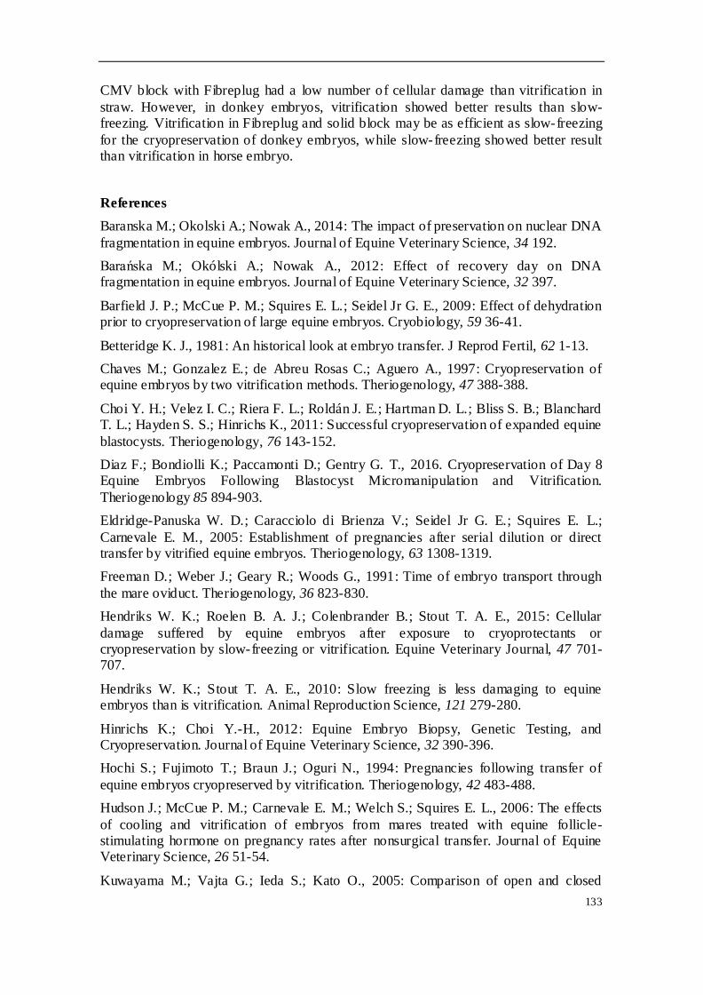
133
CMV block with Fibreplug had a low number of cellular damage than vitrification in
straw. However, in donkey embryos, vitrification showed better results than slow-freezing. Vitrification in Fibreplug and solid block may be as efficient as slow-freezing
for the cryopreservation of donkey embryos, while slow-freezing showed better result than vitrification in horse embryo.
References
Baranska M.; Okolski A.; Nowak A., 2014: The impact of preservation on nuclear DNA
fragmentation in equine embryos. Journal of Equine Veterinary Science, 34 192.
Barańska M.; Okólski A.; Nowak A., 2012: Effect of recovery day on DNA fragmentation in equine embryos. Journal of Equine Veterinary Science, 32 397.
Barfield J. P.; McCue P. M.; Squires E. L.; Seidel Jr G. E., 2009: Effect of dehydration prior to cryopreservation of large equine embryos. Cryobiology, 59 36-41.
Betteridge K. J., 1981: An historical look at embryo transfer. J Reprod Fertil, 62 1-13.
Chaves M.; Gonzalez E.; de Abreu Rosas C.; Aguero A., 1997: Cryopreservation of equine embryos by two vitrification methods. Theriogenology, 47 388-388.
Choi Y. H.; Velez I. C.; Riera F. L.; Roldán J. E.; Hartman D. L.; Bliss S. B.; Blanchard T. L.; Hayden S. S.; Hinrichs K., 2011: Successful cryopreservation of expanded equine
blastocysts. Theriogenology, 76 143-152.
Diaz F.; Bondiolli K.; Paccamonti D.; Gentry G. T., 2016. Cryopreservation of Day 8 Equine Embryos Following Blastocyst Micromanipulation and Vitrification.
Theriogenology 85 894-903.
Eldridge-Panuska W. D.; Caracciolo di Brienza V.; Seidel Jr G. E.; Squires E. L.;
Carnevale E. M., 2005: Establishment of pregnancies after serial dilution or direct transfer by vitrified equine embryos. Theriogenology, 63 1308-1319.
Freeman D.; Weber J.; Geary R.; Woods G., 1991: Time of embryo transport through
the mare oviduct. Theriogenology, 36 823-830.
Hendriks W. K.; Roelen B. A. J.; Colenbrander B.; Stout T. A. E., 2015: Cellular
damage suffered by equine embryos after exposure to cryoprotectants or cryopreservation by slow-freezing or vitrification. Equine Veterinary Journal, 47 701-707.
Hendriks W. K.; Stout T. A. E., 2010: Slow freezing is less damaging to equine embryos than is vitrification. Animal Reproduction Science, 121 279-280.
Hinrichs K.; Choi Y.-H., 2012: Equine Embryo Biopsy, Genetic Testing, and Cryopreservation. Journal of Equine Veterinary Science, 32 390-396.
Hochi S.; Fujimoto T.; Braun J.; Oguri N., 1994: Pregnancies following transfer of
equine embryos cryopreserved by vitrification. Theriogenology, 42 483-488.
Hudson J.; McCue P. M.; Carnevale E. M.; Welch S.; Squires E. L., 2006: The effects
of cooling and vitrification of embryos from mares treated with equine follicle-stimulating hormone on pregnancy rates after nonsurgical transfer. Journal of Equine Veterinary Science, 26 51-54.
Kuwayama M.; Vajta G.; Ieda S.; Kato O., 2005: Comparison of open and closed

134
methods for vitrification of human embryos and the elimination of potential
contamination. Reproductive biomedicine online, 11 608-614.
Lagares M. A.; Castanheira P. N.; Amaral D. C.; Vasconcelos A. B.; Veado J. C.;
Arantes R. M.; Stahlberg R., 2009: Addition of ficoll and disaccharides to vitrification solutions improve in vitro viability of vitrified equine embryo. Cryo Letters, 30 408-413.
Moussa M.; Bersinger I.; Doligez P.; Guignot F.; Duchamp G.; Vidament M.; Mermillod P.; Bruyas J. F., 2005: In vitro comparisons of two cryopreservation
techniques for equine embryos: Slow-cooling and open pulled straw (OPS) vitrification. Theriogenology, 64 1619-1632.
Moussa M.; Tremoleda J. L.; Duchamp G.; Bruyas J. F.; Colenbrander B.; Bevers M.
M.; Daels P. F., 2004: Evaluation of viability and apoptosis in horse embryos stored under different conditions at 5 °C. Theriogenology, 61 921-932.
Oberstein N.; O'Donovan M. K.; Bruemmer J. E.; Seidel Jr G. E.; Carnevale E. M.; Squires E. L., 2001: Cryopreservation of equine embryos by open pulled straw, cryoloop, or conventional slow cooling methods. Theriogenology, 55 607-613.
O’Donovan M.; Oberstein N.; Bruemmer J.; Lane M.; Seidel Jr G.; Gardner D.; Squires E., 2001: Metabolic activity of equine blastocysts cryopreserved in ethylene glycol and
galactose. Havemeyer Monograph Series. 2000;3:57–59..
Panzani D.; Rota A.; Crisci A.; Kindahl H.; Govoni N.; Camillo F., 2012: Embryo quality and transcervical technique are not the limiting factors in donkey embryo
transfer outcome. Theriogenology, 77 563-569.
Pomar F. J.; Teerds K. J.; Kidson A.; Colenbrander B.; Tharasanit T.; Aguilar B.;
Roelen B. A., 2005: Differences in the incidence of apoptosis between in vivo and in vitro produced blastocysts of farm animal species: a comparative study. Theriogenology, 63 2254-2268.
Scherzer J.; Davis C.; Hurley D. J., 2011: Laser-assisted vitrification of large equine embryos. Reprod Domest Anim, 46 1104-1106.
Seidel Jr G. E.; Cullingford E. L.; Stokes J. E.; Carnevale E. M.; McCue P. M., 2010: Pregnancy rates following transfer of biopsied and/or vitrified equine embryos: evaluation of two biopsy techniques. Animal Reproduction Science, 121 297-298.
Slade N.; Takeda T.; Squires E.; Elsden R.; Seidel G., 1985: A new procedure for the cryopreservation of equine embryos. Theriogenology, 24 45-58.
Smits K.; Goossens K.; Van Soom A.; Govaere J.; Hoogewijs M.; Peelman L. J., 2011: In vivo-derived horse blastocysts show transcriptional upregulation of developmentally important genes compared with in vitro-produced horse blastocysts. Reproduction,
Fertility and Development, 23 364-375.
Stout T. A. E., 2006: Equine embryo transfer: review of developing potential. Equine
Veterinary Journal, 38 467-478.
Tharasanit T.; Colenbrander B.; Stout T. A., 2005: Effect of cryopreservation on the cellular integrity of equine embryos. Reproduction, 129 789-798.

135
Tremoleda J. L.; Stout T. A.; Lagutina I.; Lazzari G.; Bevers M. M.; Colenbrander B.;
Galli C., 2003: Effects of in vitro production on horse embryo morphology, cytoskeletal characteristics, and blastocyst capsule formation. Biol Reprod, 69 1895-1906.

136

137
Figure 1. Confocal microscopy evaluation of
horse embryos frozen with ethylene glycol in
0.25ml straw (SF). Photomicrographs represent
the DAPI staining (A), the TUNEL staining (B),
cytoskeleton structure stained by phalloidine (C)
and the merge image (D). Photomicrographs at
the right side show the structure of embryos when
they were thawed.

138
Figure 2. Confocal microscopy evaluation of horse
embryos vitrified with ethylene glycol and glycerol
in Fibreplug and solid block (VF).
Photomicrographs represent the DAPI staining
(A), the TUNEL staining (B), cytoskeleton structure
stained by phalloidine (C) and the merge image
(D). Photomicrographs at the right side show the
structure of embryos when they were warmed.

139
Figure 3. Confocal microscopy
evaluation of horse embryos vitrified in
0.25ml straw (VS). Photomicrographs
represent the DAPI staining (A), the
TUNEL staining (B), cytoskeleton
structure stained by phalloidine (C)
and the merge image (D).
Photomicrographs at the right side
show the structure of embryos when
they were warmed.

140
Figure 4. Confocal microscopy
evaluation of horse donkey frozen with
ethylene glycol in 0.25ml straw (SF).
Photomicrographs represent the DAPI
staining (A), the TUNEL staining (B),
cytoskeleton structure stained by
phalloidin (C) and the merge image
(D). Photomicrographs at the right side
show the structure of embryos when
they were thawed.

141
Figure 5. Confocal microscopy
evaluation of donkey embryos
vitrified with ethylene glycol and
glycerol in Fibreplug and solid
block (VF). Photomicrographs
represent the DAPI staining (A),
the TUNEL staining (B),
cytoskeleton structure stained by
phalloidin (C) and the merge
image (D). Photomicrographs at
the right side show the structure
of embryos when they were
warmed.
142
Figure 6. Confocal microscopy
evaluation of donkey embryos
vitrified with 7M ethylene glycol
in Fibreplug and solid block
(VF7M). Photomicrographs
represent the DAPI staining (A),
the TUNEL staining (B),
cytoskeleton structure stained by
phalloidin (C) and the merge
image (D). Photomicrographs at
the right side show the structure
of embryos when they were
warmed.

143
7. DISCUSIÓN GENERAL
En el mundo actual se subestima la importancia que tiene para la biodiversidad la
recuperación y conservación de material genético de determinadas razas y especies
animales, cuya transcendencia y relevancia es mucho menor en nuestra sociedad, pero
no por ello dejan de ser imprescindibles para la riqueza genética de nuestro mundo. En
este sentido, se ideó el trabajo de la presente Tesis Doctoral utilizando diferentes
técnicas de reproducción asistida para la conservación y desarrollo de dos especies
equinas en peligro de extinción. Son muchos los factores que influyen en el desarrollo
con éxito de un programa de transferencia y criopreservación de embriones. Por ello,
en el primero de los trabajos se estudió la eficacia de tres protocolos de inducción y
sincronización de celo en yeguas en anestro. Normalmente la fase de anestro y
transición se presenta con ciclos irregulares o erráticos y es necesario controlarlos para
conseguir preñeces al comienzo de la estación reproductiva o para mejorar los
programas de transferencia de embriones. En la literatura se han descrito numerosos
tratamientos para la inducción del celo en la yegua, siendo el más frecuente el uso de
progesterona u otros progestágenos. El primero de nuestros estudios evaluó el uso
intravaginal de dispositivos durante 8 u 11 días en yeguas en anestro y se comparó con
el empleo del tratamiento vía oral. Se había reportado que el empleo de dispositivos
intravaginales provocaba hiperemia y vaginitis moderadas en la yegua (Ginger y col.,
1981; Rutten y col., 1986; Arbeiter y col., 1994; Lubbecke y col., 1994; Handler y col.,
2006). En nuestro estudio, algunas yeguas tratadas con CIDR® mostraron ligeros
cambios vaginales con una leve inflamación y secreción mucopurulenta. Otros estudios
describieron aislamientos bacterianos en yeguas tras el uso de otros dispositivos
intravaginales como PRID® (Newcombe, 2002), y recomendaban el empleo de
antibiótico local junto con la aplicación del dispositivo intravaginal para reducir las
descargas (Rutten y col., 1986). Sin embargo, en nuestro caso no se observaron
síntomas de endometritis ascendente, como son el acumulo de líquido o el
engrosamiento de la pared uterina, asociados con la aplicación del tratamiento
(Handler y col., 2006). Newcombe & Wilson (1997) sugirieron que los cambios
vaginales observados tras la aplicación de tratamientos intravaginales no estabán
asociados con la perdida de fertilidad en yeguas, ya que notaron un aumento en la tasa
de gestación tras su uso.
Durante este experimento ninguno de los dispositivos se perdieron, de acuerdo con
otras investigaciones (Newcombe, 2002; Larocca y col., 2006). Por lo tanto, desde el
punto de vista práctico, el dispositivo intravaginal es una muy buena alternativa para el
tratamiento con yeguas de difícil manejo, reduciendo la pérdida parcial de dosis que se
puede producir en los tratamientos por vía oral, y facilitando los tratamientos durante
los fines de semana. En este estudio se indujo la ciclicidad en el 88.5% de las yeguas
después del tratamiento. El patrón ecográfico varió tras el tratamiento, de manera que
los folículos más pequeños de 10 mm al principio del tratamiento, alcanzaron una talla

144
de >20 mm y llegaron a ser folículos preovulatorios en el 73.1% de las yeguas. En
nuestras latitudes, las yeguas entran en anestro entre noviembre y diciembre,
empiezan la fase de transición en febrero y marzo, y entran en época reproductiva en
abril (Handler y col., 2006). Sin embargo, se observó una gran desviación en el grupo
control, y se detectó que las yeguas podían estar ciclando a lo largo de todo año.
Nuestros resultados indicaron que el folículo que llega a ovular tienen un diámetro
significativamente más pequeño (46.5 ± 1.4 mm. vs 51.0 ± 1.1 mm; p<0.05) en yeguas
que ovularon 14 días después del tratamiento. Además, no se observaron diferencias
significativas entre grupos respecto al diámetro del folículo mayor al final del
tratamiento. Esto puede sugerir que, tras los diferentes tratamientos con
progesterona, el crecimiento del folículo dominante no se modifica y no se afecta el
momento de la ovulación. Sin embargo, yeguas que presentaron ovulación retrasada,
es decir, que ocurría más de 14 días después del tratamiento, mostraron un
incremento significativo del diámetro ovulatorio.
La comparación de los diferentes tratamientos de este estudio demuestran un efecto
similar y eficaz para la inducción del estro en yeguas en anestro estacional. Handler y
col. (2006) observaron que el dispositivo PRID® (sin estradiol) también era capaz de
inducir el estro y la ovulación en yeguas en diferentes épocas del año. Sin embargo, se
observó una deficiente sincronización de la ovulación, siendo difícil de implementar
ese tipo de tratamiento en los programas de transferencia de embriones.
En nuestro trabajo, ninguna de la yeguas ovuló antes del final del tratamiento, a
diferencia de lo obtenido por Newcombe (2002) y Handler y col. (2006) que reportaron
un 4.6% y 23.3% respectivamente. La diferencia podría explicarse por la actividad
folicular al inicio del tratamiento. Mientras que Newcombe (2002) estudió yeguas en
anestro y transición que tenían una realmente baja actividad folicular, Handler y col.
(2006) reportaron ovulaciones en el tratamiento de yeguas cíclicas. Podría ser
interesante realizar una evaluación preliminar de la actividad ovárica en yeguas y así
tener en cuenta el mes (es decir, la estación reproductiva) en el que el tratamiento se
utiliza. Se observó que el desarrollo folicular durante el tratamiento estaba asociado
con ovulaciones subsecuentes, mientras que yeguas sin desarrollo folicular fallaron en
la ovulación. Por lo tanto, diferentes enfoques pueden ser recomendados cuando
empleamos tratamientos basados en progesterona.
El porcentaje de yeguas en estro (88.5%) y la tasa de ovulación (73.1%) después del
tratamiento eran similares a los descritos por otros autores (Handler y col., 2006;
Larocca y col., 2006) y notamos que los tratamientos no afectaron a estas tasas. El
porcentaje de yeguas preñadas después de los tratamientos (47.4%) fue ligeramente
más bajo que los reportados previamente (Newcombe, 2002; Cuervo-Arango & Clark,
2010; Hanlon & Firth, 2012). Sin embargo, estos valores eran similares a los
observados en las yeguas no tratadas durante la primera ovulación de la época

145
reproductiva (46.7%). Este porcentaje fue ligeramente inferior al obtenido por Hanlon
& Firth (2012), pero mucho menor que el observado por Cuervo-Arango & Clark
(2010). Los citados estudios concluyeron que la tasa de preñez no se ve influenciada
por el tratamiento, al igual que ocurrió en nuestra experiencia.
Un estudio comparativo entre dispositivos intravaginales con 1.0 y 1.38 g de
progesterona (y 25 mg de benzoato de estradiol) concluyó que esas dosis de
progesterona tienen el mismo efecto en el manejo reproductivo en yeguas cíclicas
(Larocca y col., 2006). En este experimento se utilizó un dispositivo con 1.38 g de
progesterona, lo que supone una dosis más baja que la utilizada en otros estudios más
recientes (Handler y col., 2007; Cuervo-Arango & Clark, 2010; Hanlon & Firth 2012).
Los resultados muestran que los dispositivos con dosis de progesterona más bajas
tienen los mismos resultados que otros en los que se aplicaban dosis más elevadas, y la
disminución en tiempo de tratamiento de 11 a 8 días tampoco afectó de forma
negativa al rendimiento reproductivo futuro de yeguas en estado anovulatorio.
No se ha encontrado ningún signo que prediga la respuesta de tratamientos de
sincronización en yeguas en anestro (Handler y col., 2006). Sin embargo, en el presente
estudio hemos observado preliminarmente que la concentración plas mática de leptina
en yeguas en anestro puede ser un factor predictivo de la respuesta reproductiva a los
tratamientos de inducción del estro. Se ha observado que las yeguas que tienen
reactivación folicular después del tratamiento presentan un mayor nivel inicial de
leptina plasmática que las yeguas que prolongaron la aciclicidad después del
tratamiento. El posible papel de la leptina en yeguas ha sido estudiado por otros
autores (Ferreira-Dias y col., 2005) y se ha sugerido que esta modula ambas funciones,
tanto la reproductiva como la inmune (Drazen y col., 2000). Un incremento en la
concentración de leptina circulante podría estar asociada con una reactivación o
mantenimiento de la ciclicidad durante la estimulación de la función del hipotálamo-
hipófisis-gónada (Ferreira-Dias y col., 2005). Nuestro estudio apoya la idea de que la
leptina puede estar envuelta en la reactivación temprana de la actividad ovárica
después del tratamiento hormonal. El-Maaty y Gabr (2010) demostraron que un
incremento de leptina del día 7 al 9 postparto podría explicar su papel en la ovulación,
porque el hipotálamo parece ser el órgano sobre el que actúa la leptina, ya que sus
receptores se encuentran localizados en las áreas hipotalámicas asociadas al control de
la reproducción (Dyer y col., 1997; Lin y col., 2000). Los resultados obtenidos sugieren
que la leptina puede estar envuelta en la respuesta de las yeguas a tratamientos de
inducción de estro, lo que podría ser un indicador antes de iniciar el tratamiento. Más
estudios deberían ser enfocados a comprender mejor la acción de la leptina en la
reproducción de las yeguas. Se describe que las variaciones estacionales en yeguas
están asociadas a las concentraciones de leptina, siendo los valores de dicha hormona
más bajos durante el anestro (Ferreira-Dias y col., 2005). Estas observaciones
preliminares podrían ser confirmadas en estudios posteriores.

146
Los niveles de progesterona fueron mínimos (menores de 1 ng/ml) durante el
tratamiento en el grupo T1, lo cual indica que ninguna estructura lútea estaba
presente. Mientras que altrenogest estaba ejerciendo su acción progesteronémica, los
niveles de progesterona plasmática no se veían incrementados, ya que el test utilizado
era sensible únicamente a la molécula de progesterona. Sin embargo, los grupos en los
que se utilizaron los dispositivos intravaginales con progesterona sí mostraron un
aumento de dicha hormona a partir del segundo día después del tratamiento, con un
descenso progresivo hasta la retirada.
Como describiera anteriormente Cuervo-Arango y Clark (2010), los tratamientos con
dispositivos intravaginales de progesterona en yeguas de transición presentan ventajas
para el manejo en la reproducción durante periodos complicados del año tanto para el
veterinario y el ganadero. En el grupo T2, los niveles de progesterona cayeron por
debajo de 1 ng/ml en el día 11, mostrando que la liberación de progesterona es muy
reducida en este tiempo, sugiriendo que tratamientos más cortos podían tener efectos
similares.
Algunos estudios utilizan progesterona (y sus análogos) para inducir el estro en yeguas,
y están asociados con inductores de la ovulación como HCG o deslorelina. Dichos
inductores se administran cuando el folículo dominante alcanza un diámetro mayor de
35 mm y el dispositivo intravaginal se retira en diferentes días, dependiendo del
crecimiento folicular. En nuestro estudio no se emplearon inductores de la ovulación y
los tratamientos de progesterona se mantuvieron durante un tiempo determinado (8 ó
11 días). El uso de los 3 protocolos resultó en un crecimiento folicular, tasa de
ovulación y de preñez similar en las yeguas en anestro en que se utilizaron. Los
tratamientos demostraron una adecuada capacidad de inducir el estro, aunque no se
obtuvo una buena sincronización en las yeguas tratadas. Los resultados permiten
proponer su utilización en programas de criopreservación de embriones porque es
capaz de reducir el periodo improductivo (es decir, acortar la época no reproductiva) e
incrementa las oportunidades de tener embriones en hembras donantes. Sin embargo,
su baja capacidad de sincronización de estro podría dificultar su uso en transferencia
en fresco.
Además de evaluar la acción de la leptina como posible indicador y su influencia en la
reproducción, y de estudiar la acción de diferentes tratamientos de inducción de celo
en yeguas en anestro para mejorar la eficacia de la reproducción, se estudiaron los
factores que pueden influir en un programa de recogida y criopreservación de
embriones. Para ello se planteó, en el segundo de los trabajos realizados, un análisis
retrospectivo de los factores involucrados en dicho programa, que nos podría ayudar
en la toma de decisiones para obtener una mayor productividad. La tasa de ovulación
en yeguas fue de 1.12 por ciclo, mientras que en burras fue más alta (1.87
ovulaciones). La edad de la donante no afectó a la tasa de ovulación, lo cual estaba en

147
concordancia con estudios previos (Pérez-Marín y col., 2016), pero la tasa de ovulación
en burras donantes fue significativamente mayor en época reproductiva. Los
resultados que obtuvimos en yeguas están de acuerdo con los publicados por Ginther y
col. (1987), que afirman que la tasa de ovulación múltiple no se ve afectada por la
estacionalidad en la yegua.
Nuestros resultados mostraron una baja tasa de recogida de embriones en la raza
Hispano-árabe (35%), en comparación con otros autores que observaron tasas de
recogida del 48.1% al 64% (McCue y col., 2010; Aurich y col., 2011; Jacob y col., 2012;
Panzani y col., 2014). Douglas y col. (1985) obtuvieron una tasa del 28%, pero ellos
trabajaban con yeguas subfértiles, lo que podría sugerir que las yeguas empleadas en
nuestro estudio podrían tener reducida su fertilidad. Otros estudios describen tasas de
alrededor del 40% (Oberstein y col., 2001; Baranska y col., 2012), y cuya peculiaridad
era que empleaban una única inseminación con semen refrigerado. Debido a que la
raza Hispano-árabe está clasificada como raza en peligro de extinción, el número de
yeguas utilizadas en nuestro estudio fue limitado, lo que nos hizo imposible llevar a
cabo una buena selección de las donantes. Esto podría explicar la baja tasa de recogida
de embriones del estudio, aunque también podría ser justificado porque los lavados se
realizaron durante la época reproductiva y no reproductiva. Al analizar la variable
“época reproductiva” apreciamos que ésta afectó de manera significativa a la tasa de
recogida de embriones de nuestro estudio, incrementando en 11 puntos (46%) cuando
nos encontrábamos en estación favorable. La tasa de ovulación también afectó al
número de embriones que se recogían en la yegua. Cuando las ovulaciones fueron
dobles se notó una mayor recogida embrionaria por flushing (55% vs. 31%), al igual
que describieran otros autores (Squire y col., 1999; Stout, 2006; McCue y col., 2010), lo
que resalta la importancia de seleccionar, siempre que sea posible, yeguas donantes
que de forma natural presenten múltiple ovulación, incrementando así las
probabilidades de mejorar la eficiencia de recogida de embriones por lavado. Ninguna
de las otras variables analizadas (edad de la donante, días del flushing y calidad del
efluente) afectó a la tasa de recogida de embriones en la raza Hispano-árabe.
Una cosa parecida ocurre cuando la recogida de embriones se lleva a cabo en razas
locales de burras españolas (también razas amenazadas), aunque la tasa de recogida
fue mayor (44.7%) que la descrita en la yegua. Pocos estudios se han publicado sobre
la transferencia de embriones en burras, describiéndose tasas de recogida de 50.0% a
75.9% (Allen y col., 1985; Vendramini y col., 1997; Camillo y col., 2010; Panzani y col.,
2012; Paña-Alfaro y col., 2014). En un estudio retrospectivo previo realizado en burras
españolas (Pérez-Marín y col., 2016) se observó que la tasa media de preñez era del
56.4%, y que factores como la raza, edad y estación reproductiva no afectaban a esta
tasa de preñez, coincidiendo con las observaciones publicadas en esta Tesis Doctoral,
en la que no se apreció ninguna influencia debida a la edad de la donante, la esta ción
reproductiva, el día del lavado y la calidad del efluente uterino. Sin embargo, el

148
número de ovulaciones por ciclo sí afectó a la tasa de recogida en la burra, es decir,
más ovulaciones se traducen en más embriones recogidos, tal y como describieron
Camillo y col. (2010) en raza Pantesca. Ellos notaron que el número de ovulaciones, el
día del lavado y el número de ciclos repetidos en la misma donante influían sobre el
número de flushing positivos y sobre la tasa de recogida de embriones, mientras que el
año, la donante y la época del año no tuvieron efecto.
La tasa de recogida de embriones no varió en nuestras yeguas y burras cuando los
lavados se realizaron los días 6.5, 7.5 o 8.5 tras la ovulación. Jacob y col. (2012)
observaron una disminución significativa en dicha tasa cuando las yeguas fueron
sometidas a flushing uterino el día 6, en contraste con los días 7, 8, 9 y 10, mientras
que Panzani y col. (2014) no encontraron tales diferencias entre los días 7 y 10. En
burras, Camillo y col. (2010) observaron que se recogían más embriones en el día 8 y 9
post-ovulación, que en el día 7, aunque no encontramos más estudios que evaluaran la
influencia del día de recogida en burras. En nuestro estudio, no existieron diferencias
significativas asociadas al día del lavado, comparándose desde el día 6.5 al día 8.5.
La tasa de ovulación múltiple fue menor en yeguas que en burras, en las que
observamos ovulaciones dobles, triples e incluso cuádruples. La asincronía entre las
ovulaciones múltiple hacía difícil definir la edad exacta del embrión y este hecho fue el
responsable de la amplia variabilidad en cuanto a estado y tamaño observada entre los
embriones, especialmente en burras.
Se ha propuesto que la disminución de la tasa de recogida de embriones puede estar
por la fertilidad reducida de los sementales y la temprana recogida de los embriones
para el programa de criopreservación (alrededor del día 6) (Freeman y col., 1991). En
nuestro estudio, no existieron diferencias entre los días de lavado, y se consiguieron
más flushing positivos (considerando como flushing positivo aquel en el que al menos
se recoge un embrión) en el día 6.5, en comparación con los días 7.5 o 8.5 (aunque no
se observaron diferencias). Se observó un alto número de yeguas y burras que fallaron
en todos los intentos realizados por conseguir un embrión, lo que podría sugerir que
esas hembras tenían una baja fertilidad, y explicaría parcialmente la baja tasa de
recogida de embriones del presente estudio.
La mayoría de los embriones recogidos (92.9%) en yeguas y burras mostraron una
excelente o buena calidad, es decir, eran de calidad I o II (McCue y col., 2010; Camillo y
col., 2010; Camargo y col., 2013). Se ha descrito que los embriones normalmente
producen PGE2 (y otras sustancias) en el oviducto que promueven su paso al útero
desde el día 5 (Smits y col., 2012), mientras que los UFOs o embriones degenerados se
quedan retenidos en el oviducto (McCue y col., 2010; Aguilar y col., 2012). De la misma
manera, en estudios realizados en burras se describe que la mayoría de embriones
obtenidos eran de grado I o II (Panzani y col., 2012; Peña-Alfaro y col., 2014). En burras
españolas encontramos un 85.7% de embriones de grado I-II. En este sentido, nosotros

149
recogimos una mórula y un UFO en una donante que había ovulado un único folículo.
Eso sugiere que cuando se encuentra un UFO, debería existir un embrión normal en el
útero y en el lavado (Smits y col., 2012). Sin embargo, en nuestro estudio, en una burra
que había ovulado de un único folículo se encontraron 3 UFOs, pero ningún embrión se
halló en el lavado. Este hallazgo podría sugerir que no siempre el mecanismo de
apertura de la papila útero-tubárica necesita la presencia de un embrión normal,
aunque también podría pensarse que el embrión normal no se pudo encontrar. En este
caso es difícil explicar porqué los tres UFOs aparecieron en el lavado, pero sabemos
que en el ciclo anterior se había producido una doble ovulación (aunque ningún
embrión se había recogido).
Doce de las sesenta y una yeguas donantes tuvieron doble ovulación espontánea
(19.67%), menor del 30% que se ha descrito en yeguas donantes de otras razas como
PSI, Razas mixtas europeas, caballos trotones, etc. (Morel y col., 2005; Stout, 2006). Sin
embargo, diez de las trece burras donantes (76.9%) tuvieron ovulación múltiple en
alguno de los ciclos que se monitorizaron, de acuerdo con lo observado en estudios
previos (Pérez-Marín y col., 2016).
McCue y col. (2010) observaron que se obtenía un mayor número de embriones en
yegua que presentaban menos debris en el efluente uterino. Aunque nosotros no
detectamos diferencias significativas en nuestro estudio, ni en yeguas ni en burras, se
apreció que cuando la calidad del efluente era mala (una alta celularidad y/o turbidez),
un bajo porcentaje de embriones era obtenido.
Aunque se ha descrito que el tipo de semen es otro factor que afecta a la tasa de
recogida de embriones (Meedows y col., 2000; McCue y col., 2010; Panarace y col.,
2014; Panzani y col., 2014), en este trabajo no pudimos analizarlo, ya que todas las
hembras fueron inseminadas con semen refrigerado. Sin embargo, sí se observó un
efecto asociado al semental o garañón pues en un total de 6 sementales se apreció
una muy baja tasa de recogida de embriones (menos del 25%). Los seis sementales que
mostraron baja tasa de recogida embrionaria tras ser utilizados para inseminar a
yeguas donantes fueron los responsables del 45% de los flushing realizados (63/140). A
pesar de los fracasos repetidos, se continuaron utilizando debido a su importanc ia
desde el punto de vista genético. Eso podría sugerir que, en parte, la baja tasa de
recogida se debió a este hecho. El número de garañones empleados fue reducido (solo
5), y uno de ello mostró un claro fracaso reproductivo tras su empleo en burras
donantes; tras la IA con semen de ese burro se recogieron tres UFOs, lo que
demostraba su reducida efectividad.
El tamaño de los embriones varía ampliamente en referencia al día en que la primera
ovulación es detectada. Sin embargo, si comparamos el tamaño embrionario en los
diferentes estados en que clasificamos a los embriones, la variación es muy peque ña.
No obstante, las dificultades surgen cuando intentamos determinar la edad del

150
embrión a partir del día en que consideramos que se ha producido la ovulación, ya que
las ovulaciones múltiples y/o asincrónicas impiden establecer la edad exacta del
embrión. Este problema no es tan importante en yegua, ya que las ovulaciones fueron
en su mayoría simples. Diferentes autores han observado variaciones en el tamaño
embrionario dependiendo del día de recogida. Así, el día 6 se describe un diámetro
medio de entre 142-284 µm (Hochi y col., 1994; Eldridge-Panuska y col., 2005; Moussa
y col., 2005; Lagares y col., 2009; Choi y col., 2010; McCue y col., 2010; Baranska y col. ,
2012); en el día 7, varía desde 354 a 1000 µm (Tharasanit y col., 2005; Koblischke y
col., 2008; Choi y col., 2010; McCue y col., 2010; Panzani y col., 2014); y en el día 8, las
medidas oscilan entre 623 y 3800 µm (Choi y col., 2010; McCue y col., 2010; Baranska y
col., 2012; Herrera y col., 2014; Panzani y col., 2014). En el presente estudio, el
diámetro medio fue ligeramente más bajo que el descrito por otros autores para el día
7.5. Sin embargo, cuando los embriones eran clasificados acorde a su estatus
morfológico, el tamaño fue similar a lo descrito por otros (McCue y col., 2010).
A pesar de que existen muy pocos estudios relacionados con la obtención de
embriones en burra, estos muestran una gran variabilidad en los diámetros medidos.
Mientras que Vendramini y col. (1997) informaron de que el diámetro de los
embriones era de 187 39, 250 69 y 348 80 µm para embriones obtenidos en el día
6, día 7 y día 8 respectivamente, Panzani y col. (2012) observaron tamaños de 666
378 y 1005 545 µm en embriones recogidos en el día 8 y 9, lo que ha hecho difícil la
elaboración de una tabla predictiva. Los resultados obtenidos en embriones de burras
españolas demostraron también grandes diferencias en sus tamaños, y sugieren que el
embrión de burro debería ser clasificado de acuerdo a su estatus morfológico, en lugar
de hacerlo por la edad según el día en que se produjo la ovulación.
Un total de 9 flushing uterinos se llevaron a cabo durante el celo del potro en yeguas y
ninguno en burras, obteniéndose un único embrión. Aunque el número de yeguas
evaluadas fue muy bajo para sacar conclusiones, la recogida embrionaria realizada tras
la primera ovulación post-parto (es decir, tras el celo del potro) no ofreció buenos
resultados en nuestro estudio, en contraste con lo observado por Lopes y col. (2011),
pero de acuerdo con Metcalf & Sanchez (2012), aunque estos últimos describieron que
la fertilidad de esos embriones (obtenidos tras el celo del potro) era mayor que la
observada en embriones de yeguas no lactantes. El mayor tamaño que presenta el
útero durante ese periodo y la complejidad de la involución uterina podrían explicar
los malos resultados en la tasa de recogida de embriones durante el celo del potro.
Ante las bajas tasas de recogida de embriones observadas en nuestro estudio,
pensamos en la posibilidad de que el ambiente uterino pudiera estar alterado y fuese
el responsable de tales resultados. Con objeto de evaluar el ambiente uterino en
yeguas donantes en el mismo momento de la recogida, se llevó a cabo el tercero de los
estudios. Douglas y col. (1985) sugirieron que el ambiente uterino peri -ovulatorio

151
afecta significativamente a la tasa de gestación. El embrión entra en el útero alrededor
del día 6 después de la ovulación y en un programa de transferencia de embriones,
permanecerá allí hasta su recogida en torno al día 6-7. Pero las técnicas recomendadas
para evaluar el estado endometrial no se pueden realizar en la hembra donante
debido al alto riesgo de pérdida del embrión durante la realización de las mismas. El
objetivo de este estudio fue evaluar el estado bacteriológico y citológico en yeguas
donantes en el momento de la recogida, y determinar la utilidad de la muestra
obtenida del efluente uterino durante el flushing para determinar el estado
endometrial. Los métodos rutinarios no invasivos incluyen el citobrush, el hisopo de
algodón y el lavado con bajo volumen (LeBlanc y col., 2007; Davies Morel y col., 2013).
En resumen, este estudio analizó si el análisis del efluente retenido en el filtro después
de la recogida de embriones podría constituir un método lo suficientemente sensible y
específico como para obtener información relativa al estatus uterino de la donante.
El estudio comenzó con la comparación de los diferentes métodos de toma de muestra
en yeguas con endometritis clínica, diagnosticada por ecografía, citología y
bacteriología uterina. En este grupo de yeguas con endometritis clínica, al comparar
los resultados citológicos y bacteriológicos procedentes del hisopo uterino y los
procedentes del liquido residual del filtro, se observó que fueron prácticamente
idénticos, con lo que se confirmaba que la realización de citología y cultivo de
muestras procedentes del liquido residual del filtro tenían la misma efectividad que las
procedentes de hisopos uterinos. Estos hallazgos eran cruciales para la decisión
siguiente de usar el efluente para evaluar muestras de las yeguas donantes, con vistas
a conocer el estado del útero.
De acuerdo con la hipótesis inicial en la que ningún microorganismo debería estar
presente en el útero de la yegua donante en el momento de la recogida, se recogieron
muestras del efluente retenido en el filtro del embrión después del lavado, con vistas a
hacer un estudio citológico y bacteriológico, y evitar la posible perdida de embriones
asociada a esas técnicas. El chequeo previo de las yeguas del grupo control (yeguas con
signos de endometritis) mostraron que el cultivo del efluente del filtro tenía resultados
similares a los obtenidos por el método del hisopo de algodón. Además, se aislaron
bacterias en muestras procedentes de hembras donantes, incluyendo cuando se
obtuvieron embriones. El aislamiento bacteriano y la citología endometrial realizadas
en las yeguas donantes indicó que ninguna de ellas era positiva a ambas pruebas, lo
que podría significar que no existía una infección o inflamación activas.
LeBlanc y col. (2007) describieron que el método de lavado uterino con pequeño
volumen es una forma rápida, precisa y práctica de diagnosticar endometritis en
yeguas con infertilidad crónica, con una sensibilidad y especificidad aproximada de
0.75 y 0.9, respectivamente. El método de diagnóstico del efluente retenido en el filtro
que se testa en nuestro estudio es muy similar a la técnica antes mencionada, pero

152
permite el empleo del efluente retenido en el filtro y evita comprometer la recogida de
embriones. Los resultados indican que este método ofrece un alto grado de
sensibilidad y especificidad para el análisis citológico (1 y 0.86, respectivamente) y para
el aislamiento bacteriano (1 y 0.41, respectivamente). Mientras que en el estudio de
LeBLanc y col. (2007) se utilizaron yeguas con infertilidad crónica, las yeguas aquí
estudiadas pertenecían a un programa de criopreservación de embriones, donde se
considera crucial que exista una adecuada salud uterina. Es posible que se detecte un
menor número de microorganismos patógenos en hisopo de algodón uterino que en la
técnica de pequeño volumen o de efluente después del lavado uterino (como se usa en
transferencia de embriones), ya que los dos últimos métodos abarcan una mayor área
de superficie de muestra (LeBlanc y col., 2007).
Nielsen y col. (2005) consideran la presencia de neutrófilos en el tejido como la mejor
prueba diagnóstica de endometritis. En nuestro estudio, se identificaron células
inflamatorias solo en tres de las yeguas donantes pero, como mencionamos
anteriormente, ninguna de ellas mostró crecimiento bacteriano por lo que se descartó
la existencia de una infección reactiva.
Riddle y col. (2007) describieron que la presencia de Streptococcus equi subs.
zooepidemicus, E. coli, Staphylococcus, Micrococcus, Bacillus y haemolytic
Streptococcus son especies saprofitas en el útero de las yeguas, que no promueven la
aparición de infecciones reactivas. La mayoría de las bacterias aisladas en las yeguas
donantes (75%) pertenecían a las citadas especies, apareciendo en cultivos puros o
mixtos. LeBlanc y col. (2007) no encontraron una correlación entre el cultivo positivo
obtenido en muestras del bajo volumen y el grado de inflamación si no se observan
PMNs. Así que la presencia de PMNs en muestras de endometrio es dependiente del
tipo de bacteria aislada; la detección de PMNs en el útero está relacionada más
frecuentemente con Streptococcus β-hemoliticos y con E. coli (Nielsen y col., 2010). La
fase estral de la yegua también podría afectar a la presencia o ausencia de neutrófilos
en el útero, de manera que la migración de los neutrófilos hacia la luz uterina durante
el periodo de dominancia de progesterona es menor que durante la dominancia de
estrógenos (Ball y col., 1988; LeBlanc y col., 2007, Riddle y col., 2007). En nuestro
estudio, ninguna célula inflamatoria fue detectada en el 85% las donantes (17/20) a las
que se les realizó el lavado uterino, es decir, durante la fase de dominancia de la
progesterona.
Riddle y col. (2007) indican que el aislamiento de microorganismos en el endometrio
durante la cubrición está siempre asociado con una disminución en la tasa de preñez,
independientemente de los resultados citológicos. En un estudio reciente sobre
citología endometrial, Davies Morel y col. (2013) indican que el valor umbral de ≥1%
PMNs está asociado a una reducción de potros nacidos vivos. Los resultados obtenidos
en el presente estudio indican que la presencia de bacterias en el útero no impide la

153
recogida de embriones, ya que en cuatro donantes obtuvimos crecimiento bacteriano
pero pudimos recoger embriones.
La apariencia del efluente recogido en el grupo control (yeguas con endometritis) fue
considerablemente diferente del obtenido durante el lavado de embriones en el grupo
de donantes, estando el crecimiento bacteriano asociado a la suciedad del efluente.
LeBlanc y col. (2007) también describieron que la turbidez y mucosidad del efluente
obtenido en yeguas con endometritis estaba asociado con el aislamiento de
microorganismos. En nuestro estudio apreciamos que el aislamiento de
Staphylococcus estaba siempre asociado con efluente turbio, mientras que los
resultados para otras bacterias eran variables. En vista de los resultados obtenidos y
conforme a lo que describieron LeBlanc y col. (2007), el cultivo bacteriano positivo a
partir de muestras obtenidas del flushing se puede considerar contaminado cuando se
acompaña de un efluente claro, no hay cambios en el pH, no hay células inflamatorias
o hipocelularidad citológica, y existe ausencia de neutrófilos en el tejido endometrial
obtenido por la biopsia.
Por último, en el trabajo 4 se compararon los resultados de diferentes protocolos de
vitrificación con los de congelación convencional en embriones de caballo de raza
Hispano-árabe y asnos de raza Andaluza y Zamorano-leones. Hay que resaltar que
existen muy pocas publicaciones sobre criopreservación de embriones de burro y se
desconoce cómo los diferentes métodos pueden afectar a su calidad. Además en este
estudio, se muestran resultados empleando la vitrificación en superficie sólida, que
también ha sido muy poco investigada en equinos.
Los resultados in vitro muestran que los porcentajes de células muertas obtenidos tras
someter a los embriones de caballo y asno a congelación lenta (5.0% y 4.6%,
respectivamente) fueron similares al 3.3% descrito por Hendriks y col. (2015), y más
bajos que los publicados por otros autores, que oscilaron entre 14.6% y 42.0%
(Oberstein y col., 2001; Moussa y col., 2005; Tharasanit y col., 2005; Hendriks & Stout,
2010).
No hemos encontrado en la literatura ningún estudio que analizara embriones de
burro criopreservados, y solo Panzani y col. (2012) han descrito la tinción de
embriones frescos de burro con DAPI. Estos investigadores observaron que los
embriones de burro en fresco tenían un bajo número de células muertas y que dicho
número no estaba correlacionado con el tamaño embrionario. También describieron
que los embriones de pobre calidad (grado 3) presentaban un mayor porcentaje de
células muertas, pero que no alcanzaba el 20%, que había sido fijado por Moussa y col.
(2005) como el límite en embriones viables. En nuestro estudio, todos los embriones
criopreservados de burro tuvieron un porcentaje de células muertas por debajo del 20
%, aunque notamos que la congelación convencional inducía un índice mayor de
muerte celular que la vitrificación.

154
El porcentaje de muerte celular en los embriones durante el proceso de vitrificación
osciló en un rango del 14.1% al 2.2% en caballo y burro, que resultaban más bajos que
los descritos previamente por otros autores (49% al 52% en Oberstein y col., 2001;
47%, 79% y 94% en O’Donovan y col., 2001; 47% en Moussa y col., 2005; 99%, 74% y
81% en Lagares y col., 2009; 30% en Hendriks & Stout, 2010, y 20% en Hendriks y col.,
2015). Estas diferencias podrían deberse en parte al método utilizado para contar el
número total de células y para calcular el porcentaje de células muertas. En este
estudio, el recuento celular se realizó usando el software ImageJ, de manera que se
hacía un recuento automático del número de núcleos en el plano de la imágenes
captados por el microscopio confocal, consiguiendo la profundidad completa del
embrión, como describieran Hendriks & Stout (2010) y Hendriks y col. (2015). A pesar
de todo, cuando el porcentaje de muerte celular en nuestro estudio fue calculado
según la fórmula empleada por Moussa y col. (2005), los resultados continuaron
siendo más bajos que en otros estudios. En contraste con lo observado en embriones
de burro, los resultados obtenidos en este estudio utilizando congelación lenta indican
que es menos dañina para los embriones de caballo que la vitrificación.
En el caso de embriones frescos, se ha descrito un muy bajo número o incluso nulo, de
células apoptóticas (Moussa y col., 2004; Pomar y col., 2005; Baranska y col., 2012 y
2014). En cuanto al porcentaje de células apoptóticas de nuestro estudio, se obtuvo
entre 1.5% y 0.5% en embriones de caballo y de burro respectivamente, sometidos a
congelación lenta. Sin embargo, no hemos encontrado ningún trabajo de la literatura
consultada que describiera el uso de TUNEL en embriones equinos tras su congelación
lenta. En el presente estudio, el 80% de los embriones equinos congelados tuvieron al
menos una célula positiva a TUNEL, con una media de un 1.0% de células apoptóticas.
En el caso de los embriones de burro, el 75% de los embriones mostraron alguna célula
positiva o teñida por TUNEL, y el porcentaje medio de células apoptóticas fue aún
menor, del 0.5%. En los embriones de caballo vitrificados se observó que un 80% de
ellos mostraban alguna célula positiva cuando se utilizó el protocolo de Fibreplug en
bloque de vitrificación, y un 75% cuando se vitrificó en pajuela. La media del
porcentaje de células apoptóticas fue mayor cuando la vitrificación se hizo empleando
Fibreplug que cuando lo hicimos en pajuela (9.9% vs. 4.9%). Estos resultados difieren
de los obtenidos por Baranska y col. (2014), que emplearon un dispositivo de
vitrificación denominado Rapid-i y una pajuela; compararon la tasa de apoptosis tras la
vitrificación en ambos sistemas, encontrando valores de 0.3% y 2.3% para Rapid-i y
pajuela, respectivamente. En el caso de los embriones vitrificados de burro con
Fibreplug, todos mostraron alguna célula apoptótica, pero los porcentajes eran muy
bajos, rondando el 1.8% cuando utilizamos EG-Glicerol, y el 0.15% cuando se empleó
7M EG. En nuestro estudio se observó que los embriones de caballo vitrificados tienen
un índice de apoptosis más alto que los embriones congelados, lo que sugiere que la
vitrificación podría tener un efecto más negativo para la viabilidad del embrión equino,

155
aunque algunos autores afirman que la apoptosis es un mecanismo celular para
eliminar células no funcionales, anormales o en mala posición en embriones obtenidos
de forma in vivo (Tremoleda y col., 2003). Los embriones de burro congelados en
pajuela mostraron un bajo índice de apoptosis, en contraste con los embriones
vitrificados en etilenglicol y glicerol. La comparación entre los embriones de las dos
especies mostró una diferencia significativa en el porcentaje de células apoptóticas,
siendo este más bajo para los embriones de burro que los de caballo (p<0.05), aunque,
a pesar de lo que dicen otros autores, nosotros pensamos que el índice de apoptosis
no es un buen indicador de viabilidad embrionaria tras un proceso de vitrificación ya
que mostró un índice alto en embriones con bajo porcentaje de células muertas y con
buena calidad morfológica.
Respecto a la calidad del citoesqueleto, en este estudio solo se observaron embriones
de calidad I o II, tras la descongelación/calentamiento. No encontramos ningún
embrión de mala calidad (grado III), a diferencia de otros estudios que describen hasta
un 25% de embriones de ese tipo tras la vitrificación (Hendriks y col., 2015) o
congelación (Tharasanit y col., 2005). Apreciamos una relación positiva entre el
porcentaje de células muertas y el grado del citoesqueleto, que apoya la teoría de
otros autores que consideran la evaluación del citoesqueleto como un buen indicador
de la calidad in vitro de los embriones equinos (Tharasanit y col., 2005). No debemos
olvidar que el citoesqueleto es una estructura necesaria para el crecimiento normal de
la célula, y los daños producidos por la congelación (como la formación de cristales de
hielo) podría provocar su desorganización. En el caso de los embriones de burro,
también mostraron buena calidad del citoesqueleto tras el proceso de
criopreservación.
En este trabajo solo realizamos transferencia de embriones de caballo vitrificados en
Fibreplug y los resultados fueron más bajos de lo que a priori esperábamos,
basándonos en otros estudios. Aunque se alcanzaron porcentajes similares a los
descritos por Seidel y col. (2011) y Hochi y col. (1994), éstos fueron más bajos que los
publicados por Eldridge-Panuska y col. (2005) o Hudson y col. (2006). Sin duda, existen
muchos factores que pueden influir en el éxito de una transferencia de embriones tras
la criopreservación, por lo que debemos incrementar el número de embriones
transferidos en un futuro.

156

157
8. CONCLUSIONES
Los dispositivos intravaginales de progesterona son una alternativa aceptable para
inducir el estro en yeguas donantes frente a los tratamientos orales, ya que muestran
ventajas esenciales como la disminución de los días de tratamiento, la reducción de
manejo de los animales o la menor pérdida del principio activo empleado.
Los niveles de leptina podrían ser un buen indicador, previo al tratamiento, de la
eficacia de los programas de inducción de celo en las yeguas.
La tasa de recogida de embriones en yegua se ve afectada por la estación
reproductiva y el número de ovulaciones por ciclo, mientras que en la burra solo se
apreció la influencia del número de ovulaciones por ciclo.
La calidad de los embriones no mostró variaciones debidos a los diferentes factores
externos analizados, pero su diámetro sí estuvo influenciado en yeguas por el día del
lavado y por el estatus morfológico, mientras que en burras el tamaño embrionario
estuvo afectó por el número de ovulaciones por ciclo y el estatus morfológico del
embrión.
El análisis del efluente retenido en el filtro después de la recogida de embriones en
yeguas es un método eficiente para evaluar es estado bacteriológico y citológico del
útero de las yeguas donantes.
Los embriones obtenidos en yeguas son más susceptibles al proceso de vitrificación
que al de congelación convencional, ya que presentan un mayor daño a nivel celular,
como se manifesta por la mayor muerte celular, mayor apoptosis y peor calidad en la
ultraestructura embrionaria observadas.
La vitrificación indujo una menor muerte celular sobre los embriones recogidos en
burra que la congelación lenta, por lo que se postula como el método más interesante,
al menos desde el punto de vista in vitro.
Respecto a los diferentes protocolos de vitrificación empleados en este estudio, el
uso de Fibreplug junto a un bloque sólido ofreció mejores resultados in vitro para la
vitrificación de embriones equinos, y demostró su eficacia in vivo tras obtenerse dos
gestaciones.

158

159
CONCLUSIONS
Intravaginal progesterone-releasing devices are a valid alternative to oral
treatments in donor mares, because it reduces the days of treatment, the animal
management is easier, and diminishes the loss of drugs.
Leptin levels would be a good sensor of the efficiency of oestrus induction protocol
in mares when it is measured before treatment.
The embryo recovery rate in mares is affected by the breeding season and the
number of ovulation per cycle, whereas in jennies, the number of ovulation per cycle
was the only factor that modified the number of collected embryos.
Embryo diameter was affected by day of flushing and embryo morphological status
in mares, whereas in jenny was affected by the number of ovulation per cycle and
morphological status of the embryos.
Analysis of the efflux retained in the filter after embryo collection in mares is an
efficient method to evaluate the bacteriological and cytological uterine status in
donors.
Embryos recovered from mares are more susceptible to vitrification than to slow
freezing, because more cellular injuries are induced, provoking more death cells,
higher apoptosis and defects on the embryo cytoskeleton.
Vitrification induces lower cellular death on donkey embryos than slow freezing, and
it could be considered as a good cryopreservation method for these embryos, as
demonstrated after in vitro assesment.
In relation to the vitrification protocols tested in this study, Fibreplug used with solid
block shows better in vitro results for equine embryo vitrification than others, and two
pregnancies were obtained after embryo tranfer.

160

161
9. BIBLIOGRAFIA
Abe H.; Hoshi H., 2003: Evaluation of bovine embryos produced in high performance serum-free media. J Reprod Dev, 49 193-202.
Aguilar J.; Fraser H. M.; Wilson H.; Clutton E.; Shaw D. J.; Watson E. D., 2006: Temporal relationship between proliferating and apoptotic hormone-producing and endothelial cells in the equine corpus luteum. Reproduction, 132 111-118.
Aguilar J. J.; Cuervo-Arango J.; Mouguelar H.; Losinno L., 2012: Histological Characteristics of the Equine Oviductal Mucosa at Different Reproductive Stages. Journal of Equine Veterinary Science, 32 99-105.
Allen W., 1982: Embryo transfer in the horse. Mammalian egg transfer, 135-154.
Allen W.; KYDD J.; Boyle M.; Antczak D., 1985: Between‐species transfer of horse and
donkey embryos: A valuable research tool. Equine Veterinary Journal, 17 53-62.
Allen W.; Stewart F., 2001: Equine placentation. Reproduction, Fertility and Development, 13 623-634.
Alm H.; Torner H.; Blottner S.; Nürnberg G.; Kanitz W., 2001: Effect of sperm cryopreservation and treatment with calcium ionophore or heparin on in vitro
fertilization of horse oocytes. Theriogenology, 56 817-829.
Almodin C. G.; Minguetti-Camara V. C.; Paixao C. L.; Pereira P. C., 2010: Embryo development and gestation using fresh and vitrified oocytes. Human Reproduction, 25 1192-1198.
Alvarenga F. L.; Alvarenga M.; Meira C., 1993: Transmission electron microscopy of equine embryos cryopreserved by different methods. Equine Veterinary Journal, 25
67-70.
Alvarenga M. A.; McCue P. M.; Bruemmer J.; Neves Neto J. R.; Squires E. L., 2001:
Ovarian superstimulatory response and embryo production in mares treated with equine pituitary extract twice daily. Theriogenology, 56 879-887.
Andrabi S. M. H.; Maxwell W. M. C., 2007: A review on reproductive biotechnologies for conservation of endangered mammalian species. Animal Reproduction Science, 99 223-243.
Araujo G. H. M.; Rocha Filho A. N.; Burns S. D.; Burns C. M.; Moya-Araujo C. F.; C. M., 2010: Pregnancy rates after vitrification, warming and transfer of equine embryos.
Animal Reproduction Science, 121 299-300.
Arav A.; Yavin S.; Zeron Y.; Natan D.; Dekel I.; Gacitua H., 2002: New trends in gamete's cryopreservation. Molecular and cellular endocrinology, 187 77-81.
Arbeiter K.; Barth U.; Jöchle W., 1994: Observations on the use of progesterone
intravaginally and of deslorelin STI in acyclic mares for induction of ovulation. Journal of Equine Veterinary Science, 14 21-25.
Ataman M.; Günay A.; Günay Ü.; Baran A.; Uzman M., 2000: Oestrous synchronization

162
with progesterone impregnated device and prostaglandin F2α both combined with human chorionic gonadotropin in transitional mares. Revue Méd. Vét, 151 1031-1034.
Aurich C., 2011: Reproductive cycles of horses. Animal Reproduction Science, 124 220-228.
Aurich C.; Daels P. F.; Ball B. A.; Aurich J. E., 1995: Effects of gonadal steroids on the opioid regulation of LH and prolactin release in ovariectomized pony mares. Journal of
Endocrinology, 147 195-202.
Bae S.-E.; Watson E. D., 2003: A light microscopic and ultrastructural study on the
presence and location of oxytocin in the equine endometrium. Theriogenology, 60 909-921.
Ball B. A.; Shin S. J.; Patten V. H.; Lein D. H.; Woods G. L., 1988: Use of a low-volume uterine flush for microbiologic and cytologic examination of the mare's endometrium. Theriogenology, 29 1269-1283.
Baranska M.; Okolski A.; Nowak A., 2014: The impact of preservation on nuclear DNA fragmentation in equine embryos. Journal of Equine Veterinary Science, 34 192.
Barańska M.; Okólski A.; Nowak A., 2012a: Effect of recovery day on DNA
fragmentation in equine embryos. Journal of Equine Veterinary Science, 32 397.
Barańska M.; Okólski A.; Nowak A., 2012b: Effect of recovery day on DNA
fragmentation in equine embryos. Journal of Equine Veterinary Science, 32 397.
Barfield J. P.; McCue P. M.; Squires E. L.; Seidel Jr G. E., 2009: Effect of dehydration prior to cryopreservation of large equine embryos. Cryobiology, 59 36-41.
Barlund C. S.; Carruthers T. D.; Waldner C. L.; Palmer C. W., 2008: A comparison of diagnostic techniques for postpartum endometritis in dairy cattle. Theriogenology, 69 714-723.
Barry B. E.; Thompson D. L.; White K. L.; Wood T. C.; Hehnke K. E.; Rabb M. H.; Colborn D. R., 1989: Viability of inner cell mass versus trophectodermal cells of frozen-thawed horse embryos. Equine Veterinary Journal, 21 82-83.
Bass L.; Denniston D.; Maclellan L.; McCue P.; Seidel G.; Squires E., 2004: Methanol as a cryoprotectant for equine embryos. Theriogenology, 62 1153-1159.
Becker S.; Johnson A., 1992: Effects of gonadotropin-releasing hormone infused in a pulsatile or continuous fashion on serum gonadotropin concentrations and ovulation
in the mare. Journal of animal science, 70 1208-1215.
Beg M. A.; Ginther O. J., Follicle selection in cattle and horses: role of intrafollicular factors.
Bergfelt D. R.; Mann B. G.; Schwartz N. B.; Ginther O. J., 1991: Circulating concentrations of immunoreactive inhibin and FSH during the estrous cycle of mares.
Equine Vet J, 11 319-322.
Betteridge K. J., 1981: An historical look at embryo transfer. J Reprod Fertil, 62 1-13.
Betteridge K. J., 2001: Comparative aspects of conceptus growth: a historical perspective. Reproduction, 122 11-19.

163
Betteridge K., 2006: Embryo ‘talk’in horses: its comparative relevance. Embryo Talk, 1 131-139.
Betteridge K. J., 2007: Equine embryology: An inventory of unanswered questions. Theriogenology, 68, Supplement 1 S9-S21.
Boerboom D.; Brown K. A.; Vaillancourt D.; Poitras P.; Goff A. K.; Watanabe K.; Doré M.; Sirois J., 2004: Expression of key prostaglandin synthases in equine endometrium
during late diestrus and early pregnancy. Biology of reproduction, 70 391-399.
Boyle M.; Allen W.; Tischner M.; CZLONKOWSKA M., 1985: Storage and international
transport of horse embryos in liquid nitrogen. Equine Veterinary Journal, 17 36-39.
Braverman I., 2014: Conservation without nature: the trouble with in situ versus ex situ conservation. Geoforum, 51 47-57.
Cabrera P.; Fernández A.; Bastidas P.; Molina M.; Bethencourt A.; Díaz T., 2006: Vitrificación: una alternativa para la criopreservación de embriones. Rev. Fac. Cienc. Vet, 47 9-23.
Camargo C. E.; Weiss R. R.; Kozicki L. E.; Duarte M. P.; Garcia Duarte M. C.; Lunelli D.; Weber S.; de Abreu R. A., 2013: Some Factors Affecting the Rate of Pregnancy after
Embryo Transfer Derived from the Brazilian Jumper Horse Breed. Journal of Equine Veterinary Science, 33 924-929.
Camillo F.; Panzani D.; Scollo C.; Rota A.; Crisci A.; Vannozzi I.; Balbo S., 2010: Embryo
recovery rate and recipients’ pregnancy rate after nonsurgical embryo transfer in donkeys. Theriogenology, 73 959-965.
Campos-Chillon L. F.; Suh T. K.; Barcelo-Fimbres M.; Seidel G. E.; Carnevale E. M., 2009: Vitrification of early-stage bovine and equine embryos. Theriogenology, 71 349-354.
Camus A.; Clairaz P.; Ersham A.; Van Kappel A.-L.; Savić G.; Staub C., 2006: Principe de la vitrification: cinétiques comparatives. Gynécologie obstétrique & fertilité, 34 737-745.
Carmo M.; Losinno L.; Aquilar J.; Araujo G.; Alvarenga M. A., 2006: Oocyte transport to the oviduct of superovulated mares. Animal Reproduction Science, 337-339.
Carnevale E.; da Silva M. C.; Preis K.; Stokes J.; Squires E., 2004: Establishment of
pregnancies from oocytes collected from the ovaries of euthanized mares. Proceedings of the 50th Annual Convention of the American Association of Equine Practitioners,
Denver, Colorado, USA, 4-8 December, 2004. American Association of Equine Practitioners (AAEP), pp. 531-533.
Carnevale E. M.; Maclellan L. J., 2006: Collection, evaluation, and use of oocytes in
equine assisted reproduction. Veterinary Clinics of North America: Equine Practice, 22 843-856.
Carnevale E. M.; Sessions D. R., 2012: In Vitro Production of Equine Embryos. Journal of Equine Veterinary Science, 32 367-371.
Causey R. C., 2006: Making sense of equine uterine infections: the many faces of physical clearance. Vet J, 172 405-421.

164
Chaves M.; Gonzalez E.; de Abreu Rosas C.; Aguero A., 1997: Cryopreservation of equine embryos by two vitrification methods. Theriogenology, 47 388-388.
Chian R.; Son W.; Huang J.; Cui S.; Buckett W.; Tan S., 2005: High survival rates and pregnancies of human oocytes following vitrification: preliminary report. Fertility and Sterility, 84 S36.
Choi Y.; Love C.; Love L.; Varner D.; Brinsko S.; Hinrichs K., 2002: Developmental
competence in vivo and in vitro of in vitro-matured equine oocytes fertilized by intracytoplasmic sperm injection with fresh or frozen-thawed spermatozoa.
Reproduction, 123 455-465.
Choi Y. H.; Gustafson-Seabury A.; Velez I. C.; Hartman D. L.; Bliss S.; Riera F. L.; Roldán J.
E.; Chowdhary B.; Hinrichs K., 2010: Viability of equine embryos after puncture of the capsule and biopsy for preimplantation genetic diagnosis. Reproduction, 140 893-902.
Choi Y. H.; Velez I. C.; Riera F. L.; Roldán J. E.; Hartman D. L.; Bliss S. B.; Blanchard T. L.; Hayden S. S.; Hinrichs K., 2011: Successful cryopreservation of expanded equine blastocysts. Theriogenology, 76 143-152.
Cocchia N.; Paciello O.; Auletta L.; Uccello V.; Silvestro L.; Mallardo K.; Paraggio G.; Pasolini M. P., 2012: Comparison of the cytobrush, cottonswab, and low-volume uterine flush techniques to evaluate endometrial cytology for diagnosing endometritis
in chronically infertile mares. Theriogenology, 77 89-98.
Collins S.; Zieba D.; Williams G., 2007: Continuous administration of low-dose GnRH in mares: II. Pituitary and ovarian responses to uninterrupted treatment beginning near the autumnal equinox and continuing throughout the anovulatory season. Theriogenology, 68 673-681.
Contri A.; Robbe D.; Gloria A.; De Amicis I.; Veronesi M. C.; Carluccio A., 2014: Effect of the season on some aspects of the estrous cycle in Martina Franca donkey.
Theriogenology, 81 657-661.
Coutinho da Silva M.; Carnevale E.; Maclellan L.; Seidel G.; Squires E., 2002: Effect of
time of oocyte collection and site of insemination on oocyte transfer in mares. Journal of animal science, 80 1275-1279.
Crowell-Davis S. L., 2007: Sexual behavior of mares. Hormones and Behavior, 52 12-17.
Cuervo-Arango J.; Newcombe J., 2008: Repeatability of preovulatory follicular diameter
and uterine edema pattern in two consecutive cycles in the mare and how they are influenced by ovulation inductors. Theriogenology, 69 681-687.
Cuervo-Arango J.; Clark A., 2010: The first ovulation of the breeding season in the mare: the effect of progesterone priming on pregnancy rate and breeding management (hCG response rate and number of services per cycle and mare). Animal reproduction science, 118 265-269.
Cullingford E. L.; Squires E. L.; McCue P. M.; Seidel Jr G. E., 2010: Attempts at
Superovulation of Mares With Porcine Follicle Stimulating Hormone and Recombinant Equine Follicle Stimulating Hormone. Journal of Equine Veterinary Science, 30 305-309.
Czlonkowska M.; Boyle M.; Allen W., 1985: Deep freezing of horse embryos. Journal of
165
reproduction and fertility, 75 485-490.
da Costa R. R.; Branco V.; Pessa P.; Silva J. R.; Ferreira-Dias G., 2005: Progesterone receptors and proliferating cell nuclear antigen expression in equine luteal tissue. Reproduction, Fertility and Development, 17 659-666.
Davies Morel M. C. G.; Lawlor O.; Nash D. M., 2013: Equine endometrial cytology and bacteriology: Effectiveness for predicting live foaling rates. The Veterinary Journal, 198
206-211.
Dell'Aquila M. E.; De Felici M.; Massari S.; Maritato F.; Minoia P., 1999: Effects of fetuin
on zona pellucida hardening and fertilizability of equine oocytes matured in vitro. Biology of reproduction, 61 533-540.
Derar R. I.; Maeda Y.; Hoque M. S.; Osawa T.; Watanabe G.; Taya K.; Miyake Y.-I., 2004: Effect of Active Immunization of Pony Mares against Recombinant Porcine Inhibin α Subunit on Ovarian Follicular Development and Plasma Steroids and Gonadotropins. Journal of Veterinary Medical Science, 66 31-35.
Diaz F.; Bondiolli K.; Paccamonti D.; Gentry G. T., 2016. Cryopreservation of Day 8 Equine Embryos Following Blastocyst Micromanipulation and Vitrification. Theriogenology 85 894-903
Dinger J. E.; Noiles E. E.; Bates M. J. L., 1982: Ovarian activity in progesterone treated
mares following administration of pregnant mare serum gonadotropin. Theriogenology, 17 305-311.
Dinnyés A.; Dai Y.; Jiang S.; Yang X., 2000: High developmental rates of vitrified bovine
oocytes following parthenogenetic activation, in vitro fertilization, and somatic cell nuclear transfer. Biology of Reproduction, 63 513-518.
Dippert K.; Hofferer S.; Palmer E.; Jasko D.; Squires E., 1992: Initiation of superovulation in mares 5 or 12 days after ovulation using equine pituitary extract with
or without GnRH analogue. Theriogenology, 38 695-710.
Dobrinsky J., 1996: Cellular approach to cryopreservation of embryos. Theriogenology, 45 17-26.
Dobrinsky J. R.; Nagashima H., 2003: Cryopreservation of swine embryos. Google Patents.
Douglas R. H.; Burns P. J.; Hershman L., 1985: Physiological and commercial
parameters for producing progeny from subfertile mares by embryo transfer. Equine Veterinary Journal, 17 111-114.
Drazen D. L.; Kriegsfeld L. J.; Schneider J. E.; Nelson R. J., 2000: Leptin, but not immune
function, is linked to reproductive responsiveness to photoperiod. American Journal of Physiology-Regulatory, Integrative and Comparative Physiology, 278 R1401-R1407.
Duchamp G.; Allard A.; Grizelj J.; Plotto A.; Bruneau B.; Mermillod P.; Me riaux J. C.; Bruyas J. F.; Vidament M., 2006: Effect of conditions of incorporation and
concentration of cryoprotectants on equine embryo viability after freezing. International Symposium on Equine Reproduction (9; 2006).
Dyer C.; Simmons J.; Matteri R.; Keisler D., 1997: Leptin receptor mRNA is expressed in

166
ewe anterior pituitary and adipose tissues and is differentially expressed in hypothalamic regions of well-fed and feed-restricted ewes. Domestic animal
endocrinology, 14 119-128.
El-Maaty A. M. A.; Gabr F. I., 2010: Relation between leptin and estradiol levels in Egyptian lactating Arab mares during foaling heat. Animal reproduction science, 117 95-98.
Eldridge-Panuska W. D.; Caracciolo di Brienza V.; Seidel Jr G. E.; Squires E. L.; Carnevale E. M., 2005: Establishment of pregnancies after serial dilution or direct transfer by
vitrified equine embryos. Theriogenology, 63 1308-1319.
Farinasso A.; Faria C.; Mariante A.; Bem A., 1989: Embryo technology applied to the
conservation of equids. Equine Veterinary Journal, 21 84-86.
Ferreira J.; Meira C. d.; Papa F. O.; Alvarenga F. L.; Alvarenga M. A.; Buratini J., 1997: Cryopreservation of equine embryos with glycerol plus sucrose and glycerol plus 1, 2‐propanediol. Equine Veterinary Journal, 29 88-93.
Ferreira-Dias G.; Claudino F.; Carvalho H.; Agrícola R.; Alpoim-Moreira J.; Silva J. R., 2005: Seasonal reproduction in the mare: possible role of plasma leptin, body weight and immune status. Domestic animal endocrinology, 29 203-213.
Ferreira-Dias G.; Mateus L.; Costa A. S.; Solá S.; Ramalho R. M.; Castro R. E.; Rodrigues
C. M. P., 2007: Progesterone and caspase-3 activation in equine cyclic corpora lutea. Reproduction in Domestic Animals, 42 380-386.
Flood P.; Betteridge K.; Diocee M., 1981: Transmission electron microscopy of horse
embryos 3-16 days after ovulation. Journal of reproduction and fertility. Supplement, 32 319-327.
Foglia R.; McCue P.; Squires E.; Jöchle W., 1999: Stimulation of follicular development in transitional mares using a progesterone vaginal insert (CIDR-B). Proceedings of the
Annual Meeting of the Society of Theriogenology, Vol. 33.
Fowler A.; Toner M., 2006: Cryo-Injury and Biopreservation. Annals of the New York Academy of Sciences, 1066 119-135.
Freeman D.; Weber J.; Geary R.; Woods G., 1991: Time of embryo transport through the mare oviduct. Theriogenology, 36 823-830.
Fuller B.; Paynter S., 2004: Fundamentals of cryobiology in reproductive medicine.
Reproductive biomedicine online, 9 680-691.
Fuller B. J.; Paynter S. J., 2007: Cryopreservation of mammalian embryos.
Cryopreservation and Freeze-Drying Protocols, 325-339.
Galli C., 2002: Frozen-thawed embryos produced by ovum pick up of immature oocytes and ICSI are capable to establish pregnancies in the horse. Theriogenology, 58 705-
708.
Galli C.; Colleoni S.; Duchi R.; Lagutina I.; Lazzari G., 2007: Developmental competence of equine oocytes and embryos obtained by in vitro procedures ranging from in vitro maturation and ICSI to embryo culture, cryopreservation and somatic cell nuclear
transfer. Animal Reproduction Science, 98 39-55.

167
Galli C.; Duchi R.; Colleoni S.; Lagutina I.; Lazzari G., 2014: Ovum pick up, intracytoplasmic sperm injection and somatic cell nuclear transfer in cattle, buffalo and
horses: from the research laboratory to clinical practice. Theriogenology, 81 138-151.
Gastal E.; Gastal M.; Bergfelt D.; Ginther O., 1997: Role of diameter differences among follicles in selection of a future dominant follicle in mares. Biology of reproduction, 57 1320-1327.
Gastal E.; Gastal M.; Ginther O., 1999: Experimental assumption of dominance by a smaller follicle and associated hormonal changes in mares. Biology of reproduction, 61
724-730.
Ginger J.; Noiles E.; Bates M., 1981: Effect of progesterone impregnated vaginal
sponges and PMSG administration on estrus synchronization in mares. Theriogenology, 16 231-237.
Ginther O.; Scraba S.; Bergfelt D., 1987: Reproductive seasonality of the jenney. Theriogenology, 27 587-592.
Ginther O. J., 2000: Selection of the dominant follicle in cattle and horses. Animal Reproduction Science, 60–61 61-79.
Ginther O.; Gastal E.; Gastal M.; Beg M., 2008: Dynamics of the equine preovulatory follicle and periovulatory hormones: What's new? Journal of Equine Veterinary
Science, 28 454-460.
Ginther O. J.; Almamun M.; Shahiduzzaman A. K. M.; Beg M. A., 2010: Disruption of the periovulatory LH surge by a transient increase in circulating 17β-estradiol at the time
of ovulation in mares. Animal Reproduction Science, 117 178-182.
Goldman B. D., 2001: Mammalian photoperiodic system: formal properties and
neuroendocrine mechanisms of photoperiodic time measurement. Journal of biological rhythms, 16 283-301.
Grimmett J.; Hanlon D.; Duirs G.; Jochle W., 2002: A new intra-vaginal progesterone-releasing device (Cue-Mare) for controlling the estrous cycle in mares. Theriogenology, 58 585-587.
Handler J.; Schönlieb S.; Hoppen H.-O.; Aurich C., 2006: Seasonal effects on attempts to synchronize estrus and ovulation by intravaginal application of progesterone-releasing device (PRID™) in mares. Theriogenology, 65 1145-1158.
Handler J.; Schönlieb S.; Hoppen H.-O.; Aurich C., 2007: Influence of reproductive stage at PRID™ insertion on synchronization of estrus and ovulation in mares. Animal
reproduction science, 97 382-393.
Hanlon D.; Evans M.; Firth E., 2010: Effects of intravaginal progesterone on follicular dynamics and FSH, LH and progesterone concentrations in transitional mares. Anim
Reprod Sci, 121 32-34.
Hanlon D.; Firth E., 2012: The reproductive performance of Thoroughbred mares
treated with intravaginal progesterone at the start of the breeding season. Theriogenology, 77 952-958.
Hasler J. F., 2003: The current status and future of commercial embryo transfer in

168
cattle. Animal Reproduction Science, 79 245-264.
Hazlerigg D.; Morgan P.; Messager S., 2001: Decoding photoperiodic time and melatonin in mammals: what can we learn from the pars tuberalis? Journal of biological rhythms, 16 326-335.
He X.; Park E. Y.; Fowler A.; Yarmush M. L.; Toner M., 2008: Vitrification by ultra-fast cooling at a low concentration of cryoprotectants in a quartz micro-capillary: a study
using murine embryonic stem cells. Cryobiology, 56 223-232.
Heidler B.; Aurich J.; Pohl W.; Aurich C., 2004: Body weight of mares and foals, estrous
cycles and plasma glucose concentration in lactating and non-lactating Lipizzaner mares. Theriogenology, 61 883-893.
Hendriks W. K.; Roelen B. A. J.; Colenbrander B.; Stout T. A. E., 2015: Cellular damage suffered by equine embryos after exposure to cryoprotectants or cryopreservation by slow-freezing or vitrification. Equine Veterinary Journal, 47 701-707.
Hendriks W. K.; Stout T. A. E., 2010: Slow freezing is less damaging to equine embryos than is vitrification. Animal Reproduction Science, 121 279-280.
Herrera C.; Morikawa M. I.; Bello M. B.; von Meyeren M.; Eusebio Centeno J.; Dufourq
P.; Martinez M. M.; Llorente J., 2014: Setting up equine embryo gender determination by preimplantation genetic diagnosis in a commercial embryo transfer program.
Theriogenology, 81 758-763.
Herrler A.; Beier H., 2000: Early embryonic coats: morphology, function, practical applications. Cells Tissues Organs, 166 233-246.
Hinrichs K., 2010: In vitro production of equine embryos: state of the art. Reproduction in Domestic Animals, 45 3-8.
Hinrichs K.; Betschart R.; McCue P.; Squires E., 1999: Effect of timing of follicle aspiration on pregnancy rate after oocyte transfer in mares. Journal of reproduction and fertility. Supplement, 493-498.
Hinrichs K.; Choi Y.-H., 2012: Equine Embryo Biopsy, Genetic Testing, and Cryopreservation. Journal of Equine Veterinary Science, 32 390-396.
Hinrichs K.; Love C.; Brinsko S.; Choi Y.; Varner D., 2002: In vitro fertilization of in vitro-
matured equine oocytes: effect of maturation medium, duration of maturation, and sperm calcium ionophore treatment, and comparison with rates of fertilization in vivo
after oviductal transfer. Biology of reproduction, 67 256-262.
Hochi S.; Fujimoto T.; Braun J.; Oguri N., 1994: Pregnancies following transfer of equine
embryos cryopreserved by vitrification. Theriogenology, 42 483-488.
Hochi S.; Maruyama K.; Oguri N., 1996: Direct transfer of equine blastocysts frozen-thawed in the presence of ethylene glycol and sucrose. Theriogenology, 46 1217-1224.
Hudson J.; McCue P. M.; Carnevale E. M.; Welch S.; Squires E. L., 2006: The effects of cooling and vitrification of embryos from mares treated with equine follicle-stimulating hormone on pregnancy rates after nonsurgical transfer. Journal of Equine Veterinary Science, 26 51-54.

169
Huhtinen M.; Lagneaux D.; Koskinen E.; Palmer E., 1997: The effect of sucrose in the thawing solution on the morphology and mobility of frozen equine embryos. Equine
Veterinary Journal, 29 94-97.
Huhtinen M.; Paranko J., 2000: Confocal microscopic analysis of microfilament damage induced by cryopreservation of equine embryos. EUROPEAN EQUINE GAMETE GROUP (EEGG), 55.
Hunter M. G.; Robinson R. S.; Mann G. E.; Webb R., 2004: Endocrine and paracrine control of follicular development and ovulation rate in farm species . Animal
Reproduction Science, 82–83 461-477.
Hutz R.; DeMayo F.; Dukelow W., 1985: The use of vital dyes to assess embryonic
viability in the hamster, Mesocricetus auratus. Stain technology, 60 163-167.
Ignácio F. S.; Bergfelt D. R.; Mendes D.; Orlandi C.; Araújo G. H. M.; Oliveira J. V.; Montechiesi D. F.; Carvalho L. R.; Meira C., 2013: Ovarian response to porcine FSH in association with ablation-induced or spontaneous follicular wave development during the estrous cycle in crossbred and Brazilian Warmblood mares. Animal Reproduction Science, 137 82-87.
Irvine C.; McKeough V. L.; Turner J.; Alexander S.; Taylor T., 2002: Effectiveness of a two‐dose regimen of prostaglandin administration in inducing luteolysis without
adverse side effects in mares. Equine veterinary journal, 34 191-194.
Isachenko E.; Isachenko V.; Weiss J. M.; Kreienberg R.; Katkov, II; Schulz M.; Lulat A. G.; Risopatron M. J.; Sanchez R., 2008: Acrosomal status and mitochondrial activity of human spermatozoa vitrified with sucrose. Reproduction, 136 167-173.
Jacob J. C.; Gastal E. L.; Gastal M. O.; Carvalho G. R.; Beg M. A.; Ginther O. J., 2009:
Follicle Deviation in Ovulatory Follicular Waves with One or Two Dominant Follicles in Mares. Reproduction in Domestic Animals, 44 248-254.
Jacob J. C. F.; Haag K. T.; Santos G. O.; Oliveira J. P.; Gastal M. O.; Gastal E. L., 2012: Effect of embryo age and recipient asynchrony on pregnancy rates in a commercial
equine embryo transfer program. Theriogenology, 77 1159-1166.
Jasko D.; Farlin M.; Hutchinson H.; Moran D.; Squires E.; Burns P., 1993: Progesterone and estradiol in biodegradable microspheres for control of estrus and ovulation in mares. Theriogenology, 40 465-478.
Jochle W.; Hamm D.; Sieme H.; Merkt H., 1991: Clinical experiences in anestrous mares with EAZI breed CIDR-B, an intravaginal progesterone delivering device, used in the
transition phase and during the season. Reproduction in Domestic Animals, 26 183.
Kardymowicz O., 1972: A method of vital staining for determining the viability of fertilized sheep ova stored in vitro. Proceedings of the Seventh International Congress on Animal Reproduction and Artificial Insemination, Vol. 1. pp. 503-506.
Karow A., 1997: Pharmacological interventions in vitro. Reproductive Tissue Banking.
Academic Press, London, 167-227.
Kasai M., 2002: Advances in the cryopreservation of mammalian oocytes and embryos:
development of ultrarapid vitrification. Reproductive Medicine and Biology, 1 1-9.

170
Kefena E.; Mekasha Y.; Han J. L.; Rosenbom S.; Haile A.; Dessie T.; Beja-Pereira A., 2012: Discordances between morphological systematics and molecular taxonomy in
the stem line of equids: A review of the case of taxonomy of genus Equus. Livestock Science, 143 105-115.
King W. A.; Bezard J.; Bousquet D.; Palmer E.; Betteridge K. J., 1987: The meiotic stage of preovulatory oocytes in mares. Genome, 29 679-682.
Klug E.; Jöchle W., 2001: Advances in synchronizing estrus and ovulations in the mare: A mini review. Journal of Equine Veterinary Science, 21 474-479.
Koblischke P.; Kindahl H.; Budik S.; Aurich J.; Palm F.; Walter I.; Kolodziejek J.; Nowotny N.; Hoppen H. O.; Aurich C., 2008a: Embryo transfer induces a subclinical endometritis
in recipient mares which can be prevented by treatment with non-steroid anti-inflammatory drugs. Theriogenology, 70 1147-1158.
Kraemer D. C., 2013: A History of Equine Embryo Transfer and Related Technologies. Journal of Equine Veterinary Science, 33 305-308.
Kuwayama M.; Kato O., 2000: All round vitrification for human oocytes and embryos.
Kuwayama M.; Vajta G.; Ieda S.; Kato O., 2005: Comparison of open and closed
methods for vitrification of human embryos and the elimination of potential contamination. Reproductive biomedicine online, 11 608-614.
Lagares M. A.; Castanheira P. N.; Amaral D. C.; Vasconcelos A. B.; Veado J. C.; Arantes
R. M.; Stahlberg R., 2009: Addition of ficoll and disaccharides to vitrification solutions improve in vitro viability of vitrified equine embryo. Cryo Letters, 30 408-413.
Lagneaux D.; Palmer E., 1991: Non-surgical recovery of morulae in the mare for freezing purposes using induction of ovulation. Theriogenology, 35 228.
Lagneaux D.; Pomarici A.; Sattler M.; Bruneau B.; Duchamp G.; Camillo F.; Palmer E., 1999: Effect of L-glutamine for freezing equine embryos: evaluation by DAPI staining and transfer of multiple embryos to recipient mares. Journal of reproduction and fertility. Supplement, 561-568.
Lane M.; Schoolcraft W. B.; Gardner D. K.; Phil D., 1999: Vitrification of mouse and human blastocysts using a novel cryoloop container-less technique. Fertility and sterility, 72 1073-1078.
Larman M.; Gardner D., 2010: Vitrifying mouse oocytes and embryos with super-
cooled air. Human Reproduction, Vol. 25. pp. I265-I266.
Larocca C.; Saralegui G.; Crispo M.; Devincenzi J. C. B., 2006: Aplicación de dispositivos
intravaginales impregnados con progesterona para la sincronización del estro y
ovulación en yeguas pura sangre de carrera. Revista Científica, 16 62-66.
Lascombes F.; Pashen R., 2001: Results from embryo freezing and post ovulation
breeding in a commercial embryo transfer programme. Equine Embryo Transfer, Havemeyer Foundation Monograph Series, 3 95-96.
LeBlanc M. M., 2010. Advances in the Diagnosis and Treatment of Chronic Infectious and Post–Mating-Induced Endometritis in the Mare.
171
LeBlanc M. M.; Magsig J.; Stromberg A. J., 2007: Use of a low-volume uterine flush for diagnosing endometritis in chronically infertile mares. Theriogenology, 68 403-412.
Legrand E.; Bencharif D.; Barrier-Battut I.; Delajarraud H.; Corniere P.; Fieni F.; Tainturier D.; Bruyas J.-F., 2002a: Comparison of pregnancy rates for days 7-8 equine embryos frozen in glycerol with or without previous enzymatic treatment of their capsule. Theriogenology, 58 721-723.
Leibo S. P., 2008: Cryopreservation of oocytes and embryos: Optimization by theoretical versus empirical analysis. Theriogenology, 69 37-47.
Leibo S.; Pool T. B., 2011: The principal variables of cryopreservation: solutions, temperatures, and rate changes. Fertility and sterility, 96 269-276.
Lelong I., 2007: La congélation de l'embryon équin: comparaison in vivo de la vitrification par la méthode OPS à la méthode de congélation lente.
Lewis N.; Sturmey R., 2015: Embryo metabolism: what does it really mean? Animal Reproduction, 12 521-528.
Lin J.; Barb C.; Matteri R.; Kraeling R.; Chen X.; Meinersmann R.; Rampacek G., 2000a: Long form leptin receptor mRNA expression in the brain, pituitary, and other tissues in
the pig. Domestic animal endocrinology, 19 53-61.
Lin J.; Barb C.; Matteri R.; Kraeling R.; Chen X.; Meinersmann R.; Rampacek G., 2000b:
Long form leptin receptor mRNA expression in the brain, pituitary, and other tissues in
the pig. Domestic animal endocrinology, 19 53-61.
Lincoln G.; Andersson H.; Hazlerigg D., 2003: Clock genes and the long‐term regulation
of prolactin secretion: evidence for a photoperiod/circannual timer in the pars tuberalis. Journal of neuroendocrinology, 15 390-397.
Logan N. L.; McCue P. M.; Alonso M. A.; Squires E. L., 2007: Evaluation of three equine FSH superovulation protocols in mares. Animal Reproduction Science, 102 48-55.
Logan N. L.; McCue P. M.; Squires E. L., 2006: How to use equine FSH efficiently to enhance embryo recovery. Proceedings of the... annual convention.
Lopes E. d. P.; Siqueira J. B.; Pinho R. O.; Guimarães J. D.; Rocha A. N.; de Carvalho G. R.; Torres C. A. A., 2011: Reproductive Parameters of Mangalarga Marchador Mares in
a Commercial Embryo Transfer Programme.
Lübbecke M.; Klug E.; Hoppen H.; Jöchle W., 1994: Attempts to Synchronize Estrus and
Ovulation in Mares Using Progesterone (CIDR‐B) and GnRH‐Analog Deslorelin. Reproduction in Domestic Animals, 29 305-314.
Maclellan L.; Bass L.; McCue P.; Squires E., 2003: Effect of cooling large and small
equine embryos prior to cryopreservation on pregnancy rates after transfer. Theriogenology, 59 306.
Maclellan L.; Carnevale E.; Coutinho da Silva M.; McCue P.; Seidel G.; Squires E., 2002: Cryopreservation of small and large equine embryos pre-treated with cytochalasin-B and/or trypsin. Theriogenology, 58 717-720.
MAGRAMA, 2016: Ministerio de Agricultura, Alimentación y Medio Ambiente.

172
http://www.magrama.gob.es/es/ganaderia/temas/zootecnia/razas-ganaderas/razas/catalogo.aspx [accessed 03.03.16].
Majumdar A., 1990: Embryo transfer in rabbit: assessment of viability of embryo using dye technique. Journal of Veterinary Physiology and Allied Sciences, 9 15-20.
Martino A.; Songsasen N.; Leibo S., 1996: Development into blastocysts of bovine oocytes cryopreserved by ultra-rapid cooling. Biology of Reproduction, 54 1059-1069.
Martino N. A.; Dell’Aquila M. E.; Cardone R. A.; Somoskoi B.; Lacalandra G. M.; Cseh S., 2013: Vitrification preserves chromatin integrity, bioenergy potential and oxidative
parameters in mouse embryos. Reproductive Biology and Endocrinology, 11 27.
Massip A.; Mermillod P.; Dinnyes A., 1995: Morphology and biochemistry of in-vitro produced bovine embryos: implications for their cryopreservation. Human Reproduction, 10 3004-3011.
Mazur P., 1977: The role of intracellular freezing in the death of cells cooled at supraoptimal rates. Cryobiology, 14 251-272.
Mazur P., 2004: Principles of cryobiology. Vol. 672. CRC, Boca Raton, FL.
McCue P. M.; Carney N. J.; Hughes J. P.; Rivier J.; Vale W.; Lasley B. L., 1992: Ovulation
and embryo recovery rates following immunization of mares against an inhibin alpha-subunit fragment. Theriogenology, 38 823-831.
McCue P. M.; Ferris R. A.; Lindholm A.; DeLuca C.; Moyer W., 2010: Embryo recovery
procedures and collection success: results of 492 embryo-flush attempts. Proceedings of the 56th Annual Convention of the American Association of Equine Practitioners,
Baltimore, Maryland, USA, 4-8 December, 2010. American Association of Equine Practitioners (AAEP), pp. 318-321.
McPartlin L.; Suarez S.; Czaya C.; Hinrichs K.; Bedford-Guaus S., 2009: Hyperactivation of stallion sperm is required for successful in vitro fertilization of equine oocytes. Biology of Reproduction, 81 199-206.
Meadows S., Lisa, H., & Welsh, C., 2000 Factors affecting embryo recovery, embryo development and pregnancy rate in a commercial embryo transfer programme. . Proceedings of the 1st European Equine Gamete Group, 1 61-62.
Meira C.; Alvarenga M.; Papa F.; Oba E.; Alvarenga F. L., 1993: Cryopreservation of equine embryos using glycerol and 1, 2‐propanediol as cryoprotectants. Equine
Veterinary Journal, 25 64-66.
Meira C.; Ferreira J.; Papa F.; Tornero M.; Bicudo S., 1995: Study of the estrous cycle in
donkeys (Equus asinus) using ultrasonography and plasma progesterone
concentrations. Society for the Study of Reproduction.
Metcalf E. S.; Sanchez R., 2012: The use of mares on their first postpartum estrus (“foal
heat”) in embryo transfer programs. Journal of Equine Veterinary Science, 32 413-414.
Meyers-Brown G.; Bidstrup L.; Famula T.; Colgin M.; Roser J., 2011: Treatment with recombinant equine follicle stimulating hormone (reFSH) f ollowed by recombinant equine luteinizing hormone (reLH) increases embryo recovery in superovulated mares.
Animal reproduction science, 128 52-59.

173
Meyers-Brown G. A.; McCue P. M.; Niswender K. D.; Squires E. L.; DeLuca C. A.; Bidstrup L. A.; Colgin M.; Famula T. R.; Roser J. F., 2010: Superovulation in Mares Using
Recombinant Equine Follicle Stimulating Hormone: Ovulation Rates, Embryo Retrieval, and Hormone Profiles. Journal of Equine Veterinary Science, 30 560-568.
Mohr L. R.; Trounson A., 1980: The use of fluorescein diacetate to assess embryo viability in the mouse. Journal of reproduction and fertility, 58 189-196.
Morel M. C. G. D.; Newcombe J. R.; Swindlehurst J. C., 2005: The effect of age on multiple ovulation rates, multiple pregnancy rates and embryonic vesicle diameter in
the mare. Theriogenology, 63 2482-2493.
Moreno E. A.; Cuéllar C. G.; González A. D., 2000: Métodos de detección de la
apoptosis, aplicaciones y limitaciones. Rev Inst Nal Cancerol (Mex), 46.
Mortensen C. J.; Choi Y. H.; Hinrichs K.; Ing N. H.; Kraemer D. C.; Vogelsang S. G.; Vogelsang M. M., 2009: Embryo recovery from exercised mares. Animal Reproduction Science, 110 237-244.
Moussa M.; Bersinger I.; Doligez P.; Guignot F.; Duchamp G.; Vidament M.; Mermillod P.; Bruyas J. F., 2005: In vitro comparisons of two cryopreservation techniques for equine embryos: Slow-cooling and open pulled straw (OPS) vitrification. Theriogenology, 64 1619-1632.
Moussa M.; Tremoleda J. L.; Duchamp G.; Bruyas J. F.; Colenbrander B.; Bevers M. M.; Daels P. F., 2004: Evaluation of viability and apoptosis in horse embryos stored under different conditions at 5 °C. Theriogenology, 61 921-932.
Muthukumar K.; Mangalaraj A.; Kamath M.; George K., 2008: Blastocyst cryopreservation: vitrification or slow freeze. Fertility and Sterility, 90 S426-S427.
Newcombe J.; Wilson M., 1997: The use of progesterone releasing intravaginal devices to induce estrus and ovulation in anestrous Standardbred mares in Australia. Equine
practice (USA).
Newcombe J. R., 2002: Field observations on the use of a progesterone-releasing intravaginal device to induce estrus and ovulation in seasonally anestrous mares. Journal of Equine Veterinary Science, 22 378-382.
Nielsen J. M., 2005: Endometritis in the mare: A diagnostic study comparing cultures from swab and biopsy. Theriogenology, 64 510-518.
Nielsen J. M.; Troedsson M. H.; Pedersen M. R.; Bojesen A. M.; Lehn-Jensen H.; Zent W. W., 2010: Diagnosis of Endometritis in the Mare Based on Bacteriological and
Cytological Examinations of the Endometrium: Comparison of Results Obtained by Swabs and Biopsies. Journal of Equine Veterinary Science, 30 27-30.
Niswender K.; Alvarenga M. A.; McCue P.; Hardy Q.; Squires E., 2003: Superovulation in
cycling mares using equine follicle stimulating hormone (eFSH). Journal of Equine Veterinary Science, 23 497-500.
Oberstein N.; O'Donovan M. K.; Bruemmer J. E.; Seidel Jr G. E.; Carnevale E. M.; Squires E. L., 2001: Cryopreservation of equine embryos by open pulled straw, cryoloop, or
conventional slow cooling methods. Theriogenology, 55 607-613.

174
Oriol J.; Sharom F.; Betteridge K., 1993: Developmentally regulated changes in the glycoproteins of the equine embryonic capsule. Journal of reproduction and fertility,
99 653-664.
Ousey J.; Rossdale P.; Palmer L.; Grainger L.; Houghton E., 2000: Effects of maternally administered Depot ACTH1–24 on fetal maturation and the timing of parturition in the mare. Equine veterinary journal, 32 489-496.
Overbeck W.; Jäger K.; Schoon H. A.; Witte T. S., 2013: Comparison of cytological and histological examinations in different locations of the equine uterus —an in vitro study.
Theriogenology, 79 1262-1268.
Overström E. W., 1996: In vitro assessment of embryo viability. Theriogenology, 45 3-
16.
O’Donovan M.; Oberstein N.; Bruemmer J.; Lane M.; Seidel Jr G.; Gardner D.; Squires E., 2001: Metabolic activity of equine blastocysts cryopreserved in ethylene glycol and galactose. Havemeyer Monograph Series, 57.
Palmer E.; Bezard J.; Magistrini M.; Duchamp G., 1990: In vitro fertilization in the horse. A retrospective study. Journal of reproduction and fertility. Supplement, 44 375-384.
Panarace M.; Pellegrini R. O.; Basualdo M. O.; Belé M.; Ursino D. A.; Cisterna R.;
Desimone G.; Rodríguez E.; Medina M. J., 2014: First field results on the use of stallion sex-sorted semen in a large-scale embryo transfer program. Theriogenology, 81 520-525.
Panzani D.; Rota A.; Crisci A.; Kindahl H.; Govoni N.; Camillo F., 2012: Embryo quality and transcervical technique are not the limiting factors in donke y embryo transfer
outcome. Theriogenology, 77 563-569.
Panzani D.; Rota A.; Marmorini P.; Vannozzi I.; Camillo F., 2014: Retrospective study of
factors affecting multiple ovulations, embryo recovery, quality, and diameter in a commercial equine embryo transfer program. Theriogenology, 82 807-814.
Panzani D.; Zicchino I.; Taras A.; Marmorini P.; Crisci A.; Rota A.; Camillo F., 2011: Clinical use of dopamine antagonist sulpiride to advance first ovulation in transitional mares. Theriogenology, 75 138-143.
Payan Carreira R.; Quaresma M., 2014: Characterization of the estrous cycle during
breeding season on Asinina de Miranda donkeys. Reproduction In Domestic Animals, Vol. 49. Wiley-Blackwell 111 River St, Hoboken 07030-5774, Nj USA, Pp. 87-87.
Peres K.; Fernandes C.; Alvarenga M. A.; Landim-Alvarenga F., 2007: Effect of eFSH on ovarian cyclicity and embryo production of mares in spring transitional phase. Journal of Equine Veterinary Science, 27 176-180.
Perez-Marin C. C.; Galisteo I.; Perez-Rico A.; Galisteo J., 2016: Effects of breed, age, season, and multiple ovulations on cyclic, PGF2α-induced, and postpartum estrus
characteristics in Spanish jennies. Theriogenology, 85 1045-1052.
Pessoa M. A.; Cannizza A. P.; Reghini M. F. S.; Alvarenga M. A., 2011: Embryo Transfer
Efficiency of Quarter Horse Athletic Mares. Journal of Equine Veterinary Science, 31

175
703-705.
Petters R. M.; Lucy M., 1987: Rhodamine 123 as a cytoplasmic stain for mammalian zygotes. Theriogenology, 28 639-646.
Peña-Alfaro C.; Barros L.; Carneiro G.; Gastal M.; Gastal E., 2014: Embryo transfer in
Pega donkeys (Equus asinus) in Brazil. Journal of Equine Veterinary Science, 34 185.
Pfaff R.; Seidel G.; Squires E.; Jasko D., 1993: Permeability of equine blastocysts to
ethylene glycol and glycerol. Theriogenology, 39 284.
Pomar F. J.; Teerds K. J.; Kidson A.; Colenbrander B.; Tharasanit T.; Aguilar B.; Roelen B. A., 2005: Differences in the incidence of apoptosis between in vivo and in vitro produced blastocysts of farm animal species: a comparative study. Theriogenology, 63 2254-2268.
Portmann M.; Nagy Z.; Behr B., 2010: Evaluation of blastocyst survival following vitrification/warming using two different closed carrier systems. Human Reproduction, Vol. 25. Oxford Univ Press Great Clarendon St, Oxford Ox2 6dp, England, pp. I261-I261.
Preis K. A.; Carnevale E.; da Silva M. C.; di Brienza V. C.; Gomes G.; Maclellan L.; Squires E., 2004: In vitro maturation and transfer of equine oocytes after transport of ovaries
at 12 or 22 C. Theriogenology, 61 1215-1223.
Prieto M., Barrio, M., Quintela, L. A., Perez-Marin, C. C., Becerra, J. J., Vigo, M., Diaz, C.,
Cainzos, J., Prieto, A., Fernandez,F.I., Martinez, D., Herradon, P.G., 2012: Validation of a
simple method for the interpretation of uterine cytology in cows. Veterinarni Medicina, 57 360-363.
Pruitt J.; Wilson J.; Kraemer D.; Forrest D.; Evans J., 1988: Viability of equine embryos following fluorescein diacetate staining. Theriogenology, 29 291.
Quaresma M.; Payan-Carreira R., 2015: Characterization of the estrous cycle of Asinina de Miranda jennies (Equus asinus). Theriogenology, 83 616-624.
Rall W.; Fahy G., 1985: Vitrification: a new approach to embryo cryopreservation. Theriogenology, 23 220.
Raz T.; Amorim M. D.; Stover B. C.; Card C. E., 2010: Ovulation, Pregnancy Rate and Early Embryonic Development in Vernal Transitional Mares Treated with Equine- or
Porcine-FSH. Reproduction in Domestic Animals, 45 287-294.
Riddle W. T.; LeBlanc M. M.; Stromberg A. J., 2007: Relationships between uterine
culture, cytology and pregnancy rates in a Thoroughbred practice. Theriogenology, 68 395-402.
Riha J.; Landa V.; Kneissl J.; Matus J.; Jindra J.; Kloucek Z., 1991: Vitrification of cattle
embryos by direct dropping into liquid nitrogen and embryo survival after nonsurgical transfer. Zivocisna Vyroba-UVTIZ (CSFR).
Risco R.; Elmoazzen H.; Doughty M.; He X.; Toner M., 2007: Thermal performance of quartz capillaries for vitrification. Cryobiology, 55 222-229.
Roser J. F.; Meyers-Brown G., 2012: Superovulation in the Mare: A Work in Progress. Journal of Equine Veterinary Science, 32 376-386.

176
Royall J. A.; Ischiropoulos H., 1993: Evaluation of 2 ′, 7′-dichlorofluorescin and dihydrorhodamine 123 as fluorescent probes for intracellular H 2 O 2 in cultured
endothelial cells. Archives of biochemistry and biophysics, 302 348-355.
Ruiz J.; Correa J.; Martínez M., 2010: Vitrificación de ovocitos bovinos y su uso en el desarrollo partenogenético de embriones. Archivos de medicina veterinaria, 42 79-83.
Rutten D.; Chaffaux S.; Valon M.; Deletang F.; De Haas V., 1986: Progesterone therapy
in mares with abnormal oestrous cycles. The Veterinary record, 119 569-571.
Samper f. c. J. C.; Picock J. F.; Mckinnon A. O., 2007: Current Therapy in Equine
Reproduction. Saunder Elsevier.
Samper J. C., 2009: Equine breeding management and artificial insemination. Elsevier Health Sciences.
Saragusty J.; Arav A., 2011: Current progress in oocyte and embryo cryopreservation by slow freezing and vitrification. Reproduction, 141 1-19.
Saragusty J.; Gacitua H.; Rozenboim I.; Arav A., 2009: Do physical forces contribute to
cryodamage? Biotechnology and bioengineering, 104 719-728.
Scherzer J.; Davis C.; Hurley D. J., 2011: Laser-assisted vitrification of large equine
embryos. Reprod Domest Anim, 46 1104-1106.
Scherzer J.; Fayrer-Hosken R. A.; Ray L.; Hurley D. J.; Heusner G. L., 2008a: Advancements in Large Animal Embryo Transfer and Related Biotechnologies.
Reproduction in Domestic Animals, 43 371-376.
Scherzer J.; Fayrer-Hosken R. A.; Ray L.; Hurley D. J.; Heusner G. L., 2008b:
Advancements in Large Animal Embryo Transfer and Related Biotechnologies. Reproduction in Domestic Animals, 43 371-376.
Schilling E.; Smidt D.; Sacher B.; Petac D.; El Kaschab S., 1979: Diagnosis of the viability of early bovine embryos by fluorescence microscopy. Annales de Biologie Animale Biochimie Biophysique, Vol. 19. EDP Sciences, pp. 1625-1629.
Scoggin C.; Meira C. d.; McCue P.; Carnevale E.; Nett T.; Squires E., 2002: Strategies to
improve the ovarian response to equine pituitary extract in cyclic mares. Theriogenology, 58 151-164.
Scraba S.; Ginther O., 1985: Effects of lighting programs on onset of the ovulatory season in mares. Theriogenology, 24 667-679.
Seidel Jr G. E.; Cullingford E. L.; Stokes J. E.; Carnevale E. M.; McCue P. M., 2010: Pregnancy rates following transfer of biopsied and/or vitrified equine embryos: evaluation of two biopsy techniques. Animal Reproduction Science, 121 297-298.
Sharp D. C., 2000: The early fetal life of the equine conceptus. Animal Reproduction Science, 60–61 679-689.
Skidmore J.; Boyle M.; Allen W., 1991: A Comparison Of 2 Different Methods Of Freezing Horse Embryos. Journal of Reproduction and Fertility, pp. 714-716.
Slade N.; Takeda T.; Squires E.; Elsden R.; Seidel G., 1985: A new procedure for the cryopreservation of equine embryos. Theriogenology, 24 45-58.

177
Smits K.; Goossens K.; Van Soom A.; Govaere J.; Hoogewijs M.; Peelman L. J., 2011: In vivo-derived horse blastocysts show transcriptional upregulation of developmentally
important genes compared with in vitro-produced horse blastocysts. Reproduction, Fertility and Development, 23 364-375.
Smits K.; Govaere J.; Peelman L. J.; Goossens K.; de Graaf D. C.; Vercauteren D.; Vandaele L.; Hoogewijs M.; Wydooghe E.; Stout T.; Van Soom A., 2012: Influence of the uterine environment on the development of in vitro-produced equine embryos. Reproduction, 143 173-181.
Squires E.; Seidel G.; McKinnon A., 1989: Transfer of cryopreserved equine embryos to progestin‐treated ovariectomised mares. Equine Veterinary Journal, 21 89-91.
Squires E.; Wilson J.; Kato H.; Blaszczyk A., 1996: A pregnancy after intracytoplasmic sperm injection into equine oocytes matured in vitro. Theriogenology, 1 306.
Squires E. L.; McCue P. M.; Vanderwall D., 1999: The current status of equine embryo transfer. Theriogenology, 51 91-104.
Squires E. L.; Carnevale E. M.; McCue P. M.; Bruemmer J. E., 2003: Embryo technologies in the horse. Theriogenology, 59 151-170.
Squires E. L.; McCue P. M., 2007: Superovulation in mares. Animal Reproduction Science, 99 1-8.
Staempfli S. A.; Clavier S.; Thompson D. L.; Burns P. J.; Lyle S. K.; McKinnon A. O., 2011:
Effect of a single injection of long-acting progesterone on the first ovulation in early and late spring transitional mares. Journal of Equine Veterinary Science, 31 744-748.
Stich K.; Wendt K.; Blanchard T.; Brinsko S., 2004: Effects of a new injectable short-term release deslorelin in foal-heat mares. Theriogenology, 62 831-836.
Stolzing A.; Naaldijk Y.; Fedorova V.; Sethe S., 2012: Hydroxyethylstarch in cryopreservation – Mechanisms, benefits and problems. Transfusion and Apheresis Science, 46 137-147.
Stout T.; Allen W., 2001: Role of prostaglandins in intrauterine migration of the equine conceptus. Reproduction, 121 771-775.
Stout T. A. E., 2006: Equine embryo transfer: review of developing potential. Equine
Veterinary Journal, 38 467-478.
Stout T. A. E.; Meadows S.; Allen W. R., 2005: Stage-specific formation of the equine
blastocyst capsule is instrumental to hatching and to embryonic survival in vivo. Animal Reproduction Science, 87 269-281.
Stout T. A., 2012: Cryopreservation of equine embryos: current state-of-the-art.
Reprod Domest Anim, 47 Suppl 3 84-89.
Takeda T.; Elsden R.; Squires E., 1984: In vitro and in vivo development of frozen-
thawed equine embryos. 10. international congress on animal reproduction and artificial insemination, University of Illinois (USA), 10-14 Jun 1984. University of Illinois at Urbana-Champaignd.
Tharasanit T.; Colenbrander B.; Stout T. A., 2005: Effect of cryopreservation on the

178
cellular integrity of equine embryos. Reproduction, 129 789-798.
Tortonese D. J.; Gregory S. J.; Eagle R. C.; Sneddon C. L.; Young C. L.; Townsend J., 2001: The equine hypophysis: A gland for all seasons. Reproduction, Fertility and Development, 13 591-597.
Tremoleda J. L.; Stout T. A.; Lagutina I.; Lazzari G.; Bevers M. M.; Colenbrander B.; Galli C., 2003: Effects of in vitro production on horse embryo morphology, cytoskeletal
characteristics, and blastocyst capsule formation. Biol Reprod, 69 1895-1906.
Ulrich P.; Nowshari M., 2002: [Successful direct transfer of a deep frozen-thawed
equine embryo]. DTW. Deutsche tierarztliche Wochenschrift, 109 61-62.
Vajta G.; Booth P. J.; Holm P.; Greve T.; Calleson H., 1997: Successful vitrification of early stage bovine in vitro produced embryos with the open pulled straw (OPS) method.
Vajta G.; Kuwayama M., 2006: Improving cryopreservation systems. Theriogenology, 65 236-244.
Vanderzwalmen P.; Bertin G.; Debauche C.; Standaart V.; Schoysman E., 2000: “In vitro” survival of metaphase II oocytes (MII) and blastocysts after vitrification in a
hemi-straw (HS) system. Fertility and Sterility, 74 S215-S216.
Vendramini O. M.; Bruyas J. F.; Fieni F.; Battut I.; Tainturier D., 1997: Embryo transfer
in poitou donkeys, preliminary results. Theriogenology, 47 409.
von Schilling E.; Doepke H., 1978: A rapid diagnostic test for the viability of early cattle and rabbit embryos using diacetyl-fluorescein. Naturwissenschaften, 65 658.
Watson E.; Bae S.-E.; Hogg C.; Armstrong D., 2005: Expression of mRNA encoding insulin-like growth factor binding protein-2 (IGFBP-2) during induced and natural regression of equine corpora lutea. Theriogenology, 64 1371-1380.
Watson E.; Fraser H., 2003: Angiogenesis and vascular endothelial growth factor expression in the equine corpus luteum. Reproduction, 125 259-270.
Weber J.; Woods G.; Lichtenwalne A., 1995: Relaxatory effect of prostaglandin E2 on
circular smooth muscle isolated from the equine oviductal isthmus. Society for the Study of Reproduction.
Weber J. A.; Freeman D. A.; Vanderwall D. K.; Woods G. L., 1991: Prostaglandin E2 hastens oviductal transport of equine embryos. Biology of reproduction, 45 544-546.
Welch S. A.; Denniston D. J.; Hudson J. J.; Bruemmer J. E.; McCue P. M.; Squires E. L., 2006: Exogenous eFSH, follicle coasting, and hCG as a novel superovulation regimen in mares. Journal of Equine Veterinary Science, 26 262-270.
Wiepz G.; Squires E.; Chapman P., 1988: Effects of norgestomet, altrenogest, and/or estradiol on follicular and hormonal characteristics of late transitional mares.
Theriogenology, 30 181-193.
Williams G. L.; Thorson J. F.; Prezotto L. D.; Velez I. C.; Cardoso R. C.; Amstalden M., 2012: Reproductive seasonality in the mare: neuroendocrine basis and pharmacologic control. Domestic Animal Endocrinology, 43 103-115.

179
Wilmut I.; Rowson L. E. A., 1973: Experiments on the low temperature preservation of cow embryos. Veterinary Record, 92 686-690.
Wilson J.; Kraemer D.; Potter G.; Welsh T., 1986: Non-surgical transfer of cryopreserved equine embryos to pony mares treated with exogenous progestin. Theriogenology, 25 217.
Yamamoto Y Fau - Oguri N.; Oguri N Fau - Tsutsumi Y.; Tsutsumi Y Fau - Hachinohe Y.;
Hachinohe Y., Experiments in the freezing and storage of equine embryos.
Yamamoto Y.; Hachinohe Y., 1985: Frozen storage of equine embryos. Journal of the
Faculty of Agriculture, Hokkaido University, 62 182-210.
Young C.; Squires E.; Seidel G.; Kato H.; McCue P., 1997: Cryopreservation procedures for Day 7–8 equine embryos. Equine Veterinary Journal, 29 98-102.

180

181
10. AGRADECIMIENTOS
Bueno señores, al final parece que sí va a ser posible. Por fin estoy ante mi Tesis
Doctoral. No es fácil explicar lo que siento ahora mismo han sido muchos los
recuerdos, sentimientos y anécdotas vividos durante este tiempo. Me gustaría
agradecérselo a todos los que han hecho posible, de forma consciente e inconsciente,
la realización de este trabajo.
Todo esto comenzó hace mucho tiempo, cuando sin saber, cómo ni por qué, tras
terminar la carrera y sin saber muy bien para dónde tirar, me dirigí hacia el despacho
de Carlos, “a ver si tenía algo para mí”. Tras decirme que no podía ofrecerme nada, se
le escapó un “bueno tú vente por aquí si puedes que algo podremos hacer en el
departamento”. Al poco tiempo empecé a trabajar en San Julián, un sitio donde había
poco tiempo para aburrirse. Después de unos meses aprendiendo, decidí hacer “El
Máster” y por qué no solicitar la beca FPU. No sabía muy bien en qué consistía dicha
beca, pero lo cierto es que, tras un durísimo año de trabajo entre la granja y el máster,
me llega la noticia de que me han concedido la beca. Y ahí empieza este camino.
Esto hubiera sido imposible sin la inestimable ayuda de mi director de tesis Carlos C.
Pérez Marín, principal responsable de todo lo que me ha pasado durante mi formación
y mi vida en el mundo pre-doctoral. Creo que es imposible agradecerte todo lo que has
hecho y has contribuido a mi formación: cursos, docencias, clases, congresos,
prácticas, estancias en el extranjero, “¡uno de Malcocinado en Hanover y en Utrecht!”,
encontrando soluciones a todo; pero principalmente por mostrarme valores como la
humildad, la constancia, el trabajo y todo ello sin esperar nada a cambio… Por todo eso
y muchísimas cosas más, gracias.
A Laura, por enseñarme todo lo que sé sobre vacuno, por todo lo vivido durante
nuestros años en San Julián y por tu incombustible sonrisa, por apoyarme en tantos y
tantos momentos. A Sánchez, ya que es difícil hablar de uno sin la otra, gracias por tu
tranquilidad y sensatez en los momentos en los que hemos coincidido.
A Juan e Inma Galisteo, por su ayuda en este proyecto.
A mis compañeros de departamento, Ander, Bea, Emilia, Isa, Dani, “Mari Jo”, por hacer
el día a día mucho más ameno y por compartir el sufrimiento de la tesis.
A todos los inquilinos de la posada El Laurel, Rafa (informático-veterinario), por tantos
momentos vividos juntos y por las ayudas con los “problemitas” informáticos, a Jordi,
Ignaçi, Naia y Dani, por tantas horas compartidas, charlas, risas y algún que otro llanto.
A Raúl y Pili, parte fundamental de mi última etapa en Córdoba que, junto con Ana y
Rafa, me han acogido en sus humildes moradas. Gracias Ana, porque siempre es bueno
tener a un paisano tan cerca, para lo que sea. Al resto de inquilinos de la posada,
gracias por tan buenos ratos.

182
A los “manis”, ¡vaya grupo! Si es verdad que la risa son años de vida, a mí me quedan
unos cuantos por vivir.
A Raquel, gracias por prestarme tu ayuda y compañía desde el primer día que nos
conocimos en esa clase de física, por haber compartido tanto y tantos momentos de
nuestras vidas, está claro que sin tu ayuda esto tampoco habría sido posible.
Al resto de compañeros de promoción y de doctorado que continuaron con la idea de
escribir una tesis, Milito, Antonio, Mari Ángeles, María Esgueva, Irene, Fúnes, Sete,
Balta… Gracias por tantos ratos de cafés y anécdotas compartidas.
A la gente de Cáceres, especialmente a Patri y María, que junto con “mis niños”
convirtieron una época que pintaba complicada, en una de los períodos que recodaré
con más cariño. Muchos de vosotros formáis parte de esa otra gran familia con la que
siempre podré contar cuando os necesite.
A Azahara y mi colega “el pollo” por resolverme tantas dudas sobre química y siempre
con esos toques de humor.
A mis padres, Antonio y Adoración, por el cariño y apoyo moral que siempre me han
brindado, aunque muchas veces viniera acompañado de un “y eso para qué sirve”.
Todo lo que soy os lo debo a vosotros. Gracias por enseñarme que el sacrificio y el
trabajo siempre tienen recompensa, que las cosas hay que afrontarlas como vienen y
que todos los días sale el sol. A mi hermano, por su apoyo incondicional, por su forma
de ser, por su buen humor y su forma de vivir la vida, creo que pocas personas han
influido tanto en mi vida. A mis tíos, primos, y resto de familia, sin vosotros todo
hubiera sido mucho más complicado.
A Marta, está claro que no existen palabras para agradecerte lo que haces por mí. Tus
palabras de ánimo y fe incondicional, tu paciencia y comprensión. Por apoyarme en
todas las decisiones complicadas que hemos tenido que tomar. Por ser la primera en
ayudarme a levantarme después de cada tropiezo. Por todo lo que nos queda por vivir.
Gracias.
Related Documents