Neuropsychologia 45 (2007) 853–859 Impaired motor imagery in right hemiparetic cerebral palsy Marcel Mutsaarts ∗ , Bert Steenbergen, Harold Bekkering Nijmegen Institute for Cognition and Information, PO Box 9104, Radboud University Nijmegen, 6500 HE Nijmegen, The Netherlands Received 18 January 2006; received in revised form 14 August 2006; accepted 19 August 2006 Available online 13 October 2006 Abstract It is generally assumed that movements of a part of the body (e.g., hands) are simulated in motor imagery (MI) tasks. This is evidenced by a linear increase in reaction time as a function of the angular rotation of the stimulus. Under the assumption that MI plays a critical role for anticipatory motor planning, which is known to be impaired in individuals with right hemiparetic cerebral palsy (right HCP; left congenital brain damage), but to a lesser extent in individuals with left HCP, we hypothesized that MI is impaired in the participants with right HCP. In the present study, 8 participants with right and 11 participants with left congenital brain damage and 9 neurologically healthy controls were presented with two MI tasks to study this supposed relation between hemispheric processes and behaviour. Participants were instructed to make a laterality judgment on the basis of displayed pictures of hands (either holding a hammer or not) presented in different orientations. For both the control group and the left HCP group, a linear increase in reaction time as a function of angle of rotation was found. Interestingly, no such relationship was observed for the right HCP group, suggesting a disorder in MI for these participants. Collectively, these findings provide new insights into the cause of the anticipatory planning deficits in right HCP individuals. © 2006 Elsevier Ltd. All rights reserved. Keywords: Motor planning; Congenital brain damage; Hemispheric dominance 1. Introduction Cerebral Palsy (CP) is a condition caused by congenital, non-progressive brain damage. A variety of motor disorders are associated with CP (i.e., spasticity, athetosis, and ataxia), impairing muscle coordination of the affected limb(s). In the case of spastic CP, motor function is characterized by slower movements that consist of more submovements (Chang, Wu, Wu, & Su, 2005; Trombly, 1992, 1993; Utley & Sugden, 1998), a stereotypical shoulder–elbow recruitment order (Steenbergen, van Thiel, Hulstijn, & Meulenbroek, 2000), more variable hand trajectories (van Thiel, Meulenbroek, Smeets, & Hulstijn, 2002), and increased trunk involvement (van Roon, Steenbergen, & Meulenbroek, 2004). Despite spastic CP being thought of primarily as a motor execution disorder, several recent studies involving participants with spastic Hemiparetic CP (HCP; the group under investiga- tion in the present study) showed deficits in anticipatory plan- ning as well. More specifically, in tasks that involve complex ∗ Corresponding author. Tel.: +31 24 361 21 48; fax: +31 24 361 60 66. E-mail address: [email protected] (M. Mutsaarts). action sequences, individuals with HCP inadequately anticipate the forthcoming perceptual-motor demands of the task goals (Mutsaarts, Steenbergen, & Bekkering, 2005, 2006; Mutsaarts, Steenbergen, & Meulenbroek, 2004; Steenbergen, Hulstijin, & Dortmans, 2000; Steenbergen, Meulenbroek, & Rosenbaum, 2004). In the majority of these studies, the tasks were per- formed with the relatively unimpaired hand (see Steenbergen & Meulenbroek, 2006), thereby ruling out possible explanations related to (neuro)motor problems. Steenbergen et al. (2004), using two object manipulation tasks, examined the differential roles of both hemispheres for motor planning by comparing participants with HCP with left and right brain damage. They showed anticipatory planning problems in participants with right HCP (left brain damage), whereas planning was relatively unaffected in HCP participants with left HCP (right brain damage). More specifically, partici- pants with right HCP did not select an initial grip that allowed them to end the task in a comfortable final posture (i.e., end- state comfort effect; see Rosenbaum, van Heugten, & Caldwell, 1996; Rosenbaum et al., 1990; Rosenbaum, Vaughan, Jorgensen, Barnes, & Stewart, 1993). Also, the combined findings of the studies of Mutsaarts et al. are in line with left cerebral dominance for motor planning, as these studies were exclusively (Mutsaarts 0028-3932/$ – see front matter © 2006 Elsevier Ltd. All rights reserved. doi:10.1016/j.neuropsychologia.2006.08.020

Welcome message from author
This document is posted to help you gain knowledge. Please leave a comment to let me know what you think about it! Share it to your friends and learn new things together.
Transcript
A
imb8ttlfa©
K
1
naicmWavtaM
ewtn
0d
Neuropsychologia 45 (2007) 853–859
Impaired motor imagery in right hemiparetic cerebral palsy
Marcel Mutsaarts ∗, Bert Steenbergen, Harold BekkeringNijmegen Institute for Cognition and Information, PO Box 9104, Radboud University Nijmegen, 6500 HE Nijmegen, The Netherlands
Received 18 January 2006; received in revised form 14 August 2006; accepted 19 August 2006Available online 13 October 2006
bstract
It is generally assumed that movements of a part of the body (e.g., hands) are simulated in motor imagery (MI) tasks. This is evidenced by a linearncrease in reaction time as a function of the angular rotation of the stimulus. Under the assumption that MI plays a critical role for anticipatory
otor planning, which is known to be impaired in individuals with right hemiparetic cerebral palsy (right HCP; left congenital brain damage),ut to a lesser extent in individuals with left HCP, we hypothesized that MI is impaired in the participants with right HCP. In the present study,participants with right and 11 participants with left congenital brain damage and 9 neurologically healthy controls were presented with two MI
asks to study this supposed relation between hemispheric processes and behaviour. Participants were instructed to make a laterality judgment onhe basis of displayed pictures of hands (either holding a hammer or not) presented in different orientations. For both the control group and the
eft HCP group, a linear increase in reaction time as a function of angle of rotation was found. Interestingly, no such relationship was observedor the right HCP group, suggesting a disorder in MI for these participants. Collectively, these findings provide new insights into the cause of thenticipatory planning deficits in right HCP individuals.2006 Elsevier Ltd. All rights reserved.
at(SD2fMr
tmapww
eywords: Motor planning; Congenital brain damage; Hemispheric dominance
. Introduction
Cerebral Palsy (CP) is a condition caused by congenital,on-progressive brain damage. A variety of motor disordersre associated with CP (i.e., spasticity, athetosis, and ataxia),mpairing muscle coordination of the affected limb(s). In thease of spastic CP, motor function is characterized by slowerovements that consist of more submovements (Chang, Wu,u, & Su, 2005; Trombly, 1992, 1993; Utley & Sugden, 1998),stereotypical shoulder–elbow recruitment order (Steenbergen,an Thiel, Hulstijn, & Meulenbroek, 2000), more variable handrajectories (van Thiel, Meulenbroek, Smeets, & Hulstijn, 2002),nd increased trunk involvement (van Roon, Steenbergen, &eulenbroek, 2004).Despite spastic CP being thought of primarily as a motor
xecution disorder, several recent studies involving participants
ith spastic Hemiparetic CP (HCP; the group under investiga-ion in the present study) showed deficits in anticipatory plan-ing as well. More specifically, in tasks that involve complex
∗ Corresponding author. Tel.: +31 24 361 21 48; fax: +31 24 361 60 66.E-mail address: [email protected] (M. Mutsaarts).
pts1Bsf
028-3932/$ – see front matter © 2006 Elsevier Ltd. All rights reserved.oi:10.1016/j.neuropsychologia.2006.08.020
ction sequences, individuals with HCP inadequately anticipatehe forthcoming perceptual-motor demands of the task goalsMutsaarts, Steenbergen, & Bekkering, 2005, 2006; Mutsaarts,teenbergen, & Meulenbroek, 2004; Steenbergen, Hulstijin, &ortmans, 2000; Steenbergen, Meulenbroek, & Rosenbaum,004). In the majority of these studies, the tasks were per-ormed with the relatively unimpaired hand (see Steenbergen &
eulenbroek, 2006), thereby ruling out possible explanationselated to (neuro)motor problems.
Steenbergen et al. (2004), using two object manipulationasks, examined the differential roles of both hemispheres for
otor planning by comparing participants with HCP with leftnd right brain damage. They showed anticipatory planningroblems in participants with right HCP (left brain damage),hereas planning was relatively unaffected in HCP participantsith left HCP (right brain damage). More specifically, partici-ants with right HCP did not select an initial grip that allowedhem to end the task in a comfortable final posture (i.e., end-tate comfort effect; see Rosenbaum, van Heugten, & Caldwell,
996; Rosenbaum et al., 1990; Rosenbaum, Vaughan, Jorgensen,arnes, & Stewart, 1993). Also, the combined findings of thetudies of Mutsaarts et al. are in line with left cerebral dominanceor motor planning, as these studies were exclusively (Mutsaarts
8 psych
epdR(aG1S2d
m(wCccr11&(aetmedetpptaabttwhWsiiiafiws
ratttw
tuabpw(pOdhiwt
(tloprmpt1gHI
2
2
(ifwdofiNHt
aHascTpoW
54 M. Mutsaarts et al. / Neuro
t al., 2005), or predominantly (Mutsaarts et al., 2004, 2006)erformed with participants with right HCP. Substantial evi-ence in participants without brain damage (Schluter, Krams,ushworth, & Passingham, 2001), with left hemispheric stroke
Rushworth, Nixon, Wade, Renowden, & Passingham, 1998; seelso Haaland, Elsinger, Mayer, Durgerian, & Rao, 2004; Sabate,onzalez, & Rodriguez, 2004), and with apraxia (Goldenberg,996; Harrington & Haaland, 1992; Hermsdorfer et al., 1996;underland & Sluman, 2000; Tomasino, Rumiati, & Umilta,003; Weiss et al., 2001) further corroborate the left hemisphericominance for movement planning.
A growing body of evidence suggests that internal move-ent simulation of part(s) of the body, or motor imagery
MI), involves the same neural mechanisms as those activatedhen planning and executing overt movements (e.g., Johnson,orballis, & Gazzaniga, 2001; Wohlschlager, 2001). The spe-ific areas that are found to be activated during MI include theerebellum, premotor area, supplementary motor area, poste-ior parietal cortex (Kosslyn, Digirolamo, Thompson, & Alpert,998; Lang, Cheyne, Hollinger, Gerschkager, & Lindinger,996; Parsons & Fox, 1998; Rao et al., 1993; Wolbert, Weiller,
Buchel, 2003), and possibly even the primary motor cortexGanis, Keenan, Kosslyn, & Pascual-Leone, 2000; Kosslyn etl., 1998; Porro et al., 1996; but see Parsons et al., 1995; Sirigut al., 1996). As a possible interpretation it has been proposedhat MI reflects the conscious experience of an inhibited pre-
otor plan, which would be non-conscious if it were normallyxecuted (Jeannerod, 1994, 1995). However, rather than beingependent on the existence of a completed premotor plan, sev-ral studies indicate that MI is critically involved in predictinghe consequences of an action, thus contributing to movementlanning processes. Firstly, Johnson (2000b) had participantsrospectively judge hand-object interactions. In one task, par-icipants were asked to asses how they would grasp objects with
certain orientation, while in another task they were told toctually grasp the objects. The results showed great similarityetween the mental and actual performance of the task on bothhe selection of grips (underhand versus overhand) and the reac-ion times. Secondly, in a study using a similar task, patientsith ideomotor apraxia were shown to be very limited in judgingand–object interactions (Buxbaum, Johnson-Frey, & Bartlett-illiams, 2005). Finally, in a study using transcranial magnetic
timulation (TMS), it was shown that corticospinal excitabil-ty – an indirect measure of MI – increased in an object–handnteraction judgment task in comparison with control tasks notnvolving the prediction of the sensory-motor consequences ofn action (Pelgrims, Andres, & Olivier, 2005). Based on thesendings, we reasoned that the anticipatory planning deficits thatere found previously in individuals with congenital left hemi-
pheric damage may be due to disorders at the level of MI.The present study was designed to examine this supposed
elation between hemispheric processes (participants with leftnd right hemispheric damage) and behaviour (motor imagery
ask). Specifically, we presented participants with HCP withasks that necessitate the mental simulation of movements ofheir hands. We used two variations on the standard MI task,hich involves a hand laterality judgment through mental rota-2
ls
ologia 45 (2007) 853–859
ion of pictures of hands (Parsons, 1987, 1994). We were partic-larly interested in possible cerebral dominance for this task. InPET study, Kosslyn et al. (1998) demonstrated unilateral leftrain activation in motor and premotor areas when participantserformed a MI task. Similarly, in a study with stroke patientsith unilateral brain-damage, Tomasino, Toraldo, and Rumiati
2003) showed that patients with left brain damage – and notatients with right brain damage – were impaired on a MI task.n the basis of these studies and the studies on left-hemisphericominance for anticipatory planning in HCP individuals, weypothesize that MI will be particularly impaired in the partic-pants with right HCP. A neurologically healthy control groupas used to establish baseline measures in the different condi-
ions.For analyzing MI abilities, our main focus is on reaction time
RT) patterns. We assume a lack of linear increase in reactionime as a function of angle of rotation of the stimuli to indicateack of MI ability. However, we will also analyze the numberf incorrect responses. This is done to examine whether thearticipants with HCP respond above chance level, to rule outandom responding. Also, the number of incorrect responsesight shed light (albeit more indirectly) on the MI capacity of the
articipants with HCP. Finally, we will also analyze scores fromhe Wechsler Intelligence Scale for Children (revised; Wechsler,974), to make sure that there is no systematic difference ineneral intelligence between the participants with left and rightCP. Also, we will examine possible correlations between the
Q scores and the number of incorrect responses.
. Methods
.1. Participants
A total of 19 adolescents with spastic hemiparesis as a result of cerebral palsy11 males and 8 females, mean age = 16.2 years, S.D. = 2.0 years) participatedn the study, after signing a written informed consent form. They received 5 Dor their participation. The HCP group consisted of 11 participants diagnosedith right spastic hemiparesis (left brain damage; right HCP) and 8 participantsiagnosed with left spastic hemiparesis (right brain damage; left HCP). A groupf nine neurologically healthy right-handed participants (one male and eightemales, mean age = 24.3 years, S.D. = 2.5 years) served as control participantsn the study. They were psychology students from the Radboud University inijmegen who participated as part of a college research credit requirement.andedness was established by asking the participants prior to testing what
heir hand preference was.This study was approved by the local ethics committee and performed in
ccordance with the ethical standards laid down in the 1964 Declaration ofelsinki. All participants with HCP followed an adapted educational program
t the Werkenrode Institute (Groesbeek, The Netherlands), where they weretudents at the moment of testing. Since they were not patients in a medicallinic, only limited information on individual neuropathology was available.able 1 lists additional information on the participants with HCP. To examineossible correlations between task performance and general cognitive abilityf the participants with HCP, we report the results of the Dutch version of theechsler Intelligence Scale for Children-Revised (WISC-R; Wechsler, 1974).
.2. Experimental stimuli and apparatus
Photographs of identical left and right hands, and photographs of identicaleft and right hands holding a hammer were used as stimuli (see Fig. 1). Alltimuli measured approximately 8 cm in diameter when displayed on a monitor.
M. Mutsaarts et al. / Neuropsychologia 45 (2007) 853–859 855
Table 1Participant information (standard deviations in parentheses)
HCP Participant Gender Age Cause Total IQ Performance IQ Verbal IQ
Left
JA M 17 Polyomyelitis 48 48 48JH M 17 Cerebral Palsy 82 – 79JU F 17 Cerebral Palsy – – –LM M 17 Cerebral Palsy 98 81 –MS M 16 Cerebral Palsy 68 67 75MSCH M 18 Cerebral Palsy 58 65 57PS M 14 Cerebral Palsy 72 70 81SL F 16 Cerebral Palsy – 64 90
Mean left 16.5 (1.2) 71 (18) 66 (11) 72 (16)
Right
CJ F 14 Cerebral Palsy – – –DV F 17 Cerebral Palsy 71 75 72GB M 13 Cerebral Palsy 70 71 74HT F 20 Cerebral Palsy 75 79 77JF M 15 Cerebral Palsy – 58 101MK M 14 – 58 – –NH F 14 Cerebral Palsy 98 114 86PE M 15 Cerebral Palsy – – –RG F 19 Cerebral Palsy 61 68 61RT M 15 Cerebral Palsy – 58 94SW F 19 Cerebral Palsy 63 57 75
M
T ISC
((rd
2
wfiowmwmttffo
F0c
pt
3(owwowtppa
fi
ean right 15.9 (2.4)
he IQ scores are from the Wechsler Intelligence Scale for Children-Revised (W
A custom made button box, consisting of a rectangular shaped metal casingmeasuring 30 cm in length and 22.5 cm in width) with two adjacent buttonsmeasuring 17 mm × 17 mm) placed at the centre of the apparatus, was used toecord responses with an accuracy of 1 ms. Also, a 17 in. monitor was used toisplay the stimuli.
.3. Experimental procedure and design
The participants were seated on a chair positioned in front of a table uponhich the button box was placed. All participants responded with the indexnger and middle finger of their dominant/unimpaired hand by pressing onef the two buttons on the button box. That is, the participants who respondedith the right hand placed the right index finger on the left button and the rightiddle finger on the right button. Vice versa, the participants who respondedith the left hand placed the left index finger on the right button and the leftiddle finger on the left button. During testing, the participants were instructed
o maintain this position. For the participants who responded with the right hand,he button box was placed on the right side of the body’s midline, so that theorearm was perpendicular to the body in the saggital plane. In a similar vein,or the participants who responded with the left hand, the button box was placedn the left side of the body’s midline. The distance between the eyes of the
ig. 1. Two examples of stimuli used. The left stimulus is a right hand in the◦ orientation and the right stimulus is a left hand holding a hammer in thelockwise 150◦ orientation.
0iobh
Apv
2
utSdHwir
71 (13) 73 (19) 80 (13)
-R).
articipants and the monitor, which was placed behind the button box on theable, was approximately 50 cm.
Stimuli were displayed in 12 orientations: upright, upside-down, clockwise0◦, 60◦, 90◦, 120◦, or 150◦, and counterclockwise 30◦, 60◦, 90◦, 120◦, or 150◦see Fig. 1 for two examples of stimuli in the 0◦ orientation and clockwise 150◦rientation, respectively). All 24 stimuli (12 orientations × 2 different pictures)ere presented six times yielding a total of 144 trials. The total number of trialsas divided in two blocks for the two different pictures. The presentation orderf the blocks was counterbalanced across participants. Trials within each blockere randomized. Each block of 72 trials was again divided in six blocks of 12
rials. After every block of 12 trials, presentation of stimuli paused and partici-ants could start the next block of 12 trials by pressing one of the buttons. Thisrocedure was followed so that ample rest periods were present for participants,nd the experiment was self-paced to a large degree.
Each trial started with a blank screen for a period of 2 s. Subsequently, axation cross appeared in the center of the screen for a random period (between.8 and 1.3 s), immediately followed by the presentation of the stimulus. Partic-pants were instructed to press the right button when the stimulus was a picturef a right hand (with or without a hammer). Likewise, the left button had toe pressed when the stimulus was a picture of a left hand (with or without aammer). No feedback was given regarding the correctness of the responses.
Prior to each block of 72 trials, participants were allowed six practice trials.fter each practice trial, visual feedback on the correctness of the responses wasresented on the monitor. After the practice trials, each participant confirmederbally that the task instructions were understood.
.4. Statistical analysis
We analyzed the mean RTs of the correct responses across the replicationssing repeated measures analysis of variance (ANOVA). Prior to the use ofhe ANOVAs we confirmed the normality of the RT distribution by means ofhapiro–Wilk tests for each of the three groups of participants separately. The
esign consisted of one between-subjects factor (Group) with three levels (rightCP, left HCP, and controls), and one within-subjects factor (Angle of Rotation)ith seven levels (0◦, 30◦, 60◦, 90◦, 120◦, 150◦, and 180◦). Since we werenterested in possible linear relations between RT and angle of rotation, weeport the results of the linear polynomial contrasts. Furthermore, we analyzed
8 psychologia 45 (2007) 853–859
two1wpptTpssr
3
3
tilpRPfptfri
3
etttr
Ftdpwa
Fig. 3. Incorrect responses (as a percentage of total trials). Error bars representbpr
arggnp
tffrtthat they made significantly more errors for the left hand stimulithan for the right hand stimuli. No such hand related differences
56 M. Mutsaarts et al. / Neuro
he data of the incorrect responses by means of a Kruskal–Wallis H-test. Thisas done to examine possible between-subjects effects regarding the amountf errors. Because the number of trials performed for the angles 30◦, 60◦, 90◦,20◦, and 150◦, was twice as much as for the angles 0◦ and 180◦, we calculatedeighted percentages of incorrect responses across the angles for each individualarticipant. Also, we used a Wilcoxon Sign–Rank test to statistically analyzeossible differences in incorrect responses between left and right hand stimuli forhe three different groups. An alpha level of .05 was used for all statistical tests.o examine potential differences between the participants with left HCP and thearticipants with right HCP with respect to the IQ-scores, we used independent-amples t-tests. Finally, we used two-tailed Spearmann rank-order correlations totatistically analyze the correlations between IQ-scores and number of incorrectesponses across all HCP participants.
. Results
.1. Reaction time
Fig. 2 represents the mean RTs of the correct responses forhe three groups (controls, left HCP, right HCP) for the sevenndividual angles of rotation. The ANOVA showed a significantinear group × angle of rotation interaction effect (F(2) = 4.139,= .028, η2 = .25), indicating that the linear relation betweenT and angle of rotation was different among the three groups.ost hoc analyses showed a linear effect of angle of rotationor both the controls and the left HCP group (F(1) = 33.896,< .001, η2 = .81, and F(1) = 12.33, p < .01, η2 = .64, respec-
ively). However, for the right HCP group no such effect wasound (F(1) = 3.154, p = ns, η2 = .24). Thus, for participants withight HCP there was no linear increase of RT as a function ofncreased angle of rotation.
.2. Incorrect responses
First, we established that the participants in the differ-nt groups responded better than chance level. Independent
-tests showed that all three groups made significantly lesshan 50% errors (t(8) = 12.043, p < .001; t(7) = 3.28, p = .007;(10) = 5.321, p < .001, for the controls, left HCP, and right HCP,espectively).ig. 2. Mean reaction times of the correct responses at the seven different orien-ations. Error bars represent standard errors of the means. The solid line with theiamonds represents the controls, the dashed line with the squares represents thearticipants with left HCP, and the solid/dashed line represents the participantsith right HCP. An * indicates that there is a linear relation between RT and
ngle of rotation.
w(
Fbibwt
etween-subjects variability (S.D.s). The left bar represent the control partici-ants, the middle bar represent the participants with left HCP, and the right barepresent the participants with right HCP.
Next, we calculated the incorrect responses (as a percent-ge of total trials) for each group separately (see Fig. 3). Meananks for the right HCP group (mean rank = 11.77) and left HCProup (mean rank = 13.00) were much lower than the controlroup (mean rank = 19.17). A Kruskal–Wallis H-test indicatedo significant difference between the groups with respect to theercentage of incorrect responses, χ2(2) = 4.374, p = ns.
Finally, in Fig. 4 the incorrect responses (as a percentage ofotal trials) for the left and right hand stimuli are representedor the three groups. A Wilcoxon Sign–Ranks test showed thator the left HCP group the mean scores for left hands (meanank = 4.42) were significantly higher (z = 2.12, p = .034.) thanhe mean scores for right hands (mean rank = 1.5). This indicates
ere found for the controls (z < 1) and the right HCP groupz = 1.067, p = ns).
ig. 4. Incorrect responses (as a percentage of total trials). Error bars representetween-subjects variability (S.D.s). The left bars represent the control partic-pants, the middle bars represent the participants with left HCP, and the rightars represent the participants with right HCP. The * indicates that participantsith left HCP made significantly more errors for the left hand stimuli than for
he right hand stimuli.
M. Mutsaarts et al. / Neuropsych
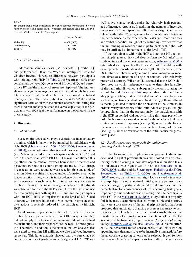
Table 2Spearmann Rank-order correlations (p-values between parentheses) betweennumber of errors and scores on the Wechsler Intelligence Scale for Children-Revised (WISC-R) for all HCP participants
Number of errors
Verbal IQ −.009 (.967)PT
3
aCwcmsltsttp
4
4
pra(nhblrlerwtwdpH
rdtiwrc
warrtatm
tsedDtpdosasiobrtcl(t
4p
dii(S(tepIwfitpftst
erformance IQ −.44 (.101)otal IQ −.551 (.052)
.3. Clinical measures
Independent-samples t-tests (t < 1 for total IQ, verbal IQ,nd performance IQ) on the Wechsler Intelligence Scale forhildren-Revised showed no difference between participantsith left and right HCP. In Table 2 the Spearmann rank-order
orrelations between IQ scores (total IQ, verbal IQ, and perfor-ance IQ) and the number of errors are displayed. The analyses
howed no significant negative correlations, although the corre-ation between total IQ and number of errors showed a statisticalrend (p = .052). The verbal subtest of the WISC-R showed noignificant correlation with the number of errors, indicating thathere is no relationship between the verbal capacities of the par-icipants with HCP and the performance on the MI tasks in theresent study.
. Discussion
.1. Main results
Based on the idea that MI plays a critical role in anticipatorylanning, which is known to be impaired in individuals withight HCP (Mutsaarts et al., 2004, 2005, 2006; Steenbergen etl., 2004), we hypothesized that internal movement simulationi.e., MI) is impaired in the participants with right HCP, andot in the participants with left HCP. The results confirmed thisypothesis on the relation between hemispheric processes andehaviour. For both the control group and the left HCP group,inear relations were found between reaction time and angle ofotation. More specifically, larger angles of rotation resulted inonger reaction times, which is in accordance with what is gen-rally observed in such tasks. In contrast, no linear increase ineaction time as a function of the angular distance of the stimulias observed for the right HCP group. From this we conclude
hat the participants with right HCP (and not the participantsith left HCP) have an impairment at the level of MI. Statedifferently, it appears that the ability to internally simulate com-lex actions is severely reduced in the participants with rightCP.An alternative explanation for the lack of linear increase of
eaction times in participants with right HCP may be that theyid not comply with task instruction and/or did not understandhe instruction, and their responses are the result of mere guess-
ng. Therefore, in addition to the main RT pattern analyses thatere used to examine MI abilities, we also analyzed incorrectesponses. This latter analysis showed that the percentage oforrect responses of participants with right and left HCP was
eutt
ologia 45 (2007) 853–859 857
ell above chance level, despite the relatively high percent-ge of incorrect responses. In addition, the number of incorrectesponses of all participants with HCP was not significantly cor-elated with verbal IQ, suggesting a lack of relationship betweenhe performance on the experimental tasks (i.e., reaction time)nd verbal capacities. In light of these findings, we believe thathe null-finding on reaction time in participants with right HCP
ay be attributed to impairments at the level of MI.If the participants with right HCP did not use MI and nei-
her simply guessed, how did they perform the MI tasks? In atudy on internal movement representation, Wilson et al. (2004)stablished a comparable effect on a MI task in children withevelopmental coordination disorder (DCD). Specifically, theCD children showed only a small linear increase in reac-
ion times as a function of angle of rotation, with relativelyreserved accuracy. Wilson et al. assumed that the DCD chil-ren used viewpoint-independent cues to determine lateralityf the hand stimuli, without subsequently mentally rotating thetimuli. Indeed, Parsons (1994) proposed that in the hand later-lity judgment task, first an ‘educated guess’ is made, based onuch viewpoint-independent cues. Next, the hypothesized hands mentally rotated to match the orientation of the stimulus, inrder to verify the veracity of the initial educated guess. It mighte speculated that, in the present study, the participants withight HCP responded without performing this latter part of theask. Such a strategy would account for the relatively high per-entage of incorrect responses (see Fig. 3), as well as the lack ofinear increase in reaction times as a function of angle of rotationsee Fig. 2), since no verification of the initial ‘educated guess’akes place.
.2. Possible processes responsible for anticipatorylanning deficits in right HCP
In the following, the implications of present findings areiscussed in light of previous studies that showed lack of antic-patory motor planning in complex object manipulation tasksn individuals with right HCP. In both the Mutsaarts et al.2004, 2006) studies and the Steenbergen, Hulstijn, et al. (2000),teenbergen, van Thiel, et al. (2000), and Steenbergen et al.2004) studies, participants with right HCP showed a tendencyo grasp objects using an optimal initial grasping pattern. How-ver, in doing so, participants failed to take into account theerceptual-motor consequences of the upcoming task goals.mportantly, this tendency was so strong that the participantsith HCP in the Mutsaarts et al. (2006) study could not properlynish the task, due to biomechanically impossible end-postures
hat were a consequence of the initial grip selected. It has beenroposed that anticipatory planning processes necessary to per-orm such complex object manipulation tasks involve the mentalransformation of a somatomotor representation of the effectorystem, in order to select a proper response (Imagery as planningheory; Johnson, 2000a; see also Parsons, 2003). Stated differ-
ntly, the perceptual-motor consequences of an initial grip onpcoming task demands have to be internally simulated, beforehe appropriate grasping pattern can be selected. It then followshat a severely reduced capacity to internally simulate move-
8 psych
mgosaawca
4
ffstwdTrfsttfiwcBmtcwtpijJoelipacinIoir
4
mi
piwair
A
sNsd
R
B
C
C
D
D
G
G
H
H
H
J
J
J
J
J
J
J
58 M. Mutsaarts et al. / Neuro
ents would seriously complicate the selection of an appropriaterasping pattern. In such case, a strategy might be to select anptimal initial grasping pattern, as indeed observed in severaltudies (Mutsaarts et al., 2004, 2006; Steenbergen, Hulstijn, etl., 2000; Steenbergen, van Thiel, et al., 2000; Steenbergen etl., 2004). Hence, in light of the imagery as planning theory,e believe that the present finding of a severely reduced MI
apacity may provide new insight into a potential cause of thenticipatory planning deficits of individuals with right HCP.
.3. Hemiparetic disadvantage
Participants with left HCP showed more incorrect responsesor left hand stimuli (corresponding to their affected side) thanor right hand stimuli (corresponding to their non-affected side;ee Fig. 4). This finding suggests that it is more difficult forhe participants with left HCP to mentally simulate movementsith their affected hand than with their non-affected hand. Weid not find such an effect for the participants with right HCP.hat is, these participants did not make more errors for the
ight hand stimuli (corresponding to their affected side) thanor the left hand stimuli (corresponding to their non-affectedide). This is not surprising, since we established that – con-rary to the participants with left HCP – they appear unableo mentally simulate movements of their hands. Regarding thending of the participants with left HCP, comparable resultsere established in a study with seven patients with asymmetri-
al (right side affected) Parkinson’s disease (Dominey, Decety,roussolle, Chazot, & Jeannerod, 1995). They observed thatental simulation of movements in these patients was slower for
he affected right hand compared to the non-affected left hand. Inontrast, Johnson, Sprehn, and Saykin (2002) found that patientsith chronic hemiplegia were more accurate on MI tasks when
he affected hand was involved, an effect they termed “hemi-legic advantage”. Surprisingly, they did not observe this effectn patients with acute hemiplegia (Johnson, 2000a), healthy sub-ects (Johnson, 2000b), and patients recovered from hemiplegia.ohnson et al. (2002) speculated that the ‘hemiplegic advantage’bserved in patients with chronic hemiplegia reflects a constantffort of these patients to imagine movements with the paralyzedimb that they are no longer able to execute. If this speculations true, this might explain why we did not established a ‘hemi-aretic advantage’ for the participants with left HCP, but rather‘hemiparetic disadvantage’. In contrast to stroke patients withhronic hemiplegia, individuals with HCP have neuromotor lim-tations at their affected side from birth onwards. As such, theyever experienced ‘normal’ movement with their affected side.n light thereof, we suggest that the ‘hemiparetic disadvantage’bserved for the participants with left HCP in the present study,s a result of focusing predominantly on the non-affected side,ather than on the affected side.
.4. Conclusions
We established a reduced capacity to internally simulateovements in participants with right HCP. We believe this find-
ng might shed new light on the processes responsible for theJ
ologia 45 (2007) 853–859
reviously observed anticipatory planning deficits in right HCPndividuals. Furthermore, for the participants with left HCP itas more difficult to internally simulate movements with their
ffected hand, a ‘hemiparetic disadvantage’. As a final remark,t must be noted that the groups participating in this study wereelatively small, which constitutes a study limitation.
cknowledgements
We thank those who kindly volunteered to participate in thetudy. This research was supported by a grant awarded by Theetherlands Organization for Scientific Research (NWO) to the
econd author for the research project ‘Adaptation in movementisorder’.
eferences
uxbaum, L. J., Johnson-Frey, S. H., & Bartlett-Williams, M. (2005). Deficientinternal models for planning hand-object interactions in apraxia. Neuropsy-chologia, 43, 917–929.
hang, J. J., Wu, T. I., Wu, W. L., & Su, F. C. (2005). Kinematical measure forspastic reaching in children with cerebral palsy. Clinical Biomechanics, 20,381–388.
rammond, D. J. (1997). Motor imagery: Never in your wildest dream. Trendsin Neuroscience, 20, 54–57.
ecety, J., Perani, D., Jeannerod, M., Bettinardi, V., Tadary, B., Woods, R., etal. (1994). Mapping motor representations with PET. Nature, 371, 600–602.
ominey, P., Decety, J., Broussolle, E., Chazot, G., & Jeannerod, M. (1995).Motor imagery of a lateralized sequential task is asymmetrically slowed inhemi-Parkinson’s patients. Neuropsychologia, 33, 727–741.
anis, G., Keenan, J. P., Kosslyn, S. M., & Pascual-Leone, A. (2000). Transcra-nial magnetic stimulation of primary motor cortex affects mental rotation.Cerebral Cortex, 10, 175–180.
oldenberg, G. (1996). Defective imitation of hand and finger postures inpatients with damage in the left or right hemispheres. Journal of Neurol-ogy Neurosurgery and Psychiatry, 61, 176–180.
aaland, K. Y., Elsinger, C. L., Mayer, A. R., Durgerian, S., & Rao, S. M. (2004).Motor sequence complexity and performing hand produce differential pat-terns of hemispheric lateralization. Journal of Cognitive Neuroscience, 16,621–636.
arrington, D. L., & Haaland, K. Y. (1992). Motor sequencing with left-hemisphere damage—are some cognitive deficits specific to limb apraxia.Brain, 115, 857–874.
ermsdorfer, J., Mai, N., Spatt, J., Marquardt, C., Veltkamp, R., & Goldenberg,G. (1996). Kinematic analysis of movement imitation in apraxia. Brain, 119,1575–1586.
eannerod, M. (1994). The representing brain: Neural correlates of motor inten-tion and imagery. Behavioral and Brain Sciences, 17, 187–245.
eannerod, M. (1995). Mental imagery in the motor context. Neuropsychologia,33, 1419–1432.
eannerod, M. (2001). Neural simulation of action: A unifying mechanism formotor cognition. Neuroimage, 14, S103–S109.
ohnson, S. H. (1998). Cerebral organization of motor imagery: Contralateralcontrol of grip selection in mentally represented prehension. PsychologicalScience, 9, 219–222.
ohnson, S. H. (2000a). Imagining the impossible: Intact motor representationsin hemiplegics. Neuroreport, 11, 729–732.
ohnson, S. H. (2000b). Thinking ahead: The case for motor imagery in prospec-tive judgments of prehension. Cognition, 74, 33–70.
ohnson, S. H., Corballis, P. M., & Gazzaniga, M. S. (2001). Within grasp but
out of reach: Evidence for a double dissociation between imagined handand arm movements in the left cerebral hemisphere. Neuropsychologia, 39,36–50.ohnson, S. H., Sprehn, G., & Saykin, A. J. (2002). Intact motor representa-tions in chronic upper limb hemiplegics: Evidence for activity-independent
psych
K
L
M
M
M
P
P
P
P
P
P
P
R
R
R
R
R
S
S
S
S
S
S
S
S
T
T
T
T
U
v
v
W
W
W
M. Mutsaarts et al. / Neuro
action representations. Journal of Cognitive Neuroscience, 14, 841–852.
osslyn, S. M., Digirolamo, G. J., Thompson, W. L., & Alpert, N. M. (1998).Mental rotation of objects versus hands: Neural mechanisms revealed bypositron emission tomography. Psychophysiology, 35, 151–161.
ang, W., Cheyne, D., Hollinger, P., Gerschkager, W., & Lindinger, G. (1996).Electric and magnetic fields of the brain accompanying internal simulationof movement. Cognitive Brain Research, 3, 125–129.
utsaarts, M., Steenbergen, B., & Meulenbroek, R. G. J. (2004). Assessing therigidity of the grasping movements of three adolescents with spastic hemi-paresis due to a cerebral palsy. Experimental Brain Research, 156, 293–304.
utsaarts, M., Steenbergen, B., & Bekkering, H. (2005). Anticipatory planningof movement sequences in hemiparetic cerebral palsy. Motor Control, 9,435–454.
utsaarts, M., Steenbergen, B., & Bekkering, H. (2006). Anticipatory planningdeficits and context effects in hemiparetic cerebral palsy. Experimental BrainResearch, 172, 151–162.
arsons, L. M. (1987). Imagined spatial transformation of ones body. Journalof Experimental Psychology: General, 116, 172–191.
arsons, L. M. (1994). Temporal and kinematic properties of motor behaviourreflected in mentally simulated action. Journal of Experimental Psychology:Human Perception and Performance, 20, 709–730.
arsons, L. M. (2003). Superior parietal cortices and varieties of mental rotation.Trends in Cognitive Sciences, 7, 515–517.
arsons, L. M., & Fox, P. T. (1998). The neural basis of the implicit movementsused in recognizing hand shape. Cognitive Neuropsychology, 15, 583–615.
arsons, L. M., Fox, P. T., Downs, J. H., Glass, T., Hirsch, T. B., Martin, C. C.,et al. (1995). Use of implicit motor imagery for visual shape discriminationas revealed by PET. Nature, 375, 54–58.
elgrims, B., Andres, M., & Olivier, E. (2005). Motor imagery while judgingobject-hand interactions. Neuroreport, 16, 1193–1196.
orro, C. A., Francescato, M. P., Cettolo, V., Diamond, M. E., Baaraldi, P.,Zuiani, C., et al. (1996). Primary motor and sensory cortex activation duringmotor performance and motor imagery: A functional magnetic resonanceimaging study. Journal of Neuroscience, 16, 7688–7698.
ao, S. M., Binder, J. R., Bandettini, P. A., Hemmeke, T. A., Yetkin, F. Z.,Jesmanowixz, A., et al. (1993). Functional magnetic resonance imaging ofcomplex human movements. Neurology, 43, 2311–2318.
osenbaum, D. A., van Heugten, C. M., & Caldwell, G. E. (1996). Fromcognition to biomechanics and back: The end-state comfort effect and themiddle-is-faster effect. Acta Psychologica, 94, 59–85.
osenbaum, D. A., Marchak, F., Barnes, H. J., Vaughan, J., Slotta, J. D., &Jorgensen, M. J. (1990). Constraints for action selection: Overhand versusunderhand grips. In M. Jeannerod (Ed.), Attention and performance XIII.Hillsdale, New York: Lawrence Erlbaum Associates.
osenbaum, D. A., Vaughan, J., Jorgensen, M. J., Barnes, H. J., & Stewart, E.(1993). Plans for object manipulation. In D. E. Meyer, & S. Kornblum (Eds.),Attention and performance XIV: synergies in experimental psychology, arti-ficial intelligence, and cognitive neuroscience (pp. 803–820). Cambridge,MA: MIT Press.
ushworth, M. F. S., Nixon, P. D., Wade, S., Renowden, S., & Passingham, R.
E. (1998). The left hemisphere and the selection of learned actions. Neu-ropsychologia, 36, 11–24.abate, M., Gonzalez, B., & Rodriguez, M. (2004). Brain lateralization of motorimagery: Motor planning asymmetry as a cause of movement lateralization.Neuropsychologia, 42, 1041–1049.
W
W
ologia 45 (2007) 853–859 859
chluter, N. D., Krams, M., Rushworth, M. F. S., & Passingham, R. E. (2001).Cerebral dominance for action in the human brain: The selection of actions.Neuropsychologia, 39, 105–113.
irigu, A., Duhamel, J. R., Cohen, L., Pillon, B., Dubois, B., & Agid, Y. (1996).The mental representation of hand movements after parietal cortex damage.Science, 273, 1564–1568.
teenbergen, B., Hulstijn, W., & Dortmans, S. (2000). Constraints on grip selec-tion in cerebral palsy: Minimizing discomfort. Experimental Brain Research,134, 385–397.
teenbergen, B., van Thiel, E., Hulstijn, W., & Meulenbroek, R. G. J. (2000).The coordination of reaching and grasping in spastic hemiparesis. HumanMovement Science, 19, 75–105.
teenbergen, B., Meulenbroek, R. G. J., & Rosenbaum, D. A. (2004). Con-straints on grip selection in hemiparetic cerebral palsy: Effects of lesionalside, end-point accuracy, and context. Cognitive Brain Research, 19, 145–159.
teenbergen, B., & Meulenbroek, R. G. J. (2006). Deviations in upper-limb func-tion of the less-affected side in congenital hemiparesis. Neuropsychologia,44, 2296–2307.
underland, A., & Sluman, S. M. (2000). Ideomotor apraxia, visuomotor con-trol and the explicit representation of posture. Neuropsychologia, 38, 923–934.
omasino, B., Rumiati, R. I., & Umilta, C. A. (2003). Selective deficit of motorimagery as tapped by a left-right decision of visually presented hands. Brainand Cognition, 53, 376–380.
omasino, B., Toraldo, A., & Rumiati, R. I. (2003). Dissociation between themental rotation of visual images and motor images in unilateral brain-damaged patients. Brain and Cognition, 51, 368–371.
rombly, C. A. (1992). Deficits of reaching in subjects with left hemiparesis: Apilot study. American Journal of Occupational Therapy, 46, 887–897.
rombly, C. A. (1993). Observations of improvement of reaching in five subjectswith hemiparesis. Journal of Neurology, Neurosurgery and Psychiatry, 56,40–45.
tley, A., & Sugden, D. A. (1998). Interlimb coupling in children with hemi-plegic cerebral palsy during reaching and grasping at speed. DevelopmentalMedicine and Child Neurology, 40, 396–404.
an Roon, D., Steenbergen, B., & Meulenbroek, R. G. J. (2004). Trunk recruit-ment during spoon use in tetraparetic cerebral palsy. Experimental BrainResearch, 155, 186–195.
an Thiel, E., Meulenbroek, R. G. J., Smeets, J. B. J., & Hulstijn, W. (2002).Fast adjustments of ongoing movements in hemiparetic cerebral palsy. Neu-ropsychology, 40, 16–27.
echsler, D. (1974). Manual for the Wechsler Intelligence Scale for Children-Revised. San Antonio, TX: Psychological Corporation.
eiss, P. H., Dohle, C., Binkofski, F., Schnitzler, A., Freund, H. J., & Hefter, H.(2001). Motor impairment in patients with parietal lesions: Disturbancesof meaningless arm movement sequences. Neuropsychologia, 39, 397–405.
ilson, P. H., Maruff, P., Butson, M., Williams, J., Lum, J., & Thomas, P. R.(2004). Internal representation of movement in children with developmentalcoordination disorder: A mental rotation task. Developmental Medicine and
Child Neurology, 46, 754–759.ohlschlager, A. (2001). Mental object rotation and the planning of hand move-ments. Perception and Psychophysics, 63, 709–718.
olbert, T., Weiller, C., & Buchel, C. (2003). Contralateral coding of imaginedbody parts in the superior parietal lobe. Cerebral Cortex, 13, 392–399.
Related Documents