Immunosuppression of experimental autoimmune myasthenia gravis by mycophenolate mofetil Sofie P.M. Janssen a,1 , Marko Phernambucq a,1 , Pilar Martinez-Martinez a , Marc H. De Baets a,b , Mario Losen a, ⁎ a Department of Neuroscience, School of Mental Health and Neuroscience, Faculty of Health, Medicine and Life Sciences, Maastricht University, Universiteitssingel 50, P.O. Box 616, 6200 MD Maastricht, The Netherlands b Neuroimmunology Group, Biomedical Research Institute (BIOMED), Hasselt University, Diepenbeek, Belgium Received 20 March 2008; received in revised form 13 May 2008; accepted 13 May 2008 Abstract Currently used non-specific immunosuppressive drugs often require intervention in myasthenia gravis (MG) and clinical improvement varies widely. To analyze the therapeutic effect of mycophenolate mofetil (MMF) in experimental autoimmune MG (EAMG), rats were immunized with acetylcholine receptors (AChRs) and subsequently treated with MMF or vehicle. MMF treatment resulted in a significant suppression of anti-rat AChR antibody titers. Interestingly, no abnormalities of neuromuscular transmission and adverse side effects were detected in MMF-treated EAMG animals. Moreover, anti-rat AChR antibody titers correlated to an improvement of clinical outcome. In conclusion, our data suggest that MMF acts as a potent immunosuppressant drug in EAMG. © 2008 Elsevier B.V. All rights reserved. Keywords: Myasthenia gravis; Mycophenolate mofetil; Immunosuppression; Acetylcholine receptor; Neuromuscular junction 1. Introduction Myasthenia gravis (MG) is a potentially life-threatening but treatable organ specific autoimmune disorder, characterized by weakness and fatigability of voluntary muscles, i.e. ocular, bulbar, limb and respiratory muscles. In about 85% of patients (Lindstrom et al., 1976), auto-antibodies against the nicotinic acetylcholine receptor (AChR) in the postsynaptic membrane of the neuromuscular junction are responsible for these symptoms, and have been shown to destroy neuromuscular transmission by different mechanisms: cross-linking of AChRs leading to increased AChR turnover, functional blockade of the acetylcho- line-binding sites, complement-mediated damage to the post- synaptic membrane (Drachman, 1994), and destruction of proteins involved in neuromuscular formation (Martinez- Martinez et al., 2007). Moreover, auto-antibodies that bind to the muscle specific protein kinase (MuSK), are present in a subgroup of patients with MG who do not have antibodies against the AChR (Hoch et al., 2001). Currently many therapies are available for MG patients, all intervening with different targets of the disease. Thymectomy is nowadays a widely accepted and practiced form of treatment for MG and is performed in an attempt to induce complete remission or to reduce long-term need for immunosuppression (Gronseth and Barohn, 2000). In contrast, anti-cholinesterase agents are frequently used in the early disease course to decrease MG symptoms as they inhibit acetylcholine esterase from hydrolyz- ing acetylcholine. The prolonged exposure time of acetylcholine to AChRs in turn leads to improved neuromuscular transmission and muscle strength, however without affecting the autoimmune attack itself (Richman and Agius, 2003). Intravenous immuno- globulin (Gajdos et al., 2006) and plasmapheresis (Gajdos et al., 2006; Lehmann et al., 2006) are mostly temporary treatments used to produce rapid improvement during myasthenic crises or acute exacerbations. Finally, long-term immunosuppressives like corticosteroids, azathioprine, cyclosporine A, tacrolimus, Journal of Neuroimmunology 201–202 (2008) 111 – 120 www.elsevier.com/locate/jneuroim ⁎ Corresponding author. Tel.: +31 43 38 810 39; fax: +31 43 36 710 96. E-mail address: [email protected] (M. Losen). 1 These authors contributed equally to this work. 0165-5728/$ - see front matter © 2008 Elsevier B.V. All rights reserved. doi:10.1016/j.jneuroim.2008.05.029

Welcome message from author
This document is posted to help you gain knowledge. Please leave a comment to let me know what you think about it! Share it to your friends and learn new things together.
Transcript
01–202 (2008) 111–120www.elsevier.com/locate/jneuroim
Journal of Neuroimmunology 2
Immunosuppression of experimental autoimmune myasthenia gravisby mycophenolate mofetil
Sofie P.M. Janssen a,1, Marko Phernambucq a,1, Pilar Martinez-Martinez a,Marc H. De Baets a,b, Mario Losen a,⁎
a Department of Neuroscience, School of Mental Health and Neuroscience, Faculty of Health, Medicine and Life Sciences, Maastricht University,Universiteitssingel 50, P.O. Box 616, 6200 MD Maastricht, The Netherlands
b Neuroimmunology Group, Biomedical Research Institute (BIOMED), Hasselt University, Diepenbeek, Belgium
Received 20 March 2008; received in revised form 13 May 2008; accepted 13 May 2008
Abstract
Currently used non-specific immunosuppressive drugs often require intervention in myasthenia gravis (MG) and clinical improvement varieswidely. To analyze the therapeutic effect of mycophenolate mofetil (MMF) in experimental autoimmune MG (EAMG), rats were immunized withacetylcholine receptors (AChRs) and subsequently treated with MMF or vehicle. MMF treatment resulted in a significant suppression of anti-ratAChR antibody titers. Interestingly, no abnormalities of neuromuscular transmission and adverse side effects were detected in MMF-treatedEAMG animals. Moreover, anti-rat AChR antibody titers correlated to an improvement of clinical outcome. In conclusion, our data suggest thatMMF acts as a potent immunosuppressant drug in EAMG.© 2008 Elsevier B.V. All rights reserved.
Keywords: Myasthenia gravis; Mycophenolate mofetil; Immunosuppression; Acetylcholine receptor; Neuromuscular junction
1. Introduction
Myasthenia gravis (MG) is a potentially life-threatening buttreatable organ specific autoimmune disorder, characterized byweakness and fatigability of voluntary muscles, i.e. ocular,bulbar, limb and respiratory muscles. In about 85% of patients(Lindstrom et al., 1976), auto-antibodies against the nicotinicacetylcholine receptor (AChR) in the postsynaptic membrane ofthe neuromuscular junction are responsible for these symptoms,and have been shown to destroy neuromuscular transmission bydifferent mechanisms: cross-linking of AChRs leading toincreased AChR turnover, functional blockade of the acetylcho-line-binding sites, complement-mediated damage to the post-synaptic membrane (Drachman, 1994), and destruction ofproteins involved in neuromuscular formation (Martinez-Martinez et al., 2007). Moreover, auto-antibodies that bind to
⁎ Corresponding author. Tel.: +31 43 38 810 39; fax: +31 43 36 710 96.E-mail address: [email protected] (M. Losen).
1 These authors contributed equally to this work.
0165-5728/$ - see front matter © 2008 Elsevier B.V. All rights reserved.doi:10.1016/j.jneuroim.2008.05.029
the muscle specific protein kinase (MuSK), are present in asubgroup of patients with MG who do not have antibodiesagainst the AChR (Hoch et al., 2001).
Currently many therapies are available for MG patients, allintervening with different targets of the disease. Thymectomy isnowadays a widely accepted and practiced form of treatment forMG and is performed in an attempt to induce complete remissionor to reduce long-term need for immunosuppression (Gronsethand Barohn, 2000). In contrast, anti-cholinesterase agents arefrequently used in the early disease course to decrease MGsymptoms as they inhibit acetylcholine esterase from hydrolyz-ing acetylcholine. The prolonged exposure time of acetylcholineto AChRs in turn leads to improved neuromuscular transmissionand muscle strength, however without affecting the autoimmuneattack itself (Richman and Agius, 2003). Intravenous immuno-globulin (Gajdos et al., 2006) and plasmapheresis (Gajdos et al.,2006; Lehmann et al., 2006) are mostly temporary treatmentsused to produce rapid improvement during myasthenic crisesor acute exacerbations. Finally, long-term immunosuppressiveslike corticosteroids, azathioprine, cyclosporine A, tacrolimus,
112 S.P.M. Janssen et al. / Journal of Neuroimmunology 201–202 (2008) 111–120
rituximab, etanercept, leflunomide and cyclophosphamide arenowadays considered to be the most consistently effective andeasily usable therapeutic agents as they induce improvement inapproximately 80% of patients. However, the onset of clinicalimprovement varies widely among these immunosuppressiveagents and side effects are often very severe due to toxicity ofthe therapeutics (Ciafaloni, 2005; Garcia-Carrasco et al., 2007;Sieb, 2005). Currently, the best evidence based immunosup-pressive therapy for MG is the combination of prednisone withazathioprine. This combined treatment allows a reduction of re-quired prednisone and concomitantly reduces, albeit incomple-tely, side effects such as “malaise, back pain, visual deterioration,and rash” (Palace et al., 1998). From this point of view, neweffective and safe immunosuppressive drugs for MG patientsare urgently required.
Mycophenolate mofetil (MMF) is a potent immunosuppres-sive drug that was primarily licensed to prevent rejection oftransplanted organs (European Mycophenolate Mofetil Coop-erative Study Group, 1995) and then introduced for the therapyof autoimmune diseases of skin (Enk and Knop, 1997), gut(Neurath et al., 1999), and eye (Larkin and Lightman, 1999), inrheumatoid arthritis (Goldblum, 1993), lupus nephritis (Ginzleret al., 2005) and Wegener's granulomatosis (Nowack et al.,1999). In the last decennium it has also been used in patientswith neuromuscular diseases like inflammatory myopathy andchronic inflammatory demyelinating polyradiculoneuropathy toachieve either symptom stabilization or remission (Chaudhryet al., 2001). MMF operates through reduction of intracellularpurine synthesis in both B- and T-lymphocytes, leading toinhibition of lymphocyte proliferation. Moreover, MMF doesnot only augment apoptosis of lymphocytic and monocytic celllines (Cohn et al., 1999) and reduces the levels of immunoglo-bulin isotypes M, G and A produced by polyclonal activated B-lymphocytes (Eugui et al., 1991), it also inhibits glycosylationof adhesion molecules (Blaheta et al., 1998) and regulates thesecretion of inflammatory and anti-inflammatory cytokines(Durez et al., 1999). Consequently, MMF acts as a pluripotentimmunomodulator in numerous types of immunologicaldiseases (Schneider-Gold et al., 2006).
The successful use of MMF in MG has already beendescribed in several small clinical trials, with the main advantageof MMF being its tolerability and safety profile (Caponnettoet al., 2001; Chaudhry et al., 2001; Ciafaloni et al., 2001; Hauseret al., 1998; Lim et al., 2007; Meriggioli et al., 2003a; Meriggioliet al., 2003b; Mowzoon et al., 2001; Prakash et al., 2007;Schneider et al., 2001). However, data from 2 recently com-pleted large phase 3 prospective, multicenter, placebo-controlledtrials showed only little or no effect of the drug when used as anadjunctive treatment to corticosteroids (Hampton, 2007; Sanderset al., 2008; The Muscle Study Group, 2008). This suggests thatfurther studies are needed to assess the benefits of MMF asprimary or long-term therapy and therefore encourages theexamination of its precise mechanism in an experimental animalmodel of MG.
Experimental autoimmune myasthenia gravis (EAMG) inrats induced by immunization with AChRs from the electricorgan of Electrophorus electricus or Torpedo californica, is
already known for decades to be a reproducible and character-istic chronic model of MG (De Baets, 2003). Indeed, thepresence of antibodies directed to rat muscle AChRs in thecirculation of rats with EAMG provides evidence for theexistence of autoimmunity in this experimental disease model.Moreover, similar to MG, antigenic modulation and comple-ment-mediated focal damage of the postsynaptic membrane arethe main pathogenic mechanisms (Losen et al., 2008) that lead tomuscular weakness, hunched posture, weight loss and electro-physiological abnormalities in these animals, and can be relievedby anti-cholinesterases (Lennon et al., 1975). For these reasons,EAMG in rats is an excellent model to examine the therapeuticeffect of MMF.
2. Materials and methods
2.1. Animal model
Eight-week old female Lewis rats were obtained from theDepartment of Experimental Animal Services, University ofMaastricht, The Netherlands, with permission of the Committeeon Animal Welfare, according to Dutch governmental rules.
For immunization and intubation, rats were anesthetized byinhalation of 3–5% isoflurane in air enriched with 50% oxygen,supplied by a cylindrical cap held over the head. Euthanasiawas performed by CO2/air inhalation and subsequent cervicaldislocation.
2.2. Induction of chronic EAMG
Animals were immunized at the base of the tail with 10 µgT. californica AChR (tAChR) in 0.1 mL of phosphate-bufferedsaline (PBS), emulsified in an equal amount of Freund'sadjuvant with 0.1% Mycobacterium tuberculosis (DifcoLaboratories, Detroit, USA) (Lennon et al., 1975). Bloodsamples were taken weekly from the vena saphena magna tomeasure anti-rat AChR antibody titers. Sham-immunizationwas performed under similar conditions using 0.1 mL ofPBS and an equal volume of Freund's adjuvant with 0.1%M. tuberculosis (CFA) (Lennon et al., 1975).
2.3. Administration of immunosuppressive drugs
Mycophenolate mofetil (MMF; Roche Palo Alto LLC,California) was administered orally at a dose of 30 mg/kg/day ina 900 µL autoclaved vehicle solution consisting of distilled water,0.9% benzyl alcohol, 0.4%polysorbate 80, 0.9% sodium chloride,0.5% carboxymethylcellulose and 5.0% sucrose (pH 3.5).
2.4. Experimental design
Two different treatment regimes of MMF were investigatedusing 62 rats in total. For the first treatment regime, two groupsof rats immunized with tAChR were given either vehicle(vehicle-treated EAMG rats, n=10) or MMF (MMF-treatedEAMG rats, n=10) daily for 3 weeks, starting at the time ofimmunization, and were compared to sham-immunized controls
113S.P.M. Janssen et al. / Journal of Neuroimmunology 201–202 (2008) 111–120
(n=10) 8 weeks after immunization. For this series in particular,immunohistochemical analyses were performed together withdetermination of total muscle AChR content.
For the second treatment regime, two groups of rats im-munized with tAChR received either vehicle (vehicle-treatedEAMG rats, n=18) or MMF (MMF-treated EAMG rats, n=10)daily for 8 weeks, starting at the time of immunization. Foursham-immunized ratswere used as controls andwere compared tothe above-mentioned groups 8 weeks after immunization. Moreprecisely, electromyographic measurements (EMG) during con-tinuously intravenous curare infusions to induce decrement ofcompound muscle action potentials (CMAP) were performed.
For all rats, the body weights, clinical scores and anti-ratAChR antibody titers were measured regularly.
2.5. Clinical scoring
The severity of clinical signs in EAMG was scored weeklyby measuring rats' muscular weakness through assessing theirability to grasp and lift repeatedly a 300-g rack from the tablewhile suspended manually by the base of the tail for 30 s(Hoedemaekers et al., 1997). Clinical scoring was based on thepresence of tremor, hunched posture, muscle strength andfatigability. Disease severity was expressed as 0, no obviousabnormalities; I, no abnormalities before testing but reducedstrength at the end; II, clinical signs present before testing i.e.tremor, hunched posture, head down, weak grip; III, severeclinical signs present before testing, no grip, moribund (Lennonet al., 1975).
2.6. Measurement of serum anti-rat AChR antibody titers
Anti-rat AChR antibody titer measurements were carried outat 4 °C by a double-antibody radioimmunoassay (RIA) asdescribed previously (Losen et al., 2005; Martinez-Martinezet al., 2007). Briefly, antibodies directed against rat AChRswere measured using a crude extract of denervated rat muscle(approximately 5 nmol/L AChR), labeled with an excess of 125I-α-bungarotoxin (IM109, 6.12 TBq/mmol, GE Healthcare,Amersham Place, UK), and incubated overnight with 5 µL rattest serum. Precipitation was induced by addition of 100 µLgoat anti-rat IgG serum, followed by 3 washing steps in PBSwith 0.5% Triton X-100. Radioactivity counts were measured ina γ-counter (1480 Wizard 3″, PerkinElmer, USA) and correctedfor background measurements using normal rat serum. Anti-body titers were expressed as nmoles of α-bungarotoxinbinding sites/L.
2.7. Immunohistochemical staining
Left tibialis anterior muscles were frozen in isopentanecooled with liquid nitrogen to perform immunohistochemicalstaining as described previously (Losen et al., 2005; Martinez-Martinez et al., 2007) with the following modifications: 10 µmcryosections were incubated with mouse anti-rapsyn mAb 1234(1:500 in PBS with 2% bovine serum albumin (PBSA), Sigma,USA), rabbit anti-vesicular acetylcholine transporter (VAChT,
1:500 in PBSA, Sigma, USA), and Alexa 594-conjugatedα-bungarotoxin (1:300 in PBSA, Molecular Probes, USA)for 1 h. Subsequently, sections were incubated for 45 min withthe appropriate secondary antibodies: biotinylated goat anti-mouse IgG (1:400 in PBSA; Jackson Immunoresearch, USA)and Alexa 350-conjugated goat anti-rabbit (1:100 in PBSA;Molecular Probes, USA). Finally, the biotinylated secondaryantibody was stained for 30 min with Alexa 488-conjugatedstreptavidin (1:2000 in PBSA; Molecular Probes, USA). Allincubation steps were performed at room temperature.
2.8. Immunofluorescence microscopy
Pictures of triple stained muscle sections were taken using aProvis AX70 fluorescent microscope (Olympus, Hamburg,Germany) connected to a black and white digital video camera(U-CMAD-2, Olympus), equipped with AnalySIS software(Soft Imaging Systems, Münster, Germany). All microscopesettings were maintained constant during photographing, withthe exposure time of the camera set to values that preventsaturation. Pictures were subsequently analyzed using ImageJsoftware (version 1.37k; http://rsb.info.nih.gov/ij/). Neuromus-cular junctions were identified as regions with presynapticVAChT staining.
2.9. Measurement of total muscle AChR
The right tibialis anterior muscle of each rat was minced andhomogenized with an Ultra-Turrax (3 times 30 s at 4 °C) in10 mL extraction buffer (PBS mixed with 10 mM sodium azide,10 mM ethylenediaminetetraacetic acid, 10 mM iodoacetamide,and 1 mM phenylmethyl sulfonyl fluoride). The homogenatewas centrifuged at 22,100 g for 30 min at 4 °C and the resultingpellet was resuspended in 2.5 mL extraction buffer with anadditional 2% Triton X-100. Extraction was performed on areciprocal shaker for 1 h at 4 °C, followed by centrifugation at22,100 g for 30 min at 4 °C. AChR concentrations were thenmeasured by RIA using an excess of 125I-α-bungarotoxin(IM209, 74 TBq/mmol, GE healthcare, Amersham Place, UK)as described previously (Losen et al., 2005). Total musclemembrane AChR concentration was calculated per g of freshmuscle and thus expressed in fmol/g.
2.10. Electromyography
Decrement of CMAP during repetitive nerve stimulation wasmeasured during concurrently continuous curare (D-tubocur-arine chloride, ICN Biomedicals, Illkirch France) infusions inleft tibialis anterior muscles of rats 8 weeks after immunizationusing the EMG system Viking IV (Nicolet Biomedicals Inc.,Madison, USA) as previously described (Losen et al., 2005;Martinez-Martinez et al., 2007; Seybold et al., 1976) with thefollowing modifications: curare infusions (Terfusion syringepump, model STC-521, Terumo, Japan; 1 mL/h, 0.33 µg curare/min) were proceeded after intubation and catheterization of thetail vene. To detect a decrementing response, series of 8supramaximal stimuli were given to the nervus tibialis at 3 Hz
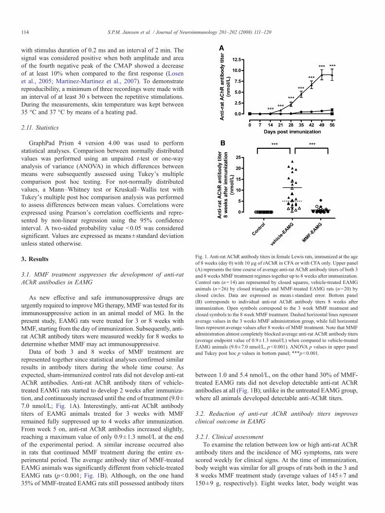
Fig. 1. Anti-rat AChR antibody titers in female Lewis rats, immunized at the ageof 8 weeks (day 0) with 10 µg of tAChR in CFA or with CFA only. Upper panel(A) represents the time course of average anti-rat AChR antibody titers of both 3and 8 weeks MMF treatment regimes together up to 8 weeks after immunization.Control rats (n=14) are represented by closed squares, vehicle-treated EAMGanimals (n=26) by closed triangles and MMF-treated EAMG rats (n=20) byclosed circles. Data are expressed as mean±standard error. Bottom panel(B) corresponds to individual anti-rat AChR antibody titers 8 weeks afterimmunization. Open symbols correspond to the 3 week MMF treatment andclosed symbols to the 8 week MMF treatment. Dashed horizontal lines representaverage values in the 3 weeks MMF administration group, while full horizontallines represent average values after 8 weeks of MMF treatment. Note that MMFadministration almost completely blocked average anti-rat AChR antibody titers(average endpoint value of 0.9±1.3 nmol/L) when compared to vehicle-treatedEAMG animals (9.0±7.0 nmol/L, pb0.001). ANOVA p values in upper paneland Tukey post hoc p values in bottom panel; ⁎⁎⁎pb0.001.
114 S.P.M. Janssen et al. / Journal of Neuroimmunology 201–202 (2008) 111–120
with stimulus duration of 0.2 ms and an interval of 2 min. Thesignal was considered positive when both amplitude and areaof the fourth negative peak of the CMAP showed a decreaseof at least 10% when compared to the first response (Losenet al., 2005; Martinez-Martinez et al., 2007). To demonstratereproducibility, a minimum of three recordings were made withan interval of at least 30 s between the repetitive stimulations.During the measurements, skin temperature was kept between35 °C and 37 °C by means of a heating pad.
2.11. Statistics
GraphPad Prism 4 version 4.00 was used to performstatistical analyses. Comparison between normally distributedvalues was performed using an unpaired t-test or one-wayanalysis of variance (ANOVA) in which differences betweenmeans were subsequently assessed using Tukey's multiplecomparison post hoc testing. For not-normally distributedvalues, a Mann–Whitney test or Kruskall–Wallis test withTukey's multiple post hoc comparison analysis was performedto assess differences between mean values. Correlations wereexpressed using Pearson's correlation coefficients and repre-sented by non-linear regression using the 95% confidenceinterval. A two-sided probability value b0.05 was consideredsignificant. Values are expressed as means±standard deviationunless stated otherwise.
3. Results
3.1. MMF treatment suppresses the development of anti-ratAChR antibodies in EAMG
As new effective and safe immunosuppressive drugs areurgently required to improveMG therapy, MMFwas tested for itsimmunosuppressive action in an animal model of MG. In thepresent study, EAMG rats were treated for 3 or 8 weeks withMMF, starting from the day of immunization. Subsequently, anti-rat AChR antibody titers were measured weekly for 8 weeks todetermine whether MMF may act immunosuppressive.
Data of both 3 and 8 weeks of MMF treatment arerepresented together since statistical analyses confirmed similarresults in antibody titers during the whole time course. Asexpected, sham-immunized control rats did not develop anti-ratAChR antibodies. Anti-rat AChR antibody titers of vehicle-treated EAMG rats started to develop 2 weeks after immuniza-tion, and continuously increased until the end of treatment (9.0±7.0 nmol/L; Fig. 1A). Interestingly, anti-rat AChR antibodytiters of EAMG animals treated for 3 weeks with MMFremained fully suppressed up to 4 weeks after immunization.From week 5 on, anti-rat AChR antibodies increased slightly,reaching a maximum value of only 0.9±1.3 nmol/L at the endof the experimental period. A similar increase occurred alsoin rats that continued MMF treatment during the entire ex-perimental period. The average antibody titer of MMF-treatedEAMG animals was significantly different from vehicle-treatedEAMG rats (pb0.001; Fig. 1B). Although, on the one hand35% of MMF-treated EAMG rats still possessed antibody titers
between 1.0 and 5.4 nmol/L, on the other hand 30% of MMF-treated EAMG rats did not develop detectable anti-rat AChRantibodies at all (Fig. 1B); unlike in the untreated EAMG group,where all animals developed detectable anti-AChR titers.
3.2. Reduction of anti-rat AChR antibody titers improvesclinical outcome in EAMG
3.2.1. Clinical assessmentTo examine the relation between low or high anti-rat AChR
antibody titers and the incidence of MG symptoms, rats werescored weekly for clinical signs. At the time of immunization,body weight was similar for all groups of rats both in the 3 and8 weeks MMF treatment study (average values of 145±7 and150±9 g, respectively). Eight weeks later, body weight was
115S.P.M. Janssen et al. / Journal of Neuroimmunology 201–202 (2008) 111–120
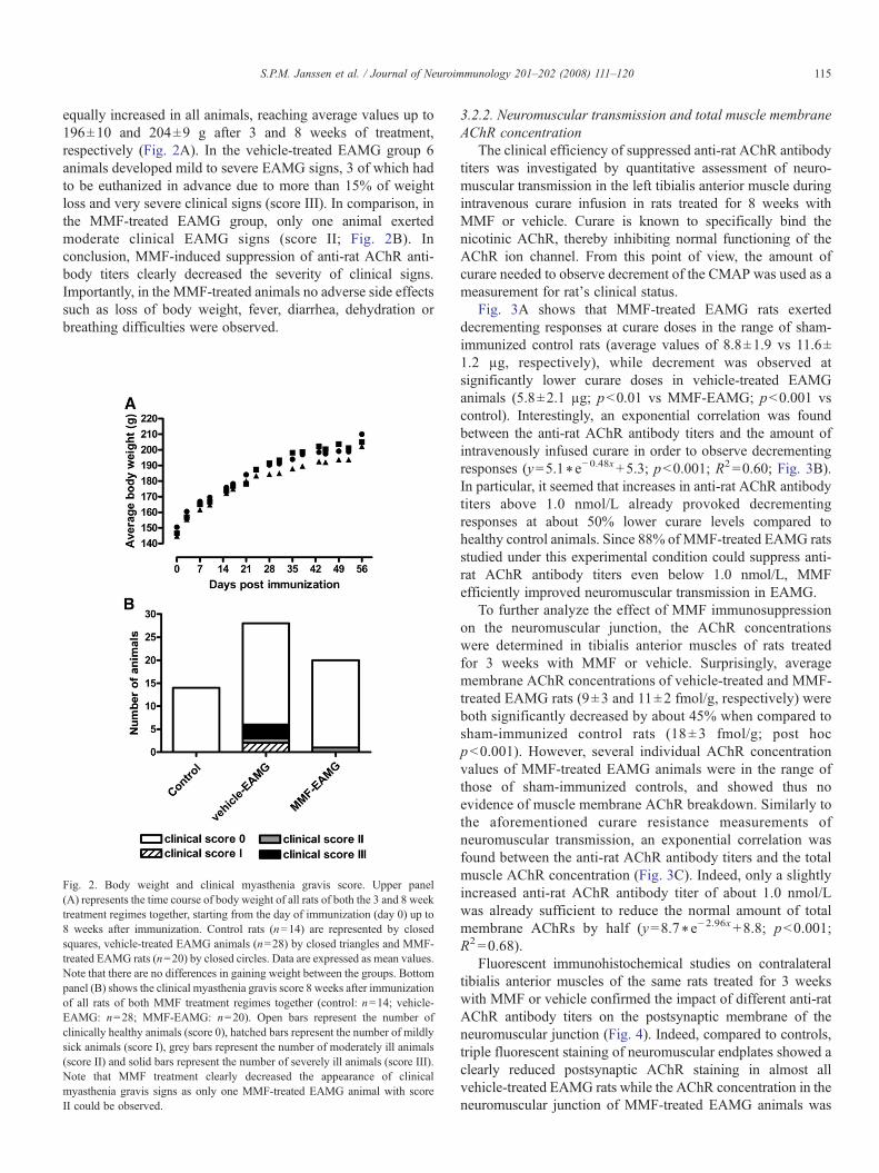
equally increased in all animals, reaching average values up to196±10 and 204±9 g after 3 and 8 weeks of treatment,respectively (Fig. 2A). In the vehicle-treated EAMG group 6animals developed mild to severe EAMG signs, 3 of which hadto be euthanized in advance due to more than 15% of weightloss and very severe clinical signs (score III). In comparison, inthe MMF-treated EAMG group, only one animal exertedmoderate clinical EAMG signs (score II; Fig. 2B). Inconclusion, MMF-induced suppression of anti-rat AChR anti-body titers clearly decreased the severity of clinical signs.Importantly, in the MMF-treated animals no adverse side effectssuch as loss of body weight, fever, diarrhea, dehydration orbreathing difficulties were observed.
Fig. 2. Body weight and clinical myasthenia gravis score. Upper panel(A) represents the time course of body weight of all rats of both the 3 and 8 weektreatment regimes together, starting from the day of immunization (day 0) up to8 weeks after immunization. Control rats (n=14) are represented by closedsquares, vehicle-treated EAMG animals (n=28) by closed triangles and MMF-treated EAMG rats (n=20) by closed circles. Data are expressed as mean values.Note that there are no differences in gaining weight between the groups. Bottompanel (B) shows the clinical myasthenia gravis score 8 weeks after immunizationof all rats of both MMF treatment regimes together (control: n=14; vehicle-EAMG: n=28; MMF-EAMG: n=20). Open bars represent the number ofclinically healthy animals (score 0), hatched bars represent the number of mildlysick animals (score I), grey bars represent the number of moderately ill animals(score II) and solid bars represent the number of severely ill animals (score III).Note that MMF treatment clearly decreased the appearance of clinicalmyasthenia gravis signs as only one MMF-treated EAMG animal with scoreII could be observed.
3.2.2. Neuromuscular transmission and total muscle membraneAChR concentration
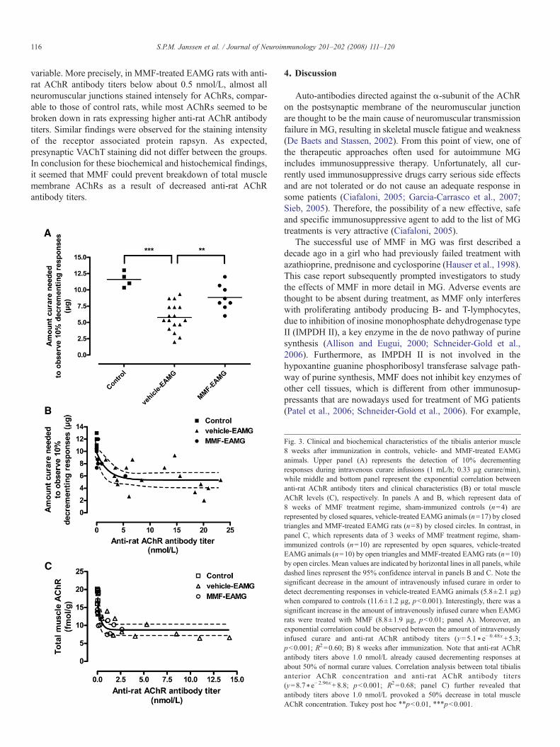
The clinical efficiency of suppressed anti-rat AChR antibodytiters was investigated by quantitative assessment of neuro-muscular transmission in the left tibialis anterior muscle duringintravenous curare infusion in rats treated for 8 weeks withMMF or vehicle. Curare is known to specifically bind thenicotinic AChR, thereby inhibiting normal functioning of theAChR ion channel. From this point of view, the amount ofcurare needed to observe decrement of the CMAP was used as ameasurement for rat's clinical status.
Fig. 3A shows that MMF-treated EAMG rats exerteddecrementing responses at curare doses in the range of sham-immunized control rats (average values of 8.8±1.9 vs 11.6±1.2 µg, respectively), while decrement was observed atsignificantly lower curare doses in vehicle-treated EAMGanimals (5.8±2.1 µg; pb0.01 vs MMF-EAMG; pb0.001 vscontrol). Interestingly, an exponential correlation was foundbetween the anti-rat AChR antibody titers and the amount ofintravenously infused curare in order to observe decrementingresponses (y=5.1⁎e−0.48x+5.3; pb0.001; R2 =0.60; Fig. 3B).In particular, it seemed that increases in anti-rat AChR antibodytiters above 1.0 nmol/L already provoked decrementingresponses at about 50% lower curare levels compared tohealthy control animals. Since 88% of MMF-treated EAMG ratsstudied under this experimental condition could suppress anti-rat AChR antibody titers even below 1.0 nmol/L, MMFefficiently improved neuromuscular transmission in EAMG.
To further analyze the effect of MMF immunosuppressionon the neuromuscular junction, the AChR concentrationswere determined in tibialis anterior muscles of rats treatedfor 3 weeks with MMF or vehicle. Surprisingly, averagemembrane AChR concentrations of vehicle-treated and MMF-treated EAMG rats (9±3 and 11±2 fmol/g, respectively) wereboth significantly decreased by about 45% when compared tosham-immunized control rats (18± 3 fmol/g; post hocpb0.001). However, several individual AChR concentrationvalues of MMF-treated EAMG animals were in the range ofthose of sham-immunized controls, and showed thus noevidence of muscle membrane AChR breakdown. Similarly tothe aforementioned curare resistance measurements ofneuromuscular transmission, an exponential correlation wasfound between the anti-rat AChR antibody titers and the totalmuscle AChR concentration (Fig. 3C). Indeed, only a slightlyincreased anti-rat AChR antibody titer of about 1.0 nmol/Lwas already sufficient to reduce the normal amount of totalmembrane AChRs by half (y=8.7⁎e−2.96x+8.8; pb0.001;R2 =0.68).
Fluorescent immunohistochemical studies on contralateraltibialis anterior muscles of the same rats treated for 3 weekswith MMF or vehicle confirmed the impact of different anti-ratAChR antibody titers on the postsynaptic membrane of theneuromuscular junction (Fig. 4). Indeed, compared to controls,triple fluorescent staining of neuromuscular endplates showed aclearly reduced postsynaptic AChR staining in almost allvehicle-treated EAMG rats while the AChR concentration in theneuromuscular junction of MMF-treated EAMG animals was
116 S.P.M. Janssen et al. / Journal of Neuroimmunology 201–202 (2008) 111–120
variable. More precisely, in MMF-treated EAMG rats with anti-rat AChR antibody titers below about 0.5 nmol/L, almost allneuromuscular junctions stained intensely for AChRs, compar-able to those of control rats, while most AChRs seemed to bebroken down in rats expressing higher anti-rat AChR antibodytiters. Similar findings were observed for the staining intensityof the receptor associated protein rapsyn. As expected,presynaptic VAChT staining did not differ between the groups.In conclusion for these biochemical and histochemical findings,it seemed that MMF could prevent breakdown of total musclemembrane AChRs as a result of decreased anti-rat AChRantibody titers.
4. Discussion
Auto-antibodies directed against the α-subunit of the AChRon the postsynaptic membrane of the neuromuscular junctionare thought to be the main cause of neuromuscular transmissionfailure in MG, resulting in skeletal muscle fatigue and weakness(De Baets and Stassen, 2002). From this point of view, one ofthe therapeutic approaches often used for autoimmune MGincludes immunosuppressive therapy. Unfortunately, all cur-rently used immunosuppressive drugs carry serious side effectsand are not tolerated or do not cause an adequate response insome patients (Ciafaloni, 2005; Garcia-Carrasco et al., 2007;Sieb, 2005). Therefore, the possibility of a new effective, safeand specific immunosuppressive agent to add to the list of MGtreatments is very attractive (Ciafaloni, 2005).
The successful use of MMF in MG was first described adecade ago in a girl who had previously failed treatment withazathioprine, prednisone and cyclosporine (Hauser et al., 1998).This case report subsequently prompted investigators to studythe effects of MMF in more detail in MG. Adverse events arethought to be absent during treatment, as MMF only interfereswith proliferating antibody producing B- and T-lymphocytes,due to inhibition of inosine monophosphate dehydrogenase typeII (IMPDH II), a key enzyme in the de novo pathway of purinesynthesis (Allison and Eugui, 2000; Schneider-Gold et al.,2006). Furthermore, as IMPDH II is not involved in thehypoxantine guanine phosphoribosyl transferase salvage path-way of purine synthesis, MMF does not inhibit key enzymes ofother cell tissues, which is different from other immunosup-pressants that are nowadays used for treatment of MG patients(Patel et al., 2006; Schneider-Gold et al., 2006). For example,
Fig. 3. Clinical and biochemical characteristics of the tibialis anterior muscle8 weeks after immunization in controls, vehicle- and MMF-treated EAMGanimals. Upper panel (A) represents the detection of 10% decrementingresponses during intravenous curare infusions (1 mL/h; 0.33 µg curare/min),while middle and bottom panel represent the exponential correlation betweenanti-rat AChR antibody titers and clinical characteristics (B) or total muscleAChR levels (C), respectively. In panels A and B, which represent data of8 weeks of MMF treatment regime, sham-immunized controls (n=4) arerepresented by closed squares, vehicle-treated EAMG animals (n=17) by closedtriangles and MMF-treated EAMG rats (n=8) by closed circles. In contrast, inpanel C, which represents data of 3 weeks of MMF treatment regime, sham-immunized controls (n=10) are represented by open squares, vehicle-treatedEAMG animals (n=10) by open triangles and MMF-treated EAMG rats (n=10)by open circles. Mean values are indicated by horizontal lines in all panels, whiledashed lines represent the 95% confidence interval in panels B and C. Note thesignificant decrease in the amount of intravenously infused curare in order todetect decrementing responses in vehicle-treated EAMG animals (5.8±2.1 µg)when compared to controls (11.6±1.2 µg, pb0.001). Interestingly, there was asignificant increase in the amount of intravenously infused curare when EAMGrats were treated with MMF (8.8±1.9 µg, pb0.01; panel A). Moreover, anexponential correlation could be observed between the amount of intravenouslyinfused curare and anti-rat AChR antibody titers (y=5.1⁎ e− 0.48x+5.3;pb0.001; R2=0.60; B) 8 weeks after immunization. Note that anti-rat AChRantibody titers above 1.0 nmol/L already caused decrementing responses atabout 50% of normal curare values. Correlation analysis between total tibialisanterior AChR concentration and anti-rat AChR antibody titers(y=8.7⁎e− 2.96x+8.8; pb0.001; R2=0.68; panel C) further revealed thatantibody titers above 1.0 nmol/L provoked a 50% decrease in total muscleAChR concentration. Tukey post hoc ⁎⁎pb0.01, ⁎⁎⁎pb0.001.
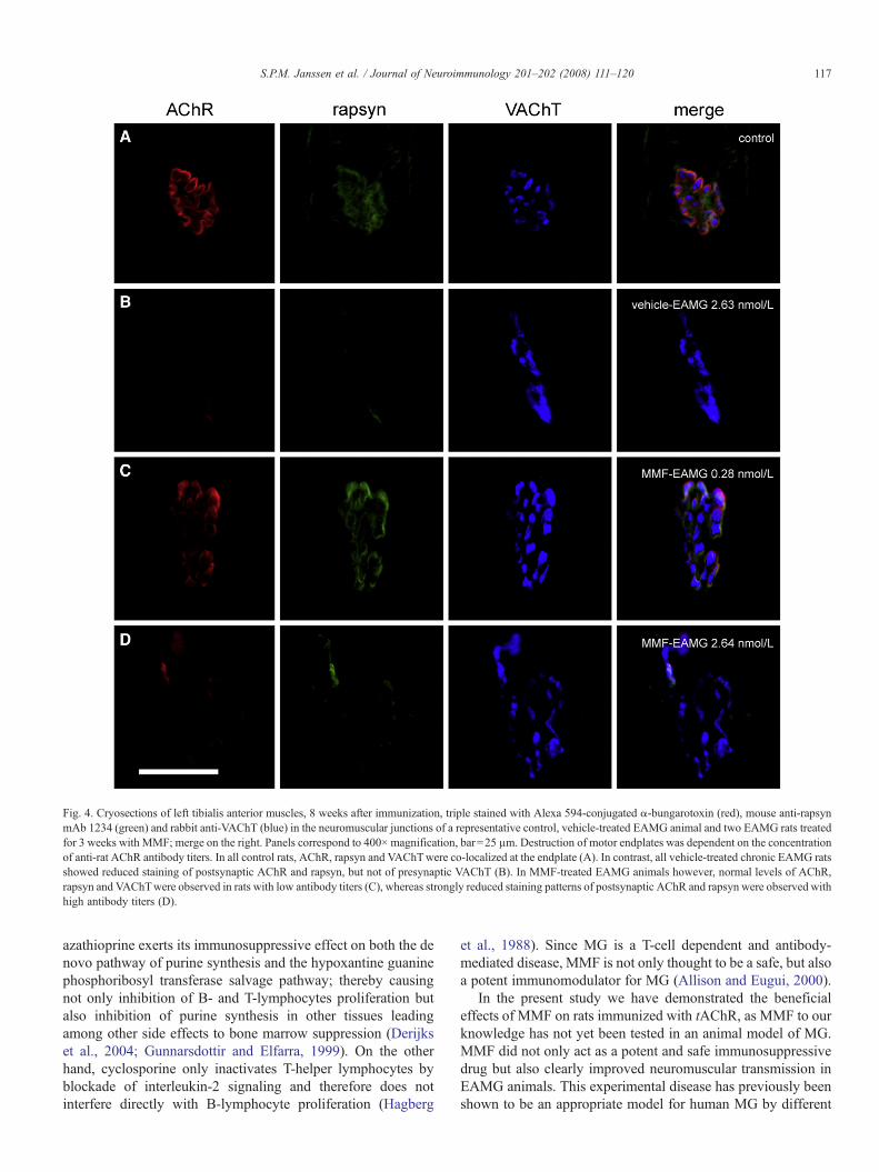
Fig. 4. Cryosections of left tibialis anterior muscles, 8 weeks after immunization, triple stained with Alexa 594-conjugated α-bungarotoxin (red), mouse anti-rapsynmAb 1234 (green) and rabbit anti-VAChT (blue) in the neuromuscular junctions of a representative control, vehicle-treated EAMG animal and two EAMG rats treatedfor 3 weeks with MMF; merge on the right. Panels correspond to 400× magnification, bar=25 µm. Destruction of motor endplates was dependent on the concentrationof anti-rat AChR antibody titers. In all control rats, AChR, rapsyn and VAChTwere co-localized at the endplate (A). In contrast, all vehicle-treated chronic EAMG ratsshowed reduced staining of postsynaptic AChR and rapsyn, but not of presynaptic VAChT (B). In MMF-treated EAMG animals however, normal levels of AChR,rapsyn and VAChTwere observed in rats with low antibody titers (C), whereas strongly reduced staining patterns of postsynaptic AChR and rapsyn were observed withhigh antibody titers (D).
117S.P.M. Janssen et al. / Journal of Neuroimmunology 201–202 (2008) 111–120
azathioprine exerts its immunosuppressive effect on both the denovo pathway of purine synthesis and the hypoxantine guaninephosphoribosyl transferase salvage pathway; thereby causingnot only inhibition of B- and T-lymphocytes proliferation butalso inhibition of purine synthesis in other tissues leadingamong other side effects to bone marrow suppression (Derijkset al., 2004; Gunnarsdottir and Elfarra, 1999). On the otherhand, cyclosporine only inactivates T-helper lymphocytes byblockade of interleukin-2 signaling and therefore does notinterfere directly with B-lymphocyte proliferation (Hagberg
et al., 1988). Since MG is a T-cell dependent and antibody-mediated disease, MMF is not only thought to be a safe, but alsoa potent immunomodulator for MG (Allison and Eugui, 2000).
In the present study we have demonstrated the beneficialeffects of MMF on rats immunized with tAChR, as MMF to ourknowledge has not yet been tested in an animal model of MG.MMF did not only act as a potent and safe immunosuppressivedrug but also clearly improved neuromuscular transmission inEAMG animals. This experimental disease has previously beenshown to be an appropriate model for human MG by different
118 S.P.M. Janssen et al. / Journal of Neuroimmunology 201–202 (2008) 111–120
criteria such as cellular and humoral immunological responses,decrementing responses of muscle action potentials in electro-myographic examinations, as well as transitory improvement byanti-cholinesterase drugs (Lennon et al., 1975).
To test whether MMF could prevent the onset of MG in rats,MMF was administered orally for 3 or 8 weeks, starting fromthe day of immunization. A dose of 30 mg/kg/day was chosenas this dose has already been proven to be efficient in otherexperimental diseases (Gibson and Hayden, 2007; Tran et al.,2001; Zandman-Goddard and Shoenfeld, 2005). Moreover, MGpatients daily receive an oral dose of 1.0–2.0 g, which is in therange of the body weight related dose of MMF used in thepresent study (Schneider-Gold et al., 2006). The results showedthat MMF was able to suppress anti-rat AChR antibody titers tovalues to 0.9±1.3 nmol/L 8 weeks after immunization in bothexperimental regimes. The immunosuppressive properties ofMMF in the present study were moreover consistent withfindings in MG case reports where antibody titers could alreadybe reduced up to 50% after one month of MMF treatment(Meriggioli et al., 2003b; Schneider et al., 2001).
Compared to other immunosuppressive agents, the improve-ment in tolerability and safety profile of MMF is has alreadybeen established clearly in several small studies. Indeed, whilemost therapeutic immunosuppressive drugs are known to exertsevere nephrotoxic and hepatotoxic side effects, clear adverseeffects of MMF treatment have been shown to be absent in mostMG patients (Garcia-Carrasco et al., 2007). Although we didnot rigorously test for side effects of MMF in our experimentalmodel, none of the MMF-treated animals in this studydeveloped overt side effects such as fever, diarrhea, dehydra-tion, breathing difficulties or immunosuppression relatedinfections. Furthermore, MMF seemed to be highly efficientin preventing clinical MG symptoms in 95% of immunizedanimals. Single case reports and several small clinical studiesconcerning MMF treatment in MG confirm the above-mentioned observations as most patients were able to reachpharmacological remission, and improved manual muscletesting and quantitative MG score after several months oftreatment (Caponnetto et al., 2001; Chaudhry et al., 2001;Ciafaloni et al., 2001; Hauser et al., 1998; Lim et al., 2007;Meriggioli et al., 2003a; Meriggioli et al., 2003b; Mowzoonet al., 2001; Prakash et al., 2007; Schneider et al., 2001). Incontrast, two recently completed large phase 3 prospective,multicenter, placebo-controlled trials showed little or no effectof MMF when it was used as an adjunctive treatment tocorticosteroids. Indeed, no improvement was observed inbaseline Quantitative Myasthenia Gravis score for diseaseseverity and in the ability to decrease the dose of corticosteroidsand cholinesterase inhibitors, as AChR antibody titersdecreased equally in both groups by about 50%. However, theobtained results are likely due to a higher than expectedresponse to prednisone alone (Hampton, 2007).
To study the effects of MMF-induced low anti-rat AChRantibody titers on clinical characteristics in more detail, EMGmeasurements of tibialis anterior muscles were performedduring continuous curare infusions. The results indicated asignificant improvement in neuromuscular transmission after
MMF administration to EAMG rats. Moreover, no significantdifferences could be observed between MMF-treated EAMGanimals and sham-immunized control rats, indicating thatthe immunosuppressant action of MMF was indeed highlyefficient in improving neuromuscular transmission, which is inaccordance with the clinical EAMG score discussed earlier.Patient data fully support these findings as improvements infunctional status, manual muscle testing and jitter in single fiberEMG were reported after MMF treatment in MG (Chaudhryet al., 2001; Ciafaloni et al., 2001; Hampton, 2007; Meriggioliet al., 2003a; Meriggioli et al., 2003b; Mowzoon et al., 2001;Prakash et al., 2007). Interestingly, the anti-rat AChR antibodytiters observed in the present study were moreover expo-nentially correlated with curare resistance of neuromusculartransmission. Indeed, values above 1.0 nmol/L of anti-rat AChRantibodies seemed to be responsible for provocation ofdecrementing responses at about 50% of the normal curaredose. This indicates that in the present setup only 30% of allMMF-treated EAMG animals would suffer from as severeneuromuscular deficits as occurring in vehicle-treated EAMGrats, since short-term administration of MMF was able tosuppress anti-rat AChR antibody titers below 1.0 nmol/L in70% of immunized rats (Fig. 1B). These findings suggest thepossibility that very low, or even undetectable anti-AChRantibody levels can cause the neuromuscular junction transmis-sion defect in MG patients. At the basis of a high susceptibilityof some patients to low levels of auto-antibodies might begenetic differences in the expression levels of postsynapticproteins. As a case in point, it has been shown that rapsyn,utrophin and s-laminin expression determine disease severity inrats susceptible to EAMG (Hoedemaekers et al., 1998; Losenet al., 2005; Martinez-Martinez et al., 2007).
In relation to neuromuscular transmission, tibialis anteriorAChR concentrations were also determined. The results wererather surprising as AChR concentrations of both vehicle-treated and MMF-treated EAMG animals were equallydecreased by about 45% when compared to sham-immunizedcontrols, whereas anti-rat AChR antibody titers of MMF-treatedEAMG rats were 10 times lower in comparison with vehicle-treated EAMG animals. On the other hand, correlation analysisrevealed that the anti-rat AChR antibody titer is an importantdeterminant in breaking down the AChRs of the muscle. Indeed,a similar relation was observed between antibody titers andmuscle AChR concentration when compared to the previouslydiscussed correlation between anti-rat AChR antibodies andneuromuscular transmission. Severe AChR breakdown up to50% of normal values already seemed to occur with an anti-rat AChR antibody titer of about 1.0 nmol/L. Immunohisto-chemical analyses of endplate regions fully confirmed thesefindings. Vehicle-treated EAMG rats seemed to have an overallbreakdown of neuromuscular junction AChRs, while endplateAChRs of MMF-treated EAMG animals were not equallydestructed. In particular, it seemed that overall endplate AChRswere intact with extremely low antibody titers while AChRbreakdown was observed in several neuromuscular junctions ofMMF-treated animals with higher anti-rat AChR antibodytiters. It is possible that the membrane AChR loss measured in
119S.P.M. Janssen et al. / Journal of Neuroimmunology 201–202 (2008) 111–120
muscle extracts is in part due to loss of unclustered and moresusceptible extrasynaptic AChRs. The electromyographicalmeasurements during challenge with curare are howevercertainly suggestive of AChR protection at the neuromuscularjunction as a result of MMF treatment.
In summary, these data show that short-term administration ofMMF is able to act as a highly immunosuppressive drug in ananimal model of MG and consequently improves clinicalcharacteristics by enhancing neuromuscular transmission. More-over, these data strengthen the case for using MMF as a treatmentof humanMG, although further large clinical trials are required todetermine the exact use of MMF as a sole therapeutic or as anadjunctive treatment to other immunosuppressive drugs.
Acknowledgements
The authors sincerely thank Aspreva Pharmaceuticals, PaloAlto, United States for their financial support and for providingthe MMF.
References
Allison, A.C., Eugui, E.M., 2000. Mycophenolate mofetil and its mechanisms ofaction. Immunopharmacology 47, 85–118.
Blaheta, R.A., Leckel, K., Wittig, B., Zenker, D., Oppermann, E., Harder, S.,Scholz, M., Weber, S., Schuldes, H., Encke, A., Markus, B.H., 1998.Inhibition of endothelial receptor expression and of T-cell ligand activity bymycophenolate mofetil. Transpl. Immunol. 6, 251–259.
Caponnetto, C., Rossi, E., Primavera, A., 2001. Mycophenolate mofetil: a newimmunosuppressive approach. Successful treatment in a case of myastheniagravis associated with incomplete lupus erythematosus syndrome andhepatitis C virus infection. Eur. Neurol. 46, 53–54.
Chaudhry, V., Cornblath, D.R., Griffin, J.W., O'Brien, R., Drachman, D.B.,2001. Mycophenolate mofetil: a safe and promising immunosuppressant inneuromuscular diseases. Neurology 56, 94–96.
Ciafaloni, E., 2005. Mycophenolate mofetil and myasthenia gravis. Lupus 14(Suppl 1), s46–49.
Ciafaloni, E., Massey, J.M., Tucker-Lipscomb, B., Sanders, D.B., 2001.Mycophenolate mofetil for myasthenia gravis: an open-label pilot study.Neurology 56, 97–99.
Cohn, R.G., Mirkovich, A., Dunlap, B., Burton, P., Chiu, S.H., Eugui, E.,Caulfield, J.P., 1999. Mycophenolic acid increases apoptosis, lysosomes andlipid droplets in human lymphoid and monocytic cell lines. Transplantation68, 411–418.
De Baets, M., Stassen, M.H., 2002. The role of antibodies in myasthenia gravis.Ann. N.Y. Acad. Sci. 202, 5–11.
De Baets, M., Stassen, M., Losen, M., Zhang, X., Machiels, B., 2003.Immunoregulation in experimental autoimmune myasthenia gravis–about Tcells, antibodies, and endplates. Ann. N.Y. Acad. Sci. 998, 308–317.
Derijks, L.J., Gilissen, L.P., Engels, L.G., Bos, L.P., Bus, P.J., Lohman, J.J.,Curvers, W.L., Van Deventer, S.J., Hommes, D.W., Hooymans, P.M., 2004.Pharmacokinetics of 6-mercaptopurine in patients with inflammatory boweldisease: implications for therapy. Ther. Drug. Monit. 26, 311–318.
Drachman, D.B., 1994. Myasthenia gravis. N. Engl. J. Med. 330, 1797–1810.Durez, P., Appelboom, T., Pira, C., Stordeur, P., Vray, B., Goldman, M., 1999.
Antiinflammatory properties of mycophenolate mofetil in murine endotox-emia: inhibition of TNF-alpha and upregulation of IL-10 release. Int. J.Immunopharmacol. 21, 581–587.
Enk, A.H., Knop, J., 1997. Treatment of pemphigus vulgaris with mycophe-nolate mofetil. Lancet 350, 494.
Eugui, E.M., Mirkovich, A., Allison, A.C., 1991. Lymphocyte-selectiveantiproliferative and immunosuppressive activity of mycophenolic acid andits morpholinoethyl ester (RS-61443) in rodents. Transplant. Proc. 23, 15–18.
European Mycophenolate Mofetil Cooperative Study Group, 1995. Placebo-controlled study of mycophenolate mofetil combined with cyclosporinand corticosteroids for prevention of acute rejection. Lancet 345,1321–1325.
Gajdos, P., Chevret, S., Toyka, K., 2006. Intravenous immunoglobulin formyasthenia gravis. Cochrane. Database. Syst. Rev. CD002277.
Garcia-Carrasco, M., Escarcega, R.O., Fuentes-Alexandro, S., Riebeling, C.,Cervera, R., 2007. Therapeutic options in autoimmune myasthenia gravis.Autoimmun. Rev. 6, 373–378.
Gibson, W.T., Hayden, M.R., 2007. Mycophenolate mofetil and atherosclerosis:results of animal and human studies. Ann. N. Y. Acad. Sci. 1110, 209–221.
Ginzler, E.M., Dooley, M.A., Aranow, C., Kim, M.Y., Buyon, J., Merrill, J.T.,Petri, M., Gilkeson, G.S., Wallace, D.J., Weisman, M.H., Appel, G.B., 2005.Mycophenolate mofetil or intravenous cyclophosphamide for lupusnephritis. N. Engl. J. Med. 353, 2219–2228.
Goldblum, R., 1993. Therapy of rheumatoid arthritis with mycophenolatemofetil. Clin. Exp. Rheumatol. 11 (Suppl 8), S117–119.
Gronseth, G.S., Barohn, R.J., 2000. Practice parameter: thymectomy forautoimmune myasthenia gravis (an evidence-based review): report of theQuality Standards Subcommittee of the American Academy of Neurology.Neurology 55, 7–15.
Gunnarsdottir, S., Elfarra, A.A., 1999. Glutathione-dependent metabolism ofcis-3-(9H-purin-6-ylthio)acrylic acid to yield the chemotherapeutic drug 6-mercaptopurine: evidence for two distinct mechanisms in rats. J. Pharmacol.Exp. Ther. 290, 950–957.
Hagberg, R.C., Hoyt, E.G., Billingham, M.E., Sibley, R.K., Starnes, V.A.,Baldwin, J.C., 1988. Comparison of cyclosporin A and G with and withoutazathioprine regarding immunosuppressive efficacy, toxicity, and pharma-cokinetics in Lewis rats. J. Heart. Transplant. 7, 359–369.
Hampton, T., 2007. Trials assess myasthenia gravis therapies. Jama 298, 29–30.Hauser, R.A., Malek, A.R., Rosen, R., 1998. Successful treatment of a patient
with severe refractory myasthenia gravis using mycophenolate mofetil.Neurology 51, 912–913.
Hoch, W., McConville, J., Helms, S., Newsom-Davis, J., Melms, A., Vincent,A., 2001. Auto-antibodies to the receptor tyrosine kinase MuSK in patientswith myasthenia gravis without acetylcholine receptor antibodies. Nat. Med.7, 365–368.
Hoedemaekers, A., Graus, Y., van Breda Vriesman, P., de Baets, M., 1997. Age-and sex-related resistance to chronic experimental autoimmune myastheniagravis (EAMG) in Brown Norway rats. Clin. Exp. Immunol. 107, 189–197.
Hoedemaekers, A., Bessereau, J.L., Graus, Y., Guyon, T., Changeux, J.P.,Berrih-Aknin, S., van Breda Vriesman, P., De Baets, M.H., 1998. Role of thetarget organ in determining susceptibility to experimental autoimmunemyasthenia gravis. J. Neuroimmunol. 89, 131–141.
Larkin, G., Lightman, S., 1999. Mycophenolate mofetil. A useful immunosup-pressive in inflammatory eye disease. Ophthalmology 106, 370–374.
Lehmann, H.C., Hartung, H.P., Hetzel, G.R., Stuve, O., Kieseier, B.C., 2006.Plasma exchange in neuroimmunological disorders: part 2. Treatment ofneuromuscular disorders. Arch. Neurol. 63, 1066–1071.
Lennon, V.A., Lindstrom, J.M., Seybold, M.E., 1975. Experimental auto-immune myasthenia: a model of myasthenia gravis in rats and guinea pigs.J. Exp. Med. 141, 1365–1375.
Lim, A.K., Donnan, G., Chambers, B., Ierino, F.L., 2007. Mycophenolatemofetil substitution for cyclosporine-dependent myasthenia gravis andnephrotoxicity. Intern. Med. J. 37, 55–59.
Lindstrom, J.M., Seybold, M.E., Lennon, V.A., Whittingham, S., Duane, D.D.,1976. Antibody to acetylcholine receptor in myasthenia gravis. Prevalence,clinical correlates, and diagnostic value. Neurology 26, 1054–1059.
Losen, M., Stassen, M.H., Martinez-Martinez, P., Machiels, B.M., Duimel, H.,Frederik, P., Veldman, H., Wokke, J.H., Spaans, F., Vincent, A., De Baets,M.H., 2005. Increased expression of rapsyn in muscles prevents acetylcho-line receptor loss in experimental autoimmune myasthenia gravis. Brain 128,2327–2337.
Losen, M., Martínez-Martínez, P., Phernambucq, M., Schuurman, J., Parren, P.W.,De Baets, M.H., 2008. Treatment of myasthenia gravis by preventingacetylcholine receptor modulation. Ann. N.Y. Acad. Sci. 1132, 174–179.
Martinez-Martinez, P., Losen, M., Duimel, H., Frederik, P., Spaans, F.,Molenaar, P., Vincent, A., De Baets, M.H., 2007. Overexpression of rapsyn

120 S.P.M. Janssen et al. / Journal of Neuroimmunology 201–202 (2008) 111–120
in rat muscle increases acetylcholine receptor levels in chronic experimentalautoimmune myasthenia gravis. Am. J. Pathol. 170, 644–657.
Meriggioli, M.N., Ciafaloni, E., Al-Hayk, K.A., Rowin, J., Tucker-Lipscomb,B., Massey, J.M., Sanders, D.B., 2003a. Mycophenolate mofetil formyasthenia gravis: an analysis of efficacy, safety, and tolerability. Neurology61, 1438–1440.
Meriggioli, M.N., Rowin, J., Richman, J.G., Leurgans, S., 2003b. Mycophe-nolate mofetil for myasthenia gravis: a double-blind, placebo-controlledpilot study. Ann. N. Y. Acad. Sci. 998, 494–499.
Mowzoon, N., Sussman, A., Bradley, W.G., 2001. Mycophenolate (CellCept)treatment of myasthenia gravis, chronic inflammatory polyneuropathy andinclusion body myositis. J. Neurol. Sci. 185, 119–122.
Neurath, M.F., Wanitschke, R., Peters, M., Krummenauer, F., Meyer zumBuschenfelde, K.H., Schlaak, J.F., 1999. Randomised trial of mycopheno-late mofetil versus azathioprine for treatment of chronic active Crohn'sdisease. Gut 44, 625–628.
Nowack, R., Gobel, U., Klooker, P., Hergesell, O., Andrassy, K., van derWoude, F.J., 1999. Mycophenolate mofetil for maintenance therapy ofWegener's granulomatosis and microscopic polyangiitis: a pilot study in 11patients with renal involvement. J. Am. Soc. Nephrol. 10, 1965–1971.
Palace, J., Newsom-Davis, J., Lecky, B., 1998. A randomized double-blind trialof prednisolone alone or with azathioprine in myasthenia gravis. MyastheniaGravis Study Group. Neurology 50, 1778–1783.
Patel, A.A., Swerlick, R.A., McCall, C.O., 2006. Azathioprine in dermatology:the past, the present, and the future. J. Am. Acad. Dermatol. 55, 369–389.
Prakash, K.M., Ratnagopal, P., Puvanendran, K., Lo, Y.L., 2007. Mycopheno-late mofetil — as an adjunctive immunosuppressive therapy in refractory
myasthenia gravis: the Singapore experience. J. Clin. Neurosci. 14,278–281.
Richman, D.P., Agius, M.A., 2003. Treatment of autoimmune myastheniagravis. Neurology 61, 1652–1661.
Sanders, D.B., Hart, I.K., Mantegazza, R., Shukla, S.S., Siddiqi, Z.A., De Baets,M.H., Melms, A., Nicolle, M.W., Solomons, N., Richman, D.P., 2008. Aninternational, phase III, randomized trial of mycophenolate mofetil inmyasthenia gravis. Neurology. doi:10.1212/01.wnl.0000312374.95186.cc.
Schneider-Gold, C., Hartung, H.P., Gold, R., 2006. Mycophenolate mofetil andtacrolimus: new therapeutic options in neuroimmunological diseases.Muscle. Nerve. 34, 284–291.
Schneider, C., Gold, R., Reiners, K., Toyka, K.V., 2001. Mycophenolate mofetilin the therapy of severe myasthenia gravis. Eur. Neurol. 46, 79–82.
Seybold, M.E., Lambert, E.H., Lennon, V.A., Lindstrom, J.M., 1976.Experimental autoimmune myasthenia: clinical, neurophysiologic, andpharmacologic aspects. Ann. N. Y. Acad. Sci. 274, 275–282.
Sieb, J.P., 2005. Myasthenia gravis: emerging new therapy options. Curr. Opin.Pharmacol. 5, 303–307.
The Muscle Study Group, 2008. A trial of mycophenolate mofetil withprednisone as initial immunotherapy in myasthenia gravis. Neurology.doi:10.1212/01.wnl.0000312374.95186.cc.
Tran, G.T., Carter, N., Hodgkinson, S.J., 2001. Mycophenolate mofetil treatmentaccelerates recovery from experimental allergic encephalomyelitis. Int.Immunopharmacol. 1, 1709–1723.
Zandman-Goddard, G., Shoenfeld, Y., 2005. Mycophenolate mofetil in animalmodels of autoimmune disease. Lupus 14 (Suppl 1), s12–16.
Related Documents