INTRODUCTION In insulin-dependent diabetes mellitus (IDDM), the b-cell loss and subsequent hyperglycaemia are the outcome of an autoim- mune process leading to inflammatory reaction with accumula- tion of macrophages and T cells in the islets [1–3]. Although results obtained with pancreas or islet transplantation are pro- mising, the interference with deleterious autoimmune response by using immunosuppressive or immunomodulating agents still remains a conceivable strategy for IDDM treatment. However, conventional calcineurine-inhibiting immunosuppressants CsA and FK506 are not appropriate for such a task, because their actions include decreased insulin secretion, increased insulin resistance or direct toxic effect on the b-cells [4,5]. Accordingly, both drugs can cause post-transplantation diabetes mellitus [6], while CsA was also found to exacerbate the disease symptoms in an animal model of IDDM [7]. Mycophenolate mofetil (MMF) is a novel xenobiotic immunosuppressant that has proved effective in the therapy of transplant rejection, as well as in various animal models of autoimmunity (for review, see [8]). Through its active metabolite mycophenolic acid (MPA), MMF inhibits the activity of inosine monophosphate dehydrogenase (IMPDH), a rate-limiting enzyme in the synthesis of guanosine nucleotides [9]. MPA exerts more potent antiprolifferative effects on lymphocytes than on other cell types, as it is fairly selective for the isoform II of IMPDH, which is expressed mainly in activated T and B cells [10]. Moreover, through depletion of intracellular GTP levels and sub- sequent suppression of glycosylation and the expression of some adhesion molecules, MPA interferes with the recruitment of lymphocytes and monocytes into sites of inflammation [11,12]. Hao et al. [13] reported that MMF prevented spontaneous development of autoimmune diabetes in genetically predisposed bio-breeding (BB) rats, but the protection ceased with the termi- 214 Down-regulation of multiple low dose streptozotocin-induced diabetes by mycophenolate mofetil D. MAKSIMOVIC-IVANIC*, V. TRAJKOVIC†, DJ. MILJKOVIC*, M. MOSTARICA STOJKOVIC† & S. STOSIC-GRUJICIC* *Institute for Biological Research ‘Sinisa Stankovic’, Belgrade, and †Institute of Microbiology and Immunology, School of Medicine, Belgrade, Yugoslavia (Accepted for publication 5 April 2002) SUMMARY The new immunosuppressive agent mycophenolate mofetil (MMF) has been shown recently to exert a protective effects in certain animal models of autoimmunity, including diabetes in diabetes-prone bio- breeding (BB) rats. In the present study, the immunomodulatory potential of MMF was investigated in autoimmune diabetes induced by multiple low doses of streptozotocin (MLD-STZ) in genetically susceptible DA rats [20mg STZ/kg body weight (b.w.) for 5 days] and CBA/H mice (40mg STZ/kg b.w. for 5 days). In both species, short time treatment of animals with MMF (25 mg/kg) during the early development of the disease, as well as continuous MMF treatment, prevented the appearance of hyper- glycaemia and inflammatory infiltrates in the pancreatic tissue. Moreover, clinical manifestations of dia- betes were suppressed by application of the drug after the onset of clinical symptoms. Treatment with guanosine (1 mg/kg) in parallel with MMF completely reversed MMF activity in vivo, indicating that inhibition of inosine monophosphate dehydrogenase (IMPDH) was responsible for the observed sup- pressive effects. MMF-mediated protection from diabetes correlated with reduced ex vivo spontaneous spleen mononuclear cell (MNC) proliferation and defective adhesive cell interactions. MMF-treated animals also had lower local production of IFN-g, as well as IL-12 and nitric oxide (NO) production by peripheral tissues (spleen and peritoneal cells), compared to that in control diabetic groups, while IL- 10 level was elevated. Together, these data demonstrate that MMF interferes with autoimmune process in streptozotocin-induced diabetes at multiple levels, including lymphocyte proliferation and adhesion, as well as pro/anti-inflammatory cytokine balance. Keywords adhesion molecules autoimmune diabetes cytokines mycophenolate mofetil streptozotocin Clin Exp Immunol 2002; 129:214 –223 Correspondence: Dr Stanislava Stosic-Grujicic, Institute for Biological Research ‘Sinisa Stankovic’, 29 Novembra 142, 11000 Belgrade, Yugoslavia. E-mail: [email protected] © 2002 Blackwell Science

Welcome message from author
This document is posted to help you gain knowledge. Please leave a comment to let me know what you think about it! Share it to your friends and learn new things together.
Transcript
INTRODUCTION
In insulin-dependent diabetes mellitus (IDDM), the b-cell lossand subsequent hyperglycaemia are the outcome of an autoim-mune process leading to inflammatory reaction with accumula-tion of macrophages and T cells in the islets [1–3]. Althoughresults obtained with pancreas or islet transplantation are pro-mising, the interference with deleterious autoimmune responseby using immunosuppressive or immunomodulating agents stillremains a conceivable strategy for IDDM treatment. However,conventional calcineurine-inhibiting immunosuppressants CsAand FK506 are not appropriate for such a task, because theiractions include decreased insulin secretion, increased insulinresistance or direct toxic effect on the b-cells [4,5]. Accordingly,both drugs can cause post-transplantation diabetes mellitus [6],
while CsA was also found to exacerbate the disease symptoms inan animal model of IDDM [7].
Mycophenolate mofetil (MMF) is a novel xenobioticimmunosuppressant that has proved effective in the therapy oftransplant rejection, as well as in various animal models ofautoimmunity (for review, see [8]). Through its active metabolitemycophenolic acid (MPA), MMF inhibits the activity of inosinemonophosphate dehydrogenase (IMPDH), a rate-limitingenzyme in the synthesis of guanosine nucleotides [9]. MPA exertsmore potent antiprolifferative effects on lymphocytes than onother cell types, as it is fairly selective for the isoform II ofIMPDH, which is expressed mainly in activated T and B cells [10].Moreover, through depletion of intracellular GTP levels and sub-sequent suppression of glycosylation and the expression of someadhesion molecules, MPA interferes with the recruitment of lymphocytes and monocytes into sites of inflammation [11,12]. Hao et al. [13] reported that MMF prevented spontaneous development of autoimmune diabetes in genetically predisposed bio-breeding (BB) rats, but the protection ceased with the termi-
214
Down-regulation of multiple low dose streptozotocin-induced diabetes bymycophenolate mofetil
D. MAKSIMOVIC-IVANIC*, V. TRAJKOVIC†, DJ. MILJKOVIC*, M. MOSTARICA STOJKOVIC† & S. STOSIC-GRUJICIC**Institute for Biological Research ‘Sinisa Stankovic’, Belgrade, and †Institute of Microbiology and Immunology, School of Medicine,
Belgrade, Yugoslavia
(Accepted for publication 5 April 2002)
SUMMARY
The new immunosuppressive agent mycophenolate mofetil (MMF) has been shown recently to exert aprotective effects in certain animal models of autoimmunity, including diabetes in diabetes-prone bio-breeding (BB) rats. In the present study, the immunomodulatory potential of MMF was investigatedin autoimmune diabetes induced by multiple low doses of streptozotocin (MLD-STZ) in geneticallysusceptible DA rats [20 mg STZ/kg body weight (b.w.) for 5 days] and CBA/H mice (40 mg STZ/kg b.w.for 5 days). In both species, short time treatment of animals with MMF (25 mg/kg) during the earlydevelopment of the disease, as well as continuous MMF treatment, prevented the appearance of hyper-glycaemia and inflammatory infiltrates in the pancreatic tissue. Moreover, clinical manifestations of dia-betes were suppressed by application of the drug after the onset of clinical symptoms. Treatment withguanosine (1 mg/kg) in parallel with MMF completely reversed MMF activity in vivo, indicating thatinhibition of inosine monophosphate dehydrogenase (IMPDH) was responsible for the observed sup-pressive effects. MMF-mediated protection from diabetes correlated with reduced ex vivo spontaneousspleen mononuclear cell (MNC) proliferation and defective adhesive cell interactions. MMF-treatedanimals also had lower local production of IFN-g, as well as IL-12 and nitric oxide (NO) production byperipheral tissues (spleen and peritoneal cells), compared to that in control diabetic groups, while IL-10 level was elevated. Together, these data demonstrate that MMF interferes with autoimmune processin streptozotocin-induced diabetes at multiple levels, including lymphocyte proliferation and adhesion,as well as pro/anti-inflammatory cytokine balance.
Keywords adhesion molecules autoimmune diabetes cytokines mycophenolate mofetilstreptozotocin
Clin Exp Immunol 2002; 129:214–223
Correspondence: Dr Stanislava Stosic-Grujicic, Institute for BiologicalResearch ‘Sinisa Stankovic’, 29 Novembra 142, 11000 Belgrade,Yugoslavia.
E-mail: [email protected]
© 2002 Blackwell Science
Mycophenolate mofetil in streptozotocin-induced diabetes 215
nation of the therapy. Furthermore, the mechanisms responsiblefor the beneficial action of MMF in this diabetes model were notinvestigated.
In the present study we have explored the effect of MMF inthe autoimmune diabetes induced in rats and mice by multiplelow doses of streptozotocin (MLD-STZ), which offers a numberof experimental advantages. The islet lesions of MLD-STZ diabetes, initiated mainly by infiltrating T cells and macrophages,resemble closely those of human disease [14]. Moreover, the onsetof disease is controlled and the animals involved do not haveimmune abnormalities that complicate studies in models of spontaneous autoimmune diabetes [15]. Here we present evi-dence that MMF provides a long-lasting protection in rodentMLD-STZ-induced diabetes, interfering with autoimmune diabetogenic process at several levels, including lymphocyte proliferation and adhesiveness, as well as the production of proinflammatory mediators.
MATERIALS AND METHODS
ReagentsStreptozotocin (STZ, S-0130), mycophenolic acid (MPA), sul-phanilamide and naphthylethylenediamine dichidrochloride werepurchased from Sigma (St Louis, MO, USA). Mycophenolatemofetil (MMF, CellCept®) was obtained from Roche (Basel,Switzerland). RPMI-1640 supplemented with 1 mm HEPESbuffer, 5% fetal calf serum (FCS), 1% sodium pyruvate, 2 mm l-glutamine (all from Flow Laboratories, Irvine, UK), penicillin/streptomycin (ICN, Yugoslavia) and 5 ¥ 10–5 m 2-mercaptoethanol (Sigma) was used as a culture medium. Recombinant rat IL-1b was from Genzyme (Boston, MA, USA).Mouse MoAbs specific for rat CD4 (W3/25, Serotec, Oxford,UK), MHC class II (OX6, HBT, Leiden, the Netherlands), IFN-g (DB-1, HBT) and monocytes/macrophages (ED-1, kindlyprovided by Dr C. D. Dijkstra, Medical Faculty, Free University,Amsterdam, the Netherlands), as well as rat MoAbs specific formouse CD4 (GK1·5) and CD8 (YTS 169) (both from Serotec)were used for immunohistochemical staining.
Animals and treatmentsMale Dark Agouti (DA) rats and CBA/H mice were obtainedfrom our own facility (Institute for Biological Research ‘SinisaStankovic’, Belgrade, Yugoslavia) and used when 10–16 weeksold. Diabetes was induced into two rodent species with multiplesubtoxic doses of STZ [20 mg/kg body weight (b.w.)/day in rats,and 40 mg/kg b.w./day in mice], given i.p. for 5 consecutive days.In order to evaluate the effect of MMF on the disease develop-ment in rats, the drug was administrated i.m. at a dose of25 mg/kg/day by three treatment protocols: continuously through-out the experiment (7 weeks), or as a 10-day treatment startedeither jointly with MLD-STZ doses (the first injection of MMFgiven at day 0), or after MLD-STZ (starting at day 5 in relationto the last STZ dose). Mice were treated with the same dose ofMMF, with or without guanosine (1 mg/kg b.w./day), for the 2weeks starting jointly with MLD-STZ. Control animals receivedsaline in the same way as MMF. Plasma glucose was determinedby a glucose-oxidase method using a glucometer (Glucotronic C;Macherey-Nagel, Duren, Germany) once a week for up to7 weeks. Clinical diabetes was defined by hyperglycaemia in non-fasted animals (blood glucose >11 mmol/l). In order to collect cellsand tissues for further analysis, a groups of rats and mice were
sacrificed on day 10 or 15, respectively, when the treatment withMMF was completed.
Immunohistochemical analysis of pancreasAt sacrifice, pancreata were taken out and fixed overnight in 4%paraformaldehyde in PBS and subsequently transferred to 30%sucrose at 4°C. Fixed pancreata were embedded in PolyFreezetissue freezing medium (Polyscience, Warrington, PA, USA) andsections were cut in a cryomicrotome. The air-dried specimens(10–12 mm thick) on gelatinized slides were fixed for 10–15 min inacetone at –20°C. After blocking of endogenous peroxidase with0·3% H2O2 in methanol, sections were incubated for 1 h withprimary antibodies (W3/25, ED-1, DB-1, OX6, GK1·5, YTS 169).Staining of rat tissue was performed using mouse ExtrAvidin peroxidase staining kit (Sigma), according to the manufacturer’sinstruction. For mouse tissue, peroxidase-conjugated rabbit anti-rat IgG (DAKO, Hamburg, Germany) was used as a sec-ondary antibody. All incubations were performed at room temperature (RT) and between each step slides were washed with PBS. Finally, sections were incubated for 2–5 min withdiaminobenzamidine (DAB, Dako) as a substrate diluted in 0·02 m PBS pH 7·2 and then counterstained with Mayer’s haema-toxylin. Slides were mounted with Canada balsam and analysedunder the light microscope. Immunohistochemical examinationwas undertaken in a blind fashion by two independent observers.A minimum of 100 islets per animal were analysed for eachmarker. Islets were graded as follows according to mononuclearcell infiltration: grade 0, normal islets totally free of any mononu-clear cells stained for a given marker; grade 1, two to five posi-tively stained mononuclear cells within the islet; grade 2, morethan five positively stained mononuclear cells within the islet. Thehistology score gives the mean percentages of infiltration grade ineach category of the islets analysed.
Ex vivo lymphoproliferative responseSpleen mononuclear cells (MNC) were obtained as previouslydescribed [16]. The cells (5 ¥ 105/well in 200 ml) were cultured intriplicate in flat-bottom 96-well plates with 1 mCi [3H]-thymidine(specific activity 5 Ci/mmol; ICN, Costa Mesa, CA, USA) at 37°Cin 5% CO2 for 24 h, harvested on an automatic harvester and [3H]-thymidine incorporation was measured in a liquid scintila-tion counter. In order to determine ConA-induced proliferativeresponse, cells were cultured with or without ConA (1 mg/ml) for48 h, then pulsed with [3H]-thymidine for the last 18 h and countedby liquid scintilation.
Quantitative cell aggregation assayThe analysis of spontaneous cell aggregation was performed asdescribed previously [17]. Briefly, spleen MNC (1 ¥ 105/well in100 ml) were placed in flat-bottom 96-well plates and incubated at 37°C alone or in the presence of MPA (10 mm). After 2 h ofincubation, free cells were counted in a haemocytometer. The percentage of cells in aggregates was calculated as follows: 100 ¥[1 – (number of free cells in the test/number of free cells incontrol], where control represented the total number of free cells(1 ¥ 106/ml).
Colorimetric assay for splenocyte adherence to fibroblasts and to plasticTo measure the adhesion of spleen MNC to fibroblasts or plastic,we used a modification of crystal violet assay for adherent cells
© 2002 Blackwell Science Ltd, Clinical and Experimental Immunology, 129:214–223
216 D. Maksimovic-Ivanic et al.
© 2002 Blackwell Science Ltd, Clinical and Experimental Immunology, 129:214–223
staining [18]. Pancreatic fibroblasts were obtained as previouslydescribed [19], incubated overnight in 96-well flat-bottom plates(6 ¥ 103/well) and stimulated with IL-1b (10 ng/ml) for additional24 h. Afterwards, fibroblast cultures were washed and spleenMNC (2·5 ¥ 105 in 100 ml) were added. Alternatively, MNC wereadded to empty wells to test their adherence to plastic. Following2 h of incubation at 37°C, non-adherent cells were removed bygentle washing with warm Hanks’s medium supplemented with2% FCS. Adherent cells were then fixed with methanol andstained with 1% crystal violet solution for 10 min. Plates werewashed thoroughly with PBS, 100 ml 33% acetic acid was addedto each well and the absorbance of dissolved dye, correspondingto the number of adherent cells, was measured in a microplatereader at 570 nm.
Cytokine and nitric oxide measurementSpleen MNC, pancreatic islets and peritoneal macrophages usedfor ex vivo detection of cytokines and nitric oxide (NO) have beenprepared as previously described [16]. Cells were incubated inculture medium for 48 h at 37°C in 5% CO2 and supernatants werecollected for cytokine and nitrite determination. Alternatively,nitrites were measured in PEC supernatants after 24 h of incuba-tion. Nitrite accumulation, an indicator of NO production, wasmeasured using Griess reagent [20]. Briefly, 50 ml of culture super-natants were mixed with an equal volume of Griess reagent(mixture at 1 : 1 of 0·1% naphthyletilenediamine dihydrochlorideand 1% sulphanilamide in 5% H3PO4), and incubated at RT for10 min. The absorbance at 570 nm was measured in a microplatereader. Nitrite concentration was calculated from a NaNO2
standard curve. Rat IFN-g was determined by enzyme-linkedimmunosorbent assay (ELISA) detection kit (Biosource Interna-tional, Camarillo, CA, USA) according to manufacturer’s instruc-tions. Mouse IL-12p40, IFN-g and IL-10 were measured byELISA with paired antibodies (all from Pharmingen, San Diego,CA, USA). The assay sensitivity limit was <13 pg/ml for rat IFN-g, and < 20 pg/ml for all other cytokines.
Statistical analysisThe statistical significance of differences between examinedgroups was evaluated using the analysis of variance (anova), fol-lowed by Student–Newman–Keuls test for multiple comparisons.A P-value less than 0·05 was considered significant.
RESULTS
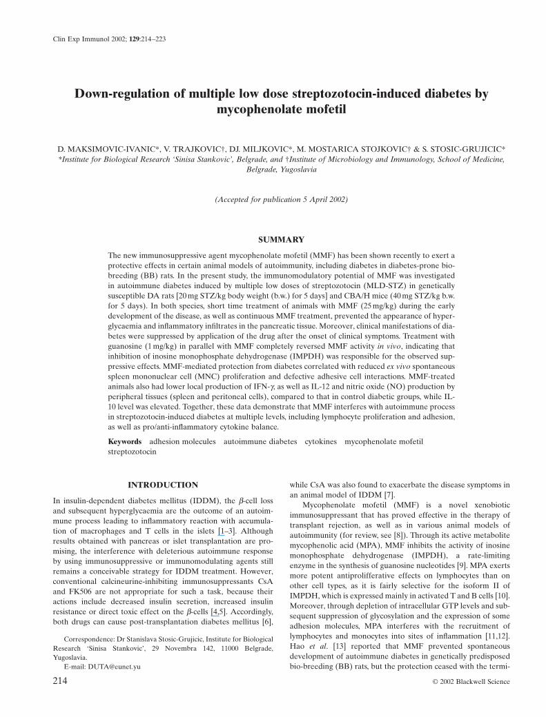
MMF reduces clinical and histological parameters of MLD-STZ-induced diabetesPreliminary experiments have shown that MMF administrationfor 2 weeks did not significantly alter blood glucose level, norglucose tolerance in healthy animals, indicating that the drug itselfwas not toxic to b-cells (data not shown). In order to evaluate theability of MMF to interfere with the diabetogenic process inducedby external agents, streptozotocin, we used both murine and ratmodel of autoimmune diabetes. Although the dose regimens ofb-cell toxin STZ for mice and rats were not identical in bothrodent species, as reported previously [21,22], repeated injectionswith five subtoxic daily doses of STZ induced delayed and chronichyperglycaemia 10–15 days after completion of the treatment(Fig. 1). Continuous application of MMF during the 7 weeks significantly reduced blood glucose level in DA rats (Fig. 1a).Moreover, animals exposed to either early or late 10-day MMFtreatment kept normoglycaemic status even after the treatmentwas terminated (Fig. 1a). Similarly, blood glucose level was significantly lower in CBA/H mice receiving MMF for 2 weeksconcomitantly with MLD-STZ, compared to control diabeticanimals.
The interference of MMF with a pathological process leadingto islet disfunction has further been studied at the level of targettissue. Rats and mice were killed on days 10 and 15 in relation tothe first STZ injection, respectively, the time at which histologicalchanges were maximal [1,3], and immunostaining of pancreata was performed. At this time, analysis of control MLD-STZ-induced diabetic mice revealed heavy mononuclear infiltrates in
25
20
15
10
5
00 1 2 3 4 5 6 7
(a) (b)
Pla
sma
gluc
ose
(mm
ol/l)
15
13
11
9
7
50 1 2 3 4 5 6 7
Weeks
*
*
****
**
*
**
***
** ****
**
Fig. 1. Effects of MMF treatment of rats and mice on MLD-STZ-induced hyperglycaemia. Plasma glucose levels in control DA rats (a),or CBA/H mice (b) receiving MLD-STZ only, or treated with MLD-STZ in conjunction with MMF (25 mg/kg/day) for different timeregimes, as indicated. Results from a representative experiment are presented as mean + s.e.m. for five to six animals/group. *P < 0·05 refersto treatment with MLD-STZ. (a) �, Control; �, MMF 0–9 days; �, MMF 0-49 days; �, MMF 10–19 days. (b) �, Control; �, MMF 0–14days.

Mycophenolate mofetil in streptozotocin-induced diabetes 217
© 2002 Blackwell Science Ltd, Clinical and Experimental Immunology, 129:214–223
100
80
60
40
20
0
ED-1
100
80
60
40
20
0
MHC II
100
80
60
40
20
0
IFN-g
100
80
60
40
20
0
CD4
100
80
60
40
20
0
CD8
100
80
60
40
20
0
CD4
MLD-STZ MLD-STZ+
MMF
Isle
ts i
n s
co
rin
g c
ate
go
ry (
%)
Fig. 2. Effects of MMF treatment of rats and mice on inflammatory infiltration of the islets. Immunohistological analysis of islet-infiltrating cells in DA rats (a) or CBA/H mice (b) receiving MLD-STZ only versus animals treated with MLD-STZ plus MMF from day0 to day 9 (a), or from days 0–14 (b). One day after termination of MMF treatments the sections of pancreata from five animals per groupwere stained with monoclonal antibodies specific for rat (a) ED-1 (a, b, c), MHC II (d, e), CD4 (f,g) and IFN-g (h, i), or murine (b) CD4(j, k, l) and CD8 (m, n) and were graded as described in Materials and methods. Bars indicated the mean of islets + s.e.m. in each cate-gory of mononuclear cell infiltration scored. Photos are paired and represent the staining pattern of control MLD-STZ-treated animals(left and middle panel), or MLD-STZ plus MMF-treated animals (right panel). Note phagocytosis of one cell undergoing apoptosis by theadjacent non-endocrine cell (a), and apoptotic cell surrounded by the infiltrated lymphocytes (j). �, Gr0; �, Gr1; , Gr2.

218 D. Maksimovic-Ivanic et al.
© 2002 Blackwell Science Ltd, Clinical and Experimental Immunology, 129:214–223
some of the islets (Fig. 2b), while in rats small infiltrates were seenrarely and infiltrating cells were scattered throughout the islets(Fig. 2a). Immunohistochemical analysis confirmed our earlierfindings [3,21] that in rats the majority of mononuclear cells par-ticipating in insulitis process were blood-borne ED-1+ and MHCclass II+ macrophages, while CD4+ and IFN-g+ cells were alsodetected (Fig. 2a). In comparison to diabetic rats, pancreatic isletsobtained from animals treated with MMF during the induction ofdiabetes were mainly without infiltrated cells, and only few spa-tially distributed cells positive for these markers could be found ina small number of the islets (Fig. 2a). Similarly, in MMF treatedmice there were much larger numbers of normal islets withoutinflammatory cells, or islets with only mild infiltrates of CD4+ andCD8+ cells (Fig. 2b) in comparison to diabetic control mice. Apop-totic cells with characteristic condensation of nuclear chromatin orfragmented into small apoptotic bodies were noticed in the pan-creatic sections of both diabetic rats (Fig. 2a) and mice (Fig. 2b),but were almost completely absent in MMF-treated animals.Although we were not able to confirm the origin of apoptotic cells,they were most probably b-cells because they did not stain for the markers characteristic for the infiltrated lymphocytes ormacrophages. In general, although MMF prophylaxis could not
suppress insulitis development and b-cell damage completely, it drastically reduced its severity, which was sufficient for the maintenance of normoglycaemic status (Fig. 1).
MMF down-regulates the proliferation and adhesive propertiesof spleen MNCWe next assessed the impact of MMF treatment on spontaneous,as well as mitogen-stimulated lymphoproliferative response inMLD-STZ-treated animals. As shown in Fig. 3 spontaneous pro-liferation of mononuclear spleen cells obtained from diabetic ratsand mice was elevated significantly in comparison with healthyanimals, probably reflecting the activation of autoreactive lym-phocytes [16]. The MMF treatment almost completely revertedthe proliferative activity of splenocytes to the control level.Because it was previously shown that MMF treatment could causeleukopenia in transplant recipients [23], there was a possibilitythat similar MMF effect might be operative in our experiments.However, while diabetic rats were leukopenic, the leucocytenumber in MMF-treated rats was similar to that of healthyanimals (Fig. 3c). Moreover, MMF treatment did not affectConA-induced proliferative response of spleen MNC (Fig. 3d). Toexamine the influence of the drug on adhesive interactions, we
200
150
100
50
150
50
0
100
0
* * *
*
(a)
(c) (d)
(b)
* *
Control MLD-STZ MLD-STZ+
MMF
Control MLD-STZ MLD-STZ+
MMF
% o
f con
trol
Fig. 3. Effect of MMF on spleen mononuclear cell proliferation and peripheral blood leucocytes number. Incorporation of [3H]-thymidineby rat (a) and mouse (b) splenic MNC, as well as ConA-induced proliferation (d) of rat spleen MNC were determined as indicated inMaterials and methods. Results from a representative experiment, presented as percentage of control (controls were 1749 cpm for (a), 8977 cpm for (b) and 254527 cpm for (d)) are given as mean ± s.e.m. for three to five animals per group. Peripheral blood leucocytes number(c) was counted after the completion of continuous MMF treatment in rats. Results, presented as percentage of control (value for controlhealthy animals was 14·59 ¥ 109/l), are given as mean ± s.e.m. for five to six rats per group. *P < 0·05 refers to MLD-SZ treatment.
Mycophenolate mofetil in streptozotocin-induced diabetes 219
© 2002 Blackwell Science Ltd, Clinical and Experimental Immunology, 129:214–223
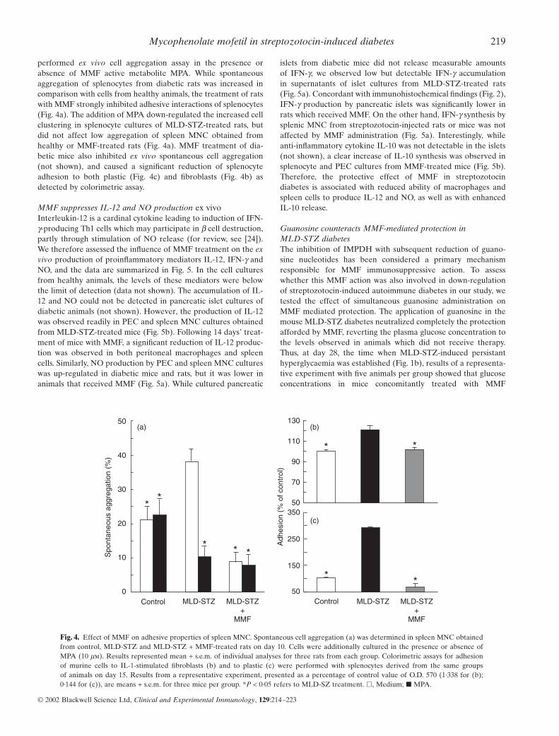
performed ex vivo cell aggregation assay in the presence orabsence of MMF active metabolite MPA. While spontaneousaggregation of splenocytes from diabetic rats was increased incomparison with cells from healthy animals, the treatment of ratswith MMF strongly inhibited adhesive interactions of splenocytes(Fig. 4a). The addition of MPA down-regulated the increased cellclustering in splenocyte cultures of MLD-STZ-treated rats, butdid not affect low aggregation of spleen MNC obtained fromhealthy or MMF-treated rats (Fig. 4a). MMF treatment of dia-betic mice also inhibited ex vivo spontaneous cell aggregation(not shown), and caused a significant reduction of splenocyteadhesion to both plastic (Fig. 4c) and fibroblasts (Fig. 4b) asdetected by colorimetric assay.
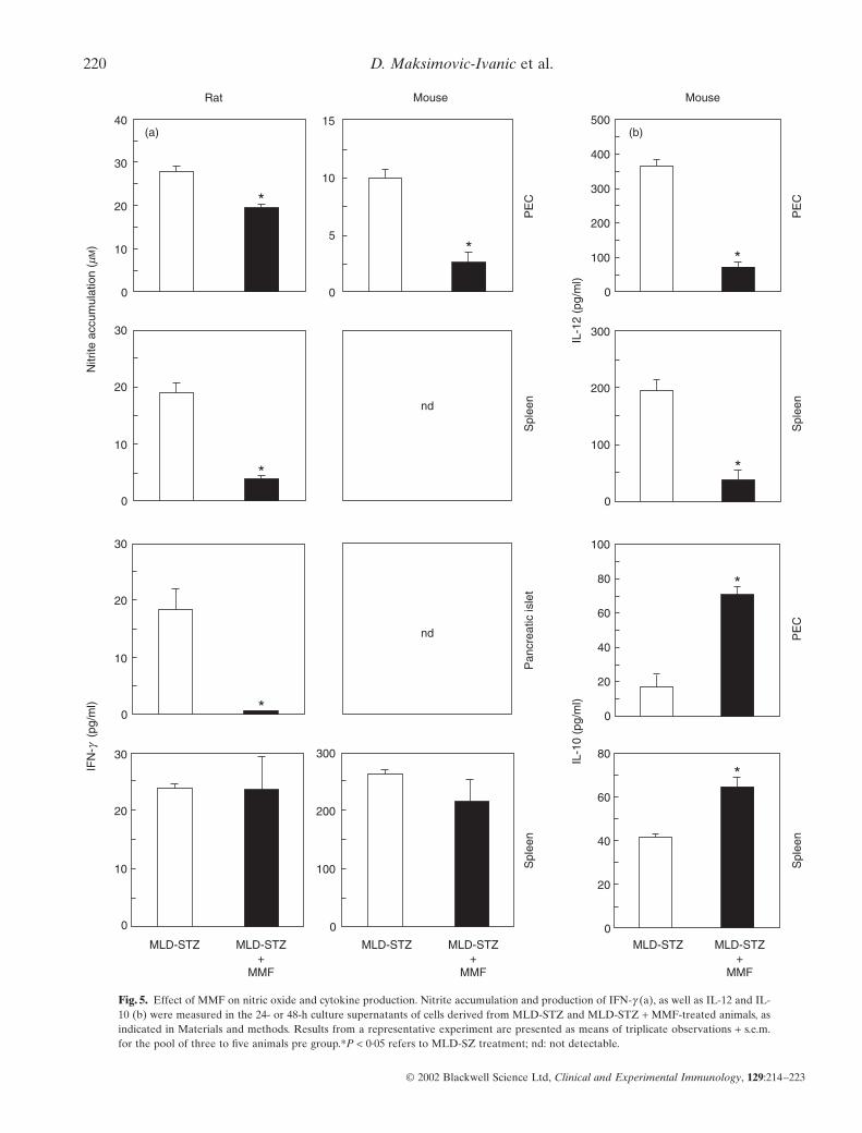
MMF suppresses IL-12 and NO production ex vivoInterleukin-12 is a cardinal cytokine leading to induction of IFN-g-producing Th1 cells which may participate in b cell destruction,partly through stimulation of NO release (for review, see [24]).We therefore assessed the influence of MMF treatment on the exvivo production of proinflammatory mediators IL-12, IFN-g andNO, and the data are summarized in Fig. 5. In the cell culturesfrom healthy animals, the levels of these mediators were belowthe limit of detection (data not shown). The accumulation of IL-12 and NO could not be detected in pancreatic islet cultures ofdiabetic animals (not shown). However, the production of IL-12was observed readily in PEC and spleen MNC cultures obtainedfrom MLD-STZ-treated mice (Fig. 5b). Following 14 days’ treat-ment of mice with MMF, a significant reduction of IL-12 produc-tion was observed in both peritoneal macrophages and spleencells. Similarly, NO production by PEC and spleen MNC cultureswas up-regulated in diabetic mice and rats, but it was lower inanimals that received MMF (Fig. 5a). While cultured pancreatic
islets from diabetic mice did not release measurable amounts of IFN-g, we observed low but detectable IFN-g accumulation in supernatants of islet cultures from MLD-STZ-treated rats(Fig. 5a). Concordant with immunohistochemical findings (Fig. 2),IFN-g production by pancreatic islets was significantly lower inrats which received MMF. On the other hand, IFN-g synthesis bysplenic MNC from streptozotocin-injected rats or mice was notaffected by MMF administration (Fig. 5a). Interestingly, whileanti-inflammatory cytokine IL-10 was not detectable in the islets(not shown), a clear increase of IL-10 synthesis was observed insplenocyte and PEC cultures from MMF-treated mice (Fig. 5b).Therefore, the protective effect of MMF in streptozotocin diabetes is associated with reduced ability of macrophages andspleen cells to produce IL-12 and NO, as well as with enhancedIL-10 release.
Guanosine counteracts MMF-mediated protection in MLD-STZ diabetesThe inhibition of IMPDH with subsequent reduction of guano-sine nucleotides has been considered a primary mechanismresponsible for MMF immunosuppressive action. To assesswhether this MMF action was also involved in down-regulationof streptozotocin-induced autoimmune diabetes in our study, wetested the effect of simultaneous guanosine administration onMMF mediated protection. The application of guanosine in themouse MLD-STZ diabetes neutralized completely the protectionafforded by MMF, reverting the plasma glucose concentration tothe levels observed in animals which did not receive therapy.Thus, at day 28, the time when MLD-STZ-induced persistanthyperglycaemia was established (Fig. 1b), results of a representa-tive experiment with five animals per group showed that glucoseconcentrations in mice concomitantly treated with MMF
50
40
30
20
10
0Control MLD-STZ MLD-STZ
+MMF
MLD-STZ MLD-STZ+
MMF
Spo
ntan
eous
agg
rega
tion
(%)
**
* * *
130
110
90
70
50350
250
150
50Control
Adh
esio
n (%
of c
ontr
ol)
(b)(a)
(c)
*
* *
*
Fig. 4. Effect of MMF on adhesive properties of spleen MNC. Spontaneous cell aggregation (a) was determined in spleen MNC obtainedfrom control, MLD-STZ and MLD-STZ + MMF-treated rats on day 10. Cells were additionally cultured in the presence or absence ofMPA (10 mm). Results represented mean + s.e.m. of individual analyses for three rats from each group. Colorimetric assays for adhesionof murine cells to IL-1-stimulated fibroblasts (b) and to plastic (c) were performed with splenocytes derived from the same groups of animals on day 15. Results from a representative experiment, presented as a percentage of control value of O.D. 570 (1·338 for (b); 0·144 for (c)), are means + s.e.m. for three mice per group. *P < 0·05 refers to MLD-SZ treatment. �, Medium; � MPA.
220 D. Maksimovic-Ivanic et al.
© 2002 Blackwell Science Ltd, Clinical and Experimental Immunology, 129:214–223
Rat
40
30
20
10
0
30
20
10
0
*
* *
*
*
*
*
30
20
10
0 *
30
20
10
0
15
10
5
0
Mouse Mouse
nd
nd
300
200
100
0
Nitr
ite a
ccum
ulat
ion
(mM
)IF
N-g
(pg
/ml)
MLD-STZ MLD-STZ+
MMF
MLD-STZ MLD-STZ+
MMF
MLD-STZ MLD-STZ+
MMF
(a) (b)500
400
300
200
100
300
200
100
100
80
60
40
20
80
60
40
20
0
0
0
0
PE
CS
plee
nS
plee
nP
ancr
eatic
isle
t
PE
CP
EC
Spl
een
Spl
een
IL-1
2 (p
g/m
l)IL
-10
(pg/
ml)
Fig. 5. Effect of MMF on nitric oxide and cytokine production. Nitrite accumulation and production of IFN-g (a), as well as IL-12 and IL-10 (b) were measured in the 24- or 48-h culture supernatants of cells derived from MLD-STZ and MLD-STZ + MMF-treated animals, asindicated in Materials and methods. Results from a representative experiment are presented as means of triplicate observations + s.e.m.for the pool of three to five animals pre group.*P < 0·05 refers to MLD-SZ treatment; nd: not detectable.
Mycophenolate mofetil in streptozotocin-induced diabetes 221
and guanosine and in control diabetic mice were similar(13·1 ± 0·5 mmol/l and 12·9 ± 1·4 mmol/l, respectively), in contrastto MMF-treated (8·1 ± 0·5 mmol/l), or normal healthy mice(8·2 ± 0·2 mmol/l), P < 0·05. It should be noted that guanosine didnot have any effect on blood glucose levels in healthy or MLD-STZ-treated animals in the absence of MMF (not shown). Thesedata strongly suggest that the beneficial effect of MMF in ourexperiments was mediated exclusively through interference withde novo synthesis of guanosine nucleotides.
DISCUSSION
In the present study the immunosuppressive agent MMF exerteda strong long-lasting protection in a streptozotocin model ofautoimmune diabetes in rodents. Destructive autoimmune re-sponse leading to loss of pancreatic b-cell function was impairedby the drug at multiple levels – the lymphocyte proliferation andadhesion, followed by their infiltration into pancreas, as well asthe production of important proinflammatory mediators IL-12and NO, were all inhibited by MMF treatment. While the bene-ficial effect of MMF in spontaneously diabetic BB rats has beenpreviously reported [13], this is the first study providing insightsinto the mechanisms behind antidiabetogenic effect of thisimmunosuppressant.
The well-documented antiproliferative action of MMF affectsmainly activated T and B cells due to drug selectivity for IMPDHisoform II, which is expressed in activated lymphocytes. Accord-ingly, the spontaneous, presumably self-antigen driven splenocyteproliferation in our study was suppressed by MMF treatment. On the other hand, the proliferative response to ConA was notimpaired in MMF-treated animals, indicating that naive T lym-phocytes retained their capacity for mitogen-induced activationfollowing the treatment applied. Moreover, the development of leukopenia in MLD-STZ-injected rats [25] was prevented completely by MMF treatment in our experiments. A similarMMF-mediated protection from leukopenia associated withautoimmunity was observed previously in a murine model of sys-temic lupus erythematosus [26]. In addition to impaired sponta-neous proliferation, the mononuclear cells from MMF-treatedanimals exhibited reduced ability for intercellular adhesion oradhesion to plastic, which is the first report on such MMF actionin an autoimmune disease. This is consistent with the findings thatMPA-mediated GTP depletion and subsequent impairment of N-linked glycosilation decreases the in vitro binding activity of Tcell and monocyte adhesion molecules [11,27]. Interestingly, theincreased adhesion of mononuclear cells from diabetic animalswas down-regulated by MMF active component MPA in vitro,indicating that the drug presence during leucocyte activation was not required for its anti-adhesive action. This ability of MPAto interfere with the adhesion of already activated lymphocytesand macrophages might be an important mechanism for theantidiabetogenic effect of late MMF administration, presumablypreventing the recruitment of activated immune cells into the site of inflammation. A similar MMF-mediated reduction ofinflammatory infiltration of the target tissue was recentlyobserved in rat model of EAE, which resulted in the recoveryfrom already established disease [28]. In addition, recentlydescribed reduction by MPA of selectin expression on endothe-lial cells [12] might contribute further to drug-mediated pro-tection of pancreatic islets from inflammatory infiltration andautoimmune destruction.
The immunohistochemical examination confirmed that MPA-mediated interference with the proliferation of autoreactive lymphocytes and leucocyte adhesiveness correlated with reducedseverity of pancreatic inflammation. The protective effect ofMMF in rats was associated with reduced number of ED-1+ andCD4+ cells in the islets, which is consistent with the findings thatED-1+ macrophages are the first cells to enter the endocrine pan-creas in rat MLD-STZ diabetes [21], while subsequent infiltrationof CD4+ cells is necessary for the development of disease [3,29].The observation that MMF-injected animals had lower number ofMHC class II+ cells in their islets is important in light of the datadescribing protection from MLD-STZ insulitis by administrationof anti-class II antibodies [30]. Because MHC class II expressionduring streptozotocin-induced insulitis seems to be restricted tomonocytes/macrophages infiltrating the pancreas and presumablypresenting b-cell antigens [31], the MHC class II+ cells in our studyare likely to correspond to ED-1+ macrophages. While CD8+ Tcells apparently do not contribute to the induction of MLD-STZdiabetes in rats [32], both CD4+ and CD8+ T lymphocytes arerequired for disease development in mouse [29,33,34], and theirnumbers were decreased markedly in the islets of MLD-STZ-treated mice after MMF treatment. Recent findings indicate thatthe death of b-cells, as a final consequence of the autoimmuneattack in diabetes, can take a form of apoptosis [35]. Indeed, wehave observed numerous cells with the morphological appearanceof apoptotic ones in the islets of diabetic, but not in MMF-treatedanimals, thus suggesting further another level of MMF mediatedprotection.
In addition to impaired lymphocyte proliferation and leuco-cyte adhesion, an important feature of MMF anti-diabetogeniceffect is the reduced production of macrophage proinflammatorymediators IL-12 and NO. Interleukin-12 is a heterodimericcytokine required for the development of destructive Th1 pheno-type of auto-reactive T cells [36], with its inducible p40 chainobserved readily during progression of auto-immune diabetes inspontaneously diabetic NOD mice and BB rats, as well as inMLD-STZ-treated mice ([37–39] and the present study). Some-what surprisingly, down-regulation of IL-12p40 production inperitoneal and spleen cells of MMF-treated mice was associatedwith unaltered splenocyte production of the signature Th1cytokine IFN-g. However, the amount of IFN-g produced duringthe priming of T cells does not necessarily correlate with theirpolarization towards Th1 phenotype. The striking example is theaction of IL-18, which in the absence of IL-12 markedly enhancesIFN-g secretion in the first contact of T cells with antigen, butleads eventually to the development on IFN-g non-producing Th2cells [40]. Therefore, the reduction of IL-12 production in MMF-treated mice could still interfere with the induction of Th1response and might partially account for the observed decreaseof IFN-g synthesis within the microenvironment of the pancreas,even in the absence of IFN-g down-regulation during priming inthe spleen. The destructive actions of Th1 cell-derived IFN-ginclude the induction of macrophage free radical NO, which isdirectly toxic to b-cells [41]. Regardless of unchanged splenicIFN-g production, the NO production by spleen and peritonealmacrophages was abolished almost completely by MMF treat-ment in our study. While, due to the low sensitivity of the methodemployed for NO detection, we could not observe any NO releasein the cultured islets, it seems conceivable to assume that impairedNO synthesis by peripheral macrophages might reflect a similardisability of the macrophages infiltrating the target tissue. Inter-
© 2002 Blackwell Science Ltd, Clinical and Experimental Immunology, 129:214–223
222 D. Maksimovic-Ivanic et al.
estingly, although MPA suppressed NO production by endothe-lial cells in vitro [42], it did not affect either IL-12 or NO pro-duction in IFN-g + LPS-stimulated macrophages or splenocytes(our unpublished data). Thus, it appears that the drug actedthrough IFN-g-independent interference with cytokine networkregulating macrophage activation, rather than direct modulationof macrophage activity. An intriguing possibility for such MMFeffect is the in vivo up-regulation of macrophage-deactivatingcytokine IL-10, observed previously in endotoxaemic mice [43],as well as in the present study.
Finally, our data indicate that MMF exerted the protection in MLD-STZ diabetes through its best defined intracellularaction – the inhibition of guanosine nucleotide synthesis, reportedpreviously to cause the inhibition of lymphocyte proliferation andadhesion in vitro (for review, see [8]). Recent evidence suggeststhat guanosine depletion might affect GTP binding protein-dependent signal transduction machinery such as MAP kinasepathway [44], which could be a mechanism for MMF interferencewith cytokine and NO synthesis. Having in mind a favourableratio between the beneficial and the side-effects of MMF, ourstudy indicate that its potential use for treatment of humanIDDM is worthy of further investigation.
ACKNOWLEDGEMENTS
This work was supported partly by grants from the Ministry of Science andTechnology, Republic of Serbia, Yugoslavia. MMF was kindly provided byRoche Representative Office, Belgrade.
REFERENCES
1 Kolb-Bachofen V, Epstein S, Kiesel U et al. Low dose streptozotocininduced diabetes in mice. Electron microscopy reveals single-cellinsulitis before diabetes onset. Diabetes 1988; 37:21–7.
2 Huang X, Yuan J, Goddard A et al. Interferon expression in the pancreas of patients with type I diabetes. Diabetes 1995; 44:658–64.
3 Lukic ML, Stosic-Grujicic S, Shahin A. Effector mechanisms in lowdose streptozotocin induced diabetes. Dev Immunol 1998; 6:119–28.
4 Drachenberg CB, Klassen DK, Weir MR et al. Islet cell damage associated with tacrolimus and cyclosporine: morphological features in pancreas allograft biopsies and clinical correlation. Transplantation1999; 68:396–402.
5 Lohmann T, List C, Lamesch P et al. Diabetes mellitus and islet cellspecific autoimmunity as adverse effects of immunsuppressive therapyby FK506/tacrolimus. Exp Clin Endocrinol Diabetes 2000; 108:347–52.
6 Jindal RM, Sidner RA, Milgrom ML. Post-transplant diabetes mellitus. The role of immunosuppression. Drug Saf 1997; 16:242–57.
7 Sai P, Maugendre D, Loreal O et al. Effects of cyclosporin on auto-immune diabetes induced in mice by streptozotocin. Diabetes Metabolisme 1988; 14:455–62.
8 Allison AC, Eugui EM. Mycophenolate mofetil and its mechanisms ofaction. Immunopharmacology 2000; 47:85–118.
9 Brazelton TR, Morris RE. Molecular mechanisms of action of newxenobiotic immunosuppressive drugs: tacrolimus (FK506), sirolimus(rapamycin), mycophenolate mofetil and leflunomide. Curr OpinImmunol 1996; 8:710–20.
10 Hager PW, Collart FR, Huberman E et al. Recombinant human inosinemonophosphate dehydrogenase type I and II proteins. Purification and characterization of inhibitor binding. Biochem Pharmacol 1995;49:1323–9.
11 Laurent AF, Dumont S, Poindron P et al. Mycophenolic acid sup-presses protein N-linked glycosylation in human monocytes and theiradhesion to endothelial cells and to some substrates. Exp Hematol1996; 24:59–67.
12 Blaheta RA, Leckel K, Wittig B et al. Mycophenolate mofetil impairstransendothelial migration of allogenic CD4 and CD8 T-cells. Trans-plant Proc 1999; 31:1250–2.
13 Hao L, Chan S-M, Lafferty KJ. Mycophenolate mofetil can prevent thedevelopment of diabetes in BB rats. Ann NY Acad Sci 1993; 696:328–32.
14 Like AA, Rosini AA. Streptozotocin-induced pancreatic insulitis. anew model of diabetes mellitus. Science 1976; 193:415–7.
15 Herold KC, Vezys V, Sun Q et al. Regulation of cytokine productionduring development of autoimmune diabetes induced with multiplelow doses of streptozotocin. J Immunol 1996; 156:3521–7.
16 Stosic-Grujicic S, Maksimovic D, Badovinac V et al. Antidiabetogeniceffect of pentoxifylline is associated with systemic and target tissuemodulation of cytokines and nitric oxide production. J Autoimmun2001; 16:47–58.
17 Stosic-Grujicic S, Dimitrijevic M, Bartlett R. Leflunomide protect micefrom multiple low dose of streptozotocin (MLD-SZ)-induced insulitisand diabetes. Clin Exp Immunol 1999; 117:44–50.
18 Draskovic-Pavlovic B, Van Der Laan LJW, Pejnovic N et al. Differen-tial effects of anti-rat CD11b monoclonal antibodies on granulocyteadhesiveness. Immunology 1999; 96:83–9.
19 Pechold K, Patterson NB, Craighead N et al. Inflammatory cytokinesIFN-g plus TNF-a induce regulated expression of CD80 (B7–1) but notCD86 (B7–2) on murine fibroblasts. J Immunol 1997; 158:4921–9.
20 Hibbs JB, Taintor Z, Vavrin E et al. Nitric oxide: a cytotoxic activatedmacrophage effector molecule. Biochem Biophys Res Commun 1989;157:87–94.
21 Lukic ML, Al-Sharif R, Mostarica M et al. Immunological basis of thestrain differences in susceptibility to low-dose streptozotocin-induceddiabetes in rats. In: Imhof BA Berrih-Aknin S Ezine S., eds Lymphatictissues and in vivo immune responses. New York: Marcel Dekker,1991:643–7.
22 Lukic ML, Stosic-Grujicic S, Ostojic N et al. Inhibition of nitric oxidegeneration affects the induction of diabetes by streptozotocin in mice.Biochem Biophys Res Commun 1991; 178:913–20.
23 Squifflet JP, Backman L, Claesson K et al. Dose optimization ofmycophenolate mofetil when administered with a low dose oftacrolimus in cadaveric renal transplant recipients. Transplantation2001; 72:63–9.
24 Santamaria P. Effector lymphocytes in autoimmunity. Curr OpinImmunol 2001; 13:663–9.
25 Oon BB, Muggleston D, Warley A. A diet enriched in essential fattyacids protects against the loss of lymphocytes which occurs in rats suffering from streptozotocin-induced diabetes. Exp Physiol 1992;77:185–90.
26 Corna D, Morigi M, Facchinetti D et al. Mycophenolate mofetil limitsrenal damage and prolong life in murine lupus autoimmune disease.Kidney Int 1997; 51:1583–9.
27 Allison AC, Kowalski WJ, Muller CJ et al. Mycophenolic acid and brequinar, inhibitors of purine and pyrimidine synthesis, block the glycosylation of adhesion molecules. Transplant Proc 1993; 25:67–70.
28 Tran GT, Carter N, Hodgkinson SJ. Mycophenolate mofetil treatmentaccelerates recovery from experimental allergic encephalomyelitis. IntImmunopharmacol 2001; 1:1709–23.
29 Elliott JI, Dewchand H, Altmann DM. Streptozotocin-induced diabetes in mice lacking ab T cells. Clin Exp Immunol 1997; 109:116–20.
30 Kiesel U, Kolb H. Suppressive effect of antibodies to immune responsegene product on the development of low-dose streptozotocin-induceddiabetes. Diabetes 1983; 32:869–71.
31 Farr AG, Mannschreck JW, Anderson SK. Expression of class II MHCantigens in murine pancreas after streptozotocin-induced insulitis. Diabetes 1988; 37:1373–9.
32 Stosic-Grujicic S, Mijatovic S, Ejdus L et al. Enhancement of Th2-typeactivity downregulated diabetes induction. Transplant Proc 1996; 28:3259.
33 Herold KC, Montag AG, Fitch FW. Treatment with anti-T-lymphocyte
© 2002 Blackwell Science Ltd, Clinical and Experimental Immunology, 129:214–223
Mycophenolate mofetil in streptozotocin-induced diabetes 223
© 2002 Blackwell Science Ltd, Clinical and Experimental Immunology, 129:214–223
antibodies prevents induction of insulitis in mice given multiple dosesof streptozotocin. Diabetes 1987; 36:796–801.
34 Herold KC, Bluestone JA, Montag AG et al. Prevention of auto-immune diabetes with nonactivating anti-CD3 monoclonal antibody.Diabetes 1992; 41:385–91.
35 O’Brien BA, Harmon BV, Cameron DP et al. Beta-cell apoptosis isresponsible for the development of IDDM in the multiple low-dosestreptozotocin model. J Pathol 1996; 178:176–81.
36 Gately MK, Renzetti LM, Magram J et al. The interleukin-12/interleukin-12-receptor system. Role in normal and pathologicimmune responses. Annu Rev Immun 1998; 16:495–521.
37 Rothe H, Burkart V, Faust A et al. Interleukin-12 gene expression isassociated with rapid development of diabetes mellitus in non-obesediabetic mice. Diabetologia 1996; 39:119–22.
38 Zipris D, Greiner DL, Malkani S et al. Cytokine gene expression inislets and thyroids of BB rats. IFN-gamma and IL-12p40 mRNAincrease with age in both diabetic and insulin-treated nondiabetic BBrats. J Immunol 1996; 156:1315–21.
39 Li X, Kaminski NE, Fischer LJ. Examination of the immunosuppres-
sive effect of delta9-tetrahydrocannabinol in streptozotocin-inducedautoimmune diabetes. Int Immunopharmacol 2001; 1:699–712.
40 Xu D, Trajkovic V, Hunter D et al. IL-18 induces the differentiation ofTh1 or Th2 cells depending upon cytokine milieu and genetic back-ground. Eur J Immunol 2000; 30:3147–56.
41 Rabinovitch A, Suarez-Pinzon WL. Cytokines and their role in pan-creatic islet b-cell destruction and insulin-dependent diabetes mellitus.Biochem Pharmacol 1998; 55:1139–49.
42 Senda M, DeLustro B, Eugui E et al. Mycophenolic acid, an inhibitorof IMP dehydrogenase that is also an immunosuppressive agent, sup-presses the cytokine- induced nitric oxide production in mouse and ratvascular endothelial cells. Transplantation 1995; 60:1143–8.
43 Durez P, Appelboom T, Pira C et al. Antiinflammatory properties ofmycophenolate mofetil in murine endotoxemia: inhibition of TNF-aand upregulation of IL-10 release. Int J Immunopharmac 1999;21:581–7.
44 Vallee S, Fouchier F, Braguer D et al. Ribavirin-induced resistance toheat shock, inhibition of the Ras-Raf-1 pathway and arrest in G(1).Eur J Pharmacol 2000; 404:49–62.
Related Documents