PO Box 2345, Beijing 100023, China World J Gastroenterol 2005;11(42):6571-6576 www.wjgnet.com World Journal of Gastroenterology ISSN 1007-9327 [email protected] © 2005 The WJG Press and Elsevier Inc. All rights reserved. ELSEVIER • REVIEW • HEPATIC TUMORS Although relatively uncommon in Western countries, hepatocellular carcinoma (HCC) is probably the most common solid cancer in the world, with an estimated incidence of at least one million new patients per year [1,2] . The optimal treatment for HCC is surgical excision with a curative intent, but only 5-15% of newly diagnosed patients undergo potentially curative resection [3] . Patients with disease confined to the liver may not be candidates for resection because of multifocal disease, or an inadequate hepatic functional reserve capacity related to co-existent cirrhosis may contraindicate resection. As there are few other curative treatment options for patients with unresectable liver disease, HCC is one of the most lethal human malignancies, with a mortality rate of 94% [4] . The liver is second only to lymph nodes as a site of metastases from other solid cancers [5] , and may be the only site of metastatic disease particularly in patients with colorectal adenocarcinoma [6] . However, fewer than 10-15% of patients with liver metastases are candidates for resection for the same reasons as those regarding HCC. The majority of patients with primary or metastatic hepatic malignancies who are not candidates for complete surgical resection therefore require novel treatment modalities to control and potentially cure their disease [2,7] . Cirrhosis may be another variable that places such patients at the highest risk [2] . Patients in class C of the Child-Pugh Classification (Table 1) have the highest mortality and morbidity rate following all treatments, particularly surgical procedures [8,9] , and so most centers have shifted away from open liver surgery and are attempting other approaches. The treatment of hepatic tumors in cirrhotic and non-cirrhotic patients is a major decision-making issue for oncologists and surgeons, and the high mortality rate of open liver surgery in cirrhotic patients has spurred physicians to seek new modalities [10] . We here outline the immunological and genetic techniques available for the treatment of liver tumors, and propose a new immunologico-clinical algorithm using immunological therapy to debulk the mass, kill micro- metastases, and allow a lower dose of chemotherapy to achieve better cytoreduction. Maurizio Chiriva-Internati, Department of Microbiology and Immunology, Texas Tech University Health Sciences Center and Southwest Cancer Treatment and Research Center, Lubbock, TX 79430, United States Fabio Grizzi, Scientific Direction, Istituto Clinico Humanitas, 20089 Rozzano, and Foundation “M. Rodriguez” – Institute for Quantitative Measures in Medicine, 20100 Milan, Italy Cynthia A Jumper, Department of Internal Medicine, Texas Tech University Health Sciences Center and Southwest Can- cer Treatment and Research Center, Lubbock, TX 79430, United States Everardo Cobos, Department of Internal Medicine, Texas Tech University Health Sciences Center and Southwest Can- cer Treatment and Research Center, Lubbock, TX 79430, United States Paul L Hermonat, Department of Internal Medicine, University of Arkansas for Medical Sciences, Little Rock, Arkansas 72205, United States Eldo E Frezza, Department of Surgery, Texas Tech University Health Sciences Center and Southwest Cancer Treatment and Research Center, Lubbock, TX 79430, United States Correspondence to: Maurizio Chiriva-Internati, PhD, Department of Microbiology and Immunology, Texas Tech University Health Sciences Center, Room 5B191, Lubbock, TX 79430-6591, United States. [email protected] Telephone: +1-806-743-4057 Fax: +1-806-743-2334 Received: 2005-01-22 Accepted: 2005-02-18 Abstract Although multiple options for the treatment of liver tumors have often been described in the past, including liver resection, radiofrequency ablation with or without hepatic pump insertion, laparoscopic liver resection and the use of chemotherapy, the potential of immunotherapy and gene manipulation is still largely unexplored. Immunological therapy by gene manipulation is based on the interaction between virus-based gene delivery systems and dendritic cells. Using viruses as vectors, it is possible to transduce dendritic cells with genes encoding tumor-associated antigens, thus inducing strong humoral and cellular immunity against the antigens themselves. Both chemotherapy and radiation therapy have the disadvantage of destroying healthy cells, thus causing severe side-effects. We need more precisely targeted therapies capable of killing cancer cells while sparing healthy cells. Our goal is to establish a new treatment for solid liver tumors based on the concept of cytoreduction, and propose an innovative algorithm. © 2005 The WJG Press and Elsevier Inc. All rights reserved. Key words: Liver; Tumors; Surgery; Dendritic cell; Cytor- eduction; Immunotherapy; Gene manipulation Immunological treatment of liver tumors Maurizio Chiriva-Internati, Fabio Grizzi, Cynthia A Jumper, Everardo Cobos, Paul L Hermonat, Eldo E Frezza Chiriva-Internati M, Grizzi F, Jumper CA, Cobos E, Hermonat PL, Frezza EE. Immunological treatment of liver tumors. World J Gastroenterol 2005; 11(42): 6571-6576 http://www.wjgnet.com/1007-9327/11/6571.asp

Welcome message from author
This document is posted to help you gain knowledge. Please leave a comment to let me know what you think about it! Share it to your friends and learn new things together.
Transcript
PO Box 2345, Beijing 100023, China World J Gastroenterol 2005;11(42):6571-6576www.wjgnet.com World Journal of Gastroenterology ISSN [email protected] © 2005 The WJG Press and Elsevier Inc. All rights reserved.E L S E V I E R
• REVIEW •
HEPATIC TUMORSAlthough relatively uncommon in Western countries, hepatocellular carcinoma (HCC) is probably the most common solid cancer in the world, with an estimated incidence of at least one million new patients per year[1,2]. The optimal treatment for HCC is surgical excision with a curative intent, but only 5-15% of newly diagnosed patients undergo potentially curative resection[3]. Patients with disease confi ned to the liver may not be candidates for resection because of multifocal disease, or an inadequate hepatic functional reserve capacity related to co-existent cirrhosis may contraindicate resection. As there are few other curative treatment options for patients with unresectable liver disease, HCC is one of the most lethal human malignancies, with a mortality rate of 94%[4].
The liver is second only to lymph nodes as a site of metastases from other solid cancers[5], and may be the only site of metastatic disease particularly in patients with colorectal adenocarcinoma[6]. However, fewer than 10-15% of patients with liver metastases are candidates for resection for the same reasons as those regarding HCC. The majority of patients with primary or metastatic hepatic malignancies who are not candidates for complete surgical resection therefore require novel treatment modalities to control and potentially cure their disease[2,7].
Cirrhosis may be another variable that places such patients at the highest risk[2]. Patients in class C of the Child-Pugh Classification (Table 1) have the highest mortality and morbidity rate following all treatments, particularly surgical procedures[8,9], and so most centers have shifted away from open liver surgery and are attempting other approaches. The treatment of hepatic tumors in cirrhotic and non-cirrhotic patients is a major decision-making issue for oncologists and surgeons, and the high mortality rate of open liver surgery in cirrhotic patients has spurred physicians to seek new modalities[10].
We here outline the immunological and genetic techniques available for the treatment of liver tumors, and propose a new immunologico-clinical algorithm using immunological therapy to debulk the mass, kill micro-metastases, and allow a lower dose of chemotherapy to achieve better cytoreduction.
Maurizio Chiriva-Internati, Department of Microbiology and Immunology, Texas Tech University Health Sciences Center and Southwest Cancer Treatment and Research Center, Lubbock, TX 79430, United StatesFabio Grizzi, Scientific Direction, Istituto Clinico Humanitas, 20089 Rozzano, and Foundation “M. Rodriguez” – Institute for Quantitative Measures in Medicine, 20100 Milan, ItalyCynthia A Jumper, Department of Internal Medicine, Texas Tech University Health Sciences Center and Southwest Can-cer Treatment and Research Center, Lubbock, TX 79430, United StatesEverardo Cobos, Department of Internal Medicine, Texas Tech University Health Sciences Center and Southwest Can-cer Treatment and Research Center, Lubbock, TX 79430, United StatesPaul L Hermonat, Department of Internal Medicine, University of Arkansas for Medical Sciences, Little Rock, Arkansas 72205, United StatesEldo E Frezza, Department of Surgery, Texas Tech University Health Sciences Center and Southwest Cancer Treatment and Research Center, Lubbock, TX 79430, United StatesCorrespondence to: Maurizio Chiriva-Internati , PhD, Department of Microbiology and Immunology, Texas Tech University Health Sciences Center, Room 5B191, Lubbock, TX 79430-6591, United States. [email protected]: +1-806-743-4057 Fax: +1-806-743-2334Received: 2005-01-22 Accepted: 2005-02-18
AbstractAlthough multiple options for the treatment of liver tumors have often been described in the past, including liver resection, radiofrequency ablation with or without hepatic pump insertion, laparoscopic liver resection and the use of chemotherapy, the potential of immunotherapy and gene manipulation is still largely unexplored. Immunological therapy by gene manipulation is based on the interaction between virus-based gene delivery systems and dendritic cells. Using viruses as vectors, it is possible to transduce dendritic cells with genes encoding tumor-associated antigens, thus inducing strong humoral and cellular immunity against the antigens themselves. Both chemotherapy and radiation therapy have the disadvantage of destroying healthy cells, thus causing severe side-effects. We need more precisely targeted therapies capable of killing cancer cells while sparing healthy cells. Our goal is to establish a new treatment for solid liver tumors based on the concept of cytoreduction, and propose an innovative algorithm.
© 2005 The WJG Press and Elsevier Inc. All rights reserved.
Key words: Liver; Tumors; Surgery; Dendritic cell; Cytor-eduction; Immunotherapy; Gene manipulation
Immunological treatment of liver tumors
Maurizio Chiriva-Internati, Fabio Grizzi, Cynthia A Jumper, Everardo Cobos, Paul L Hermonat, Eldo E Frezza
Chiriva-Internati M, Grizzi F, Jumper CA, Cobos E, Hermonat PL, Frezza EE. Immunological treatment of liver tumors. World J Gastroenterol 2005; 11(42): 6571-6576 http://www.wjgnet.com/1007-9327/11/6571.asp

6572 ISSN 1007-9327 CN 14-1219/ R World J Gastroenterol November 14, 2005 Volume 11 Number 42
WHAT IS THE ROLE OF SURGERY?Open surgeryComplete surgical resection of primary or secondary liver tumors is the gold standard of surgical therapy[2,8,9], but it has fallen out of favor because of complications related to bleeding and liver failure. Furthermore, the time of the associated hospitalization is not cost-effective in the context of the new health plan insurance capitation systems.
Underlying anatomical and physiological limitations may exclude the use of complete surgical resection but, when complete or partial resection is plausible, the approach of choice is either the traditional open technique (wedge resection, segmentectomy or major lobectomy) or the laparoscopic technique. Laparoscopic liver surgery has become feasible with the improvement in laparoscopic techniques and the development of new and dedicated technologies[9]. There are benefi ts common to all endoscopic procedures, and the choice of the approach to hepatic resection is usually made by both the surgeon and the patient.
Laparoscopic surgeryThe laparoscopic method is useful in oncological therapy, as it allows abdominal exploration and the visualization of the tumor itself. Specimen collection is another key benefi t, and can range from a lymph node biopsy in the peritoneum or retro-peritoneum, to scraping the peritoneum in the abdominal wall. Laparoscopy allows direct visualization of the organs and biopsy. The liver is a large organ, and can therefore be visualized quite well, particularly the anterior section, although it is laparoscopically more difficult to visualize the posterior section of the retroperitoneal area of the right lobe. Anatomically, the left side of the liver is not hard to mobilize by dissecting the left triangular ligament and flipping the left side of the liver over the midline, but it is more complicated to achieve the same result on the right side where segments VI and VII (the lateral segments) and segment VIII are harder to visualize posteriorly, and so intraoperative ultrasound has been introduced to improve the visualization of tumors in these segments[9].
Radiofrequency ablationThis is a thermal technique designed to cause localized tumor destruction by heating the tumoral tissue to temperatures of more than 50 °C. The methodology has been previously described by our group[8,11], and has been found to be safe and effective in the treatment of single
tumors of <5 cm with curative intent, or the cytoreduction of multiple or larger tumors.
Percutaneous ethanol injection (PEI)This is usual ly perfor med under transabdominal ultrasonographic guidance, and consists of intra-tumorally injecting 5-10 mL of ethanol twice a week. Patient compliance has been a problem because of the number of injections required and the associated pain. As PEI requires multiple treatment sessions and is associated with a high local recurrence rate, it should only be considered in the case of tumors with a diameter of less than 1.5 cm.
CryosurgeryThis has been used to treat patients with unresectable primary and metastatic liver tumors for the last 20 years. Most of the scientific data concerning local tumor recurrences and complications after cryosurgery comes from patients treated for colorectal cancer liver metastases[8].
CLINICAL ALGORITHM FOR SOLID LIVER TUMORSThe pros and cons of liver surgery and the new clinical algorithm used for the treatment of liver tumors will be briefl y discussed[8], considering only the patients with Child–Pugh class A or B cirrhosis, because those with advanced liver cirrhosis (Child–Pugh class C) would probably receive no survival benefi t and would be at a disproportionately increased risk of interventional therapy. The patients in the two groups will belong to one of the following four categories: (1) Those with stage I, primary liver tumors will be evaluated for liver resection or radiofrequency ablation (RFA); (2) Those with stage II and III primary liver tumors will undergo complete resection, if anatomically possible, or partial resection with RFA, or RFA alone; the patients with vascular invasion will also receive a hepatic arterial pump (HAP); (3) The patients with stage IV primary liver tumors or liver metastases of other than colorectal origin (endocrine, breast) will only be treated with RFA and a HAP; (4) The patients with colorectal metastases will undergo complete resection if possible, or partial resection with RFA, or RFA alone, and all will receive a HAP.
After a median follow-up of 20 mo in patients with unresectable liver disease, the addition of adjuvant HAP therapy to cryoreduction decreased all recurrences from 77% to 49% and decreased liver recurrences from 67% to 38%. This, and other multi-approaches (RFA and HAP therapy) to the treatment of partially resectable or unresectable liver disease, is promising and deserves further investigation.
IMMUNOTHERAPY AND NEOPLASTIC LIV-ER DISEASEMost cancer pat ients a re cur rent ly t rea ted wi th some combination of surgery, radiation therapy and
A B CAscites None Controlled UncontrolledBilirubin (mmol/L) <2.0 2.0-2.5 >3.0Encephalopathy None Minimal AdvancedPT (s prolonged) <4.0 4.0-6.0 >6.0INR <2.0 2.0-3.0 >3Albumin (g/L) >3.5 3.0-3.5 <3.0
Table 1 Child-Pugh classifi cation
Chiriva-Internati M et al. Liver cancer immunotherapy, biological therapy 6573
chemotherapy, but both chemo- and radiation-therapy have the disadvantage of destroying healthy cells and this causes severe side effects. The possibility of destroying more cancer cells by increasing the chemotherapeutic dose or radiation exposure is limited by the non-specifi c organ toxicity of these therapies and the relatively old age of most patients. We therefore need more precisely targeted therapies capable of killing cancer cells while sparing healthy cells.
One possible answer is immunological therapy, which is not only more specific and less toxic, but may also induce memory responses that could yield long-term tumor immunosurveillance and reduce the incidence of relapses, thus increasing long-term disease-free survival. Immunological therapy may be adoptive[10,12] in which case the patients’ white blood cells are coupled with a naturally producing growth factor to enhance their cancer fi ghting capacity, or passive[13], with immunity being acquired as a result of the transfer of antibodies from a healthy donor. However, the possibility of successfully implementing these therapies rests on the existence of tumor-specific antigens, and suitable antigens have been hard to come by because of the complex process required to validate them[14–18].
Immunotherapy refers to any approach aimed at mo-bilizing or manipulating a patient’s immune system to treat or cure disease[19], and immunological therapy by means of gene manipulation is based on the interaction between virus-based gene delivery systems and dendritic cells (DCs). Using viruses as vectors, it is possible to transduce DCs with genes encoding tumor-associated antigens (TAA), thus inducing a robust immune response[20,21].
A number of studies have established the role played by DCs in the immune system, and provided a rationale for using them as natural adjuvants for cancer im-munotherapy[20-22]. Previous studies have concentrated on identifying the proliferating progenitors of DCs within the small CD34+ sub-fraction of cells in human blood[23]. These cells can be stimulated by cytokines (particularly by GM-CSF and TNF-alpha) to differentiate into DCs in vitro over a period of 1 wk[24]. It has also been more recently found that the combination of GM-CSF and IL-4 facilitates the generation of signifi cantly larger numbers of DCs from monocytes/macrophages, which have equal or greater stimulatory activity in mixed lymphocyte reactions, and a greater capacity to present soluble protein antigens
than CD34+ cell-derived DCs[23,24].
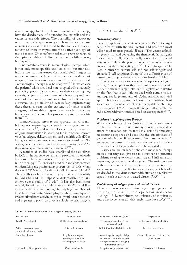
Gene manipulationGene manipulation transmits new genes/DNA into target cells infected with the viral vector, and has been most widely used to treat genetic diseases. The vector unloads its genetic material containing the therapeutic human gene into the target cell, which is fi nally restored to its normal state as a result of the generation of a functional protein encoded by the therapeutic gene[24,25]. The technique can be used in cancer to activate self and non-self antigens and enhance T cell responses. Some of the different types of viruses used as gene therapy vectors are listed in Table 2.
There are also various non-viral options for gene delivery. The simplest method is to introduce therapeutic DNA directly into target cells, but its application is limited by the fact that it can only be used with certain tissues and requires large amounts of DNA. Another non-viral approach involves creating a liposome (an artificial lipid sphere with an aqueous core), which is capable of shuttling the therapeutic DNA through the target cell's membrane, and a further delivery system is based on electroporation[25-28].
Problems in applying gene therapyWhenever a foreign body (antigen, bacteria, etc.) enters the human tissue, the immune system is prompted to attack the invader, and so there is a risk of stimulating an immune response and reducing the effectiveness of gene manipulation. Furthermore, the immune system's enhanced response to previously encountered invaders makes it diffi cult for gene therapy to be repeated.
Viruses are the carriers of choice in most gene therapy studies, but they can give rise to a number of potential problems relating to toxicity, immune and inflammatory responses, gene control, and targeting. The main concern is that, once inside the patients, the viral vector may somehow recover its ability to cause disease, which is why we decided to use virus vectors with little or no replicative capacity, such as adeno-associated viruses (AAV)[29-31].
Viral delivery of antigen genes into dendritic cellsThere are various ways of inserting antigen genes and proteins into DCs via protein pulses or viral vector loading[27-32]. Recombinant retroviruses, adenoviruses, and poxviruses can all efficiently transduce DCs[29-31],
Retroviruses Adenoviruses Adeno-associated virus (AAV) Herpes virus
8kb, RNA enveloped 35 kb, DNA, non-enveloped 5 kb, single stranded DNA, non-enveloped
61 kb, double-stranded DNA
Activate proto-oncogene by insertional mutagenesis
Episomal, transient Stable integration, high infectivity Infect mainly neurons
Cause lymphoma Highly immunogenic, causing infl ammation
and anaphylactic shock
Non-pathogenic; requires helper viruses such as Adenoviruses for replication and packaging
in mammalian cells.
Cause cold sores or blisters in the genital areas
Inactivation of transgene in vivo One case of death Long-term expression in vivo Cutaneous skin lesions
Table 2 Commonest viruses used as gene therapy vectors
6574 ISSN 1007-9327 CN 14-1219/ R World J Gastroenterol November 14, 2005 Volume 11 Number 42
but they all have well-known and serious disadvantages. Retroviruses can integrate chromosomally, but any residual contaminating wild-type virus can lead to significant disease and malignancy in the host. Furthermore, as they can also integrate gonadally and alter the germ line, their use may be restricted by the FDA[30,31].
Adenoviruses carry many genes in addition to the transgene, and the viral particle contains several proteins; the delivered antigen gene would therefore be only one of the many genes/proteins and epitopes to which a CTL response would be generated.
Unlike these viruses, AAVs are non-pathogenic, and various studies have shown that they are effective gene delivery vectors for both immortalized tissue culture cells and primary hematopoietic cells[33-36]. The helper-dependent parvovirus AAV can latently infect cells via stable chromosomal integration. Early studies demonstrated that 15-30% of immortalized cells could be latently infected with wild-type AAV, and the AAV genome was chromosomally integrated[21]. After the mapping of AAV genes and their functions[32-34], recombinant AAV virus vectors proved to have a similar capacity in immortalized tissue culture cells[33,34], and the recombinant AAV transduction of primary hematopoietic stem cells was achieved in 1988[35].
We have demonstrated that AAVs can be highly efficiently (>90%) used to transduce antigen genes into primary human monocytes (Mo) and Mo-derived DCs[36,37]. Unlike cells transduced using adenoviruses, retroviruses and other pathogenic viruses, AAV-transduced cells are not usually signifi cant targets of the host immune system[37]. The use of the rAAV-based DC loading of human papillomavirus type 16, E6, and E7 antigen genes leads to robust and rapid antigen-specifi c, MHC class I-restricted CTL responses with one stimulation (one DC addition) and a 7-10 d co-incubation period[37,38]. Our data therefore strongly suggest that AAVs may be effective vectors for manipulating DCs[20,36,39].
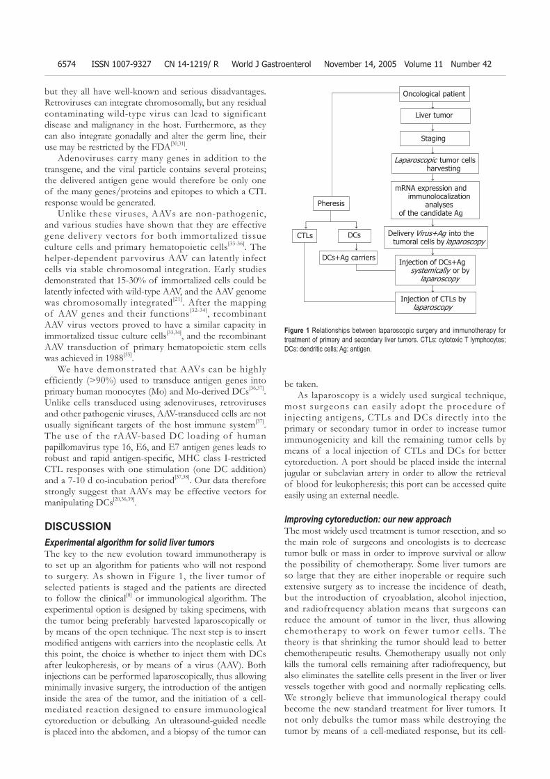
DISCUSSIONExperimental algorithm for solid liver tumorsThe key to the new evolution toward immunotherapy is to set up an algorithm for patients who will not respond to surgery. As shown in Figure 1, the liver tumor of selected patients is staged and the patients are directed to follow the clinical[8] or immunological algorithm. The experimental option is designed by taking specimens, with the tumor being preferably harvested laparoscopically or by means of the open technique. The next step is to insert modifi ed antigens with carriers into the neoplastic cells. At this point, the choice is whether to inject them with DCs after leukopheresis, or by means of a virus (AAV). Both injections can be performed laparoscopically, thus allowing minimally invasive surgery, the introduction of the antigen inside the area of the tumor, and the initiation of a cell-mediated reaction designed to ensure immunological cytoreduction or debulking. An ultrasound-guided needle is placed into the abdomen, and a biopsy of the tumor can
be taken.As laparoscopy is a widely used surgical technique,
most surgeons can easi ly adopt the procedure of injecting antigens, CTLs and DCs directly into the primary or secondary tumor in order to increase tumor immunogenicity and kill the remaining tumor cells by means of a local injection of CTLs and DCs for better cytoreduction. A port should be placed inside the internal jugular or subclavian artery in order to allow the retrieval of blood for leukopheresis; this port can be accessed quite easily using an external needle.
Improving cytoreduction: our new approachThe most widely used treatment is tumor resection, and so the main role of surgeons and oncologists is to decrease tumor bulk or mass in order to improve survival or allow the possibility of chemotherapy. Some liver tumors are so large that they are either inoperable or require such extensive surgery as to increase the incidence of death, but the introduction of cryoablation, alcohol injection, and radiofrequency ablation means that surgeons can reduce the amount of tumor in the liver, thus allowing chemotherapy to work on fewer tumor cel ls. The theory is that shrinking the tumor should lead to better chemotherapeutic results. Chemotherapy usually not only kills the tumoral cells remaining after radiofrequency, but also eliminates the satellite cells present in the liver or liver vessels together with good and normally replicating cells. We strongly believe that immunological therapy could become the new standard treatment for liver tumors. It not only debulks the tumor mass while destroying the tumor by means of a cell-mediated response, but its cell-
Figure 1 Relationships between laparoscopic surgery and immunotherapy for treatment of primary and secondary liver tumors. CTLs: cytotoxic T lymphocytes; DCs: dendritic cells; Ag: antigen.
Oncological patient
Liver tumor
Staging
Laparoscopic tumor cells harvesting
mRNA expression and immunolocalization
analyses of the candidate Ag
Delivery Virus+Ag into the tumoral cells by laparoscopy
Injection of DCs+Ag systemically or by
laparoscopy
Injection of CTLs by laparoscopy
Pheresis
CTLs DCs
DCs+Ag carriers
↓
↓
↓
↓
↓
↓
↓
↓ ↓
↓
Chiriva-Internati M et al. Liver cancer immunotherapy, biological therapy 6575
specific nature should enable it to kill satellite lesions and micro-metastases more efficiently (thus leading to better cytoreduction) without killing normal cells, and allow the use of low-dose chemotherapy to avoid or reduce undesirable side effects. Furthermore, the possible activation of memory responses could lead to much-needed long-term tumor immunosurveillance, which should reduce the incidence of relapses.
CONCLUSIONSEvidence of practical immunotherapy treatmentOur initial clinical results suggest that there is an urgent need to explore further therapeutic options for liver tumors. This study introduces several innovations and a methodology that will help establish critical clinical assays for assessing immune responses targeting liver tissue, and verify the relationship between host response and liver tumor regression/progression.
Relevance to liver cancer researchWe believe that immunological therapy can improve our overall understanding of how the host immune system interacts with primary and secondary liver cancer tissue, and will further elucidate useful methods for assessing this potential interaction.
Relevance to tumor immunology/immunotherapyThe importance of breaking host tolerance in order to achieve a tissue-specific mediated response and facilitate a favorable response to antitumor immunotherapy has recently been stressed in the literature[40]. For this reason, we believe that the use of a mini-invasive surgical approach, the immunotherapy and a clinical treatment will have a signifi cant impact on liver tumors.
Costs and applicationsImmunological therapy seems a very promising treatment for liver tumors. However, its specifi city and cost makes it indicated for patients with tumors that cannot be resected by any of the different ablation methods, and small tumors in cirrhotic patients who are unsuitable candidates for standard surgery. Improved cytoreductionWe believe that immunological therapy could become a new standard treatment of liver tumor for mainly three reasons: (1) it can debulk the tumor mass, while destroying the tumor by means of a cellular response; (2) it should be able to control the satellite lesions and micro-metastases more effi ciently because of its more cell-specifi c nature, thus improving cytoreduction, avoiding the killing of normal cells, and reducing the use of chemotherapy and therefore its side effects; and (3) the possible activation of memory responses could lead to much-needed long-term tumor immunosurveillance, and thus reduce the incidence of relapses.
REFERENCES1 Di Bisceglie AM, Rustgi VK, Hoofnagle JH, Dusheiko GM,
Lotze MT. NIH conference. Hepatocellular carcinoma. Ann Intern Med 1988; 108: 390-401
2 Carr BI. Hepatocellular carcinoma: current management and future trends. Gastroenterology 2004; 127: S218-S224
3 Tsuzuki T, Sugioka A, Ueda M, Iida S, Kanai T, Yoshii H, Nakayasu K. Hepatic resection for hepatocellular carcinoma. Surgery 1990; 107: 511-520
4 Di Bisceglie AM. Hepatocellular carcinoma: molecular biology of its growth and relationship to hepatitis B virus infection. Med Clin North Am 1989; 73: 985-997
5 Liu LX, Zhang WH, Jiang HC. Current treatment for liver metastases from colorectal cancer. World J Gastroenterol 2003; 9: 193-200
6 Arya SC, Ashraf SJ, Parande CM, Tobeiqi MS, Ageel AR. Hepatitis B and delta markers in primary hepatocellular carcinoma patients in the Gizan area of Saudi Arabia. APMIS Suppl 1988; 3: 30-34
7 Curley SA, Izzo F, Delrio P, Ellis LM, Granchi J, Vallone P, Fiore F, Pignata S, Daniele B, Cremona F. Radiofrequency ablation of unresectable primary and metastatic hepatic malignancies: results in 123 patients. Ann Surg 1999; 230: 1-8
8 Frezza EE. Therapeutic management algorithm in cirrhotic and noncirrhotic patients in primary or secondary liver masses. Dig Dis Sci 2004; 49: 876-871
9 Frezza EE. Extensive liver resection: can it be applicable to laparoscopic surgery? J Laparoendosc Adv Surg Tech A 2001; 11: 141-145
10 Sun HC, Tang ZY. Preventive treatments for recurrence after curative resection of hepatocellular carcinoma--a literature review of randomized control trials. World J Gastroenterol 2003; 9: 635-640
11 Frezza EE. Laparoscopic radiofrequency ablation of solitary hepatic gastrinoma metastases. Dig Dis Sci 2004; 49: 224-227
12 Butterfield LH. Immunotherapeutic strategies for hepa-tocellular carcinoma. Gastroenterology 2004; 127: S232-S241
13 Haigwood NL, Montefiori DC, Sutton WF, McClure J, Watson AJ, Voss G, Hirsch VM, Richardson BA, Letvin NL, Hu SL, Johnson PR. Passive immunotherapy in simian immunodeficiency virus-infected macaques accelerates the development of neutralizing antibodies. J Virol 2004; 78: 5983-5995
14 Zhao L, Mou DC, Leng XS, Peng JR, Wang WX, Huang L, Li S, Zhu JY. Expression of cancer-testis antigens in hepatocellular carcinoma. World J Gastroenterol 2004; 10: 2034-2038
15 Chiriva-Internati M, Wang Z, Salati E, Bumm K, Barlogie B, Lim SH. Sperm protein 17 (Sp17) is a suitable target for immunotherapy of multiple myeloma. Blood 2002; 100: 961-965
16 Chiriva-Internati M, Wang Z, Salati E, Timmins P, Lim SH. Tumor vaccine for ovarian carcinoma targeting sperm protein. Cancer 2002; 94: 2447-2453
17 Kast WM, Levitsky H, Marincola FM. Synopsis of the 6th Walker's Cay Colloquium on Cancer Vaccines and Im-munotherapy. J Transl Med 2004; 2: 20
18 Chiriva-Internati M, Grizzi F, Bright RK, Martin Kast W. Cancer immunotherapy: avoiding the road to perdition. J Transl Med 2004; 2: 26
19 Steinman RM, Mellman I. Immunotherapy: bewitched, bothered, and bewildered no more. Science 2004; 305: 197-200
20 Chiriva-Internati M, Liu Y, Salati E, Zhou W, Wang Z, Grizzi F, Roman JJ, Lim SH, Hermonat PL. Efficient generation of cytotoxic T lymphocytes against cervical cancer cells by adeno-associated virus/human papillomavirus type 16 E7 antigen gene transduction into dendritic cells. Eur J Immunol 2002; 32: 30-38
21 Fisher-Adams G, Wong KK Jr, Podsakoff G, Forman SJ, Chatterjee S. Integration of adeno-associated virus vectors in CD34+ human hematopoietic progenitor cells after
6576 ISSN 1007-9327 CN 14-1219/ R World J Gastroenterol November 14, 2005 Volume 11 Number 42
transduction. Blood 1996; 88: 492-50422 Steinman RM. The dendritic cell system and its role in
immunogenicity. Annu Rev Immunol 1991; 9: 271-29623 Romani N, Gruner S, Brang D, Kampgen E, Lenz A,
Trockenbacher B, Konwalinka G, Fritsch PO, Steinman RM, Schuler G. Proliferating dendritic cell progenitors in human blood. J Exp Med 1994; 180: 83-93
24 Sallusto F, Lanzavecchia A. Effi cient presentation of soluble antigen by cultured human dendritic cells is maintained by granulocyte/macrophage colony-stimulating factor plus interleukin 4 and downregulated by tumor necrosis factor alpha. J Exp Med 1994; 17: 1109-1118
25 Arthur JF, Butterfi eld LH, Roth MD, Bui LA, Kiertscher SM, Lau R, Dubinett S, Glaspy J, McBride WH, Economou JS. A comparison of gene transfer methods in human dendritic cells.Cancer Gene Ther 1997; 4: 17-25
26 Meyer zum Buschenfelde C, Nicklisch N, Rose-John S, Peschel C, Bernhard H. Generation of tumor-reactive CTL against the tumor-associated antigen HER2 using retrovirally transduced dendritic cells derived from CD34+ hemopoietic progenitor cells. J Immunol 2000; 165: 4133-4140
27 Szabolcs P, Gallardo HF, Ciocon DH, Sadelain M, Young JW. Retrovirally transduced human dendritic cells express a normal phenotype and potent T-cell stimulatory capacity. Blood 1997; 90: 2160-2167
28 Yoshida J, Mizuno M. Clinical gene therapy for brain tumors. Liposomal delivery of anticancer molecule to glioma. J Neurooncol 2003; 65: 261-267
29 Grimm D, Kay MA. From virus evolution to vector revolution: use of naturally occurring serotypes of adeno-associated virus (AAV) as novel vectors for human gene therapy. Curr Gene Ther 2003; 3: 281-304
30 Ponnazhagan S. Parvovirus vectors for cancer gene therapy. Expert Opin Biol Ther 2004; 4: 53-64
31 Shih A, Coutavas EE, Rush MG. Evolutionary implications of primate endogenous retroviruses. Virology 1991; 182: 495-502
32 Daly TM. Overview of adeno-associated viral vectors. Methods
Mol Biol 2004; 246: 157-16533 Hermonat PL, Labow MA, Wright R, Berns KI, Muzyczka N.
Genetics of adeno-associated virus: isolation and preliminary characterization of adeno-associated virus type 2 mutants. J Virol 1984; 51: 329-339
34 Tratschin JD, Miller IL, Carter BJ. Genetic analysis of adeno-associated virus: properties of deletion mutants constructed in vitro and evidence for an adeno-associated virus replication function. J Virol 1984; 51: 611-619
35 LaFace D, Hermonat P, Wakeland E, Peck A. Gene transfer into hematopoietic progenitor cells mediated by an adeno-associated virus vector. Virology 1988; 162: 483-486
36 Chiriva-Internati M, Liu Y, Weidanz JA, Grizzi F, You H, Zhou W, Bumm K, Barlogie B, Mehta JL, Hermonat PL. Testing recombinant adeno-associated virus-gene loading of dendritic cells for generating potent cytotoxic T lymphocytes against a prototype self-antigen, multiple myeloma HM1.24. Blood 2003; 102: 3100-3107
37 Liu Y, Santin AD, Mane M, Chiriva-Internati M, Parham GP, Ravaggi A, Hermonat PL. Transduction and utility of the granulocyte-macrophage colony-stimulating factor gene into monocytes and dendritic cells by adeno-associated virus. J Interferon Cytokine Res 2000; 20: 21-30
38 Tillman BW, Hayes TL, DeGruijl TD, Douglas JT, Curiel DT. Adenoviral vectors targeted to CD40 enhance the effi cacy of dendritic cell-based vaccination against human papillomavirus 16-induced tumor cells in a murine model. Cancer Res 2000; 60: 5456-5463
39 Liu Y, Chiriva-Internati M, You C, Luo R, You H, Prasad CK, Grizzi F, Cobos E, Klimberg VS, Kay H, Mehta JL, Hermonat PL. Use and specifi city of breast cancer antigen/milk protein BA46 for generating anti-self-cytotoxic T lymphocytes by recombinant adeno-associated virus-based gene loading of dendritic cells. Cancer Gene Ther 2005; 12: 304-312
40 Crittenden MR, Thanarajasingam U, Vile RG, Gough MJ. Intratumoral immunotherapy: using the tumour against itself. Immunology 2005; 114: 11-22
Science Editor Guo SY Language Editor Elsevier HK

PO Box 2345, Beijing 100023, China World J Gastroenterol 2005;11(42):6577-6581www.wjgnet.com World Journal of Gastroenterology ISSN [email protected] © 2005 The WJG Press and Elsevier Inc. All rights reserved.E L S E V I E R
• REVIEW •
INTRODUCTIONSymptoms of upper gastrointestinal distress are of world-wide interest and very common in the general population. In developing countries the important form of dyspepsia is organic dyspepsia, whereas the problem of functional dyspepsia (FD) seems to be mainly confi ned to industrialized Western countries though convincing data for underdeveloped countries are still lacking[1]. It is estimated that the annual prevalence of recurrent upper abdominal discomfort in the United States and other Western countries is approximately 25%, about 2% to 5% of all primary care consultations are related to dyspeptic symptoms[2]. For many patients the symptoms are of short duration or mild severity[3] and are therefore self-manageable. Less than half of these patients consult their general practitioner[2]. Moreover, patients with upper gastrointestinal problems frequently suffer from recurrent affections. However, several long-term studies showed that high percentages of patients with dyspeptic symptoms at entry report similar symptoms of dyspepsia after some years[3,4]. Repetitive diagnostic measures and medical treatments with low success rates lead to high costs and frustrating results. Thus, FD represents not only a clinical challenge but also a major socio-economical problem. In recent years, a lot of efforts have been made by national and international consensus meetings to work out precise defi nitions as well as adequate management strategies for dyspepsia. Still unsolved problems and new perspectives for both research work and disease management in clinical practice are summarized and discussed in more detail in this review.
Defi nition of functional dyspepsia Several defi nitions of dyspepsia have been proposed in the past decades[5] demonstrating the diffi culties in categorizing
Ahmed Madisch, Stephan Miehlke, Medical Department I, Technical University Hospital Dresden, Germany Joachim Labenz, Medical Department, Ev. Jung-Stilling-Hospital, Academic Teaching Hospital of the University of Bonn, Siegen, GermanyCorrespondence to: Ahmed Madisch, MD, Medical Department I, Technical University Hospital, Fetscherstrasse 74, D-01307 Dresden, Germany. [email protected]: +49-351-4584780 Fax: +49-351-4584394Received: 2004-11-12 Accepted: 2005-02-18
AbstractThe common characteristic criteria of all functional gastrointestinal (GI) disorders are the persistence and recurrence of variable gastrointestinal symptoms that cannot be explained by any structural or biochemical abnormalities. Functional dyspepsia (FD) represents one of the important GI disorders in Western countries because of its remarkably high prevalence in general population and its impact on quality of life. Due to its dependence on both subjective determinants and diverse country-specifi c circumstances, the defi nition and management strategies of FD are still variably stated. Clinical trials with several drug classes (e.g., proton pump inhibitors, H2-blockers, prokinetic drugs) have been performed frequently without validated disease-specifi c test instruments for the outcome measurements. Therefore, the interpretation of such trials remains diffi cult and controversial with respect to comparability and evaluation of drug effi cacy, and defi nite conclusions can be drawn neither for diagnostic management nor for efficacious drug therapy so far. In view of these unsolved problems, guidelines both on the clinical management of FD and on the performance of clinical trials are needed. In recent years, increasing research work has been done in this area. Clinical trials conducted in adequately diagnosed pat ients that provided validated outcome measurements may result in better insights leading to more effective treatment strategies. Encouraging perspectives have been recently performed by methodologically well-designed treatment studies with herbal drug preparations. Herbal drugs, given their proven effi cacy in clinical trials, offer a safe therapeutic alternative in the treatment of FD which is often favored by both patients and physicians. A fi xed combination of peppermint oil and caraway oil in patients suffering from FD could be proven effective by well-designed clinical trials.
Management of functional dyspepsia: Unsolved problems and new perspectives
Ahmed Madisch, Stephan Miehlke, Joachim Labenz
© 2005 The WJG Press and Elsevier Inc. All rights reserved.
Key words: Dyspepsia; Functional dyspepsia; Defi nition; Diagnosis; Management; Drug efficacy; Clinical trials; Outcome measurements; Herbal drugs
Madisch A, Miehlke S, Labenz J. Management of functional dyspepsia: Unsolved problems and new perspectives. World J Gastroenterol 2005; 11(42): 6577-6581http://www.wjgnet.com/1007-9327/11/6577.asp
6578 ISSN 1007-9327 CN 14-1219/ R World J Gastroenterol November 14, 2005 Volume 11 Number 42
dyspepsia as a clearly pathologically defi ned entity based on the variability of symptoms. According to the proposition of an international committee meeting in Rome in 1991, the term "dyspepsia" refers to pain or discomfort centered in the upper abdomen[6] while discomfort refers to a subjective negative (or aversive) feeling that is distinct from pain. Discomfort may include several specifi c bothersome but non-painful symptoms, such as early satiety, fullness, bloating and nausea (the so-called Rome criteria). In Rome I and more recent Rome II reports[1,7-9], the symptoms of heartburn, acid regurgitation, and belching are excluded from the definition of dyspepsia because they are more likely related to gastroesophageal refl ux disease (GERD) and aerophagia[1,9]. It is important to distinguish subjects with uninvestigated dyspepsia from patients with dyspepsia after adequate diagnostic procedure. Patients who have neither defi nite structural or biochemical explanation for their symptoms are considered to have FD. Thus, FD is defi ned as a persistent or recurrent dyspepsia for at least 12 wk in the preceding 12 mo if there is no evidence for organic disease (including upper endoscopy) that could cause the symptoms. The Rome II definitions of FD also exclude patients who report a relief of symptoms by defecation or symptoms associated with the onset of a change in stool frequency or stool form[9]. In the latter case, irritable bowel syndrome (IBS) is the diagnosis by defi nition. Coexistence of FD and IBS can be considered if there is pain or discomfort in the upper abdomen that is unrelated to bowel pattern and if there is other pain or discomfort that is related to bowel pattern[7].
Management of dyspepsiaDue to geographical, cultural, educational, social, and psychological aspects, universally applicable guidelines on diagnostic and therapeutical measures are difficult to implement[1,10]. Management strategies should be individualized and developed for each major community taking into account the prevalence of risk factors for gut diseases such as prevalence of H pylori infection, use of non-steroidal anti-inflammatory drugs, dietary habits, tobacco smoking and alcohol consumption[1,10]. Beyond these patient-related factors, the available financial and technical resources in each particular country may dictate the individual steps in the management of dyspepsia[1].
Nevertheless, useful recommendations regarding the management of dyspepsia are concluded in a recent systematic review of the literature[11]. To date, five management strategies can be offered to the physicians treating dyspeptic patients: (1) wait and see-strategy without diagnostic and therapeutic interventions; (2) empiric medical therapy with any subsequent investigation reserved for treatment failures; (3) immediate diagnostic evaluation in all cases; (4) testing for H pylori infection and reserving endoscopy for H pylori-positive cases to look for organic diseases (test-and-scope strategy); and (5) testing for H pylori infection by serology or urea breath test and treating all positive cases with H pylori eradication therapy (test-and-treat strategy).
For adult patients in Western countries with new onset of dyspepsia, endoscopy is the gold standard approach providing a firm diagnosis and facilitating decisions on treating or excluding organic diseases. In elderly patients or in those with alarm symptoms such as weight loss, immediate endoscopy is strongly advised. In respect of cost-effectiveness, a repeated endoscopy in those with an initially negative result should be avoided. An alternative management strategy in young dyspeptic patients under 45 years is non-invasive testing for H pylori infection and antibacterial treatment of positive cases[10-12]. Because of many substantial disadvantages such as antibiotic resistance, overtreatment, or undertreatment, there is ongoing discussion about the benefi t of this strategy.
Management of functional dyspepsia Patients with FD typically present an array of painful and non-painful symptoms demonstrating the multifactorial nature of this syndrome[13,14]. In order to identify pathophysiological abnormalities with subsequent targeted treatment and to promote more homogeneity, patients can be subdivided into ulcer-like, dysmotility-like and unspecified dyspepsia subgroups based on the concept of a cluster of symptoms[13,15]. Several studies have shown that this arbitrary classifi cation seems to be unsustainable because of the considerable overlap of the subgroups, the lack of stability over time, and the inconsistent responses to therapy[13,16]. Currently, the existence of subgroups among dyspeptic patients is neither endorsed nor categorically disproved[7,8,13].
Another approach to a subdivision of patients with FD is the sus pected association with H pylori infection. Between 30% and 60% of patients suffering from FD have H pylori-induced gastritis. However, H pylori infection is also common in the asymptomatic background population[17,18]. Even most recent trials with prolonged follow-up, analyzing the association between H pylori status and specifi c symptom profi les in FD have produced inconsistent and conflicting results. To date, there is no convincing evidence for the relief of specific dyspeptic symptoms after an eradication therapy[5,13,19,20]. Thus, a benefi t of anti- H pylori therapy in FD is not established [5,11,19].
Drug therapy for functional dyspepsia The wide range of therapies refl ects the uncertainty about the pathogenesis and the lack of satisfactory treatment. The pathophysiology of FD remains inadequately understood, even though various mechanisms may play a role in the development of symptoms. As yet, there is no cure for this disorder and available treatments are aimed at the relief of symptoms. Even though the effi cacy of some currently established treatments (e.g., antisecretory agents or prokinetics) has been proven in placebo-controlled trials, these treatments yield suffi cient relief of symptoms only in a proportion of patients[5].
In ulcer-like (pain predominating) functional dyspepsia, H2-receptor antagonists have produced inconsistent response rates[21]. Patients with dysmotility-like symptoms

Madisch A et al. Management of functional dyspepsia 6579
(upper abdominal discomfort predominating) may benefi t from prokinetic drug treatment[22-24]. Proton pump inhibitors appear to be efficacious especially in patients with ulcer-like pain and accompanying reflux symptoms. The majority of controlled clinical trials have shown only minor advantages of these drugs compared to placebo[25,26].
Thus, efforts should be made to identify and develop new effective treatments. Various herbal medications are used in many countries for the treatment of patients with FD. While some clinicians believe that clinical experience appears to support the use of these remedies, randomized controlled studies supporting the efficacy of these treatments have been lacking in the past decades. Recently, several well-designed placebo-controlled clinical trials have provided evidence for the effi cacy of herbal preparations used in the treatment of dyspepsia[27]. Particularly, patients with dysmotility-like dyspeptic symptoms, such as postprandial sensations of fullness, premature feelings of repleteness, non-acid eructation, or epigastric pain, experience a notable amelioration of their complaints[28,29].
Problems with evaluating drug efficacy in functional dyspepsia Clinical trials in functional GI disorders remain a challenge due to a variable placebo response ranging 20-60%[30], marked spontaneous fl uctuations of symptoms and a lack of widely accepted primary response variables. In addition, patients recruited at tertiary referral centers may represent a highly selected population that is less likely to respond to therapy[31]. It is likely that patients with FD present to general practitioners when their symptoms are worse. Therefore, spontaneous improvement may partially explain at least part of the placebo response[18].
Beside these well-known problems, the differences in the design of clinical drug trials in FD call for caution when interpreting their results. A systematic analysis of more than fifty eligible published placebo-controlled clinical trials testing prokinetics[32-35], cytoprotectives[36,37] or anti-ulcer agents[38-40] and other drugs[36,37] used in the treatment of functional dyspepsia revealed that single substantial items for the consistency of clinical studies such as inclusion and exclusion criteria for trial design and outcome measures are common but differ quite defi nitively in specifi c determinations[41]. Particularly, it is of importance how investigators deal with symptomatic GERD and other organic diseases. In 50% of the analyzed studies other upper GI disorders such as esophagitis and duodenal or gastric ulcer were not excluded; only 27% of the trials exclude or account for patients with over t ir r itable bowel syndrome as an overlapping functional disorder. The study design varies from parallel group, cross-over to multiple cross-over design[41]. The majority of analyzed trials fail to fulfi ll the indispensable requirement for efficacy evaluation and comparability of drug classes, i.e. use of clearly defi ned patient groups according to the consensual definition of FD and the use of validated outcome measures regarding described symptoms, their severity, and quality of life yielded with
validated categorical and visual analog scales (VAS). Thus, the authors concluded that convincing conclusions for effi cacious drug therapy in the treatment of FD cannot be drawn. Promising outcome measures for clinical trialsAlthough some research work has been done to develop validated outcome measures of symptoms[42] which can be used in FD, no generally accepted scales are available. Categorical scales (often referred to as Likert Scales) and VAS (horizontal line, usually 10 cm with endpoints on which the patient must place a mark) have been extensively applied[29,39,43-45] and qualifi ed as most eligible measurement scales by their reproducibility and ability to detect changes in a wide variety of clinical trials of different diseases. The usefulness of a reasonable combination of a categorical scale and a VAS is demonstrated by the dyspeptic discomfort score (DDS) which records the existence, frequency and severity of the symptoms of functional dyspepsia[28,29]. Integrating the dyspeptic, intestinal and extraintestinal autonomic discomforts assessed by means of numerical scales, the DDS seems to consider the entire complexity of this syndrome. Nevertheless, the DDS has not been validated yet.
A noteworthy measurement instrument to be me-ntioned is the clinical global impression (CGI) scale consisting of three items, namely severity of illness, global improvement and efficacy index. The first and second items are rated on a point scale while the third is a rating of the interaction of therapeutic effectiveness and adverse reactions. Originally conceived for schizophrenic studies, the CGI scale facilitates prognosis, survey and assessment of drug effi cacy during the treatment period[28,29,44].
During the last years, attention has been drawn to the fact that in diseases without obvious biological or clinical markers such as functional dyspepsia, the use of quality of life instruments and psychometric documentation as an outcome measure can refl ect treatment effi cacy evaluated by its impact on symptoms as well as on patient well-being and functioning[41,46]. The underlying philosophy is that quality of life is affected by the severity of disease-specifi c symptoms. Hence, the reciprocal conclusion can be drawn by any change of symptom severity. Recently, validation data of the new disease-specifi c Nepean dyspepsia index (NDI)[46,47] and the quality of life in refl ux and dyspepsia patient (QOLRAD) questionnaire[48] measuring frequency, intensity, and bothersomeness of upper gastrointestinal symptoms have been presented. The remarkable feature of the NDI is the consideration not only of a subject’s ability to perform or engage in an aspect of life but also the enjoyment of that aspect of life. In a systematic review of full-length publications during 1980-2002 reporting studies in patients with FD and measuring health-related quality of life, none of the studies used dyspepsia-specifi c health-related quality of life instruments[49]. However, recently a fi rst methodologically well-designed clinical study proving effi cacy of the study drug by use of the NDI was reported by Holtmann and colleagues[50], which demonstrates a
6580 ISSN 1007-9327 CN 14-1219/ R World J Gastroenterol November 14, 2005 Volume 11 Number 42
statistically signifi cant and clinically relevant superiority of a fixed combination of peppermint oil and caraway oil (PCC) in comparison to placebo. The reported outcome confirms the results formerly obtained with this herbal preparation in placebo-controlled clinical trials[28,44] and in a double-blind equivalence study with the prokinetic drug cisapride[29], measured by VAS, CGI and the DDS.
Recommendations for future trialsIn view of the mentioned weaknesses in present trials, the most essential recommendations are summarized as follows.
According to the consensus for a diagnosis of FD, a minimum set of diagnostic measures including upper endoscopy, an abdominal ultrasound and basic laboratory is obligatory[6]. At the time of enrolment for a treatment study, eligible patients must have persistent symptoms that are of a suffi cient degree to seek medical attention. Any definite structural abnormalities of the upper GI tract, explaining the symptoms, e.g., peptic ulcer confi rmed by endoscopic evidence and biochemical agents such as daily use of NSAID or high dose aspirin must be excluded. To avoid an overlap with gastroesophageal refl ux disease, patients in whom heartburn or acid regurgitation are the predominant symptoms or patients suffering from irritable bowel syndrome and other known organic diseases that might explain the dyspepsia symptoms must not be enrolled.
Despite some well recognized problems such as the occurrence of period-by-treatment interactions of cross-over trials resulting in ambiguous interpretation of data, the randomized, double-blind, placebo-controlled parallel group design is strongly advocated as the trial design of choice.
It is not to deny that even among physicians there is great variation in the definitions of common dyspeptic symptoms. In addit ion, terminology and possibly also the sensations experienced vary between cultures and countries. Therefore, it is advisable that clinical investigators use definitions of symptoms suggested by the Rome Working Party report and accommodated to common parlance in the respective study population.
As validated outcome measures like the NDI and the QOLRAD questionnaire are now available, their use is strongly recommended regarding described symptoms, their severity, and aspects of quality of life. In order to support the results obtained with these validated disease specifi c questionnaires, categorical scales, VAS and the CGI could be used as secondary outcome measures. Promising outcome measures such as DDS, should be validated soon in order to broaden the range of appropriate devices for evaluating drug effi cacy in functional dyspepsia.
Further research using well-val idated outcome instruments for measurement of individual symptoms as well as their severity and their impact on quality of life may perhaps result in a valid symptom-related categorization of functional dyspepsia that may be used to improve treatment strategies.
Causally determined by the aforementioned unsolved
problems concerning the definition and management of FD as well as the listed weaknesses in trial methodology of present treatment studies, convincing conclusions for efficacious drug therapy cannot be drawn yet. However, it is very likely that effective drug therapies are av-ailable. Further research on well-validated measurement in-struments for outcome data permitting comparability of drug classes may perhaps result in better insights with respect to effective treatment strategies. Quite recently, new perspectives have been arising from presented effi cacy of a fixed peppermint oil/caraway oil preparation in a methodologically adequate clinical trial.
ACKNOWLEDGMENTThis paper is dedicated to Professor Jürgen Hotz, a friend, colleague, and academic teacher, who passed away in 2002.
REFERENCES1 Malfertheiner P. Current concepts in dyspepsia: a world
perspective. Eur J Gastroenterol Hepatol 1999; 11 Suppl 1: S25-S29
2 Knill-Jones RP. Geographical differences in the prevalence of dyspepsia. Scand J Gastroenterol Suppl 1991; 182: 17-24
3 Johannessen T, Petersen H, Kristensen P, Kleveland PM, Dybdahl J, Sandvik AK, Brenna E, Waldum H. The intensity and variability of symptoms in dyspepsia. Scand J Prim Health Care 1993; 11: 50-55
4 Jones R, Lydeard S. Dyspepsia in the community: a follow-up study. Br J Clin Pract 1992; 46: 95-97
5 Talley NJ. Helicobacter pylori and dyspepsia. Yale J Biol Med 1999; 72: 145-151
6 Talley NJ, Stanghellini V, Heading RC, Koch KL, Malagelada JR, Tytgat GN. Functional gastroduodenal disorders. Gut 1999; 45 Suppl 2: II37-II42
7 Talley NJ, Stanghellini V, Heading RC, Koch KL, Malagelada JR, Tytgat GN. Functional gastroduodenal disorders. Gut 1999; 45 Suppl 2: II37-II42
8 Thompson WG, Longstreth GF, Drossman DA, Heaton KW, Irvine EJ, Muller-Lissner SA. Functional bowel disorders and functional abdominal pain. Gut 1999; 45 Suppl 2: II43-II47
9 Spiller R. Rome II: the functional gastrointestinal disorders. Diagnosis, pathophysiology and treatment: a multinational consensus. Gut 2000; 46: 741B
10 Mullins PD, Colin-Jones DG. Guidelines for the management of dyspepsia. Eur J Gastroenterol Hepatol 1999; 11: 215-217
11 Talley NJ, Silverstein MD, Agreus L, Nyren O, Sonnenberg A, Holtmann G. AGA technical review: evaluation of dyspepsia. American Gastroenterological Association. Gastroenterology 1998; 114: 582-595
12 Moayyedi P, Zilles A, Clough M, Hemingbrough E, Chalmers DM, Axon AT. The effectiveness of screening and treating Helicobacter pylori in the management of dyspepsia. Eur J Gastroenterol Hepatol 1999; 11: 1245-1250
13 Holtmann G, Stanghellini V, Talley NJ. Nomenclature of dyspepsia, dyspepsia subgroups and functional dyspepsia: clarifying the concepts. Baillieres Clin Gastroenterol 1998; 12: 417-433
14 Mansi C, Mela GS, Pasini D, Grosso M, Corti L, Moretti M, Celle G. Patterns of dyspepsia in patients with no clinical evidence of organic diseases. Dig Dis Sci 1990; 35: 1452-1458
15 Gotthard R, Bodemar G, Brodin U, Jonsson KA. Treatment with cimetidine, antacid, or placebo in patients with dyspepsia of unknown origin. Scand J Gastroenterol 1988; 23: 7-18
16 Talley NJ, Weaver AL, Tesmer DL, Zinsmeister AR. Lack of
Madisch A et al. Management of functional dyspepsia 6581
discriminant value of dyspepsia subgroups in patients referred for upper endoscopy. Gastroenterology 1993; 105: 1378-1386
17 Verdu EF, Armstrong D, Idstrom JP, Labenz J, Stolte M, Borsch G, Blum AL. Intragastric pH during treatment with omeprazole: role of Helicobacter pylori and H pylori-associated gastritis. Scand J Gastroenterol 1996; 31: 1151-1156
18 Talley NJ, Hunt RH. What role does Helicobacter pylori play in nonulcer dyspepsia? Arguments for and against H. pylori being associated with dyspeptic symptoms. Gastroenterology 1997; 113: S67-S77
19 Talley NJ. A critique of therapeutic trials in Helicobacter pylori-positive functional dyspepsia. Gastroenterology 1994; 106: 1174-1183
20 El-Omar EM, Oien K, El-Nujumi A, Gillen D, Wirz A, Dahill S, Williams C, Ardill JE, McColl KE. Helicobacter pylori infection and chronic gastric acid hyposecretion. Gastroenterology 1997; 113: 15-24
21. Farup PG, Wetterhus S, Osnes M, Ulshagen K. Ranitidine effectively relieves symptoms in a subset of patients with functional dyspepsia. Scand J Gastroenterol 1997; 32: 755-759
22 Holtmann G, Gschossmann J, Karaus M, Fischer T, Becker B, Mayr P, Gerken G. Randomised double-blind comparison of simethicone with cisapride in functional dyspepsia. Aliment Pharmacol Ther 1999; 13: 1459-1465
23 Halter F, Staub P, Hammer B, Guyot J, Miazza BM. Study with two prokinetics in functional dyspepsia and GORD: domperidone vs. cisapride. J Physiol Pharmacol 1997; 48: 185-192
24 Carvalhinhos A, Fidalgo P, Freire A, Matos L. Cisapride compared with ranitidine in the treatment of functional dyspepsia. Eur J Gastroenterol Hepatol 1995; 7: 411-417
25 Hansen JM, Bytzer P, Schaffalitzky de Muckadell OB. Placebo-controlled trial of cisapride and nizatidine in unselected patients with functional dyspepsia. Am J Gastroenterol 1998; 93: 368-374
26 Talley NJ, Meineche-Schmidt V, Pare P, Duckworth M, Raisanen P, Pap A, Kordecki H, Schmid V. Efficacy of omeprazole in functional dyspepsia: double-blind, randomized, placebo-controlled trials (the Bond and Opera studies). Aliment Pharmacol Ther 1998; 12: 1055-1065
27 Pu RT, Osmani SA. Mitotic destruction of the cell cycle regulated NIMA protein kinase of Aspergillus nidulans is required for mitotic exit. EMBO J 1995; 14: 995-1003
28 May B, Kohler S, Schneider B. Efficacy and tolerability of a fixed combination of peppermint oil and caraway oil in patients suffering from functional dyspepsia. Aliment Pharmacol Ther 2000; 14: 1671-1677
29 Madisch A, Heydenreich CJ, Wieland V, Hufnagel R, Hotz J. Treatment of functional dyspepsia with a fixed peppermint oil and caraway oil combination preparation as compared to cisapride. A multicenter, reference-controlled double-blind equivalence study. Arzneimittelforschung 1999; 49: 925-932
30 Talley NJ, Phillips SF. Non-ulcer dyspepsia: potential causes and pathophysiology. Ann Intern Med 1988; 108: 865-879
31 Veldhuyzen van Zanten SJ, Talley NJ, Bytzer P, Klein KB, Whorwell PJ, Zinsmeister AR. Design of treatment trials for functional gastrointestinal disorders. Gut 1999; 45 Suppl 2: II69-II77
32 Hausken T, Berstad A. Wide gastric antrum in patients with non-ulcer dyspepsia. Effect of cisapride. Scand J Gastroenterol 1992; 27: 427-432
33 Hausken T, Berstad A. Cisapride treatment of patients with non-ulcer dyspepsia and erosive prepyloric changes. A double-blind, placebo-controlled trial. Scand J Gastroenterol 1992; 27: 213-217
34 Sarin SK, Sharma P, Chawla YK, Gopinath P, Nundy S. Clinical trial on the effect of domperidone on non-ulcer dyspepsia. Indian J Med Res 1986; 83: 623-628
35 De Loore I, Van Ravensteyn H, Ameryckx L. Domperidone
drops in the symptomatic treatment of chronic paediatric vomiting and regurgitation. A comparison with meto-clopramide. Postgrad Med J 1979; 55 Suppl 1: 40-2
36 Hausken T, Stene-Larsen G, Lange O, Aronsen O, Nerdrum T, Hegbom F, Schulz T, Berstad A. Misoprostol treatment exacerbates abdominal discomfort in patients with non-ulcer dyspepsia and erosive prepyloric changes. A double-blind, placebo-controlled, multicentre study. Scand J Gastroenterol 1990; 25: 1028-1033
37 Skoubo-Kristensen E, Funch-Jensen P, Kruse A, Hanberg-Sorensen F, Amdrup E. Controlled clinical trial with sucralfate in the treatment of macroscopic gastritis. Scand J Gastroenterol 1989; 24: 716-720
38 Johannessen T, Kristensen P, Petersen H, Fosstvedt D, Loge I, Kleveland PM, Dybdahl J. The symptomatic effect of 1-day treatment periods with cimetidine in dyspepsia. Combined results from randomized, controlled, single-subject trials. Scand J Gastroenterol 1991; 26: 974-980
39 Farup PG, Larsen S, Ulshagen K, Osnes M. Ranitidine for non-ulcer dyspepsia. A clinical study of the symptomatic effect of ranitidine and a classification and characterization of the responders to treatment. Scand J Gastroenterol 1991; 26: 1209-1216
40 Smith PM, Troughton AH, Gleeson F, Walters J, McCarthy CF. Pirenzepine in non-ulcer dyspepsia: a double-blind multicentre trial. J Int Med Res 1990; 18: 16-20
41 Veldhuyzen van Zanten SJ, Cleary C, Talley NJ, Peterson TC, Nyren O, Bradley LA, Verlinden M, Tytgat GN. Drug treatment of functional dyspepsia: a systematic analysis of trial methodology with recommendations for design of future trials. Am J Gastroenterol 1996; 91: 660-673
42 Leidy NK, Farup C, Rentz AM, Ganoczy D, Koch KL. Patient-based assessment in dyspepsia: development and validation of Dyspepsia Symptom Severity Index (DSSI). Dig Dis Sci 2000; 45: 1172-1179
43 Madisch A, Melderis H, Mayr G, Sassin I, Hotz J. A plant extract and its modifi ed preparation in functional dyspepsia. Results of a double-blind placebo controlled comparative study. Z Gastroenterol 2001; 39: 511-517
44 May B, Kuntz HD, Kieser M, Kohler S. Efficacy of a fixed peppermint oil/caraway oil combination in non-ulcer dyspepsia. Arzneimittelforschung 1996; 46: 1149-1153
45 Corazza GR, Biagi F, Albano O, Porro GB, Cheli R, Mazzacca G, Miglio F, Naccarato R, Quaglino D, Surrenti C, Verme G, Gasbarrini G. Levosulpiride in functional dyspepsia: a multicentric, double-blind, controlled trial. Ital J Gastroenterol 1996; 28: 317-323
46 Talley NJ, Haque M, Wyeth JW, Stace NH, Tytgat GN, Stanghell ini V, Holtmann G, Verl inden M, Jones M. Development of a new dyspepsia impact scale: the Nepean Dyspepsia Index. Aliment Pharmacol Ther 1999; 13: 225-235
47 Talley NJ, Verlinden M, Jones M. Validity of a new quality of life scale for functional dyspepsia: a United States multicenter trial of the Nepean Dyspepsia Index. Am J Gastroenterol 1999; 94: 2390-2397
48 Wiklund IK, Junghard O, Grace E, Talley NJ, Kamm M, Veldhuyzen van Zanten S, Pare P, Chiba N, Leddin DS, Bigard MA, Colin R, Schoenfeld P. Quality of Life in Reflux and Dyspepsia patients. Psychometric documentation of a new disease-specific questionnaire (QOLRAD). Eur J Surg Suppl 1998; 583: 41-49
49 El-Serag HB, Talley NJ. Health-related quality of life in functional dyspepsia. Aliment Pharmacol Ther 2003; 18: 387-393
50 Holtmann G, Haag S , Adam B, Funk P, Wieland V, Heydenreich CJ. Effects of a fi xed combination of peppermint oil and caraway oil on symptoms and quality of life in patients suffering from functional dyspepsia. Phytomedicine 2003; 10 Suppl 4: 56-57
Science Editor Wang XL and Guo SY Language Editor Elsevier HK
PO Box 2345, Beijing 100023, China World J Gastroenterol 2005;11(42):6582-6586www.wjgnet.com World Journal of Gastroenterology ISSN [email protected] © 2005 The WJG Press and Elsevier Inc. All rights reserved.E L S E V I E R
• ESOPHAGEAL CANCER •
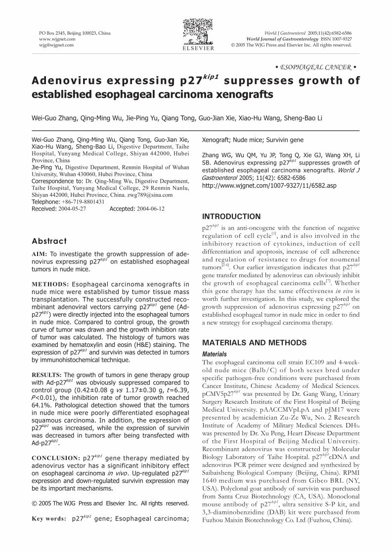
INTRODUCTIONp27kip1 is an anti-oncogene with the function of negative regulation of cell cycle[1], and is also involved in the inhibitory reaction of cytokines, induction of cell differentiation and apoptosis, increase of cell adherence and regulation of resistance to drugs for noumenal tumors[2-6]. Our earlier investigation indicates that p27kip1 gene transfer mediated by adenovirus can obviously inhibit the growth of esophageal carcinoma cells[7]. Whether this gene therapy has the same effectiveness in vivo is worth further investigation. In this study, we explored the growth suppression of adenovirus expressing p27kip1 on established esophageal tumor in nude mice in order to fi nd a new strategy for esophageal carcinoma therapy.
MATERIALS AND METHODS MaterialsThe esophageal carcinoma cell strain EC109 and 4-week-old nude mice (Balb/C) of both sexes bred under specific pathogen-free conditions were purchased from Cancer Institute, Chinese Academy of Medical Sciences. pCMV5p27kip1 was presented by Dr. Gang Wang, Urinary Surgery Research Institute of the First Hospital of Beijing Medical University. pAACCMVpLpA and pJM17 were presented by academician Zu-Ze Wu, No. 2 Research Institute of Academy of Military Medical Sciences. DH5a was presented by Dr. Xu Peng, Heart Disease Department of the First Hospital of Beijing Medical University. Recombinant adenovirus was constructed by Molecular Biology Laboratory of Taihe Hospital. p27kip1cDNA and adenovirus PCR primer were designed and synthesized by Saibaisheng Biological Company (Beijing, China). RPMI 1640 medium was purchased from Gibco BRL (NY, USA). Polyclonal goat antibody of survivin was purchased from Santa Cruz Biotechnology (CA, USA). Monoclonal mouse antibody of p27kip1, ultra sensitive S-P kit, and 3,3-diaminobenzidine (DAB) kit were purchased from Fuzhou Maixin Biotechnology Co. Ltd (Fuzhou, China).
Wei-Guo Zhang, Qing-Ming Wu, Qiang Tong, Guo-Jian Xie, Xiao-Hu Wang, Sheng-Bao Li, Digestive Department, Taihe Hospital, Yunyang Medical College, Shiyan 442000, Hubei Province, ChinaJie-Ping Yu, Digestive Department, Renmin Hospital of Wuhan University, Wuhan 430060, Hubei Province, ChinaCorrespondence to: Dr. Qing-Ming Wu, Digestive Department, Taihe Hospital, Yunyang Medical College, 29 Renmin Nanlu, Shiyan 442000, Hubei Province, China. [email protected]: +86-719-8801431Received: 2004-05-27 Accepted: 2004-06-12
AbstractAIM: To investigate the growth suppression of ade-novirus expressing p27kip1 on established esophageal tumors in nude mice.
METHODS: Esophageal carcinoma xenografts in nude mice were established by tumor tissue mass transplantation. The successfully constructed reco-mbinant adenoviral vectors carrying p27kip1 gene (Ad-p27kip1) were directly injected into the esophageal tumors in nude mice. Compared to control group, the growth curve of tumor was drawn and the growth inhibition rate of tumor was calculated. The histology of tumors was examined by hematoxylin and eosin (H&E) staining. The expression of p27kip1 and survivin was detected in tumors by immunohistochemical technique. RESULTS: The growth of tumors in gene therapy group with Ad-p27kip1 was obviously suppressed compared to control group (0.42±0.08 g vs 1.17±0.30 g, t =6.39, P<0.01), the inhibition rate of tumor growth reached 64.1%. Pathological detection showed that the tumors in nude mice were poorly differentiated esophageal squamous carcinoma. In addition, the expression of p27kip1 was increased, while the expression of survivin was decreased in tumors after being transfected with Ad-p27kip1.
CONCLUSION: p27kip1 gene therapy mediated by adenovirus vector has a significant inhibitory effect on esophageal carcinoma in vivo . Up-regulated p27kip1 expression and down-regulated survivin expression may be its important mechanisms.
© 2005 The WJG Press and Elsevier Inc. All rights reserved.
Key words: p27kip1 gene; Esophageal carcinoma;
Adenovirus expressing p27kip1 suppresses growth of established esophageal carcinoma xenografts
Wei-Guo Zhang, Qing-Ming Wu, Jie-Ping Yu, Qiang Tong, Guo-Jian Xie, Xiao-Hu Wang, Sheng-Bao Li
Xenograft; Nude mice; Survivin gene
Zhang WG, Wu QM, Yu JP, Tong Q, Xie GJ, Wang XH, Li SB. Adenovirus expressing p27kip1 suppresses growth of established esophageal carcinoma xenografts. World J Gastroenterol 2005; 11(42): 6582-6586http://www.wjgnet.com/1007-9327/11/6582.asp

Zhang WG et al. p27kip1 suppresses esophageal carcinoma xenografts 6583
Construction of recombinant adenovirus Ad-p27kip1 The process was the same as described in our previous work[7].
Cell culture Human esophageal carcinoma cell strain EC109 was maintained in RPMI 1640 medium supplemented with 100 mL/L fetal calf serum (FCS), 100 kU/L penicillin, 100 mg/L streptomycin, 2 mmol/L L-glutamine, and 50 mL/L CO2 in a humidifi ed incubator at 37 ℃. The medium was changed every 2-3 d.
Establishment of esophageal carcinoma xenograftsEC109 cells growing exponentially were selected. The fi nal concentration was adjusted to 107 cells/mL. Nude mice (Balb/C) of 4 wk old received injections into the dorsal midline in a 100 mL volume to establish tumors. The transplanted tumors were reproduced among the animals continually when the original grafts were growing well. Then esophageal carcinoma xenografts were established by transplanting the tumor tissue mass into the subcutaneous tissue of 36 nude mice. They were ready for use when the tumor diameter reached about 0.7 cm.
Therapeutic effect of intratumoral injection of Ad-p27kip1 into established tumorsThe animals were randomized into three groups, and each group had seven mice with comparable tumor size within and among the groups. Intratumoral injection of Ad-p27kip1, Ad-LacZ (1.0×10¹⁰ pfu) or PBS was made every other day for totally four times. The growth curve of tumor was drawn and the growth inhibitory rate of tumor was calculated after the animals were killed at wk 4. Tumor sizes were calculated by the formula: tumor volume = 1/2×length×width2. The growth inhibitory rate of tumor was calculated by the formula: inhibitory rate = (tumor mass of control group–tumor mass of experimental group)/tumor mass of control group. HistologyThe tumor tissues were fixed in 10% neutral formalin and embedded in paraffin. Sections of 5 μm thickness were used for morphological and immunohistochemical examinations. Paraffin sections were stained with hematoxylin and eosin (H&E) to demonstrate esophageal carcinoma tissue components.
Expression of p27kip1
The paraffin sections were washed with phosphate-buffered saline (PBS, pH 7.4) and incubated in 3% hydrogen peroxide for 10 min to block endogenous per-oxidase. After being heated for 10 min in 0.01 mol/L citrate buffer (pH 6.0) using a microwave oven, the sections were incubated with normal animal serum for 10 min and then with monoclonal mouse antibody of p27kip1 overnight at 4 ℃. Biotinylated antimouse immunoglobulin and streptavidin conjugated to horseradish peroxidase were subsequently applied. Finally, DAB was used for
color development, and hematoxylin was used for coun-terstaining. As a negative control, the sections were processed in the absence of primary antibody. A scoring method was used to quantitate the p27kip1 expression in samples examined. A mean percentage of positive tumor cells was determined in at least five areas at 400-fold magnification. Samples with scores less than 50% were defi ned as low expression, otherwise as high expression[8]. These scorings were performed in a blinded fashion.
Expression of survivinThe sections carrying survivin protein were stained according to SP immunohistochemical staining method as aforementioned. The primary antibody was polyclonal goat antibody of survivin (dilution 1:200). The mean percentage of positive cells for the expression of survivin was determined in at least fi ve areas at 400-fold magnifi cation, and the samples with less than 10% positively stained cells were defi ned as negative. Samples with 10-29% positively stained cells were defi ned as +, 30-59% as ++, and 60% or more than 60% as +++[9]. Statistical analysis The data were expressed as mean±SD. The difference between each group was analyzed by t-test. P<0.05 was considered statistically signifi cant.
RESULTSGrowth suppression of established esophageal carcinoma xenografts by intratumoral injection of Ad-p27kip1
Intratumoral injection of Ad-p27kip1 into established tumors induced partial growth suppression. The growth of tumors in gene therapy group with p27kip1 was obviously suppressed, being significantly different from that in control group and Ad-LacZ group (P<0.01). The growth inhibitory rate (IR) of tumor reached 64.1% (Figures 1 and 2, Table 1).
Histological evaluation The result of hematoxylin and eosin staining showed poorly differentiated esophageal squamous carcinoma (Figure 3).
Figure 1 The growth curves of tumor.
ControlAd-LacZAd-p27kip13
2.5
2
1.5
1
0.5
00 3 6 9 12 15 18 21 24 27
Time (d)
Tum
or v
olum
e (c
m3 )

6584 ISSN 1007-9327 CN 14-1219/ R World J Gastroenterol November 14, 2005 Volume 11 Number 42
tumor tissues is due to post-transcriptional degradation[11]. p27kip1 protein belongs to the family of proteins called cyclin-dependent kinase inhibitors (CDKIs). These proteins play an important role as negative regulators of cell cycle-dependent kinases during the progression of cell cycle. p27kip1 regulates the progression from G1 into S phase by binding to and inhibiting the cyclin E/Cdk2 complex, which is required for entry into the S phase. It also interacts with various other cyclin complexes and is therefore designated as a universal CDKI[12-16]. p27kip1 expression decreases in esophageal cancer and may correlate with the histologic differentiation. Reduction of p27kip1 is considered to be an independent prognostic indicator of esophageal cancer[17-20].
In this study, we found that the growth of established esophageal carcinoma xenografts was obviously depressed and the inhibitory rate reached 64.1% after transfection with Ad-p27kip1. The result of immunohistochemical staining demonstrated that Ad-p27kip1 could efficiently express p27kip1 in esophageal carcinoma. Ad-p27kip1 constructed in the present study is a kind of replication de-fective adenoviral vector, which has only one opportunity for infection in target cells without any dup-lication ability to fulfi ll the functions of adenoviral carrier, thus avoiding damage of adenovirus itself to target cells and reaching gene conversion.
We also observed that the expression of survivin was decreased in tumors of nude mice, indicating that p27kip1 might downregulate survivin expression. Survivin is a newly identified gene in inhibitors of apoptosis protein (IAP) family and is characterized by a unique structure with a single baculovirus IAP repeat and no zinc-binding domain known as Ring finger[21-23]. Survivin is an oncogene, which has been implicated in inhibition of apoptosis and control of mitotic progression. It is not usually detectable in normal adult tissues, but is prominently expressed in almost all common human cancers and most transformed cell lines. It is a short-lived protein degraded by ubiquitin proteasome pathway and interferes with the activation of caspases, called "cell death executioners"[24-28]. Disruption of survivin-microtubule interactions results in loss of survivin's anti-apoptosis function and increase of caspase-3 activity, a mechanism involved in cell death during mitosis. Survivin functions as a dimer and is regulated in a cell-cycle-dependent manner, peaking at G2/M, nearly not detectable at G1, and is associated with the mitotic spindle, centromeres, and the midbody in dividing cells[29-31]. p27kip1, a down-regulating survivin may be associated with G1 blocking of p27kip1, which has been identifi ed in our previous studies[7].
Expression of p27kip1
Immunohistochemical staining showed that the expression of p27kip1 was increased in established esophageal carcinoma xenografts after being transfected with Ad-p27kip1 (Figure 4).
Expression of survivinSurvivin was prominently found in control group by immunohistochemistry and decreased in established esophageal carcinoma xenografts after being transfected with Ad-p27kip1 (Figure 5).
DISCUSSIONp27kip1 has been mapped to the short arm of chromosome 12 at the 12p12-12p13.1 boundary containing two introns and two exons[10]. Mutations of gene p27kip1 occur rarely in human tumors and the decrease in p27kip1 expression in
Groups (%)
Tumor mass (g) IR
1 2 3 4 5 6 7 mean±SD
Control 1.02 0.93 1.31 1.47 0.86 1.63 0.95 1.17±0.30 Ad-LacZ 0.72 1.36 1.12 0.87 0.92 1.23 0.85 1.00±0.23 Ad-p27kip1 0.32 0.46 0.35 0.43 0.57 0.38 0.40 0.42±0.08b 64.1
Table 1 Tumor mass of three groups treated with Ad-p27kip1, Ad-LacZ (1.0×10¹⁰ pfu) or PBS at wk 4
t = 6.39, bP<0.01 vs control group. IR: inhibition rate.
Figure 3 Hematoxylin and eosin (H&E) staining of established esophageal carcinoma xenografts (×200).
Figure 2 Growth suppression of Ad-p27kip1 on established esophageal carcinoma xenografts. 1: control group; 2: Ad-LacZ group; 3: Ad-p27kip1 group.
1
2
3

Zhang WG et al. p27kip1 suppresses esophageal carcinoma xenografts 6585
In conclusion, p27kip1 gene therapy mediated by adenovirus vector has significant inhibitory effect on esophageal carcinoma in vivo. Upregulated p27kip1 expression and downregulated survivin expression may be its important mechanisms.
REFERENCES1 Polyak K, Kato JY, Solomon MJ, Sherr CJ, Massague J,
Roberts JM, Koff A. p27Kip1, a cyclin-CDK inhibitor, links transforming growth factor-beta and contact inhibition to cell cycle arrest. Genes Dev 1994; 8: 9-22
2 Zhang WG, Wu QM, Tong Q, Yu JP, Wang XH, Xie GJ. The effect of p27kip1 mediated by adenovirus on the cell cycle and DNA synthesis in gastric cancer cells. Weichang Bingxue 2003; 8: 210-214
3 Kiyokawa H, Kineman RD, Manova-Todorova KO, Soares VC, Hoffman ES, Ono M, Khanam D, Hayday AC, Frohman LA, Koff A. Enhanced growth of mice lacking the cyclin-dependent kinase inhibitor function of p27(Kip1). Cell 1996; 85: 721-732
4 Eguchi H, Carpentier S, Kim SS, Moss SF. P27kip1 regulates the apoptotic response of gastric epithelial cells to Helicobacter pylori. Gut 2004; 53: 797-804
5 Ghanem MA, Van der Kwast TH, Sudaryo MK, Mathoera RB, van den Heuvel MM, Al-Doray AA, Nijman RM, van Steenbrugge GJ. MIB-1 (KI-67) proliferation index and cyclin-dependent kinase inhibitor p27(Kip1) protein expression in nephroblastoma. Clin Cancer Res 2004; 10: 591-597
6 Center DM, Cruikshank WW, Zhang Y. Nuclear pro-IL-16 regulation of T cell proliferation: p27(KIP1)-dependent G0/G1 arrest mediated by inhibition of Skp2 transcription. J Immunol 2004; 172: 1654-1660
7 Zhang WG, Wu QM, Tong Q, Yu JP, Wang XH, Xie GJ, Wang
WM. Effect of p27kip1 gene transfere mediated by adenovirus on cell cycle of esophageal carcinoma cells. Zhongguo Yishi Zazhi 2004; 6: 456-458
8 Singh SP, Lipman J, Goldman H, Ellis FH Jr, Aizenman L, Cangi MG, Signoretti S, Chiaur DS, Pagano M, Loda M. Loss or altered subcellular localization of p27 in Barrett's associated adenocarcinoma. Cancer Res 1998; 58: 1730-1735
9 Zhang WG, Wu QM, Wang XH, Xie GJ, Yu JP. Relationship between expression of Survivin gene and biological character in esophageal carcinoma. Zhongguo Yishi Zazhi 2003; 5: 1378-1380
10 Ponce-Castaneda MV, Lee MH, Latres E, Polyak K, Lacombe L, Montgomery K, Mathew S, Krauter K, Sheinfeld J, Massague J. p27Kip1: chromosomal mapping to 12p12-12p13.1 and absence of mutations in human tumors. Cancer Res 1995; 55: 1211-1214
11 Shin JY, Kim HS, Lee KS, Kim J, Park JB, Won MH, Chae SW, Choi YH, Choi KC, Park YE, Lee JY. Mutation and expression of the p27KIP1 and p57KIP2 genes in human gastric cancer. Exp Mol Med 2000; 32: 79-83
12 Dimberg A, Bahram F, Karlberg I, Larsson LG, Nilsson K, Oberg F. Retinoic acid-induced cell cycle arrest of human myeloid cell lines is associated with sequential down-regulation of c-Myc and cyclin E and posttranscriptional up-regulation of p27(Kip1). Blood 2002; 99: 2199-2206
13 Lim MS, Adamson A, Lin Z, Perez-Ordonez B, Jordan RC, Tripp S, Perkins SL, Elenitoba-Johnson KS. Expression of Skp2, a p27(Kip1) ubiquitin ligase, in malignant lymphoma: correlation with p27(Kip1) and proliferation index. Blood 2002; 100: 2950-2956
14 Taguchi T, Kato Y, Baba Y, Nishimura G, Tanigaki Y, Horiuchi C, Mochimatsu I, Tsukuda M. Protein levels of p21, p27, cyclin E and Bax predict sensitivity to cisplatin and paclitaxel in head and neck squamous cell carcinomas. Oncol Rep 2004; 11: 421-426
15 Leung-Pineda V, Pan Y, Chen H, Kilberg MS. Induction of
Figure 4 Result of immunohistochemical staining of tumors in nude mice. A: control group. p27kip1 protein located in cytoplasm of a few cells showing low expression; B: Ad-LacZ group. The result showed low expression also; C: Ad-p27kip1 group. p27kip1 protein located in cytoplasm and nucleus showing high expression (×200).
Figure 5 Result of immunohistochemical staining of tumors in nude mice. A: control group. Survivin protein located in cytoplasm showing high expression; B: Ad-LacZ group. The result showed high expression also; C: Ad-p27kip1 group. Survivin protein located in cytoplasm of a few cells showing low expression (×200).
A B C
A B C

6586 ISSN 1007-9327 CN 14-1219/ R World J Gastroenterol November 14, 2005 Volume 11 Number 42
p21 and p27 expression by amino acid deprivation of HepG2 human hepatoma cells involves mRNA stabilization. Biochem J 2004; 379: 79-88
16 Sekimoto T, Fukumoto M, Yoneda Y. 14-3-3 suppresses the nuclear localization of threonine 157-phosphorylated p27(Kip1). EMBO J 2004; 23: 1934-1942
17 Yasunaga M, Tabira Y, Nakano K, Iida S, Ichimaru N, Nagamoto N, Sakaguchi T. Accelerated growth signals and low tumor-infi ltrating lymphocyte levels predict poor outcome in T4 esophageal squamous cell carcinoma. Ann Thorac Surg 2000; 70: 1634-1640
18 Shamma A, Doki Y, Tsujinaka T, Shiozaki H, Inoue M, Yano M, Kawanishi K, Monden M. Loss of p27(KIP1) expression predicts poor prognosis in patients with esophageal squamous cell carcinoma. Oncology 2000; 58: 152-158
19 Shibata H, Matsubara O, Wakiyama H, Tanaka S. The role of cyclin-dependent kinase inhibitor p27 in squamous cell carcinoma of the esophagus. Pathol Res Pract 2001; 197: 157-164
20 Taniere P, Martel-Planche G, Saurin JC, Lombard-Bohas C, Berger F, Scoazec JY, Hainaut P. TP53 mutations, amplifi cation of P63 and expression of cell cycle proteins in squamous cell carcinoma of the oesophagus from a low incidence area in Western Europe. Br J Cancer 2001; 85: 721-726
21 Ambrosini G, Adida C, Altieri DC. A novel anti-apoptosis gene, survivin, expressed in cancer and lymphoma. Nat Med 1997; 3: 917-921
22 Verdecia MA, Huang H, Dutil E, Kaiser DA, Hunter T, Noel JP. Structure of the human anti-apoptotic protein survivin reveals a dimeric arrangement. Nat Struct Biol 2000; 7: 602-608
23 Muchmore SW, Chen J, Jakob C, Zakula D, Matayoshi ED, Wu W, Zhang H, Li F, Ng SC, Altieri DC. Crystal structure and mutagenic analysis of the inhibitor-of-apoptosis protein survivin. Mol Cell 2000; 6: 173-182
24 Shariat SF, Lotan Y, Saboorian H, Khoddami SM, Roehrborn CG, Slawin KM, Ashfaq R. Survivin expression is associated with features of biologically aggressive prostate carcinoma. Cancer 2004; 100: 751-757
25 Pennati M, Binda M, Colella G, Zoppe' M, Folini M, Vignati S, Valentini A, Citti L, De Cesare M, Pratesi G, Giacca M, Daidone MG, Zaffaroni N. Ribozyme-mediated inhibition of survivin expression increases spontaneous and drug-induced apoptosis and decreases the tumorigenic potential of human prostate cancer cells. Oncogene 2004; 23: 386-394
26 Lo Muzio L, Pannone G, Staibano S, Mignogna MD, Rubini C, Mariggio MA, Procaccini M, Ferrari F, De Rosa G, Altieri DC. Survivin expression in oral squamous cell carcinoma. Br J Cancer 2003; 89: 2244-2248
27 Altieri DC. Survivin, versatile modulation of cell division and apoptosis in cancer. Oncogene 2003; 22: 8581-8589
28 Carter BZ, Kornblau SM, Tsao T, Wang RY, Schober WD, Milella M, Sung HG, Reed JC, Andreeff M. Caspase-independent cell death in AML: caspase inhibition in vitro with pan-caspase inhibitors or in vivo by XIAP or Survivin does not affect cell survival or prognosis. Blood 2003; 102: 4179-4186
29 Li F, Ambrosini G, Chu EY, Plescia J, Tognin S, Marchisio PC, Altieri DC. Control of apoptosis and mitotic spindle checkpoint by survivin. Nature 1998; 396: 580-584
30 Fukuda S, Pelus LM. Regulation of the inhibitor-of-apoptosis family member survivin in normal cord blood and bone marrow CD34(+) cells by hematopoietic growth factors: implication of survivin expression in normal hematopoiesis. Blood 2001; 98: 2091-2100
31 Song Z, Liu S, He H, Hoti N, Wang Y, Feng S, Wu M. A single amino acid change (Asp 53 --> Ala53) converts Survivin from anti-apoptotic to pro-apoptotic. Mol Biol Cell 2004; 15: 1287-1296
Science Editor Wang XL and Zhu LH Language Editor Elsevier HK
PO Box 2345, Beijing 100023, China World J Gastroenterol 2005;11(42):6587-6592www.wjgnet.com World Journal of Gastroenterology ISSN [email protected] © 2005 The WJG Press and Elsevier Inc. All rights reserved.E L S E V I E R
• GASTRIC CANCER •
INTRODUCTIONGastric cancer, quite uncommon in the developed countries, is still the second leading cause of cancer death in the world[1]. Surgical resection is the only effective therapy to secure curability of this fatal disease. Therefore, overestimation of the stage to render surgery being given up is critically hazardous, as it could deprive a patient of a chance for cure.
The two most frequent conditions in which gastric cancer is regarded incurable are when distant metastasis and malignant peritoneal seeding are demonstrated[2]. Dynamic CT is an excellent modality in clinical staging for gastric cancer and reliably detects metastasis to distant organs such as liver or lung[3,4]. However, the accuracy of dynamic CT in assessing malignant peritoneal involvement is somewhat questionable[4]. The presence of ascites, intestinal wall thickening, contrast-enhanced density in peritoneal adipose tissues, or implanted peritoneal, mesenteric or omental nodules are commonly stated CT findings suggestive of peritoneal carcinomatosis[5,6]. Among these, ascites is assumed to be the most frequently occurring clue for malignant seeding[5].
The nature of ascites is easily disclosed by aspiration cytology as long as the quantity of intra-abdominal fl uid enables paracentesis[7,8]. Even small amount of ascites could be recovered for analysis by ultrasonography-guided needle aspiration[9,10]. However, dynamic CT has now become extremely sensitive and may occasionally detect subtle and equivocal amount of ascites in the pelvis, too little for preoperative aspiration-based examination. Without a cytological study or other strong evidences of peritoneal carcinomatosis, the signifi cance of CT-defi ned minimal ascites might be ambiguous.
In clinical practice, some degree of hesitation is unavoidable in proceeding to surgery, when peritoneal seeding is equivocally suspected. The aim of this study is to make obvious whether the minimal ascites, which was
Dong Kyung Chang, Ji Won Kim, Byung Kwan Kim, Kook Lae Lee, Chi Sung Song, In Sung Song, Department of Internal Medicine, Seoul National University, College of Medicine, Seoul National University Hospital, and Seoul Municipal Boramae Hospital, Seoul, KoreaJoon Koo Han, Department of Radiology, Seoul National University, College of Medicine, Seoul National University Hospital, and Seoul Municipal Boramae Hospital, Seoul, KoreaCorrespondence to: Ji Won Kim, MD, Department of Internal Medicine, Seoul Municipal Boramae Hospital, 395, Shindaebang 2-Dong, Dongjak-Gu, Seoul, Korea. [email protected]: +82-2-840-2414 Fax:+82-2-831-0714 Received: 2005-03-17 Accepted: 2005-05-12
AbstractAIM: To study the clinical signifi cance of minimal ascites, which was only defi ned by the CT and whose nature was not determined preoperatively, in the relationship with the peritoneal carcinomatosis.
METHODS: The medical records and the dynamic CT fi lms of 118 patients with gastric cancer were reviewed. Factors associated with peritoneal carcinomatosis were analyzed in 40 patients who had CT-defi ned ascites of which the nature was surgically confi rmed.
RESULTS: Only 12.5-25% of the CT-defined minimal ascites, whose volume was estimated to be less than 50 mL, were associated with peritoneal carcinomatosis. When the estimated CT-defined ascitic volume was 50 mL or more, peritoneal carcinomatosis was identifi ed in 75–100%. When CT-defi ned lymph node enlargements were not found beyond the regional gastric area, perigastric invasions were not suspected, and the size of tumor was less than 3 cm, peritoneal carcinomatosis seemed signifi cantly less accompanied at the univariate analysis. However, except for the minimal volume of CT-defined ascites in comparison with the mild or more, other factors were not confi rmed multivariately.
CONCLUSION: In the patients with gastric cancer, CT-defi ned minimal ascites alone is rarely associated with peritoneal carcinomatosis, if it does not accompany other signs suggestive of malignant seeding. Therefore, consideration of active curative resection should not be hesitated, if CT-defined minimal ascites is the only delusive sign.
© 2005 The WJG Press and Elsevier Inc. All rights reserved.
Clinical signifi cance of CT-defi ned minimal ascites in patients with gastric cancer
Dong Kyung Chang, Ji Won Kim, Byung Kwan Kim, Kook Lae Lee, Chi Sung Song, Joon Koo Han, In Sung Song
Key words: Ascites; Peritoneal carcinomatosis; Gastric cancer
Chang DK, Kim JW, Kim BK, Lee KL, Song CS, Han JK, Song IS. Clinical significance of CT-defined minimal ascites in patients with gastric cancer. World J Gastroenterol 2005; 11(42): 6587-6592http://www.wjgnet.com/1007-9327/11/6587.asp
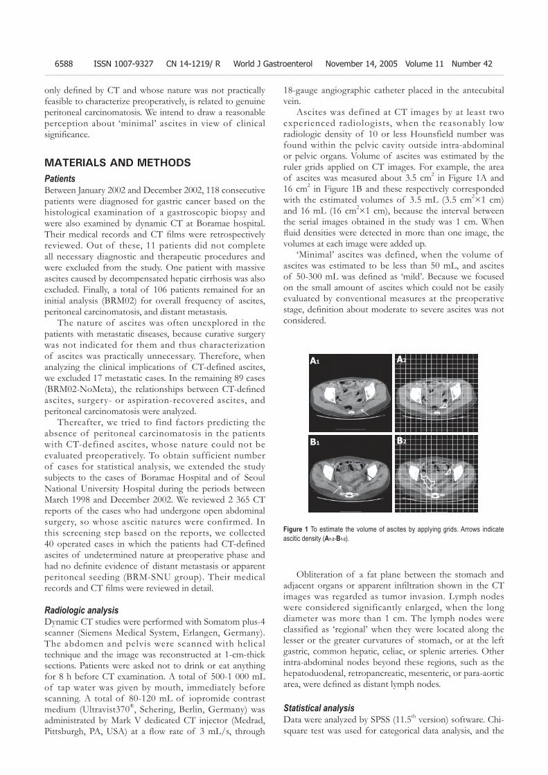
6588 ISSN 1007-9327 CN 14-1219/ R World J Gastroenterol November 14, 2005 Volume 11 Number 42
only defi ned by CT and whose nature was not practically feasible to characterize preoperatively, is related to genuine peritoneal carcinomatosis. We intend to draw a reasonable perception about ‘minimal’ ascites in view of clinical signifi cance.
MATERIALS AND METHODSPatientsBetween January 2002 and December 2002, 118 consecutive patients were diagnosed for gastric cancer based on the histological examination of a gastroscopic biopsy and were also examined by dynamic CT at Boramae hospital. Their medical records and CT fi lms were retrospectively reviewed. Out of these, 11 patients did not complete all necessary diagnostic and therapeutic procedures and were excluded from the study. One patient with massive ascites caused by decompensated hepatic cirrhosis was also excluded. Finally, a total of 106 patients remained for an initial analysis (BRM02) for overall frequency of ascites, peritoneal carcinomatosis, and distant metastasis.
The nature of ascites was often unexplored in the patients with metastatic diseases, because curative surgery was not indicated for them and thus characterization of ascites was practically unnecessary. Therefore, when analyzing the clinical implications of CT-defined ascites, we excluded 17 metastatic cases. In the remaining 89 cases (BRM02-NoMeta), the relationships between CT-defi ned ascites, surgery- or aspiration-recovered ascites, and peritoneal carcinomatosis were analyzed.
Thereafter, we tried to find factors predicting the absence of peritoneal carcinomatosis in the patients with CT-defined ascites, whose nature could not be evaluated preoperatively. To obtain sufficient number of cases for statistical analysis, we extended the study subjects to the cases of Boramae Hospital and of Seoul National University Hospital during the periods between March 1998 and December 2002. We reviewed 2 365 CT reports of the cases who had undergone open abdominal surgery, so whose ascitic natures were confirmed. In this screening step based on the reports, we collected 40 operated cases in which the patients had CT-defined ascites of undetermined nature at preoperative phase and had no defi nite evidence of distant metastasis or apparent peritoneal seeding (BRM-SNU group). Their medical records and CT fi lms were reviewed in detail.
Radiologic analysisDynamic CT studies were performed with Somatom plus-4 scanner (Siemens Medical System, Erlangen, Germany). The abdomen and pelvis were scanned with helical technique and the image was reconstructed at 1-cm-thick sections. Patients were asked not to drink or eat anything for 8 h before CT examination. A total of 500-1 000 mL of tap water was given by mouth, immediately before scanning. A total of 80-120 mL of iopromide contrast medium (Ultravist370®, Schering, Berlin, Germany) was administrated by Mark V dedicated CT injector (Medrad, Pittsburgh, PA, USA) at a fl ow rate of 3 mL/s, through
18-gauge angiographic catheter placed in the antecubital vein.
Ascites was defined at CT images by at least two experienced radiologists, when the reasonably low radiologic density of 10 or less Hounsfield number was found within the pelvic cavity outside intra-abdominal or pelvic organs. Volume of ascites was estimated by the ruler grids applied on CT images. For example, the area of ascites was measured about 3.5 cm2 in Figure 1A and 16 cm2 in Figure 1B and these respectively corresponded with the estimated volumes of 3.5 mL (3.5 cm2×1 cm) and 16 mL (16 cm2×1 cm), because the interval between the serial images obtained in the study was 1 cm. When fl uid densities were detected in more than one image, the volumes at each image were added up.
‘Minimal’ ascites was defined, when the volume of ascites was estimated to be less than 50 mL, and ascites of 50-300 mL was defi ned as ‘mild’. Because we focused on the small amount of ascites which could not be easily evaluated by conventional measures at the preoperative stage, defi nition about moderate to severe ascites was not considered.
Obliteration of a fat plane between the stomach and adjacent organs or apparent infi ltration shown in the CT images was regarded as tumor invasion. Lymph nodes were considered significantly enlarged, when the long diameter was more than 1 cm. The lymph nodes were classified as ‘regional’ when they were located along the lesser or the greater curvatures of stomach, or at the left gastric, common hepatic, celiac, or splenic arteries. Other intra-abdominal nodes beyond these regions, such as the hepatoduodenal, retropancreatic, mesenteric, or para-aortic area, were defi ned as distant lymph nodes.
Statistical analysisData were analyzed by SPSS (11.5th version) software. Chi-square test was used for categorical data analysis, and the
Figure 1 To estimate the volume of ascites by applying grids. Arrows indicate ascitic density (A1-2-B1-2).
A1 A2
B1 B2
↑
↑
↑
Chang DK et al. Clinical signifi cance of CT-defi ned ascites in gastric cancer 6589
univariate analysis and the multivariate logistic regression modeling were employed for assessing the predictive factors for the absence of peritoneal carcinomatosis in patients with CT-defi ned ascites.
RESULTSOverall proportion of CT-defined ascites, peritoneal carcinomatosis and distant metastasis in the patients with gastric cancer (BRM02 group)One hundred and six patients committed to the initial analysis consisted of 70 men and 36 women with a mean age of 63.3 years. The stage of gastric cancer at diagnosis was stage I, 23.6%; stage II, 7.5%; stage III, 34.0%; and stage IV, 34.9%. Patients with early gastric cancers were 27.4%.
Of the BRM02 patients, 22 (20.7%) had ascites defi ned by CT images. Based on the estimated volume of ascites, 12 patients belonged to the category of minimal ascites and 10 patients had mild or more ascites (11.3% and 9.4% of all patients, respectively).
Twenty patients out of the 106 BRM02 group (18.9%) were found to have peritoneal carcinomatosis that was confirmed by preoperative aspiration cytology or laparotomy no matter, whether they had CT-defi ned ascites or not. Seventeen patients (16%) had metastatic diseases to distant organs. Among these, four patients (3.8%) were confi rmed to have both distant metastasis and malignant peritoneal seeding (Table 1).
Clinical realities of CT-defi ned ascites in the metastasis-free patients (BRM02-NoMeta)Out of the remaining 89 patients (BRM02-NoMeta) after excluding those with metastatic diseases, 8 had minimal ascites, and 7 had mild ascites. There were no patients having ascites greater than mild category in the metastasis-free group. The nature of ascites was explored by surgery, in all 8 cases with minimal ascites, and in 5 out of 7 cases with mild ascites. The remaining two patients’ ascites were proven malignant by aspiration cytology, and surgery was not performed.
Ascites defi ned by CT images were not always identifi ed as peritoneal fluid on surgery. As for the CT-defined minimal ascites, ascitic fl uid was recovered at the surgical fi eld in only one (12.5%) of the cases. However, as CT-
estimated ascitic volume was higher, surgical correlation improved. In the cases of ‘mild’ ascites which had more volume than ‘minimal’, the concordance rate between CT-defi ned ascites and surgery- or aspiration-recovered ascites was as high as 85.7% (6 out of 7). On the other hand, 5 (6.8%) out of 74 patients who had no CT-identifi ed ascites preoperatively demonstrated ascites at the time of surgery (Table 2A).
The relationship between CT-defined ascites and peritoneal carcinomatosis was analyzed (Table 2B). Among the patients who did not show ascites at the CT images, seven (9.5%) had peritoneal carcinomatosis on surgery. Seven out of sixteen (43.8%) patients with peritoneal carcinomatosis did not demonstrate radiological signs of ascites at the preoperative CT. In the patients with CT-defined ‘minimal’ ascites, only 25% were concluded by surgery to have peritoneal carcinomatosis. This was contrast to the cases with CT-defined ‘mild’ ascites, in which all of the CT-defined ascites turned out to have occurred in the association with peritoneal carcinomatosis.
The presence of surgery- or aspiration-recovered ascites was not always accompanied with malignant peritoneal seeding, too. Four out of twelve patients (33.3%) with surgically or cytologically recovered ascites did not accompany peritoneal carcinomatosis, and 8% of ascites-free patients were actually positive for malignant peritoneal seeding (Table 2C).
Factors favoring absence of peritoneal carcinomatosis
Peritoneal carcinomatosis
Distant metastasis Negative Positive Unknown Total
Negative 73 (67/6/0)1 11 (6/2/3) 0 84 (73/8/3)
Positive 5 (4/1/0) 4 (1/0/3) 8 (5/3/0) 17 (10/4/3)Unknown 0 5 (1/0/4) 0 5 (1/0/4)
Total 78 (71/7/0) 20 (8/2/10) 8 (5/3/0) 106 (84/12/10)
Table 1 Proportion of peritoneal carcinomatosis, distant metastasis, and CT-defined ascites in the patients with gastric cancer that completed diagnostic work-up including dynamic CT in January 2002–December 2002 at Boramae Hospital (BRM02)
1CT-defined ascites (none/minimal/more than minimal).
Surgery- or aspiration-recovered ascitesCT-defi ned ascites Negative (%) Positive (%) Unknown (%) Total (%)
No 68 (91.9) 5 (6.8) 1 (1.4) 74 (100)
Minimal 7 (87.5) 1 (12.5) 0 (0.0) 8 (100)
Mild 0 (0.0) 6 (85.7) 1 (14.3) 7 (100)
Total 75 12 2 89
Peritoneal carcinomatosis
CT-defi ned ascites Negative (%) Positive (%) Total (%)
No 67 (90.5) 7 (9.5) 74 (100)
Minimal 6 (75.0) 2 (25.0) 8 (100)Mild 0 (0.0) 7 (100) 7 (100)Total 73 16 89
Peritoneal carcinomatosis
Surgery- or aspiration- Recovered ascites Negative (%) Positive (%) Total (%)
Negative 69 (92.0) 6 (8.0) 75 (100)Positive 4 (33.3) 8 (66.7) 12 (100)Unknown 0 (0.0) 21 (100) 2 (100)Total 73 16 89
c
b
a
Table 2 Clinical realities of CT-defi ned, preoperatively malignancy-undetermined ascites in the metastasis-free patients (BRM02-NoMeta)1
1Presence of ascites was not examined because operation was im-mediately ceased following observation of malignant omental cakes.

6590 ISSN 1007-9327 CN 14-1219/ R World J Gastroenterol November 14, 2005 Volume 11 Number 42
in the patients with CT-defi ned ascites which was not yet determined whether malignant or not.
We tried to search factors favoring absence of peritoneal carcinomatosis in the gastric cancer patients with CT-defi ned ascites in the larger number of cases. The sex and age distributions in these 40 patients (BRM-SNU) were not statistically different from those of the initial 106 gastric cancer patients (BRM02).
Majority of CT-defined ascites, of which the na-ture could not be preoperatively characterized, had vo-lumes of less than 10 mL with a left-skewed pattern (Skewness = 2.768, Figure 2). The range of volume was 1-300 mL; the mean was 34.8 mL and the median was 9.9 mL. Thirty-two cases were categorized as ‘minimal’ ascites and the rest eight were ‘mild’ ascites. Age and sex were not related to the volume of CT-defi ned ascites (data not shown), but the stage of cancer seemed lower in the patients with minimal ascites than those with mild ascites (Table 3).
The CT-defi ned ‘minimal’ ascites were associated with peritoneal carcinomatosis in only 12.5% and demonstrated surgically recovered ascites in only 9.4% (Table 4). These data confi rmatively reproduced the results obtained from the fewer cases in BRM02 (Table 2). Contrary to the CT-defined ‘minimal’ ascites, the CT-defined ‘mild’ ascites were accompanied with peritoneal carcinomatosis in as high as 75%, and were proved to have genuine fluid collection in 62.5%.
In addition to the ascitic volume, several other factors suggestive of negative peritoneal carcinomatosis were tested by the univariate analysis in the patients with CT-defined minimal or mild ascites (Table 5). When
enlargement of peritoneal lymph nodes were absent or confi ned to the regional perigastric area at the CT images, the negative predictive value for peritoneal carcinomatosis was 88.9%. Free of CT-defined perigastric invasion also negatively predicted peritoneal carcinomatosis in 90.9%. None of the patients with tumors with less than 3 cm were accompanied with malignant peritoneal seeding. However, except for the CT-defi ned ‘minimal’ ascites in comparison with the more abundant ascites, none of the above factors obtained statistical significance by multivariate analysis as an independent predictor for absence of malignant seeding.
DISCUSSION CT has been established as the most popular staging modality in gastric cancer although conventional or endoscopic ultrasonography, or laparoscopy can also be used[3,4,10-14]. The role of CT in detecting distant metastasis has been particularly well recognized; overall sensitivity for assessing liver metastasis reached about 62-89%[4,13,14].
However, CT is less reliable in identifying ascites or peritoneal carcinomatosis; the sensitivity was merely
CT-defi ned Peritoneal carcinomatosis Signifi cance Predictive valueascites Negative PositiveMinimal 28 (27/1)1 4 (2/2) NPV: 87.5%2
Mild 2 (1/1) 6 (2/4) P<0.01
Stage Minimal ascites (%) Mild ascites (%)1 9 (28.1) 0 (0)2 4 (12.5) 1 (12.5)3 5 (15.6) 1 (12.5)4 14 (43.8) 6 (75.0)Total 32 (100) 8 (100)
Table 3 Proportion of the minimal or mild CT-defined ascites at each UICC stage of gastric cancer
25
20
15
10
5
0
0-10
10-2
0
20-3
030
-40
40-5
050
-100
100-
200
200-
300
Estimated CT-defi ned ascites volume
Num
ber
of p
atie
nts
Figure 2 Frequency of patients based on the estimated CT-defined ascites volume.
Table 4 Peritoneal carcinomatosis and surgically recovered ascites in the patients with minimal or mild CT-defi ned ascites
1Surgically recovered ascites (negative/positive). 2NPV: negative predictive value of CT-defined ‘minimal’ ascites for peritoneal carcinomatosis, in comparison with CT-defi ned ‘mild’ ascites.
Peritoneal carcinomatosis Signifi cance Predictive value Negative Positive CT-defi ned enlarged lymph node (L/N) None or Regional L/N 24 3 NPV: 88.9%1
Distant L/N 6 7 P<0.01
CT-defi ned perigastric invasion
Negative 20 2 NPV: 90.9% Positive 10 8 P<0.05
Tumor size defi ned by endoscopy or UGIS
<3 cm 10 0 NPV: 100% 3-10 cm 16 6 >10 cm 4 4 P<0.05EGC vs AGC2
EGC 7 0 NPV: 100% AGC 23 10 NS
Table 5 Peritoneal carcinomatosis in the patients with CT-defi ned, yet preoperatively malignancy-undetermined ascites. Univariate analysis
1NPV: negative predictive value for peritoneal carcinomatosis. 2EGC: early gastric cancer; AGC: advanced gastric cancer.
(mL)

Chang DK et al. Clinical signifi cance of CT-defi ned ascites in gastric cancer 6591
36-46.7% for ascites and was 13-30% for malignant peritoneal seeding[4,12,14,15]. While the full blown peritoneal carcinomatosis demonstrating all the relevant radiologic features is diagnosed unambiguously, early and tiny malignant implantation cannot be easily decided. Although ascites has been regarded an important sign suggesting peritoneal carcinomatosis[5], the meaning of ascites may become ambiguous, as the dynamic CT detects subtle amounts of peritoneal fluid collection with increased sensitivity.
Then, what is the clinical significance of minimal ascites found in the CT image? Our data showed that minimal amounts of suspicious ascites defined by the dynamic CT were associated by peritoneal carcinomatosis in only 12.5-25%. This data may suggest that the patients need not hesitate to go through surgery, if the CT-defi ned minimal ascites is the only delusive clue for peritoneal seeding.
In the past, these amounts of minimal ascites might have never been detected by the CT and, therefore, might have never become a clinical issue. It may be argued whether the CT-defined minimal ascites is true or false-positive. However, this kind of question seems clinically out of point. Confirmation of real ascites may hardly be possible. Even surgery cannot be a gold standard in judging the presence of ascites because some blood or irrigated saline might be inevitably mixed with minimal, if any, ascites during surgery. Moreover, peritoneal cavity physiologically contains small amount of serous fluid which has been produced by permeable mesothelium[16], although this disperses diffusely in the peritoneal cavity and is not usually detected by the CT. It remains uncertain, if our CT-defined ‘minimal’ ascites is exaggerated physiologic fl uid, or pathologic ascites.
On the other hand, high index of suspicion may have rendered observers to detect phantom radiologic ascites in the cancer patients. However, an article by Chen et al. also reported that small amounts of ascites were detected at the perigastric area in 39% of patients with gastric cancers by endoscopic ultrasonography and they were not signifi cantly correlated with macroscopic peritoneal carcinomatosis[17]. Revealing the mechanism in the development of minimal ascites may be beyond the scope of this study.
No matter whether CT-defined minimal ascites is true or false, the apparent existence of minimal ascites reasonably defined by the dynamic CT may confuse a physician in determining the patient’s operability. This may be particularly critical in the patients with marginally poor condition. It is possible that, for example, elderly patients may give up radical surgery, when they are imprudently suggested the worse prognosis, because of the suspicious ascites. Adoption of laparoscopic staging in patients with CT-defined minimal ascites is another issue. Although laparoscopic examination may reveal the nature of minimal ascites more clearly[12], this procedure is not always routinely available at every hospital. In the most common practice setting, a physician cannot help but judge the operability according to the CT fi nding and other clinical manifestations.
If CT-defi ned minimal ascites has just low probability of peritoneal carcinomatosis, what additional factors could enhance the possibility of free peritoneum? Of course, it should be explored first whether peritoneal nodules, fat strands, pleated soft tissue, thickening of omental, mesentery and/or bowel wall, or other radiologic clues for malignant seeding were accompanied with or not. As our data suggested, when CT-defined lymph node enlargements were not found beyond the regional gastric area, CT-defi ned perigastric invasions were not detected, and the size of tumor was less than 3 cm, the probability of true peritoneal carcinomatosis may be very low, at least based on the univariate analysis.
In conclusion, majority of malignancy-undetermined CT-defi ned minimal ascites that was estimated to be less than 50 mL at the preoperative phase are not signifi cantly related to the peritoneal carcinomatosis. Therefore, if the peritoneal carcinomatosis was not definitely established, passive therapeutic strategy should not be applied to those patients simply because ascites is suspected. Definite meaning of CT-defined minimal ascites may need to be reinterpreted by the fi nal effect on survival after long-term follow-up, and this study is under way.
ACKNOWLEDGMENTSWe thank Dr. Yaron Niv for helpful discussion and critical reading of the manuscript; and Dr. Ajay Goel for editing the manuscript.
REFERENCES1 Plummer M, Franceschi S, Munoz N. Epidemiology of gastric
cancer. IARC Sci Publ 2004; 157: 311-3262 Adachi Y, Kitano S, Sugimachi K. Surgery for gastric cancer:
10-year experience worldwide. Gastric Cancer 2001; 4: 166-1743 Paramo JC, Gomez G. Dynamic CT in the preoperative
evaluation of patients with gastric cancer: correlation with surgical findings and pathology. Ann Surg Oncol 1999; 6: 379-384
4 D'Elia F, Zingarelli A, Palli D, Grani M. Hydro-dynamic CT preoperative staging of gastric cancer: correlation with pathological fi ndings. A prospective study of 107 cases. Eur Radiol 2000; 10: 1877-1885
5 Yoshikawa T, Kanari M, Tsuburaya A, Kobayahi O, Sairenji M, Motohashi H. Clinical and diagnostic significance of abdominal CT for peritoneal metastases in patients with primary gastric cancer. Gan To Kagaku Ryoho 2002; 29: 1925-1928
6 Villanueva A, Perez C, Sabate JM, Llauger J, Gimenez A, Sanchis E, Garcia T, Moreno A. Peritoneal carcinomatosis. Review of CT fi ndings in 107 cases. Rev Esp Enferm Dig 1995; 87: 707-714
7 Nakajima T, Harashima S, Hirata M, Kajitani T. Prognostic and therapeutic values of peritoneal cytology in gastric cancer. Acta Cytol 1978; 22: 225-229
8 Kodera Y, Nakanishi H, Yamamura Y, Shimizu Y, Torii A, Hirai T, Yasui K, Morimoto T, Kato T, Kito T, Tatematsu M.. Prognostic value and clinical implication of disseminated cancer cells in the peritoneal cavity detected by reverse transcriptase-polymerase chain reaction and cytology. Int. J. Cancer 1998: 79: 429-433
9 Doust BD. The use of ultrasound in the diagnosis of gastroenterological disease. Gastroenterology 1976; 70: 602-610
6592 ISSN 1007-9327 CN 14-1219/ R World J Gastroenterol November 14, 2005 Volume 11 Number 42
10 Habermann CR, Weiss F, Riecken R, Honarpisheh H, ohnacker S, Staedtler C, Dieckmann C, Schoder V, Adam G. Preoperative staging of gastric adenocarcinoma: comparison of helical CT and endoscopic US. Radiology 2004; 230: 465-471
11 Lavonius MI, Gullichsen R, Salo S, Sonninen P, Ovaska J. Staging of gastric cancer: a study with spiral computed tomography, ultrasonography, laparoscopy, and laparoscopic ultrasonography. Surg Laparosc Endosc Percutan Tech 2002; 12: 77-81
12 Stell DA, Carter CR, Stewart I, Anderson JR. Prospective comparison of laparoscopy, ultrasonography and computed tomography in the staging of gastric cancer. Br J Surg 1996; 83: 1260-1262
13 Adachi Y, Sakino I, Matsumata T, Iso Y, Yoh R, Kitano S, Okudaira Y. Preoperative assessment of advanced gastric carcinoma using computed tomography. Am J Gastroenterol
1997; 92: 872-87514 Kayaalp C, Arda K, Orug T, Ozcay N. Value of computed
tomography in addition to ultrasound for preoperative staging of gastric cancer. Eur J Surg Oncol 2002; 28: 540-543.
15 Nozoe T, Matsumata T, Sugimachi K. Usefulness of preoperative transvaginal ultrasonography for women with advanced gastric carcinoma. Am J Gastroenterol 1999; 94: 2509-2512
16 Nance FC. Diseases of the peritoneum, retroperitoneum, mesentery, and omentum. In: Haubrich WS, Schaffner F, Berk JE, eds. Bockus Gastroenterology. 5th ed. Philadelphia: W.B. Saunders, 1995: 3061-3096
17 Chen CH, Yang CC, Yeh YH. Preoperative staging of gastric cancer by endoscopic ultrasound: the prognostic usefulness of ascites detected by endoscopic ultrasound. J Clin Gastroenterol 2002; 35: 321-327
Science Editor Pravda J and Guo SY Language Editor Elsevier HK
PO Box 2345, Beijing 100023, China World J Gastroenterol 2005;11(42):6593-6600www.wjgnet.com World Journal of Gastroenterology ISSN [email protected] © 2005 The WJG Press and Elsevier Inc. All rights reserved.E L S E V I E R
• GASTRIC CANCER •
INTRODUCTIONDNA repair pathways are responsible for maintaining the integrity of the genome in face of environmental insults and general DNA replication errors, playing a role in pro-tecting it against mutations that lead to cancer[1]. So, poly-morphisms of DNA repair enzymes, which may alter the function or effi ciency of the DNA repair, may contribute to an increased risk of environmental carcinogenesis[2]. These low-penetrance susceptibility genes have common variants and interact with environmental factors, contrib-uting as a major factor to the populational incidence of cancer[3]. Several polymorphisms in genes that participate in different DNA repair pathways, such as XPD, XPF, ERCC1, XRCC1, XRCC3[4], hOGG1[5], XPA, XPB[6] and XPC[7], have been identifi ed and related to cancer suscep-tibility. The XRCC1 gene is responsible for a scaffolding pro-tein that directly associates with other proteins such as DNA polymerase β, PARP (ADP-ribose polymerase) and DNA ligase III in a complex, to facilitate the proc-esses of base excision repair (BER) or single-strand break repair[8]. The BER pathway repairs DNA damage caused by a variety of endogenous and exogenous factors, includ-ing oxidation, alkylating agents and ionizing radiation[1,9]. The XRCC1 protein can bind directly to both gapped and nicked DNA, as well as to gapped DNA associated with DNA
Márcia Cristina Duarte, Jucimara Colombo, Ana Elizabete Silva, UNESP - São Paulo State University, Department of Biology, Campus São José do Rio Preto, SP, BrazilAndrea Regina Baptista Rossit, FAMERP- São José do Rio Preto School of Medicine, Microorganism Investigation Center, São José do Rio Preto, SP, BrazilAlaor Caetano, Aldenis Albaneze Borim, FAMERP- São José do Rio Preto School of Medicine, Hospital de Base, São José do Rio Preto, SP, BrazilDurval Wornrath, Pio XII Foundation, Barretos, SP, BrazilSupported by Brazilian Agency CAPESCorrespondence to: Ana Elizabete Silva, Departamento de Biologia, UNESP, Rua Cristóvão Colombo, 2265, Jardim Nazareth, CEP: 15054-000, São José do Rio Preto, SP, Brazil. [email protected]: +55-17-32212384 Fax: +55-17-32212390Received: 2005-02-24 Accepted: 2005-04-09
AbstractAIM: To evaluate the association between poly-morphisms XRCC1 Arg194Trp and Arg399Gln and XRCC3 Thr241Met and the risk for chronic gastritis and gastric cancer, in a Southeastern Brazilian population.
METHODS: Genotyping by PCR-RFLP was carried out on 202 patients with chronic gastritis (CG) and 160 patients with gastric cancer (GC), matched to 202 (C1) and 150 (C2) controls, respectively.
RESULTS: No differences were observed among the studied groups with regard to the genotype distribution of XRCC1 codons 194 and 399 and of XRCC3 codon 241. However, the combined analyses of the three variant alleles (194Trp, 399Gln and 241Met) showed an increased risk for chronic gastritis when compared to the GC group. Moreover, an interaction between the polymorphic alleles and demographic and environmental factors was observed in the CG and GC groups. XRCC1 194Trp was associated with smoking in the CG group, while the variant alleles XRCC1 399Gln and XRCC3 241Met were related with gender, smoking, drinking and H pylori infection in the CG and GC groups.
CONCLUSION: Our results showed no evidence of a rela-tionship between the polymorphisms XRCC1 Arg194Trp and Arg399Gln and XRCC3 Thr241Met and the risk of chronic gastritis and gastric cancer in the Brazilian
Polymorphisms of DNA repair genes XRCC1 and XRCC3 , interaction with environmental exposure and risk of chronic gastritis and gastric cancer
Márcia Cristina Duarte, Jucimara Colombo, Andrea Regina Baptista Rossit, Alaor Caetano, Aldenis Albaneze Borim, Durval Wornrath, Ana Elizabete Silva
population, but the combined effect of these variants may interact to increase the risk for chronic gastritis, considered a premalignant lesion. Our data also indicate a gene-environment interaction in the susceptibility to chronic gastritis and gastric cancer.
© 2005 The WJG Press and Elsevier Inc. All rights reserved.
Key words: Gastric cancer; Gastritis; XRCC1 ; XRCC3 ; Polymorphism; Environmental exposure
Duarte MC, Colombo J, Rossit ARB, Caetano A, Borim AA, Wornrath D, Silva AE. Polymorphisms of DNA repair genes XRCC1 and XRCC3, interaction with environmental exposure and risk of chronic gastritis and gastric cancer. World J Gastroenterol 2005; 11(42): 6593-6600http://www.wjgnet.com/1007-9327/11/6593.asp
6594 ISSN 1007-9327 CN 14-1219/ R World J Gastroenterol November 14, 2005 Volume 11 Number 42
polymerase β, suggesting that this protein might be inde-pendently involved in DNA damage recognition[10]. Two polymorphisms, more often found in XRCC1’ conserved sites, lead to a C→T substitution at codon 194 in exon 6 and to a G→A substitution at codon 399 in exon 10 of the gene, leading to the amino acid alterations arginine (Arg) to tryptophan (Trp) and arginine (Arg) to glutamine (Gln), respectively. These changes in conserved protein sites may alter the BER capacity, increasing the chances of DNA damage[4].
The Arg399Gln variant is more frequent and has been associated mainly with head and neck[11], colorectal[12], gastric[13], esophageal[14,15], breast[16] and lung[17,18] cancers. The Arg194Trp polymorphism has been related to color-ectal[12], gastric[13], head and neck[19] and skin[20] cancers.
Protein XRCC3 functions in the DNA double-strand break (DSB) and cross-link repair[21] and interacts and stabilizes Rad51[22], one of the key components of the homologous repair (HR) pathway. The HR pathway uses a second intact copy of a homologous chromosome as a template to copy the information lost at the DSB site, resulting in a high-fi delity process and preventing chromosomal aberrations[9]. The main polymorphism in this gene involves the change of threonine (Thr) to methionine (Met) at co-don 241 in exon 7[4]. Little is known about the functional consequences of this variation, although some studies observed a positive relation between the Thr241Met poly-morphism and an increased risk for skin[23], bladder[24], breast[25] and lung[26] cancers.
So far, the investigations about interactions between XRCC1 and XRCC3 polymorphisms and environmental carcinogenesis have produced scarce and conflicting results[27,28,29,30], showing the functional complexity of these variants, that can include their interaction with en-vironmental factors, thus modulating the susceptibility to cancer. Regarding gastric cancer, only a few studies were conducted to investigate its association with XRCC1 and XRCC3 variants[13, 27,29,31].
In Brazil, gastric cancer is still one of the most fre-quent types of cancer. The estimate for 2005 points to the fourth place in incidence and mortality, with about 23 000 new cases and 12 000 deaths[32]. However, multiple factors are thought to play a role in gastric carcinogenesis, including diet[33], lifestyle[34], pathological changes in the stomach such as chronic gastritis[35], and genetic alterations[36,37], besides the infection by Helicobacter pylori, the fi rst bacterium to be termed as a defi nitive cause of cancer[38].
Thus, we conducted a study to evaluate the association between the polymorphisms XRCC1 Arg194Trp and Arg-399Gln and XRCC3 Thr241Met and the risk of chronic gastritis and gastric cancer in a Brazilian population, as well as the interaction between these polymorphisms and environmental factors involved in gastric carcinogenesis.
MATERIALS AND METHODSSubjects This was a case-control study on chronic gastritis and gastric cancer. The case groups comprised 202 patients
with a histopathologically confi rmed diagnosis of chronic gastritis (100 men and 102 women), with a mean age of 52 years (range 19-86 years), and 160 patients with a histopathologically confirmed diagnosis of gastric adenocarcinoma (118 men and 42 women), with a mean age of 61 years (range 28-93 years). All subjects were recruited from the Hospital de Base in São José do Rio Preto, SP, and from the Pio XII Foundation in Barretos, SP, Brazil. Gastric adenocarcinomas were classified as diffuse or intestinal types, according to the classifi cation proposed by Lauren[39], and the chronic gastritis cases according to the Sidney System[40]. H pylori infection was histologically established by the Giemsa staining technique. Two cancer-free control groups with no previous history of gastric disease were matched to the case groups with respect to age, gender and ethnicity. The control group for chronic gastritis (C1) was composed of 202 healthy individuals (100 men and 102 women) with a mean age of 51 years (range 20-85 years), and the control group for gastric cancer (C2) consisted of 150 healthy volunteers (108 men and 42 women) with a mean age of 59 years (range 22-93 years). Epidemiological data on the study population were collected using a standard interviewer-administered questionnaire, with questions about current and past occupation, smoking habits, alcohol intake and family history of cancer. This work was approved by the National Research Ethics Committee and written informed consent was obtained from all individuals.
DNA extraction and genotype analysesAbout 5 mL of whole blood were collected from all study participants in sterile EDTA-coated vacutainers. The samples were assigned a unique identifi er code. DNA was extracted according to Abdel-Rahman et al.[41] and stored at -20 ºC until used for genotyping.
Genotypic analyses of the XRCC1 gene were carried out by multiplex PCR-RFLP, using primers for codons 399 (F 5’-TTGTGCTTTCTCTGTGTCCA-3’ and R 5’-TCCTCCAGCCTTTTCTGATA-3’) and 194 (F 5’-GCCCCGTCCCAGGTA-3’ and R 5’-AGCCCCAAGACCCTTTCACT-3’), which generate a fragment of 615 and 491 bp, respectively, as previously described[12] with modifications. Briefly, PCR was performed in 25 μL reaction buffer containing 12.5 pmol each primer, 0.2 mmol/L of dNTPs, 3 mmol/L of MgCl2, about 100 ng DNA and 1 U of Taq DNA polymerase. The PCR products were digested overnight with 10 U of MspI at 37 ºC. The wild-type Arg allele for codon 194 is identifi ed by the presence of a 293 bp band, and the mutant Trp allele by the presence of a 313 bp band (indicative of the absence of the MspI cutting site). For codon 399, the presence of two bands of 375 and 240 bp, respectively, identifies the wild-type Arg allele, while the uncut 615 bp band identifies the mutant Gln allele (indicative of the absence of the MspI cutting site). A 178 bp band, resulting from an additional invariant MspI cutting site in the 491 bp amplified fragment, is always present and serves as an internal control for complete enzyme digestion.
Polymorphism of the XRCC3 gene was det e r -

Duarte MC et al. DNA repair gene polymorphisms and gastric lesions 6595
mined by PCR-RFLP, us ing codon 241 pr imers ( F - 5 ’ G C C T G G T G G T C A T C G A C T C 3 ’ e R - 5 ’ A C A G G G C T C T G G A A G G C A C T GCTCAGCTCACGCACC 3’), as previously described by David-Beabes et al.[42]. The 25 μL PCR mixture con-tained about 100 ng of DNA, 12.5 pmol of each primer, 0.2 mmol/L of dNTPs, 2 mmol/L of MgCl2 and 1 U of Taq DNA polymerase. The 552 bp amplifi ed product was digested overnight with 5 U of NlaIII at 37 ºC. The wild-type allele Thr was identifi ed by the presence of two 239 and 313 bp bands, while the mutant allele Met was represented by 105, 208, and 239 bp bands.
Statistical analysisChi-square or Fisher’s exact tests were util ized to compare the groups with regard to genotype frequencies and putative risk factors such as age, gender, ethnicity, smoking, drinking, H pylori infection and histological type of adenocarcinoma. To investigate the gene-environment interactions, the odds ratios (OR) and their 95% confidence intervals (95% CI) were calculated,
according to a combination of the XRCC1 and XRCC3 polymorphisms with putative risk factors. The statistical analyses were performed using Statidisk and GraphPad InStat computer software programs. A probability level (P) of less than 0.05 was used as criterion of signifi cance.
RESULTSCases and controls did not show any statistically signifi cant difference with regard to age, gender and ethnicity, indicating a well-matched study population. Gastric cancer (GC) patients were more likely to be cigarette smokers or alcohol drinkers than chronic gastritis (CG) patients and controls (C2), and this difference was statistically signifi cant (P<0.05), the same occurring with CG patients when compared to controls (C1). The XRCC1 and XRCC3 genotypes and the allele frequency distributions among cases and controls are presented in Table 1. The allele frequencies of polymorphisms 194Trp, 399Gln and 241Met were similar in cases and controls, not showing any statistically signifi cant
Genotypes CG (n = 202) C1 (n = 202) P GC (n = 160) C2 (n = 150) Pn (%) n (%) n (%) n (%)
XRCC1 Arg/Arg 176 (87.1) 183 (90.6) 140 (87.5) 130 (86.7)Codon 194 Arg/Trp 24 (11.9) 19 (9.4) 20 (12.5) 19 (12.7)
Trp/Trp 2 (1.0) 0 (0.0) 0 (0.0) 1 (0.6)Arg/Trp+Trp/Trp 26 (12.9) 19 (9.4) 0.2683 20 (12.5) 20 (13.3) 0.8269
Allele frequency 0.07 0.05 0.06 0.07XRCC1 Arg/Arg 98 (48.5) 95 (47) 73 (45.6) 70 (46.7)Codon 399 Arg/Gln 91 (45.0) 82 (40.6) 67 (41.9) 57 (38.0)
Gln/Gln 13 (6.5) 25 (12.4) 0.7651 20 (12.5) 23 (15.3) 0.5035Arg/Gln+Gln/Gln 104 (51.5) 107 (53.0) 87 (54.4) 80 (53.3)
Allele frequency 0.29 0.33 0.33 0.34XRCC3 Thr/Thr 92 (45.5) 84 (41.6) 84 (52.5) 67 (44.7)Codon 241 Thr/Met 81 (40.1) 89 (44.1) 53 (33.1) 60 (40.0)
Met/Met 29 (14.4) 29 (14.3) 0.4221 23 (14.4) 23 (15.3) 0.1679Thr/Met+Met/Met 110 (54.5) 118 (58.4) 76 (47.5) 83 (55.3)
Allele frequency 0.34 0.36 0.31 0.35
Table 1 XRCC1 and XRCC3 allele frequencies in patients with chronic gastritis (CG) and gastric cancer (GC) and in the respective control groups C1 and C2
XRCC1 XRCC3 GroupsCodon 194 Codon 399 Codon 241 CG GC OR (95%CI) P CG C1 OR (95%CI) P GC C2 OR (95%CI) P
All wide-type genotypes
Arg Arg Thr 33 32 1.0 (reference) 33 34 1.0 (reference) 32 23 1.0 (reference)One variant polymorphism
Trp Arg Thr 5 8 0.6 (0.17-2.04) 0.5469 5 6 1.2 (0.32-4.16) 1.0000 8 6 1.1 (0.32-3.44) 1.0000 Arg Gln Thr 51 38 1.4 (0.68-2.50) 0.5124 51 42 0.8 (0.42-1.51) 0.5234 38 34 1.2 (0.61-2.56) 0.5918 Arg Arg Met 50 27 2.0 (0.91-3.57) 0.1237 50 49 1.0 (0.51-1.75) 1.0000 27 34 2,0 (0.83-3.70) 0.1426 Two variant polymorphisms
Trp Gln Thr 3 5 0.6 (0.13-2.63) 0.7106 3 2 0.6 (0.1-4.16) 1.0000 5 4 1.1 (0.16-4.54) 1.0000 Arg Gln Met 43 43 1.0 (0.50-1.85) 1.0000 43 58 1.4 (0.69-2.43) 0.4309 43 39 1.2 (0.63-2.50) 0.5389Trp Arg Met 10 6 1.6 (0.52-5.00) 0.5771 10 6 0.6 (0.19-1.78) 0.4106 6 7 1.6 (0.48-5.55) 0.5999 Three variant polymorphisms
Trp Gln Met 8 1 8.3 (0.91-100) 0.0372 8 5 0.6 (0.17-2.04) 0.5400 1 3 5.0 (0.40-50.0) 0.3113
Table 2 Association between XRCC1 and XRCC3 genotype profi les and risk for chronic gastritis (CG) and gastric cancer (GC)

6596 ISSN 1007-9327 CN 14-1219/ R World J Gastroenterol November 14, 2005 Volume 11 Number 42
Table 3 shows the associations of the different genotypes with the variables gender, smoking, drinking and H pylori infection, among the groups. For this analysis, we combined the heterozygous and mutant homozygous genotypes. Allele 194Trp was associated with smoking, with an increased OR for chronic gastritis (4.16, 95%CI = 1.16-16.66), when we compared the CG and C1 groups. The comparison between the CG and GC groups revealed that in men who were smokers and drinkers there was an association with increased OR’s for gastric cancer in the individuals with the alleles XRCC1 399Gln (3.03,
Figure 1 PCR-RFLP of XRCC1 and XRCC3 genes. A: XRCC1 gene. Lane 1: molecular weight marker. Lane 2: wild-type homozygous codons 194 and 399. Lane 3: wild-type homozygous codon 194 and heterozygous codon 399. Lane 4: mutant homozygous codon 194 and wild-type homozygous codon 399. Lane 5: heterozygous codon 194 and wild type-homozygous codon 399. Lane 6: wild-type homozygous codon 194 and mutant homozygous codon 399; B: XRCC3 gene. Lane 1: molecular weight marker. Lanes 2 and 5: heterozygous codon 241. Lane 3: wild-type homozygous codon 241. Lane 4: mutant homozygous codon 241.
differences (P>0.05). However, a combined analysis of the XRCC1 Arg194Trp and Arg399Gln polymorphisms and the XRCC3 Thr241Met polymorphism (Table 2), and the assessment of the inter- and intra-gene interactions of these three polymorphisms revealed a statistically significant (P = 0.0372) association when the three variant alleles interacted in the chronic gastritis group, as compared to the gastric cancer group. Other combinations did not show any significant difference. The banding patterns of XRCC1 Arg194Trp and Arg399Gln and of XRCC3 Thr241Met are represented in Figure 1.
Table 3 Association of heterozygous and mutant homozygous to the polymorphisms of codons 194 and 399 of the XRCC1 gene and to the codon 241 of the XRCC3 gene with demographic and environmental risk factors in chronic gastritis (CG) and gastric cancer (GC) patients and the respective control groups C1 and C2
Groups Variable
Gender Smoke Drink H pylori infectionMale Female No Yes No Yes Positive Negativen (%) n (%) n (%) n (%) n (%) n (%) n (%) n (%)
XRCC1 CG 11 (42.3) 15 (57.7) 9 (34.6) 17 (65.4) 20 (77) 6 (23)Arg194Trp
+ C1 10 (52.6) 9 (47.4) 13 (68.4) 6 (31.6) 17 (89.5) 2 (10.5) Trp194Trp P = 0.4929 P = 0.0250 P = 0.4355
XRCC1 CG 55 (53) 49 (47) 48 (46.2) 56 (53.8) 71 (68.3) 33 (31.7) 45 (53.6) 39 (46.4)Arg399Gln GC 67 (77) 20 (23) 24 (27.6) 63 (72.4) 35 (40.2) 52 (59.8) 9 (23.7) 29 (76.3)
+ P = 0.0005 P = 0.0084 P = 0.0001 P = 0.0021Gln399Gln CG 55 (52.9) 49 (47.1) 48 (46.2) 56 (53.8) 71 (68.3) 33 (31.7)
C1 52 (48.5) 55 (51.4) 68 (63.6) 39 (36.4) 91 (85) 16 (15)P = 0.5335 P = 0.0111 P = 0.0039
GC 67 (77) 20 (23) 24 (27.6) 63 (72.4) 35 (40.2) 52 (59.8)C2 56 (70) 24 (30) 46 (57.5) 34 (42.5) 63 (78.8) 17 (21.2)
P = 0.3042 P = 0.0001 P = 0.0000XRCC3 CG 57 (51.8) 53 (48.2) 46 (41.8) 64 (58.2) 77 (70) 33 (30) 44 (52.4) 40 (47.6)
Thr241Met GC 59 (77.6) 17 (22.4) 21 (27.6) 55 (72.4) 36 (47.4) 40 (52.6) 10 (28.6) 25 (71.4)+ P = 0.0004 P = 0.0494 P = 0.0019 P = 0.0175
Met241Met CG 57 (51.8) 53 (48.2) 46 (41.8) 64 (58.2) 77 (47.4) 33 (52.6)C1 59 (50) 59 (50) 75 (63.6) 43 (36.4) 98 (79.6) 20 (20.4)
P = 0.7838 P = 0.0010 P = 0.0110GC 59 (77.6) 17 (22.4) 21 (27.6) 55 (72.4) 36 (47.4) 40 (52.6)C2 59 (71.1) 24 (28.9) 42 (50.6) 41 (49.4) 66 (79.6) 17 (20.5)
P = 0.3458 P = 0.0031 P = 0.0000
A
Codon 194 ArgArg Arg/Arg Trp/Trp Arg/Trp Arg/ArgCodon 399 Arg/Arg Arg/Gln Arg/Arg Arg/Arg Gln/Gln
615 bp
375 bp313 bp293 bp240 bp
178 bp
1 2 3 4 5 6B
Codon 241 Thr/Met Thr/Thr Met/Met Thr/Met
313 bp239 bp208 bp
105 bp
1 2 3 4 5
Duarte MC et al. DNA repair gene polymorphisms and gastric lesions 6597
95%CI = 1.58-5.55; 2.27, 95%CI = 1.22-4.16 and 3.22, 95%CI = 1.75-5.88, respectively) and XRCC3 241Met (3.22, 95%CI = 1.66-6.25; 1.88, 95%CI = 1.01-3.57 and 2.63, 95%CI = 1.41-4.76, respectively). Comparing the CG and GC groups further, we found increased OR’s for gastric cancer in the association between H pylori-negative subjects and the alleles XRCC1 399Gln (0.26, 95%CI = 0 .11-0 .63 ) and XRCC3 241Met (0 .36 , 95%CI = 0.15-0.85). An association between smoking and drinking and the polymorphisms 399Gln (2.04, 95%CI = 1.17-3.57 and 2.63, 95%CI = 1.35-5.26, respectively) and 241Met (2.44, 95%CI = 1.42-4.16 and 2.13, 95%CI = 1.10-4.00, respectively) was observed when we compared the CG and C1 groups, with increased OR’s for chronic gastritis. Likewise, when we compared GC and C2, we observed an association between smoking and drinking, with an increased OR for stomach cancer, and alleles 399Gln (3.57, 95%CI = 1.85-6.66 and 5.55, 95%CI = 2.77-10.99) and 241Met (2.70, 95%CI = 1.38-5.26 and 4.34, 95%CI = 2.17-9.09). The other evaluated parameters as age and ethnicity, presented no association with the studied polymorphisms.
DISCUSSIONThere is increasing evidence that genetic variation leads to different DNA repair capacities in the human population. So, common polymorphisms can play a role in the individual genetic susceptibility to cancer[43]. Very few studies have investigated the role of polymorphisms of the DNA repair genes XRCC1 and XRCC3 in the risk of gastric cancer, and, to our knowledge, so far no study examined both gene polymorphisms in this type of cancer and in chronic gastritis. We conducted the first case-control study to investigate the relationship between the polymorphisms XRCC1 Arg194Trp and Arg399Gln and XRCC3 Thr241Met and the risk of chronic gastritis and gastric cancer, in a Southeastern Brazilian population. Our data showed no association between the XRCC1 and XRCC3 polymorphisms and an increased risk for chronic gastritis and gastric cancer. To date, there are three published reports on investigations of the association between XRCC1 polymorphisms and gastric cancer risk[13,27,31], and only one that examined the infl uence of an XRCC3 polymorphism[29], with conflicting results. While Shen et al.[13] found the wild-genotype Arg194Arg and the mutant-genotype Arg399Gln in the XRCC1 gene to be associated with an increased risk of gastric cardia cancer, Ratnasinghe et al.[27] observed a signifi cant reduction in the risk of this type of cancer, both in Chinese populations. Similarly, Lee et al.[31] did not find any association with the risk of gastric cancer in a Korean population, but suggested that the 194Trp allele might be a protective allele with regard to gastric antral cancer. The only study that investigated the role of the XRCC3 polymorphism in gastric cancer in a Chinese population found no evidence of an association between this polymorphism and an increased risk of gastric cancer[29]. The differences observed in these reports may be due
to the different types of gastric cancer studied (cardia and antrum), which may have a distinct pathogenesis and ethnical differences[31]. In the Brazilian population, most gastric cancers are located in the antral region, as in the Korean population, and are commonly associated with H. pylori infection. We also found that in the gastric cancer group the variant allele frequency of 194Trp was lower (0.06) and those of 399Gln and 241Met were higher (0.33 and 0.31, respectively) than those reported in Asian populations[13,27,29,31]. These differences can yet be due to the presence of variants of the common susceptibility polymorphisms, not just a single one, but DNA repair genes or activation and detoxification genes may jointly contribute to the susceptibility of gastric and other cancers. Thus, it is important to include more gene polymorphisms for the same or other DNA repair pathways to verify the gene-gene interactions, as well as the gene-environment interactions that may be important in the etiology of the disease. When we assessed the association of XRCC1 Arg194trp and Arg399Gln and XRCC3 Thr241Met with the risk of chronic gastritis and gastric cancer, we found an increased risk for chronic gastritis when all three variant alleles were present at the same time, supporting the hypothesis of an additive effect of these three polymorphisms. There are no studies on chronic gastritis regarding its association with DNA repair gene polymorphisms, as there are for several metabolizing genes, such as GSTM1, GSTT1 and CYP2E1, in Brazilian[44] and Chinese[45] populations. Chronic gastritis, a frequent infl ammation of the stomach[46], is considered a premalignant lesion[47]. Gastritis may start after an H pylori infection and progress over time from an initially superficial form to more severe forms, including severe atrophic gastritis with intestinal metaplasia[48]. About 10% of patients with gastric atrophy develop gastric cancer within a time period of 15 years[46]. Therefore, a reduced DNA repair capacity due to variant alleles may allow mutations to accumulate in the DNA of the epithelial cells of the stomach, resulting from the infl ammatory process caused by H pylori or from environmental factors such as dietary habits and lifestyle, increasing the risk of gastric cancer. Gastric cancer has a complex etiology in which genetic and environmental factors play an important role. In this study, we observed a statistically significant association between variant alleles and demographic and environmental factors such as gender, smoking, drinking and H pylori infection. It is known that H pylori infection has a very important role in the development of chronic gastritis and in its development into gastric cancer[48]. Various mechanisms were proposed for H pylori-associated carcinogenesis, such as the formation of DNA adducts, the generation of free radicals, and a dysregulation of the gastric epithelial cell cycle[49,50]. So, these factors, associated with a decreased DNA repair capacity, may increase the risk for gastric cancer. Differently from these findings, we found a high frequency of variants 399Gln and 241Met
6598 ISSN 1007-9327 CN 14-1219/ R World J Gastroenterol November 14, 2005 Volume 11 Number 42
in H pylori-negative gastric cancer patients, as compared to chronic gastritis patients. However, these data must have been influenced by the great number of H pylori-negative individuals, as compared to the H. pylori-positive individuals found in the gastric cancer group, probably due to an underestimate of the histological diagnosis in these patients.
In smokers, the presence of XRCC1 194Trp was more frequent in chronic gastritis cases, while polymorphisms XRCC1 399Gln and XRCC3 241Met were more frequent in both the chronic gastritis and the gastric cancer groups, compared with healthy controls. Whether the mechanisms of tobacco carcinogens act in human gastric cancer is currently uncertain. The main carcinogens contained in tobacco smoke include polyaromatic hydrocarbons (PAH), N-nitrosamines and aromatic amines. Cigarette smoking increases the number of single-strand breaks and DNA adducts, which, if left unrepaired, can lead to gene mutation[51]. These DNA damages can be repaired by BER, in which the XRCC1 protein has an important role. Functional studies of XRCC1 variants observed a significantly elevated level of sister chromatid exchange (SCE) in peripheral blood lymphocytes after in vitro exposure to the tobacco-specifi c NNK in carriers of the 399Gln allele, but the same was not observed for the 194Trp polymorphism[52]. Duell et al.[53] reported higher frequencies of SCE for current smokers with the 399Gln polymorphism than for smokers with the Arg/Arg genotype.
Protein XRCC3 participates in the DSB repair by the homologous repair pathway and the 241Met variant may lead to biological implications for the enzyme’s function and/or the interaction with other proteins involved in DNA damage repair. Matullo et al.[54] associated the 241Met polymorphism with 32P-DNA adduct levels, indicating a possible role of the XRCC3 gene in the repair of bulky DNA adducts. Thus, variations in DNA repair capacity caused by polymorphisms of DNA repair genes may modulate the genotoxic effect of tobacco smoking.
Excessive alcohol consumption can also lead to DNA damage through the production of free radical intermediates, such as reactive oxygen species, which are produced during the ethanol metabolism[55]. The frequency of DNA single-strand breaks also increases with chronic exposure to alcohol[56]. We observed an association with alcohol consumption in the patients with chronic gastritis and gastric cancer, and found an increased risk for these diseases when polymorphisms XRCC1 399Gln and XRCC3 241Met were present.
In conclusion, in the Brazilian population studied, we did not find evidence of a relationship between the polymorphisms XRCC1 Arg194Trp and Arg399Gln and XRCC3 Thr241Met and the development of chronic gastritis and gastric cancer. However, intra- and inter-gene interactions may contribute to the development of chronic gastritis, a precursor lesion of stomach cancer. We also verifi ed a gene-environment interaction between the XRCC1 and XRCC3 polymorphisms, mainly with the habits of smoking and drinking, in the chronic gastritis
and gastric cancer patients. Our study is an important addition to the small number of previously published reports on DNA repair gene variants in gastric cancer and shows the need for further studies in different populations, to elucidate the role of these polymorphisms in carcinogenesis.
ACKNOWLEDGMENTSWe thank Prof. Dr. Antonio José Manzato for assistance with the statistical analysis.
REFERENCES1 Lindahl T. Suppression of spontaneous mutagenesis in
human cells by DNA base excision-repair. Mutat Res 2000; 462: 129-135
2 Mohrenweiser HW, Jones IM. Variation in DNA repair is a factor in cancer susceptibility: a paradigm for the promises and perils of individual and population risk estimation? Mutat Res 1998; 400: 15-24
3 Shields PG, Harris CC. Cancer risk and low-penetrance susceptibility genes in gene-environment interactions. J Clin Oncol 2000; 18: 2309-2315
4 Shen MR, Jones IM, Mohrenweiser H. Nonconservative amino acid substitution variants exist at polymorphic frequency in DNA repair genes in healthy humans. Cancer Res 1998; 58: 604-608
5 Ishida T, Takashima R, Fukayama M, Hamada C, Hippo Y, Fujii T, Moriyama S, Matsuba C, Nakahori Y, Morita H, Yazaki Y, Kodama T, Nishimura S, Aburatani H. New DNA polymorphisms of human MMH/OGG1 gene: prevalence of one polymorphism among lung-adenocarcinoma patients in Japanese. Int J Cancer 1999; 80: 18-21
6 Butkiewicz D, Rusin M, Harris CC, Chorazy M. Identifi cation of four single nucleotide polymorphisms in DNA repair genes: XPA and XPB (ERCC3) in Polish population. Hum Mutat 2000; 15: 577-578
7 Chavanne F, Broughton BC, Pietra D, Nardo T, Browitt A, Lehmann AR, Stefanini M. Mutations in the XPC gene in families with xeroderma pigmentosum and consequences at the cell, protein, and transcript levels. Cancer Res 2000; 60: 1974-1982
8 Caldecott KW, Aoufouchi S, Johnson P, Shall S. XRCC1 polypeptide interacts with DNA polymerase beta and possibly poly (ADP-ribose) polymerase, and DNA ligase III is a novel molecular 'nick-sensor' in vitro. Nucleic Acids Res 1996; 24: 4387-4394
9 Christmann M, Tomicic MT, Roos WP, Kaina B. Mechanisms of human DNA repair: an update. Toxicology 2003; 193: 3-34
10 Marintchev A, Mullen MA, Maciejewski MW, Pan B, Gryk MR, Mullen GP. Solution structure of the single-strand break repair protein XRCC1 N-terminal domain. Nat Struct Biol 1999; 6: 884-893
11 Sturgis EM, Castillo EJ, Li L, Zheng R, Eicher SA, Clayman GL, Strom SS, Spitz MR, Wei Q. Polymorphisms of DNA repair gene XRCC1 in squamous cell carcinoma of the head and neck. Carcinogenesis 1999; 20: 2125-2129
12 Abdel-Rahman SZ, Soliman AS, Bondy ML, Omar S, El-Badawy SA, Khaled HM, Seifeldin IA, Levin B. Inheritance of the 194Trp and the 399Gln variant alleles of the DNA repair gene XRCC1 are associated with increased risk of early-onset colorectal carcinoma in Egypt. Cancer Lett 2000; 159: 79-86
13 Shen H, Xu Y, Qian Y, Yu R, Qin Y, Zhou L, Wang X, Spitz MR, Wei Q. Polymorphisms of the DNA repair gene XRCC1 and risk of gastric cancer in a Chinese population. Int J Cancer 2000; 88: 601-606
14 Lee JM, Lee YC, Yang SY, Yang PW, Luh SP, Lee CJ, Chen CJ, Wu MT. Genetic polymorphisms of XRCC1 and risk of the
Duarte MC et al. DNA repair gene polymorphisms and gastric lesions 6599
esophageal cancer. Int J Cancer 2001; 95: 240-246 15 Yu HP, Zhang XY, Wang XL, Shi LY, Li YY, Li F, Su YH, Wang
YJ, Lu B, Sun X, Lu WH, Xu SQ. DNA repair gene XRCC1 polymorphisms, smoking, and esophageal cancer risk. Cancer Detect Prev 2004; 28: 194-199
16 Duell EJ, Millikan RC, Pittman GS, Winkel S, Lunn RM, Tse CK, Eaton A, Mohrenweiser HW, Newman B, Bell DA. Polymorphisms in the DNA repair gene XRCC1 and breast cancer. Cancer Epidemiol Biomarkers Prev 2001; 10: 217-222
17 Divine KK, Gilliland FD, Crowell RE, Stidley CA, Bocklage TJ, Cook DL, Belinsky SA. The XRCC1 399 glutamine allele is a risk factor for adenocarcinoma of the lung. Mutat Res 2001; 461: 273-278
18 Zhou W, Liu G, Miller DP, Thurston SW, Xu LL, Wain JC, Lynch TJ, Su L, Christiani DC. Polymorphisms in the DNA repair genes XRCC1 and ERCC2, smoking, and lung cancer risk. Cancer Epidemiol Biomarkers Prev 2003; 12: 359-365
19 Olshan AF, Watson MA, Weissler MC, Bell DA. XRCC1 polymorphisms and head and neck cancer. Cancer Lett 2002; 178: 181-186
20 Han J, Hankinson SE, Colditz GA, Hunter DJ. Genetic variation in XRCC1, sun exposure, and risk of skin cancer. Br J Cancer 2004; 91: 1604-1609
21 Thompson LH, Schild D. Recombinational DNA repair and human disease. Mutat Res 2002; 509: 49-78
22 Schild D, Lio YC, Collins DW, Tsomondo T, Chen DJ. Evidence for simultaneous protein interactions between human Rad51 paralogs. J Biol Chem 2000; 275: 16443-16449
23 Winsey SL, Haldar NA, Marsh HP, Bunce M, Marshall SE, Harris AL, Wojnarowska F, Welsh KI. A variant within the DNA repair gene XRCC3 is associated with the development of melanoma skin cancer. Cancer Res 2000; 60: 5612-5616
24 Matullo G, Guarrera S, Carturan S, Peluso M, Malaveille C, Davico L, Piazza A, Vineis P. DNA repair gene polymorphisms, bulky DNA adducts in white blood cells and bladder cancer in a case-control study. Int J Cancer 2001; 92: 562-567
25 Smith TR, Miller MS, Lohman K, Lange EM, Case LD, Mohrenweiser HW, Hu JJ. Polymorphisms of XRCC1 and XRCC3 genes and susceptibility to breast cancer. Cancer Lett 2003; 190: 183-190
26 Jacobsen NR, Raaschou-Nielsen O, Nexo B, Wallin H, Overvad K, Tjonneland A, Vogel U. XRCC3 polymorphisms and risk of lung cancer. Cancer Lett 2004; 213: 67-72
27 Ratnasinghe LD, Abnet C, Qiao YL, Modali R, Stolzenberg-Solomon R, Dong ZW, Dawsey SM, Mark SD, Taylor PR. Polymorphisms of XRCC1 and risk of esophageal and gastric cardia cancer. Cancer Lett 2004; 216: 157-64
28 Duan Z, Shen H, Lee JE, Gershenwald JE, Ross MI, Mansfi eld PF, Duvic M, Strom SS, Spitz MR, Wei Q. DNA repair gene XRCC3 241Met variant is not associated with risk of cutaneous malignant melanoma. Cancer Epidemiol Biomarkers Prev 2002; 11: 1142-1143
29 Shen H, Wang X, Hu Z, Zhang Z, Xu Y, Hu X, Guo J, Wei Q. Polymorphisms of DNA repair gene XRCC3 Thr241Met and risk of gastric cancer in a Chinese population. Cancer Lett 2004; 206: 51-58
30 Sanyal S, Festa F, Sakano S, Zhang Z, Steineck G, Norming U, Wijkstrom H, Larsson P, Kumar R, Hemminki K. Polymorphisms in DNA repair and metabolic genes in bladder cancer. Carcinogenesis 2004; 25: 729-734
31 Lee SG, Kim B, Choi J, Kim C, Lee I, Song K. Genetic polymorphisms of XRCC1 and risk of gastric cancer. Cancer Lett 2002; 187: 53-60
32 INCA – Instituto Nacional do Câncer, Ministério da Saúde. Estimativa 2005 – Estimativas de incidência por câncer no Brasil, 2004. Disponível em http://www.inca.org.br.
33 Kobayashi M, Tsubono Y, Sasazuki S, Sasaki S, Tsugane S. Vegetables, fruit and risk of gastric cancer in Japan: a 10-year follow-up of the JPHC Study Cohort I. Int J Cancer 2002; 102: 39-44
34 Sasazuki S, Sasaki S, Tsugane S. Cigarette smoking, alcohol
consumption and subsequent gastric cancer risk by subsite and histologic type. Int J Cancer 2002; 101: 560-566
35 Correa P. Human gastric carcinogenesis: a multistep and multifactorial process--First American Cancer Society Award Lecture on Cancer Epidemiology and Prevention. Cancer Res 1992; 52: 6735-6740
36 Chen X, Leung SY, Yuen ST, Chu KM, Ji J, Li R, Chan AS, Law S, Troyanskaya OG, Wong J, So S, Botstein D, Brown PO. Variation in gene expression patterns in human gastric cancers. Mol Biol Cell 2003; 14: 3208-3215
37 Tahara E. Genetic pathways of two types of gastric cancer. IARC Sci Publ 2004; 157: 327-349
38 IARC–Working Group on the Evaluation of Carcinogenic Risks to Humans. Lyon, 7-14 June 1994. Schistosomes, liver flukes and Helicobacter pylori. IARC Monogr Eval Carcinog Risks Hum 1994; 61: 1-241
39 Lauren P. The two histological main types of gastric carcinoma: diffuse and so-called intestinal-type carcinoma. an attempt at a histo-clinical classifi cation. Acta Pathol Microbiol Scand 1965; 64: 31-49
40 Price AB. The Sydney System: histological division. J Gastroenterol Hepatol 1991; 6: 209-222
41 Abdel-Rahman SZ, Nouraldeen AM, Ahmed AE. Molecular interaction of [2,3-14C] acrylonitrile with DNA in gastric tissue of rat. J Biochem Toxicol 1994; 9: 191-198
42 David-Beabes GL, Lunn RM, London SJ. No association between the XPD (Lys751G1n) polymorphism or the XRCC3 (Thr241Met) polymorphism and lung cancer risk. Cancer Epidemiol Biomarkers Prev 2001; 10: 911-912
43 Hu JJ, Mohrenweiser HW, Bell DA, Leadon SA, Miller MS. Symposium overview: genetic polymorphisms in DNA repair and cancer risk. Toxicol Appl Pharmacol 2002; 185: 64-73
44 Colombo J, Rossit AR, Caetano A, Borim AA, Wornrath DR, Silva AE. GSTT1, GSTM1 and CYP2E1 genetic polymorphisms in gastric cancer and chronic gastritis in a Brazilian population. World J Gastroenterol 2004; 10: 1240-1245
45 Setiawan VW, Zhang ZF, Yu GP, Li YL, Lu ML, Tsai CJ, Cordova D, Wang MR, Guo CH, Yu SZ, Kurtz RC. GSTT1 and GSTM1 null genotypes and the risk of gastric cancer: a case-control study in a Chinese population. Cancer Epidemiol Biomarkers Prev 2000; 9: 73-80
46 Cheli R, Giacosa A. Chronic atrophic gastritis and gastric mucosal atrophy--one and the same. Gastrointest Endosc 1983; 29: 23-25
47 Genta RM. Review article: Gastric atrophy and atrophic gastritis--nebulous concepts in search of a defi nition. Aliment Pharmacol Ther 1998; 12 Suppl 1: 17-23
48 Correa P. Helicobacter pylori and gastric carcinogenesis. Am J Surg Pathol 1995; 19 Suppl 1: S37-S43
49 Moss SF. The carcinogenic effect of H. pylori on the gastric epithelial cell. J Physiol Pharmacol 1999; 50: 847-856
50 Zhang ZW, Patchett SE, Farthing MJ. Role of Helicobacter pylori and p53 in regulation of gastric epithelial cell cycle phase progression. Dig Dis Sci 2002; 47: 987-995
51 Newcomb PA, Carbone PP. The health consequences of smoking. Cancer. Med Clin North Am 1992; 76: 305-331
52 Abdel-Rahman SZ, El-Zein RA. The 399Gln polymorphism in the DNA repair gene XRCC1 modulates the genotoxic response induced in human lymphocytes by the tobacco-specifi c nitrosamine NNK. Cancer Lett 2000; 159: 63-71
53 Duell EJ, Wiencke JK, Cheng TJ, Varkonyi A, Zuo ZF, Ashok TD, Mark EJ, Wain JC, Christiani DC, Kelsey KT. Polymorphisms in the DNA repair genes XRCC1 and ERCC2 and biomarkers of DNA damage in human blood mononuclear cells. Carcinogenesis 2000; 21: 965-971
54 Matullo G, Palli D, Peluso M, Guarrera S, Carturan S, Celentano E, Krogh V, Munnia A, Tumino R, Polidoro S, Piazza A, Vineis P. XRCC1, XRCC3, XPD gene polymorphisms, smoking and (32)P-DNA adducts in a sample of healthy subjects. Carcinogenesis 2001; 22: 1437-1445
55 Brooks PJ. DNA damage, DNA repair, and alcohol toxicity - a review. Alcohol Clin Exp Res 1997; 21: 1073-1082

6600 ISSN 1007-9327 CN 14-1219/ R World J Gastroenterol November 14, 2005 Volume 11 Number 42
56 Daiker DH, Shipp BK, Schoenfeld HA, Klimpel GR, Witz G, Moslen MT, Ward JB Jr. Effect of CYP2E1 induction by ethanol
on the immunotoxicity and genotoxicity of extended low-level benzene exposure. J Toxicol Environ Health A 2000; 59: 181-196
Science Editor Guo SY Language Editor Elsevier HK
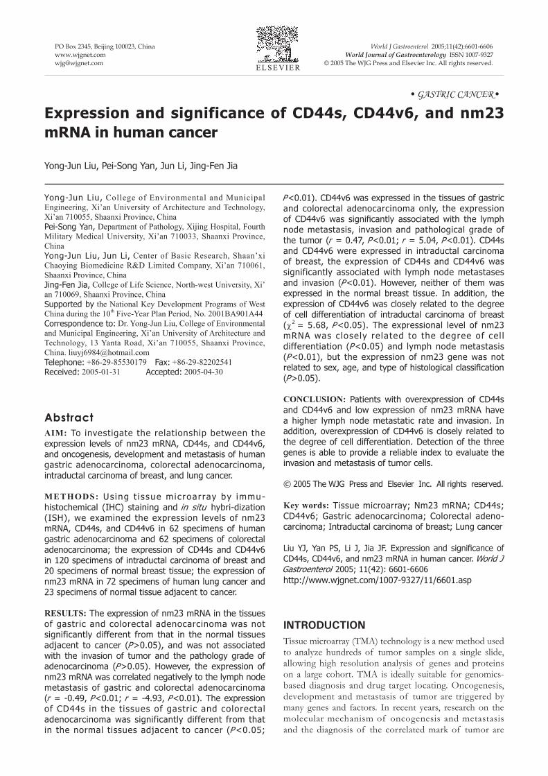
PO Box 2345, Beijing 100023, China World J Gastroenterol 2005;11(42):6601-6606www.wjgnet.com World Journal of Gastroenterology ISSN [email protected] © 2005 The WJG Press and Elsevier Inc. All rights reserved.E L S E V I E R
• GASTRIC CANCER•
INTRODUCTIONTissue microarray (TMA) technology is a new method used to analyze hundreds of tumor samples on a single slide, allowing high resolution analysis of genes and proteins on a large cohort. TMA is ideally suitable for genomics-based diagnosis and drug target locating. Oncogenesis, development and metastasis of tumor are triggered by many genes and factors. In recent years, research on the molecular mechanism of oncogenesis and metastasis and the diagnosis of the correlated mark of tumor are
Yong-Jun Liu, College of Environmental and Municipal Engineering, Xi’an University of Architecture and Technology, Xi’an 710055, Shaanxi Province, ChinaPei-Song Yan, Department of Pathology, Xijing Hospital, Fourth Military Medical University, Xi’an 710033, Shaanxi Province, ChinaYong-Jun Liu, Jun Li, Center of Basic Research, Shaan’xi Chaoying Biomedicine R&D Limited Company, Xi’an 710061, Shaanxi Province, ChinaJing-Fen Jia, College of Life Science, North-west University, Xi’an 710069, Shaanxi Province, ChinaSupported by the National Key Development Programs of West China during the 10th Five-Year Plan Period, No. 2001BA901A44Correspondence to: Dr. Yong-Jun Liu, College of Environmental and Municipal Engineering, Xi’an University of Architecture and Technology, 13 Yanta Road, Xi’an 710055, Shaanxi Province, China. [email protected]: +86-29-85530179 Fax: +86-29-82202541Received: 2005-01-31 Accepted: 2005-04-30
AbstractAIM: To investigate the relationship between the expression levels of nm23 mRNA, CD44s, and CD44v6, and oncogenesis, development and metastasis of human gastric adenocarcinoma, colorectal adenocarcinoma, intraductal carcinoma of breast, and lung cancer.
METHODS: Us ing t i ssue microar ray by immu-histochemical (IHC) staining and in situ hybri-dization (ISH), we examined the expression levels of nm23 mRNA, CD44s, and CD44v6 in 62 specimens of human gastric adenocarcinoma and 62 specimens of colorectal adenocarcinoma; the expression of CD44s and CD44v6 in 120 specimens of intraductal carcinoma of breast and 20 specimens of normal breast tissue; the expression of nm23 mRNA in 72 specimens of human lung cancer and 23 specimens of normal tissue adjacent to cancer.
RESULTS: The expression of nm23 mRNA in the tissues of gastric and colorectal adenocarcinoma was not significantly different from that in the normal tissues adjacent to cancer (P>0.05), and was not associated with the invasion of tumor and the pathology grade of adenocarcinoma (P>0.05). However, the expression of nm23 mRNA was correlated negatively to the lymph node metastasis of gastric and colorectal adenocarcinoma (r = -0.49, P<0.01; r = -4.93, P<0.01). The expression of CD44s in the tissues of gastric and colorectal adenocarcinoma was significantly different from that in the normal tissues adjacent to cancer (P <0.05;
Expression and significance of CD44s, CD44v6, and nm23 mRNA in human cancer
Yong-Jun Liu, Pei-Song Yan, Jun Li, Jing-Fen Jia
P<0.01). CD44v6 was expressed in the tissues of gastric and colorectal adenocarcinoma only, the expression of CD44v6 was signifi cantly associated with the lymph node metastasis, invasion and pathological grade of the tumor (r = 0.47, P<0.01; r = 5.04, P<0.01). CD44s and CD44v6 were expressed in intraductal carcinoma of breast, the expression of CD44s and CD44v6 was significantly associated with lymph node metastases and invasion (P<0.01). However, neither of them was expressed in the normal breast tissue. In addition, the expression of CD44v6 was closely related to the degree of cell differentiation of intraductal carcinoma of breast (χ2 = 5.68, P <0.05). The expressional level of nm23 mRNA was c losely re lated to the degree of cel l differentiation (P <0.05) and lymph node metastasis (P <0.01), but the expression of nm23 gene was not related to sex, age, and type of histological classifi cation (P>0.05).
CONCLUSION: Patients with overexpression of CD44s and CD44v6 and low expression of nm23 mRNA have a higher lymph node metastatic rate and invasion. In addition, overexpression of CD44v6 is closely related to the degree of cell differentiation. Detection of the three genes is able to provide a reliable index to evaluate the invasion and metastasis of tumor cells.
© 2005 The WJG Press and Elsevier Inc. All rights reserved.
Key words: Tissue microarray; Nm23 mRNA; CD44s; CD44v6; Gastric adenocarcinoma; Colorectal adeno-carcinoma; Intraductal carcinoma of breast; Lung cancer
Liu YJ, Yan PS, Li J, Jia JF. Expression and signifi cance of CD44s, CD44v6, and nm23 mRNA in human cancer. World J Gastroenterol 2005; 11(42): 6601-6606http://www.wjgnet.com/1007-9327/11/6601.asp
6602 ISSN 1007-9327 CN 14-1219/ R World J Gastroenterol November 14, 2005 Volume 11 Number 42
important objects in clinical research in oncology. CD44 is one of the transmembrane proteins on the surface of cells. Its distribution is very extensive and can be detected in lymphocytes and fibroblasts[1]. The expression of CD44v, which is related to the progression, metastasis and prognosis of tumor, has been detected in lung cancer, carcinoma of colon, esophageal cancer, carcinoma of breast, carcinoma of urinary bladder, liver cancer, cervix cancer, carcinoma of kidney and reticulosarcoma[2-5]. Nm23 was fi rst found by Steeg in 1988. Lower expression of nm23 is related to the metastasis of tumors, such as carcinoma of breast, lung cancer, gastric carcinoma, malignant melanoma, and ovary cancer, and has been regarded as the gene of transfer inhibition. In order to investigate the relationship between the expression level of nm23 mRNA, CD44s and CD44v6 and the oncogenesis, pathological grade, invasion, and metastasis of tumor, we detected the expression of nm23 mRNA, CD44s, and CD44v6 in tissues of human gastric adenocarcinoma, colorectal adenocarcinoma, intraductal carcinoma of breast and lung cancer, and also in tissues adjacent to cancer using tissue microarray by immunohistochemical (IHC) staining and in situ hybridization (ISH).
MATERIALS AND METHODSTissue specimensThe following tissue specimens were enrolled in this study: 40 specimens of colorectal adenocarcinoma (including 16 specimens of moderately differentiated colorectal adenocarcinoma, 24 poorly differentiated adenocarcinoma, 14 lymph node metastatic carcinoma, and 17 invasive carcinoma) and 22 specimens of normal colorectal tissue, 40 specimens of gastric adenocarcinoma (including 20 specimens of moderately differentiated gastric adenocarcinoma, 20 poorly differentiated adenocarcinoma, 15 lymph node metastatic carcinoma, and 23 invasive carcinoma) and 22 specimens of normal gastric tissue, 120 specimens of intraductal carcinoma of breast (including 86 specimens of moderately differentiated intraductal carcinoma, 34 poorly differentiated intraductal carcinoma, 30 lymph node metastatic carcinoma and 58 invasive carcinoma) and 20 specimens of normal breast tissue, 72 specimens of lung cancer (including 13 specimens of moderately differentiated carcinoma, 19 poorly differentiated carcinoma, 24 lymph node metastatic carcinoma) and 23 specimens of normal lung tissue. All patients underwent surgery at Xijing Hospital of the Fourth Military University, China in 2003. The resected specimens were fi xed in 10% formaldehyde.
ReagentsCD44s and CD44v6 monoclonal antibodies were obtained from MBI. SABC kit was bought from Sina-America Biotechnology Company. nm23 oligonucleotide probe and CSA test kit were purchased from Boshide Biotechnology Company. Biotin labeled anti-digitoxin (DIG) antibody was obtained from Sigma.
Tissue microarraySamples were fixed in buffered formaldehyde and subsequently paraffin-embedded. Histological sections (5 μm) were prepared from the specimens, and then diagnosed and labeled by the pathologist. Tissue microarray was performed on the instrument of microarray from Beecher, USA. A hole (diameter 0.5 mm) was made on the recipient block (blank paraffi n block), then the tissue core was taken on the supply block (labeled tissue block) and put in the hole. The former procedure was repeated and tissue microarray was completed. Histological sections (3-5 μm) were prepared with the instrument of Leica, Germany, and then rediagnosed by the pathologist.
Immunohistochemical analysis The sections for tissue microarray were dewaxed in water by normal technique, and then the antigen was restored in high pressure (at 121 °C for 10 min). Immunohistochemistry was performed with the SABC kit. The primary antibodies were CD44s and CD44v6 monoclonal antibodies. Staining was performed following the instructions of SABC kit.
Detection of nm23 mRNA expression by in situ hy-bridization All reagents and containers were treated by diethy-lprocarbonate (DEPC). In brief, the sections for tissue microarray were dewaxed in water by normal technique, immersed into 3% H2O2 at room temperature for 10 min, and then washed twice with distilled water. Twenty microgram per milliliter of freshly diluted protease K was added and digested for 20 min at 37 °C to expose mRNA nucleic acid segments. Twenty milliliter of glycerine (20%) was added to the dry bottom of the test kits to keep humidity, 20 μL of the pre-hybridization solution was added to each section and kept at 37 °C for 4 h. Then the supernumerary liquid was absorbed without washing, hybridization solution was added as mentioned above. The sections were then covered with protective membrane and put in homeothermia at 40 °C overnight. After the coverglass was removed, the sections were washed twice at 37 °C in 2×SSC for 5 min, once in 0.5×SSC for 15 min, once in 0.2×SSC for 15 min, and kept in 3% BSA at 37 °C for 30 min and in seal solution at 37 °C for 30 min. After being washed with 0.5 mol/L PBS, the biotin labeled anti-digitoxin antibody was added for 60 min at 37 °C, followed by biotin-peroxidase for 20 min at 37 °C and then washed with PBS. Color was showed by DAB. Finally, the sections were restained, dehydrated, pellucidated, and sealed. Negative controls were designed.
Assessment of immunohistochemical staining and in situ hybridizationSpecimens were considered positive when >50% of the tissue components were immunohistochemically stained brown-yellow in appropriate cellular compartment. Specimens were considered positive for ISH when >50% of the tissue components were stained blue in appropriate cellular compartment.
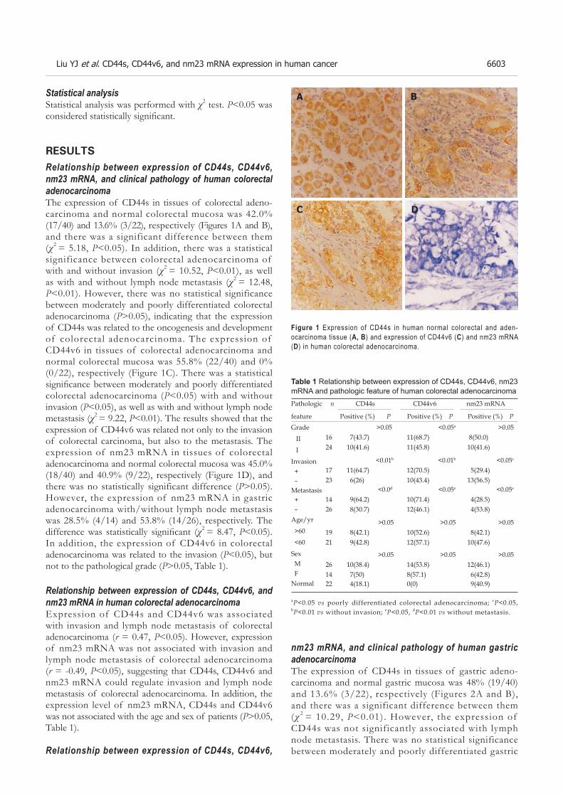
Liu YJ et al. CD44s, CD44v6, and nm23 mRNA expression in human cancer 6603
Statistical analysis Statistical analysis was performed with χ2 test. P<0.05 was considered statistically signifi cant.
RESULTSRelationship between expression of CD44s, CD44v6, nm23 mRNA, and clinical pathology of human colorectal adenocarcinoma The expression of CD44s in tissues of colorectal adeno-carcinoma and normal colorectal mucosa was 42.0% (17/40) and 13.6% (3/22), respectively (Figures 1A and B), and there was a significant difference between them (χ2 = 5.18, P<0.05). In addition, there was a statistical significance between colorectal adenocarcinoma of with and without invasion (χ2 = 10.52, P<0.01), as well as with and without lymph node metastasis (χ2 = 12.48, P<0.01). However, there was no statistical significance between moderately and poorly differentiated colorectal adenocarcinoma (P>0.05), indicating that the expression of CD44s was related to the oncogenesis and development of colorectal adenocarcinoma. The expression of CD44v6 in tissues of colorectal adenocarcinoma and normal colorectal mucosa was 55.8% (22/40) and 0% (0/22), respectively (Figure 1C). There was a statistical signifi cance between moderately and poorly differentiated colorectal adenocarcinoma (P<0.05) with and without invasion (P<0.05), as well as with and without lymph node metastasis (χ2 = 9.22, P<0.01). The results showed that the expression of CD44v6 was related not only to the invasion of colorectal carcinoma, but also to the metastasis. The expression of nm23 mRNA in tissues of colorectal adenocarcinoma and normal colorectal mucosa was 45.0% (18/40) and 40.9% (9/22), respectively (Figure 1D), and there was no statistically significant difference (P>0.05). However, the expression of nm23 mRNA in gastric adenocarcinoma with/without lymph node metastasis was 28.5% (4/14) and 53.8% (14/26), respectively. The difference was statistically signifi cant (χ2 = 8.47, P<0.05). In addition, the expression of CD44v6 in colorectal adenocarcinoma was related to the invasion (P<0.05), but not to the pathological grade (P>0.05, Table 1).
Relationship between expression of CD44s, CD44v6, and nm23 mRNA in human colorectal adenocarcinoma Expression of CD44s and CD44v6 was associated with invasion and lymph node metastasis of colorectal adenocarcinoma (r = 0.47, P<0.05). However, expression of nm23 mRNA was not associated with invasion and lymph node metastasis of colorectal adenocarcinoma (r = -0.49, P<0.05), suggesting that CD44s, CD44v6 and nm23 mRNA could regulate invasion and lymph node metastasis of colorectal adenocarcinoma. In addition, the expression level of nm23 mRNA, CD44s and CD44v6 was not associated with the age and sex of patients (P>0.05, Table 1).
Relationship between expression of CD44s, CD44v6,
nm23 mRNA, and clinical pathology of human gastric adenocarcinoma The expression of CD44s in tissues of gastric adeno-carcinoma and normal gastric mucosa was 48% (19/40) and 13.6% (3/22), respectively (Figures 2A and B), and there was a significant difference between them (χ 2 = 10.29, P<0.01). However, the expression of CD44s was not significantly associated with lymph node metastasis. There was no statistical significance between moderately and poorly differentiated gastric
Table 1 Relationship between expression of CD44s, CD44v6, nm23 mRNA and pathologic feature of human colorectal adenocarcinomaPathologic n CD44s CD44v6 nm23 mRNA
feature Positive (%) P Positive (%) P Positive (%) PGrade
II
I
Invasion + –Metastasis + –Age/yr
>60
<60
Sex M FNormal
1624
710
>0.05(43.7)(41.6)
<0.05a
11(68.7) 11(45.8)
>0.05 8(50.0) 10(41.6)
1723
116
<0.01b
(64.7)(26)
<0.01b
12(70.5) 10(43.4)
<0.05c
5(29.4) 13(56.5)
1426
98
<0.0d
(64.2)(30.7)
<0.05e
10(71.4) 12(46.1)
<0.05e
4(28.5) 4(53.8)
1921
89
>0.05(42.1)(42.8)
>0.05 10(52.6) 12(57.1)
>0.05 8(42.1) 10(47.6)
2614
107
>0.05(38.4)(50)
>0.05 14(53.8) 8(57.1)
>0.05 12(46.1) 6(42.8)
22 4(18.1) 0(0) 9(40.9)
aP<0.05 vs poorly differentiated colorectal adenocarcinoma; cP<0.05, bP<0.01 vs without invasion; eP<0.05, dP<0.01 vs without metastasis.
Figure 1 Expression of CD44s in human normal colorectal and aden-ocarcinoma tissue (A, B) and expression of CD44v6 (C) and nm23 mRNA (D) in human colorectal adenocarcinoma.
A B
C D

6604 ISSN 1007-9327 CN 14-1219/ R World J Gastroenterol November 14, 2005 Volume 11 Number 42
adenocarcinoma with/without invasion (P>0.05). The expression of CD44v6 in tissues of gastric adeno-carcinoma and normal gastric mucosa was 63.3% (25/40) and 0% (0/22), respectively (Figure 2C). There was a statistical significance between moderately and poorly differentiated gastric adenocarcinoma (χ2 = 9.19, P<0.01) with and without invasion (χ2 = 22.22, P<0.01), as well as with and without lymph node metastasis (χ2 = 10.36, P<0.01). The expression of nm23 mRNA in tissues of gastric adenocarcinoma and normal gastric mucosa was 47% (19/40) and 43% (9/22), respectively (Figure 2D) and there was no statistically signifi cant difference between them (P>0.05). However, the expression of nm23 mRNA in gastric adenocarcinoma with/without lymph node metastasis was 26.7% (4/15) and 60% (15/25) respectively, the difference was signifi cant (χ2 = 18.47, P<0.01, Table 2).
Relat ionship between expression of CD44v6 and nm23mRNA in human gastric adenocarcinoma The expression of CD44v6 was associated with lymph node metastasis of gastric adenocarcinoma (r = 5.04). However, the expression of nm23 mRNA was not associated with lymph node metastasis of gastric adenocarcinoma (r = -4.93, Table 2).
Relationship between the expression of CD44s, CD44v6, and clinical pathology of human intraductal carcinoma of breastThe expression of CD44s and CD44v6 in tissues of intraductal carcinoma of breast was 45.8% (55/120) and 53.3% (64/120), respectively (Figures 3A and B), but neither of them was expressed in normal breast t issue. The expression of CD44s and CD44v6 in
intraductal breast carcinoma with and without invasion had a signifi cant statistical difference (χ2 = 9.52, P<0.01; χ 2 = 22.89, P<0.001), as well as with and without lymph node metastasis (χ2 = 9.41, P<0.01; χ2 = 8.75, P<0.01). Furthermore, the expression of CD44s was not significantly associated with moderately and poorly differentiated intraductal breast carcinoma (P>0.05). However, the expression of CD44v6 was not only related to invasion and lymph node metastasis of intraductal breast carcinoma, but also related to its pathological grade (χ2 = 5.68, P<0.05, Table 3).
Relationship between expression of nm23 mRNA and clinical pathology of human lung cancer The expression of nm23 mRNA in tissues of lung cancer and normal lung tissues was 55.2% (40/72) and 82.6% (19/23), respectively (Figure 4), and there was a significant difference between them (χ2 = 5.42, P<0.05). The expression of nm23 mRNA in tissues of lung cancer with and without lymph node metastasis was 25.0% (6/24) and 70.8% (34/48), respectively and there was a signifi cant difference between them (χ2 = 13.61, P<0.001), as well as between moderately and poorly differentiated lung cancer (χ2 = 9.61, P<0.01), indicating that the expression of nm23 mRNA was associated with the oncogenesis, lymph node metastasis and pathological grade of lung cancer. In addition, the expression of nm23 mRNA in lung cancer was not associated with the age and sex of patients and the pathologic feature of lung cancer (P>0.05, Table 4).
DISCUSSION CD44s, a hyaluronic acid receptor, is important in regulating invasion and metastasis of tumor[3-7]. Previous studies demonstrated that the expression of CD44s is an important biological marker for predicting metastatic potential. Invasion and metastasis of tumor are a very complicated process, which is regulated by many correlated genes. CD44v6 may take part in the invasion and meta-
Pathologic n CD44s CD44v6 nm23 mRNA
feature Positive % P Positive % P Positive % PGrade II IIIInvasion + –Metastasis + –Age/year >60 <60 Normal
2020
810
>0.0540.050.0
b<0.0110 50.015 75.0
>0.05 9 45.0 10 50.0
2317
11 7
>0.0547.841.1
d<0.0118 78.2 7 41.0
>0.05 11 47.8 8 47.1
1525
612
>0.0540.048.0
f<0.0112 80.013 52.0
f<0.01 4 26.7 15 60.0
2119
10 8
>0.0547.242.1
>0.0513 61.912 63.1
>0.05 9 42.9 10 52.6
22 3 13.6 0 0 9 43.0
Table 2 Relationship between expression of CD44s, CD44v6, nm23 mRNA, and pathologic feature of human gastric adenocarcinoma
bP <0.01 vs well-differentiated gastric adenocarcinoma; dP <0.01 vs without invasion; fP <0.01 vs without metastasis.
A B
C D
Figure 2 Expression of CD44s in human normal gastric tissue (A, B) and expression CD44v6 (C) and nm23 mRNA (D) in human gastric adenocarcinoma
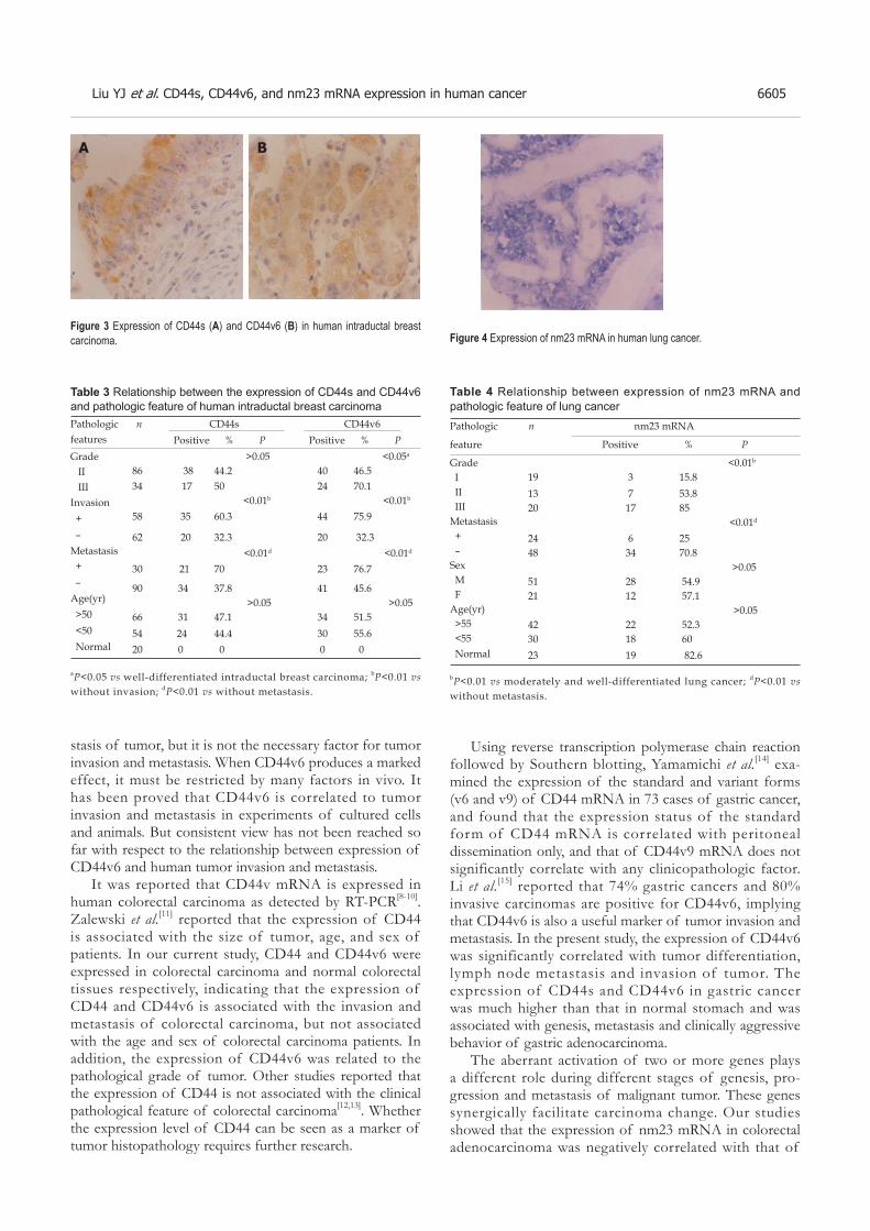
Liu YJ et al. CD44s, CD44v6, and nm23 mRNA expression in human cancer 6605
Using reverse transcription polymerase chain reaction followed by Southern blotting, Yamamichi et al.[14] exa-mined the expression of the standard and variant forms (v6 and v9) of CD44 mRNA in 73 cases of gastric cancer, and found that the expression status of the standard form of CD44 mRNA is correlated with peritoneal dissemination only, and that of CD44v9 mRNA does not significantly correlate with any clinicopathologic factor. Li et al.[15] reported that 74% gastric cancers and 80% invasive carcinomas are positive for CD44v6, implying that CD44v6 is also a useful marker of tumor invasion and metastasis. In the present study, the expression of CD44v6 was significantly correlated with tumor differentiation, lymph node metastasis and invasion of tumor. The expression of CD44s and CD44v6 in gastric cancer was much higher than that in normal stomach and was associated with genesis, metastasis and clinically aggressive behavior of gastric adenocarcinoma.
The aberrant activation of two or more genes plays a different role during different stages of genesis, pro-gression and metastasis of malignant tumor. These genes synergically facilitate carcinoma change. Our studies showed that the expression of nm23 mRNA in colorectal adenocarcinoma was negatively correlated with that of
stasis of tumor, but it is not the necessary factor for tumor invasion and metastasis. When CD44v6 produces a marked effect, it must be restricted by many factors in vivo. It has been proved that CD44v6 is correlated to tumor invasion and metastasis in experiments of cultured cells and animals. But consistent view has not been reached so far with respect to the relationship between expression of CD44v6 and human tumor invasion and metastasis.
It was reported that CD44v mRNA is expressed in human colorectal carcinoma as detected by RT-PCR[8-10]. Zalewski et al.[11] reported that the expression of CD44 is associated with the size of tumor, age, and sex of patients. In our current study, CD44 and CD44v6 were expressed in colorectal carcinoma and normal colorectal tissues respectively, indicating that the expression of CD44 and CD44v6 is associated with the invasion and metastasis of colorectal carcinoma, but not associated with the age and sex of colorectal carcinoma patients. In addition, the expression of CD44v6 was related to the pathological grade of tumor. Other studies reported that the expression of CD44 is not associated with the clinical pathological feature of colorectal carcinoma[12,13]. Whether the expression level of CD44 can be seen as a marker of tumor histopathology requires further research.
Figure 3 Expression of CD44s (A) and CD44v6 (B) in human intraductal breast carcinoma.
A B
Pathologic nfeatures
CD44s CD44v6 Positive % P Positive % P
Grade II IIIInvasion + –Metastasis + –Age(yr) >50 <50 Normal
>0.05 <0.05a
8634
38 17
44.2 50
4024
46.5 70.1
<0.01b <0.01b
58 35 60.3 44 75.9
62 20 32.3 20 32.3 <0.01d <0.01d
30 21 70 23 76.7
90 34 37.8 41 45.6 >0.05 >0.05
66 31 47.1 34 51.554 24 44.4 30 55.620 0 0 0 0
Table 3 Relationship between the expression of CD44s and CD44v6 and pathologic feature of human intraductal breast carcinoma
aP<0.05 vs well-differentiated intraductal breast carcinoma; bP<0.01 vs without invasion; dP<0.01 vs without metastasis.
Figure 4 Expression of nm23 mRNA in human lung cancer.
Table 4 Relationship between expression of nm23 mRNA and pathologic feature of lung cancer
bP<0.01 vs moderately and well-differentiated lung cancer; dP<0.01 vs without metastasis.
Pathologic n nm23 mRNA
feature Positive % P
Grade I II IIIMetastasis + –Sex M FAge(yr) >55 <55 Normal
<0.01b
19 3 15.813 7 53.820 17 85 <0.01d
24 6 2548 34 70.8 >0.0551 28 54.921 12 57.1 >0.0542 22 52.330 18 6023 19 82.6

6606 ISSN 1007-9327 CN 14-1219/ R World J Gastroenterol November 14, 2005 Volume 11 Number 42
CD44 and CD44v6, which was closely associated with the invasion and metastasis of colorectal adenocarcinoma. These results indicate that nm23 and CD44v6 synergically play positive and negative roles during lymph node metastasis of colorectal, and gastric adenocarcinoma. Further studies are needed to investigate the relationship between nm23 and CD44v6, which is important for tumor metastasis.
Mammary cancer is one of the most common malignant tumors. Although 50% of mammary cancers are surgically curable, 50% patients have metastasis within 5 years after surgery. Kaufmann et al.[16] reported that the positive expression rate of CD44v6 is 80% in primary tumor and 100% in focal tumor, but no expression is found in normal mammary tissue. The patients with positive expression of CD44v6 have a poor prognosis. There are still many correlative reports[17–20]. Our results showed that the expression of CD44s and CD44v6 in intraductal carcinoma was related with tumor invasion and metastasis, but not with the age and sex of patients.
Since the gene of nm23 was found, lower expression of nm23 has been found to be associated with high metastatic potential and poor prognosis of human mammary cancer, gastric cancer, lung cancer, melanoma, and ovary cancer. Nm23H1 is more important than nm23H2. Gazzeri et al.[21] reported that the expression of nm23H1 is not associated with clinicopathological features of adenocarcinoma, while it is associated with the development of squamous carcinoma. Lai et al.[22] reported that the expression of nm23H1 is positively correlated with tumor metastasis and prognosis. The lower the nm23H1 expression is, the poorer the prognosis is. Our studies showed that the expression of nm23 mRNA was correlated with lymph node metastasis (r = -4.93). The expression of nm23 mRNA was gradually reduced in normal lung tissue adjacent to cancer. In conclusion, nm23H1 may take part in the regulation of metastasis as a prohibitive gene, and may become a valuable target in the evaluation of tumor development and prognosis.
REFERENCES1 Gu H, Ni C, Zhan R. The expression of CD15 mRNA CD44v6
mRNA and nm23H1 mRNA in breast cancer and their clinical signifi cance. Zhonghua Yixue Zazhi 2000; 80: 854-857
2 Wang FL, Wei LX. Expression of CD44 variant exon 6 in lung cancers. Zhongguo Yixue Kexueyuan Xuebao 2001; 23: 401-402
3 Yan RL, Qian XH, Xin XY, Jin M, Hui HX, Wang DT, Wang J. Experimental study of anti-VEGF hairpin ribozyme gene inhibiting expression of VEGF and proliferation of ovarian cancer cells. Ai Zheng 2002; 21: 39-44
4 Ylagan LR, Scholes J, Demopoulos R. Cd44: a marker of squamous differentiation in adenosquamous neoplasms. Arch Pathol Lab Med 2000; 124: 212-215
5 Shimabukuro K, Toyama-Sorimachi N, Ozaki Y, Goi T, Furukawa K, Miyasaka M, Aso T. The expression patterns of standard and variant CD44 molecules in normal uterine cervix and cervical cancer. Gynecol Oncol 1997; 64: 26-34
6 Frank S, Rihs HP, Stocker W, Muller J, Dumont B, Baur X,
Schackert HK, Schackert G. Combined detection of CD44 isoforms by exon-specifi c RT-PCR and immunohistochemistry in primary human brain tumors and brain metastases. Biochem Biophys Res Commun 1996; 222: 794-801
7 Montgomery E, Abraham SC, Fisher C, Deasel MR, Amr SS, Sheikh SS, House M, Lilliemoe K, Choti M, Brock M, Ephron DT, Zahuruk M, Chadburn A. CD44 loss in gastric stromal tumors as a prognostic marker. Am J Surg Pathol 2004; 28: 168-177
8 Kuniyasu H, Oue N, Tsutsumi M, Tahara E, Yasui W. Heparan sulfate enhances invasion by human colon carcinoma cell lines through expression of CD44 variant exon 3. Clin Cancer Res 2001; 7: 4067-4072
9 Yamada Y, Itano N, Narimatsu H, Kudo T, Hirohashi S, Ochiai A, Tohnai I, Ueda M, Kimata K. CD44 variant exon 6 expressions in colon cancer assessed by quantitative analysis using real time reverse transcriptase-polymerase chain reaction. Oncol Rep 2003; 10: 1919-1924
10 Masaki T, Goto A, Sugiyama M, Matsuoka H, Abe N, Sakamoto A, Atomi Y. Possible contribution of CD44 variant 6 and nuclear beta-catenin expression to the formation of budding tumor cells in patients with T1 colorectal carcinoma. Cancer 2001; 92: 2539-2546
11 Zalewski B, Famulski W, Sulkowska M, Sobaniec-Lotowska M, Piotrowski Z, Kisielewski W, Sulkowski S. CD44 expression in colorectal cancer. An immunohistochemical study including correlation with cathepsin D immunoreactivity and some tumour clinicopathological features. Folia Histochem Cytobiol 2001; 39 Suppl 2: 152-153
12 Sokmen S, Lebe B, Sarioglu S, Fuzun M, Terzi C, Kupelioglu A, Ellidokuz H. Prognostic value of CD44 expression in colorectal carcinomas. Anticancer Res 2001; 21: 4121-4126
13 Ishida T. Immunohistochemical expression of the CD44 variant 6 in colorectal adenocarcinoma. Surg Today 2000; 30: 28-32
14 Yamamichi K, Uehara Y, Kitamura N, Nakane Y, Hioki K. Increased expression of CD44v6 mRNA signifi cantly correlates with distant metastasis and poor prognosis in gastric cancer.Int J Cancer 1998; 79: 256-262
15 Li H, Li J, Guo L. Characteristics of expression of CD44v and receptor for HA-mediated motility (RHAMM) in multi-step gastrocarcinogenesis. Zhonghua Zhongliu Zazhi 1999; 21: 329-331
16 Kaufmann M, Heider KH, Sinn HP, von Minckwitz G, Ponta H, Herrlich P. CD44 variant exon epitopes in primary breast cancer and length of survival. Lancet 1995; 345: 615-619
17 Kopp R, Classen S, Wolf H, Gholam P, Possinger K, Wilmanns W. Predictive relevance of soluble CD44v6 serum levels for the responsiveness to second line hormone- or chemotherapy in patients with metastatic breast cancer. Anticancer Res 2001; 21: 2995-3000
18 Saddik M, La i R . CD44s as a surrogate marker for distinguishing intraductal papilloma from papillary carcinoma of the breast. J Clin Pathol 1999; 52: 862-864
19 Sanchez Lockhart M, Hajos SE, Basilio FM, Mongini C, Alvarez E. Splice variant expression of CD44 in patients with breast and ovarian cancer. Oncol Rep 2001; 8: 145-151
20 Berner HS, Nesland JM. Expression of CD44 isoforms in infi ltrating lobular carcinoma of the breast. Breast Cancer Res Treat 2001; 65: 23-29
21 Gazzeri S, Brambilla E, Negoescu A, Thoraval D, Veron M, Moro D, Brambilla C.Overexpression of nucleoside diphosphate/kinase A/nm23-H1 protein in human lung tumors:association with tumor progression in squamous carcinoma. Lab Invest 1996; 74: 158-167
22 Lai WW, Wu MH, Yan JJ, Chen FF. Immunohistochemical analysis of nm23-H1 in stage I non-small cell lung cancer:a useful marker in prediction of metastases. Ann Thorac Surg 1996; 62: 1500-1504
Science Editor Wang XL and Guo SY Language Editor Elsevier HK
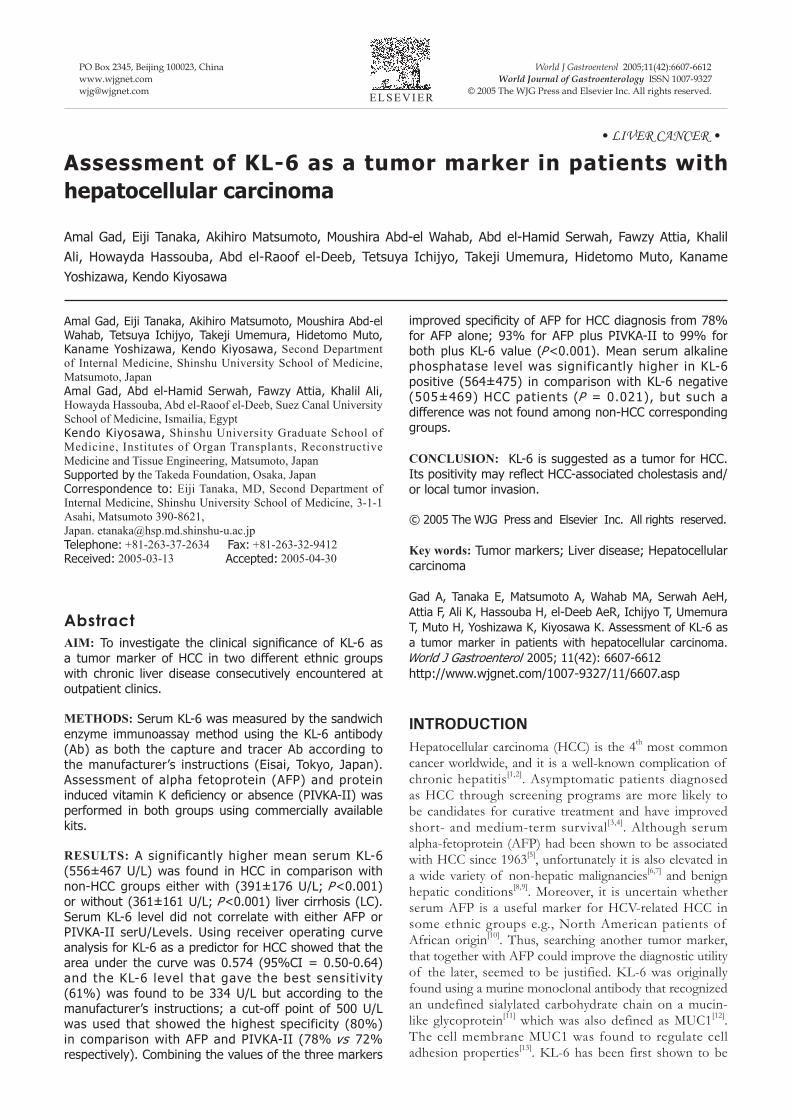
PO Box 2345, Beijing 100023, China World J Gastroenterol 2005;11(42):6607-6612www.wjgnet.com World Journal of Gastroenterology ISSN [email protected] © 2005 The WJG Press and Elsevier Inc. All rights reserved.E L S E V I E R
• LIVER CANCER •
INTRODUCTIONHepatocellular carcinoma (HCC) is the 4th most common cancer worldwide, and it is a well-known complication of chronic hepatitis[1,2]. Asymptomatic patients diagnosed as HCC through screening programs are more likely to be candidates for curative treatment and have improved short- and medium-term survival[3,4]. Although serum alpha-fetoprotein (AFP) had been shown to be associated with HCC since 1963[5], unfortunately it is also elevated in a wide variety of non-hepatic malignancies[6,7] and benign hepatic conditions[8,9]. Moreover, it is uncertain whether serum AFP is a useful marker for HCV-related HCC in some ethnic groups e.g., North American patients of African origin[10]. Thus, searching another tumor marker, that together with AFP could improve the diagnostic utility of the later, seemed to be justified. KL-6 was originally found using a murine monoclonal antibody that recognized an undefined sialylated carbohydrate chain on a mucin-like glycoprotein[11] which was also defined as MUC1[12]. The cell membrane MUC1 was found to regulate cell adhesion properties[13]. KL-6 has been first shown to be
Amal Gad, Eiji Tanaka, Akihiro Matsumoto, Moushira Abd-el Wahab, Tetsuya Ichijyo, Takeji Umemura, Hidetomo Muto, Kaname Yoshizawa, Kendo Kiyosawa, Second Department of Internal Medicine, Shinshu University School of Medicine, Matsumoto, JapanAmal Gad, Abd el-Hamid Serwah, Fawzy Attia, Khalil Ali, Howayda Hassouba, Abd el-Raoof el-Deeb, Suez Canal University School of Medicine, Ismailia, EgyptKendo Kiyosawa, Shinshu University Graduate School of Medicine, Institutes of Organ Transplants, Reconstructive Medicine and Tissue Engineering, Matsumoto, JapanSupported by the Takeda Foundation, Osaka, JapanCorrespondence to: Eiji Tanaka, MD, Second Department of Internal Medicine, Shinshu University School of Medicine, 3-1-1 Asahi, Matsumoto 390-8621, Japan. [email protected]: +81-263-37-2634 Fax: +81-263-32-9412Received: 2005-03-13 Accepted: 2005-04-30
AbstractAIM: To investigate the clinical signifi cance of KL-6 as a tumor marker of HCC in two different ethnic groups with chronic liver disease consecutively encountered at outpatient clinics.
METHODS: Serum KL-6 was measured by the sandwich enzyme immunoassay method using the KL-6 antibody (Ab) as both the capture and tracer Ab according to the manufacturer’s instructions (Eisai, Tokyo, Japan). Assessment of alpha fetoprotein (AFP) and protein induced vitamin K defi ciency or absence (PIVKA-II) was performed in both groups using commercially available kits.
RESULTS: A significantly higher mean serum KL-6 (556±467 U/L) was found in HCC in comparison with non-HCC groups either with (391±176 U/L; P<0.001) or without (361±161 U/L; P<0.001) liver cirrhosis (LC). Serum KL-6 level did not correlate with either AFP or PIVKA-II serU/Levels. Using receiver operating curve analysis for KL-6 as a predictor for HCC showed that the area under the curve was 0.574 (95%CI = 0.50-0.64) and the KL-6 level that gave the best sensit ivity (61%) was found to be 334 U/L but according to the manufacturer’s instructions; a cut-off point of 500 U/L was used that showed the highest specificity (80%) in comparison with AFP and PIVKA-II (78% vs 72% respectively). Combining the values of the three markers
Assessment of KL-6 as a tumor marker in patients with hepatocellular carcinoma
Amal Gad, Eiji Tanaka, Akihiro Matsumoto, Moushira Abd-el Wahab, Abd el-Hamid Serwah, Fawzy Attia, Khalil Ali, Howayda Hassouba, Abd el-Raoof el-Deeb, Tetsuya Ichijyo, Takeji Umemura, Hidetomo Muto, Kaname Yoshizawa, Kendo Kiyosawa
improved specifi city of AFP for HCC diagnosis from 78% for AFP alone; 93% for AFP plus PIVKA-II to 99% for both plus KL-6 value (P<0.001). Mean serum alkaline phosphatase level was significantly higher in KL-6 positive (564±475) in comparison with KL-6 negative (505±469) HCC patients (P = 0.021), but such a difference was not found among non-HCC corresponding groups.
CONCLUSION: KL-6 is suggested as a tumor for HCC. Its positivity may refl ect HCC-associated cholestasis and/or local tumor invasion.
© 2005 The WJG Press and Elsevier Inc. All rights reserved.
Key words: Tumor markers; Liver disease; Hepatocellular carcinoma
Gad A, Tanaka E, Matsumoto A, Wahab MA, Serwah AeH, Attia F, Ali K, Hassouba H, el-Deeb AeR, Ichijyo T, Umemura T, Muto H, Yoshizawa K, Kiyosawa K. Assessment of KL-6 as a tumor marker in patients with hepatocellular carcinoma. World J Gastroenterol 2005; 11(42): 6607-6612http://www.wjgnet.com/1007-9327/11/6607.asp

6608 ISSN 1007-9327 CN 14-1219/ R World J Gastroenterol November 14, 2005 Volume 11 Number 42
elevated in patients with interstitial pneumonia[14]. It was also reported to have a high positive rate in different non-hepatic malignancies and its expression was also correlated with metastatic potential of the primary tumor in some of them[15-17]. It has also been studied as a fi brosis marker in patients with HCV-related chronic liver disease[18] and was found to correlate with the degree of irregular regeneration of hepatocytes[19]. A recent study addressed its clinical signifi cance as a tumor marker in HCV-related HCC[20]. However, all these studies investigated KL-6 in HCV-related disease only so that its actual significance as a marker for screening HCC in patients with different chronic liver disease is not yet fully understood. In this study, we aimed to investigate KL-6 as a tumor marker in consecutive patients with chronic liver disease seen at outpatient settings in two different ethnic groups of possible different risk factors for HCC, so that we could get a wider spectrum of disease in order to assess KL-6 validity for HCC screening.
MATERIALS AND METHODSStudy populationWe conducted a cross-sectional study between October 2001 and November 2002. Data were gathered from two Affiliations; Shinshu University (Japan) and Suez Canal University (Egypt) Hospitals. A total of 334 consecutive patients with chronic liver disease seen at outpatient liver clinics in the two settings (who met our inclusion/exclusion criteria) were included; of them: 110 patients were diagnosed as HCC with a mean age of 61±11 years and M:F (4:1). Sixty-fi ve were Egyptians and 45 Japanese with viral-related liver disease accounting for 94% and 98% of them respectively. Non-HCC patients were 234 with a mean age of 56±13 years; M:F (7/3). One hundred and six were Egyptians and 128 Japanese with viral-related liver disease accounting for 91% and 84% of them respectively
(Table 1).Chronic liver disease and cirrhosis were identified
and diagnosed according to liver biopsy fi ndings, clinical and/or radiological evidence of portal hypertension. HCC was excluded by imaging studies (abdominal ultrasound (US), computed tomography (CT), magnetic resonance imaging (MRI) and/or hepatic angiography), one of which must have been performed at least 6 months following the measurement of AFP.
HCC was diagnosed when meeting our inclusion criteria of positive cytology and/or histology or by the presence of characteristic hepatic masses on liver CT, MRI and/or hepatic angiography (i.e., enlarging tumors and/or tumors with typical arterial vascularization).
We excluded patients with alcoholic and schistosomal liver diseases from our study populations. We had also excluded patients known from their medical history to have interstitial lung fibrosis or any other lung disease from our study population.
Tumor markers measurementSerum KL-6 was measured by the sandwich enzyme immunoassay method using the KL-6 antibody (Ab) as both the capture and tracer Ab (14) according to the manufacturer’s instructions (Eisai, Tokyo, Japan). KL-6 cut-off point was set at 500 U/L for this study. Assessment of alpha fetoprotein (AFP) and protein-induced vitamin K deficiency or absence (PIVKA-II) was performed using commercially available kits. Cut-off points were set at 10 ng/mL for AFP and 40 mAU/L for PIVKA-II.
Statistical analysisUnivariate statistical analysis was performed using Student's t-test for quantitative and χ2 test with Yates’ correction for qualitative data. Fisher’s exact test was used
Egyptian Japanese
HCC (+) HCC (–) P HCC (+) HCC (–) P
n = 65 n = 106 n = 45 n = 128
Mean age (SD, yr) 57±11b 47±9 <0.001 66±10b 63±10 NS
Age <50 yr 16 (25)d 65 (61) <0.001 3 (7)d 17 (13) NS
Male 50 (77) 82 (77) NS 38 (84) 87 (68) 0.024Liver disease
Viral 61 (94) 96 (91) 44 (98) 107 (84)HCV-related 59 (91) 92 (87) NS 36 (80) 81 (63) 0.031HBV-related 2 (3)1 4 (4) NS 8 (18)1 28 (22) NSNon-viral 4 (6) 10 (9) NS 1 (2) 20 (16) 0.010Cirrhosis 46 (71) 45 (42) <0.001 40 (89) 40 (31) <0.001Child’s C 25 (38)f 17 (16) 0.001 4 (9)f 1 (1) 0.017Mean±(SD)ALT (IU/L) 73±95 66±45 0.08 55±35 50±39 NSSerum Albumin (g/L) 3.0±0.7 3.0±0.5 NS 3.6 ±0.5 4.2±0.4 <0.001Platelet count×1 000/mL3 186±107h 89±53 0.001 130±51h 170±71 <0.001AFP >10 ng/mL (+) 64 (99) 28 (26) <0.001 30 (67) 23 (18) <0.001PIVIKA>40 mAU/L (+) 51 (79) 38 (36) <0.001 16 (36) 27 (21) 0.047
Table 1 Background data of the study groups
bP<0.001, dP<0.001, fP<0.001, hP<0.001 vs Japannese, 1P = 0.001.
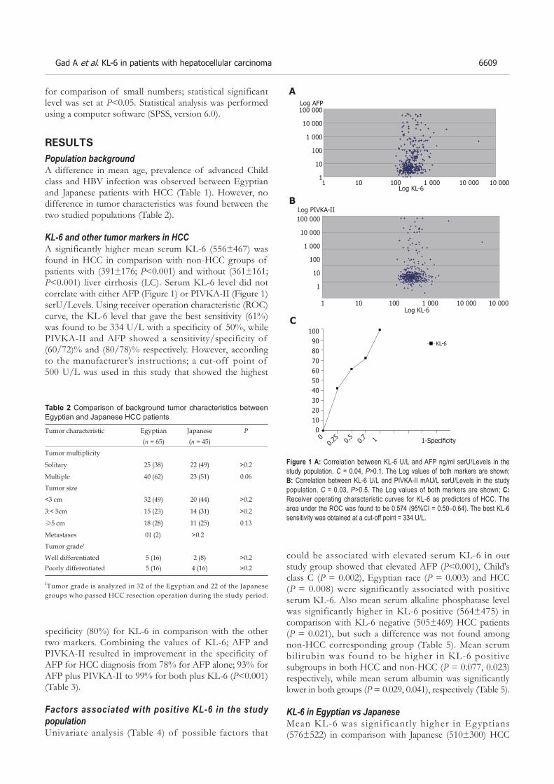
Gad A et al. KL-6 in patients with hepatocellular carcinoma 6609
for comparison of small numbers; statistical significant level was set at P<0.05. Statistical analysis was performed using a computer software (SPSS, version 6.0).
RESULTSPopulation backgroundA difference in mean age, prevalence of advanced Child class and HBV infection was observed between Egyptian and Japanese patients with HCC (Table 1). However, no difference in tumor characteristics was found between the two studied populations (Table 2).
KL-6 and other tumor markers in HCCA significantly higher mean serum KL-6 (556±467) was found in HCC in comparison with non-HCC groups of patients with (391±176; P<0.001) and without (361±161; P<0.001) liver cirrhosis (LC). Serum KL-6 level did not correlate with either AFP (Figure 1) or PIVKA-II (Figure 1) serU/Levels. Using receiver operation characteristic (ROC) curve, the KL-6 level that gave the best sensitivity (61%) was found to be 334 U/L with a specifi city of 50%, while PIVKA-II and AFP showed a sensitivity/specificity of (60/72)% and (80/78)% respectively. However, according to the manufacturer’s instructions; a cut-off point of 500 U/L was used in this study that showed the highest
could be associated with elevated serum KL-6 in our study group showed that elevated AFP (P<0.001), Child's class C (P = 0.002), Egyptian race (P = 0.003) and HCC (P = 0.008) were significantly associated with positive serum KL-6. Also mean serum alkaline phosphatase level was significantly higher in KL-6 positive (564±475) in comparison with KL-6 negative (505±469) HCC patients (P = 0.021), but such a difference was not found among non-HCC corresponding group (Table 5). Mean serum bil irubin was found to be higher in KL-6 positive subgroups in both HCC and non-HCC (P = 0.077, 0.023) respectively, while mean serum albumin was significantly lower in both groups (P = 0.029, 0.041), respectively (Table 5). KL-6 in Egyptian vs JapaneseMean KL-6 was signif icantly higher in Egyptians (576±522) in comparison with Japanese (510±300) HCC
specifi city (80%) for KL-6 in comparison with the other two markers. Combining the values of KL-6; AFP and PIVKA-II resulted in improvement in the specificity of AFP for HCC diagnosis from 78% for AFP alone; 93% for AFP plus PIVKA-II to 99% for both plus KL-6 (P<0.001) (Table 3).
Factors associated with positive KL-6 in the study populationUnivariate analysis (Table 4) of possible factors that
Table 2 Comparison of background tumor characteristics between Egyptian and Japanese HCC patients
Tumor characteristic Egyptian Japanese P(n = 65) (n = 45)
Tumor multiplicity
Solitary 25 (38) 22 (49) >0.2
Multiple 40 (62) 23 (51) 0.06
Tumor size
<3 cm 32 (49) 20 (44) >0.2
3:< 5cm 15 (23) 14 (31) >0.2
≥5 cm 18 (28) 11 (25) 0.13
Metastases 01 (2) >0.2
Tumor grade1
Well differentiated 5 (16) 2 (8) >0.2Poorly differentiated 5 (16) 4 (16) >0.2
1Tumor grade is analyzed in 32 of the Egyptian and 22 of the Japanese groups who passed HCC resection operation during the study period.
1009080706050403020100
00.2
5 0.5 0.7 1 1-Specifi city
KL-6
100 000
10 000
1 000
100
10
11 10 100 1 000 10 000 10 000
Log KL-6
Log AFP
100 000
10 000
1 000
100
10
1
1 10 100 1 000 10 000 10 000Log KL-6
Log PIVKA-II
Figure 1 A: Correlation between KL-6 U/L and AFP ng/ml serU/Levels in the study population. C = 0.04, P>0.1. The Log values of both markers are shown; B: Correlation between KL-6 U/L and PIVKA-II mAU/L serU/Levels in the study population. C = 0.03, P>0.5. The Log values of both markers are shown; C: Receiver operating characteristic curves for KL-6 as predictors of HCC. The area under the ROC was found to be 0.574 (95%CI = 0.50–0.64). The best KL-6 sensitivity was obtained at a cut-off point = 334 U/L.
A
B
C

6610 ISSN 1007-9327 CN 14-1219/ R World J Gastroenterol November 14, 2005 Volume 11 Number 42
patients (P = 0.041) (Table 6). Although a significant difference in mean KL-6 level between HCC and non-HCC was observed in both Egyptian and Japanese patients with chronic liver disease (P<0.001 respectively), the difference was not statistically signifi cant among Japanese patients with HCV-related disease (Table 6). No difference in mean KL-6 level was found between cirrhotic and non-cirrhotic in either HCC or non-HCC patients.
KL-6 and tumor characteristicsIn the HCC groups of both Egyptian and Japanese patients; KL-6 showed no significant association with tumor site, echogenicity or multiplicity. However, a significantly lower mean KL-6 (Table 7) was noticed in larger size tumors of >5 cm (371±168 U/L) in comparison with tumors of less than or equal to 5 cm (537±323) (P<0.05 in the Japanese group).
DISCUSSIONKL-6 was studied as a tumor marker in different mali-gnancies like breast, lung and pancreatic cancer and it was reported to be elevated in up to 50% of these mali-gnancies[14]. Two previous studies by Moriyama et al.[19, 20] addressed KL-6 as a tumor marker for HCC in patients with HCV-related chronic liver disease, and his results showed that the estimated cumulative incidence of HCC development in HCV-related chronic liver disease patients was signifi cantly greater in patients with positive KL-6[19] and suggested KL-6 to be used as a serological marker for HCC development in HCV-positive patients[20]. In our study, we included consecutive patients with chronic liver disease seen at outpatient settings in two different ethnic groups of possible different risk factors for HCC[22, 23] in order to have a wider spectrum of disease to judge KL-6 validity as a diagnostic test for HCC; however, one limitation was that most of the encountered patients in the two settings were actually with HCV-related disease with low proportion of HBV and non-viral-related disease. Our results showed a significantly higher mean KL-6 in HCC compared with non-HCC; either with or without LC; in addition no difference in mean KL-6 was found among HCC patients with and without LC; such fi ndings together point to KL-6 association with HCC independent on the presence or absence of LC. A signifi cantly higher mean KL-6 level was found in HBV-related in comparison with HCV-related HCC in both Egyptian and Japanese populations; a finding that deserves future study on a larger population of HBV-related disease. Our results also showed a significantly higher mean KL-6 level in HCC patients of Egyptian compared with Japanese race. The fi nding of a difference in the clinical background between both in terms of lower mean age and lower prevalence of HBV-related HCC could refl ect a difference in the risk factors for HCC in both groups. Also, a higher prevalence of advanced Child class in the HCC Egyptian patients was observed that could stand behind the finding of higher mean KL-6 level in this group compared to their corresponding Japanese group. Although we excluded patients with overt schistosomal from this study, still some Egyptian patients had a past history of schistosomiasis with US evidence of hepatic periportal fi brosis (denoting a background of schistosomal liver disease) that could also explain the finding of higher mean KL-6 level in Egyptian HCC patients, if we consider the possibility that KL-6 could be a fi brosis marker too[21]. This topic is highly suggested for future study.
Table 4 Factors associated with KL-6 positivity in the study population
Factors Total KL-6 (+) KL-6 (–)Age (yr) >=50 244 60 (24) 184 (75) <50 100 24(24) 76 (76) P NSSex Male 257 61 (24) 196 (76) Female 87 23 (26) 64 (73) P NSEthnicity Egyptian 171 53 (31) 118 (69) Japanese 173 31 (18) 142 (82) P 0.003Underlying liver disease HCV-related 267 71 (27) 196 (73) HBV-related 42 5 (12) 37 (88) Non-viral 35 8 (23) 27 (77) P NSCirrhosis (+) 169 46 (27) 123 (73) (–) 175 38 (22) 137 (78) P NSChild’s class C 47 20 (43) 27 (57) A&B 297 64 (21) 233 (78) P 0.002HCC: (+) 110 37 (34) 73 (66) (–) 234 47 (20) 187 (80) P 0.008AFP (+) 145 49 (34) 96 (66) (–) 199 35 (17) 164 (82) P <0.001
Tumor marker (cut-off point) Sensitivity Specifi city% %
AFP (10 ng/mL) 86 78PIVKA-II (35 mAU/L) 61 72KL-6 (500 U/L) 34 80AFP+PIVKA-II 861 93AFP+KL-6 872 94AFP+PIVKA-II+KL-6 87 99
Table 3 Comparison of the result of different tumor markers between HCC and non-HCC group
1All PIVKA-II (+) HCC patients are AFP (+). 2One KL-6 (+) HCC patient is AFP (–).

Gad A et al. KL-6 in patients with hepatocellular carcinoma 6611
We used a cut-off point of 500 U/L for KL-6 posi-tivity in this study; however, applying the ROC analysis showed that a cut-off point of 334 U/L would give the best sensitivity in our study population of 60% compared with only 32% for a cut-off (500 U/L); however, the best specificity was obtained using the later. Moriyama et al. used a cut-off point of 300 U/L in his analysis of KL-6 in HCV-related disease[19,20]. KL-6 serU/Level did not correlate with either serum AFP or PIVKA-II levels, which points to its behavior independently from either of them and this may justify its clinical significance as an independent tumor marker for HCC diagnosis when considered with both AFP and PIVKA-II. Our results also supported this finding as AFP specificity for HCC diagnosis improved from 78% for AFP alone and 93% of both AFP and PIVKA-II to 99% when combined with KL-6. Univariate analysis showed that low serum albumin, hyperbilirubinemia and elevated ALP were significantly
associated with positive KL-6 in HCC patients, while KL-6 showed no association with LC in turn, and this denotes a possible association between positive KL-6 and deterioration of hepatic condition in HCC patients independent from their cirrhotic status; a finding that might point to KL-6 as a predictor of tumor aggression and/or local or systemic metastasizing potential. A follow-up study is needed to confi rm its exact role in this regard.
ACKNOWLEDGMENTSWe would like to thank Takeda Foundation, Osaka, Japan for their financial support. We also thank Dr. Alla Sad, Dr. Essam Abd Alla, Dr. Khaled Gad for their help with various laboratory techniques.
REFERENCES1 Sherman M. Hepatocellular carcinoma: epidemiology, risk
factors, and screening. Semin Liver Dis 2005; 25: 143-1542 Szilagyi A, Alpert L. Clinical and histopathological variation in
hepatocellular carcinoma. Am J Gastroenterol 1995; 90: 15-23 3 Yuen MF, Cheng CC, Lauder IJ, Lam SK, Ooi CG, Lai CL.
Early detection of hepatocellular carcinoma increases the chance of treatment: Hong Kong experience. Hepatology 2000; 31: 330-335
4 Wong LL, Limm WM, Severino R, Wong LM. Improved survival with screening for hepatocellular carcinoma. Liver Transpl 2000; 6: 320-325
5 Johnson PJ. The role of serum alpha-fetoprotein estimation in the diagnosis and management of hepatocellular carcinoma.
Egyptian P1 value Japanese P value
(n = 65) (n = 45)
Tumor size<3 cm 485±227 618±3613:<5 cm 643±685 >0.1 456±285 0.17≥5 cm 581±420 >0.1 371±168 0.04
Table 5 Comparison of the clinical profi le of KL-6 positive and negative patients with and without HCC
1Data is shown as mean±SD. Other data is shown as n (%).
Table 7 Difference in mean KL-6 level according to HCC size
1P value is shown for the difference group (<3 cm) and the other two groups.
HCC (+) HCC (–)
KL-6 (+) KL-6 (–) P KL-6 (+) KL-6 (–) Pn = 37 n = 73 n = 47 n = 187
Mean age (yr)1 59±12 62±10 NS 57±12 56±13 NSCirrhosis 39 (81) 55 (76) NS 16 (34) 65 (35) NSChild’s C 13 (35) 15 (21) NS 7 (15) 11 (6) 0.045Mean ALT 74±101 62±61 NS 59±33 57±45 NSSerumAlbumin (g/L)1 2.9±0.7 3.3±0.7 0.029 3.5±0.9 3.8±0.8 0.041Bilirubin (mmol/L)1 2.7±2.8 2.5±3.0 0.077 2.4±2.9 1.4±1.9 0.023ALP (IU/L)1 564±475 505±469 0.021 316±139 299±152 NSAFP (+) 36 (97) 58 (80) 0.013 13 (28) 37 (20) NSPIVIKA (+) 23 (62) 44 (60) NS 14 (30) 50 (27) NS
Egyptian Japanese
HCC (+) HCC (-) P HCC (+) HCC (-) P
Chronic liver disease1: 576 (522) 398 (185) 0.001 510 (300) 350 (147) <0.001HCV-related 558 (524) 400 (172) 0.008 356 (290) 382 (209) >0.2
HBV-related 778 (663) 246 (72) >0.2 877 (292) 340 (163) <0.001Non-viral 729 (538) 446 (309) >0.2 2622 () 357 (160) –Cirrhotics 599 (586) 406 (159) 0.035 510 (350) 374 (196) 0.035
Non-cirrhotics 518 (325) 398 (185) 0.045 225 (73) 349 (222) <0.001
Table 6 Comparison of mean serum KL-6 level among different study sub-groups
1The KL-6 values are shown as mean (SD) U/L. 2Only one patient’s data.

6612 ISSN 1007-9327 CN 14-1219/ R World J Gastroenterol November 14, 2005 Volume 11 Number 42
Clin Liver Dis 2001; 5: 145-1596 Iwai M, Kashiwadani M, Takino T, Ibata Y. Demonstration
by light and ultrastructural immunoperoxidase study of alpha-fetoprotein-positive non-hepatoma cells and hepatoma cel ls during 3 ' -methyl-4-dimethylaminoazobenzene hepatocarcinogenesis. Virchows Arch B Cell Pathol Incl Mol Pathol 1988; 55: 117-123
7 McIntire KR, Waldmann TA, Moertel CG, Go VL, Serum alpha-fetoprotein in patients with neoplasms of the gastrointestinal tract. Cancer Res 1975; 35: 991-996
8 Gallo V, Cerutti E, Riberi A, Re M, Petrino R, Pecchio F. Alpha-fetoprotein and tissue polypeptide antigen in non neoplastic hepatic disorders. J Nucl Med Allied Sci 1989; 33: 89-93
9 Alpert E, Feller ER. Alpha-fetoprotein (AFP) in benign liver disease. Evidence that normal liver regeneration does not induce AFP synthesis. Gastroenterology 1978; 74: 856-858
10 Nguyen MH, Garcia RT, Simpson PW, Wright TL, Keeffe EB. Racial differences in effectiveness of alpha-fetoprotein for diagnosis of hepatocellular carcinoma in hepatitis C virus cirrhosis. Hepatology 2002; 36: 410-417
11 Kohno N, Kyoizumi S, Awaya Y, Fukuhara H, Yamakido M, Akiyama M. New serum indicator of interstitial pneumonitis activity. Sialylated carbohydrate antigen KL-6. Chest 1989; 96: 68-73
12 Stahel RA, Gilks WR, Lehmann HP, Schenker T. Third International Workshop on Lung Tumor and Differentiation Antigens: overview of the results of the central data analysis. Int J Cancer Suppl 1994; 8: 6-26
13 Wesseling J, van der Valk SW, Vos HL, Sonnenberg A, Hilkens J. Episialin (MUC1) overexpression inhibits integrin-mediated cell adhesion to extracellular matrix components. J Cell Biol 1995; 129: 255-265
14 Kohno N. Serum marker KL-6/MUC1 for the diagnosis and management of interstitial pneumonitis. J Med Invest 1999; 46: 151-158
15 Sagara M, Yonezawa S, Nagata K, Tezuka Y, Natsugoe S, Xing PX, McKenzie IF, Aikou T, Sato E. Expression of mucin
1 (MUC1) in esophageal squamous-cell carcinoma: its relationship with prognosis. Int J Cancer 1999; 84: 251-257
16 Utsunomiya T, Yonezawa S, Sakamoto H, Kitamura H, Hokita S, Aiko T, Tanaka S, Irimura T, Kim YS, Sato E. Expression of MUC1 and MUC2 mucins in gastric carcinomas: its relationship with the prognosis of the patients. Clin Cancer Res 1998; 4: 2605-2614
17 Tanimoto T, Tanaka S, Haruma K, Yoshihara M, Sumii K, Kajiyama G, Shimamoto F, Kohno N. MUC1 expression in intramucosal colorectal neoplasms. Possible involvement in histogenesis and progression. Oncology 1999; 56: 223-231
18 Suzuki K, Takada H, Oka S, Kanouzawa S, Iimuro M, Kitazumi Y, Arima T, Ohyama R, Kuwayama H. Clinical significance of KL-6, a marker of interstitial pneumonia, in cases of HCV-associated chronic liver disease. Intern Med 2003; 42: 650-654
19 Moriyama M, Matsumura H, Mikuni M, Arkawa Y, Ohshiro S, Aoki H, Yamagami H, Kaneko M, Shioda A, Saito H, Tanaka N, Arakawa Y. The clinical signifi cance of serum KL-6 levels in patients with type C liver diseases. Hepatol Res 2003; 25: 385-395
20 Moriyama M, Matsumura H, Watanabe A, Nakamura H, Arakawa Y, Oshiro S, Aoki H, Shimizu T, Yamagami H, Kaneko M, Shioda A, Tanaka N, Arakawa Y. Detection of serum and intrahepatic KL-6 in anti-HCV positive patients with hepatocellular carcinoma. Hepatol Res 2004; 30: 24-33
21 Hirasawa Y, Kohno N, Yokoyama A, Inoue Y, Abe M, Hiwada K. KL-6, a human MUC1 mucin, is chemotactic for human fi broblasts. Am J Respir Cell Mol Biol 1997; 17: 501-507
22 Hassan MM, Zaghloul AS, El-Serag HB, Soliman O, Patt YZ, Chappell CL, Beasley RP, Hwang LY. The role of hepatitis C in hepatocellular carcinoma: a case control study among Egyptian patients. J Clin Gastroenterol 2001; 33: 123-126
23 Tanaka K, Sakai H, Hashizume M, Hirohata T. A long-term follow-up study on risk factors for hepatocellular carcinoma among Japanese patients with liver cirrhosis. Jpn J Cancer Res 1998; 89: 1241-1250
Science Editor Guo SY Language Editor Elsevier HK

PO Box 2345, Beijing 100023, China World J Gastroenterol 2005;11(42):6613-6619www.wjgnet.com World Journal of Gastroenterology ISSN [email protected] © 2005 The WJG Press and Elsevier Inc. All rights reserved.E L S E V I E R
• LIVER CANCER •
INTRODUCTIONHuman hepatocellular carcinoma (HCC) is one of the most prevalent malignancies in China. Patients with HCC often die of tumor metastasis and recurrence even after curative resection. Recently, a metastatic human HCC model in nude mice (LCI-D20) and a series of HCC cell lines (MHCC97, MHCC97-H, MHCC97-L) with different metastatic potentials derived from LCI-D20 have been established in our institute[1,2]. Using this model, IFN-α significantly inhibits tumor growth and metastasis of MHCC97 xenografts has been found[3-5]. However, the underlying molecular mechanisms are still unclear.
IFN-α is a multifunctional cytokine capable of inter-fering with viral infection, inhibiting cell proliferation, regulating cell differentiation, as well as modulating immune response[6-9]. It is well known that these pleiotropic effects of IFN-α are mediated primarily through the tran-scriptional regulation of many different functional genes. Thanks to the rapid progress in human genetic projects; many functional human genes and expressed sequence tags (ESTs) are identifi ed and released, which make us possible to use cDNA microarray to survey IFN-α-modulated genes in MHCC97 cells. In this study, we identified 190 differentially expressed genes from 8 464 known human genes, which might mediate various biological functions of IFN-α. These data provide us useful clues for further studying the anti-tumor mechanisms of IFN-α and fi nding the IFN-α mimics for HCC therapy.
MATERIALS AND METHODSCell cultureMHCC97, a metastatic HCC cell line derived from LCI-D20 xenografts, was cultured in high glucose Dulbecco’s modified Eagle’s medium (Gibco-BRL, NY, USA) supplemented with 10% fetal calf serum (Hyclone, UT, USA), 100 U/mL penicillin and 100 μg/mL streptomycin in 20-cm2 tissue culture flasks. Cells were grown at 37 °C in a humidifi ed atmosphere of 50 mL/L CO2 and passaged every 3 d.
cDNA microarray analysisA total of 8 464 cDNAs of known human genes (United Gene Holding, Ltd, Shanghai) were amplified by polymerase chain reaction (PCR) using universal primers and spotted onto silylated slides (CEL Associates,
Wei-Zhong Wu, Hui-Chuan Sun, Lu Wang, Jie Chen, Kang-Da Liu, Zhao-You Tang, Liver Cancer Institute and Zhongshan Hospital, Fudan University, 136 Yi Xue Yuan Road, Shanghai 200032, ChinaSupported by the Key Projects for the Clinical Medicine from the Ministry of Public Health of China (2002–2005)Correspondence to: Zhao-You Tang, Liver Cancer Institute and Zhongshan Hospital, Fudan University, 136 Yi Xue Yuan Road, Shanghai 200032, China. [email protected]: +86-21-6403-7181 Fax: +86-21-6403-7181Received: 2005-03-17 Accepted: 2005-04-30
AbstractAIM: To elucidate the molecular mechanisms of the inhibitory effects of IFN-α on tumor growth and metastasis in MHCC97 xenografts.
METHODS: Three thousand international units per milliliter of IFN-α-treated and -untreated MHCC97 cells were enrolled for gene expression analysis using cDNA microarray. The mRNA levels of several differentially expressed genes in cDNA microarray were further identifi ed by Northern blot and RT-PCR.
RESULTS: A total of 190 differentially expressed genes including 151 IFN-α-repressed and 39 -stimulated genes or expressed sequence tags from 8 464 known human genes were found to be regulated by IFN-α in MHCC97. With a few exceptions, mRNA levels of the selected genes in RT-PCR and Northern blot were in good agreement with those in cDNA microarray.
CONCLUSION: IFN-α might exert its complicated anti-tumor effects on MHCC97 xenografts by regulating the expression of functional genes involved in cell metabolism, proliferation, morphogenesis, angiogenesis, and signaling.
© 2005 The WJG Press and Elsevier Inc. All rights reserved.
Key words: Interferon α; cDNA microarray; Gene expression profi le; HCC
Wu WZ, Sun HC, Wang L, Chen J, Liu KD, Tang ZY. Modulation of gene expression in MHCC97 cells by interferon alpha. World J Gastroenterol 2005; 11(42): 6613-6619http://www.wjgnet.com/1007-9327/11/6613.asp
Modulation of gene expression in MHCC97 cells by interferon alpha
Wei-Zhong Wu, Hui-Chuan Sun, Lu Wang, Jie Chen, Kang-Da Liu, Zhao-You Tang

6614 ISSN 1007-9327 CN 14-1219/ R World J Gastroenterol November 14, 2005 Volume 11 Number 42
Houston, TX, USA) using a Cartesian PixSys 7500 motion control robot (Cartesian Tech, Irvine, CA, USA) fitted with ChipMaker micro-spotting technology (TeleChem, Sunnyvale, CA, USA). After being hydrated, dried, cross linked and washed, the microarray was ready for use. Total RNA was isolated from IFN-α-treated and untreated (3 000 IU/mL, 16 h) cells using TRIzol (Gibco-BRL). cDNA probes were prepared by reverse transcription and purified according to the methods described by Schena et al.[10]. Then equal amount of cDNA from IFN-α-untreated and treated MHCC97 cells was labeled with Cy3-dUTP and Cy5-dUTP, respectively. The mixed Cy3/Cy5 probes were purifi ed and dissolved in 20 μL of hybridization solution (0.75 mol/L NaCl, 0.075 mol/L sodium citrate, 0.4% SDS, 50% formamide, 0.1% Ficoll, 0.1% polyvinylpyrrolidone and 0.1% BSA). Microarrays were pre-hybridized with 0.5 mg/mL salmon sperm DNA at 42 °C for 6 h. After being extensively washed, the denatured (95 °C, 5 min) fluorescent-labeled probe mixture was applied onto the pre-hybridized chips and further hybridized at 42 °C for 15-17 h under a cover glass. Subsequently, chips were sequentially washed for 10 min at 60 °C with 2×SSC+0.2% SDS, 0.1×SSC+0.2% SDS and 0.1×SSC solutions and dried at room temperature (1×SSC: 150 mmol/L NaCl, 15 mmol/L sodium citrate). Both Cy3 and Cy5 fl uorescent signals of hybridized chips were scanned by ScanArray 4000 (GSI Lumonics, MA, USA) and analyzed using Genepix Pro 3.0 software (BioDiscovery Inc., CA, USA). To minimize artifacts arising from low expression, only genes whose Cy3 and Cy5 fluorescent intensities were both over 200 counts, or genes whose Cy3 or Cy5 fluorescent intensity was over 800 were selected for calculating the normalization cofactor (ln(Cy5/Cy3)). Genes were identified as differentially expressed, if the ratio of Cy5/(Cy3×normalization cofactor) (Cy5/Cy3*) was more than 2 or less than 0.5.
Reverse transcription and polymerase chain reactionMHCC97 cells (106) cultured in 20-cm2 fl asks were treated with 3 000 IU/mL IFN-α (Roche, Shanghai) for 0 or 16 h, and total RNA was extracted (RNeasy Mini Kit, QIAGEN Inc., CA, USA). One microgram RNA was used to set-up reverse transcription reactions (Gibco-BRL, NY, USA). Nine differentially expressed genes identified by cDNA microarray were selected for analysis by semi-quantitative PCR. Appropriate primers were designed using Primer3 software (http://www-genome.wi.mit.edu). γ-Actin was used as an internal standard. PCR reaction conditions and primer sequences are summarized in Table 1.
Northern blot analysisTotal RNA of 3 000 IU/mL IFN-α-treated or untreated MHCC97 cells was isolated as described above. Thirty microgram was separated by 1% agarose formaldehyde gel electrophoresis and transferred to a nylon membrane (Millipore, MA, USA) in 10×SSC by capillary blotting. The membrane was hybridized with the appropriate cDNA probe prepared from the human library of cDNA
clones (Biostar Genechip Inc., Shanghai) and labeled with [α-32P]dCTP (Yahui Biomedical, Beijing) using random primer (Ambion Inc., Austin, TX, USA).
RESULTSGene expression profi le identifi ed by cDNA microarrayIt is well known that the gene expression pattern of cells often varies with time and differentiation status and that cells derived from different individuals often have different genetic expression profi les. As a result, it is often difficult to extract useful information on the possible causes of phenotypic differences by comparing the genetic expression profi les of different cell lines. To minimize such complicated factors, we compared the gene expression profiles in 3 000 IU/mL IFN-α-treated and untreated (0 IU/mL) MHCC97 cells in two independent cDNA microarray analyses. We reasoned that such an internally consistent comparison might provide useful information on explaining the anti-tumor molecular mechanism of IFN-α in MHCC97 xenografts.
In 8 464 tested genes and ESTs, 190 genes were ide-ntifi ed to be modulated by 3 000 IU/mL IFN-α treatment in MHCC97 cells. Among them the ex-pression of 151 genes was downregulated by IFN-α and the expression of 39 genes was upregulated by IFN-α. All differentially expressed genes are listed in Table 2 and the gene expression profi les obtained by cDNA microarray analysis are shown in Figure 1.
Nine differentially expressed genes evaluated by RT-PCR and Northern blotTo validate the results of cDNA microarray, we selected
60000
50000
40000
30000
20000
10000
0 0 10000 20000 30000 40000 50000 60000
Cy 5
Figure 1 Representative hybrid result (A) and scatter plots (B) of cDNA microarray analysis in IFN-α treated MHCC97.
A
B
Cy 3
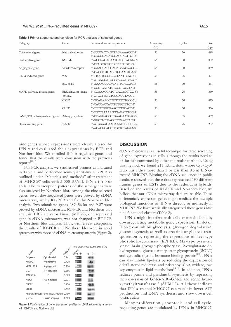
Wu WZ et al. IFN-α-regulated genes in MHCC97 6615
nine genes whose expressions were clearly altered by IFN-α and evaluated their expressions by PCR and Northern blot. We enrolled IFN-α-regulated genes and found that the results were consistent with the previous reports[11,12].
For PCR analysis, we synthesized primers as indicated in Table 1 and performed semi-quantitative RT-PCR as outlined under “Materials and methods” after treatment of MHCC97 cells with 3 000 IU/mL IFN-α for 0 or 16 h. The transcription patterns of the same genes were also analyzed by Northern blot. Among the nine selected genes, seven downregulated genes were proved by cDNA microarray, six by RT-PCR and five by Northern blot analysis. Two stimulated genes, ISG-56 ku and 9-27 were proved by cDNA microarray, RT-PCR and Northern blot analysis. ERK activator kinase (MEK2), one repressed gene in cDNA microarray, was not changed in RT-PCR or Northern blot analysis. Thus, with a few exceptions, the results of RT-PCR and Northern blot were in good agreement with those of cDNA microarray analysis (Figure 2).
DISCUSSIONcDNA microarray is a useful technique for rapid screening of gene expressions in cells, although the results need to be further confi rmed by other molecular methods. Using this method, we found 211 hybrid dots, whose Cy5/Cy3* ratio was either more than 2 or less than 0.5 in IFN-α-treated MHCC97. Blasting the cDNA sequences in public database showed that these dots represented 190 different human genes or ESTs due to the redundant hybrids. Based on the results of RT-PCR and Northern blot, we believe that our cDNA microarray data are reliable. These differentially expressed genes might mediate the multiple biological functions of IFN-α directly or indirectly in MHCC97. We have artifi cially categorized these genes into nine functional clusters (Table 2).
IFN-α might interfere with cellular metabolisms by downregulating metabolic gene expression. In detail, IFN-α can inhibit glycolysis, glycogen degradation, gluconeogenesis as well as creatine or glucose tran-sportation by repressing the expressions of liver-type phosphofructokinase (hPFKL), M2-type pyruvate kinase, brain glycogen phosphorylase, 2-oxoglutarate de-hydrogenase, glucose transporter glycoprotein (SGLT) and cytosolic thyroid hormone-binding protein[13]. IFN-α can also inhibit lipolysis by reducing the expression of delta7-sterol reductase and pristanoyl-CoA oxidase, two key enzymes in lipid metabolism[14,15]. In addition, IFN-α reduces purine and pyridine biosynthesis by repressing the expression of GARs-AIRs-GART and serine hydro-xymethyltransferase 2 (SHMT2). All these indicate that IFN-α-treated MHCC97 can result in lower ATP production and DNA synthesis, and slow down cell proliferation.
Many proliferation-, apoptosis- and cel l cycle-regulating genes are modulated by IFN-α in MHCC97.
Category Gene Sense and antisense primers Annealing(°C)
Cycles Size (bp)
Cytoskeletal gene Neutral calponin 5’-TGGCACCAGCTAGAAAACCT-3’; 5’-CAGGGACATGGAGGAGTTGT-3’
56 26 498
Proliferative gene hMCM2 5’-ACCGAGACAATGACCTACGG-3’;5’-CTAGCTGTCTGCCCCTTGTC-3’
56 30 382
Angiogenic gene VEGF165 receptor 5’-GAAGCACCGAGAGAACAAGG-3; 5’-CACCTGTGAGCTGGAAGTCA-3’
56 30 359
IFN-α-induced genes 9-27 5’-TTGGTCCCTGGCTAATTCAC-3’; 5’-ATGAGGATGCCCAGAATCAG-3’
53 35 491
ISG-56 ku 5’-AAAAGCCCACATTTGAGGTG-3’; 5’-GGCTGATATCTGGGTGCCTA-3’
54 30 451
MAPK pathway-related genes ERK activator kinase (MEK2)
5’-CGAAAGGATCTCAGAGCTGG-3’; 5’-GTGCTTCTCTCGGAGGTACG-3’
56 26 349
G3BP2 5’-GCAGAACCTGTTTCTCTGCC-3’; 5’-CACCACCACCTCTGGTTTCT-3’
56 30 475
CHED 5’-TCCTTGGCGAACTCTTCACT-3’; 5’-TGCCATAAAGGGAGATCTGG-3’
56 30 336
cAMP/PI3 pathway-related gene Adenylyl cyclase 5’-CCAGGAGCCTGAAGAATGAG-3’; 5’-GGCTTCTGAGCTCCAATCAC-3’
53 35 439
Housekeeping gene γ-Actin 5’-ATGGAAGAAGAAATCGCCGC-3’; 5’-ACACGCAGCTCGTTGTAGAA-3’
55 25 287
Table 1 Primer sequence and condition for PCR analysis of selected genes
Calponin Cytoskeletal 0.141
hMCM2 Proliferative 0.428
VEGF165 R Angiogenetic 0.250
9-27 IFN inducible 2.356
ISG-56 Ku '' 3.829
MEK2 MAPK related 0.271
G3BP2 '' 0.296
CHED 0.412
Adenylyl cyclase cAMP/PI3K related 0.409
γ-actin House keeping 1.083
Gene
Categ
ory
Cy5/
Cy3*
marke
r
0 160 16
Time after 3,000 IU/mL IFN-α (h)
Figure 2 Confi rmation of gene expression profi les in cDNA microarray analysis with RT-PCR and Northern blot.

6616 ISSN 1007-9327 CN 14-1219/ R World J Gastroenterol November 14, 2005 Volume 11 Number 42
Table 2 Gene expression profi le of MHCC97 cells induced by IFN-α
Category GenBank ID Gene description Cy5/Cy3* (average)
2.1 Metabolism related genes
HUMCRTR Creatine transporter 0.251HSGAGMR GARs-AIRs-GART 0.289HUM2OGDH 2-Oxoglutarate
dehydrogenase0.298
AF034544 Delta7-sterol reductase 0.318HUMTK Thymidine kinase 0.333HSU12778 Acyl-CoA dehydrogenase 0.341HUMTHBP Thyroid hormone-binding
protein(p55)0.349
AF067127 7-Dehydrocholesterol reductase (DHCR)
0.356
HSPRCOX Pristanoyl-CoA oxidase 0.364AF035429 Cytochrome oxidase
subunit 10.372
AF070544 Glucose transporter glycoprotein (SGLT)
0.379
HSPFKLA Liver-type1-phosphofructokianse (PFKL)
0.392
HUMSHMT Serine hydroxymethyltransferase 2 (SHMT2)
0.407
HUMMGPHB Brain glycogen phosphorylase
0.413
HUMTCBA Cytosolic thyroid hormone-binding protein (p58)
0.415
D88152 Acetyl-coenzyme A transporter
0.451
HUMPKM2L M2-type pyruvate kinase 0.456HSLDHBR Lactate dehydrogenase B 2.156AF108211 Inorganic pyrophosphatase 2.25HSCOXVII Cytochrome C oxidase VII 2.279HUMCYCPSK Cytochrome C (HS7) 2.574HUMDBI Diazepam binding inhibitor 2.628
2.2 Proliferation, apoptosis and damaged DNA repairing related genes
HSATPBR Na/K ATPase beta subunit 0.208HUMP53T Mutant p53 protein 0.233HSMITG Mitochondrial DNA 0.309HSNUMAMR Nuclear mitotic apparatus
protein0.325
HSDNALIG3 DNA ligase III 0.34G28520 STS HSGC-31478 (homolog
to Rad23a)0.341
AF096870 Estrogen-responsive B box protein
0.352
AF001609 EXT like protein 3 0.367AF015283 Selenoprotein W 0.369AF011905 Putative checkpoint control
protein hRad10.398
HUMHMAM2 Minichromosome maintenance 2
0.408
HUMRNAPII RNA polymerase II 23 ku subunit
0.408
AF007790 Inversely correlated with estrogen receptor Expression (ICERE-1)
0.413
HSU78310 Pescadillo 0.43AF004162 Nickel-specifi c induction
protein (Cap43)0.434
HSU3298 UV-damaged DNA binding factor
0.437
HUMP1CDC47 P1cdc47 0.442HSU72649 B cell translocation gene 2 0.444AF031523 bcl-xL/bcl-2 associated
death promoter (BAD)0.481
AF132973 CGI-39 (homolog to GRIM-19)
2.079
remodeling related genes
AF070593 Beta tublin 0.236HSU35622 EWS-E1A-F chimeric
protein0.255
AF049259 Keratin 13 0.335HSPRO4HY Prolyl 4-hydoxylase beta 0.337HUMCN4GEL Collagenase type IV 0.36AF005654 Actin-binding double zinc-
fi nger protein0.378
HSTEST Testican 0.379HUMEPSURAN Surface antigen 0.389AF004841 CAM-related/down-
regulated by oncogenes0.398
HUMGLBA Co-beta-glucosidase 0.402HUMCA1XIA Alpha-1 type XI collagen 0.423HUMMCPGV Macrophage capping
protein0.461
HUMNID Nidogen 0.497HSTUMP Translationally controlled
tumor protein2.022
2.4 Signal transmitting related genes
HUMEPHT2R Protein tyrosine kinase (NET PTK)
0.248
HUMMEK2NF ERK activator kinase (MEK2)
0.271
HUMBADPTA Beta-adaptin 0.273HUMP2A Alpha-PR65 0.282HUMHRGAA rab GDI alpha 0.285AF053535 ras-GAP/RNA binding
protein G3BP20.296
HSRING3GE RING 3 0.316HSU45973 Pt Ins (4,5) P(2)
5-phosphatase0.324
HSU07139 Voltage-gated calcium channel beta
0.329
HUMFTPB Farnesyl-protein transferase beta
0.345
HSU33053 Lipid-activated protein kinase (PRK1)
0.352
HUMHK1A Calcium-ATPase (HK1) 0.386HSU66406 EPH-related PTK receptor
ligand LERK-80.386
HSPP15 Placental protein 15 0.387HSADCYCL Adenylyl cyclase 0.409HUMCHED cdc2-related protein kinase
(CHED)0.412
AF093265 Homer 3 0.415HSU40282 Integrin-linked kinase 0.416HUMGKAS Stimulatory G protein 0.416HSU43939 Nuclear transport factor 2 0.429HUMCAK Tyrosine protein kinase
(CAK)0.439
HUMGNOS48 Endothelial nitric oxide synthase
0.443
HUMCDPKIV Calmodulin-dependent protein kinase IV
0.449
HSPKX1MR Protein kinase, PKX1 0.469D83760 Mother against dpp (Mad)
related protein0.472
HUMEGFGRBA EGF receptor binding protein GRB2
0.481
HSU51004 Protein kinase C inhibitor (PKCI-1)
2.223
2.5 Tumor angiogenesis related genes
HUMRNAMBPE Golli-mbp 0.236AF016050 VEGF 165 receptor/
neuropilin0.25
2.3 Morphogenesis, adhesion, and cytoskeleton
D38735 Neutral calponin 0.141AF006082 Actin-related protein Arp2 0.197U01244 Fibulin 1D 0.212
AF001307 Aryl hydrocarbon receptor nuclear translocator
0.27
HSU64791 Golgi membrane sialoglycoprotein MG 160
0.355
HUMPTPRZ Protein tyrosine phosphatase Zeta-polypeptide
0.363
HSU28811 Cysteine-rich FGFR (CFR1) 0.414HSU20758 Osteopontin 2.193
Wu WZ et al. IFN-α-regulated genes in MHCC97 6617
Downregulating the expression of mutant p53, mito-chondrial DNA, nuclear mitotic apparatus protein (NuMA), and RNA polymerase II 23 ku subunit (polR2) might cause cell cycle arrest[16,17]. Downregulating the expression of DNA ligase III, hRad1, minichromosome maintenance 2 (hMCM2) as well as UV-damaged DNA binding factor might hinder damaged DNA repairing[18,19]. Stimulating retinoid-IFN-induced mortality 19 (GRIM-19) expression might promote IFN-α-induced apoptosis[20].
Several genes functionally related to cell morphogenesis,
HUMTR107 DNA-binding protein, TAXREB107
2.24
HUMNEPPON Nephropontin 2.4132.6 Transcriptional activity related genes
S66431 Retinoblastoma binding protein 2
0.182
HUMANT61K Medium antigen-associated 61 ku protein
0.183
HSU58197 Interleukin enhancer binging factor 2
0.226
HSUBP Upstream binding factor 0.2664758315 ets-related molecule, ETV5 0.267AF099013 Glucocorticoid modulatory
element binding protein 10.309
HSU72621 Lost on transformation 1(LOT1)
0.313
HUMFOS Oncogene protein, c-fos 0.361AB019524 Nuclear receptor co-
repressor0.369
HS14AGGRE Conserved gene telomeric to alpha globin cluster
0.398
HSU74667 tat interactive protein (tip60)
0.404
AF114816 KRAB-zinc fi nger protein SZF1-1
0.406
HSU80456 Drosophila single-minded, SIM2
0.409
AF117756 TRAP 150 0.41HSU15306 Cysteine rich DNA binding
protein NFX10.417
S57153 Retinoblastoma binding protein 1
0.469
HUM56KDAPR IEF SSP 9502 2.183HUMTR107 DNA binding protein.
TAXREB 1072.24
HUMMSS1 Mammalian suppressor of sgv 1, MSS 1
2.313
2.7 mRNA and protein processing, secretory, proteolysis related genes
HSU39412 Alpha SNAP 0.141HSU47927 Isopeptidase T (ISOT) 0.229HSU72355 hsp27 ERE-TATA bind
protein, HET0.231
AF077039 TIM17 homolog 0.238HUMHRH1 RNA helicase, HRH1 0.251AF206402 U5 SnRNP 100 ku protein 0.255D85429 Heat shock protein 40 0.344HSU85946 hSec 10p 0.378HSY10806 Arginine methyltransferase 0.412AB002135 Glycophosphatidylinositol
anchor attachment 10.428
AB007510 PRP8 protein 0.436HSU24105 Coatomer protein (COPA) 0.455HSCANPX Calpain-like protease
(CANPX)0.456
HSRBPRL7A Ribosomal protein L7 2.067
HUMRPS7A Ribosomal protein S7 2.289AF106622 TIM17a 2.312HSUCEH3 Ubiquitin-conjugated
enzyme UbCH22.323
HUMPSC3 Proteasome subunit HC3 2.368HUMTCP20 Chaperonin protein, TCP20 2.5724504522 Chaperonin protein, hsp10 2.686
2.8 Tumor antigen processing, anti-viral infection related genes
HUMSAPC1 Cerebroside sulfate activator protein
0.211
AF077011 Interleukin 16 0.23AF057307 Prosaposin 0.26HUMSIATA Sialyltransferase 0.26AF055008 Epithelin 1 and 2 0.363HSU58766 FX protein 0.393HUMOSF1 OSF1 0.407HSU46194 RAGE 4 0.43HSU18121 136 ku double-stranded
RNA binding protein0.469
AF021315 Reverse transcriptase 0.483S74095 Preproenkephalin A 2.115HUM927A Interferon inducible protein
9-272.356
HSIFI56R Interferon inducible protein 56 ku
3.829
HUMHCAMAP1 Interferon inducible protein 44 ku
4.03
2.9 Genes with unknown biological functions
D50928 KIAA0138 0.23AF132942 CGI08 0.269AB020677 KIAA0870 0.271AB011110 KIAA0538 0.277AB028956 KIAA1033 0.28HSU10362 GB36b glycoprotein 0.3354579277 A homolog of proteasome
regulatory S20.352
AB002356 KIAA0358 0.3714505130 A homolog of MCM3 0.371AB029020 KIAA1097 0.381HS130N43 0.383HSU66406 Eplg8 0.386HSNIPSNA1 NIPSNAP1 protein 0.391AB002378 KIAA0380 0.405HSU90907 Regulatory subunit of P55
PIK0.407
AB208959 KIAA1036 0.414AB020658 KIAA0851 0.416AF035282 0.416AF000136 0.419HUMORFFA KIAA0120 0.424D13699 KIAA0019 0.43HUMORFB1 KIAA0123 0.432AF151830 CGI72 0.436AB007900 KIAA0440 0.437AB014595 KIAA0695 0.439HSM800064 0.439HUMORFA04 KIAA0115 0.457HSU79287 0.462AF007149 0.473AF007135 2.147AF151875 CGI117 2.184AF151857 CGI99 2.326HUMRSC508 KIAA0020 2.45
D89678 A+U-rich element RNA-binding protein
2.069
HSU14966 Ribosomal protein L5 2.113HSRPL31 Ribosomal protein L31 2.142HUMPSC9 Proteasome subunit HC9 2.179HSU26312 Heterochromatin protein
HP1 HS-gamma2.182
HUMRPS7A Ribosomal protein S7 2.289AF106622 TIM17a 2.312HSUCEH3 Ubiquitin-conjugated
enzyme UbCH22.323
HUMRPS25 Ribosomal protein S25 2.326HUMRPSA3A Ribosomal protein S3a 2.328HSRNASMG Sm protein G 2.334HUMRPS18 Ribosomal protein S18 2.341HUMRP4SX Ribosomal protein S4
isoform2.346

6618 ISSN 1007-9327 CN 14-1219/ R World J Gastroenterol November 14, 2005 Volume 11 Number 42
adhesion, and cytoskeleton remodeling are also modulated by IFN-α in MHCC97. For example, decreasing the expression of calponin, actin-related protein 2 (Arp2), fi bulin 1D, beta-tublin and epidermal surface antigen (ESA), etc., might damage mitotic spindle formation and might interfere with actin-based cell motility, migration, adhesion and morphogenesis[21-24]. Reducing the expression of prolyl 4-hydroxylase beta, a key enzyme in collagen biosynthesis and type IV collagenase, a tumor-derived extracellular matrix metalloproteases might block tumor invasion and metastasis. Although most genes in this category were first identified as IFN-α regulating genes, their roles in mediating IFN-α functions need to be further studied.
In this study, we found that many genes functionally related to signal transmitting were affected by IFN-α in MHCC97. By repressing the expressions of discoidin domain receptor, integrin-linked kinase, EPH-related tyrosine kinase (EPT2) and MEK2, etc., IFN-α might block cellular signaling initiated by tyrosine-kinase receptors[25,26]. By modulating the expressions of Rab GDI, Ras-related GTP-binding proteins and farnesyl-protein transferase and nuclear transport factor (NTF2) and G3BP2, a Ras-GAP/RNA binding protein, IFN-α might interfere with GTP/GDP exchange and nuclear import, thus infl uencing the recycles and activities of ras and its homologs[27-29]. By attenuating the expressions of adenylyl cyclase (AC) and phosphatidylinositol 4,5-bisphosphate 5-phosphatase (PtdIns (4,5)P(2)5- phospharase), a catalyzer of pho-sphatidylinositol 4,5-bisphosphate and PRK1, IFN-α might decrease inositol polyphosphate levels in cytosol and might inhibit the serine/threonine-kinase activities through cAMP/ PI3P signal pathway[30,31]. All these changes might exert inhibitory effects of IFN-α on MAPK and PI3K signaling. In addition, other signaling pathways such as Ca(2+), NO and TGFβ/hMAD-dependent signaling pathways are suppressed by IFN-α as well[32,33]. Plausibly Jak/STATs pathway, the most important IFN-α signaling pathway, is confi rmed not to be regulated in IFN-α-treated MHCC97. The deficient expression of p48 (ISGF3γ) in this cell line may be the possible mechanism for the non-response of IFN-α priming via Jak/STATs pathway (data not shown).
In this study, we found that many angiogenic-related genes were regulated by IFN-α . By attenuating the expressions of Golli-MBP[34], VEGF 165 receptor and aryl hydrocarbon receptor nuclear translocator (ARNT)[35] as well as Golgi membrane sialoglycoprotein MG 160, a bFGF binding protein and cysteine-rich FGF receptor (CFR-1)[36], IFN-α may destroy the balance between pro- and anti-angiogenic factors and exert its inhibitory effects on tumor angiogenesis.
It is well known that cells usually respond to various stimuli by rapidly shifting the functions of transcriptional factors. Using this strategy, IFN-α might impose its anti-proliferative functions and hormone response by fl uctuating the expression of several transcriptional factors or their cofactors such as retinoblastoma binding protein2 (RBP2), interleukin enhancer binding factor 2, lost on transformation 1 (LOT1) and KRAB-zinc fi nger protein
(SZF1)[37-40].In addition, IFN-α might hinder with mRNA/rRNA
spicing and maturation by downregulating RNA helicase (HRH1), U5 snRNP[41] and affect protein transportation, secretion and proteolysis by downregulating alpha SNAP, GPAA1, hSec10p, hsp40 and isopeptidase T, a putative molecular in ubiquitin–proteasome pathway[42-44]. Meanwhile IFN-α might evoke anti-viral or tumor immune response by upregulating 9-27, 56 ku protein and p44 expressions.
Except for functionally definite genes, many ESTs with unknown functions were identified as IFN-α-regulated genes in our study (Table 2). In conclusion, cDNA microarray is a useful, rapid method for screening transcriptome of cells and potentially paves a way for elucidating IFN-α effects on tumor growth and metastasis.
ACKNOWLEDGMENTWe thank Shanghai Biostar Genechip Inc. for cDNA microarray service.
REFERENCES1 Sun FX, Tang ZY, Liu KD, Ye SL, Xue Q, Gao DM, Ma ZC.
Establishment of a metastatic model of human hepatocellular carcinoma in nude mice via orthotopic implantation of histologically intact tissue. Int J Cancer 1996; 66: 239-243
2 Tian J, Tang ZY, Ye SL, Liu YK, Lin ZY, Chen J, Xue Q. New human hepatocellular carcinoma (HCC) cell line with highly metastatic potential (MHCC97) and its expressions of the factors associated with metastasis. Br J Cancer 1999; 81: 814-821
3 Wang L, Tang ZY, Qin LX, Wu XF, Sun HC, Xue Q, Ye SL. High-dose and long-term therapy with interferon-alfa inhibits tumor growth and recurrence in nude mice bearing human hepatocellular carcinoma xenografts with high metastatic potential. Hepatology 2000; 32: 43-48
4 Wu WZ, Sun HC, Gao YQ, Li Y, Wang L, Zhou K, Liu KD, Iliakis G, Tang ZY. Reduction in p48-ISGFgamma levels confers resistance to interferon-alpha2a in MHCC97 cells. Oncology 2004; 67: 428-440
5 Wu WZ, Sun HC, Shen YF, Chen J, Wang L, Tang ZY, Iliakis G, Liu KD. Interferon alpha 2a downregulates VEGF expression through PI3 kinase and MAP kinase signaling pathways. J Cancer Res Clin Oncol 2005; 131: 169-178
6 Tough DF, Borrow P, Sprent J. Induction of bystander T cell proliferation by viruses and type I interferon in vivo. Science 1996; 272: 1947-1950
7 Albini A, Marchisone C, Del Grosso F, Benelli R, Masiello L, Tacchetti C, Bono M, Ferrantini M, Rozera C, Truini M, Belardelli F, Santi L, Noonan DM. Inhibition of angiogenesis and vascular tumor growth by interferon-producing cells: A gene therapy approach. Am J Pathol 2000; 156: 1381-1393
8 Slaton JW, Perrotte P, Inoue K, Dinney CPN, Fidler IJ. Interferon-α-mediated down-regulation of angiogenesis-related genes and therapy of bladder cancer are dependent on optimization of biological dose and schedule. Clin Cancer Res 1999; 5: 2726-2734
9 Hong YK, Chung DS, Joe YA, Yang YJ, Kim KM, Park YS, Yung WK, Kang JK. Efficient inhibition of in vivo human malignant glioma growth and angiogenesis by interferon-beta treatment at early stage of tumor development. Clin Cancer Res 2000; 6: 3354-3360
10 Schena M, Shalon D, Davis RW, Brown PO. Quantitative monitoring of gene expression patterns with a complementary DNA microarray. Science 1995; 270: 467-470
11 Elco CP, Guenther JM, Williams BR, Sen GC. Analysis of

Wu WZ et al. IFN-α-regulated genes in MHCC97 6619
genes induced by Sendai virus infection of mutant cell lines reveals essential roles of interferon regulatory factor 3, NF-kappaB, and interferon but not toll-like receptor 3. J Virol 2005; 79: 3920-3929
12 Martensen PM, Justesen J. Small ISGs coming forward. J Interferon Cytokine Res 2004; 24: 1-19
13 Ishikawa N, Oguri T, Isobe T, Fujitaka K, Kohno N. SGLT gene expression in primary lung cancers and their metastatic lesions. Jpn J Cancer Res 2001; 92: 874-879
14 Witsch-Baumgartner M, Loffl er J, Utermann G. Mutations in the human DHCR7 gene. Hum Mutat 2001; 17: 172-182
15 Jia Y, Qi C, Zhang Z, Hashimoto T, Rao MS, Huyghe S, Suzuki Y, Van Veldhoven PP, Baes M, Reddy JK. Overexpression of peroxisome proliferator-activated receptor-alpha (PPARalpha)-regulated genes in liver in the absence of peroxisome proliferation in mice deficient in both L- and D-forms of enoyl-CoA hydratase/dehydrogenase enzymes of peroxisomal beta-oxidation system. J Biol Chem 2003; 278: 47232-47239
16 Wang J, Silva JP, Gustafsson CM, Rustin P, Larsson NG. Increased in vivo apoptosis in cells lacking mitochondrial DNA gene expression. Proc Natl Acad Sci U S A 2001; 98: 4038-4043
17 Taimen P, Viljamaa M, Kallajoki M. Preferential expression of NuMA in the nuclei of proliferating cells. Exp Cell Res 2000; 256: 140-149
18 Maiorano D, Lemaitre JM, Mechali M. Stepwise regulated chromatin assembly of MCM2-7 proteins. J Biol Chem 2000; 275: 8426-8431
19 Brand M, Moggs JG, Oulad-Abdelghani M, Lejeune F, Dilworth FJ, Stevenin J, Almouzni G, Tora L. UV-damaged DNA-binding protein in the TFTC complex links DNA damage recognition to nucleosome acetylation. EMBO J 2001; 20: 3187-3196
20 Chidambaram NV, Angell JE, Ling W, Hofmann ER, Kalvakolanu DV. Chromosomal localization of human GRIM-19, a novel IFN-beta and retinoic acid-activated regulator of cell death. J Interferon Cytokine Res 2000; 20: 661-665
21 Curtis M, Nikolopoulos SN, Turner CE. Actopaxin is phosphorylated during mitosis and is a substrate for cyclin B1/cdc2 kinase. Biochem J 2002; 363: 233-242
22 Kovacs EM, Goodwin M, Ali RG, Paterson AD, Yap AS. Cadherin-directed actin assembly: E-cadherin physically associates with the Arp2/3 complex to direct actin assembly in nascent adhesive contacts. Curr Biol 2002; 12: 379-382
23 Roof DJ, Hayes A, Adamian M, Chishti AH, Li T. Molecular characterization of abLIM, a novel actin-binding and double zinc fi nger protein. J Cell Biol 1997; 138: 575-588
24 Bickel PE, Scherer PE, Schnitzer JE, Oh P, Lisanti MP, Lodish HF. Flotillin and epidermal surface antigen define a new family of caveolae-associated integral membrane proteins. J Biol Chem 1997; 272: 13793-13802
25 Hannigan GE, Leung-Hagesteijn C, Fitz-Gibbon L, Coppolino MG, Radeva G, Filmus J, Bell JC, Dedhar S. Regulation of cell adhesion and anchorage-dependent growth by a new beta 1-integrin-linked protein kinase. Nature 1996; 379: 91-96
26 Tang XX, Biegel JA, Nycum LM, Yoshioka A, Brodeur GM, Pleasure DE, Ikegaki N. cDNA cloning, molecular character izat ion , and chromosomal loca l izat ion of NET(EPHT2), a human EPH-related receptor protein-tyrosine kinase gene preferentially expressed in brain. Genomics 1995; 29: 426-437
27 Ishizaki H, Miyoshi J, Kamiya H, Togawa A, Tanaka M, Sasaki T, Endo K, Mizoguchi A, Ozawa S, Takai Y. Role of rab GDP dissociation inhibitor alpha in regulating plasticity of hippocampal neurotransmission. Proc Natl Acad Sci USA 2000; 97: 11587-11592
28 Prigent M, Barlat I, Langen H, Dargemont C. IkappaBalpha
and IkappaBalpha /NF-kappa B complexes are retained in the cytoplasm through interaction with a novel partner, RasGAP SH3-binding protein 2. J Biol Chem 2000; 275: 36441-36449
29 Brassard DL, English JM, Malkowski M, Kirschmeier P, Nagabhushan TL, Bishop WR. Inhibitors of farnesyl protein transferase and MEK1,2 induce apoptosis in fibroblasts transformed with farnesylated but not geranylgeranylated H-Ras. Exp Cell Res 2002; 273: 138-146
30 Tu JC, Xiao B, Yuan JP, Lanahan AA, Leoffert K, Li M, Linden DJ, Worley PF. Homer binds a novel proline-rich motif and links group 1 metabotropic glutamate receptors with IP3 receptors. Neuron 1998; 21: 717-726
31 Di Pasquale G, Stacey SN. Adeno-associated virus Rep78 protein interacts with protein kinase A and its homolog PRKX and inhibits CREB-dependent transcriptional activation. J Virol 1998; 72: 7916-7925
32 Tamura N, Tai Y, Sugimoto K, Kobayashi R, Konishi R, Nishioka M, Masaki T, Nagahata S, Tokuda M. Enhanced expression and activation of Ca(2+)/calmodulin-dependent protein kinase IV in hepatocellular carcinoma. Cancer 2000; 89: 1910-1916
33 Mostert V, Dreher I, Kohrle J, Wolff S, Abel J. Modulation of selenoprotein P expression by TGF-beta(1) is mediated by Smad proteins. Biofactors 2001; 14: 135-142
34 Baron P, Constantin G, Meda L, Scarpini E, Scarlato G, Trinchieri G, Monastra G, Rossi F, Cassatella MA. Cultured human monocytes release proinflammatory cytokines in response to myelin basic protein. Neurosci Lett 1998; 252: 151-154
35 Onita T, Ji PG, Xuan JW, Sakai H, Kanetake H, Maxwell PH, Fong GH, Gabril MY, Moussa M, Chin JL. Hypoxia-induced, perinecrotic expression of endothelial Per-ARNT-Sim domain protein-1/hypoxia-inducible factor-2alpha correlates with tumor progression, vascularization, and focal macrophage infi ltration in bladder cancer. Clin Cancer Res 2002; 8: 471-480
36 Shen B, Arese M, Gualandris A, Rifkin DB. Intracellular association of FGF-2 with the ribosomal protein L6/TAXREB107. Biochem Biophys Res Commun 1998; 252: 524-528
37 Lopez-Fernandez LA, Parraga M, del Mazo J. Ilf2 is regulated during meiosis and associated to transcriptionally active chromatin. Mech Dev 2002; 111: 153-157
38 Cao X, Sudhof TC. A transcriptionally [correction of transcriptively] active complex of APP with Fe65 and histone acetyltransferase Tip60. Science 2001; 293: 115-120
39 Peng H, Begg GE, Harper SL, Friedman JR, Speicher DW, Rauscher FJ 3rd. Biochemical analysis of the Kruppel-associated box (KRAB) transcriptional repression domain. J Biol Chem 2000; 275: 18000-18010
40 Woods SL, Whitelaw ML. Differential activities of murine single minded 1 (SIM1) and SIM2 on a hypoxic response element. Cross-talk between basic helix-loop-helix/per-Arnt-Sim homology transcription factors. J Biol Chem 2002; 277: 10236-10243
41 Teigelkamp S, Mundt C, Achsel T, Will CL, Luhrmann R. The human U5 snRNP-specific 100-kD protein is an RS domain-containing, putative RNA helicase with signifi cant homology to the yeast splicing factor Prp28p. RNA 1997; 3: 1313-1326
42 Moro F, Sirrenberg C, Schneider HC, Neupert W, Brunner M. The TIM17.23 preprotein translocase of mitochondria: composition and function in protein transport into the matrix. EMBO J 1999; 18: 3667-3675
43 Hiroi Y, Chen R, Sawa H, Hosoda T, Kudoh S, Kobayashi Y, Aburatani H, Nagashima K, Nagai R, Yazaki Y, Medof ME, Komuro I. Cloning of murine glycosyl phosphatidylinositol anchor attachment protein, GPAA1. Am J Physiol Cell Physiol 2000; 279: C205-C212
44 Hernandez MP, Chadli A, Toft DO. HSP40 binding is the fi rst step in the HSP90 chaperoning pathway for the progesterone receptor. J Biol Chem 2002; 277: 11873-11881
Science Editor Wang XL and Guo SY Language Editor Elsevier HK
PO Box 2345, Beijing 100023, China World J Gastroenterol 2005;11(42):6620-6623www.wjgnet.com World Journal of Gastroenterology ISSN [email protected] © 2005 The WJG Press and Elsevier Inc. All rights reserved.E L S E V I E R
• COLORECTAL CANCERr •
INTRODUCTIONhMLH1 and hMSH2 are the two most important genes for HNPCC, which is the most common hereditary colon syn-drome accounting for 10% of all colorectal cancers. It is autosomally dominant with a penetrance rate of 80-90%. HNPCC occurrence is closely associated with defi ciency or loss of function of mismatch repair (MMR) genes. Af-fected individuals have an approximately 70% lifetime risk of colon cancer with a mean onset age of 44 years and an approximately 40% lifetime risk of endometrial cancer in females. At least 5 MMR genes, hMLH1, hMSH2, hMSH6, hPMS1, and hPMS2, have been implicated in HNPCC[1,2]. Information of genetic linkage analysis shows that germ-line mutations of hMLH1 and hMSH2 account for nearly 90% of all germline mutations found in HNPCC[3]. Germ-line mutations in MMR genes predispose to colorectal and other HNPCC associated epithelial cancers. Identifi cation of MMR gene germline mutations has direct clinical impli-cations in counseling and management of HNPCC. Methods such as microsatellite instability (MSI), immu-nohistochemistry (IHC)[4-6], and sequencing of genes are employed to screen HNPCC. The most specifi c method is to detect the germline mutations of MMR. Its cost and sensitivity limitations can be overcome at least in part by RNA-based analysis[7]. It is the fi rst time in China that we identified HNPCC families by detecting germline muta-tions of hMLH1 and hMSH2 genes based on cDNA sequencing with special primers and heat-resistant reverse transcriptase.
MATERIALS AND METHODSSubjectsFourteen anticipants from 12 unrelated families fulfi lling Amsterdam criteria II for HNPCC were studied. Personal and family cancer history was obtained from the patients and their relatives. Pathological diagnosis and death were confirmed by review of medical records, pathological reports or death certifi cates.
SamplesThree microliters of peripheral blood was taken from each participant. Total RNA was extracted using TRIzol (Sigma Company) according to the manufacturer’s instructions.
Chao-Fu Wang, Xiao-Yan Zhou, Tai-Ming Zhang, Meng-Hong Sun, Da-Ren Shi, Laboratory of Molecular Pathology, Cancer Hospital of Fudan University; Department of Oncology, Shanghai Medical College of Fudan University, Shanghai 200032, ChinaSupported by the Key Programs of Shanghai Medical Subjects, No. 05 Ш 004Correspondence to: Dr. Xiao-Yan Zhou, Laboratory of Molecular Pathology, Cancer Hospital of Fudan University, 270 Dongan Road, Shanghai 20032, China. [email protected]: +86-21-64175590-3646Received: 2005-03-18 Accepted: 2005-04-18
AbstractAIM: To detect the germline mutations of hMLH1 and hMSH2 based on mRNA sequencing to identify hereditary non-polyposis colorectal cancer (HNPCC) families.
METHODS: Total RNA was extracted from peripheral blood of 14 members from 12 different families fulfi lling Amsterdam criteria II. mRNA of hMLH1 and hMSH2 was reversed with special primers and heat-resistant reverse transcriptase. cDNA was amplified with expand long template PCR and cDNA sequencing analysis was followed.
RESULT: Seven germline mutations were found in 6 families (6/12, 50%), in 4 hMLH1 and 3 hMSH2 mutations (4/12, 33.3%); (3/12, 25%). The mutation types involved 4 missense, 1 silent and 1 frame shift mutations as well as 1 mutation in the non-coding area. Four out of the seven mutations have not been reported previously. The 4 hMLH1 mutations were distributed in exons 8, 12, 16, and 19. The 3 hMSH2 mutations were distributed in exons 1 and 2. Six out of the 7 mutations were pathological, which were distributed in 5 HNPCC families.
CONCLUSION: Germline mutations of hMLH1 and hMSH2 can be found based on cDNA sequencing so as to identify HNPCC family, which is highly sensitive and has the advantages of cost and time saving.
© 2005 The WJG Press and Elsevier Inc. All rights reserved.
Key words: hMLH1; hMSH2; Colorectal cancer; Hereditary non-polyposis; Reverse transcription; Germline mutation
Wang CF, Zhou XY, Zhang TM, Sun MH, Shi DR. Detection of germline mutations of hMLH1 and hMSH2 based on cDNA sequencing in China. World J Gastroenterol 2005; 11(42): 6620-6623
Detection of germline mutations of hMLH1 and hMSH2 based on cDNA sequencing in China
Chao-Fu Wang, Xiao-Yan Zhou, Tai-Ming Zhang, Meng-Hong Sun, Da-Ren Shi
http://www.wjgnet.com/1007-9327/11/6620.asp
Wang CF et al. cDNA sequencing in China 6621
RT-PCRcDNA was synthesized with transcriptor reverse tran-scriptase (Roche Diagnostics) using 0.5 μg of total RNA and specific primers complementary to the 3’ end of hMLH1 (2484-TATGTTAAGACACATCTATTTATTTA-2459) and to the 3’ end of hMSH2 (3145-CCACCAAACTACATGATTTTATTTATAAAATTC-3114). RT was performed at 60 °C for 60 min. cDNA of hMLH1 and hMSH2 was amplifi ed in two overlapping fragments using primers (Table 1) to generate products of ~2 000 bp. PCR was performed using expand long template PCR (Roche Diagnostics) at 94 °C for 5 min; then 10 cycles at 94 °C for 30 s, at 59 °C for 30 s, at 68 °C for 3 min; 32 cycles at 94 °C for 30 s, at 57 °C for 30 s, at 68 °C for 3 min with a fi nal elongation at 68 °C for 7 min. PCR products were size fractionated by agarose gel electrophoresis and analyzed by ethidium bromide staining.
SequencingPurifi ed PCR fragments were sequenced directly using a DNA sequencing kit according to Applied Biosystems
from USA with BigDye Terminators on an ABI3700 automated DNA sequencer. cDNA of hMLH1 (2 484 bp) was sequenced in six overlapping fragments and cDNA of hMS H2 (3 145 bp) was sequenced in eight overlapping fragments using primers (Table 2).
RESULTSThe sizes of amplified hMLH1 and hMSH2 segments were respected (Figure 1). Seven germline mutations were found in 6 out of 12 families, 4 hMLH1 and 3 hMSH2 mutations (4/12, 33.3%); (3/12, 25%). The mutation types involved 4 missense, 1 silent and 1 frame shift mutations as well as 1 mutation in non-coding area, including hMLH1 mutation in family H2 at 649 codon 217 exon 8: CGC→TGC; hMLH1 mutation in family H31 at 1742 codon 581 exon 16: CCG→CTG; hMLH1 missense mutation in family H114 at 1151 codon 384 exon 12: GTT→GAT; family H111 hMLH1 non-coding area at 2438 exon 19: A→C; family H11 hMSH2 at 14 codon 5: CCG→CAG; family H38 hMSH2 mutations at 295 and 296 codon 99 exon 2: 295: A→C, 296:del.G (Table 3, Figure 2).
DISCUSSIONColorectal cancer (CRC) is one of the most common malignant tumors and its incidence is increasing gradually. According to the different molecular mechanism, CRC is divided into sporadic and genetic types. The latter type HNPCC is characterized by its early onset[8-10], location in the proximal colon and an increased risk of neoplasms in extracolonic organs including endometrium, stomach, urothelium, small intestine, ovary and multiple
Table 1 Sequence and localization of primers used for amplifi cation of cDNA of hMLH1 and hMSH2 Sense AntisensehMLH1-1F(1-18) hMLH1-5R(2198-2175)CTTGGCTCTTCTGGCGCC GAGCGCAAGGCTTTATAGACAATGhMLH1-4F(1333-1353) hMLH1-6R(2484-2459)GCTGAAGTGGCTGCCAAAAAT TATGTTAAGACACATCTATTTATTTAhMSH2-1F(1-21) hMSH2-7R(2753-2732)GGCGGGAAACAGCTTAGTGGG GGGCATTTGTTTCACCTTGGAChMSH2-6F(1898-1920) hMSH2-8R(3145-3114)CGTGTCAAATGGAGCACCTGTTC CCACAAACTACATGATTTTATTTATAAAATTC
Table 2 hMLH1 and hMSH2 primers used for sequencing of cDNA Sense AntisensehMLH1-1F CCTGGCTCTTCTGGCGCC hMLH1-1R CTTTTCTCCTCGTGGCTATGTTGThMLH1-2F ATGTGCTGGCAATCAAGGGA hMLH1-2R GGTGCACATTAACATCCACATTCThMLH1-3F CCAAAAACACACACCCATTCCT hMLH1-3R CCTTTGTTGTATCCCCCTCCAhMLH1-4F GCTGAAGTGGCTGCCAAAAAT hMLH1-4R CATCTTCCTCTGTCCAGCCACTChMLH1-5F TTGCCATGCTTGCCTTAGATAGTC hMLH1-5R GAGCGCAAGGCTTTATAGACAATGhMLH1-6F GCTCCATTCCAAACTCCT hMLH1-6R TATGTTAAGACACATCTATTTATTTAhMSH2-1F GGCGGGAAACAGCTTAGTGGG hMSH2-1R CTCTGGCCATCAACTGCGGAChMSH2-2F GGCTTCTCCTGGCAATCTCTCTCA hMSH2-2R CTTGATTACCGCAGACAGTGATGAAAChMSH2-3F GCAAAAAGGGAGAGCAGATGAATAGTG hMSH2-3R GGCAAGTCGGTTAAGATCTGGGAAThMSH2-4F AGATGCAGAATTGAGGCAGACTTTACA hMSH2-4R GGACTTTTTCTTCCTTACAGGTTACACGhMSH2-5F CAGAGATCTTGGCTTGGACCCT hMSH2-5R TTCAACACAAGCATGCCTGGAThMSH2-6F CGTGTCAAATGGAGCACCTGTTC hMSH2-6R GATTGGCCAAGGCAGTAAGTTCAThMSH2-7F AATCATAGATGAATTGGGAAGAGGAACT hMSH2-7R GGGCATTTGTTTCACCTTGGAChMSH2-8F CTATCTGGAAAGAGAGCAAGGTGAA hMSH2-8R CCACAAACTACATGATTTTATTTATAAAATTC
Table 3 hMLH1 and hMSH2 mutations detected by cDNA sequencingFamilies Genes Exon Codons affected DNA change Amino acid change Mutation types
H31 hMLH1 16 581 T>C, at 1742 Pro→Leu MissenseH111 hMLH1 19 Non-coding area A>T, at 2438H114 hMLH1 12 384 T→A, at 1151 Val→Asp Missense
H2 hMLH1 8 217 T->C, at 649 Arg→Cyc MissenseH11 hMLH1 1 5 A>C, at 14 Pro→His MissenseH38 hMLH1 2 99 A>C, at 295 Arg→Arg SilentH38 hMLH1 2 99 Del G, at 296 Frame shift Frame shift

6622 ISSN 1007-9327 CN 14-1219/ R World J Gastroenterol November 14, 2005 Volume 11 Number 42
Figure 1 Amplifi ed segments of hMLH1 and hMSH2. “M” is mark. Lanes 1-4 sizes of segments.
M 1 1 1 1 2 2 2 2 3 3 3 3 4 4 4 4
in two over fragments using specific primers followed by sequencing analysis of cDNA. The technique can successfully avoid the infl uence of introns. Additionally, it is well known that RNA is easily decayed. If RNA samples are stored too long, reverse transcription with random primers and common reverse transcription enzyme often fails, while the new technique employs specific primers and heat-resistant reverse trancriptase, the limitations can be overcome at least in part, thus improving the specifi city and effi ciency. Anna et al.[7] compared the two techniques and found that cDNA-based sequencing not only has the advantage of specificity and efficiency, but also a lower cost, being 2.5-3 times less expensive than gDNA-based sequencing. We used 35 pair primers to amplify the two genes in the past, and only 4 pair primers were used in the present study, the procedure is greatly simplifi ed. We detected 7 germline mutations in 14 anticipants with HNPCC from 12 different families employing the new technique. The 3 mutations, at sites 1151, 14, and 217 in hMLH1 reported, the fi rst two have been verifi ed to be pathological. Moreover, the mutation at 1151 in hMLH1 has been found only in Japan and Korea, which is likely to be a hot mutation site in East Asia. The mutation at site 217 in hMLH1 occurs at a less conserved region is in 80 healthy Japanese. Whether it is pathological or not needs further study. None of the 4 unreported mutations belongs to polymorphism[17]. The 6 pathological mutations (2 reported, 4 unreported) were distributed in 5 HNPCC families in our study. Of course, all mutations cannot be detected by the improved technique. For example, mutations in the promoter and 3'-untranslated regions of hMLH1 and hMSH2 cannot be detected. Sequencing of individual exons of gDNA also has such limitations. Up to now, there is no optimal method to screen HNPCC patients or their families. The new technique can be utilized to screen HNPCC patients and their families, which may achieve a better result.
REFERENCES1 Huang D, Chen C, Sun W, Strom CM, Bender RA. High-
throughput gene sequencing assay development for hereditary nonpolyposis colon cancer. Clin Colorectal Cancer 2004; 4: 275-279
2 Wijnen J, de Leeuw W, Vasen H, van der Klift H, Moller P, Stormorken A, Meijers-Heijboer H, Lindhout D, Menko F, Vossen S, Moslein G, Tops C, Brocker-Vriends A, Wu Y, Hofstra R, Sijmons R, Cornelisse C, Morreau H, Fodde R. Familial endometrial cancer in female carriers of MSH6
Figure 2 hMLH1 and hMSH2 mutations (A, C, E, G) and wild-type sequence (B, D, F, H) in families H31, H2, H114, and H38 at different codons. Arrows indicate the corresponding sites of mutation.
A B
C D
E F
G H
↓
↓ ↓↓
400 410C C A G C A C C G C T C T T T
metachronous CRCs[9,11-14]. Its prognosis is better than sporadic type of CRC[15]. HNPCC is closely associated with the defi ciency or loss of MMR gene function. Iden-tification of MMR gene germline mutations has direct clinical implications in counseling and management of HNPCC. Methods are available for the identification of HNPCC. The most specifi c method is to detect the ger-mline mutations of MMR genes. Up to now, the germline mutations are mainly detected by genomic DNA-based sequencing (gDNA). A lot of information shows that the germline mutations of MMR genes associated with HNPCC are mainly localized in exons[3,5,7,13]. The gDNA-based sequencing is invariably affected by introns. cDNA-based sequencing of MMR genes has been reported recently. The new technique utilizes specifi c primers and heat-resistant reverse trancriptase to specifi cally synthesize cDNA of MMR genes, then full-length cDNA is amplifi ed
400 410C A G C A C N G C T C T T T
2 7 0 T G G T T C G T A
2 6 0 A T G G A T C G T
9 0 2 0 0A T A T T C G C T C C A
190 A T A T T N G C T C C
340C A G T A T A G A G T T C
310 3 2 0 A G T A T N N A G T T G
↓ ↓
↓ ↓
↓
↓
Wang CF et al. cDNA sequencing in China 6623
germline mutations. Nat Genet 1999; 23: 142-144 3 Peltomaki P, Vasen H. Mutations associated with HNPCC
predisposition -- Update of ICG-HNPCC/INSiGHT mutation database. Dis Markers 2004; 20: 269-276
4 Shin KH, Shin JH, Kim JH, Park JG. Mutational analysis of promoters of mismatch repair genes hMSH2 and hMLH1 in hereditary nonpolyposis colorectal cancer and early onset colorectal cancer patients: identifi cation of three novel germ-line mutations in promoter of the hMSH2 gene. Cancer Res 2002; 62: 38-42
5 Peltomaki P, Gao X, Mecklin JP. Genotype and phenotype in hereditary nonpolyposis colon cancer: a study of families with different vs. shared predisposing mutations. Fam Cancer 2001; 1: 9-15
6 Wahlberg SS, Schmeits J, Thomas G, Loda M, Garber J, Syngal S, Kolodner RD, Fox E. Evaluation of microsatellite instability and immunohistochemistry for the prediction of germ-line MSH2 and MLH1 mutations in hereditary nonpolyposis colon cancer families. Cancer Res 2002; 62: 3485-3492
7 Jakubowska A, Gorski B, Kurzawski G, Debniak T, Hadaczek P, Cybulski C, Kladny J, Oszurek O, Scott RJ, Lubinski J. Optimization of experimental conditions for RNA-Based sequencing of MLH1 and MSH2 Genes. Hum Mutat 2001; 17: 52-60
8 Anwar S, Hall C, White J, Deakin M, Farrell W, Elder JB. Hereditary non-polyposis colorectal cancer: an updated review. Eur J Surg Oncol 2000; 26: 635-645
9 Jass JR. HNPCC and sporadic MSI-H colorectal cancer: a review of the morphological similarities and differences. Fam Cancer 2004; 3: 93-100
10 Aarnio M, Sankila R, Pukkala E, Salovaara R, Aaltonen LA, de la Chapelle A, Peltomaki P, Mecklin JP, Jarvinen HJ. Cancer risk in mutation carriers of DNA-mismatch-repair genes. Int J Cancer 1999; 81: 214-218
11 Lucci-Cordisco E, Zito I, Gensini F, Genuardi M. Hereditary nonpolyposis colorectal cancer and related conditions. Am J Med Genet A 2003; 122: 325-334
12 Park YJ, Shin KH, Park JG. Risk of gastric cancer in hereditary nonpolyposis colorectal cancer in Korea. Clin Cancer Res 2000; 6: 2994-2998
13 Ericson K, Halvarsson B, Nagel J, Rambech E, Planck M, Piotrowska Z, Olsson H, Nilbert M. Defective mismatch-repair in patients with multiple primary tumours including colorectal cancer. Eur J Cancer 2003; 39: 240-248
14 Lynch HT, de la Chapelle A. Genetic susceptibility to non-polyposis colorectal cancer. J Med Genet 1999; 36: 801-818
15 Watson P, Lin KM, Rodriguez-Bigas MA, Smyrk T, Lemon S, Shashidharan M, Franklin B, Karr B, Thorson A, Lynch HT. Colorectal carcinoma survival among hereditary nonpolyposis colorectal carcinoma family members. Cancer 1998; 83: 259-266
16 Wang Y, Friedl W, Lamberti C, Nothen MM, Kruse R, Propping P. A novel missense mutation in the DNA mismatch repair gene hMLH1 present among East Asians but not among Europeans. Hum Hered 1998; 48: 87-91
17 Peltomaki P, Vasen HF. Mutations predisposing to hereditary nonpolyposis colorectal cancer: database and results of a collaborative study. The International Collaborative Group on Hereditary Nonpolyposis Colorectal Cancer. Gastroenterology 1997; 113: 1146-1158
Science Editor Wang XL and Guo SY Language Editor Elsevier HK
PO Box 2345, Beijing 100023, China World J Gastroenterol 2005;11(42):6624-6630www.wjgnet.com World Journal of Gastroenterology ISSN [email protected] © 2005 The WJG Press and Elsevier Inc. All rights reserved.E L S E V I E R
• VIRAL HEPATITIS •
INTRODUCTIONHepatitis C virus(HCV) is a common cause of hepa-tocellular injury that is associated with complex and vigorous immunologic mechanisms. Both humoral and cell-mediated immune responses participate in the host defense against HCV infection, but it is increasingly recognized that cellmediated response to the cytokine system plays a role in the im-munopathogenesis of chronic hepatitis C[1].
Cytokines constitute a complex network of molecules involved in the regulation of the infl ammatory response and the homeostasis of organ functions. Moreover, cytokines coordinate physiologic and pathologic processes going on in the liver, such as liver growth and regeneration, infl ammatory processes including viral liver disease, liver fi brosis and cirrhosis. Liver growth and regeneration are regulated by several cytokines. The cell-mediated immune response plays a central role in hepatocellular necrosis and in the immunopathogenic mechanisms involved in viral clearance and persistence in liver disease of viral etiology[2].
T lymphocytes and immunoregulatory cytokines are of critical importance in the host defense against HCV infection. T-helper type 1 (Th1) cytokines (interleukin-2 [IL-2], interferon-γ [IFN-γ]) are required for host anti-viral responses, while T-helper type 2 (Th2) cytokines (IL-4, IL-10) can inhibit the development of these effectors[3]. It has been demonstrated that pro-infl ammatory IL-6 is able
Abdel-Rahman N Zekri, Hanaa M Alam El-Din, Virology and Immunology Unit, Cancer Biology Department, National Cancer Institute, Cairo University, EgyptMohammed S El-Din Ashour, Maha A Abu-Shady, Microbiology Department, Faculty of Pharmacy, Azhar University, EgyptAhmed Hassan, Microbiology and Immunology Department, Sohag Faculty of Medicine, South Valley University, EgyptAmal MR El-Shehaby, Biochemistry Department, El-Kaser El-Aini School of Medicine, Cairo University, EgyptSupported by the USA project BIO-8-002-009 and by the Grant offi ce of National Cancer Institute, Cairo UniversityCorrespondence to: Abdel-Rahman N Zekri, MSc, PhD, Virology and Immunology Unit, Cancer Biology Department, National Cancer Institute, Fom El-Khalig, Cairo 11796, Egypt. [email protected]: +20-10-1413-521 Fax: +20-2-3644-720 Received: 2004-05-25 Accepted: 2004-07-19
AbstractAIM: To observe the imbalance between T helper cell Th1 and Th2 cytokines in several chronic hepatitis disease at different stages of disease progression.
METHODS: We measured the cytokine levels of Th1 (IL-2 and IL-2R), Th2 (IL-10) and the pro-infl ammatory cytokines (IL-6 and IL-6R and TNF and TNF-RI and II) by the ELISA technique in the sera of 33 hepatocellular carcinoma (HCC) patients and 20 chronic liver disease (CLD) patients. In addition, 20 asymptomatic hepatitis C virus carriers and 20 healthy subjects negative for hepatitis C virus(HCV) markers served as controls.
RESULTS: Anti-HCV antibodies were found to be positive in 94% of HCC cases and 75% of CLD cases. On the other hand, HCV viremia was detected using RT-PCR in 67% of HCC cases and 65% of CLD cases. HBsAg was positive in 9% of HCC cases and 30% of CLD cases. Also bilharzial-Ab was positive in 55% of HCC cases, 65% of CLD cases and in 70% of asymptomatic carriers (ASC). HCC patients had signifi cantly higher values of IL-2R, TNF-RII (P<0.001), and TNF-RI (P>0.05), but lower TNFα (P<0.001) and IL-6 (P = 0.032) in comparison to ASC. But, in comparison to non-cancer controls, HCC patients had higher values of IL-2R, IL-6R, TNF-RI and TNF-RII, but lower TNF-α (P <0.001). CLD patients had higher IL-2R, TNF-RI, and TNF-RII (P<0.001) than ASC. But, in comparison to non-cancer controls, CLD patients had higher values of IL-2R, TNF-RI and TNF-
Cytokine profile in Egyptian hepatitis C virus genotype-4 in relation to liver disease progression
Abdel-Rahman N Zekri, Mohammed S El-Din Ashour, Ahmed Hassan, Hanaa M Alam El-Din, Amal MR El-Shehaby, Maha A Abu-Shady
RII, but lower TNF-α (P <0.001). IL-10 was higher (though not signifi cantly) in HCC and CLD patients than in symptomatic carriers and non-cancer controls.
CONCLUSION: Liver disease progression from CLD to HCC due to HCV genotype-4 infection is associated with an imbalance between Th1 and Th2 cytokines. IL-2R, TNF-RI, and TNF-RII could be used as potential markers.
© 2005 The WJG Press and Elsevier Inc. All rights reserved.
Key words: Cytokine; HCV; Genotype-4; Liver disease
Zekri ARN, Ashour MSE, Hassan A, Alam El-Din MA, El-Shehaby AMR, Abu-Shady MA. Cytokine profi le in Egyptian HCV genotype-4 in relation to liver disease progression. World J Gastroenterol 2005; 11(42): 6624-6630http://www.wjgnet.com/1007-9327/11/6624.asp
Zekri ARN et al. Cytokines in Egyptian HCV genotype-4 6625
to infl uence hepatocarcinoma progression in patients with liver cancer[4]. Tumor necrosis factor (TNF) plays a role in the pathogenesis of chronic hepatitis C[5]. Also, chronic HCV infection is associated with an increase in the levels of soluble TNF receptors I and II[6].
Screening high-risk populations with ultrasonography and serum αFP levels produces diagnosis of only 40-60% of patients with hepatocellular carcinoma (HCC) at a stage where the tumor can be resected or treated with curative intent[7].
In this study, we aimed to characterize serum cytokine levels of IL-2 and its receptor (IL-2R), IL-6 and its re-ceptor (IL-6R), IL-10, TNF-α and its soluble receptors (TNF-RI and TNF-RII) by enzyme immunosorbent assay in HCC patients, chronic liver disease (CLD) patients, and HCV asymptomatic carriers (ASC), to fi gure out the possible imbalance between Th1 and Th2-like cytokines and their possible relation to hepatocarcinogenesis, as well as to evaluate the clinical signifi cance of these cytokines in different stages of HCV infection, and their possible use as markers of disease progression, since there are no reliable markers for disease progression from CLD to HCC.
PATIENTS AND METHODSPatients and controlsThis study was conducted on 33 histologically proven HCC and 20 CLD patients (i.e., chronic active hepatitis with or without cirrhosis). All patients were presented before treatment to the specialized liver clinic of the National Cancer Institute (NCI), Cairo University, between April 2000 and June 2001. The study also included 40 control subjects: 20 asymptomatic carriers of HCV infection (ASC) as positive controls (positive for both HCV-AB and HCV RT-PCR), and 20 subjects without infection with HCV (NC) as negative controls (negative for HCV by both anti-HCV-Ab and HCV RT-PCR). The criteria for inclusion in the study groups were as follows: (a) ASC group: persistently normal alanine aminotransferase (ALT) values for 6 mo and no detectable liver changes by sonography except for a bright fatty liver, which is common in the Egyptian population. (b) CLD group: (1) persistent increase of the ALT values more than three times the normal value for atleast 6 mo; (2) exclusion of other causes of CLD such as alcoholism or hepatotoxic drugs; (3) histopathological examination of core needle biopsies. Accordingly, patients were classifi ed into 8 mild, 7 moderate, and 5 severe cases of CLD. (c) HCC group: HCC neoplastic cells were identified histopathologically in H&E-stained sections of a core needle biopsy. Cases were classifi ed into G1 (8 cases), G2 (22 cases) and G3 (3 cases). A detailed history and physical examination of the patients were carried out with special emphasis on history of bilharzias, prior parenteral therapy, infective hepatitis and jaundice or other signs of liver cell failure. Complete clinical examination, which includes the manifestations of hepatitis and liver cell failure such as jaundice,
hepatomegaly, tenderness in the right hypochondrium, ascites, spleenomegaly, lower limb edema as well as abdominal ultrasonography was also done side by side with routine laboratory investigations including complete blood picture, liver and kidney function tests.
ELISA and HA assaysSera collected from 5 mL of coagulated blood was aliquoted and stored at -80 ℃ until use. All the sera of patients and controls were tested for HCV antibody and HBsAg by the third-generation ELISA using kits from Innogenetics (Belgium) and the Equipar (Saronno, Italy). They were also tested for antibodies of Schistosomal infestation by quantitative indirect hemagglutination kits from Fumouze Laboratories (Paris, France). All tests were done according to the manufacturer’s instructions.
RT-PCR of HCVNucleic acid extraction was done by QIAGEN viral RNA Mini-extraction kit (QIAGEN) using 140 μL of patient serum according to the manufacturer’s procedure.
RT and PCR were done as previously described by Zekri et al.[8]. After completion of the amplification reaction, 10 μL of each PCR reaction product was analyzed by electrophoresis through an agarose 1.2% gel stained by ethidium bromide in Tris-acetate-EDTA buffer (pH 8.0) and DNA was transferred from the gel onto a nitrocellulose fi lter with alkaline buffer (4 N NaOH). The transferred DNA was cross-linked by incubation for 2-3 h at 80 ℃ and the blot was then hybridized with an internal probe[8].
HCV genotypingThe line immuno-probe assay was used to determine the HCV genotype as described previously[9] using INNO-LiPA II and III provided by Innogenetics (Belgium).
ELISA for cytokine assayThe following cytokines were assayed for all study groups using quantitative ELISA plate method: IL-2 (Quantikine, R&D Systems, Inc., Minneapolis, USA), soluble IL-2 receptor (sIL-2R) (Diaclone Research, France), IL-10 (Quantikine R&D Systems, Inc., Minneapolis, USA), IL-6 (Accucyte, Cytimmune Sciences Inc., MD, USA), soluble IL-6 receptor (sIL-6R) (Diaclone Research, France), tumor necrosis factor alpha (TNF-α) (Accucyte, Cytimmune Sciences Inc., MD, USA), as well as their soluble receptors (sTNF-RI, sTNF-RII ; Immunotech, France) . We considered the cut-off values for the studied cytokines as mean+2SD of the negative controls.
Histological studies of liverLiver core needle biopsies (atleast 10 mm long) from CAH and HCC patients who participated in the study were examined by two independent pathologists. Biopsy specimens were assessed for fibrosis (score 0-4) and activity (score 0-18) according to the scoring system of
6626 ISSN 1007-9327 CN 14-1219/ R World J Gastroenterol November 14, 2005 Volume 11 Number 42
Knodell. Chronic hepatitis C was defi ned as mild, if the total score was 6, moderate, if the score was between 6 and 9, and severe, if the score was 9.
Statistical analysisSPSS package (version 10) was used. Mean and standard deviation were estimates of quantitative data. Non-parametric t test (Mann-Whitney test) or non-parametric ANOVA (Kruskal-Wallis test) was used to compare means of more than two independent groups. Fisher’s exact and chi-square tests were used to validate the hypothesis of proportional independency. Correlation analysis was used to detect the association between quantitative data.
RESULTSThe clinical characteristics of the studied groups are shown in Table 1. HCC patients had signifi cantly higher cirrhosis, irregular surface of liver, jaundice, serum AST and HCV-Ab positivity than in CLD cases (0.05, 0.007, 0.002, 0.006, and 0.05 respectively) and only HCC patients had signifi cantly lower HBsAg than CLD patients. All the studied cases showed HCV genotype-4 by INNO-LiPA.
No significant difference was found in the history of bilharziasis, prior to the parenteral anti-bilharzial therapy, smoking, diabetes and hypertension in relation to the presence of HCV-Ab was found in HCC and CLD patients. HCV viremia by RT-PCR was positive in 22 of 31 (71%) HCV-Ab positive HCC cases, and in 11 of 15 (73%) HCV-Ab positive CLD cases.
Scatter diagram representing the values of IL-2, IL-2R, IL-6, IL-6R, IL-10, TNF-α and their soluble receptors TNF-RI and TNF-RII in HCC, CLD cases, ASC and normal controls around the cut-off value are shown in Figures 1A-G.
Concentrations of IL-2R, IL-6R and IL-10 were higher in HCC patients than in other groups (16.7±5.4 ng/mL, 56.5±17.97 ng/mL, 9±26.07 pg/mL respectively). On the other hand, the mean concentrations of TNF-RI and TNF-RII were higher (1.87±1.5 and 9.160±0.4 ng/mL) in CLD patients than in other groups. Schistosomal-Ab and TNF-α were higher (1205±120 and 811±5.8 ng/mL) in asymptomatic HCV carriers than in other groups. IL-6R was signifi cantly higher in HCC, CLD and ASC than NC group and there is a consistent increase in the IL-10 level with the disease progression from NC to HCC (Table 2).
In HCC group, positive HCV-RT-PCR cases had higher values of IL-2R, IL-6, IL-10 and TNF-RII (P = 0.689, P = 0.925, P = 0.636 and P = 0.05 respectively) than non-viremic cases, whereas positive HCV-RT-PCR in CLD cases had higher values of IL-6R, IL-10 and TNF-α (P = 0.28, P = 0.08 and P = 0.966) (results not shown).
No significant difference was noticed in any of the clinical characteristics of the patients with cytokine values above the cut-off, when compared to those below the cut-off in both HCC and CLD patients (results not shown). Scatter diagram representing the values of IL-1, IL-2R, IL-6, IL-6R, IL-10, TNF-RI and TNF-RII in HCC, CLD cases, ASC and non-cancer controls (NC) around the
cut-off value are shown in Figures 1A-G. Mean serum cytokine levels in the different study groups are shown in Table 2. Regarding IL-2, no difference in its level was observed among the four groups. However, HCC patients had signifi cantly higher values of IL-2R, TNFRII (P<0.001), and TNF RI (P>0.05), but lower TNF-α (P<0.001) and IL-6 (P = 0.032) in comparison to ASC.
Cut-off=3.087
3.15
3.19
3.11
3.09
3.07
3.05HCC CLD AC NC
IL-2HCC CLD AC NC
302826242220181614121086420
IL-2R
Cut-off=3557.7
HCC CLD AC NC
14
12
10
8
6
4
2
0
IL-6
Cut-off=10.06
HCC CLD AC NC
100
90
80
70
60
50
40
30
20
10
0
IL-6R
Cut-off=30650.7
TNF-R2 (Thousands)
191817161514131211109876543210
Cut-off=4741.9
HCC CLD AC NC
6 0005 5005 0004 5004 0003 5003 0002 5002 0001 5001 000
5000
Cut-off=599.5
TNF-R1HCC CLD AC NC
Figure 1 Scatter diagram represents the distribution value of (A) IL-2, (B) IL-2R (C) IL-6, (D) IL-6R, (E) IL-10, (F) TNF-R1, (G) TNF-R2 in HCC, CLD, ASC and normal controls around the cut-off value.
35
30
25
20
15
10
5
0HCC CLD AC NC
IL-10
Cut-off=4.71
A B
C D
E F
G
Zekri ARN et al. Cytokines in Egyptian HCV genotype-4 6627
But, in comparison to non-cancer controls, HCC patients had higher values of IL-2R, IL-6R, TNF-RI and TNF-RII, but less TNF-α (P<0.001). CLD patients had higher IL-2R, TNF-RI and TNF-RII (P<0.001) than in ASC. But, in comparison to non-cancer controls, CLD patients had higher IL-2R, TNF-RI and TNF-RII, but lower TNF-α (P<0.001). IL-10 was higher (though not signifi cantly) in HCC and CLD patients than in symptomatic carriers and non-cancer controls. No signifi cant difference was noticed in any of the clinical characteristics of patients with cytokines values above the cut-off, when compared with those below the cut-off in both HCC and CLD patients.
The most sensitive cytokines as markers for disease progression in HCV-infected patients were IL-2R (91% of
HCC and 80% of CLD patients had values above the cut-off), IL-6R (88%, 95%, and 85% of HCC, CLD and ASC respectively, had values above the cut-off) and also TNF-RII (81% of HCC and 85% of CLD cases had values above the cut-off).
DISCUSSIONIt has been reported that hepatotropic viruses HBV, HDV, HCV are associated with HCC, and more than 80% of HCCs that occur worldwide are thought to be associated with chronic viral hepatitis[10]. In our series, the prevalence of anti-HCV antibodies by the third-generation ELISA was 93.9% in HCC patients. HCV-AB
P
0.050.0070.270.0020.850.380.590.450.050.050.8
0.060.0060.4140.34
VariablesAge (mean±SD)Gender (M/F)Liver state: Cirrhosis Irregular surface Ascites Jaundice LL edema1 MegalySpleenomegaly Schistosomal-Ab HCV-Ab HBsAg HCV-RNALiver function tests ALT (mean±SD) AST (mean±SD) Alk ph (mean±SD) Bil (mean±SD)
HCC (n = 33)55.3±10.127/6
30 (91%)30 (91%)13 (39.4%)27 (81.8%)19 (57.6%)33 (100%)19 (57.6%)18 (54.5%)31 (93.9%)3 (9%)22 (66.7%)
67.2±43.3126.4±57.4260.4±193.42.28±2.1
CLD (n = 20)63.3±8.314/6
14 (70%)12 (60%)11 (55%) 8 (40%)11 (55%)19 (95%)13 (65%)13 (65%)15 (75%) 6 (30%)14 (70%)
48±33.183.6±54.1221±126.72.02±1.9
ASC (n = 20)34±7.714/6
0 0 0 0 0 0 014 (70%)20 (100%) 4 (20%)20 (100%)
27.2±(7.0)26.1±(5.0) 70±26.1 1.0±0.2
NC (n = 20)31.7±11.812/8
0 0 0 0 0 0 01 (5%)1 (5%) 0 0
25.3±(4.2)19.1±(5.0) 65±24.2 0.9±0.1
Table 1 Clinical features of study groups
1LL edema: lower limb edema. P<0.05 difference is statistically signifi cant.
CytokineIL-2 Cut-off=3.087 pg/mLNumber of positive casesIL-2R Cut-off=3.5 ng/mLNumber of positive casesIL-6Cut-off=10.06 ng/mLNumber of positive casesIL-6R Cut-off=30.65 ng/mLNumber of positive casesIL-10 Cut-off=4.71 pg/mLNumber of positive casesTNF-αCut-off=17.2 ng/mLNumber of positive casesTNF-α RICut-off=0.599 ng/mLNumber of positive casesTNF-α RII Cut-off=4.74 ng/mLNumber of positive cases
HCC (n = 33)3.07±0.01
1 (3%)16.67±5.44
30 (91%)4.71±2.49
1 (3%)56.48±17.97
29 (88%)9±26.07
9 (31%)4.77±3.2
01.27±0.98
24 (72%)7.89±3.57
25 (81%)
CLD(n = 20)3.07±0.005
016.28±5.28
16 (80%)5.2±1.68
054.47±16.72
19 (95%)6.59±7.57
8 (40%)5.53±1.9
01.87±1.50
16 (80%)9.16±4.26
17 (85%)
ASC(n = 20)3.07±0.009
6 (30%)3.42±1.85
9 (45%)6.4±1.9
1 (5%)54.13±16.35
17 (85%)3.96±2.24
3 (15%)811±5.8
20 (100%)0.56±0.23
7 (35%)3.42±1.72
4 (20%)
NC(n = 20)3.08±0.004
01.39±1.09
1 (5%)4.82±2.62
1 (5%)19.62±5.52
03.13±0.79
058±196
1 (5%)0.29±0.16
1 (5%)2.17±1.29
0
P >0.05
<0.001
0.032
<0.001
>0.05
<0.001
<0.001
<0.001
Table 2 Serum levels of cytokines in the study groups (mean±SD)
1Cut-off=mean±2 SD of non-cancer controls. P<0.05 difference is statistically signifi cant.
6628 ISSN 1007-9327 CN 14-1219/ R World J Gastroenterol November 14, 2005 Volume 11 Number 42
was positive in 86.5% of 37 HCC patients in our previous study[11]. However, this ratio is higher than those reported by other researchers[12,13]. The present study showed that the prevalence of anti-HCV antibodies was 75% in CLD patients, which is also higher than those reported by other researchers[14,15].
The HCV viremia in our HCV-serpositive HCC cases was also higher than those reported by other researchers[11,16,17]. This discrepancy might be explained by the fluctuations of the amount of the viruses in the serum[18]; or the discrepancy of RT-PCR methods used by the different series[8].
In our study, HBsAg was found in 9% of HCC cases, which is similar to that in our previous studies[11,12]. HBsAg was found in 30% of our CLD cases, which is higher than that reported by Angelico et al.[14]. In the present study, history of bilharziasis and prior anti-bilharzial parenteral therapy was found in 51.6% and 41.9% of HCC patients and in 73.3% and 60% of CLD patients respectively, indicating that history of parenteral anti-schistosomal therapy is a major risk factor for transmitting HCV infection.
Anti-bilharzial antibodies were found in 55% of HCC patients in our study, which is higher than that reported in our previous studies[11]. This high incidence could be attributed to the fact that most of our HCC patients (66.7%) came from rural areas. Rural populations in both Egypt and other African areas have a higher incidence of viral hepatitis. In the rural areas, many individuals may become infected due to tattooing[19].
A characteristic feature of HCV infection is a high frequency of persistence and progression to CLD. Persistent infection upsets the balance between immunostimulatory and inhibitory cytokines, which can prolong inflammation and lead to necrosis, fibrosis, and CLD[1]. Elevated concentrations of cytokines also represent a characteristic feature of CLD, regardless of underlying etiology, which may represent a consequence of liver dysfunction instead of infl ammatory disorder[20].
Missale et al.[2] and Cacciarelli et al.[3] found that serum IL-2 is significantly elevated in HCV chronically HCV-infected patients. However, Simsek and Kadayifci[21] found that serum IL-2 has no significant change in the same group of patients. In contrast, our study showed no significant change in the levels of IL-2 among the different groups. The high level of sIL-2R might explain the apparently normal level of IL-2 in our patients as low IL-2 level could therefore be due to soluble receptor binding to IL-2. This theory is also supported by Sismek and Kadayifci[21]. On the other hand, Izzo et al.[22] found that serum levels of sIL-2R correlate with the histological severity of liver damage in patients with chronic HCV infection and may be used as a marker in patients at high risk of getting HCC and the highest levels of soluble IL-2R occur in patients with HCC[23]. We could not, however, correlate serum levels of sIL-2R with the different clinical and biochemical fi ndings in HCC in this study.
Thus, sIL-2R may play a role in the pathogenesis of chronic active hepatitis and HCC, and could be considered
as a good marker for disease progress in chronic HCV-infected patients. IL-2R was above normal in 91% of HCC and in 80% of CLD cases in our study.
Interleukin-6, a multifunctional cytokine produced by a variety of cells, plays a central role in regulating the immune system, hematopoiesis, and acute phase reaction. It interacts with a receptor complex consisting of a specific ligand-binding protein (IL-6R, gp80) and a signal transduction protein (gp130)[24,25]. Serum IL-6 levels are higher in patients with chronic HCV infection in comparison to healthy adults[26,27]. In contrast, our results showed that IL-6 was slightly higher only in asy-mptomatic HCV carriers than in non-cancer controls, but apparently normal in both HCC and CLD patients, which is in accordance with that reported by Tovey et al.[28]. Altered IL-6 gene expression is a characteristic feature of advanced stage of severe liver disease. McGuinness et al.[29] also found that IL-6 mRNA is down-regulated in chronic HCV-infected patients.
On the other hand, Steffen et al.[30] revealed that IL-6 is inversely correlated with sIL-6R, as IL-6 may play a role in the decrease of sIL-6R either by the direct inhibitory effect on the expression of the IL-6R gene or by the formation of IL-6R/sIL-6R complexes followed by their internalization in target cells. This might partially explain the apparently normal levels of IL-6 in our HCC and CLD patients by the increase in their sIL-6R levels.
The pro-infl ammatory cytokine tumor necrosis factor-α plays an important role in the pathophysiology of liver disease. Specific antagonists of this cytokine have been found in recent years. TNF soluble receptors p55 and p57 derived from the cell surface are naturally occurring substances that inhibit the biological effects of tumor necrosis factor.
The striking elevation of pro-inflammatory cytokine TNF-α in asymptomatic HCV carriers may reflect both insuffi ciency of HCV elimination and a failure to control the cytokine cascade. Our result is opposite to those reported by Goyal A[20] and Toyoda et al.[31]. This could be attributed also to the difference in the epitopes of the ELISA system used by the different groups or to the difference in genotypes. All our cases showed HCV genotype-4.
The promoter in the IL-6 gene has been shown to contain regulatory regions to which DNA-binding proteins can bind. These DNA-binding proteins, induced by IL-1 and TNF-α, stimulate transcription of the IL-6 gene. Thus, as IL-1 and TNF-α levels increase, production of IL-6 also increases[32]. Our result is in accordance with the above fi ndings, and the decrease in TNF-α is associated with a decrease in IL-6 in HCC and CLD cases.
In our study, HCC cases had significantly higher values of TNF-RII and TNF-RII (P<0.001) than ASC and NC subjects. Thus, the rise in concentrations of TNF receptors I and II in our patients suggests that HCV-related liver disease involves immunological mechanisms including activation of the TNF system and may reflect the degree of inflammation and development of HCC. These results are in accordance with other studies[33,34].
Zekri ARN et al. Cytokines in Egyptian HCV genotype-4 6629
Tai et al.[5] also showed that sTNF-RI levels correlate with liver infl ammation in all patients, whereas this correlation cannot be found with sTNF-RII, IL-2, IL-10 and TNF-α.
Accordingly, we can use TNF-RII as a marker in HCV-infected cases at high risk of getting CLD and HCC.
Delpuech et al. [35] showed that even if the infection of H9 T cell line with HCV does not result in any viral progeny, HCV induces the activation of IL-10 secretion, which supports the role of IL-10 in HCV pathogenesis. Cacciarelli et al.[3] and Kakumu et al.[33] found that serum IL-10 levels are significantly higher in all CLD groups than in controls, indicating that IL-10 refl ects the degree of inflammation in the liver and may be related to the development of HCC. In our series, IL-10 levels were increased (though not significantly) in HCC, CLD in comparison to non-cancer controls. The positive HCV-viremic CLD cases also had higher values of IL-10 (P = 0.08) than non-viremic cases. In addition to the production at the site of inflammatory changes with activated infiltrating mononuclear cells in the liver, the high serum IL-10 levels in patients with HCC presumably also result from the secretion of IL-10 by tumor cells. Its secretion by human hepatocellular tumors has been observed previously[36]. Thus, a high IL-10 level is suggested to contribute to a relative state of immuno-suppression, and in patients with HCC, may help the tumor cells escape host immune surveillance and potentiate tumor cells to metastasize[37].
It has been postulated that an imbalance between Th1 and Th2 cytokine production is implicated in disease progression or inability to clear infections. It was reported that HCV-infected patients who develop chronicity have a predominant Th2 response, but a weak Th1 response, suggesting that this immune response imbalance can result from HCV interaction with dendritic cell functions[38]. These results agree with ours and support the notion that Th-lymphocyte polarization may play an important pathophysiologic role in infl uencing the outcome of HCV infection. All these immunological findings are mostly due to HCV infection rather than schistosomal infection, because patients with no schistosomal antibody had the same elevation of the same cytokines, late Schistosoma mansoni cases showed a suppressed cell-mediated immunity and a signifi cant depletion of T-helper/inducer subset[39].
In conclusion, the most sensitive cytokines as markers for disease progression in HCV-infected patients are IL-2R, IL-6R and TNF-RII. Accordingly, we may use serum IL-2R, IL-6R, TNF-RII as markers in HCV-infected cases at high risk of getting CLD and HCC. Thus, disease progression due to HCV infection is associated with decrease of circulating Th1 cytokines (IL-2) and increase of Th2 cytokine (IL-10). Persistent infection upsets the balance between immunostimulatory and inhibitory cytokines, which can prolong inflammation and lead to necrosis, fi brosis, and CLD.
REFERENCES1 Jacobson Brown PM, Neuman MG. Immunopathogenesis of
hepatitis C viral infection: Th1/Th2 responses and the role of cytokines. Clin Biochem 2001; 34: 167-171
2 Missale G, Ferrari C, Fiaccadori F. Cytokine mediators in acute infl ammation and chronic course of viral hepatitis. Ann Ital Med Int 1995; 10: 14-18
3 Cacciarelli TV, Martinez OM, Gish RG, Villanueva JC, Krams SM. Immunoregulatory cytokines in chronic hepatitis C virus infection: pre- and posttreatment with interferon alfa. Hepatology 1996; 24: 6-9
4 Malaguarnera M, Trovato BA, Laurino A, Di Fazio I, Romeo MA, Motta M. Interleukin-6 in hepatitis C cirrhosis. Med 1996; 38: 207-210
5 Tai DI, Tsai SL, Chen TC, Lo SK, Chang YH, Liaw YF. Modulation of tumor necrosis factor receptors 1 and 2 in chronic hepatitis B and C: the differences and implications in pathogenesis. J Biomed Sci 2001; 8: 321-327
6 Realdon S, Pontisso P, Adami F, Trentin L, Noventa F, Ferrari A, Migliorato I, Gatta A, Alberti A. High levels of soluble tumor necrosis factor superfamily receptors in patients with hepatitis C virus infection and lymphoproliferative disorders. J Hepatol 2001; 34: 723-729
7 Curely SA, Levin B, Rish TA. Liver and Bile ducts. In: Clinical oncology. Abdeloff, MD, Armitage, JO, Lichter, AS and Niederhuber, J E (eds.), Churchill livingstone, USA, 1995; 1305-1372
8 Zekri AR, Bahnassy AA, Ramadan AS, El-Bassuoni M, Badran A, Madwar MA. Hepatitis C virus genotyping versus serotyping in Egyptian patients. Infection 2001; 29: 24-26
9 Zekri AR, Bahnassy AA, Shaarawy SM, Mansour OA, Maduar MA, Khaled HM, El-Ahmadi O. Hepatitis C virus genotyping in relation to neu-oncoprotein overexpression and the development of hepatocellular carcinoma. J Med Microbiol 2000; 49: 89-95
10 Brooks GF, Butel JS, Morse SA. Hepatitis viruses. In: Medical microbiology. Brooks, GF, Butel, JS, and Morse, S A (eds.). Twenty-second edition, Lange medical books. McGraw-Hill, USA 2001
11 Zekri AR, Sedkey L, el-Din HM, Abdel-Aziz AO, Viazov S. The pattern of transmission transfusion virus infection in Egyptian patients. Int J Infect Dis 2002; 18: 107-110
12 Hassan MM, Zaghloul AS, El-Serag HB, Soliman O, Patt YZ, Chappell CL, Beasley RP, Hwang LY. The role of hepatitis C in hepatocellular carcinoma: a case control study among Egyptian patients. J Clin Gastroenterol 2001; 33: 123-126
13 Waked IA, Saleh SM, Moustafa MS, Raouf AA, Thomas DL, Strickland GT. High prevalence of hepatitis C in Egyptian patients with chronic liver disease. Gut 1995; 37: 105-107
14 Angelico M, Renganathan E, Gandin C, Fathy M, Profi li MC, Refai W, De Santis A, Nagi A, Amin G, Capocaccia L, Callea F, Rapicetta M, Badr G, Rocchi G. Chronic liver disease in the Alexandria governorate, Egypt: contribution of schistosomiasis and hepatitis virus infections. J Hepatol 1997; 26: 236-243
15 El-Medany OM, El-Din Abdel Wahab KS, Abu Shady EA, Gad El-Hak N. Chronic liver disease and hepatitis C virus in Egyptian patients. Hepatogastroenterology 1999; 46: 1895-1903
16 Mabrouk GM. Prevalence of hepatitis C infection and schistosomiasis in Egyptian patients with hepatocellular carcinoma. Dis Markers 1997; 13: 177-182
17 Yates SC, Hafez M, Beld M, Lukashov VV, Hassan Z, Carboni G, Khaled H, McMorrow M, Attia M, Goudsmit J. Hepatocellular carcinoma in Egyptians with and without a history of hepatitis B virus infection: association with hepatitis C virus (HCV) infection but not with (HCV) RNA level. Am J Trop Med Hyg 1999; 60: 714-720
18 Brechot C. Polymerase chain reaction for the diagnosis of hepatitis B and C viral hepatitis. J Hepatol 1993; 17 Suppl 3:

6630 ISSN 1007-9327 CN 14-1219/ R World J Gastroenterol November 14, 2005 Volume 11 Number 42
S35-S4119 Attia MA. Prevalence of hepatitis B and C in Egypt and Africa.
In: “Therapies for viral hepatitis”. Schinazi R F , Sommadossi J P and Thomas H C (eds.), International Medical Press. London, UK, 1998: 15-24
20 Goyal A, Kazim SN, Sakhuja P, Malhotra V, Arora N, Sarin SK. Association of TNF-beta polymorphism with disease severity among patients infected with hepatitis C virus. J Med Virol 2004; 72: 60-65
21 Simsek H, Kadayifci A. Serum interleukin 2 and soluble interleukin 2 receptor in chronic active hepatitis C: effect of interferon therapy. J Int Med Res 1996; 24: 239-245
22 Izzo F, Curley S, Maio P, Leonardi E, Imparato L, Giglio S, Cremona F, Castello G. Correlation of soluble interleukin-2 receptor levels with severity of chronic hepatitis C virus liver injury and development of hepatocellular cancer. Surgery 1996; 120: 100-105
23 Izzo F, Cremona F, Delrio P, Leonardi E, Castello G, Pignata S, Daniele B, Curley SA. Soluble interleukin-2 receptor levels in hepatocellular cancer: a more sensitive marker than alfa fetoprotein. Ann Surg Oncol 1999; 6: 178-185
24 Blum AM, Metwali A, Elliott D, Li J, Sandor M, Weinstock JV. IL-6 defi cient mice form granulomas in murine schistosomiasis that exhibit an altered B cell response. Cell Immunol 1998; 188: 64-72
25 Giannitrapani L, Cervello M, Soresi M, Notarbartolo M, La Rosa M, Virruso L, D'Alessandro N, Montalto G. Circulating IL-6 and sIL-6R in patients with hepatocellular carcinoma. Ann N Y Acad Sci 2002; 963: 46-52
26 Malaguarnera M, Di Fazio I, Romeo MA, Restuccia S, Laurino A, Trovato BA. Elevation of interleukin 6 levels in patients with chronic hepatitis due to hepatitis C virus. J Gastroenterol 1997; 32: 211-215
27 Oyanagi Y, Takahashi T, Matsui S, Takahashi S, Boku S, Takahashi K, Furukawa K, Arai F, Asakura H. Enhanced expression of interleukin-6 in chronic hepatitis C. Liver 1999; 19: 464-472
28 Tovey MG, Gugenheim J, Guymarho J, Blanchard B, Vanden Broecke C, Gresser I, Bismuth H, Reynes M. Genes for interleukin-1, interleukin-6, and tumor necrosis factor are expressed at markedly reduced levels in the livers of patients with severe liver disease. Autoimmunity 1991; 10: 297-310
29 McGuinness PH, Painter D, Davies S, McCaughan GW.
Increases in intrahepatic CD68 positive cells MAC387 positive cells, and proinfl ammatory cytokines (particularly interleukin 18) in chronic hepatitis C infection. Gut 2000; 46: 260-269
30 Steffen M, Pichlmeier U, Zander A. Inverse correlation of interleukin-6 with soluble interleukin-6 receptor after transplantation of bone marrow or peripheral blood stem cells. Bone Marrow Transplant 1997; 20: 715-720
31 Toyoda M, Kakizaki S, Horiguchi N, Sato K, Takayama H, Takagi H, Nagamine T, Mori M. Role of serum soluble Fas/soluble Fas ligand and TNF-alpha on response to interferon-alpha therapy in chronic hepatitis C. Liver 2000; 20: 305-311
32 Akira S, Taga T, Kishimoto T. Interleukin-6 in biology and medicine. Adv Immunol 1993; 54: 1-78
33 Kakumu S, Okumura A, Ishikawa T, Yano M, Enomoto A, Nishimura H, Yoshioka K, Yoshika Y. Serum levels of IL-10, IL-15 and soluble tumour necrosis factor-alpha (TNF-alpha) receptors in type C chronic liver disease. Clin Exp Immunol 1997; 109: 458-463
34 Zylberberg H, Rimaniol AC, Pol S, Masson A, De Groote D, Berthelot P, Bach JF, Brechot C, Zavala F. Soluble tumor necrosis factor receptors in chronic hepatitis C: a correlation with histological fi brosis and activity. J Hepatol 1999; 30: 185-191
35 Delpuech O, Buffello-Le Guillou DB, Rubinstein E, Feray C, Petit MA. The hepatitis C virus (HCV) induces a long-term increase in interleukin-10 production by human CD4+ T cells (H9). Eur Cytokines Netw 2001; 12: 69-77
36 Matsuguchi I, Okamura S, Kawasaki C, Niho Y. Production of interleukin 6 from human liver cell lines: production of interleukin 6 is not concurrent with the production of alpha-fetoprotein. Cancer Res 1990; 50: 7457-7459
37 Chau GY, Wu CW, Lui WY, Chang TJ, Kao HL, Wu LH, King KL, Loong CC, Hsia CY, Chi CW. Serum interleukin-10 but not interleukin-6 is related to clinical outcome in patients with resectable hepatocellular carcinoma. Ann Surg 2000; 231: 552-558
38 Stoll-Keller F, Schvoerer E, Thumann C, Navas MC, Aubertin AM. Immunomodulating effect of HCV during the development of chronic hepatitis C: toward new therapeutic approaches. Bull Acad Natl Med 2003; 187: 1147-1160 discussion 1160-1
39 Elrefaei M, El-Sheikh N, Kamal K, Cao H. HCV-specific CD27- CD28- memory T cells are depleted in hepatitis C virus and Schistosoma mansoni co-infection. Immunology 2003; 110: 513-518
Science Editor Wang XL and Ma JY Language Editor Elsevier HK
PO Box 2345, Beijing 100023, China World J Gastroenterol 2005;11(42):6631-6637www.wjgnet.com World Journal of Gastroenterology ISSN [email protected] © 2005 The WJG Press and Elsevier Inc. All rights reserved.E L S E V I E R
• VIRAL HEPATITIS •
INTRODUCTIONHepatit is B virus (HBV) is an enveloped, double-strand DNA virus, and its replication is through an RNA intermediate that requires reverse transcriptase activity[1]. HBV infection in human beings can cause chronic hepatitis and is associated with liver cirrhosis and hepatocellular carcinoma[2,3]. One-third of the global population has been infected with HBV and about 350 million people are chronic carriers of HBV[4]. HBV is non-cytopathic to hepatocytes and the hepatitis it causes is thought to be mediated by immune mechanism. However, the innate immune response to HBV infection is not fully understood.
Toll receptors are type I membrane proteins that was first identified in Drosophila and play a key role in antifungal immunity of Drosophila[5]. The mammalian homologs of Drosophila Toll protein are called Toll-like receptors (TLRs), and there are 10 human (TLR1 to TLR10) and murine TLRs (TLR 1 to TLR9 and TLR11)[5,6]. TLRs play a key role in host defense against microbial infection by regulating both innate and acquired immunity[7,8]. For example, TLR4 is the receptor of Gram-negative bacterial lipopolysaccharide (LPS). After binding, the MyD88, interleukin-1 receptor-associated kinase, and tumor necrosis factor receptor associated factor 6 are activated, and then through MAP kinases and NF-κ B transcription factors[6,9] to turn on the genes expression, which were involved in the inflammatory responses[10]. TLR4 also plays a role in viral infections. Respiratory syncytial virus (RSV) persists longer in the lung of TLR4-defi cient mice than normal mice, and RSV fusion protein can activate the human monocytes through TLR4[11].
Hydrodynamics-based in vivo transfection has been
Wen-Wei Chang, Institute of Basic Medical Science, National Cheng Kung University Medicine College, Tainan, Taiwan, ChinaIh-Jen Su, Division of Clinical Research, National Health Research Institute, Tainan, Taiwan, ChinaMing-Derg Lai, Wen-Tsan Chang, Department of Biochemistry, National Cheng Kung University Medicine College, Tainan, Taiwan, ChinaWenya Huang, Department of Medical Laboratory Science and Biotechnology, National Cheng Kung University Medicine College, Tainan, Taiwan, ChinaHuan-Yao Lei, Department of Microbiology and Immunology, National Cheng Kung University Medicine College, Tainan, Taiwan, ChinaSupported by grant form National Science Council of Taiwan, No. NSC 93-2320-B006-026Correspondence to: Dr. Huan-Yao Lei, Department of Microbiology and Immunology, College of Medicine, National Cheng Kung University, Tainan, Taiwan, China. [email protected]: +886-6-2353535 Ext. 5643 Fax: +886-6-2097825Received: 2005-04-29 Accepted: 2005-06-18
AbstractAIM: Toll-l ike receptor 4 (TLR4) has been shown to be important for bacterial infection, especially to lipopolysaccharide signaling. Its possible role in HBV infection is studied in the present study.
MATERIALS AND METHODS: pHBV3.6 plasmid, containing full-length HBV genome was used in the murine model of acute HBV expression by hydrodynamics in vivo transfection. TLR4 normal or mutant mouse strain was compared to investigate the possible role of TLR4 in acute HBV expression.
RESULTS: After pHBV3.6 injection, the infiltrating leukocytes expressed TLR4 were observed nearby the HBsAg-expressing hepatocytes. The HBV antigenemia as well as the replication and transcription were higher in TLR4-mutant C3H/HeJ mice than in normal C3H/HeN mice. The HBV-specific immune responses were impaired in the liver or spleen of the C3H/HeJ mice. Their inducible nitric oxide synthase (iNOS) expression on the hepatic infi ltrating cells was also impaired. When adoptively transferring splenocytes from C3H/HeN mice to C3H/HeJ mice, the HBV replication was inhibited to the level as that of C3H/HeN.
CONCLUSION: These results suggest that TLR4 plays an anti-HBV role in vivo through the induction of iNOS
Toll-like receptor 4 plays an anti-HBV role in a murine model of acute hepatitis B virus expression
Wen-Wei Chang, Ih-Jen Su, Ming-Derg Lai, Wen-Tsan Chang, Wenya Huang, Huan-Yao Lei
expression and HBV-specific immune responses after HBV expression.
© 2005 The WJG Press and Elsevier Inc. All rights reserved.
Key words: TLR4; Rodent; HBV; iNOS; Liver
Chang WW, Su IJ, Lai MD, Chang WT, Huang W, Lei HY. Toll-like receptor 4 plays an anti-HBV role in a murine model of acute hepatitis B virus expression. World J Gastroenterol 2005; 11(42): 6631-6637http://www.wjgnet.com/1007-9327/11/6631.asp
6632 ISSN 1007-9327 CN 14-1219/ R World J Gastroenterol November 14, 2005 Volume 11 Number 42
recently described. With this procedure, naked DNA can be introduced and expressed signifi cantly in liver[12,13]. This property allows investigators to develop hepatitis virus infection model in mouse[14]. Recently, a murine acute HBV expression model was generated by hydrodynamics-based injection of plasmid containing full-length HBV genome by our group[15] or others[16]. After hydrodynamic injection of pHBV3.6, including full-length HBV genome, the HBV transcript and replicative intermediate were induced in the liver whereas the HBV-antigens, HBV-DNA, and HBV-specifi c antibody were detected in the sera[15].
We are interested in the role of TLR4 during HBV infection. Using the murine model of acute HBV expression in this study, we reported that HBV expression-induced TLR4 expression has anti-HBV activity by upregulating the iNOS expression and HBV-specific immune response to help clearing the virus.
MATERIALS AND METHODSMiceBreeder mice of C3H/HeN and C3H/HeJ strain were purchased from The Jackson Laboratory (Bar Harbor, ME, USA) or Charles River Japan, Inc. (Atsugi, Japan). They were fed standard laboratory chow and water ad libitum in the animal facility. The animals were raised and cared for according to the guidelines set up by the National Science Council of the Republic of China. Eight- to twelve-week-old male mice were used in all experiments.
PlasmidspHBV3.6 containing all HBV open-reading frames was provided by Dr LP Ting (Department of Microbiology and Immunology, National Yang-Ming University), p(3A)SAg that encodes HBsAg was provided by Dr CC Lu (Department of Pathology, National Cheng Kung University) and pHBV^PSX that encodes HBcAg was provided by Dr SJ Lo (Department of Microbiology and Immunology, National Yang-Ming University). pEGFP-N1 was obtained from Clontech (Palo Alto, CA, USA). All plasmids were prepared with Hi-speed Plasmid Midi Kit (Qiagen, Hilden, Germany).
Cells and transfectionThe C3H/He bladder cancer cell line, MBT-2, was kindly provided by Dr MD Lai (Department of Biochemistry, National Cheng Kung University). Cells were maintained in Dulbecco’s modifi ed Eagle medium (Gibco BRL, Grand Island, NY, USA) and 10% fetal bovine serum (HyClone, Logan, UT, USA) at 37 ℃ under 50 mL/L CO2. The cells were plated at a density of 3×10⁵ cells/well in six well-culture plate. One day later, cells were transfected with 1 μg of p(3A)SAg and pHBV^PSX using Lipofectamine 2000 (Invitrogen, Carlsbad, CA, USA) according to the manufacturer’s protocol. The medium was replaced with a fresh medium 8 h after transfection, and cells were used for T cell stimulation at 36 h after transfection.
Hydrodynamics-based in vivo transfection
Ten micrograms of plasmid, dissolved in Ringer’s solution (NaCl 0.154 mol/L, KCl 5.63 mmol/L, CaCl2 2.25 mmol/L), were injected in the mouse tail vein, within 5 to 7 s, at a 12% of mouse bodyweight (around 3.0 mL) following the hydrodynamics-based transfection protocol described previously[15].
Immunohistochemical analysis of HBsAg, TLR4, and iNOS expressionMouse liver tissues were embedded in OCT compound (Miles Inc., Elkhart, IN, USA) and frozen in liquid nitrogen. Four micrometer cryosections were made using cryostats (Leica CM 1800, Nussloch, Germany). For TLR4 and HBsAg double staining, sections were fixed by 3.7% formaldehyde/PBS and then fi rstly stained with sheep-anti-HBsAg antibody (Serotec, Oxford, UK) and FITC-conjugated donkey-anti-sheep antibody (Jackson Laboratories, West Grove, PA, USA). After washing with PBS, the sections were further stained with rat-anti-mouse TLR4 antibody (Santa Cruz Biotechnology, Inc., Santa Cruz, CA, USA) and Rhodamine-conjugated donkey-anti-rat antibody (Jackson Laboratories, West Grove, PA, USA). The nucleuses were stained by Hoecsht 33258. For HBsAg and iNOS staining, the sections were fixed by cold acetone and endogenous peroxidase was inhibited by 3% H2O2/PBS. HBsAg and iNOS were detected with sheep polyclonal anti-HBs (Serotec, Oxford, UK) and rabbit polyclonal anti-iNOS antibodies (Chemicon, Temecula, CA, USA), respectively. Secondary antibody used was either biotinylated anti-sheep or anti-rabbit, and then incubated with avidin-peroxidase complex (Vector Laboratories, Burlingame, CA, USA). Peroxidase stain of red color was developed by aminoethyl carbazole substrate (Zymed Laboratories, San Francisco, CA, USA) and counterstained with Mayer’s hematoxylin (Merck, Darmstadt, Germany).
Northern and Southern blot hybridization of HBV tran-scription and replication Total RNA was purified from mouse l iver by TRI Reagent (Molecular Research Center, Inc, Cincinnati, OH, USA). The cytoplasmic DNA of mouse liver was purified as described previously by Guidotti et al.[17]. Thirty micrograms of total RNA or cytoplasmic DNA isolated from 30 mg of liver tissue were run on agarose gel electrophoresis, transferred to a nylon membrane and hybridized with biotin labeled HBV specific DNA probe, which was prepared by PCR amplication with the primers as: HBV1806: 5’-CCGGAAAGCTTGAGCTCTTCAAAAAGTATGGTGCTGG-3’ ; HBV1821: 5’-CCGGAAAGCTTCTTTTTCACCTCTGCCTAATCA-3’, at 45 ℃ overnight. To verify the transfection effi ciency, we injected pEGFP-N1 with pHBV3.6 simultaneously and detected by a specific biotin labeled DNA probe. The hybridized bands were detected by Detector AP Chemiluminescent Blotting Kit (KPL, Inc., Gaithersburg, MD, USA) and visualized by X-ray fi lms.
HBV DNA detection in mouse sera
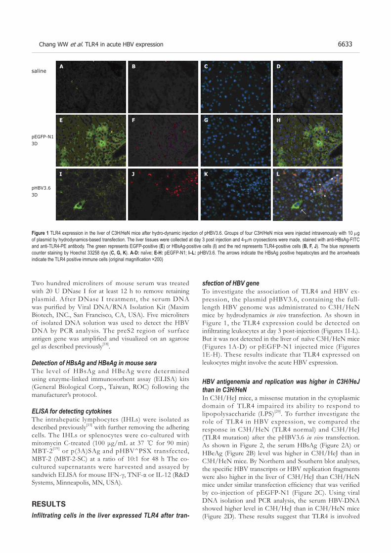
Chang WW et al. TLR4 in acute HBV expression 6633
Two hundred microliters of mouse serum was treated with 20 U DNase I for at least 12 h to remove retaining plasmid. After DNase I treatment, the serum DNA was purified by Viral DNA/RNA Isolation Kit (Maxim Biotech, INC., San Francisco, CA, USA). Five microliters of isolated DNA solution was used to detect the HBV DNA by PCR analysis. The preS2 region of surface antigen gene was amplifi ed and visualized on an agarose gel as described previously[18].
Detection of HBsAg and HBeAg in mouse sera The level of HBsAg and HBeAg were determined using enzyme-linked immunosorbent assay (ELISA) kits (General Biological Corp., Taiwan, ROC) following the manufacturer’s protocol.
ELISA for detecting cytokinesThe intrahepatic lymphocytes (IHLs) were isolated as described previously[15] with further removing the adhering cells. The IHLs or splenocytes were co-cultured with mitomycin C-treated (100 μg/mL at 37 ℃ for 90 min) MBT-2[19] or p(3A)SAg and pHBV^PSX transfected, MBT-2 (MBT-2-SC) at a ratio of 10:1 for 48 h The co-cultured supernatants were harvested and assayed by sandwich ELISA for mouse IFN-γ, TNF-α or IL-12 (R&D Systems, Minneapolis, MN, USA).
RESULTSInfiltrating cells in the liver expressed TLR4 after tran-
sfection of HBV geneTo investigate the association of TLR4 and HBV ex-pression, the plasmid pHBV3.6, containing the full-length HBV genome was administrated to C3H/HeN mice by hydrodynamics in vivo transfection. As shown in Figure 1, the TLR4 expression could be detected on infi ltrating leukocytes at day 3 post-injection (Figures 1I-L). But it was not detected in the liver of naïve C3H/HeN mice (Figures 1A-D) or pEGFP-N1 injected mice (Figures 1E-H). These results indicate that TLR4 expressed on leukocytes might involve the acute HBV expression.
HBV antigenemia and replication was higher in C3H/HeJ than in C3H/HeNIn C3H/HeJ mice, a missense mutation in the cytoplasmic domain of TLR4 impaired its ability to respond to lipopolysaccharide (LPS)[20]. To further investigate the role of TLR4 in HBV expression, we compared the response in C3H/HeN (TLR4 normal) and C3H/HeJ (TLR4 mutation) after the pHBV3.6 in vivo transfection. As shown in Figure 2, the serum HBsAg (Figure 2A) or HBeAg (Figure 2B) level was higher in C3H/HeJ than in C3H/HeN mice. By Northern and Southern blot analyses, the specifi c HBV transcripts or HBV replication fragments were also higher in the liver of C3H/HeJ than C3H/HeN mice under similar transfection effi ciency that was verifi ed by co-injection of pEGFP-N1 (Figure 2C). Using viral DNA isolation and PCR analysis, the serum HBV-DNA showed higher level in C3H/HeJ than in C3H/HeN mice (Figure 2D). These results suggest that TLR4 is involved
Figure 1 TLR4 expression in the liver of C3H/HeN mice after hydro-dynamic injection of pHBV3.6. Groups of four C3H/HeN mice were injected intravenously with 10 μg of plasmid by hydrodynamics-based transfection. The liver tissues were collected at day 3 post injection and 4-μm cryosections were made, stained with anti-HBsAg-FITC and anti-TLR4-PE antibody. The green represents EGFP-positive (E) or HBsAg-positive cells (I) and the red represents TLR4-positive cells (B, F, J). The blue represents counter staining by Hoechst 33258 dye (C, G, K). A-D: naïve; E-H: pEGFP-N1; I–L: pHBV3.6. The arrows indicate the HBsAg positive hepatocytes and the arrowheads indicate the TLR4 positive immune cells (original magnifi cation ×200)
saline
pEGFP-N13D
pHBV3.63D
A
E
I
B C D
HGF
J K L
←
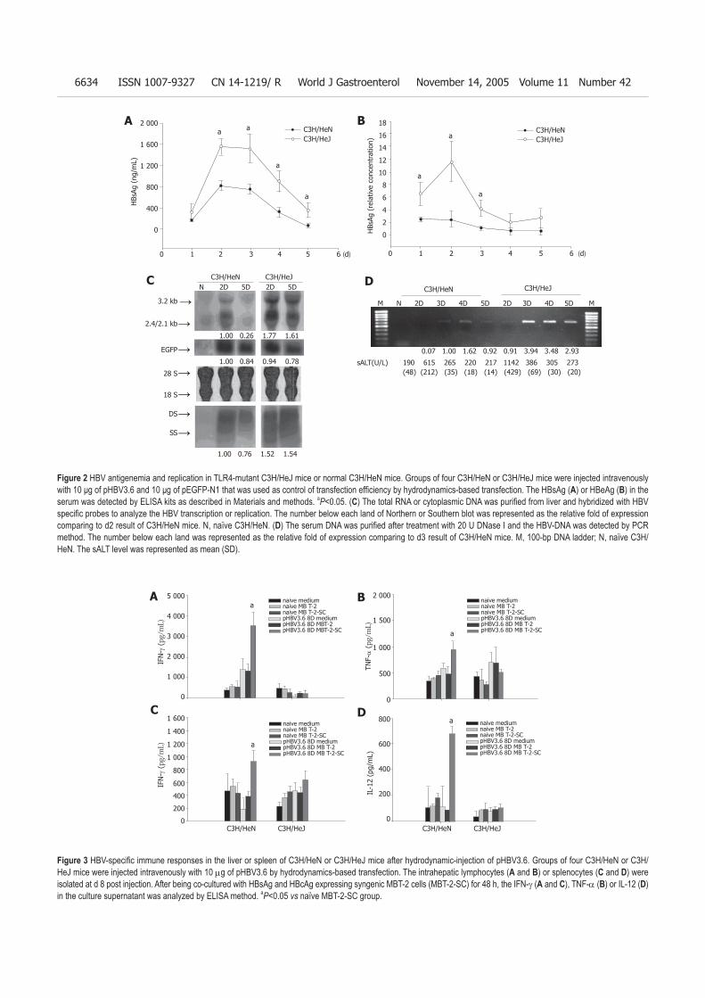
6634 ISSN 1007-9327 CN 14-1219/ R World J Gastroenterol November 14, 2005 Volume 11 Number 42
Figure 2 HBV antigenemia and replication in TLR4-mutant C3H/HeJ mice or normal C3H/HeN mice. Groups of four C3H/HeN or C3H/HeJ mice were injected intravenously with 10 μg of pHBV3.6 and 10 μg of pEGFP-N1 that was used as control of transfection effi ciency by hydrodynamics-based transfection. The HBsAg (A) or HBeAg (B) in the serum was detected by ELISA kits as described in Materials and methods. aP<0.05. (C) The total RNA or cytoplasmic DNA was purifi ed from liver and hybridized with HBV specifi c probes to analyze the HBV transcription or replication. The number below each land of Northern or Southern blot was represented as the relative fold of expression comparing to d2 result of C3H/HeN mice. N, naïve C3H/HeN. (D) The serum DNA was purifi ed after treatment with 20 U DNase I and the HBV-DNA was detected by PCR method. The number below each land was represented as the relative fold of expression comparing to d3 result of C3H/HeN mice. M, 100-bp DNA ladder; N, naïve C3H/HeN. The sALT level was represented as mean (SD).
Figure 3 HBV-specifi c immune responses in the liver or spleen of C3H/HeN or C3H/HeJ mice after hydrodynamic-injection of pHBV3.6. Groups of four C3H/HeN or C3H/HeJ mice were injected intravenously with 10 μg of pHBV3.6 by hydrodynamics-based transfection. The intrahepatic lymphocytes (A and B) or splenocytes (C and D) were isolated at d 8 post injection. After being co-cultured with HBsAg and HBcAg expressing syngenic MBT-2 cells (MBT-2-SC) for 48 h, the IFN-γ (A and C), TNF-α (B) or IL-12 (D) in the culture supernatant was analyzed by ELISA method. aP<0.05 vs naïve MBT-2-SC group.
0.07 1.00 1.62 0.92 0.91 3.94 3.48 2.93
190 615 265 220 217 1142 386 305 273(48) (212) (35) (18) (14) (429) (69) (30) (20)
M N 2D 3D 4D 5D 2D 3D 4D 5D M
C3H/HeN C3H/HeJ
sALT(U/L)
1.00 0.26 1.77 1.61
1.00 0.84 0.94 0.78
1.00 0.76 1.52 1.54
N 2D 5D 2D 5D C3H/HeN C3H/HeJ
3.2 kb
2.4/2.1 kb
EGFP
28 S
18 S
DS
SS
→
→
→
→→→
→
C3H/HeNC3H/HeJ
a a
a
a
2 000
1 600
1 200
800
400
0
0 1 2 3 4 5 6 (d)
HBs
Ag (
ng/m
L)
a
a
a
C3H/HeNC3H/HeJ
18
16
14
12
10
8
6
4
2
0
0 1 2 3 4 5 6 (d)
HBs
Ag (
rela
tive
conc
entr
atio
n)
A B
DC
a5 000
4 000
3 000
2 000
1 000
0
IFN
- γ (pg/mL)
a
a
a
naive mediumnaive MB T-2naive MB T-2-SCpHBV3.6 8D mediumpHBV3.6 8D MBT-2pHBV3.6 8D MBT-2-SC
naive mediumnaive MB T-2naive MB T-2-SCpHBV3.6 8D mediumpHBV3.6 8D MB T-2pHBV3.6 8D MB T-2-SC
naive mediumnaive MB T-2naive MB T-2-SCpHBV3.6 8D mediumpHBV3.6 8D MB T-2pHBV3.6 8D MB T-2-SC
naive mediumnaive MB T-2naive MB T-2-SCpHBV3.6 8D mediumpHBV3.6 8D MB T-2pHBV3.6 8D MB T-2-SC
1 600
1 400
1 200
1 000
800
600
400
200
0
2 000
1 500
1 000
500
0
800
600
400
200
0
IFN
- γ (pg/mL)
IL-1
2 (p
g/m
L)TN
F-α
(pg/mL)
C3H/HeN C3H/HeJ C3H/HeN C3H/HeJ
A B
DC

Chang WW et al. TLR4 in acute HBV expression 6635
in host responses to HBV replication and a mutation in C3H/HeJ impaired its ability to clear the HBV virus. HBV-specific immune responses were defective in TLR4 mutant C3H/HeJ mice after hydrodynamic injection of pHBV3.6To investigate the effect of TLR4 on immune responses to HBV, intrahepatic lymphocytes (IHLs) or splenocytes from pHBV3.6-injected C3H/HeN or C3H/HeJ mice were isolated and stimulated with HBsAg- and HBcAg-expressing syngenic MBT-2 cel ls. The IFN-γ and TNF-α production of IHLs from C3H/HeN mice was significantly increased at 48 h after HBsAg and HBcAg stimulation. But this was not observed in IHLs from C3H/HeJ mice (Figures 3A and B). The IFN-γ and IL-12 production of splenocytes of C3H/HeN mice was also significantly increased in C3H/HeN mice, but not in C3H/HeJ mice (Figures 3C and D). These data suggest that TLR4 mutation in C3H/HeJ affects the HBV specifi c immune responses (IFN-γ and TNF-α production in liver or IFN-γ and IL-12 production in spleen).
Induction of iNOS was also defective in TLR4 mutant C3H/HeJ mice after hydrodynamic injection of pHBV3.6It was reported that the activation of TLR4 signaling can induce the iNOS expression[21], and we also reported that iNOS plays an anti-HBV role in acute HBV expression[15]. Therefore, the iNOS expressed was compared in wild type and TLR4 mutant mice after pHBV3.6 transfection. The HBsAg-expressing hepatocytes were equivalent among C3H/HeN and C3H/HeJ mice after pHBV3.6 injection, indicating the transfection efficiency was similar in both strains of mice. But iNOS stainings were detected on the infiltrating leukocytes nearby the HBsAg-expressing hepatocytes in the liver of pHBV3.6-injected C3H/HeN (Figures 4C and D) whereas the expression of iNOS on
infi ltrating leukocytes was impaired in the liver of C3H/HeJ mice (Figures 4G and H). These data suggested that the mutation of TLR4 infl uences the induction of iNOS expression during acute HBV expression and may further affect the clearance of HBV.
HBV replication was reduced in C3H/HeJ mice after adoptive transfer of splenocytes from C3H/HeN miceTo further confirm, if TLR4-expressing immune cells provide the protection role to HBV expression, the splenocytes from C3H/HeN mice were injected into C3H/HeJ mice intravenously at the day before pHBV3.6 injection. The HBsAg antigenemia was higher in C3H/HeJ than in C3H/HeN mice, but the adoptive transfer of C3H/HeN splenocytes into the C3H/HeJ mice reduced the serum HBsAg to the level similar to that in C3H/HeN mice (Figure 5A). The HBV transcription or replication in liver (Figure 5B) or HBV-DNA in sera (Figure 5C) was also reduced as well under similar transfection effi ciency as verifi ed by co-injection of pEGFP-N1. But when adoptive transfer of splenocytes from C3H/HeJ mice to C3H/HeN mice was carried out, there was no effect to HBsAg in sera (Figure 5A), HBV replication in liver (Figure 5B), or HBV-DNA in sera (Figure 5C). These results indicate that the TLR4 enhances the immune functions to help clear the HBV replication.
DISCUSSIONIn this study, we investigated the role of TLR4 in a murine acute HBV expression model. The HBV antigenemia, serum HBV-DNA, as well as HBV transcription and replication in liver were higher in TLR4 mutant C3H/HeJ than TLR4 normal C3H/HeN. This is probably caused by the impaired induction of iNOS and HBV specific immune response because of TLR4 mutation in C3H/
Figure 4 iNOS expression in the liver of C3H/HeN or C3H/HeJ mice after hydrodynamic-injection of pHBV3.6. Groups of four C3H/HeN (C, D) or C3H/HeJ (G, H) mice were injected intravenously with 10 μg of pHBV3.6 by hydrodynamics-based transfection. Serial sections of frozen liver tissue were made and stained with anti-HBs antibody (B, D, F, H) or anti-iNOS antibody (A, C, E, G). Red color was developed and indicated the positive staining. The insets in (C), (D), (G), and (H) represent twofold magnifi cation of positive staining. A, B, E, F, saline injected control mice; C, D, G, H, day 2 post injection (original magnifi cation ×200).
A B DC
E F HG
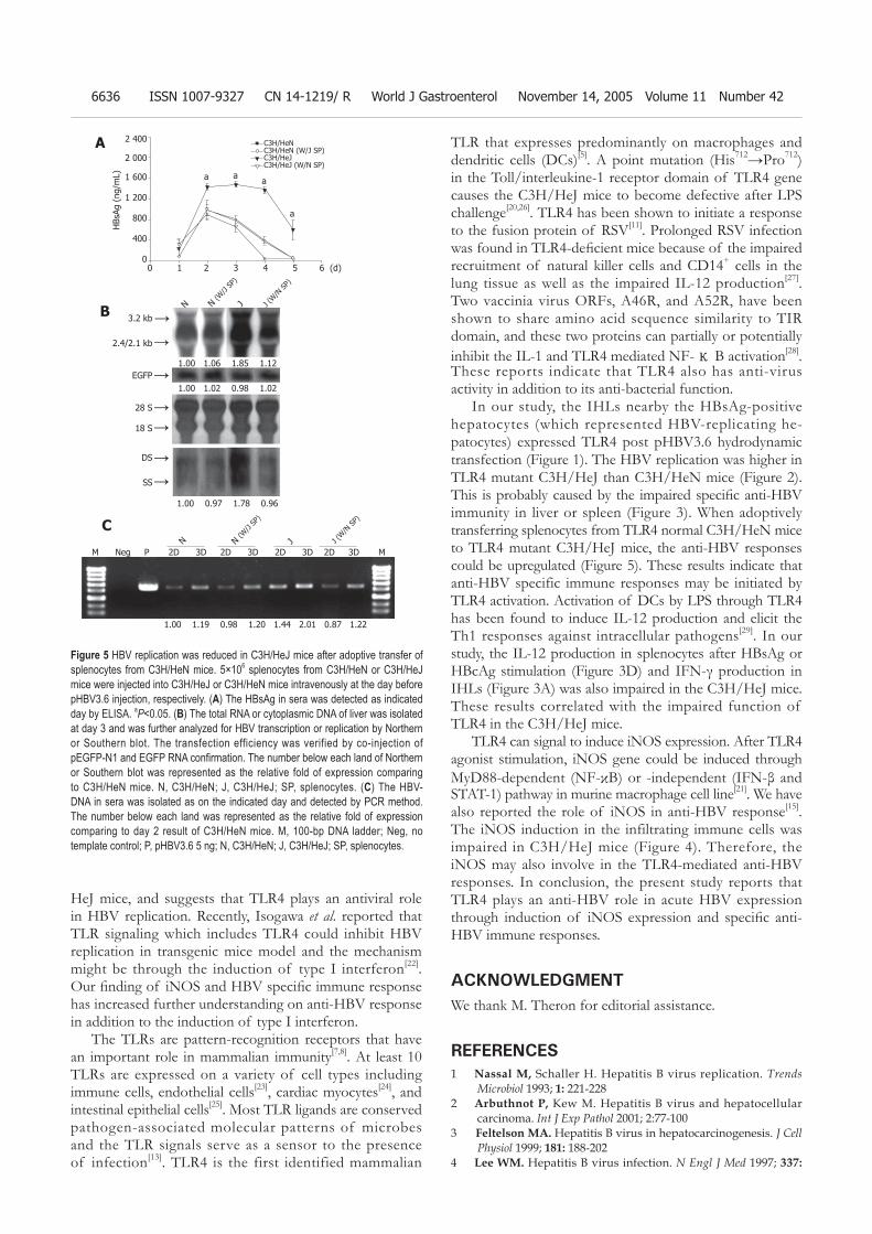
6636 ISSN 1007-9327 CN 14-1219/ R World J Gastroenterol November 14, 2005 Volume 11 Number 42
HeJ mice, and suggests that TLR4 plays an antiviral role in HBV replication. Recently, Isogawa et al. reported that TLR signaling which includes TLR4 could inhibit HBV replication in transgenic mice model and the mechanism might be through the induction of type I interferon[22]. Our fi nding of iNOS and HBV specifi c immune response has increased further understanding on anti-HBV response in addition to the induction of type I interferon.
The TLRs are pattern-recognition receptors that have an important role in mammalian immunity[7,8]. At least 10 TLRs are expressed on a variety of cell types including immune cells, endothelial cells[23], cardiac myocytes[24], and intestinal epithelial cells[25]. Most TLR ligands are conserved pathogen-associated molecular patterns of microbes and the TLR signals serve as a sensor to the presence of infection[13]. TLR4 is the first identified mammalian
TLR that expresses predominantly on macrophages and dendritic cells (DCs)[5]. A point mutation (His712→Pro712) in the Toll/interleukine-1 receptor domain of TLR4 gene causes the C3H/HeJ mice to become defective after LPS challenge[20,26]. TLR4 has been shown to initiate a response to the fusion protein of RSV[11]. Prolonged RSV infection was found in TLR4-defi cient mice because of the impaired recruitment of natural killer cells and CD14+ cells in the lung tissue as well as the impaired IL-12 production[27]. Two vaccinia virus ORFs, A46R, and A52R, have been shown to share amino acid sequence similarity to TIR domain, and these two proteins can partially or potentially inhibit the IL-1 and TLR4 mediated NF-κB activation[28]. These reports indicate that TLR4 also has anti-virus activity in addition to its anti-bacterial function.
In our study, the IHLs nearby the HBsAg-positive hepatocytes (which represented HBV-replicating he-patocytes) expressed TLR4 post pHBV3.6 hydrodynamic transfection (Figure 1). The HBV replication was higher in TLR4 mutant C3H/HeJ than C3H/HeN mice (Figure 2). This is probably caused by the impaired specifi c anti-HBV immunity in liver or spleen (Figure 3). When adoptively transferring splenocytes from TLR4 normal C3H/HeN mice to TLR4 mutant C3H/HeJ mice, the anti-HBV responses could be upregulated (Figure 5). These results indicate that anti-HBV specific immune responses may be initiated by TLR4 activation. Activation of DCs by LPS through TLR4 has been found to induce IL-12 production and elicit the Th1 responses against intracellular pathogens[29]. In our study, the IL-12 production in splenocytes after HBsAg or HBcAg stimulation (Figure 3D) and IFN-γ production in IHLs (Figure 3A) was also impaired in the C3H/HeJ mice. These results correlated with the impaired function of TLR4 in the C3H/HeJ mice.
TLR4 can signal to induce iNOS expression. After TLR4 agonist stimulation, iNOS gene could be induced through MyD88-dependent (NF-κB) or -independent (IFN-β and STAT-1) pathway in murine macrophage cell line[21]. We have also reported the role of iNOS in anti-HBV response[15]. The iNOS induction in the infiltrating immune cells was impaired in C3H/HeJ mice (Figure 4). Therefore, the iNOS may also involve in the TLR4-mediated anti-HBV responses. In conclusion, the present study reports that TLR4 plays an anti-HBV role in acute HBV expression through induction of iNOS expression and specifi c anti-HBV immune responses.
ACKNOWLEDGMENTWe thank M. Theron for editorial assistance.
REFERENCES1 Nassal M, Schaller H. Hepatitis B virus replication. Trends
Microbiol 1993; 1: 221-2282 Arbuthnot P, Kew M. Hepatitis B virus and hepatocellular
carcinoma. Int J Exp Pathol 2001; 2:77-1003 Feltelson MA. Hepatitis B virus in hepatocarcinogenesis. J Cell
Physiol 1999; 181: 188-2024 Lee WM. Hepatitis B virus infection. N Engl J Med 1997; 337:
Figure 5 HBV replication was reduced in C3H/HeJ mice after adoptive transfer of splenocytes from C3H/HeN mice. 5×10⁶ splenocytes from C3H/HeN or C3H/HeJ mice were injected into C3H/HeJ or C3H/HeN mice intravenously at the day before pHBV3.6 injection, respectively. (A) The HBsAg in sera was detected as indicated day by ELISA. aP<0.05. (B) The total RNA or cytoplasmic DNA of liver was isolated at day 3 and was further analyzed for HBV transcription or replication by Northern or Southern blot. The transfection efficiency was verified by co-injection of pEGFP-N1 and EGFP RNA confi rmation. The number below each land of Northern or Southern blot was represented as the relative fold of expression comparing to C3H/HeN mice. N, C3H/HeN; J, C3H/HeJ; SP, splenocytes. (C) The HBV-DNA in sera was isolated as on the indicated day and detected by PCR method. The number below each land was represented as the relative fold of expression comparing to day 2 result of C3H/HeN mice. M, 100-bp DNA ladder; Neg, no template control; P, pHBV3.6 5 ng; N, C3H/HeN; J, C3H/HeJ; SP, splenocytes.
C3H/HeNC3H/HeN (W/J SP)C3H/HeJC3H/HeJ (W/N SP)
2 400
2 000
1 600
1 200
800
400
0
HBs
Ag (
ng/m
L) a aa
a
0 1 2 3 4 5 6 (d)
3.2 kb
2.4/2.1 kb
EGFP
28 S
18 S
DS
SS
1.00 1.06 1.85 1.12
→
→
→→
→→
→
1.00 1.02 0.98 1.02
1.00 0.97 1.78 0.96
N N (W/J
SP)
J J (W
/N SP
)
1.00 1.19 0.98 1.20 1.44 2.01 0.87 1.22
M Neg P 2D 3D 2D 3D 2D 3D 2D 3D M N N (W
/J SP
)
J J (W
/N SP
)
A
B
C
Chang WW et al. TLR4 in acute HBV expression 6637
Science Editor Guo SY Language Editor Elsevier HK
1733-17455 Lien E, Ingalls RR. Toll-like receptors. Crit Care Med 2002; 30:
S1-S116 Zhang D, Zhang G, Hayden MS, Greenblatt MB, Bussey C,
Flavell RA, Ghosh S. A toll-like receptor that prevents infection by uropathogenic bacteria. Science 2004; 303: 1522-1526
7 Akira S, Takeda K, Kaisho T. Toll-like receptors: critical proteins linking innate and acquired immunity. Nat Immunol 2001; 2: 675-680
8 Aderem A, Ulevitch RJ. Toll-like receptors in the induction of the innate immune response. Nature 2000; 406: 782-787
9 Barton GM, Medzhitov R. Toll-like receptor signaling pathways. Science 2003; 300: 1524-1525
10 Ghosh S, May MJ, Kopp EB. NF-kappa B and Rel proteins: evolutionarily conserved mediators of immune responses. Annu Rev Immunol 1998; 16: 225-260
11 Kurt-Jones EA, Popova L, Kwinn L, Haynes LM, Jones LP, Tripp RA, Walsh EE, Freeman MW, Golenbock DT, Anderson LJ, Finberg RW. Pattern recognition receptors TLR4 and CD14 mediate response to respiratory syncytial virus. Nat Immunol 2000; 1: 398-401
12 Liu F, Song YK, Liu D. Hydrodynamics-based transfection in animals by systemic administration of plasmid DNA. Gene Ther 1999; 6: 1258-1266
13 Zhang G, Budker V, Wolff JA. High levels of foreign gene expression in hepatocytes after tail vein injections of naked plasmid DNA. Hum Gene Ther 1999; 10: 1735-1737
14 Chang J, Sigal LJ, Lerro A, Taylor J. Replication of the human hepatitis delta virus genome Is initiated in mouse hepatocytes following intravenous injection of naked DNA or RNA sequences. J Virol 2001; 75: 3469-3473
15 Chang WW, Su IJ, Lai MD, Chang WT, Huang W, Lei HY. The role of inducible nitric oxide synthase in a murine acute hepatitis B virus (HBV) infection model induced by hydrodynamics-based in vivo transfection of HBV-DNA. J Hepatol 39: 834-842
16 Yang PL, Althage A, Chung J, Chisari FV. Hydrodynamic injection of viral DNA: A mouse model of acute hepatitis B virus infection. Proc Natl Acad Sci USA 2002; 99: 13825-13830
17 Guidotti LG, Matzke B, Schaller H, Chisari FV. High level hepatitis B virus replication in transgenic mice. J Virol 1995; 69: 6158-6169
18 Fan YF, Lu CC, Chen WC, Yao WJ, Wang HC, Chang TT, Lei HY, Shiau AL, Su IJ. Prevalence and signifi cance of hepatitis B virus (HBV) pre-S mutants in serum and liver at different replicative stages of chronic HBV infection. Hepatology 2001; 33: 277-286
19 Eto M, Harada M, Tamada K, Tokuda N, Koikawa Y, Nakamura M, Nomoto K, Naito S. Antitumor activity of interleukin-12 against murine bladder cancer. J Urol 2000; 163: 1549-1552
20 Poltorak A, He X, Smirnova I, Liu MY, Van Huffel C, Du X, Birdwell D, Alejos E, Silva M, Galanos C, Freudenberg M, Ricciardi-Castagnoli P, Layton B, Beutler B. Defective LPS signaling in C3H/HeJ and C57BL/10ScCr mice: mutations in Tlr4 gene. Science 1998; 282: 2085-2088
21 Schilling D, Thomas K, Nixdorff K, Vogel SN, Fenton MJ. Toll-like receptor 4 and Toll-IL-1 receptor domain-containing adapter protein (TIRAP)/myeloid differentiation protein 88 adapter-like (Mal) contribute to maximal IL-6 expression in macrophages. J Immunol 2002; 169: 5874-5880
22 Isogawa M, Robek MD, Furuichi Y, Chisari FV. Toll-like receptor signaling inhibits hepatitis B virus replication in vivo. J Virol 2005; 79: 7269-72
23 Faure E, Thomas L, Xu H, Medvedev A, Equils O, Arditi M. Bacterial lipopolysaccharide and IFN-gamma induce Toll-like receptor 2 and Toll-like receptor 4 expression in human endothelial cells: role of NF-kappa B activation. J Immunol 2001; 166: 2018-2024
24 Frantz S, Kelly RA, Bourcier T. Role of TLR-2 in the activation of nuclear factor kappaB by oxidative stress in cardiac myocytes. J Biol Chem 2001; 276: 5197-5203
25 Cario E, Rosenberg IM, Brandwein SL, Beck PL, Reinecker HC, Podolsky DK. Lipopolysaccharide activates distinct signaling pathways in intestinal epithelial cell lines expressing Toll-like receptors. J Immunol 2000; 164: 966-972
26 Hoshino K, Takeuchi O, Kawai T, Sanjo H, Ogawa T, Takeda Y, Takeda K, et al. Toll-like receptor 4 (TLR4)-defi cient mice are hyporesponsive to lipopolysaccharide: evidence for TLR4 as the Lps gene product. J Immunol 1999; 162: 3749-3752
27 Haynes LM, Moore DD, Kurt-Jones EA, Finberg RW, Anderson LJ, Tripp RA. Involvement of toll-like receptor 4 in innate immunity to respiratory syncytial virus. J Virol 2001; 75: 10730-10737
28 Bowie A, Kiss-Toth E, Symons JA, Smith GL, Dower SK, O'Neill LA. A46R and A52R from vaccinia virus are antagonists of host IL-1 and toll-like receptor signaling. Proc Natl Acad Sci U S A 2000; 97: 10162-10167
29 Agrawal S, Agrawal A, Doughty B, Gerwitz A, Blenis J, Van Dyke T, Pulendran B. Different Toll-like receptor agonists instruct dendritic cells to induce distinct Th responses via differential modulation of extracellular signal-regulated kinase-mitogen-activated protein kinase and c-Fos. J Immunol 2003; 171: 4984-4989
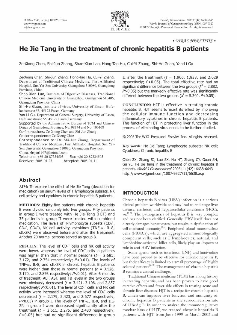
PO Box 2345, Beijing 100023, China World J Gastroenterol 2005;11(42):6638-6643www.wjgnet.com World Journal of Gastroenterology ISSN [email protected] © 2005 The WJG Press and Elsevier Inc. All rights reserved.E L S E V I E R
• VIRAL HEPATITIS •
II after the treatment (t = 1.906, 1.833, and 2.029 respectively; P>0.05). The total effective rate had no signifi cant difference between the two groups (X 2 = 2.882, P>0.05) but the markedly effective rate was signifi cantly different between the two groups (X 2 = 5.340, P<0.05).
CONCLUSION: HJT is effective in treating chronic hepatitis B. HJT seems to exert its effect by improving the ce l l u l a r immune func t i on and dec reas ing inflammatory cytokines in chronic hepatitis B patients. The function of HJT in protecting liver function in the process of eliminating virus needs to be further studied.
© 2005 The WJG Press and Elsevier Inc. All rights reserved.
Key words: He Jie Tang; Lymphocyte subsets; NK cell; Cytokines; Chronic hepatitis B
Chen ZX, Zhang SJ, Lao SX, Hu HT, Zhang CY, Guan SH, Gu YL. He Jie Tang in the treatment of chronic hepatitis B patients. World J Gastroenterol 2005; 11(42): 6638-6643http://www.wjgnet.com/1007-9327/11/6638.asp
INTRODUCTIONChronic hepatitis B virus (HBV) infection is a serious clinical problem worldwide and may lead to end-stage liver disease, cirrhosis, and hepatocellular carcinoma (HCC), etc.[1-3]. The pathogenesis of hepatitis B is very complex and has not been clarifi ed. Generally, HBV itself does not directly damages hepatocytes, but results in dysfunction of cell-mediated immunity[3-5]. Peripheral blood mononuclear cells (PBMCs), which are aggregated immunologically competent cells, such as T lymphocytes, natural, and lymphokine-activated killer cells, likely play an important role in anti-HBV infection.
Some agents such as interferon (INF) and lamivudine have been proved to be effective for chronic hepatitis B, but their effi cacy is limited to a small percentage of highly selected patients[6-13]. The management of chronic hepatitis B remains a clinical challenge.
Traditional Chinese medicine (TCM) has a long history in treating hepatitis, and has been proven to have good curative effects and fewer side effects in treating acute and chronic liver diseases. HJT is a recipe for chronic hepatitis B, which can improve liver function and immunity of chronic hepatitis B patients as the seroconversion rate of HBeAg[14]. In order to analyze the immunoregulatory mechanisms of HJT, we treated chronic hepatitis B patients with HJT from June 1999 to March 2003 and
He Jie Tang in the treatment of chronic hepatitis B patients
Ze-Xiong Chen, Shi-Jun Zhang, Shao-Xian Lao, Hong-Tao Hu, Cui-Yi Zhang, Shi-He Guan, Yan-Li Gu
Ze-Xiong Chen, Shi-Jun Zhang, Hong-Tao Hu, Cui-Yi Zhang, Department of Traditional Chinese Medicine, First Affiliated Hospital, Sun Yat-Sen University, Guangzhou 510080, Guangdong Province, ChinaShao-Xian Lao, Institute of Digestive Diseases, Traditional Chinese Medicine University of Guangzhou, Guangzhou 510405, Guangdong Province, ChinaShi-He Guan, Institute of virus, University of Essen, Hufe-landstrasse 55, 45122 Essen, GermanyYan-Li Gu, Department of General Surgery, University of Essen, Hufelandstrasse 55, 45122 Essen, GermanySupported by the Administrative Bureau of TCM and Chinese Drugs of Guangdong Province, No. 98374 and No. 100108Co-fi rst-authors: Ze-Xiong Chen and Shi-Jun ZhangCo-correspondence: Ze-Xiong ChenCorrespondence to: Dr. Shi-Jun Zhang, Department of Traditional Chinese Medicine, First Affi liated Hospital, Sun Yat-Sen University, Guangzhou 510080, Guangdong Province, China. [email protected]: +86-20-87334505 Fax: +86-20-87334505Received: 2005-01-25 Accepted: 2005-04-11
AbstractAIM: To explore the effect of He Jie Tang (decoction for medication) on serum levels of T lymphocyte subsets, NK cell activity and cytokines in chronic hepatitis B patients. METHODS: Eighty-five patients with chronic hepatitis B were divided randomly into two groups. Fifty patients in group I were treated with He Jie Tang (HJT) and 35 patients in group II were treated with combined medication. The levels of T-lymphocyte subsets (CD3
+, CD4
+, CD8+), NK cell activity, cytokines (TNF-α, IL-8,
sIL-2R) were observed before and after the treatment. Another 20 normal persons served as group 3.
RESULTS: The level of CD4+ cells and NK cell activity
were lower, whereas the level of CD8+ cells in patients
was higher than that in normal persons (t = 2.685, 3.172, and 2.754 respectively; P<0.01). The levels of TNF-α, IL-8, and sIL-2R in chronic hepatitis B patients were higher than those in normal persons (t = 3.526, 3.170, and 2.876 respectively; P<0.01). After 6 months of treatment, ALT, AST, and TB levels in the two groups were obviously decreased (t = 3.421, 3.106, and 2.857 respectively; P<0.01). The level of CD4
+ cells and NK cell activity were increased whereas the level of CD8
+ cells decreased (t = 2.179, 2.423, and 2.677 respectively; P<0.05) in group I. The levels of TNF-α, IL-8, and sIL-2R in group I were decreased significantly after the treatment (t = 2.611, 2.275, and 2.480 respectively; P <0.05) but had no significant difference in group

Chen ZX et al. He Jie Tang on chronic hepatitis B 6639
observed the clinical effect of HJT on T lymphocyte subset level, NK cell activity as well as TNF-α, IL-8, and sIL-2R level.
MATERIALS AND METHODSPatientsA total of 85 patients with chronic hepatitis B were enrolled in this study and randomly divided into two groups. There were 27 males and 23 females aged 18-60 years (mean 36.9±9.5 years) in group I. There were 19 males and 16 females aged 18-60 years (mean 38.5±9.1 years) in group II. The difference in clinical data between the two groups was insignificant. Twenty age-matched healthy donors from the Blood Center of our hospital were assigned as group III. This prospective study was approved by the local ethics committee and written consent was obtained from the participants.
Diagnostic criteriaPatients with a history of hepatitis B or HBsAg carriers for at least 6 mo, who still had symptoms and signs of hepatitis as well as abnormal liver function and positive HBsAg, HBeAg and HBV-DNA, were diagnosed as chronic hepatitis B in the present study.
Criteria for enrollmentPatients, aged 18-60 years with their serum alanine aminotransferase (ALT) level being 80-240 μ/L and who had positive serum HBeAg and HBV-DNA, were enrolled. The diagnosis of hepatitis B was made in accordance with the standards for chronic viral hepatitis issued in the Fifth National Conference on Infectious Diseases and Parasitosis (Beijing, China, 1995).
Criteria for exclusionPatients aged over 60 years or less than 18 years, patients in pregnancy or in breast feeding period; patients who had hepatitis C or other hepatic viral infection, autoimmune hepatitis and drug-induced hepatitis or alcoholic hepatitis; patients with severe complications of the cardiovascular, renal or hematopoietic system and patients with mental diseases, were excluded.
Group I was treated with HJT that consisted of 10 g Radix Bupleuri, 12 g Radix Scutellariae, 9 g Rhizoma Pinelliae, 30 g Radix Codonopsis Pilosulae, 6 g Radix Glycyrrhizae Praeparata, 9 g Fructus Ziziphi Jujubae, 30 g Rhizoma Polygoni Cuspidati, 8 g Radix Morindae Officinalis, 30 g Herba Hedyotis Diffusae. One dose was taken per day for 6 mo. Group II was treated with oxymatrine (200 mg, t.i.d.), compound vitamin B (2 tablets, t.i.d.), vitamin C (100 mg, t.i.d.), vitamin E (50 mg, t.i.d.), and ester capsule (2 tablets, t.i.d.) for 6 mo.
Patients who had normal serum ALT and sero-conversion of HBeAg and HBV DNA (quantitative PCR) after treatment were defined as responders while those with negative results as non-responders.
Recording and observation of symptoms and signsThe symptoms and signs of patients were recorded in detail using the “Clinical Observation Table” once a month before and during the treatment.
Etiological markers of hepatitis BHBV-M and anti-HAV, anti-HCV, anti-HDV, and anti-EBV marks were detected by enzyme-linked immunosorbent assay (ELISA). HBV-DNA was detected by quantitative polymerized chain reaction (PCR).
Liver functionThe patients had liver function examination every month during the treatment, including contents of serum proteins, total bilirubin (TB) and activities of ALT and AST (aspartate aminotransferase).
T-lymphocyte subsets and NK cell activityT-lymphocyte subsets were detected by the single clone antibody APAAP method, NK cell activity was assayed by MTT colorimetry.
Detection of cytokines The levels of TNF-α, sIL-2R, and IL-8 were detected by double antibody sandwich ELISA.
Statistical analysisAll statistical analyses were performed by χ2 test and Wilcoxon rank sum test using SPSS software. P<0.05 was considered statistically signifi cant.
RESULTSStandard for effi cacy evaluationThe clinical effi cacy of treatment was evaluated according to the following standards. Markedly effective: chief symptoms including right upper abdomen pain, poor appetite, and abdominal distention disappeared; HBeAg and HBV-DNA turned negative; serum levels of ALT, AST, and TBIL restored to normal. Effective: chief symptoms were alleviated or improved; the level of HBV-DNA decreased; HBeAg did not turn negative; serum levels of ALT, AST, and TBIL decreased by >50% of the original levels. Ineffective: the chief symptoms or the serum levels of ALT, AST, and TBIL or HBeAg and HBV-DNA did not show any improvement.
Clinical effi cacy of treatmentIn group I, treatment was markedly effective in 7 cases, effective in 41 and ineffective in 2, the total effective rate being 96.0%. In group II, treatment was markedly effective in 0 cases, effective in 30, and ineffective in 5, the total effective rate being 85.7%. The difference in total effective rate was insignificant between the two groups (P>0.05) and the markedly effective rate was signifi cantly different between the two groups (P<0.05).
Levels of ALT, AST, TB, and HBV-DNA before and after the treatment
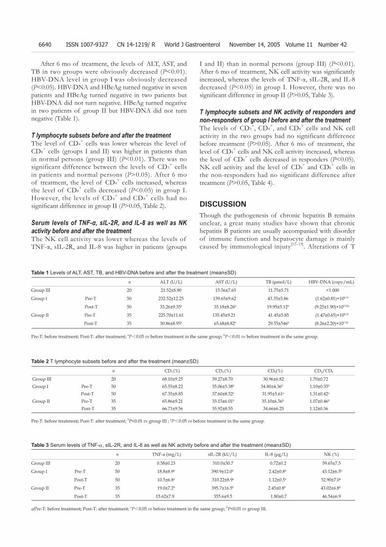
6640 ISSN 1007-9327 CN 14-1219/ R World J Gastroenterol November 14, 2005 Volume 11 Number 42
After 6 mo of treatment, the levels of ALT, AST, and TB in two groups were obviously decreased (P<0.01). HBV-DNA level in group I was obviously decreased (P<0.05). HBV-DNA and HBeAg turned negative in seven patients and HBeAg turned negative in two patients but HBV-DNA did not turn negative. HBeAg turned negative in two patients of group II but HBV-DNA did not turn negative (Table 1).
T lymphocyte subsets before and after the treatmentThe level of CD4
+ cells was lower whereas the level of CD8
+ cells (groups I and II) was higher in patients than in normal persons (group III) (P<0.01). There was no significant difference between the levels of CD3
+ cells in patients and normal persons (P>0.05). After 6 mo of treatment, the level of CD4
+ cells increased, whereas the level of CD8
+ cells decreased (P<0.05) in group I. However, the levels of CD4
+ and CD8+ cells had no
signifi cant difference in group II (P>0.05, Table 2).
Serum levels of TNF-α, sIL-2R, and IL-8 as well as NK activity before and after the treatmentThe NK cell activity was lower whereas the levels of TNF-α, sIL-2R, and IL-8 was higher in patients (groups
I and II) than in normal persons (group III) (P<0.01). After 6 mo of treatment, NK cell activity was signifi cantly increased, whereas the levels of TNF-α, sIL-2R, and IL-8 decreased (P<0.05) in group I. However, there was no signifi cant difference in group II (P>0.05, Table 3).
T lymphocyte subsets and NK activity of responders and non-responders of group I before and after the treatmentThe levels of CD3
+, CD4+, and CD8
+ cells and NK cell activity in the two groups had no significant difference before treatment (P>0.05). After 6 mo of treatment, the level of CD4
+ cells and NK cell activity increased, whereas the level of CD8
+ cells decreased in responders (P<0.05). NK cell activity and the level of CD4
+ and CD8+ cells in
the non-responders had no significant difference after treatment (P>0.05, Table 4).
DISCUSSIONThough the pathogenesis of chronic hepatitis B remains unclear, a great many studies have shown that chronic hepatitis B patients are usually accompanied with disorder of immune function and hepatocyte damage is mainly caused by immunological injury[15–19]. Alterations of T
n ALT (U/L) AST (U/L) TB (μmol/L) HBV-DNA (copy/mL)
Group III 20 21.52±8.90 15.56±7.65 11.75±5.71 <1 000
Group I Pre-T 50 232.52±12.25 139.65±9.62 43.35±5.86 (1.62±0.81)×108.31
Post-T 50 33.26±9.35b 35.18±8.26b 19.95±5.12b (9.25±1.90)×105.02a
Group II Pre-T 35 225.70±11.61 135.45±9.21 41.45±5.85 (1.47±0.65)×108.22
Post-T 35 30.86±8.95b 65.68±8.82b 29.55±546b (8.26±2.20)×107.62
Table 2 T lymphocyte subsets before and after the treatment (mean±SD)
Table 1 Levels of ALT, AST, TB, and HBV-DNA before and after the treatment (mean±SD)
Pre-T: before treatment; Post-T: after treatment; bP<0.01 vs group III ; aP<0.05 vs before treatment in the same group.
Pre-T: before treatment; Post-T: after treatment; aP<0.05 vs before treatment in the same group; bP<0.01 vs before treatment in the same group.
n TNF-a (mg/L) sIL-2R (kU/L) IL-8 (μg/L) NK (%)
Group III 20 0.58±0.23 310.0±30.7 0.72±0.2 59.65±7.5
Group I Pre-T 50 18.8±8.9b 390.9±12.0b 2.42±0.8b 43.12±6.5b
Post-T 50 10.5±6.8a 310.22±8.9a 1.12±0.5a 52.90±7.0a
Group II Pre-T 35 19.0±7.2b 395.7±16.5b 2.45±0.8b 43.02±6.8b
Post-T 35 15.62±7.9 355.6±9.5 1.80±0.7 46.54±6.9
Table 3 Serum levels of TNF-α, sIL-2R, and IL-8 as well as NK activity before and after the treatment (mean±SD)
αPre-T: before treatment; Post-T: after treatment; aP<0.05 vs before treatment in the same group; bP<0.01 vs group III.
n CD3 (%) CD4 (%) CD8(%) CD4/CD8
Group III 20 68.10±9.25 39.27±8.70 30.96±6.82 1.70±0.72 Group I Pre-T 50 65.55±8.22 35.06±5.38b 34.80±4.36b 1.10±0.35b
Post-T 50 67.35±8.85 37.60±8.52a 31.95±5.61a 1.31±0.42a
Group II Pre-T 35 65.86±9.21 35.15±6.01b 35.10±6.56b 1.07±0.46b
Post-T 35 66.71±9.56 35.92±8.55 34.66±6.25 1.12±0.36

Chen ZX et al. He Jie Tang on chronic hepatitis B 6641
lymphocyte subsets and NK cells are important reasons for the disorder of immune function due to HBV infection, TNF-α , IL-8, and sIL-2R are important cytokines associated with liver damage. Therefore, the importance of T lymphocytes and NK cells as well as cytokines in the occurrence of chronic HBV infection has received more and more attention.
CD3+, CD4
+, and CD8+ cells are major function
subgroups of T cells. An antiviral cellular immune response of CD4
+ and CD8+ is the important mechanism
of hepatocyte injury induced by HBV, the specific response of CD4
+ and CD8+ to the virus antigen is closely
related with the elimination of the virus[6,20,21]. NK cells play a critical role in host innate defense against viruses and are partly responsible for liver injury in the process of erasing viruses[22-28]. Recent studies found that NK cells are potent activators of dendritic cells (DCs), which have an impact on the magnitude and direction of DC activation of T cells under the conditions of chronic viral infection, activated NK cells can release cytokines and prevent virus from reproducing[23,29]. Therefore, T-lymphocyte subsets and NK activity can be considered as an appropriate response of immune system to inhibit viral replication and HBV eradication. In the present study, we discovered that in the outbreak period of chronic hepatitis B, NK activity and level of CD4
+ cells were lower, whereas the level of CD8
+ cells was higher in patients than in normal persons, suggesting that disorders of cellular immune function and pathologic damages occur in chronic hepatitis B patients.
The serum NK activity and CD4+ cell level in non-
responders were lower than those in normal persons, whereas the level of CD8
+ cells in non-responders was higher than that of normal persons. After treatment, the NK activity and CD4
+ cell level were increased in seven patients with the conversion of HBV-DNA and HBeAg and the liver function resumed to normal. The results suggest that T-lymphocyte subsets and NK activity are depressed rather than activated in viral hepatitis B, but levels of T lymphocyte subsets and NK activity are closely related with different courses of hepatitis B. At the same time, levels of T lymphocyte subsets and NK activity in some patients were still low in palliative period, indicating that the chance of recrudescence might increase. T lymphocyte subsets and NK cells play a critical role in response to HBV infection and their level and mutual relation can be used to identify the cellular immune level in
patients with chronic hepatitis B[11,38].TNF-α plays an indispensable role in liver injury
media ted by spec i f i c immune response to HBV infection[30]. Pretreatment with anti-TNF-α mAb in animal model strongly blocks Th1 cell-induced hepatocyte necrosis and apoptosis[32]. However, it was reported that TNF-α exerts its antiviral effects without destruction of hepatocytes[33]. IL-8 is a chemotactic factor of neutrophils and T cells and plays a role in hepatic injury in patients with chronic viral hepatitis. Remarkable increase of IL-8 leads to accumulation of cytotoxic T lymphocytes, which get direct and immediate access to the target hepatocytes and the resident intrahepatic macrophages, subsequently causing the damage of hepatocytes[34-36]. Release of sIL-2R from activated T lymphocytes may occur as a result of proteolysis of mIL-2R or as a result of alternative mRNA process. High level of sIL-2R in chronic HBV infection appears directly related to the activity of liver diseases; therefore, serum sIL-2R levels can be used to indicate the degree of liver damage in patients with chronic HBV infection[31,37,38].
In the present study, we discovered that in the outbreak period of chronic hepatitis B, the levels of IL-8, TNF-α, and sIL-2R were higher in patients than in normal persons during and after HJT treatment, significantly increased suggesting that cytokines and immunocytes may play a role in the pathogenesis of chronic hepatitis B.
HJT is a recipe for treating hepatitis in which cold and warm drugs are used to eliminate evils and restore healthy energy. Former research indicates that HJT can protect the liver from injury[20,21]. We discovered that HJT could improve liver function and NK activity, regulate T cellular immune function in chronic hepatitis B patients. The results suggest that HJT exerts its effect by improving the cellular immune function and decreasing infl ammatory cytokines in chronic hepatitis B patients.
REFERENCES1 Kagawa T, Watanabe N, Kanouda H, Takayama I, Shiba T,
Kanai T, Kawazoe K, Takashimizu S, Kumaki N, Shimamura K, Matsuzaki S, Mine T. Fatal liver failure due to reactivation of lamivudine-resistant HBV mutant. World J Gastroenterol 2004; 10: 1686-1687
2 Ohata K, Hamasaki K, Toriyama K, Ishikawa H, Nakao K, Eguchi K. High viral load is a risk factor for hepatocellular carcinoma in patients with chronic hepatitis B virus infection. J Gastroenterol Hepatol 2004; 19: 670-675
n CD3 (%) CD4 (%) CD8 (%) NK (%)
Responders Pre-T 7 66.02±8.86 35.10±4.76b 34.92±4.36b 43.52±7.1b
Post-T 7 67.80±9.11 38.85±8.85a 30.15±5.82a 55.60±8.2a
Non-responders Pre-T 43 65.50±9.08 34.92±6.30b 34.77±6.56b 42.93±6.7 b
Post-T 43 66.09±9.35 35.99±8.70 34.25±5.52 45.60±6.5
Group III 20 68.10±9.25 39.27±8.70 30.96±6.82 59.65±7.5
Table 4 T lymphocyte subsets in responders and non-responders of group I before and after the treatment (mean±SD)
aP<0.05 vs before treatment in the same group; bP<0.01 vs group III.
6642 ISSN 1007-9327 CN 14-1219/ R World J Gastroenterol November 14, 2005 Volume 11 Number 42
3 Ikeda K, Kobayashi M, Saitoh S, Someya T, Hosaka T, Akuta N, Suzuki Y, Suzuki F, Tsubota A, Arase Y, Kumada H. Significance of hepatitis B virus DNA clearance and early prediction of hepatocellular carcinogenesis in patients with cirrhosis undergoing interferon therapy: long-term follow up of a pilot study. J Gastroenterol Hepatol 2005; 20: 95-102
4 Ikeda K, Arase Y, Kobayashi M, Someya T, Saitoh S, Suzuki Y, Suzuki F, Tsubota A, Akuta N, Kumada H. Consistently low hepatitis B virus DNA saves patients from hepatocellular carcinogenesis in HBV-related cirrhosis. A nested case-control study using 96 untreated patients. Intervirology 2003; 46: 96-104
5 Torre F, Cramp M, Owsianka A, Dornan E, Marsden H, Carman W, Williams R, Naoumov NV. Direct evidence that naturally occurring mutations within hepatitis B core epitope alter CD4+ T-cell reactivity. J Med Virol 2004; 72: 370-376
6 Mutimer D. Hepatitis B virus antiviral drug resistance: from the laboratory to the patient. Antivir Ther 1998; 3: 243-246
7 Jang MK, Chung YH, Choi MH, Kim JA, Ryu SH, Shin JW, Kim IS, Park NH, Lee HC, Lee YS, Suh DJ. Combination of alpha-interferon with lamivudine reduces viral breakthrough during long-term therapy. J Gastroenterol Hepatol 2004; 19: 1363-1368
8 Jang MK, Chung YH, Choi MH, Kim JA, Ryu SH, Shin JW, Kim IS, Park NH, Lee HC, Lee YS, Suh DJ. Combination of alpha-interferon with lamivudine reduces viral breakthrough during long-term therapy. J Gastroenterol Hepatol 2004; 19: 1363-1368
9 Mutimer D. Hepatitis B virus infection: resistance to antiviral agents. J Clin Virol 2001; 21: 239-242
10 Leung N. Treatment of chronic hepatitis B: case selection and duration of therapy. J Gastroenterol Hepatol 2002; 17: 409-414
11 Fischer KP, Gutfreund KS, Tyrrell DL. Lamivudine resistance in hepatitis B: mechanisms and clinical implications. Drug Resist Updat 2001; 4: 118-128
12 Fung SK, Lok AS. Treatment of chronic hepatitis B: who to treat, what to use, and for how long? Clin Gastroenterol Hepatol 2004; 2: 839-848
13 Schiefke I, Klecker C, Maier M, Oesen U, Etzrodt G, Tannapfel A, Liebert UG, Berr F. Sequential combination therapy of HBe antigen-negative/virus-DNA-positive chronic hepatitis B with famciclovir or lamivudine and interferon-alpha-2a. Liver Int 2004; 24: 98-104
14 Zhang SJ, Chen ZX, Huang BJ. Effect of hejie decoction on T-cell receptor V beta 7 gene expression in patients of chronic hepatitis B. Zhongguo Zhongxi Yijiehe Zazhi 2002; 22: 499-501
15 Mancini-Bourgine M, Fontaine H, Scott-Algara D, Pol S, Brechot C, Michel ML. Induction or expansion of T-cell responses by a hepatitis B DNA vaccine administered to chronic HBV carriers. Hepatology 2004; 40: 874-882
16 Shimada N, Yamamoto K, Kuroda MJ, Terada R, Hakoda T, Shimomura H, Hata H, Nakayama E, Shiratori Y. HBcAg-specific CD8 T cells play an important role in virus suppression, and acute flare-up is associated with the expansion of activated memory T cells. J Clin Immunol 2003; 23: 223-232
17 Hasebe A, Akbar SM, Furukawa S, Horiike N, Onji M. Impaired functional capacities of liver dendritic cells from murine hepatitis B virus (HBV) carriers: relevance to low HBV-specifi c immune responses. Clin Exp Immunol 2005; 139: 35-42
18 Kondo Y, Kobayashi K, Asabe S, Shiina M, Niitsuma H, Ueno Y, Kobayashi T, Shimosegawa T. Vigorous response of cytotoxic T lymphocytes associated with systemic activation of CD8 T lymphocytes in fulminant hepatitis B. Liver Int 2004; 24: 561-567
19 Lee CK, Suh JH, Cho YS, Han KH, Chung JB, Chon CY, Moon YM. Direct analysis of HBV-specific CD8+ lymphocyte by tetrameric HLA-A2/core 18-27 complex in chronic Hepatitis B. Taehan Kan Hakhoe Chi 2002; 8: 139-148
20 Xuan SY, Sun Y, Zhang J. The influence to the function of
cellular immunity after being infected by HBV in the PBMC in chronic hepatitis B. Zhonghua Liuxing Bingxue Zazhi 1997; 18: 80-82
21 Ahn DS, Jang HC, Ahn JK, Yim CY, Kim DG. Impaired interleukin-2 receptor expression on lymphocytes from patients with chronic active hepatitis type B. Korean J Intern Med 1989; 4: 34-40
22 Dong Z, Wei H, Sun R, Hu Z, Gao B, Tian Z. Involvement of natural killer cells in PolyI: C-induced liver injury. J Hepatol 2004; 41: 966-973
23 Jinushi M, Takehara T, Tatsumi T, Kanto T, Miyagi T, Suzuki T, Kanazawa Y, Hiramatsu N, Hayashi N. Negative regulation of NK cell activities by inhibitory receptor CD94/NKG2A leads to altered NK cell-induced modulation of dendritic cell functions in chronic hepatitis C virus infection. J Immunol 2004; 173: 6072-6081
24 Sun R, Gao B. Negative regulation of liver regeneration by innate immunity (natural killer cells/interferon-gamma). Gastroenterology 2004; 127: 1525-1539
25 Kakimi K, Guidotti LG, Koezuka Y, Chisari FV. Natural killer T cell activation inhibits hepatitis B virus replication in vivo. J Exp Med 2000; 192: 921-930
26 Echevarria S, Casafont F, Miera M, Lozano JL, de la Cruz F, San Miguel G, Pons Romero F. Interleukin-2 and natural killer activity in acute type B hepatitis. Hepatogastroenterology 1991; 38: 307-310
27 Chemello L, Mondelli M, Bortolotti F, Schiavon E, Pontisso P, Alberti A, Rondanelli EG, Realdi G. Natural killer activity in patients with acute viral hepatitis. Clin Exp Immunol 1986; 64: 59-64
28 Lehoux M, Jacques A, Lusignan S, Lamontagne L. Murine viral hepatitis involves NK cell depletion associated with virus-induced apoptosis. Clin Exp Immunol 2004; 137: 41-51
29 Li Y, Zhang T, Ho C, Orange JS, Douglas SD, Ho WZ. Natural killer cells inhibit hepatitis C virus expression. J Leukoc Biol 2004; 76: 1171-1179
30 Bozkaya H, Bozdayi M, Turkyilmaz R, Sarioglu M, Cetinkaya H, Cinar K, Kose K, Yurdaydin C, Uzunalimoglu O. Circulating IL-2, IL-10 and TNF-alpha in chronic hepatitis B: their relations to HBeAg status and the activity of liver disease. Hepatogastroenterology 2000; 47: 1675-1679
31 Monsalve-De Castillo F, Romero TA, Estevez J, Costa LL, Atencio R, Porto L, Callejas D. Concentrations of cytokines, soluble interleukin-2 receptor, and soluble CD30 in sera of patients with hepatitis B virus infection during acute and convalescent phases. Clin Diagn Lab Immunol 2002; 9: 1372-1375
32 Tanaka Y, Takahashi A, Watanabe K, Takayama K, Yahata T, Habu S, Nishimura T. A pivotal role of IL-12 in Th1-dependent mouse liver injury. Int Immunol 1996; 8: 569-576
33 Guidotti LG, Chisari FV. Noncytolytic control of viral infections by the innate and adaptive immune response. Annu Rev Immunol 2001; 19: 65-91
34 Mahe Y , Mukaida N, Kuno K, Akiyama M, Ikeda N, Matsushima K, Murakami S. Hepatitis B virus X protein transactivates human interleukin-8 gene through acting on nuclear factor kB and CCAAT/enhancer-binding protein-like cis-elements. J Biol Chem 1991; 266: 13759-13763
35 Masumoto T, Ohkubo K, Yamamoto K, Ninomiya T, Abe M, Akbar SM, Michitaka K, Horiike N, Onji M. Serum IL-8 levels and localization of IL-8 in liver from patients with chronic viral hepatitis. Hepatogastroenterology 1998; 45: 1630-1634
36 Nobili V, Marcellini M, Giovannelli L, Girolami E, Muratori F, Giannone G, Devito R, De Benedetti F. Association of serum interleukin-8 levels with the degree of fi brosis in infants with chronic liver disease. J Pediatr Gastroenterol Nutr 2004; 39: 540-544
37 Sawayama Y, Hayashi J, Kawakami Y, Furusyo N, Ariyama I, Kishihara Y, Ueno K, Kashiwagi S. Serum soluble interleukin-2 receptor levels before and during interferon treatment in

Chen ZX et al. He Jie Tang on chronic hepatitis B 6643
patients with chronic hepatitis B virus infection. Dig Dis Sci 1999; 44: 163-169
38 Xuan SY, Sun Y, Zhang J. The influence to the function of
cellular immunity after being infected by HBV in the PBMC in chronic hepatitis B. Zhonghua Liuxing Bingxue Zazhi 1997; 18: 80-82
Science Editor Wang XL and Guo SY Language Editor Elsevier HK

PO Box 2345, Beijing 100023, China World J Gastroenterol 2005;11(42):6644-6649www.wjgnet.com World Journal of Gastroenterology ISSN [email protected] © 2005 The WJG Press and Elsevier Inc. All rights reserved.E L S E V I E R
• BRIEF REPORTS •
duodenal mucosa. There is an inverse association between HSV-1 and H pylori infection.© 2005 The WJG Press and Elsevier Inc. All rights reserved.
Key words: HSV-1; Herpes simplex virus type 1; Peptic ulcer; Duodenal ulcer; Gastric ulcer; PCR; Polymerase chain reaction; H pylori ; Non-steroidal anti-infl ammatory drugs
Tsamakidis K, Panotopoulou E, Dimitroulopoulos D, Xinopoulos D, Christodoulou M, Papadokostopoulou A, Karagiannis I, Kouroumalis E, Paraskevas E. Herpes simplex virus type 1 in peptic ulcer disease: An inverse association with H pylori . World J Gastroenterol 2005; 11(42): 6644-6649http://www.wjgnet.com/1007-9327/11/6644.asp
INTRODUCTIONDuring the last two decades, a significant progress has been made in the role of Helicobacter pylori(H pylori) in the pathogenesis of peptic ulcers, while the invention of new powerful antisecretory drugs has changed dramatically the treatment of the disease. However, the exact etiopathogenesis of peptic ulcer disease is still under investigation.
The signifi cant role of gastric acidity and infl ammation of mucosa due to H pylori cannot be disputed, but a multifactorial etiology for peptic ulcer disease seems to be emerging[1-3].
The idea of a possible correlation between HSV-1 and peptic ulcers has appeared almost 40 years before[4,5], due to many common characteristics observed in the clinical picture and the natural history of both diseases[4-7].
A possible involvement of HSV-1 in peptic ulcer disease was reported from several investigators, but a fi rm conclusion has not yet been reached. The vast majority of these studies are based on the detection of antibodies against the virus in the serum and the duodenal juice of patients with peptic ulcer[8-12], a finding also common in the apparent healthy population. There are only two studies that report the presence of HSV-1 in tissue samples obtained from gastric and duodenal ulcers, using polymerase chain reaction (PCR) methods, but the number of the examined populations is small[13,14]. On the other hand, in the studies reported above, a possible correlation between HSV-1 and H pylori has been investigated in the
Herpes simplex virus type 1 in peptic ulcer disease: An inverse association with Helicobacter pylori
Klisthenis Tsamakidis, Efstathia Panotopoulou, Dimitrios Dimitroulopoulos, Dimitrios Xinopoulos, Maria Christodoulou, Alexandra Papadokostopoulou, Ioannis Karagiannis, Elias Kouroumalis, Emmanuel Paraskevas
Kl isthenis Tsamakidis, Dimitr ios Dimitroulopoulos, Dimitrios Xinopoulos, Alexandra Papadokostopoulou, Ioannis Karagiannis, Emmanuel Paraskevas, Department of Gastroenterology, “Agios Savvas” Anticancer Hospital, Athens, GreeceEfstathia Panotopoulou, Maria Christodoulou, Papanicolaou Research Center of Oncology and Experiment Surgery, Athens, GreeceElias Kouroumalis, Department of Gastroenterology, University Hospital, Heraklion, Crete, GreeceCorrespondence to: Dr D Dimitroulopoulos, Department of Gastroenterology, “Agios Savvas” Hospital, Parnasou 35, GR-152 34 Athens, Greece. [email protected]: +30-210-6892460 Fax: +30-210-6420146Received: 2005-04-14 Accepted: 2005-05-12
AbstractAIM: To assess the frequency of herpes simplex virus type I in upper gastrointestinal tract ulcers and normal mucosa with the modern and better assays and also with a larger number of well characterized patients and controls and its relationship to Helicobacter pylori (H pylori) .
METHODS: Biopsy specimens from 90 patients (34 with gastric ulcer of the prepyloric area and 56 with duodenal ulcer) were evaluated. Biopsies from 50 patients with endoscopically healthy mucosa were considered as the control group. The method used to identify herpes simplex virus-1 (HSV-1) was polymerase chain reaction. H pylori was detected by the CLO-test and by histological method.
RESULTS: Herpes simplex virus-1 was detected in 28 of 90 patients with peptic ulcer (31%) [11 of 34 patients with gastric ulcer (32.4%) and 17 of 56 with duodenal ulcer (30.4%)] exclusively close to the ulcerous lesion. All control group samples were negative for HSV-1. The likelihood of H pylori negativity among peptic ulcer patients was signifi cantly higher in HSV-1 positive cases than in HSV-1 negative cases (P = 0.009). Gastric ulcer patients with HSV-1 positivity were strongly associated with an increased possibility of Helicobacter pylori negativity compared to duodenal ulcer patients (P = 0.010).
CONCLUSION: HSV-1 is frequent in upper gastro-intestinal tract ulcers but not in normal gastric and
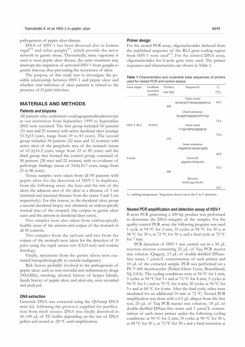
Tsamakidis K et al. HSV-1 in peptic ulcer 6645
pathogenesis of peptic ulcer disease.DNA of HSV-1 has been detected also in human
vagal[15] and celiac ganglia[16], which provide the nerve network to gastric tissue. Theoretically, since vagotomy is used to treat peptic ulcer disease, the same treatment may interrupt the migration of activated HSV-1 from ganglia to gastric mucosa, thus preventing the recurrence of ulcer.
The purpose of this study was to investigate the po-ssible relationship between HSV-1 and peptic ulcer and whether viral infection of ulcer patients is related to the presence of H pylori infection.
MATERIALS AND METHODS Patients and biopsiesAll patients who underwent esophagogastroduodenoscopy at our institution from September 1999 to September 2002 were recruited. The fi rst group included 56 patients (31 men and 25 women) with active duodenal ulcer (average 53.5±15 years, range from 19 to 83 years). The second group included 34 patients (22 men and 12 women) with active ulcer of the prepyloric area of the stomach (mean of 61.5±16.2 years, range from 22 to 89 years) and the third group that formed the control group, consisted of 50 patients (28 men and 22 women) with no evidence of pathologic fi ndings (mean of 54.8±16.7 years, range from 21 to 86 years).
Tissue samples were taken from all 90 patients with peptic ulcer for the detection of HSV-1 in duplicate, from the following areas: the base and the rim of the ulcer; the adjacent area of the ulcer at a distance of 3 cm (minimal and maximal distance from the crater 3 and 5 cm respectively). For this reason, in the duodenal ulcer group a second duodenal biopsy was obtained; an endoscopically normal area of the stomach (the corpus in gastric ulcer cases and the antrum in duodenal ulcer cases).
Two samples were also taken from endoscopically healthy areas of the antrum and corpus of the stomach in all 50 controls.
Two samples from the antrum and two from the corpus of the stomach were taken for the detection of H pylori using the rapid urease test (CLO-test) and routine histology.
Finally, specimens from the gastric ulcers were exa-mined histopathologically to exclude malignancy.
Risk factors probably involved in the pathogenesis of peptic ulcer, such as non-steroidal anti-infl ammatory drugs (NSAIDs), smoking, alcohol, history of herpes labialis, family history of peptic ulcer, and ulcer site, were recorded and analyzed.
DNA extractionGenomic DNA was extracted using the QIAamp DNA mini kit, following the protocol supplied for purifica-tion from fresh tissues. DNA was finally dissolved in 50–100 μL of TE buffer depending on the size of DNA pellets and stored at -20 °C until amplifi cation.
Primer designFor the nested PCR assay, oligonucleotides deduced from the published sequence of the RL2 gene-coding region from HSV-1 were used[17]. For the control DNA assay, oligonucleotides for b-actin gene were used. The primer sequences and characteristics are shown in Table 1.
Gene target GenBankaccession number
Product
size (bp)
Sequences1 Tm
(°C)
Outer senseagcagcgactctgaggcggagaccg 69.1
450
Outer antisensetgcgggtctgggggtcgttcacga
71.4 HSV-1: RL2 X14112 Inner sense
cccggcagttgcgggggcgc
73.4 110
Inner antisenseaaggtgtcgcagcggcaggtg
60.8 b-actin Forward
gtgatctccttctgcatcc
53.2 200
52.7
Reversectcttccagccttccttc
Nested PCR amplifi cation and detection assay of HSV-1B-actin PCR generating a 200-bp product was performed to determine the DNA integrity of the samples. For the quality control PCR assay, the following program was used 1 cycle at 94 °C for 2 min; 35 cycles at 94 °C for 30 s, at 58 °C for 30 s, at 72 °C for 30 s; and a fi nal cycle at 72 °C for 7 min.
PCR detection of HSV-1 was carried out in a 50 μL reaction mixture containing 25 μL of Taq PCR master mix solution (Qiagen), 13 μL of double-distilled DNase-free water, 1 μmol/L concentration of each primer and 10 μL of the extracted sample. PCR was performed on a PE 9 600 thermocycler (Perkin-Elmer Cetus, Branchburd, NJ, USA). The cycling conditions were at 94 °C for 1 min, 5 cycles at 94 °C for 5 s and at 72 °C for 4 min; 5 cycles at 94 °C for 5 s and at 70 °C for 4 min; 30 cycles at 94 °C for 5 s and at 68 °C for 4 min. After the fi nal cycle, tubes were incubated for an additional 10 min at 72 °C. Nested PCR amplifi cation was done with a 0.5 μL aliquot from the fi rst run, 25 μL of Taq PCR master mix solution, 18 μL of double-distilled DNase-free water and 1 μmol/L concen-tration of each inner primer under the following cycling conditions: at 94 °C for 2 min; 35 cycles at 90 °C for 30 s, at 68 °C for 30 s, at 72 °C for 30 s and a fi nal extension at
Table 1 Characteristics and nucleotide base sequences of primers used for nested PCR and control assays
Tm, melting temperature. 1Sequences shown are in the 5’ to 3’ direction.

6646 ISSN 1007-9327 CN 14-1219/ R World J Gastroenterol November 14, 2005 Volume 11 Number 42
72 °C for 10 min. Each amplifi cation run contained one negative and one positive control. The negative control consisted of blank reagent and water. For the positive con-trol, HSV-1 genomic DNA provided by Sigma was used. Consistent PCR analyses were repeated twice or more.
The PCR products were analyzed by 2% agarose gel electrophoresis in 0.5×Tris–borate EDTA buffer along with ethidium bromide. A molecular weight marker (Φ×174/Hae III, Sigma) was also run simultaneously to iden-tify the molecular size of the PCR products. The DNA bands were visualized by UV transillumination and ana-lyzed using a gel-documentation system. None of the PU and the control samples were negative in the b-actin test ultimately leaving 90 PU and 50 controls that were sub-jected to HSV-1 PCR analysis.
Helicobacter pylori testingFor the detection of H pylori, a CLO-test was used with high sensitivity and specifi city (Kimberly-Clark CLO test, Ballard Medical products, Draper, UT 84020, USA)[18,19].
An experienced pathologist also assessed the his-tological sections with Giemsa stain[20].
Statistical analysisAll associations between parameters of interest were examined either by Fisher’s exact test or Pearson’s chi square
test with continuity correction. Multivariate analysis was performed using the stepwise
logistic regression model to assess the contribution of the common risk factors to peptic ulcer development and H pylori detection.
P<0.05 was considered statistically signifi cant.
RESULTSPolymerase chain reactionThe genome of HSV-1 was present in 28 of the 90 patients with peptic ulcer (31.1%), contrary to the control group in which no positive detections were found (0%, P<0.0005, Figures 1-3). There was an equal prevalence in the two subgroups of patients, 17 of 56 patients with duodenal ulcer (30.4%) and 11 of 34 with gastric ulcer (32.4%) were tested positive (P = 0.843). In all HSV-1 positive cases, the viral genome was detected from the tissue samples obtained from the crater of the ulcer as from the samples obtained from the rim, while all samples from adjacent and distant areas were negative.
CLO testAll the H pylori positive subjects by CLO test from both groups were also positive for the bacteria with histology.
The incidence of H pylori was significantly higher in peptic ulcer patients (76/90, 84.4%) than in controls (30/50, 60%) (P = 0.002). A statistically significant difference was also found between patients with duodenal ulcer and those with gastric ulcer (P = 0.036). Negative H pylori was more frequently observed in patients with gastric ulcer (26.5%) than in patients with duodenal ulcer
(8.9%, Table 2). PCR and CLO test were signifi cantly associated with
respect to H pylori detection in all 90 peptic ulcer patients. Negative H pylori was more frequently detected in positive PCR samples (32.1%) than in negative PCR samples (8.1%) (P = 0.009) (Table 2). In the group of duodenal ulcer patients, H pylori negativity was more frequently observed in positive PCR samples (11.8%), than in negative PCR samples (7.7%). However, this difference was not
M 1 2 3
450 bp
110 bp
Figure 1 Nested PCR with HSV-1 outer and inner primers (450- and 110-bp amplicons: arrows) on 2% agarose gel, by ethidium bromide staining. Lane M: DNA molecular weight marker Φ×174/Hae III, lane 1: HSV-1 positive control (second run of nested PCR), lane 2: HSV-1 positive control (fi rst run of nested PCR), lane 3: b-actin quality control PCR peptic ulcer sample.
M 1 2 3 4 5 6 7 8 9 10 11 12
→200 bp
Figure 2 PCR quality assay control. Lane M: DNA molecular weight marker Φ×174/Hae III, lanes 1-6: positive b-actin peptic ulcer samples; lane 7: negative control (without template); lanes 8-12: positive b-actin peptic ulcer samples.
M 1 2 3 4 5 6 7 8 9 10 11 12
→110 bp
Figure 3 Nested PCR amplifi cation of HSV-1 in samples of patients with peptic ulcer. Lane M: DNA molecular weight marker Φ×174/Hae III, lane 1: HSV-1 positive control (HSV-1 genomic DNA by Sigma), lanes 2-6: positive samples from patients with peptic ulcer, lane 7: negative control (without template), lanes 8-11: positive samples from patients with peptic ulcer, lane 12: HSV-1 positive control.
→
→

Tsamakidis K et al. HSV-1 in peptic ulcer 6647
DISCUSSIONDespite the progress during the last 20 years in the understanding of the pathogenesis of peptic ulcer disease, it is clear that gastroduodenal ulcer is the result of a multifactorial process.
H pylori infection and NSAIDs have been recognized as the two most important causes of peptic ulcer disease. The proportion of peptic ulcers not associated with H pylori infection or the use of NSAIDs is increasing. Yet several studies have shown that 4.1-44% of peptic ulcers are not related to either of the two factors[21-25].
The possible involvement of HSV-1 in the process is a field of interest for several investigators, but a firm conclusion has not been reached. The presence of viral DNA in tissue samples obtained from the ulcer site is 9.5-18%[13,14]. The possible explanation for this finding is that the HSV-1 expression is prompted either by the ulcer injury or by immune cells[14,16] or HSV-1 itself might cause the ulcerative lesion by directly infecting the mucosal cells[13] or fi nally that HSV-1 expression is induced by the ulcer treatment[26-28].
H pylori (+), % H pylori (–) P Odds ratio (95%CI)
Peptic ulcer 76/90 (84.4) 14/90
0.002 3.7 (1.6–8.1)Controls 30/50 (60) 20/50 Gastric ulcer 25/34 (73.5) 9/34
0.036 3.67 (1.1–12.11)Duodenal ulcer 51/56 (91.1) 5/56 HSV-1 (+) 19/28 (67.9) 9/28
0.009 5.4 (1.61–18.11)HSV-1 (–) 57/62 (91.9) 5/62 HSV-1 (+)/GU 4/11 (36.4) 7/11
0.002 18.37 (2.75–122.94)HSV-1 (–)/ GU 21/23 (91.3) 2/23HSV-1 (+)/Duodenal ulcer
15/17 (88.2) 2/17
HSV-1(–)/Duodenal ulcer
36/39 (92.3) 3/39 0.634 -
H pylori (+), % H pylori (-) P-value Odds ratio (95% CI)
HSV-1(+)/gastric ulcer 4/11 (36.4) 5/11
0.010 13.13 (1.92–89.5)
HSV-1(–)/duodenal ulcer 15/17 (88.2) 2/17
statistically signifi cant (P = 0.634) (Table 2) whereas it was statistically significant in the subgroup of gastric ulcer patients (P = 0.002). In the gastric ulcer subgroup, H pylori, negative cases were observed in 63.6% of positive PCR samples and in 8.7% of negative PCR samples (Table 2). Finally, the likelihood of negative H pylori in HSV-1 positive samples in the group of gastric ulcer patients was significantly higher than that in the group of duodenal ulcer patients (P = 0.010) (Table 3).
Statistical analysis of other parametersWe also studied some of the common risk factors for the development of peptic ulcer disease. No statistically significant difference was found between patients and controls regarding family history of upper gastrointestinal ulcer, history of Herpes labialis, alcohol consumption, and use of NSAIDs (Table 4).
As expected, tobacco smoking was the only statistically signifi cant risk factor for the development of peptic ulcers between patients and control population (P = 0.019). Smokers were associated with a 2.57-fold increase risk of peptic ulcer development.
The performed multivariate analysis also confirmed that H pylori and tobacco smoking (OR: 3.320 and 2.619 respectively) were more likely to induce peptic ulcer (Table 5).
Peptic ulcer patients (%)
Controls (%) Odds ratio P
History of H labialis Yes 24/90 (26.7) 9/50 (18.0)
- 0.302 No 66/90 (73.3) 41/50 (82.0)Alcohol consumption Yes 32/90 (35.6) 14/50 (28.0)
- 0.469 No 58/90 (64.4) 36/50 (72.0)NSAID use Yes 23/90 (25.6) 8/50 (16.0)
- 0.275 No 67/90 (74.4) 42/50 (84.0)Smoking Yes 45/90 (50.0) 14/50 (28.0)
2–57 0.019 No 45/90 (50.0) 36/50 (72.0)Family history of peptic ulcer Yes 30/90 (33.3)
60/90 (66.7)9/50 (18.0)41/50 (82.0) 2.3 0.076
No
B S.E df Sig. Odds ratio
95%CI for Odds ratio
Smoking 0.963 0.394 1 0.015 2.619 1.209–5.673H pylori infection 1.2 0.473 1 0.011 3.32 1.313–8.389Age 0.011 0.014 1 0.432Sex 0.144 0.442 1 0.745History of H labialis 0.721 0.484 1 0.137Alcohol 0.021 0.492 1 0.966NSAID use 0.286 0.494 1 0.562Family history 0.44 0.48 1 0.359Constant –0.741 0.393 1 0.06
Table 2 Association between H pylori and HSV-1 in patients with peptic ulcer
HSV-1 (+)/gastric ulcer, patients with gastric ulcer positive for HSV-1; HSV-1 (–)/gastric ulcer, patients with gastric ulcer negative for HSV-1; HSV-1 (+)/duodenal ulcer, patients with duodenal ulcer positive for HSV-1; HSV-1 (–)/duodenal ulcer, patients with duodenal ulcer negative for HSV-1; CI, confi dence interval.
Table 3 Association between H pylori and HSV-1 in relation with the site of peptic ulcer
HSV-1 (+), patients positive for HSV-1; CI, confi dence interval.
Table 4 Risk factors and peptic ulcer disease
Table 5 Contribution of other risk factors to peptic ulcer deve-lopment
CI, confi dence interval.

6648 ISSN 1007-9327 CN 14-1219/ R World J Gastroenterol November 14, 2005 Volume 11 Number 42
In the present study, in a substantially larger number of patients than in the studies reported above (90 vs 22 and 21 respectively), the positivity for HSV-1 DNA was expressed using PCR in a greater percentage of samples (31% vs 9.5% and 18% respectively).
It should be noted that viral DNA was detected only in the tissue samples obtained from the base and the rim of the lesions, whereas all the other examined samples obtained from the adjacent ulcer areas and endoscopically healthy mucosa from the patients and the control group were negative for the viral genome.
This fi nding can be explained as follows. The HSV-1 may have initially entered the vagus ganglia through the oral pharynx or other peripheral connecting sites. Upon activation, the virus would travel down the vagal nerve to the potential site of peptic ulcer lesion. HSV-1 itself might cause ulcerative lesion in selected cases of a subset of peptic ulcer diseases by directly infecting the mucosal cells in the stomach and duodenum following virus release from neuroendocrine cells or vagal nerve terminals or both. Alternatively, peptic ulcer might activate latent HSV-1 in vagal ganglia, making replication of HSV-1, a contributing but not an initiating factor of ulcer[13].
The detection percentage of positive HSV-1 was similar between the group of patients with gastric and the group of patients with duodenal ulcer (32.4% and 30.4% respectively), in contrast to previous studies, where the viral DNA is demonstrated only in tissue samples from gastric ulcers[13,14]. The exact cellular localization of HSV-1 DNA could not be identifi ed.
On the other hand, investigating a possible association between H pylori and HSV-1 in pathogenesis of a subset of gastroduodenal ulcers, our data suggest that the PCR HSV-1 positivity is associated with a 5.4-fold increase in negative H pylori detection. Moreover, the patients with ulcer lesions infected with HSV-1 presented a similar prevalence of H pylori infection as the control group, which was significantly lower than that in the HSV-1 negative ulcer cases (P = 0.09). This finding requires further investigation.
Possible interpretations for the increased HSV-1 DNA positive detection rate in H pylori negative ulcers include the following. H pylori negativity is infl uenced by the viral expression, HSV-1 negativity infl uences H pylori infection and the virus independently causes some gastroduodenal ulcers.
According to our results, in the subgroup of patients with duodenal ulcers, the risk of H pylori infection was independent from HSV-1 DNA expression (P = 0.634). On the other hand, in the subgroup of patients with gastric ulcer disease, the possibility of H pylori negativity was 18.5-fold higher (P = 0.002). These data are in accordance with those reported by Lohr et al.[13].
Additionally, according to our results, there was not any correlation between peptic ulcer disease and age, sex, ulcer site, family history of gastroduodenal ulcers, history of H. labialis, alcohol consumption and NSAIDs use. On the contrary, statistically signifi cant difference (P = 0.019) was observed between patients with peptic ulcers and controls,
as far as smoking was concerned.Our results indicate an involvement of HSV-1 in
the pathogenesis of peptic ulcer disease. Although an opportunistic infection with the virus in the ulcer site cannot be excluded, the inverse relationship between HSV-1 detection and H pylori infection indicates a possible implication of this virus in the formation of the ulcer crater, at least in a subgroup of patients. Furthermore, experimental data support this[13,15,16,30,31]. The exact localization of the virus in ulcer tissue cells should be precisely determined in order to clarify whether the lesion is caused by HSV-1 or the virus opportunistically is established, especially in immunocompromised patients[32].
REFERENCES1 Wirth HP. Gastroduodenal ulcer disease: update on
pathogenesis. Schweiz Rundsch Med Prax 1995; 84: 570-580 2 Brooks MJ, Maxson CJ, Rubin W. The infectious etiology of
peptic ulcer disease. Diagnosis and implications for therapy. Prim Care 1996; 23: 443-454
3 Peura DA. Ulcerogenesis: integrating the roles of Helicobacter pylori and acid secretion in duodenal ulcer. Am J Gastroenterol 1997; 92: 8S-13S; discussion 13S-16S
4 Knyvett AF. Herpes simplex in peptic ulceration? Lancet (Let-ter) 1967; ii: 1040.
5 Neumann HH. Herpes simplex in peptic ulceration? Lancet (Letter) 1967; ii: 779.
6 Editorial. Viruses and duodenal ulcer. Lancet 1981; 1; 705-7067 Bader C, Crumpacker CS, Schnipper LE, Ransil B, Clark JE,
Arndt K, Freedberg IM. The natural history of recurrent facial-oral infection with herpes simplex virus. J Infect Dis 1978; 138: 897-905
8 Rand KH, Jacobson DG, Cottrell CR, Koch KL, Guild RT, McGuigan JE. Antibodies to herpes simplex type 1 in patients with active duodenal ulcer. Arch Intern Med 1983; 143: 1917-1920
9 Kottaridis SD, Mihas TA, Goula I, Mihas AA. Herpes viruses and duodenal ulcer disease. J Med Virol 1989; 29: 224-226
10 Archimandritis A, Markoulatos P, Tjivras M, Alexiou A, Kordossi A, Kordossis T, Fertakis A. Herpes simplex virus types 1 and 2 and cytomegalovirus in peptic ulcer disease and non-ulcer dyspepsia. Hepatogastroenterology 1992; 39: 540-541
11 Rune SJ, Vestergaard BF. IgA antibodies to herpes simplex virus type 1 in duodenal juice and saliva from patients with peptic ulcer and non-ulcer controls. Scand J Gastroenterol 1984; 19: 81-84
12 Hari VR, Ananthakrishnan N, Kate V, Badrinath S. Can duodenal ulcer perforation be linked to herpes simplex virus infection? Indian J Gastroenterol 2004; 23: 5-7
13 Lohr JM, Nelson JA, Oldstone MB. Is herpes simplex virus as-sociated with peptic ulcer disease? J Virol 1990; 64: 2168-2174
14 Kemker BP Jr, Docherty JJ, De Lucia A, Ruf W, Lewis RD. Herpes simplex virus: a possible etiologic agent in some gastroduodenal ulcer disease. Am Surg 1992; 58: 775-778
15 Warren KG, Brown SM, Wroblewska Z, Gilden D, Koprowski H, Subak-Sharpe J. Isolation of latent herpes simplex virus from the superior cervical and vagus ganglions of human be-ings. N Engl J Med 1978; 298: 1068-1069
16 Rand KH, Berns KI, Rayfi eld MA. Recovery of herpes simplex type 1 from the celiac ganglion after renal transplantation. South Med J 1984; 77: 403-404
17 McGeoch DJ, Dalrymple MA, Dolan A, McNab D, Perry LJ, Taylor P, Challberg MD. Structures of herpes simplex virus type 1 genes required for replication of virus DNA. J Virol 1988; 62: 444-453

Tsamakidis K et al. HSV-1 in peptic ulcer 6649
18 Marshall BJ, Warren JR, Francis GJ, Langton SR, Goodwin CS, Blincow ED. Rapid urease test in the management of Campy-lobacter pyloridis-associated gastritis. Am J Gastroenterol 1987; 82: 200-210
19 Thillainayagam AV, Arvind AS, Cook RS, Harrison IG, Tabaqchali S, Farthing MJ. Diagnostic effi ciency of an ultrar-apid endoscopy room test for Helicobacter pylori. Gut 1991; 32: 467-469
20 Stevens A. Micro-organisms. In: Bancroft J, Stevens A, eds. Theory and Practice of Histological Techniques, 3rd ed. Edin-burg-London: Churchill Livingston, 1990: 289-308.
21 Kurata JH, Nogawa AN. Meta-analysis of risk factors for peptic ulcer. Nonsteroidal antiinflammatory drugs, Helicobacter pylori, and smoking. J Clin Gastroenterol 1997; 24: 2-17
22 Sprung DJ, Apter MN. What is the role of Helicobacter pylori in peptic ulcer and gastric cancer outside the big cities? J Clin Gastroenterol 1998; 26: 60-63
23 Ciociola AA, McSorley DJ, Turner K, Sykes D, Palmer JB. He-licobacter pylori infection rates in duodenal ulcer patients in the United States may be lower than previously estimated. Am J Gastroenterol 1999; 94: 1834-1840
24 Chan HL, Wu JC, Chan FK, Choi CL, Ching JY, Lee YT, Leung WK, Lau JY, Chung SC, Sung JJ. Is non-Helicobacter pylori, non-NSAID peptic ulcer a common cause of upper GI bleeding? A prospective study of 977 patients. Gastrointest Endosc 2001; 53: 438-442
25 Xia HH, Wong BC, Wong KW, Wong SY, Wong WM, Lai KC, Hu WH, Chan CK, Lam SK. Clinical and endoscopic character-istics of non-Helicobacter pylori, non-NSAID duodenal ulcers: a long-term prospective study. Aliment Pharmacol Ther 2001; 15: 1875-1882
26 Dargan DJ, Subak-Sharpe JH. The effect of triterpenoid com-pounds on uninfected and herpes simplex virus infected cells in culture. I. Effect on cell growth, virus particles and virus replication. J Gen Virol 1985; 66 ( Pt 8): 1771-1784
27 Poswillo DE, Roberts GJ. Topical carbenoxolone for orofacial herpes simplex infections. Lancet 1981; 2: 143-144
28 Van der Spuy S, Levy DW, Levin W. Cimetidine in the treat-ment of herpesvirus infections. S Afr Med J 1980; 58: 112-116
29 Wiley CA, Schrier RD, Denaro FJ, Nelson JA, Lampert PW, Oldstone MB. Localization of cytomegalovirus proteins and genome during fulminant central nervous system infection in an AIDS patient. J Neuropathol Exp Neurol 1986; 45: 127-139
30 Alexiu O, David S, Cajal N, Gruia M, Gologan R, Nicolescu P. Gastroduodenal ulcer obtained by experimental herpes virus inoculation. Virologie 1976; 27: 61-62
31 Gesser RM, Valyi-Nagy T, Fraser NW, Altschuler SM. Oral inoculation of SCID mice with an attenuated herpes simplex virus-1 strain causes persistent enteric nervous system infection and gastric ulcers without direct mucosal infection. Lab Invest 1995; 73: 880-889
32 Howiler W, Goldberg HI. Gastroesophageal involvement in herpes simplex. Gastroenterology 1976; 70: 775-778
Science Editor Wang XL and Guo SY Language Editor Elsevier HK
PO Box 2345, Beijing 100023, China World J Gastroenterol 2005;11(42):6650-6655www.wjgnet.com World Journal of Gastroenterology ISSN [email protected] © 2005 The WJG Press and Elsevier Inc. All rights reserved.E L S E V I E R
• BRIEF REPORTS •
up of 34.9 mo, all patients remained in remission.
CONCLUSION: In the medium term, EMR is effective and safe to treat HGD and/or IMC within BE and is a valuable staging method. It could become an alternative to surgery.
© 2005 The WJG Press and Elsevier Inc. All rights reserved.
Key words: Endoscopic mucosal resection; Barrett’s esophagus; High-grade dysplasia; Intramucosal cancer
Conio M, Repici A, Cestari R, Blanchi S, Lapertosa G, Missale G, Della Casa D, Villanacci V, Calandri PG, Filiberti R. Endoscopic mucosal resection for high-grade dysplasia and intramucosal carcinoma in Barrett’s esophagus: An Italian experience. World J Gastroenterol 2005; 11(42): 6650-6655http://www.wjgnet.com/1007-9327/11/6650.asp
INTRODUCTIONThe incidence of adenocarcinoma (AC) of the esophagus has increased in the last three decades in the Western world[1-3]. Most esophageal adenocarcinomas arise in a precursor lesion, Barrett’s esophagus (BE). The esophageal cancer risk in BE patients is about 1 cancer per 200 patient-years, or 0.5% per year[4,5]. The prognosis is poor for the typical patient who presents with invasive cancer, with a 5-year survival rate of under 10%[6].
Dysplasia arising in BE is a marker of progression toward invasive cancer. High-grade dysplasia (HGD) is an uncommon but a serious problem. In 16-60% of patients found to have HGD, invasive cancer was diagnosed in the next 5-7 years[6,7], although spontaneous regression of HGD can also occur[8]. Following surgical esophagectomy for HGD, 10-50% of cases had previously undetected foci of invasive cancer found in the resected specimen[9,10].
When HGD is detected, the three options that are available are: endoscopic surveillance, esophagectomy, and endotherapy.
Surveillance in patients with HGD is controversial. The uncertainty of natural history of HGD and its slow progression rate, could justify a contemplative attitude. Schnell et al. evaluated the long-term outcome of 75 patients with HGD who were enrolled in an endoscopic
Endoscopic mucosal resection for high-grade dysplasia and intramucosal carcinoma in Barrett’s esophagus: An Italian experience
Massimo Conio, Alessandro Repici, Renzo Cestari, Sabrina Blanchi, Gabriella Lapertosa, Guido Missale, Domenico Della Casa, Vincenzo Villanacci, Pier Gigi Calandri, Rosangela Filiberti
Massimo Conio, Department of Gastroenterology, Sanremo, ItalyAlessandro Repici, Department of Gastroenterology, Molinette Hospital, Torino, ItalyRenzo Cestari, Guido Missale, Domenico Della Casa, Department of Surgical-Surgery Endoscopy, Spedali Civili, University of Brescia, ItalySabrina Blanchi, Department of Internal Medicine, University of Genova, ItalyGabriella Lapertosa, Division of Pathology, University of Genova, ItalyVincenzo Villanacci, 2nd Department of Pathology, Spedali Civili, University of Brescia, ItalyPier Gigi Calandri, Anesthesia and Resuscitation Unit, National Institute for Cancer Research, Genova, ItalyRosangela Filiberti, Epidemiology and Biostatistics, National Institute for Cancer Research, Genova, ItalyCorrespondence to: Massimo Conio, MD, Department of Gastroenterology, Sanremo Hospital, 18038 Sanremo (IM), Italy. [email protected]: +39-0184-536873 Fax: +39-0184-536873Received: 2004-12-11 Accepted: 2005-02-18
AbstractAIM: To evaluate endoscopic mucosal resection (EMR) in patients with high-grade dysplasia (HGD) and/or intramucosal cancer (IMC) in Barrett’s esophagus (BE).
METHODS: Between June 2000 and December 2003, 39 consecutive patients with HGD (35) and/or IMC (4) underwent EMR. BE >30 mm was present in 27 patients. In three patients with short segment BE (25.0%), HGD was detected in a normal appearing BE. Lesions had a mean diameter of 14.8±10.3 mm. Mucosal resection was carried out using the cap method.
RESULTS: The average size of resections was 19.7±9.4×14.6±8.2 mm. Histopathologic assessment post-resection revealed 5 low-grade dysplasia (LGD) (12.8%), 27 HGD (69.2%), 2 IMC (5.1%), and 5 SMC (-12.8%). EMR changed the pre-treatment diagnosis in 10 patients (25.6%). Three patients with SMC underwent surgery. Histology of the surgical specimen revealed 1 T0N0 and 2 T1N0 lesions. The remaining two patients were cancer free at 32.5 and 45.6 mo, respectively. A metachronous lesion was detected after 25 mo in one patient with HGD. Intra-procedural bleeding, controlled at endoscopy, occurred in four patients (10.3%). After a median follow-

Conio M et al. Endoscopic mucosal resection in Barrett’s esophagus 6651
surveillance program. After a mean follow-up of 7 years, AC occurred in 12 (16%) patients[7].
Esophagectomy has been the standard treatment of HGD and early cancer in BE. However esophagectomy is associated with surgical morbidity of 20-50% and mortality of about 3%, even at high-volume centers. In patients older than 70 years, mortality was 11%[11,12]. A less invasive treatment would be desirable. A newer alternative to esophagectomy is endoscopic mucosal resection (EMR). Improved diagnosis of early malignancy in BE, including endoscopic ultrasound (EUS), may change the therapeutic approach. The superfi cial lesions of HGD and intramucosal cancer (IMC), with minimal risk of lymph node metastasis, can be removed by EMR. This procedure allows adequate histologic assessment and definitive treatment.
The use of EMR to treat HGD and IMC in BE is increasing, but the number of published series remains less. Our aim was to evaluate EMR in the treatment of HGD and IMC in Barrett’s patients in terms of complications and recurrence rate.
PATIENTS AND METHODSPatientsBetween June 2000 and December 2003, 39 consecutive patients (mean age 62.8±11.4 years) with histologically confirmed HGD (35) or IMC (4) in BE underwent EMR in three Departments of Gastroenterology acting as regional referral centers. All patients were previously identified by endoscopic examination performed in our centers or referred from other hospitals. In this case, the original histologic slides were re-evaluated by two expert pathologists (G.L., V.V.) on BE before EMR.
This study was approved by our institutional review board. All patients gave written informed consent to endoscopic therapy. Patients were evaluated and treated using the same protocol by one of the authors representing each of the three institutions.
At endoscopy, superficial lesions were defined using the following classification: slightly elevated (0-IIa), flat (0-IIb), and slightly depressed (0-IIc)[13]. Endoscopy was performed with standard diagnostic videoendoscopes (GIF-Q145, Olympus Optical Co. Ltd., Tokyo, Japan). Patients with ulcerated lesions seen on endoscopy were excluded.
Before EMR, EUS with mechanical rotating transducer, using the water-fil l ing method (GF-UMQ130, 7.5-20 MHz, Olympus Optical Co. Ltd.), was routinely performed to assess lesion depth and mediastinal lymph node status. All patients had a CT scan of the thorax. Only lesions confined to the mucosal layer with no apparent lymph node metastases were considered for EMR.
Deep sedation with propofol was used. EMR was performed using a plastic cap (MH-594, Olympus Optical Co. Ltd, Tokyo, Japan) preloaded on the tip of a standard diagnostic forward-viewing endoscope. The cap had an outer diameter of 13 mm and a length of 15 mm. Inside the distal end of the cap was a gutter, which positions
the opened polypectomy snare. A 2-mm segment of the gutter was removed with a scalpel before placement on the endoscope tip. This modification was then aligned with the operative channel to avoid interference by the injection needle or other devices (hemoclips, biopsy forceps). Submucosal injection of epinephrine solution (1:60.000-1:100.000) to create a f luid cushion was performed using variceal injection needles (VIN-23, Wilson Cook Medical Inc; Variject Contrast Injection Needle, Boston Scientifi c) in all patients. Injected volume ranged between 8 and 30 mL, depending on the lesion diameter. Methylene blue was added to the solution for visual enhancement of the fl uid cushion in contrast to the lesion. The cap was next applied against the lesion, which was aspirated into it. A monofi lament polypectomy snare (SD-221U-25, Olympus Optical Co. Ltd, Tokyo, Japan) was then firmly secured around the tissue and resection was performed. Resection was performed by endocut mode only, using the ERBE-ICC 200 cautery device (ERBE Elektromedizin GmbH, Tubingen, Germany). The output setting predefined by the manufacturer was adopted: cut 120 W, coagulation 60 W. To minimize interobserver variability among the three endoscopists performing EMR, the diameter of the lesion and the resected areas were estimated by placing an open polypectomy snare around the lesion. In patients with lesions ≤12 mm wide “en-bloc” resection was performed. For larger lesions, piecemeal resection was completed by applying the cap close to the previously resected area. Specimens were aspirated into the cap and all materials were retrieved for histopathologic assessment. To complete EMR, multiple withdrawals and re-intubations were needed. No overtube was used.
Intra-procedural bleeding (during the EMR) was controlled by epinephrine-saline injections (1:10.000) and, when required, by placing hemoclips (HX-600-090L; rotatable clip fi xing device HX-6UR-1, Olympus Optical Co., Ltd).
All patients were hospitalized for 48 h. They were kept fasting for 24 h, then a soft diet was advised for the next two weeks. After EMR, an intravenous PPI (omeprazole, 40 mg/d) was administered for 24 h, followed by a maintenance oral dose of 40 mg/d.
SurveillanceFollowing EMR, patients were contacted by phone weekly in the first fifteen days, then monthly, to monitor for symptoms such as dysphagia. Endoscopy was repeated at 3, 6, and 12 mo and then yearly, with multiple biopsies from the EMR site, and four-quadrant biopsies from the residual BE.
Complete remission was defi ned, when well demarcated areas of squamous re-epithelialization without mucosal irregularities, were observed. A lesion was considered metachronous, when diagnosed more than 12 mo from the EMR, irrespective of its location.
HistologyFollowing the WHO guidelines, we defined HGD by cytologic and architectural changes confined to the
6652 ISSN 1007-9327 CN 14-1219/ R World J Gastroenterol November 14, 2005 Volume 11 Number 42
mucosa. IMC was defined by cytologic and architectural changes confi ned to the lamina propria. Invasive cancers were considered to be those infiltrating the submucosa (SMC)[14,15].
Statistical analysisStatistical data were expressed as mean±SD. The Kruskal-Wallis test was used to compare histologic severity with lesion size. A P value of 0.05 or less was considered statistically signifi cant.
RESULTSEMR was performed in 39 patients, 34 males and 5 females. Their mean age was 62.8±11.4 years. Thirty-six patients had type 0-IIa mucosal abnormalities and three had HGD detected by random biopsies in a normal appearing BE.
Mean Barrett’s length was 4.3±2.5 cm. Long segment BE (LSBE, ≥3 cm) was present in 27 patients. The three patients with non-visible lesions had short segment BE (SSBE). These patients underwent EMR with the aim of completely removing the metaplastic epithelium.
Lesions had a mean diameter of 14.8±10.3 mm. Histologic severity did not correlate with lesion size. The average size of reconstructed resected specimens was 19.7±9.4×14.6±8.2 mm. In all patients the EMR was completed in one session. “En-bloc” resection was performed in 19 cases with lesions of ≤12 mm. The size of the fi rst resected specimen by EMR-C method ranged between 8 and 12 mm.
Following EMR, the pre-treatment histology was re-classifi ed in 10/39 patients (25.6%). Among the 35 initially diagnosed as HGD, five were found to have only LGD, but three had SMC. Of the four patients initially diagnosed as IMC, two were re-classifi ed as SMC. EUS did not help to identify submucosal infi ltration.
Table 1 shows the characteristics of patients and lesions according to the histology found in the EMR resection specimens.
CancersThe five patients with SMC had a mean age of 73.6±8.3 years. The mean BE length was 5.8±3.1 cm, and lesion size ranged from 5 to 30 mm. The lesions were type IIa (superficial elevated). Histologic assessment detected tiny areas of low-grade differentiation, and in two of them, lymphatic permeation. Three (7.7% of all patients) underwent esophagectomy and the histopathologic assessment showed one T0N0 and two T1N0. The remaining two patients were considered unfi t for surgery due to advanced age (81 and 84 years) and/or comorbidities (cardiovascular disease). They were included in the surveillance program.
ComplicationsIntra-procedural bleeding occurred in four patients (10.3%), and was controlled with epinephrine injections
in two, and with epinephrine plus clipping in the other two. Delayed bleeding was not seen. No patient needed blood transfusion. No perforations occurred. Retrosternal pain was present in one patient. An esophageal stenosis developed 8 mo later in a patient with LSBE (7 cm). He had a 30 mm HGD lesion and the diameter of the EMR area was 40 mm. He was successfully treated by a single bougienage.
SurveillanceFollow-up endoscopy was performed in 32 of the 34 patients without invasive cancer (94.1%), two patients declining repeat examination. The follow-up ranged from 16.3 to 72.1 mo (median 34.9 mo). In one patient (3.1%), with an original lesion of 20 mm (HGD), a metachronous lesion was detected after 25 mo. It was easily removed by EMR, and the histology showed HGD. One of the two patients with SMC, who did not undergo surgery died of cardiovascular disease 45.6 mo later, and the other was alive and cancer free at a 32.5 mo surveillance.
EUS was repeated at 3 and 6 mo after EMR. CT of the thorax and upper abdomen were also performed after 6 mo, and then after 1 year to evaluate the lymph node status and the presence of metastases. Figures 1A and 1B show endoscopic appearance of a SSBE with HGD before and three months after EMR.
DISCUSSIONThe use of EMR for EC in the digestive tract was described by Inoue et al.[16]. We employed EMR to remove 39 focal esophageal HGD and IMC lesions of 3-30 mm size in BE, with few complications. Most lesions were fl at mucosal abnormalities. Systematic biopsies were taken from the remainder of the BE to exclude non-visible multifocal lesions.
The data reported in the present study represent the first experience of EMR in BE, in Italy. Three major referral centers joined in the effort of evaluating the clinical outcome of EMR. In a period of three years a relatively small number of patients has been included in our study, especially when compared with the series
LGD HGD IMC SMC (no surgery)
Age (yr) 60.6±8.8 61.76±11.8 56±4.2 73.6±8.3BE length (cm) 6.6±4.1 3.6±1.8 3.7±1.8 5.8±3.1Size of lesion (mm) 22.5±8.7 13±10.2 16±5.7 17.6±11.9Metachronous lesion (%) No 5 (100) 24 (88.9) 2 (100) 2 (100) Yes - 1 (3.7) - - No follow-up endoscopy (%) - 2 (7.4) - -Total 5 27 2 2
Table 1 Characteristics of patients and lesions according to histology of mucosa resected at EMR (mean±SD)
LGD: low-grade dysplasia; HGD: high-grade dysplasia; IMC: intramucosal cancer; SMC: submucosal cancer; BE: Barrett’s esophagus.

Conio M et al. Endoscopic mucosal resection in Barrett’s esophagus 6653
reported by German authors[17]. This difference is attributable to the still limited number of patients with early neoplastic lesions in BE, detected in endoscopic centers. Furthermore, as surgery is still the gold standard treatment for HGD and IM, the majority of these patients is referred for esophagectomy.
EMR could become a management opt ion for HGD, and also for IMC, where the risk of lymph node involvement is from 0% to 4%[18,19]. Unfortunately, in cases with SMC, the incidence of regional lymph-node metastasis is 15-50%[20-22]. Current data support surgical resection in the setting of submucosal infi ltration by AC, unless comorbidity or advanced patient age precluded it.
Table 2 displays data from selected studies on EMR in BE since 2000. Most published studies report EMR of endoscopically visible areas of HGD. Some of these studies differ in methodology and it is diffi cult to compare them. However, our results are in accordance with the data reported by other authors. EMR provides greater diagnostic precision than endoscopic biopsy, despite endoscopy with biopsies and standard EUS before EMR in all patients. In fi ve of 39 cases, undetected SMC was found on histological examination of the resected specimen. Reclassification of the histology after EMR occurred in 26% of our patients. Other authors have reported reclassifi cation in 0% to 75% of cases after EMR (Table 2). Causes may include biopsy sampling error and observer
interpretation variability.Bleeding is the most frequent adverse event with EMR,
reported in a median 10% of patients. Intra-procedural bleeding also occurred in 10% of our patients and was managed endoscopically without transfusion. Esophageal stenosis is a late complication of EMR, reported in 0-30% of cases (Table 3). In our study, one patient (2.5%) developed stenosis. Larger EMR resections may increase the risk; in a study of 137 patients, stenosis was seen only when EMR involved more than two-thirds of the esophageal circumference[23]. However, in one report of circumferential EMR, only two of 12 patients developed stenosis[24]. The perforation risk is generally less than 1%. No perforations occurred in our series. Overall, complications seem fewer for EMR than for surgical resection. In one study, complications occurred in 48% of esophagectomies vs 16% for EMR combined with photodynamic therapy (PDT)[25].
A recent controlled study of 100 mucosectomies compared the cap method and a ligation method for suction EMR. The diameter of the removed specimen, the diameter of the resected area, and the complication rate showed no signifi cant differences between the two groups, and no severe complications occurred[26].
There is limited information on the long-term effec-tiveness of EMR. May et al. followed 70 patients with HGD or early AC for a mean of 34 mo after EMR. Ten percent had minor complications. During follow-up, 21/70 patients were found to have locally recurrent or metachronous disease, treated endoscopically with success in all but one case. The only death from Barrett's AC was in a patient who had surgery for SMC[17]. In our series, follow-up for a median 35 mo was available in 94.1% of patients. One of the 39 patients (2.6%) had a metachronous lesion after 25 mo, successfully treated with another EMR. According to other authors, malignant transformation of HGD is about 34% in 6-54 mo[27], corresponding to our follow-up period. In this time range we did not fi nd invasive AC. In our two patients with SMC who did not undergo surgery, no histologic evidence of disease was detected.
Author Number of patiens
Size of lesions Technique Histology Histology Change in diagnosis (%)
Complications Follow-up Recurrence cm (mean) pre-EMR post-EMR months
(mean)
Seewald et al. 2003
Germany[24]
12 Median 5 EUSSnare
5 HGD/ IMC (visible)7 IMC
2 BE 1 LGD5 HGD
75% Bl: 33%Stricture:17%
Median:9
0
(non-visible) AC 4
Ahmad et al. 2002 19/101 0.5-3 EUS AC 6 AC 8 58% Bl: 11% >=24 0
USA[33] EMR-C HGD 6 HGD 4Snare NOS 7 LGD 1
injection Benign 6May et al. 2002 80 Nos EUS HGD 7 AC: 11/80 Nos Bl: 6% 34 24/78Germany[17] EMR±PDT EC 73 Stricture: 4%
A B
Figure 1 Endoscopic appearance of SSBE with HGD before (A) and 3 mo after EMR (B).
Table 2 Selected studies on EMR in Barrett’s esophagus
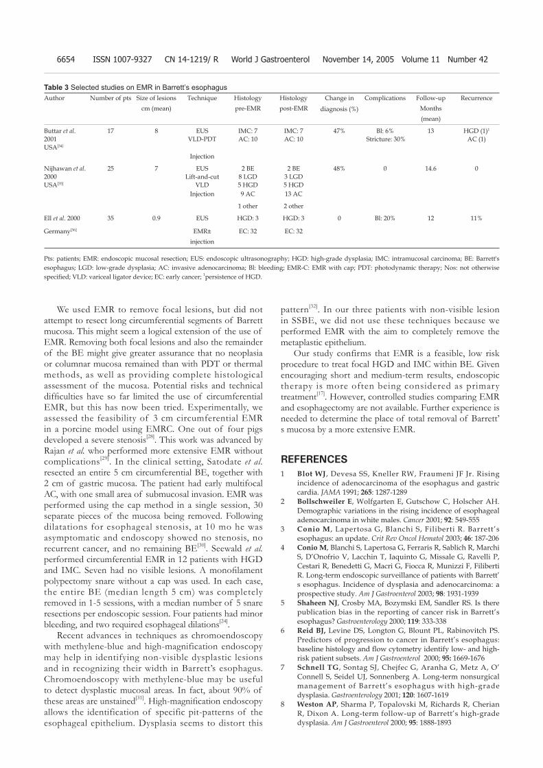
6654 ISSN 1007-9327 CN 14-1219/ R World J Gastroenterol November 14, 2005 Volume 11 Number 42
We used EMR to remove focal lesions, but did not attempt to resect long circumferential segments of Barrett mucosa. This might seem a logical extension of the use of EMR. Removing both focal lesions and also the remainder of the BE might give greater assurance that no neoplasia or columnar mucosa remained than with PDT or thermal methods, as well as providing complete histological assessment of the mucosa. Potential risks and technical diffi culties have so far limited the use of circumferential EMR, but this has now been tried. Experimentally, we assessed the feasibility of 3 cm circumferential EMR in a porcine model using EMRC. One out of four pigs developed a severe stenosis[28]. This work was advanced by Rajan et al. who performed more extensive EMR without complications[29]. In the clinical setting, Satodate et al. resected an entire 5 cm circumferential BE, together with 2 cm of gastric mucosa. The patient had early multifocal AC, with one small area of submucosal invasion. EMR was performed using the cap method in a single session, 30 separate pieces of the mucosa being removed. Following dilatations for esophageal stenosis, at 10 mo he was asymptomatic and endoscopy showed no stenosis, no recurrent cancer, and no remaining BE[30]. Seewald et al. performed circumferential EMR in 12 patients with HGD and IMC. Seven had no visible lesions. A monofi lament polypectomy snare without a cap was used. In each case, the entire BE (median length 5 cm) was completely removed in 1-5 sessions, with a median number of 5 snare resections per endoscopic session. Four patients had minor bleeding, and two required esophageal dilations[24].
Recent advances in techniques as chromoendoscopy with methylene-blue and high-magnification endoscopy may help in identifying non-visible dysplastic lesions and in recognizing their width in Barrett’s esophagus. Chromoendoscopy with methylene-blue may be useful to detect dysplastic mucosal areas. In fact, about 90% of these areas are unstained[31]. High-magnifi cation endoscopy allows the identification of specific pit-patterns of the esophageal epithelium. Dysplasia seems to distort this
pattern[32]. In our three patients with non-visible lesion in SSBE, we did not use these techniques because we performed EMR with the aim to completely remove the metaplastic epithelium.
Our study confirms that EMR is a feasible, low risk procedure to treat focal HGD and IMC within BE. Given encouraging short and medium-term results, endoscopic therapy is more often being considered as primary treatment[17]. However, controlled studies comparing EMR and esophagectomy are not available. Further experience is needed to determine the place of total removal of Barrett’s mucosa by a more extensive EMR.
REFERENCES1 Blot WJ, Devesa SS, Kneller RW, Fraumeni JF Jr. Rising
incidence of adenocarcinoma of the esophagus and gastric cardia. JAMA 1991; 265: 1287-1289
2 Bollschweiler E, Wolfgarten E, Gutschow C, Holscher AH. Demographic variations in the rising incidence of esophageal adenocarcinoma in white males. Cancer 2001; 92: 549-555
3 Conio M , Lapertosa G, Blanchi S, Filiberti R. Barrett’s esophagus: an update. Crit Rev Oncol Hematol 2003; 46: 187-206
4 Conio M, Blanchi S, Lapertosa G, Ferraris R, Sablich R, Marchi S, D’Onofrio V, Lacchin T, Iaquinto G, Missale G, Ravelli P, Cestari R, Benedetti G, Macri G, Fiocca R, Munizzi F, Filiberti R. Long-term endoscopic surveillance of patients with Barrett’s esophagus. Incidence of dysplasia and adenocarcinoma: a prospective study. Am J Gastroenterol 2003; 98: 1931-1939
5 Shaheen NJ, Crosby MA, Bozymski EM, Sandler RS. Is there publication bias in the reporting of cancer risk in Barrett’s esophagus? Gastroenterology 2000; 119: 333-338
6 Reid BJ, Levine DS, Longton G, Blount PL, Rabinovitch PS. Predictors of progression to cancer in Barrett’s esophagus: baseline histology and fl ow cytometry identify low- and high-risk patient subsets. Am J Gastroenterol 2000; 95: 1669-1676
7 Schnell TG, Sontag SJ, Chejfec G, Aranha G, Metz A, O’Connell S, Seidel UJ, Sonnenberg A. Long-term nonsurgical management of Barrett’s esophagus with high-grade dysplasia. Gastroenterology 2001; 120: 1607-1619
8 Weston AP, Sharma P, Topalovski M, Richards R, Cherian R, Dixon A. Long-term follow-up of Barrett’s high-grade dysplasia. Am J Gastroenterol 2000; 95: 1888-1893
Author Number of pts Size of lesions Technique Histology Histology Change in
diagnosis (%)
Complications Follow-up Recurrence cm (mean) pre-EMR post-EMR Months
(mean)
Buttar et al. 2001USA[34]
17 8 EUSVLD-PDT
IMC: 7AC: 10
IMC: 7AC: 10
47% Bl: 6% Stricture: 30%
13 HGD (1)1
AC (1)
Injection
Nijhawan et al. 2000USA[35]
25 7 EUSLift-and-cut
VLD
2 BE8 LGD5 HGD
2 BE3 LGD5 HGD
48% 0 14.6 0
Injection 9 AC 13 AC
1 other 2 other
Ell et al. 2000 35 0.9 EUS HGD: 3 HGD: 3 0 Bl: 20% 12 11%
Germany[36] EMR± EC: 32 EC: 32injection
Table 3 Selected studies on EMR in Barrett’s esophagus
Pts: patients; EMR: endoscopic mucosal resection; EUS: endoscopic ultrasonography; HGD: high-grade dysplasia; IMC: intramucosal carcinoma; BE: Barrett's esophagus; LGD: low-grade dysplasia; AC: invasive adenocarcinoma; Bl: bleeding; EMR-C: EMR with cap; PDT: photodynamic therapy; Nos: not otherwise specifi ed; VLD: variceal ligator device; EC: early cancer; 1persistence of HGD.
Conio M et al. Endoscopic mucosal resection in Barrett’s esophagus 6655
9 Cameron AJ, Carpenter HA. Barrett’s esophagus, high-grade dysplasia and early adenocarcinoma: a pathologic study. Am J Gastroenterol 1997; 92: 586-591
10 Korst RJ, Altorki NK. High-grade dysplasia: surveillance, mucosal ablation, or resection? World J Surg 2003; 27: 1030-1034
11 Collard JM. High-grade dysplasia in Barrett’s esophagus. The case for esophagectomy. Chest Surg Clin N Am 2002; 12: 77-92
12 McCulloch P, Ward J, Tekkis PP. Mortality and morbidity in gastro-oesophageal cancer surgery: initial results of ASCOT multicentre prospective cohort study. BMJ 2003; 327: 1192-1197
13 Schlemper RJ , Hirata I , Dixon MF. The macroscopic classifi cation of early neoplasia of the digestive tract. Endoscopy 2002; 34: 163-168
14 Jass JR , Sobin LH, Watanabe H. The World Heal th Organization’s histologic classification of gastrointestinal tumors. A commentary on the second edition. Cancer 1990; 66: 2162-2167
15 Schlemper RJ, Riddell RH, Kato Y, Borchard F, Cooper HS, Dawsey SM, Dixon MF, Fenoglio-Preiser CM, Flejou JF, Geboes K, Hattori T, Hirota T, Itabashi M, Iwafuchi M, Iwashita A, Kim YI, Kirchner T, Klimpfinger M, Koike M, Lauwers GY, Lewin KJ, Oberhuber G, Offner F, Price AB, Rubio CA, Shimizu M, Shimoda T, Sipponen P, Solcia E, Stolte M, Watanabe H, Yamabe H. The Vienna classifi cation of gastrointestinal epithelial neoplasia. Gut 2000; 47: 251-255
16 Inoue H, Takeshita K, Hori H, Muraoka Y, Yoneshima H, Endo M. Endoscopic mucosal resection with a cap-fitted panendoscope for esophagus, stomach, and colon mucosal lesions. Gastrointest Endosc 1993; 39: 58-62
17 May A, Gossner L, Pech O, Fritz A, Gunter E, Mayer G, Muller H, Seitz G, Vieth M, Stolte M, Ell C. Local endoscopic therapy for intraepithelial high-grade neoplasia and early adenocarcinoma in Barrett’s oesophagus: acute-phase and intermediate results of a new treatment approach. Eur J Gastroenterol Hepatol 2002; 14: 1085-1091
18 Baba H, Maehara Y, Okuyama T, Orita H, Anai H, Akazawa K, Sugimachi K. Lymph node metastasis and macroscopic features in early gastric cancer. Hepatogastroenterology 1994; 41: 380-383
19 Nigro JJ, Hagen JA, DeMeester TR, DeMeester SR, Theisen J, Peters JH, Kiyabu M. Occult esophageal adenocarcinoma: extent of disease and implications for effective therapy. Ann Surg 1999; 230: 433-8; discussion 438-40
20 Stein HJ, Feith M, Mueller J, Werner M, Siewert JR. Limited resection for early adenocarcinoma in Barrett’s esophagus. Ann Surg 2000; 232: 733-742
21 Holscher AH, Bollschweiler E, Schneider PM, Siewert JR. Early adenocarcinoma in Barrett’s oesophagus. Br J Surg 1997; 84: 1470-1473
22 Nigro JJ, Hagen JA, DeMeester TR, DeMeester SR, Peters JH, Oberg S, Theisen J, Kiyabu M, Crookes PF, Bremner CG. Prevalence and location of nodal metastases in distal esophageal adenocarcinoma confi ned to the wall: implications for therapy. J Thorac Cardiovasc Surg 1999; 117 : 16-23; discussion 23-25
23 Katada C, Muto M, Manabe T, Boku N, Ohtsu A, Yoshida S.
Esophageal stenosis after endoscopic mucosal resection of superficial esophageal lesions. Gastrointest Endosc 2003; 57: 165-169
24 Seewald S, Akaraviputh T, Seitz U, Brand B, Groth S, Mendoza G, He X, Thonke F, Stolte M, Schroeder S, Soehendra N. Circumferential EMR and complete removal of Barrett’s epithelium: a new approach to management of Barrett’s esophagus containing high-grade intraepithelial neoplasia and intramucosal carcinoma. Gastrointest Endosc 2003; 57: 854-859
25 Pacifico RJ , Wang KK, Wongkeesong LM, Buttar NS, Lutzke LS. Combined endoscopic mucosal resection and photodynamic therapy versus esophagectomy for management of early adenocarcinoma in Barrett’s esophagus. Clin Gastroenterol Hepatol 2003; 1: 252-257
26 May A, Gossner L, Behrens A, Kohnen R, Vieth M, Stolte M, Ell C. A prospective randomized trial of two different endoscopic resection techniques for early stage cancer of the esophagus. Gastrointest Endosc 2003; 58: 167-175
27 Sampliner RE . Practice guidelines on the diagnosis, surveillance, and therapy of Barrett’s esophagus . The Practice Parameters Committee of the American College of Gastroenterology. Am J Gastroenterol 1998; 93: 1028-1032
28 Conio M , Sorbi D, Batts KP, Gostout CJ. Endoscopic circumferential esophageal mucosectomy in a porcine model: an assessment of technical feasibility, safety and outcome. Endoscopy 2001; 33: 791-794
29 Rajan E , Gostout CJ. Widespread endoscopic mucosal resection. Gastrointest Endosc Clin N Am 2001; 11: 489-497
30 Satodate H, Inoue H, Yoshida T, Usui S, Iwashita M, Fukami N, Shiokawa A, Kudo SE. Circumferential EMR of carcinoma arising in Barrett’s esophagus: case report. Gastrointest Endosc 2003; 58: 288-292
31 Canto MI, Setrakian S, Willis JE, Chak A, Petras RE, Sivak MV. Methylene blue staining of dysplastic and nondysplastic Barrett’s esophagus: an in vivo and ex vivo study. Endoscopy 2001; 33: 391-400
32 S h a r m a P , W e s t o n A P , T o p a l o v s k i M , C h e r i a n R , Bhattacharyya A, Sampliner RE. Magnification chromoen-doscopy for the detection of intestinal metaplasia and dys-plasia in Barrett’s oesophagus. Gut 2003; 52: 24-27
33 Ahmad NA, Kochman ML, Long WB, Furth EE, Ginsberg GG. Effi cacy, safety, and clinical outcomes of endoscopic mucosal resection: a study of 101 cases. Gastrointest Endosc 2002; 55: 390-396
34 But tar NS , Wang KK, Lutzke LS , Kr i shnadath KK, Anderson MA. Combined endoscopic mucosal resection and photodynamic therapy for esophageal neoplasia within Barrett’s esophagus. Gastrointest Endosc 2001; 54: 682-688
35 Nijhawan PK, Wang KK. Endoscopic mucosal resection for lesions with endoscopic features suggestive of malignancy and high-grade dysplasia within Barrett’s esophagus. Gastrointest Endosc 2000; 52: 328-332
36 Ell C, May A, Gossner L, Pech O, Gunter E, Mayer G, Henrich R, Vieth M, Muller H, Seitz G, Stolte M. Endoscopic mucosal resection of early cancer and high-grade dysplasia in Barrett’s esophagus. Gastroenterology 2000; 118: 670-677
Science Editor Wang XL and Guo SY Language Editor Elsevier HK
PO Box 2345, Beijing 100023, China World J Gastroenterol 2005;11(42):6656-6661www.wjgnet.com World Journal of Gastroenterology ISSN [email protected] © 2005 The WJG Press and Elsevier Inc. All rights reserved.E L S E V I E R
• BRIEF REPORTS •
For IL-10 -819 genotypes, mean scores of fibrosis were A/A = 6.0, A/C = 2.2, and C/C = 1.0 (P = 0.020) though the infl ammatory activity was not much different. No significant differences in HAI were noted among polymorphisms of other cytokines. Moreover, ALT and HCV RNA levels were not signifi cantly different among different cytokine polymorphisms. There was a signifi cant correlation of HAI and HCV RNA levels with the duration of disease. TGFβ -10 genotype CC patients had a better end of treatment response than those with other genotypes (P = 0.020). Sustained virological response to the treatment was not influenced by the cytokine polymorphism. No effect of other factors like viral load, degree of fibrosis, gender, steatosis, was observed on sustained virological response in this population infected with genotype 3.
CONCLUSION: There is no significant correlation between cytokine polymorphisms and HAI except for the polymorphisms of anti-infl ammatory cytokine IL-10, which may influence hepatic inflammatory activity and fi brosis in patients with chronic hepatitis C genotype 3. Sustained virological response in this genotype does not seem to be infl uenced by cytokine gene polymorphisms.
© 2005 The WJG Press and Elsevier Inc. All rights reserved.
Key words: Interleukin; Interferon gamma; Tumor necrosis factor alpha; Transforming growth factor; Cytokines; Gene polymorphism; Hepatitis C; Alanine aminotransferase; Liver biopsy
Abbas Z, Moatter T, Hussainy A, Jafri W. Effect of cytokine gene polymorphism on histological activity index, viral load and response to treatment in patients with chronic hepatitis C genotype 3. World J Gastroenterol 2005; 11(42): 6656-6661 http://www.wjgnet.com/1007-9327/11/6656.asp
INTRODUCTIONHCV infection is a leading cause of chronic liver disease worldwide. The infection leads to viral persistence and chronic disease in a very high proportion of cases. Pathogenesis of liver injury is not fully understood. There is a complex relationship between HCV and its host. Liver lesions could be the result of immune
Effect of cytokine gene polymorphism on histological activity index, viral load and response to treatment in patients with chronic hepatitis C genotype 3
Zaigham Abbas, Tariq Moatter, Akber Hussainy, Wasim Jafri
Zaigham Abbas, Wasim Jafri, Department of Medicine, The Aga Khan University Hospital, Karachi, PakistanTariq Moatter, Akber Hussainy, Department of Pathology, The Aga Khan University Hospital, Karachi, PakistanCorrespondence to: Dr. Zaigham Abbas, Consul tant Gastroenterologist, The Aga Khan University Hospital, Stadium Road, Karachi, Pakistan. [email protected]: +92-21-4930051 Fax: +92-21-4934294Received: 2005-02-07 Accepted: 2005-04-09
AbstractAIM: To investigate the association between cytokine gene polymorphism and disease status in chronic hepatitis C genotype 3 by liver biopsy, ALT, HCV RNA levels and response to treatment. METHODS: Patients with chronic hepatitis C genotype 3 were analyzed for single nucleotide polymorphisms of interleukin (IL)-10, IL-1 beta, interferon-gamma (IFN-γ), tumor necrosis factor-alpha (TNF-α) and transforming growth factor-beta (TGF-β) by polymerase chain reaction using sequence-specific oligonucleotide primers. Liver biopsies were assessed by modifi ed histological activity index (HAI) scoring system using a scale of 0–18 for grading the necro-inflammatory activity and 0–6 for staging the fi brosis. HCV RNA levels were determined by bDNA assay. The patients were treated with interferon alpha and ribavirin for 6 mo. Sustained virological response was assessed 6 mo after the completion of the treatment.
RESULTS: Out of the 40 patients analyzed, 26 were males. Mean age was 40.5±12.5 years (range 18–65 years). The frequencies of different dimorphic polymorphisms based on single nucleotide substitution were as follows: IL-10-1082 G/A 85%, A/A 12.5%, G/G 2.5%; IL-10-819 A/C 87.5%, C/C 10%, A/A 2.5%; IL-10-592 C/A 72.5%, C/C 27.5%; IL-1 C 90%, U 10%; IFN-874 T/A 50%, T/T 27.5%, A/A 22.5%; TNF-308 A/G 95%, G/G 5%; TGF-10 T/C 52.5%, C/C 35%, T/T 12.5%. The mean grades of necro-infl ammatory activity of different genotypes of IL-10 at promoter site -1082 were A/A = 3.6, A/G = 5.0, and G/G = 10.0 and the difference was signifi cant (P = 0.029). The difference in the stage of disease at a scale of 0–6 was A/A 0.8, A/G 2.3, and G/G 4.0 (P = 0.079). The difference in the HAI seemed to be related to the presence of allele -1082G.

Abbas Z et al. Cytokine gene polymorphism and hepatitis C genotype 3 6657
responses or cytopathic action of the virus. Cytotoxic T cells and cytokines produced by both CD4+ (T helper) and cytotoxic T cells may be responsible for much of the damage that occurs in the livers of infected patients[1].
Two distinct patterns of cytokine production may occur[2]. Type 1 responses are characterized by production of interleukin-2 (IL-2), tumor necrosis factor-alpha (TNF-α) and interferon-gamma (IFN-γ), which are prime and maintain antigen-specifi c cellular immunity[3,4] and are important in defense against viruses. Type 2 responses are characterized by IL-4, IL-5, and IL-10, which promote humoral immune responses. An imbalance in helper T-cell type 1 (Th1) and type 2 (Th2) cytokines is suggested to play an important role in the pathogenesis of chronic hepatitis C. The progressive liver injury seen in chronic HCV infection is associated with the upregulation of intrahepatic Th1-like cytokines. Intrahepatic IFN-γ and IL-2 mRNA expression is upregulated in chronic hepatitis C, while the expression of IL-10, a Th2-like cytokine, is downregulated[5] .
Intrahepatic CD4+ T cells play a pathogenetic role in the hepatic injury of HCV infection[6]. Vigorous HCV-specific CD4+ Th1 response, particularly against the nonstructural proteins of the virus, may be associated with viral clearance and protection from disease progression[7]. Patients without viremia after HCV infection frequently have strong Th lymphocyte responses of the Th1 type to multiple HCV antigens many years after the onset of infection, whereas antibody responses are less marked. These results suggest that control of HCV replication may depend on effective Th lymphocyte activation[8,9]. There is also an enhanced Th2 response during chronic HCV infection, which may partly be responsible for the persistence of HCV infection.
In addition to the altered intrahepatic cytokine expression, there might be a significant correlation between circulating cytokines and degree of infl ammation in the liver. One study has shown such a correlation between baseline TNF levels and histologic grading score of hepatitis[10]. The maximal capacity of cytokine production varies between individuals and may correlate with polymorphism in cytokine gene promoters. The objectives of our study were to analyze the role of allelic or genotype variations of IL-10, IL-1 beta, IFN-γ, TNF-α and TGF-β and its association with hepatocellular injury as suggested by liver biopsy and ALT and treatment outcome. We selected genotype 3 for this study which is the main genotype in our country.
MATERIALS AND METHODSOut of the 40 patients analyzed, 26 were males. Mean age was 40.5±12.5 years (range 18-65 years). The participants did not receive interferon therapy and had neither co-infection with human immunodeficiency virus and hepatitis B virus nor other associated forms of chronic liver disease.
Quantitative serum HCV RNA was determined by
bDNA assay (Bayer, USA) according to the manufacturer’s instructions. The minimum quantifi cation limit of the assay was 3 000 HCV RNA copies/mL serum. HCV geno-typing was performed using PCR and reverse hybridization assay (Innogenetics, Belgium). All patients included in this study were of HCV genotype 3. For cytokine gene polymorphism, DNA was extracted by proteinase K digestion from peripheral mononuclear cells, followed by phenol chloroform extraction and ethanol precipitation. After amplification polymorphisms in IL-10 (592, 819, 1082), IL-1β (-511), IFNγ (874), TNFα (308) and TGF (codon25) were examined as described previously[11-13]. The number in parenthesis indicates the location of the polymorphism on the DNA sequence. Briefly, single nucleotide polymorphisms (SNPs) were determined using sequence specific oligonucleotide primers. Each PCR reaction consisted of 1× PCR buffer, 0.2 mmol dNTPs. Concentration of MgCl2 varied with the type of SNP examined, 50 ng of each polymorphism specific primer, 1 U of Taq polymerase in a fi nal volume of 10 μL. To monitor PCR inhibition, growth hormone gene was simultaneously amplifi ed as an internal control. Thermal cycling was performed in a Perkin-Elmer 9700 thermal cycler. Following PCR amplification, amplicons were stained with ethidium bromide and visualized on a UV transilluminator. The size of product generated in each PCR assay was ascertained and scored as positive/negative for the presence/absence of a particular polymorphism. The PCR product obtained with IL-1β-511 specific primers was digested with the restriction enzyme AvaII. The digested product was then visualized for the presence of restriction fragments.
Liver biopsy specimens were analyzed by a single pathologist, who was unaware of the patient’s identity, treatment regimen, response, or timing of the biopsy relative to the treatment. Liver biopsies were assessed by modifi ed histological activity index (HAI) scoring system[14] using a scale of 0-18 for grading and 0-6 for staging. Degree of steatosis was scored at a scale of 0-3. Presence or absence of lymph follicles was also documented.
Duration of disease was determined by calculating time interval from the exposure to a possible risk factor. Patients were treated with standard doses of interferon alpha (3 mega units subcutaneous, thrice a week) and ribavirin (800-1 200 mg/d) for 6 mo and followed up for another 6 mo. HCV RNA was repeated 6 mo after the treatment to document sustained response.
Statistical analysisStatistical analysis was performed by two-tailed tests. P values were calculated by one-way analysis of variance (ANOVA), Pearson’s χ2 and Spearman’s rho correlation tests. P<0.05 was considered statistically signifi cant.
RESULTSThe frequencies of different dimorphic polymorphisms based on single nucleotide substitution were as follows:
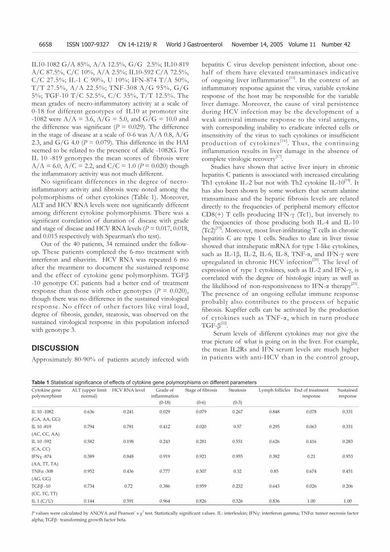
6658 ISSN 1007-9327 CN 14-1219/ R World J Gastroenterol November 14, 2005 Volume 11 Number 42
IL10-1082 G/A 85%, A/A 12.5%, G/G 2.5%; IL10-819 A/C 87.5%, C/C 10%, A/A 2.5%; IL10-592 C/A 72.5%, C/C 27.5%; IL-1 C 90%, U 10%; IFN-874 T/A 50%, T/T 27.5%, A/A 22.5%; TNF-308 A/G 95%, G/G 5%; TGF-10 T/C 52.5%, C/C 35%, T/T 12.5%. The mean grades of necro-infl ammatory activity at a scale of 0-18 for different genotypes of IL10 at promoter site -1082 were A/A = 3.6, A/G = 5.0, and G/G = 10.0 and the difference was signifi cant (P = 0.029). The difference in the stage of disease at a scale of 0-6 was A/A 0.8, A/G 2.3, and G/G 4.0 (P = 0.079). This difference in the HAI seemed to be related to the presence of allele -1082G. For IL 10 -819 genotypes the mean scores of fibrosis were A/A = 6.0, A/C = 2.2, and C/C = 1.0 (P = 0.020) though the infl ammatory activity was not much different.
No significant differences in the degree of necro-infl ammatory activity and fi brosis were noted among the polymorphisms of other cytokines (Table 1). Moreover, ALT and HCV RNA levels were not signifi cantly different among different cytokine polymorphisms. There was a signifi cant correlation of duration of disease with grade and stage of disease and HCV RNA levels (P = 0.017, 0.018, and 0.015 respectively with Spearman’s rho test).
Out of the 40 patients, 34 remained under the follow-up. These patients completed the 6-mo treatment with interferon and ribavirin. HCV RNA was repeated 6 mo after the treatment to document the sustained response and the effect of cytokine gene polymorphism. TGFβ -10 genotype CC patients had a better end of treatment response than those with other genotypes (P = 0.020), though there was no difference in the sustained virological response. No effect of other factors like viral load, degree of fi brosis, gender, steatosis, was observed on the sustained virological response in this population infected with genotype 3.
DISCUSSIONApproximately 80-90% of patients acutely infected with
hepatitis C virus develop persistent infection, about one-half of them have elevated transaminases indicative of ongoing liver inflammation[15]. In the context of an infl ammatory response against the virus, variable cytokine response of the host may be responsible for the variable liver damage. Moreover, the cause of viral persistence during HCV infection may be the development of a weak antiviral immune response to the viral antigens, with corresponding inability to eradicate infected cells or insensitivity of the virus to such cytokines or insuffi cient production of cytokines [16]. Thus, the continuing inflammation results in liver damage in the absence of complete virologic recovery[17].
Studies have shown that active liver injury in chronic hepatitis C patients is associated with increased circulating Th1 cytokine IL-2 but not with Th2 cytokine IL-10[18]. It has also been shown by some workers that serum alanine transaminase and the hepatic fibrosis levels are related directly to the frequencies of peripheral memory effector CD8(+) T cells producing IFN-γ (Tc1), but inversely to the frequencies of those producing both IL-4 and IL-10 (Tc2)[19]. Moreover, most liver-infi ltrating T cells in chronic hepatitis C are type 1 cells. Studies to date in liver tissue showed that intrahepatic mRNA for type 1-like cytokines, such as IL-1β, IL-2, IL-6, IL-8, TNF-α, and IFN-γ were upregulated in chronic HCV infection[20]. The level of expression of type 1 cytokines, such as IL-2 and IFN-γ, is correlated with the degree of histologic injury as well as the likelihood of non-responsiveness to IFN-α therapy[21]. The presence of an ongoing cellular immune response probably also contributes to the process of hepatic fi brosis. Kupffer cells can be activated by the production of cytokines such as TNF-α, which in turn produce TGF-β[22].
Serum levels of different cytokines may not give the true picture of what is going on in the liver. For example, the mean IL2Rs and IFN serum levels are much higher in patients with anti-HCV than in the control group,
Cytokine gene polymorphism
ALT (upper limit normal)
HCV RNA level Grade of infl ammation
Stage of fi brosis Steatosis Lymph follicles End of treatment response
Sustained response
(0-18) (0-6) (0-3)
IL 10 -1082 0.636 0.241 0.029 0.079 0.267 0.848 0.078 0.331(GA, AA, GG)IL 10 -819 0.794 0.781 0.412 0.020 0.57 0.295 0.063 0.331(AC, CC, AA)IL 10 -592 0.582 0.198 0.243 0.281 0.551 0.626 0.416 0.283(CA, CC)IFNγ -874 0.389 0.848 0.919 0.921 0.955 0.382 0.21 0.933(AA, TT, TA)TNFα -308 0.952 0.436 0.777 0.307 0.32 0.85 0.674 0.451(AG, GG)TGFβ -10 0.734 0.72 0.386 0.959 0.232 0.643 0.026 0.206(CC, TC, TT)IL 1 (C/U) 0.144 0.591 0.964 0.826 0.326 0.836 1.00 1.00
Table 1 Statistical signifi cance of effects of cytokine gene polymorphisms on different parameters
P values were calculated by ANOVA and Pearson’ s χ2 test. Statistically signifi cant values. IL: interleukin; IFNγ: interferon gamma; TNFα: tumor necrosis factor alpha; TGFβ: transforming growth factor beta.

Abbas Z et al. Cytokine gene polymorphism and hepatitis C genotype 3 6659
whereas the mean IL4 and IL6 levels are lower in patients infected with HCV[23]. Another study shows the higher levels of serum IL-1β, IL-4 and IL-6 (0.221, 0.104 and 1.393 pg/mL) in all HCV patients than in healthy adults (0.188, 0.025 and 0.600 pg/mL)[24].
Sustained response to interferon and ribavirin, defi ned as undetectable HCV RNA at 6 mo after discontinuation of therapy, is achievable in 30-60% of treated patients[25,26].
Predictors of response include viral factors such as viral genotypes, viral load and early disappearance of HCV RNA after initiation of therapy, while the host factors include gender, age and degree of fi brosis[25-27]. In recent years, increasing attention has been drawn to the role of host variation in cytokine levels in inflammatory and immune responses. Polymorphisms in genes encoding immunoregulatory proteins, proinflammatory cytokines, and fi brogenic factors may affect the production of these factors and influence disease progression in patients with chronic liver disease due to alcohol, primary biliary cirrhosis, or hepatitis C[28].
There might be an association of cytokine gene polymorphism and susceptibility to hepatitis C infection. Hohler et al.[29] have reported such an association with the polymorphism at the TNFα promoter. The TNF promoter variants TNF2 (-238A) and TNF3 (-308A) confer a 3.2-fold and 5.1-fold risk of cirrhosis respectively (P = 0.03 for both). Reciprocal effects have been observed with several TNF alleles and haplotypes defi ned by the -238 G/A and -308 G/A dimorphic sequences, thus polymorphisms in the TNF alpha promoter appear to be associated with the variability in the histological severity of chronic hepatitis C infection[30]. In our study done on hepatitis C genotype 3 patients, no such effect of TNFα -308 variability was observed.
Interleukin (IL)-10 is a cytokine that downregulates the proinflammatory response and has a modulatory effect on hepatic fi brogenesis and is a potent anti-infl ammatory Th2 cytokine that downregulates the expression of major histocompatibility complex (MHC) class I and class II molecules, as well as the production of Th1 cytokines[31-36]. IL-10 levels differ widely between individuals, possibly because of polymorphisms in the promoter region of the IL-10 gene[37,38]. Specifically, three SNPs in the promoter (at positions -1082, -819, and -592 relative to the transcription start site) form three SNP combinations (ATA, ACC, GCC), which are associated with differential IL-10 expression[38-40]. It is reasonable to assume that hepatitis C patients who produce high levels of IL-10 have less hepatocellular injury and less ability to control infection and patients with low secretion of IL-10 have a better ability to eliminate the hepatitis infection. Perhaps low IL-10 production can skew the immune system into the Th1 type of response, facilitating the clearance of viral load. -1082A allele is associated with reduced IL-10 production in vitro[39]. In our study, individuals who were homozygous for IL-10 AA at position -1082 had a lower HAI.
It has been shown that hepatitis C patients, genotyped as high IL-10 producers, have a poor response to IFN-α
therapy[40]. Such polymorphisms may also predict the sustained viral response to antiviral therapy[41]. These patients may benefi t from additional treatment strategies designed to enhance T-helper type 1 (Th1) response. In one study, the interleukin-10 -1082 G/G genotype was identified more frequently in patients than in controls (P = 0.048). The patients exhibiting transforming growth factor-beta 1+29 (codon 10) C/C genotype variables were less likely to respond to treatment than patients with the T/T or T/C genotypes[42]. Liver transplant recipients, who are genotyped as having a low production profi le of IL-10, are more prone to rejection and less likely to have hepatitis C recurrence[43]. The IL-1beta-31 genotype T/T or the IL-1beta-511/-31 haplotype C/T is associated with the presence of HCC in Japanese patients with chronic HCV infection[44].
However, not all the studies favor such effect of polymorphisms. Three members of the interleukin-1 gene family (IL-1A, IL-1B and IL-1RN), three polymorphic sites in the interleukin-10 gene promoter (-1082, -819, -592) and two in the TNF-α promoter (-308, -238) were studied in two independent DNA banks, each with appropriate controls. Standard PCR-based genotyping techniques were used. No signifi cant difference in the distribution of any of the polymorphisms has been found in either study set[45].
We, in this study, determined SNP at position -1082, -819 and -592 in case of IL-10, -874 for IFN-γ, -308 for TNF-α, -10 for TGF-β and IL-1 C/ U and analyzed the frequency of their distribution and correlation with the ALT and HCV RNA levels, HAI and response to treatment. In our series, we selected genotype 3 patients because this is the main genotype in our region and not enough data are available on the influence of host cytokine gene polymorphisms of this viral genotype. Another reason of selecting a single genotype was to make the group uniform as different genotypes have different response rates to antiviral therapy and infl uence of cytokines may also be different. We could not find any signifi cant difference in the cytokine genotype profi le while analyzing different variables except for some infl uence of polymorphisms of IL-10 on liver histology. These polymorphisms did not modulate the response to interferon plus ribavirin therapy. This may be due to the small sample size or the fact that viral genotype 3 is easy to treat genotype in any case.
REFERENCES1 Pawlotsky JM. Hepatitis C virus infection: virus/host
interactions. J Viral Hepat 1998; 5 Suppl 1: 3-82 Mosmann TR, Sad S. The expanding universe of T-cell
subsets: Th1, Th2 and more. Immunol Today 1996; 17: 138-1463 Biron CA. Cytokines in the generation of immune responses
to, and resolution of virus infection. Curr Opin Immunol 1994; 6: 530-538
4 Tough DF, Borrow P, Sprent J. Induction of bystander T cell proliferation by viruses and type I interferon in vivo. Science 1996; 272: 1947-1950
5 Napoli J , Bishop GA, McGuinness PH, Painter DM, McCaughan GW. Progressive liver injury in chronic hepatitis
6660 ISSN 1007-9327 CN 14-1219/ R World J Gastroenterol November 14, 2005 Volume 11 Number 42
C infection correlates with increased intrahepatic expression of Th1-associated cytokines. Hepatology 1996; 24: 759-765
6 Sobue S, Nomura T, Ishikawa T, Ito S, Saso K, Ohara H, Joh T, Itoh M, Kakumu S. Th1/Th2 cytokine profiles and their relationship to clinical features in patients with chronic hepatitis C virus infection. J Gastroenterol 2001; 36: 544-551
7 Rosen HR, Miner C, Sasaki AW, Lewinsohn DM, Conrad AJ, Bakke A, Bouwer HG, Hinrichs DJ. Frequencies of HCV-specifi c effector CD4+ T cells by fl ow cytometry: correlation with clinical disease stages. Hepatology 2002; 35: 190-198
8 Cramp ME, Carucci P, Rossol S, Chokshi S, Maertens G, Williams R, Naoumov NV. Hepatitis C virus (HCV) specifi c immune responses in anti-HCV positive patients without hepatitis C viraemia. Gut 1999; 44: 424-429
9 Chang KM, Thimme R, Melpolder JJ, Oldach D, Pemberton J, Moorhead-Loudis J, McHutchison JG, Alter HJ, Chisari FV. Differential CD4(+) and CD8(+) T-cell responsiveness in hepatitis C virus infection. Hepatology 2001; 33: 267-276
10 Fabris C, Soardo G, Falleti E, Toniutto P, Vitulli D, Federico E, Del Forno M, Mattiuzzo M, Gonano F, Pirisi M. Relationship among hepatic inflammatory changes, circulating levels of cytokines, and response to IFN-alpha in chronic hepatitis C. J Interferon Cytokine Res 1998; 18: 705-709
11 Kato S, Onda M, Yamada S, Matsuda N, Tokunaga A, Matsukura N. Association of the interleukin-1 beta genetic polymorphism and gastric cancer risk in Japanese. J Gastroenterol 2001; 36: 696-699
12 di Giovine FS, Takhsh E, Blakemore AI, Duff GW. Single base polymorphism at -511 in the human interleukin-1 beta gene (IL1 beta). Hum Mol Genet 1992; 1: 450
13 Perrey C, Turner SJ, Pravica V, Howell WM, Hutchinson IV. ARMS-PCR methodologies to determine IL-10, TNF-alpha, TNF-beta and TGF-beta 1 gene polymorphisms. Transpl Immunol 1999; 7: 127-128
14 Ishak K, Baptista A, Bianchi L, Callea F, De Groote J, Gudat F, Denk H, Desmet V, Korb G, MacSween RN. Histological grading and staging of chronic hepatitis. J Hepatol 1995; 22: 696-699
15 Alter MJ, Mast EE, Moyer LA, Margolis HS. Hepatitis C. Infect Dis Clin North Am 1998; 12: 13-26
16 Cerny A, Chisari FV. Pathogenesis of chronic hepatitis C: immunological features of hepatic injury and viral persistence. Hepatology 1999; 30: 595-601
17 Koziel MJ. The role of immune responses in the pathogenesis of hepatitis C virus infection. J Viral Hepat 1997; 4 Suppl 2: 31-41
18 Bozkaya H, Bozdayi AM, Aslan N, Turkay C, Sarioglu M, Cetinkaya H, Akdogan M, Cinar K, Erden E, Kose K, Senturk H, Akkiz H, Karayalcin S, Yurdaydin C, Uzunalimoglu O. Circulating IL-2 and IL-10 in chronic active hepatitis C with respect to the response to IFN treatment. Infection 2000; 28: 309-313
19 Prezzi C, Casciaro MA, Francavilla V, Schiaffella E, Finocchi L, Chircu LV, Bruno G, Sette A, Abrignani S, Barnaba V. Virus-specifi c CD8(+) T cells with type 1 or type 2 cytokine profi le are related to different disease activity in chronic hepatitis C virus infection. Eur J Immunol 2001; 31: 894-906
20 Llorent L, Richaud-Patin Y, Alcocer-Castillejos N, Ruiz-Soto R, Mercado MA, Orozco H, Gamboa-Dominguez A, Alcocer-Va-rela J. Cytokine gene expression in cirrhotic and non-cirrhotic human liver. J Hepatol 1996; 24: 555-563
21 Fukuda R, Ishimura N, Ishihara S, Chowdhury A, Morlyama N, Nogami C, Miyake T, Niigaki M, Tokuda A, Satoh S, Sakai S, Akagi S, Watanabe M, Fukumoto S. Intrahepatic expression of pro-infl ammatory cytokine mRNAs and interferon effi cacy in chronic hepatitis C. Liver 1996; 16: 390-399
22 Roulot D, Durand H, Coste T, Rautureau J, Strosberg AD, Benarous R, Marullo S. Quantitative analysis of transforming growth factor beta 1 messenger RNA in the liver of patients with chronic hepatitis C: absence of correlation between high levels and severity of disease. Hepatology 1995; 21: 298-304
23 Cribier B, Schmitt C, Rey D, Lang JM, Kirn A, Stoll-Keller F. Production of cytokines in patients infected by hepatitis C virus. J Med Virol 1998; 55: 89-91
24 Lapinski TW. The levels of IL-1beta, IL-4 and IL-6 in the serum and the liver tissue of chronic HCV-infected patients. Arch Immunol Ther Exp (Warsz) 2001; 49: 311-316
25 Poynard T, Marcellin P, Lee SS, Niederau C, Minuk GS, Ideo G, Bain V, Heathcote J, Zeuzem S, Trepo C, Albrecht J. Randomised trial of interferon alpha2b plus ribavirin for weeks or for 24 weeks versus interferon alpha2b plus placebo for 48 weeks for treatment of chronic infection with hepatitis C virus. International Hepatitis Interventional Therapy Group (IHIT) Lancet 1998; 352: 1426-1432
26 McHutchison JG, Gordon SC, Schiff ER, Shiffman ML, Lee WM, Rustgi VK, Goodman ZD, Ling MH, Cort S, Albrecht JK. Interferon alfa-2b alone or in combination with ribavirin as initial treatment for chronic hepatitis C. Hepatitis Interventional Therapy Group. N Engl J Med 1998; 339: 1485-1492
27 Brouwer JT, Hansen BE, Niesters HG, Schalm SW. Early prediction of response in interferon monotherapy and in interferon-ribavirin combination therapy for chronic hepatitis C: HCV RNA at 4 wk versus ALT. J Hepatol 1999; 30: 192-198
28 Bataller R, North KE, Brenner DA. Genetic polymorphisms and the progression of liver fibrosis: a critical appraisal. Hepatology 2003; 37: 493-503
29 Hohler T, Kruger A, Gerken G, Schneider PM, Meyer zum Buschenfelde KH, Rittner C. Tumor necrosis factor alpha promoter polymorphism at position -238 is associated with chronic active hepatitis C infection. J Med Virol 1998; 54: 173-177
30 Yee LJ, Tang J, Herrera J, Kaslow RA, van Leeuwen DJ. Tumor necrosis factor gene polymorphisms in patients with cirrhosis from chronic hepatitis C virus infection. Genes Immun 2000; 1: 386-390
31 Fiorentino DF, Zlotnik A, Vieira P, Mosmann TR, Howard M, Moore KW, O’Garra A. IL-10 acts on the antigen-presenting cell to inhibit cytokine production by Th1 cells. J Immunol 1991; 146: 3444-3451
32 Yue FY, Dummer R, Geertsen R, Hofbauer G, Laine E, Manolio S, Burg G. Interleukin-10 is a growth factor for human melanoma cells and down-regulates HLA class-I, HLA class-II and ICAM-1 molecules. Int J Cancer 1997; 71: 630-637
33 Tsuruma T, Yagihashi A, Torigoe T, Sato N, Kikuchi K, Watanabe N, Hirata K. Interleukin-10 reduces natural killer sensitivity and downregulates MHC class I expression on H-ras-transformed cells. Cell Immunol 1998; 184: 121-128
34 Zeller JC, Panoskaltsis-Mortari A, Murphy WJ, Ruscetti FW, Narula S, Roncarolo MG, Blazar BR. Induction of CD4+ T cell alloantigen-specific hyporesponsiveness by IL-10 and TGF-beta. J Immunol 1999; 163: 3684-3691
35 de Waal Malefyt R, Haanen J, Spits H, Roncarolo MG, te Velde A, Figdor C, Johnson K, Kastelein R, Yssel H, de Vries JE. Interleukin 10 (IL-10) and viral IL-10 strongly reduce antigen-specifi c human T cell proliferation by diminishing the antigen-presenting capacity of monocytes via downregulation of class II major histocompatibility complex expression. J Exp Med 1991; 174: 915-924
36 de Waal Malefyt R, Abrams J, Bennett B, Figdor CG, de Vries JE. Interleukin 10(IL-10) inhibits cytokine synthesis by human monocytes: an autoregulatory role of IL-10 produced by monocytes. J Exp Med 1991; 174: 1209-1220
37 Eskdale J, Gallagher G, Verweij CL, Keijsers V, Westendorp RG, Huizinga TW. Interleukin 10 secretion in relation to human IL-10 locus haplotypes. Proc Natl Acad Sci USA 1998; 95: 9465-9470
38 Eskdale J, Keijsers V, Huizinga T, Gallagher G. Microsatellite alleles and single nucleotide polymorphisms (SNP) combine to form four major haplotype families at the human interleukin-10 (IL-10) locus. Genes Immun 1999; 1: 151-155
39 Turner DM, Williams DM, Sankaran D, Lazarus M, Sinnott
Abbas Z et al. Cytokine gene polymorphism and hepatitis C genotype 3 6661
PJ, Hutchinson IV. An investigation of polymorphism in the interleukin-10 gene promoter. Eur J Immunogenet 1997; 24: 1-8
40 Edwards-Smith CJ, Jonsson JR, Purdie DM, Bansal A, Shorthouse C, Powell EE. Interleukin-10 promoter po-lymorphism predicts initial response of chronic hepatitis C to interferon alfa. Hepatology 1999; 30: 526-530
41 Yee LJ, Tang J, Gibson AW, Kimberly R, Van Leeuwen DJ, Kaslow RA. Interleukin 10 polymorphisms as predictors of sustained response in antiviral therapy for chronic hepatitis C infection. Hepatology 2001; 33: 708-712
42 Vidigal PG, Germer JJ, Zein NN. Polymorphisms in the interleukin-10, tumor necrosis factor-alpha, and transforming growth factor-beta1 genes in chronic hepatitis C patients treated with interferon and ribavirin. J Hepatol 2002; 36: 271-277
43 Tambur AR, Ortegel JW, Ben-Ari Z, Shabtai E, Klein T,
Michowiz R, Tur-Kaspa R, Mor E. Role of cytokine gene polymorphism in hepatitis C recurrence and allograft rejection among liver transplant recipients. Transplantation 2001; 71: 1475-1480
44 Wang Y, Kato N, Hoshida Y, Yoshida H, Taniguchi H, Goto T, Moriyama M, Otsuka M, Shiina S, Shiratori Y, Ito Y, Omata M. Interleukin-1beta gene polymorphisms associated with hepatocellular carcinoma in hepatitis C virus infection. Hepatology 2003; 37: 65-71
45 Constantini PK, Wawrzynowicz-Syczewska M, Clare M, Boron-Kaczmarska A, McFarlane IG, Cramp ME, Donaldson PT. Interleukin-1, interleukin-10 and tumour necrosis factor-alpha gene polymorphisms in hepatitis C virus infection: an investigation of the relationships with spontaneous viral clearance and response to alpha-interferon therapy. Liver 2002; 22: 404-412
Science Editor Wang XL and Guo SY Language Editor Elsevier HK

PO Box 2345, Beijing 100023, China World J Gastroenterol 2005;11(42):6662-6666www.wjgnet.com World Journal of Gastroenterology ISSN [email protected] © 2005 The WJG Press and Elsevier Inc. All rights reserved.E L S E V I E R
• BRIEF REPORTS •
mucosal injury, and these gastroprotective effects might be induced, at least partly by their radical scavenging activity.
© 2005 The WJG Press and Elsevier Inc. All rights reserved.
Key words: Nigella sativa ; Thymoquinone; Ulcer; Anti-oxidant; Rat
Kanter M, Demir H, Karakaya C, Ozbek H. Gastroprotective act iv i ty of Nigel la sat iva L oi l and i ts const i tuent, thymoquinone against acute alcohol-induced gastric mucosal injury in rats. World J Gastroenterol 2005; 11(42); 6662-6666http://www.wjgnet.com/1007-9327/11/6662.asp
INTRODUCTIONIntragastric application of absolute ethanol has long been used as a reproducible method to induce gastric lesions in experimental animals[1]. Gastric lesion is accompanied with the formation of free radicals (FRs) and reactive oxygen species (ROS)[2-4]. These radicals in particular seem to play an important role in ulcerative and erosive lesions of the gastrointestinal tract[5], as they attack and damage many biological molecules. Therefore, treatment with antioxidants and FR scavengers can decrease ethanol-induced gastric mucosal damage[6,7].
The black seed, Nigella sativa L (NS), a member of the family of ranunculaceae, contains more than 30% of fi xed oil and 0.4-0.45 % wt/wt of volatile oil. The volatile oil contains 18.4-24% thymoquinone (TQ) and 46% many monoterpenes such as p-cymene and α-piene[8]. Recently, clinical and animal studies have shown that extract of the black seeds have many therapeutic effects such as immunomodilative[9], antibacterial[10], hypotensive[11], hepatoprotective[12] and antidiabetic effects[13]. Ohkawa et al.[14] also reported that NS oil and its derivative TQ inhibit eicosanoid generation in leukocytes and membrane lipid peroxidation. However, the gastroprotective effect of this plant and its major constituent against ethanol-induced gastric mucosal injury remains unclear.
In the present investigation, we studied the infl uence of NS on gastric mucosal lesions and the redox state induced by ethanol and to compare its actions with those of its constituent TQ.
Gastroprotective activity of Nigella sativa L oil and its constituent, thymoquinone against acute alcohol-induced gastric mucosal injury in rats
Mehmet Kanter, Halit Demir, Cengiz Karakaya, Hanefi Ozbek
Mehmet Kanter, Department of Histology and Embryology, Faculty of Medicine, Trakya University, Edirne, TurkeyHalit Demir, Department of Chemistry, Faculty of Art and Science, Yuzuncu Yil University, Van, TurkeyCengiz Karakaya, Department of Biochemistry, Faculty of Medicine, , Yuzuncu Yil University, Van, TurkeyHanefi Ozbek, Department of Pharmacology, Faculty of Medicine, Yuzuncu Yil University, Van, TurkeyCorrespondes to: Dr. Mehmet Kanter, Department of Histology and Embryology, Faculty of Medicine, Trakya University, Edirne, Turkey. [email protected]: +902842357641 Fax: +902842352730 Received: 2004-07-23 Accepted: 2004-05-17
AbstractAIM: To evaluate the role of reactive oxygen species in the pathogenesis of acute ethanol-induced gastric mucosal lesions and the effect of Nigella sativa L oil (NS) and its constituent thymoquinone (TQ) in an exper-imental model.
METHODS: Male Wistar albino rats were assigned into 4 groups. Control group was given physiologic saline orally (10 mL/kg body weight) as the vehicle (gavage); ethanol group was administrated 1 mL (per rat) absolute alcohol by gavage; the third and fourth groups were given NS (10 mL/kg body weight) and TQ (10 mg/kg body weight p.o) respectively 1 h prior to alcohol intake. One hour af-ter ethanol administration, stomach tissues were excised for macroscopic examination and biochemical analysis.
RESULTS: NS and TQ could protect gastric mucosa against the injurious effect of absolute alcohol and promote ulcer healing as evidenced from the ulcer index (UI) values. NS prevented alcohol-induced increase in thiobarbituric acid-reactive substances (TBARS), an index of lipid peroxidation. NS also increased gastric glutathione content (GSH), enzymatic activities of gastric superoxide dismutase (SOD) and glutathione-S-transferase (GST). Likewise, TQ protected against the ulcerating effect of alcohol and mitigated most of the biochemical adverse effects induced by alcohol in gastric mucosa, but to a lesser extent than NS. Neither NS nor TQ affected catalase activity in gastric tissue.
CONCLUSION: Both NS and TQ, particularly NS can partly protect gastric mucosa from acute alcohol-induced
Kanter M et al. NS and TQ on alcohol induced gastric mucosal injury 6663
MATERIALS AND METHODSPlant materials and extraction procedure The NS seeds were purchased from a local herb store, Van, Turkey. Voucher specimens were kept at the Department of Biochemistry, Yuzuncuyil University, Van, Turkey, for the future reference. The seeds of NS were powdered in a mixer, placed in a distillation flask and the volatile oil with 0.2% yield was collected by steam distillation. TQ 2-isopropyl-5-methyl-1,4-benzoquinone, was purchased from Sigma (St. Louis, MO, USA).
Treatment of ratsForty male Wistar albino rats, weighing 200-250 g (aged 4 mo), were supplied by The Center of Medical Investigations of Yuzuncu Yil University. The animals were fed with a standard rat chaw (Murat Food Factory, Ankara, Turkey) and allowed to drink water ad libitum, but they were deprived of food 12 h before the experiment. The animals were housed in a single temperature controlled (20-25 oC) cage in dark/light cycle. All procedures were performed in sterilized conditions. All animals received human care according to the criteria outlined in the “Guide for the Care and Use of Laboratory Animals” prepared by the National Academy of Sciences and published by the National Institutes of Health.
Rats were assigned into 4 groups (each containing 10 animals). Control group was given physiologic saline orally (10 mL/kg body weight) as the vehicle (gavage), ethanol group was administrated 1 mL (per rat) absolute alcohol by gavage. The third and fourth groups were given NS (10 mL/kg body weight) and TQ (10 mg /kg body weight p.o) respectively 1 h prior to alcohol intake. One hour after ethanol administration, the animals were euthanized by cervical dislocation. The stomach was excised, cut along the greater curvature, and gently rinsed in tap water. The stomach was stretched on a piece of cork with mucosal surface up, and then examined in a standard position for macroscopic examination. Scoring of ulcer was performed with the help of magnifying glass. Lesion size (mm) was determined by measuring each lesion and its greatest diameter was recorded in the case of petechial lesions. Four such lesions were considered to be the equivalent of an 1 mm ulcer. The sum of the total severity scores in each group of rats divided by the number of animals, was expressed as the mean ulcer index (UI).
Biochemical analysisStomachs were cut into small pieces and homogenized in 0.15 mol/L ice-cold KCl using Heidolf Diax 900, type 595 (Germany) to give 20% homogenates. The homogenates were then made into aliquots and used for the assessment of antioxidant parameters.
MDA levels were determined as previously described[15]. Less than 0.2 mL of 10% (w/v) tissue homogenate, 0.2 mL of 8.1% sodium dodecyl sulfate (SDS), 1.5 mL of 20% acetic acid solution adjusted to pH 3.5 with NaOH, and 1.5 mL of 0.8% aqueous solution thiobarbituric acid (TBA) were added into the sample. The mixture was
made up to 4.0 mL with distilled water, and heated at 95 0C for 60 min. After cooling, 1.0 mL of distilled water and 5.0 mL of the mixture of n-buthanol and pyridine (15:1,v/v) were added and shaken vigorously. After centri-fugation at 4 000 r/min for 10 min, the organic layer was taken and its absorbance at 532 nm was measured. Total TBA-reactive materials were expressed as MDA, using a molar ext inct ion coeff ic ient for MDA of 1.56×105 cm/mol/L. MDA level was expressed as nmol/g.
Reduced g lu ta th ione (GSH) was de te r mined according to the method described by Sun[16]. The GSH concentration (μmol/g) was computed from a standard curve constructed using different concentrations of standard GSH.
Stomach homogenate (20%) was centrifuged at 10 000 g for 10 min for 30 min at 4 0C (Beckman XL-70, USA). Following centrifugation, the supernatant (cytosolic fraction) was carefully removed from the pellet and used directly for assay of the enzymatic activities of SOD, GST and CAT.
SOD activity was detected according to Sun and Habig[17]. One SOD unit was defined as the enzyme amount causing 50% inhibition in the NBTH2 reduction rate. SOD activity was also expressed as U/mg protein of stomach tissue sediment.
Gastric GST activity was determined according to the method of Clairborne et al. [18]. In brief, the GST activity toward 1-chloro-2,4-dinitrobenzene in the presence of glutathione as a co-substrate was measured specrophotometrically at 25 oC. The enzyme activity was determined by monitoring the changes in the absorbance at 340 nm for 4 min at 1-min intervals. The enzymatic activity was expressed as nmol min/g tissue.
CAT activity was determined according to the method of Lowr y [19]. In shor t , the super na tan t (50 μL) was added to a quartz cuvette containing 2.95 mL of 19 mmol/L H2O2 solution prepared in potassium phosphate buffer (0.1mol/L, pH 7.4). The change in absorbance was monitored at 240 nm over a 5-min period using a spectrophotometer (Shimadzu UV-1201, Japan). Commercially available CAT was used as the standard. CAT activity was expressed as U/g tissue.
The amount of protein was determined by the Lowry method[20].
Statistical analysisThe data were expressed as mean±SD and analyzed by repeated measures of variance (ANOVA). Tukey test was used to test for differences among means for which ANOVA indicated a signifi cant (P<0.05) F ratio.
RESULTSOral administration of absolute ethanol produced multiple mucosal lesions in the rat stomach. Pretreatment with NS and TQ inhibited the ethanol-induced gastric mucosal injury in rats. Pretreatment with single oral dose of NS signifi cantly reduced the ulcer index compared to alcohol
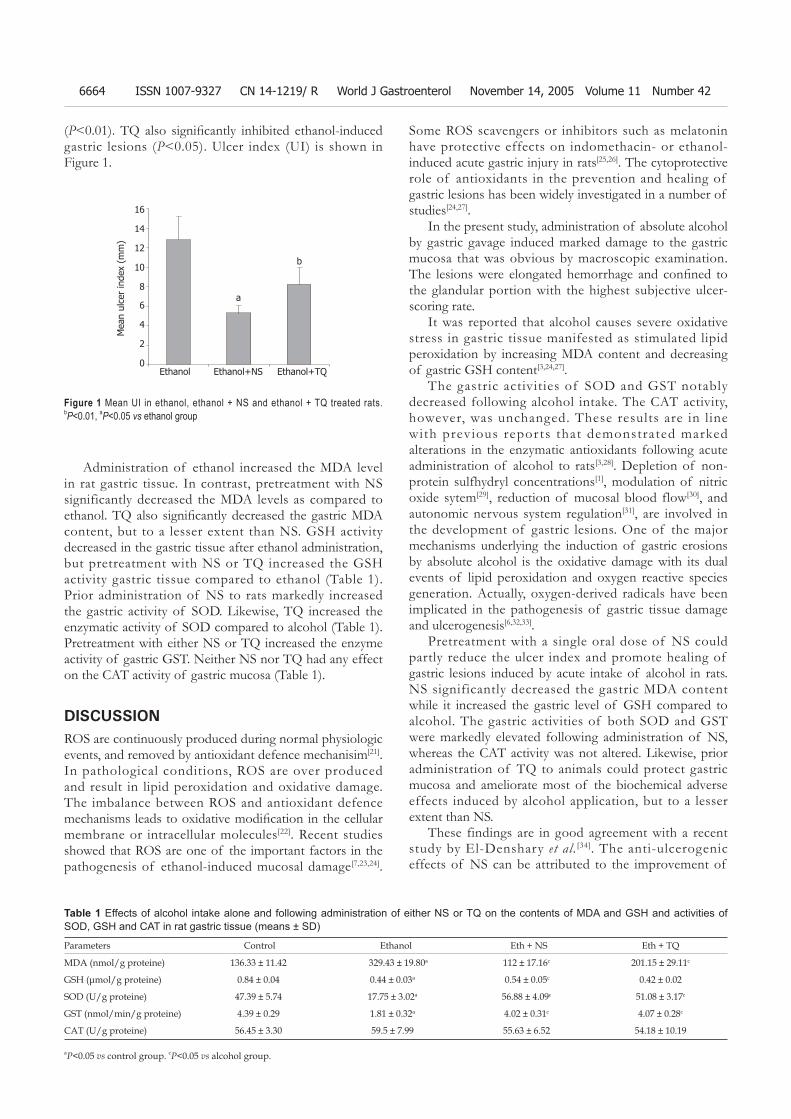
6664 ISSN 1007-9327 CN 14-1219/ R World J Gastroenterol November 14, 2005 Volume 11 Number 42
(P<0.01). TQ also signifi cantly inhibited ethanol-induced gastric lesions (P<0.05). Ulcer index (UI) is shown in Figure 1.
Some ROS scavengers or inhibitors such as melatonin have protective effects on indomethacin- or ethanol-induced acute gastric injury in rats[25,26]. The cytoprotective role of antioxidants in the prevention and healing of gastric lesions has been widely investigated in a number of studies[24,27].
In the present study, administration of absolute alcohol by gastric gavage induced marked damage to the gastric mucosa that was obvious by macroscopic examination. The lesions were elongated hemorrhage and confi ned to the glandular portion with the highest subjective ulcer-scoring rate.
It was reported that alcohol causes severe oxidative stress in gastric tissue manifested as stimulated lipid peroxidation by increasing MDA content and decreasing of gastric GSH content[3,24,27].
The gastric activities of SOD and GST notably decreased following alcohol intake. The CAT activity, however, was unchanged. These results are in l ine with previous repor ts that demonstrated marked alterations in the enzymatic antioxidants following acute administration of alcohol to rats[3,28]. Depletion of non-protein sulfhydryl concentrations[1], modulation of nitric oxide sytem[29], reduction of mucosal blood flow[30], and autonomic nervous system regulation[31], are involved in the development of gastric lesions. One of the major mechanisms underlying the induction of gastric erosions by absolute alcohol is the oxidative damage with its dual events of lipid peroxidation and oxygen reactive species generation. Actually, oxygen-derived radicals have been implicated in the pathogenesis of gastric tissue damage and ulcerogenesis[6,32,33].
Pretreatment with a single oral dose of NS could partly reduce the ulcer index and promote healing of gastric lesions induced by acute intake of alcohol in rats. NS significantly decreased the gastric MDA content while it increased the gastric level of GSH compared to alcohol. The gastric activities of both SOD and GST were markedly elevated following administration of NS, whereas the CAT activity was not altered. Likewise, prior administration of TQ to animals could protect gastric mucosa and ameliorate most of the biochemical adverse effects induced by alcohol application, but to a lesser extent than NS.
These findings are in good agreement with a recent study by El-Denshary et al. [34]. The anti-ulcerogenic effects of NS can be attributed to the improvement of
Administration of ethanol increased the MDA level in rat gastric tissue. In contrast, pretreatment with NS significantly decreased the MDA levels as compared to ethanol. TQ also signifi cantly decreased the gastric MDA content, but to a lesser extent than NS. GSH activity decreased in the gastric tissue after ethanol administration, but pretreatment with NS or TQ increased the GSH activity gastric tissue compared to ethanol (Table 1). Prior administration of NS to rats markedly increased the gastric activity of SOD. Likewise, TQ increased the enzymatic activity of SOD compared to alcohol (Table 1). Pretreatment with either NS or TQ increased the enzyme activity of gastric GST. Neither NS nor TQ had any effect on the CAT activity of gastric mucosa (Table 1).
DISCUSSIONROS are continuously produced during normal physiologic events, and removed by antioxidant defence mechanisim[21]. In pathological conditions, ROS are over produced and result in lipid peroxidation and oxidative damage. The imbalance between ROS and antioxidant defence mechanisms leads to oxidative modifi cation in the cellular membrane or intracellular molecules[22]. Recent studies showed that ROS are one of the important factors in the pathogenesis of ethanol-induced mucosal damage[7,23,24].
16
14
12
10
8
6
4
2
0Ethanol Ethanol+NS Ethanol+TQ
Figure 1 Mean UI in ethanol, ethanol + NS and ethanol + TQ treated rats. bP<0.01, aP<0.05 vs ethanol group
Parameters Control Ethanol Eth + NS Eth + TQ
MDA (nmol/g proteine) 136.33 ± 11.42 329.43 ± 19.80a 112 ± 17.16c 201.15 ± 29.11c
GSH (μmol/g proteine) 0.84 ± 0.04 0.44 ± 0.03a 0.54 ± 0.05c 0.42 ± 0.02
SOD (U/g proteine) 47.39 ± 5.74 17.75 ± 3.02a 56.88 ± 4.09c 51.08 ± 3.17c
GST (nmol/min/g proteine) 4.39 ± 0.29 1.81 ± 0.32a 4.02 ± 0.31c 4.07 ± 0.28c
CAT (U/g proteine) 56.45 ± 3.30 59.5 ± 7.99 55.63 ± 6.52 54.18 ± 10.19
aP<0.05 vs control group. cP<0.05 vs alcohol group.
Table 1 Effects of alcohol intake alone and following administration of either NS or TQ on the contents of MDA and GSH and activities of SOD, GSH and CAT in rat gastric tissue (means ± SD)
Mea
n ul
cer
inde
x (m
m)
b
a

Kanter M et al. NS and TQ on alcohol induced gastric mucosal injury 6665
the antioxidant status of animals due to an increase in mucin content of the gastric mucosa[34], or the presence of FR scavenging substances such as TQ[35]. It was also reported that NS given to sensitized quinea pigs, inhibits FR generation, and increases serum levels of SOD and glutathione[14]. NS could protect the gastric mucosa by increasing the bioavailability of arachidonic acid, resulting in biosynthesis of the cytoprotective prostaglandins in the stomach[36]. NS has also been reported to produce a marked inhibition on the release of leukotrienes, which cause mucosal tissue injury and hypoxemia[37]. Therefore, it may alter the delicate balance between prostaglandins and leukotrienes in the gastric mucosa favoring cytoprotection. TQ is the main active component of NS, and is able to inhibit lipid peroxidation[14]. Moreover, its ability to preserve the cell membrane integrity can be proven by the restoration.
In conclusion, pretreatment with NS and TQ, par-ticularly NS can partly protect the gastric mucosa against the injurious effects of absolute ethanol and promote ulcer healing. NS or TQ can also mitigate most of the biochemical adverse effects induced by alcohol instillation in gastric tissue. Further studies are required to clarify the anti- ulcer and antioxidant action of NS or TQ.
REFERENCES1 Szabo S, Trier JS, Frankel PW. Sulfhydryl compounds may
mediate gastric cytoprotection. Science 1981; 214: 200-202 2 Cho CH, Pfeiffer CJ, Misra HP. Ulcerogenic mechanism
of ethanol and the action of sulphanilyl fluoride on the rat stomach in-vivo. J Pharm Pharmacol 1991; 43: 495-498
3 Lutnicki K, Wrobel J, Ledwozyw A, Trebas-Pietras E. The effect of calcium ions on the intensity of peroxidation processes and the severity of ethanol-induced injury to the rat’s gastric mucosa. Arch Vet Pol 1992; 32: 125-132
4 Bast A, Haenen GR, Doelman CJ. Oxidants and antioxidants: state of the art. Am J Med 1991; 91: 2S-13S
5 Hirokawa M, Miura S, Yoshida H, Kurose I, Shigematsu T, Hokari R, Higuchi H, Watanabe N, Yokoyama Y, Kimura H, Kato S, Ishii H. Oxidative stress and mitochondrial damage precedes gastric mucosal cell death induced by ethanol administration. Alcohol Clin Exp Res 1998; 22: 111S-114S
6 La Casa C, Villegas I, Alarcon de la Lastra C, Motilva V, Martin Calero MJ. Evidence for protective and antioxidant properties of rutin, a natural fl avone, against ethanol induced gastric lesions. J Ethnopharmocal 2000; 71: 45-53
7 el Tahir KE, Ashour MM, al-Harbi MM. The respiratory effects of the volatile oil of the black seed (Nigella sativa) in guinea-pigs: elucidation of the mechanism(s) of action. Gen Pharmacol 1993; 24: 1115-1122
8 El-Kadi A, Kandil O. The black seed (Nigella sativa) and immunity: its effect on human T cell subset. Fed Proc 1987; 46: 1222
9 Hanafy MS, Hatem ME. Studies on the antimicrobial activity of Nigella sativa seed (black cumin). J Ethnopharmacol 1991; 34: 275-278
10 Zaoui A, Cherrah Y, Lacaille-Dubois MA, Settaf A, Amarouch H, Hassar M. Diuretic and hypotensive effects of Nigella sativa in the spontaneously hypertensive rat. Therapie 2000; 55: 379-382
11 Turkdogan MK, Agaoglu Z, Yener Z, Sekeroglu R, Akkan HA, Avci ME. The role of antioxidant vitamins (C and E), selenium and Nigella sativa in the prevention of liver fi brosis and cirrhosis in rabbits: new hopes. Dtsch Tierarztl Wochenschr
2001; 108: 71-73 12 Kanter M, Meral I, Yener Z, Ozbek H, Demir H. Partial
regeneration/proliferation of the beta-cells in the islets of Langerhans by Nigella sativa L. in streptozotocin-induced diabetic rats. Tohoku J Exp Med 2003; 201: 213-219
13 Houghton PJ, Zarka R, de las Heras B, Hoult JR. Fixed oil of Nigella sativa and derived thymoquinone inhibit eicosanoid generation in leukocytes and membrane lipid peroxidation. Planta Med 1995; 61: 33-36
14 Ohkawa H, Ohishi N, Yagi K. Reaction of linoleic acid hydroperoxide with thiobarbituric acid. J Lipid Res 1978; 19: 1053-1057
15 Ellman GL. Tissue sulfhydryl groups. Arch Biochem Biophys 1959; 82: 214-226
16 Sun Y, Oberley LW, Li Y. A simple method for clinical assay of superoxide dismutase. Clin Chem 1988; 34: 497-500
17 Habig WH, Pabst MJ, Jakoby WB. Glutathione S-transferases. The fi rst enzymatic step in mercapturic acid formation. J Biol Chem 1974; 249: 7130-7139
18 Clairborne A. Catalase activity. In: Greenwald RA, editor. Handbook of Methods for Oxygen Radical Research. Boca Raton, FL 1985; 383-384
19 Lowry OH, Rosebrough NJ, Farr AL, Randall RJ. Protein measurement with the Folin phenol reagent. J Biol Chem 1951; 193: 265-275
20 Halliwell B, Gutteridge JM, Cross CE. Free radicals, antioxidants, and human disease: where are we now? J Lab Clin Med 1992; 119: 598-620
21 El-Habit OH, Saada HN, Azab KS, Abdel-Rahman M, El-Malah DF. The modifying effect of beta-carotene on gamma radiation-induced elevation of oxidative reactions and genotoxicity in male rats. Mutat Res 2000; 466: 179-186
22 Tuncel N, Erkasap N, Sahinturk V, Ak DD, Tuncel M. The protective effect of vasoactive intestinal peptide (VIP) on stress-induced gastric ulceration in rats. Ann N Y Acad Sci 1998; 865: 309-322
23 Suzuki Y, Ishihara M, Segami T, Ito M. Anti-ulcer effects of antioxidants, quercetin, alpha-tocopherol, nifedipine and tetracycline in rats. Jpn J Pharmacol 1998; 78: 435-441
24 Brzozowski T, Konturek PC, Konturek SJ, Pajdo R, Bielanski W, Brzozowska I, Stachura J, Hahn EG. The role of melatonin and L-tryptophan in prevention of acute gastric lesions induced by stress, ethanol, ischemia, and aspirin. J Pineal Res 1997; 23: 79-89
25 Alarcon de la Lastra C, Motilva V, Martin MJ, Nieto A, Barranco MD, Cabeza J, Herrerias JM. Protective effect of melatonin on indomethacin-induced gastric injury in rats. J Pineal Res 1999; 26: 101-107
26 Bilici D, Suleyman H, Banoglu ZN, Kiziltunc A, Avci B, Ciftcioglu A, Bilici S. Melatonin prevents ethanol-induced gastric mucosal damage possibly due to its antioxidant effect. Dig Dis Sci 2002; 47: 856-861
27 El-Missiry MA, El-Sayed IH, Othman AI. Protection by metal complexes with SOD-mimetic activity against oxidative gastric injury induced by indomethacin and ethanol in rats. Ann Clin Biochem 2001; 38: 694-700
28 Kato S, Kitamura M, Korolkiewicz RP, Takeuchi K. Role of nitric oxide in regulation of gastric acid secretion in rats: effects of NO donors and NO synthase inhibitor. Br J Pharmacol 1998; 123: 839-846
29 Holzer P, Livingston EH, Saria A, Guth PH. Sensory neurons mediate protective vasodilatation in rat gastric mucosa. Am J Physiol 1981; 260: 363-370
30 Ko JK, Cho CH, Ogle CW. The vagus nerve and its non-cholinergic mechanism in the modulation of ethanol-induced gastric mucosal damage in rats. J Pharm Pharmacol 1994; 46: 29-33
31 Cho CH, Pfeiffer CJ, Misra HP. Ethanol and the antioxidant defense in the gastrointestinal tract. Acta Physiol Hung 1992; 80: 99-105
32 Ligumsky M, Sestieri M, Okon E, Ginsburg I. Antioxidants

6666 ISSN 1007-9327 CN 14-1219/ R World J Gastroenterol November 14, 2005 Volume 11 Number 42
inhibit ethanol-induced gastric injury in the rat. Role of manganese, glycine, and carotene. Scand J Gastroenterol 1995; 30: 854-860
33 El-Dakhakhny M, Barakat M, El-Halim MA, Aly SM. Effects of Nigella sativa oil on gastric secretion and ethanol induced ulcer in rats. J Ethnopharmacol 2000; 72: 299-304
34 Saleh S, El-Denshary EMS, Mahran LG, Salah N. Anti-inflammatory and antioxidant effect of Nigella sativa oil in sensitized animals. The 25th International Conference on Science and Technology, New Delhi 2000
35 Campbell WB, Halushka PV. Lipid-derived autacoids.
Eicosanoids and platelet-activating factors. In: Hardman JG, Limbird LE, Molinoff PB, Ruddon RW, Gilman AG. Goodman and Gilman’s. The Pharmacol Basis of Therapeutics, McGraw-Hill, New York 1996; 601-616
36 Tsuji S, Kawano S, Sato N, Kamada T. Mucosal blood flow stasis and hypoxemia as the pathogenesis of acute gastric mucosal injury: role of endogenous leukotrienes and prostaglandins. J Clin Gastroenterol 1990; 12 Suppl 1: S85-S91
37 Mansour MA. Protective effects of thymoquinone and desferrioxamine against hepatotoxicity of carbon tetrachloride in mice. Life Sci 2000; 66: 2583-2591
Science Editor Wang XL and Guo SY Language Editor Elsevier HK

PO Box 2345, Beijing 100023, China World J Gastroenterol 2005;11(42):6667-6670www.wjgnet.com World Journal of Gastroenterology ISSN [email protected] © 2005 The WJG Press and Elsevier Inc. All rights reserved.E L S E V I E R
• BRIEF REPORTS •
Yakoob J, Jafri W, Abid S, Jafri N, Hamid S, Shah HA, Rizvi L, Islam M, Shaikh H. Giardiasis in patients with dyspeptic symptoms. World J Gastroenterol 2005; 11(42): 6667-6670http://www.wjgnet.com/1007-9327/11/6667.asp
INTRODUCTIONGiardia lamblia is the most common protozoan isolated from the gastrointestinal tract[1]. Worldwide incidence is believed to range from 20% to 60%[2]. The incidence rate is 2-7% in industrialized nations[3]. Patients with giardiasis typically present with diarrhea, vague abdominal discomfort, nausea and distention together with mild weight loss and lassitude. The absence of these symptoms may result in a low clinical index of suspicion for the diagnosis. Giardiasis is diagnosed by signs and symptoms, as well as the presence of giardia cysts and trophozoites in the stool. Stool examination can be unreliable, as organisms may be excreted at irregular intervals which can produce a false negative test result[4]. There is no gold standard for the diagnosis of giardiasis. The initial method of diagnosis is by demonstration of the trophozoite or cysts of G lamblia in the stool by microscopy or stool antigen detection by ELISA. Other methods of diagnosis include examination of duodenal contents by aspiration or biopsy with endoscopy. A definitive diagnosis may require repeated stool examinations, fecal immunoassays, or even sampling of the upper intestinal contents. Two stool examinations can detect 80-90% of infections, while three samples detect >90%[5]. About 20% of infestations are symptomatic, and do not continue for more than 3 mo along with the passage of cysts[2]. Difficulties are often encountered in fi nding the underlying cause of recurrent abdominal pain. Giardiasis is considered as an infrequent cause of dyspepsia[6], but this might not be true in a third world country. Prevalence of giardiasis is the highest in areas of poor sanitation and drinking water treatment. It is transmitted by eating and drinking contaminated food and water or fecal-oral contact. In immunocompetent patient, small bowel biopsy may show normal histology or villous architecture, but increased intraepithelial lymphocytes and plasma cells in the lamina propria or villous atrophy and inflammatory cells[7]. Giardia trophozoites can be found on the surface or penetrating the epithelium down to the lamina propria[7]. The aim of this study was to investigate the prevalence of giardiasis in patients with dyspeptic symptoms.
Giardiasis in patients with dyspeptic symptoms
Javed Yakoob, Wasim Jafri, Shahab Abid, Nadim Jafri, Saeed Hamid, Hasnain Ali Shah, Lubna Rizvi, Muhammad Islam, Hizbullah Shaikh
Javed Yakoob, Wasim Jafri, Shahab Abid, Nadim Jafri, Saeed Hamid, Hasnain Ali Shah, Lubna Rizvi, Muhammad Islam, Section of Gastroenterology, Department of Medicine, Aga Khan University Hospital, Karachi 74800, Sindh, PakistanHizbul lah Shaikh, Department of Pathology, Aga Khan University Hospital, Karachi 74800, Sindh, PakistanCo-fi rst author: Wasim JafriCo-correspondence: Wasim JafriCorrespondence to: Dr. Javed Yakoob, MBBS, PhD, Section of Gastroenterology, Department of Medicine, Aga Khan University Hospital, Stadium Road, Karachi 74800, Pakistan. [email protected]: +92-21-48594661 Fax: +92-21-4934294 Received: 2005-03-10 Accepted: 2005-04-09
AbstractAIM: To investigate the prevalence of giardiasis in patients with dyspeptic symptoms.
METHODS: Clinical records of consecutive patients who attended Gastroenterology Department at Aga Khan University Hospital from January 2000 to June 2003 and had esophagogastroduodenoscopy (EGD) with duodenal biopsies and international classification of diseases 9th revision with clinical modifications (ICD-9-CM) coded with giardiasis were studied.
RESULTS: Two hundred and twenty patients fulfilled the above criteria. There were 44% (96/220) patients who were giardiasis positive, 72% (69/96) of them were males and 28% (27/96) of them were females. There were 65% (81/124) males and 35% (43/124) females who were giardiasis negative. The mean age of patients with giardiasis was 28±17 years, while that of giardiasis negative patients was 40±18 years (P<0.001). In patients with giardiasis, abdominal pain was present in 71% (68/96) of patients (P = 0.02) and diarrhea in 29% (28/96) (P = 0.005); duodenitis in 25% (24/96) on EGD (P = 0.006) and in 68% (65/96) on histopathology (P = 0.002).
CONCLUSION: Giardiasis occurs signifi cantly in young people with abdominal pain, while endoscopic duodenitis is seen in only 25% of giardiasis positive cases, which supports routine duodenal biopsy.
© 2005 The WJG Press and Elsevier Inc. All rights reserved.
Key words: Abdominal pain; Giardiasis; Stool exa-mination; EGD; Duodenal biopsy

6668 ISSN 1007-9327 CN 14-1219/ R World J Gastroenterol November 14, 2005 Volume 11 Number 42
MATERIALS AND METHODSPatients We carried out a retrospective analysis of medical records of all the patients who attended endoscopy unit of gastrointestinal section at the Aga Khan University Hospital from January 2000 to June 2003 consecutively for dyspeptic symptoms. They had an esophagogastroduodenoscopy (EGD) with duodenal biopsies after routine examination including stool examination. The giardiasis negative group consisted of the patients who had a stool examination and an EGD with duodenal biopsy and were not diagnosed as giardiasis. The giardiasis positive group consisted of the patients who had positive EGD with duodenal biopsy and stool examination for giardia cysts or trophozoites. Clinical symptoms at the time of presentation, diagnosis, drug treatment dosage and duration, past history of giardiasis, neutrophil and lymphocyte counts from complete blood picture, random blood glucose and stool examination were noted. Patients with diagnosis of celiac disease were excluded from the analysis.
Stool examinationStool parasitological analysis of samples collected from spontaneous bowel movement was performed. Samples were collected in a sterile container and transported soon after to the laboratory for examination. Microscopically each stool specimen was examined in a fresh normal saline smear and Lugol’s iodine preparation.
EsophagogastroduodenoscopyAll endoscopic examinations were performed by staff-members of our hospital’s gastroenterology section, using an Olympus video scope GIF xQ 140. Duodenitis was diagnosed when scattered. Reddened raised non-eroded mucosal patches were endoscopically identified in the duodenal bulb and descending duodenum.
HistopathologyHematoxylin and eosin (HE) staining of duodenal biopsy was used for pathological confi rmation of giardia trophozoites. Most organisms were tangentially cut and many were seen as small sickle-shaped objects near the epithelial surface. Statistical analysisThe SPSS (Release 11.5.0, standard version, copyright © SPSS; 1989-02) was used for data analysis. Descriptive analysis was done for demographic and clinical features. Results were presented as mean+SD for continuous variables, and number (percentage) for categorical variables. Univariate analysis was performed by using the independent sample t-test for continuous variables and Pearson’s chi-square test for categorical variables to assess the demographic and clinical parameters associated with giardiasis. Odds ratio (OR) estimates with their 95% confidence interval (CIs) and P values were calculated (Table 1). P≤0.05 was considered statistically signifi cant. All P values were two sided.
RESULTSPatientsTwo hundred and twenty patients fulfilled the criteria. There were 44% (96/220) patients who were giardiasis positive and 56% (124/220) were giardiasis negative. In patients with giardiasis, 72% (69/96) were males and 28% (27/96) were females. The mean age of patients with giardiasis was 28±17 years, while that of patients without giardiasis was 40±18 years (P<0.001). Abdominal pain was present in 71% (68/96) patients with giardiasis and in 56% (69/124) patients without giardiasis (P = 0.02), diarrhea was present in 29% (28/96) patients with giardiasis and in 14% (17/124) patients without giardiasis (P = 0.005, Table 1).
VariablesGiardiasis positive Giardiasis negative
P OR (95%CI)(n = 96) (%) (n = 124) (%)
Age groups (yr)<22 34 (35) 21 (17) 122–31 29 (30) 27 (22) 0.7 (0.3–1.5)32–48 20 (21) 35 (28) 0.4 (0.2–0.9)>48 13 (14) 41 (33) <0.001 0.2 (0.1–0.5)Gender
Female 27 (28) 43 (35)Male 69 (72) 81 (65) 0.301 1.4 (0.8–2.4)
Abdominal pain 68 (71) 69 (56) 0.021 1.9 (1.1–3.4)Diarrhea 28 (29) 17 (14) 0.005 2.6 (1.3–5.1)Weight loss 9 (9.4) 6 (5) 0.186 2.0 (0.7–5.9)Endo-duodenitis 24 (25) 53 (43) 0.007 0.4 (0.2–0.8)Histological duodenitis 65 (68) 58 (46) 0.002 2.5 (1.4–4.4)Lamina propria infl ammation 73 (76) 53 (42) <0.001 4.4 (2.4–8.1)Villus shortening 5 (5) – – –
Table 1 Clinical details of patients with and without endoscopic giardiasis
Results are presented as number (percentage), Odds ratio (95%CI).
Yakoob J et al. Diagnosis of giardiasis 6669
Stool examination Stool examination was carried out in 88.5% (85/96) cases, 6% (5/85) were positive for giardia cysts and trophozoites. EndoscopyOn EGD, antral gastritis was present in 46% (44/96) patients with giardiasis and in 75% (93/124) patients without giardiasis (P<0.001); duodenitis was present in 25% (24/96) patients with giardiasis and in 50% (53/124) patients without giardiasis (P = 0.006); duodenal ulcer was present in 4% (4/96) patients with giardiasis and in 72% (89/124) patients without giardiasis (P<0.001, Table 1). HistopathologyOn histopathology of gastric antral biopsy, H. pylori was seen in 42% (40/96) patients with giardiasis and in 54% (67/124) patients without giardiasis (P = 0.06); antral gastritis was found in 53% (51/96) patients with giardiasis and in 81% (100/124) patients without giardiasis (P<0.001). Duodenitis was seen in 68% (65/96) patients with giardiasis and in 46% (58/124) patients without giardiasis (P = 0.002); villus shortening was seen in 5% (5/96) patients with giardiasis and absent in patients giardiasis negative; lamina propria inflammatory infiltration with increased intraepithelial lymphocytes and plasma cells was seen in 76% (73/96) patients with giardiasis and in 42% (53/124) patients without giardiasis (P<0.001, Table 1).
DISCUSSIONIt is sometimes difficult to establish the diagnosis of giardiasis. Tests for parasitic antigen in stool are at least as sensitive and specifi c as good microscopic examination and are easier to perform. All these methods occasionally yield false-negative results.
The implications of this study are that no symptom complex is associated with the giardiasis. Giardiasis may present with abdominal pain alone and it should be considered even in the absence of diarrhea. Abdominal pain is the most common presentation and diagnosis, and investigations carried out are more often for gastritis or peptic ulcer disease with or without H pylori infection. Abdominal pain has also been previously found to be signifi cantly associated with the presence of giardiasis[8]. In our study, the incidence of giardiasis was higher in males, which is in agreement with the other studies[9-11]. However, the mean age of our patients was lower and might be attributed to more frequent exposure to this water borne infection because of social activities that involve frequent restaurants beside others. Diarrhea was seen in only 29% of cases. G lamblia infestation or other infectious causes were not considered because of the absence of diarrhea in most of these cases and hence a single stool examination was carried out. It is generally held that giardiasis may present with atypical gastrointestinal symptoms but this would be expected in combination with diarrhea[12]. The sensitivity for examination of a single random stool specimen is only 30-50%[13-14]. Hence this might explain
the 6% low yield of stool examination in the cases for giardia cysts and trophozoites. Microscopic examination of a single stool sample cannot exclude G lamblia infection; therefore, at least three stool specimens should be examined before other diagnostic procedures. As only saline or Lugol’s iodine examination of fecal smear was employed, it may have been insuffi cient in the absence of numerous parasites[14]. Other useful methods such as zinc sulfate fl oatation, a concentration technique for cysts, were also not employed in our study.
Duodenal ulcer is not a feature of giardiasis, while duodenitis was seen in 25% on endoscopy and in 68% on histology (P = 0.002). EGD with duodenal biopsy helps ruling out peptic ulcer as the only cause of symptoms. Giardiasis was associated with the normal endoscopic fi ndings in the duodenum in 75% cases (Table 1). Hence duodenal biopsy should be considered with EGD in patients with abdominal pain as antral biopsy alone may prove inadequate for determining the cause of abdominal pain in a developing country. This is in agreement with the opinion held by some investigators that routine duodenal biopsies should be done in patients undergoing upper intestinal endoscopy[15,16]. In this study, most of our cases on EGD showed normal duodenal mucosa but duodenal biopsy with H&E staining demonstrated G lamblia trophozoites (Table 1). Although special stains such as Giemsa or phosphotungstic acid hematoxylin can occasionally be helpful, routine hematoxylin and eosin staining is almost always satisfactory[17].
The data suggesting the role of giardiasis in dyspeptic patients are in contradiction to a previous prospective study[17]. Carr et al.[18] carried out a study in an area which is not endemic for Giardia, and demonstrated Giardia in 15.5% of patients presenting with dyspepsia and its prevalence is similar with or without obvious lesions at endoscopy. In their study, only 52% patients presented with abdominal pain, and patients with vomiting and diarrhea as presentation had a significantly increased prevalence of Giardia. However, our data originates from a developing country located in an area endemic for the acquisition of Giardia. In our study, patients more commonly presented with abdominal pain than diarrhea (Table 1). The discrepancy describing a lower frequency of endoscopic duodenitis and higher frequency of histologic duodenitis in infected patients than in non-infected group might be due to a higher prevalence of H pylori infection in the latter. However, this was a retrospective observational study with its selection bias and limitations in diagnostic methods. Serial stool examinations were not carried in all patients.
The presentation of giardiasis varies and for diagnosis it requires a high degree of suspicion in the appropriate clinical setting. Examination of duodenal biopsy from patients presenting with abdominal pain should be considered so as to prevent missing diagnosis of giardiasis. Giardiasis is diagnosed more often on EGD with duodenal biopsy rather than on stool examination. As cyst excretion is variable and may be undetectable at times, repeated

6670 ISSN 1007-9327 CN 14-1219/ R World J Gastroenterol November 14, 2005 Volume 11 Number 42
examination of properly preserved stool samples and biopsy of the small intestine may be required to detect the parasites. A prospective study is under way to confi rm the results of this study.
REFERENCES1 Eckmann L, Gillin FD. Microbes and microbial toxins: paradigms
for microbial-mucosal interactions I. Pathophysiological aspects of enteric infections with the lumen-dwelling protozoan pathogen Giardia lamblia. Am J Physiol Gastrointest Liver Physiol 2001; 280: G1-G6
2 Tripathi DM, Gupta N, Lakshmi V, Saxena KC, Agrawal AK. Antigiardial and immunostimulatory effect of Piper longum on giardiasis due to Giardia lamblia. Phytother Res 1999; 13: 561-565
3 Upcroft P, Upcroft JA. Drug targets and mechanisms of resis-tance in the anaerobic protozoa. Clin Microbiol Rev 2001; 14: 150-164
4 Beers MH, Berkow R. The Merk Manual. NJ: Whitehouse Station. Merck Research Laboratories; 1999: 1257-1258.
5 Gardner TB, Hill DR. Treatment of giardiasis. Clin Microbiol Rev 2001; 14: 114-128
6 Heikkinen M, Pikkarainen P, Takala J, Rasanen H, Julkunen R. Etiology of dyspepsia: four hundred unselected consecutive patients in general practice. Scand J Gastroenterol 1995; 30: 519-523
7 Ferguson A, Gillon J, Munro G. Pathology and pathogenesis of the intestinal mucosal damage in giardiasis. In: Meyer EA, ed. Giardiasis. New York: Elsevier Publishing Co, 1990: 55-173.
8 Minvielle MC, Pezzani BC, Cordoba MA, De Luca MM, Apezteguia MC, Basualdo JA. Epidemiological survey of Giardia spp. and Blastocystis hominis in an Argentinian rural community. Korean J Parasitol 2004; 42: 121-127
9 Zafar MN, Baqai R, Lodi TZ, Ahmad S, Ahmed W, Qureshi H, Zuberi SJ, Jamal Q, Alam SM. Giardia lamblia in patients undergoing upper G.I. endoscopy. J Pak Med Assoc 1991; 41: 74-75
10 Abbas Z, Qureshi AA, Sheikh H, Jafri SM, Khan AH. Peculiar histopathological features of giardiasis in distal duodenal biopsies. J Pak Med Assoc 1994; 44: 206-209
11 Shenoy S, Urs S, Prabhu G, Mathew B, Antony G, Bharati B. Giardiasis in the adult population of Dakshina Kannada district of south India. Trop Doct 1998; 28: 40-42
12 Kori M, Gladish V, Ziv-Sokolovskaya N, Huszar M, Beer-Gabel M, Reifen R. The significance of routine duodenal biopsies in pediatric patients undergoing upper intestinal endoscopy. J Clin Gastroenterol 2003; 37: 39-41
13 Farrar WE. Giardiasis. In: Farrar WE, Wood MJ, eds. Atlas of Gastrointestinal and Hepatobiliary infections. London: Gower Medical Publishing, 1992: 62-65.
14 Smith HV. Intestinal protozoa. In: Gillespie SH, Hawkey PM, eds. Medical parasitology: a practical approach. Oxford: Oxford University Press, 1995: 79-118.
15 Hopper AD , Cross SS, McAlindon ME, Sanders DS. Symptomatic giardiasis without diarrhea: further evidence to support the routine duodenal biopsy? Gastrointest Endosc 2003; 58: 120-122
16 Hanson KL, Cartwright CP. Use of an enzyme immunoassay does not eliminate the need to analyze multiple stool specimens for sensitive detection of Giardia lamblia. J Clin Microbiol 2001; 39: 474-477
17 Yardley JH. Pathology of chronic gastritis and duodenitis. In: Goldman H, Appelman HD, Kaufman N, eds. Gastrointestinal Pathology. Washington DC: Williams and Wilkins, 1995: 69-143
18 Carr MF Jr, Ma J, Green PHR. Giardia lamblia in patients undergoing endoscopy: lack of evidence for a role in nonulcer dyspepsia. Gastroenterology 1988; 95: 972-974
Science Editor Wang XL and Guo SY Language Editor Elsevier HK

PO Box 2345, Beijing 100023, China World J Gastroenterol 2005;11(42):6671-6675www.wjgnet.com World Journal of Gastroenterology ISSN [email protected] © 2005 The WJG Press and Elsevier Inc. All rights reserved.E L S E V I E R
• BRIEF REPORTS •
INTRODUCTIONThe adult celiac disease (CD) is a complex autoim-mune type of gastrointestinal disorder which can be induced by gluten as a nutritional etiological factor in genetically susceptible persons[1,2]. Metabolism of lipids and lipoproteins is disturbed in the disease[3-7]. The therapy includes withdrawal of the alimentary gluten, introduction of the diet usually results in dramatic clinical improvement and normalization of numerous metabolic deteriorations[1-3]. However, in the case of certain nutriments the diet alone is not enough and sup-plementation is also necessary.
The primary biochemical function of carnitine is related to its ester-forming capability[8]. In addition to its involvement in ß-oxidation of the long-chain fatty acids, it can form ester with several medium- and short-chain endogenous or exogenous fatty acids[8,9]. In mammals, the body stores of carnitine have exogenous and endogenous origin[10,11]. Several lines of evidence suggest that in human carnitine should be considered as a vitamin-like compound, since the majority of the body stores are of exogenous origin[10,12,13]. The sites of absorption are located in the small intestine[14,15]. These considerations prompted us to obtain information on plasma carnitine esters in patients with CD using tandem mass spectrometry profi ling.
MATERIALS AND METHODSPatientsWe examined 33 patients with classic form of celiac disease (9 males, 24 females, mean age: 32.2 ± 2.5 years) and 35 carefully selected clinically healthy age, sex, weight and height matched control subjects (22 males, 13 females, mean age: 31.0 ± 1.9 years; Table 1).
The diagnostic criteria of established CD in our pa-tients included: verification of the specific histological features in small intestinal biopsy specimens, according to
Judit Bene, Katalin Komlósi, Béla Melegh, Department of Medical Genetics and Child Development, School of Medicine, University of Pécs, HungaryJudit Bene, MTA PTE Clinical Genetics Research Group of Hungarian Academy of Sciences at the University of Pécs, HungaryBeáta Gasztonyi, 1st Department of Medicine, School of Medicine, University of Pécs, HungaryMárk Juhász, Zsolt Tulassay, 2nd Department of Medicine, Semmelweis University, Budapest, HungarySupported by the grant of Hungarian Science Foundation OTKA T 35026, T 49589 and by the grant of Ministry of Health ETT 325/2003Correspondence to: Dr. Béla Melegh, Professor of Medical Genetics and Pediatrics, Department of Medical Genetics and Child Development, University of Pécs, H-7624 Pécs, Szigeti 12., Hungary. [email protected] Telephone: +36-72-536-427 Fax: +36-72-536-427Received: 2005-01-12 Accepted: 2005-04-30
AbstractAIM: To determine the fasting plasma carnitine ester in patients with celiac disease. METHODS: We determined the fast ing p lasma carnitine ester profile using ESI triple quadrupol mass spectrometry in 33 adult patients with biopsy-confi rmed maturity onset celiac disease maintained on long term gluten free diet.
RESULTS: The level of free carnitine did not differ as the celiac disease patients were compared with the healthy controls, whereas the acetylcarnitine level was markedly reduced (4.703 ± 0.205 vs 10.227 ± 0.368 nmol/mL, P <0.01). The level of propionylcarnitine was 61.5%, butyrylcarnit ine 56.9%, hexanoylcarnit ine 75%, octanoylcarnitine 71.1%, octenoylcarnitine 52.1%, decanoylcarnitine 73.1%, cecenoylcarnitine 58.3%, lauroylcarnitine 61.5%, miristoylcarnitine 66.7%, miristoleylcarnitine 62.5% and oleylcarnitine 81.1% in the celiac disease patients compared to the control values, respectively (P<0.01).
CONCLUSION: The marked decrease of circulating acetylcarnitine with 50-80 % decrease of 11 other carnitine esters shows that the carnitine ester metabolism can be influenced even in clinically asymptomatic and well being adult celiac disease patients, and gluten withdrawal alone does not necessarily normalize all elements of the disturbed carnitine homeostasis.
Plasma carnitine ester profi le in adult celiac disease patients maintained on long-term gluten free diet
Judit Bene, Katalin Komlósi, Beáta Gasztonyi, Márk Juhász, Zsolt Tulassay, Béla Melegh
© 2005 The WJG Press and Elsevier Inc. All rights reserved.
Key words: Plasma carnitine ester profi le; Celiac disease
Bene J, Komlósi K, Gasztonyi B, Juhász M, Tulassay Zs, Melegh B. Plasma carnitine ester profile in adult celiac disease patients maintained on long-term gluten free diet. World J Gastroenterol 2005; 11(42): 6671-6675http://www.wjgnet.com/1007-9327/11/6671.asp

6672 ISSN 1007-9327 CN 14-1219/ R World J Gastroenterol November 14, 2005 Volume 11 Number 42
a filter paper, then the plasma dot was excised and the excised piece was placed into an Eppendorf tube. Then 200 μL of methanolic stock solution of internal deuterated standards (containing 0.76 μmol/L [2H3]-free carnitine, 0.04 μmol/L [2H3]-propionylcarnitine, 0.04 μmol/L [2H3]-octanoylcarnitine and 0.08 μmol/L [2H3]-palmitoylcarnitine) was added. After 20 min of agitation the supernatant was dried under nitrogen at 40 °C. Derivatization was carried out at 65 °C for 15 min with an addition of 100 μL 3mol/L butanolic HCl. The resulting mixtures were dried again under nitrogen at 40 °C and redissolved in 100 μL mobile phase (acetonitrile:water 80:20). With the help of the autosampler 10 μL of sample aliquots was injected into the mass spectrometer. During the ESI-MS/MS analysis free carnitine and acylcarnitines were measured by positive precursor ion scan of m/z 85, with a scan range of m/z: 200-550. The applied capillary voltage, cone voltage and collision energy were 2.52 kV, 55 V and 26 eV, respectively. The flow rate was 100 μL/min and the total analysis time was 4 min per sample. For each sample the measurements were performed in triplicates beginning with the injection step and the means of the three determinations were used for further calculations.
For statistics Student’s t test for unpaired samples was used. The values were expressed as means ± SE, in three decimals for the carnitine esters with respect to the low levels of the long-chain carnitine esters.
RESULTS Major clinical and laboratory parameters, including those regarded generally as activity markers of CD[19-23] are shown in Table 1. The levels of plasma iron and Hb, and the value of MCV and BMI determined at the time of diagnosis were signifi cantly lower in patients with CD as compared either to the values of the CD patients in the present study, or to the control subjects. In the current study all the previous parameters increased compared with the initial values, but decreased for the plasma iron, Hb and MCV (Table 1).
The plasma circulating carnitine ester profiles are shown in Table 2. The plasma level of free carnitine did not differ between CD patients and controls. By contrast, a marked decrease was found in the acetylcarnitine level in CD patients, which corresponded to 46% of the control value. A signifi cant decrease was also found in the levels of propionyl- (61.5%), butyryl- (56.9%), hexanoyl- (75%), octanoyl- (71.1%), octenoyl- (52.1%), decanoyl- (73.1%) cecenoyl- (58.3%), lauroyl- (61.5%), miristoyl- (66.7%), miristoleyl- (62.5%) and oleylcarnitine (81.1%) in the CD patients as compared with the controls (the rates of decrease are expressed throughout as percent in parentheses taking the controls as 100%).
As a result of the decrease of individual carnitine esters, the plasma level of total esters was lower in CD patients than in controls (6.087 + 0.571 vs 12.166 + 0.978, P<0.001). The ratio of acetylcarnitine/total carnitine esters was 0.773 in the patients and was 0.841 in the controls.
the modified Marsh classification[16], positive serological results (antiendomysial antibody and tissue transgluta-minase), unequivocally favorable clinical response to the administration of gluten free diet. Patients with any of the rare manifestations of the disease were excluded. All the CD patients were at least 17 years old upon diagnosis, and adhered to gluten free diet for at least one year. All patients received long-term oral iron replacement therapy. Exclusion criteria in both groups were as follows: second-ary causes of intestinal atrophy, systemic diseases, any malformations, endocrine disorders, consumption of any drugs, evidence of intestinal bacterial infection, history or evidence for any inherited metabolic disease including those with impairment of glucose and lipid metabolism, smoking, hepatic or renal disease, and pregnancy.
The clinical and laboratory data from the time of diag-nosis were from the records of the patients, while the ac-tual results of the current study were from measurements performed from sample aliquots of a blood collection done after an overnight fast precisely between 8:00 and 8:30 AM, both in the celiac disease patients and in the healthy control subjects. This strict postailmental time scheduling was introduced to prevent the diet or fasting time induced dynamic changes of carnitine esters in the circulation[17].
Informed consent was obtained from each participant of the study and the study design was approved by the departmental ethics committee.
MethodsPlasma calcium, iron and albumin levels were determined by routine methods. The blood pictures, including hemoglobin (Hb), mean corpuscular volume (MCV), red blood cell distribution width (RDW) were measured by automated analysis (SYSMEX XE 2100, Japan). The body mass index (BMI) was calculated as body weight/height2 (in kilograms/m2).
Acylcarnitines were analyzed as butyl esters using a Micromass Quattro Ultima ESI triple-quadrupole mass spectrometer, combined with a Waters 2795 HPLC system for sample introduction. The procedure was a modifi ed method described previously by Vreken et al.[18]. Essentially, 10 μL plasma was fi rst spotted and dried onto
Celiac disease patients Controlsn = 33 n = 35
Females/males 24/9 13 / 22
at diagnosis in current study
Age (yrs) 27.4 ± 3.0 32.2 ± 2.5 31.0 ± 1.9Iron (μmol/L) 13.0 ± 1.5a 17.6±1.3c 23.1 ± 2.1Hb (g/dL) 12.6 ± 0.4a 13.9±0.3c 15.8 ± 0.5MCV (fL) 85.5 ± 1.7a 88.4±1.0c 94.3 ± 2.7RDW (%) 15.7 ± 0.6 14.4±0.4 13.9 ± 0.5BMI (kg/m2) 20.0 ± 0.7 a 22.8±0.6 23.1 ± 1.1
Table 1 Selected clinical and laboratory parameters of patients with celiac disease and control subjects (means ± SE)
aP< 0.05 vs same group and controls at the time of the study. cP < 0.05 vs controls
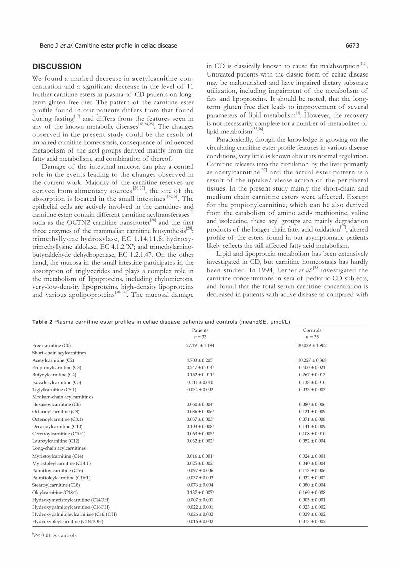
Bene J et al. Carnitine ester profi le in celiac disease 6673
DISCUSSIONWe found a marked decrease in acetylcarnitine con-centration and a significant decrease in the level of 11 further carnitine esters in plasma of CD patients on long-term gluten free diet. The pattern of the carnitine ester profile found in our patients differs from that found during fasting[17] and differs from the features seen in any of the known metabolic diseases[18,24,25]. The changes observed in the present study could be the result of impaired carnitine homeostasis, consequence of infl uenced metabolism of the acyl groups derived mainly from the fatty acid metabolism, and combination of thereof.
Damage of the intestinal mucosa can play a central role in the events leading to the changes observed in the current work. Majority of the carnitine reserves are derived from alimentary sources[26,27], the site of the absorption is located in the small intestines[14,15]. The epithelial cells are actively involved in the carnitine- and carnitine ester: contain different carnitine acyltransferases[8]
such as the OCTN2 carnitine transporter[28] and the fi rst three enzymes of the mammalian carnitine biosynthesis[29]: trimethyllysine hydroxylase, EC 1.14.11.8; hydroxy-trimethyllysine aldolase, EC 4.1.2.’X’; and trimethylamino- butyraldehyde dehydrogenase, EC 1.2.1.47. On the other hand, the mucosa in the small intestine participates in the absorption of triglycerides and plays a complex role in the metabolism of lipoproteins, including chylomicrons, very-low-density lipoproteins, high-density lipoproteins and various apolipoproteins[30-34]. The mucosal damage
in CD is classically known to cause fat malabsorption[1,2]. Untreated patients with the classic form of celiac disease may be malnourished and have impaired dietary substrate utilization, including impairment of the metabolism of fats and lipoproteins. It should be noted, that the long-term gluten free diet leads to improvement of several parameters of lipid metabolism[3]. However, the recovery is not necessarily complete for a number of metabolites of lipid metabolism[35,36].
Paradoxically, though the knowledge is growing on the circulating carnitine ester profi le features in various disease conditions, very little is known about its normal regulation. Carnitine releases into the circulation by the liver primarily as acetylcarnitine[37] and the actual ester pattern is a result of the uptake/release action of the peripheral tissues. In the present study mainly the short-chain and medium chain carnitine esters were affected. Except for the propionylcarnitine, which can be also derived from the catabolism of amino acids methionine, valine and isoleucine, these acyl groups are mainly degradation products of the longer chain fatty acid oxidation[17], altered profi le of the esters found in our asymptomatic patients likely refl ects the still affected fatty acid metabolism.
Lipid and lipoprotein metabolism has been extensively investigated in CD, but carnitine homeostasis has hardly been studied. In 1994, Lerner et al.[38] investigated the carnitine concentrations in sera of pediatric CD subjects, and found that the total serum carnitine concentration is decreased in patients with active disease as compared with
bP< 0.01 vs controls
Table 2 Plasma carnitine ester profiles in celiac disease patients and controls (mean±SE, μmol/L)Patients Controlsn = 33 n = 35
Free carnitine (C0) 27.191 ± 1.194 30.029 ± 1.902Short-chain acylcarnitines Acetylcarnitine (C2) 4.703 ± 0.205b 10.227 ± 0.368Propionylcarnitine (C3) 0.247 ± 0.014b 0.400 ± 0.021Butyrylcarnitine (C4) 0.152 ± 0.011b 0.267 ± 0.013Isovalerylcarnitine (C5) 0.111 ± 0.010 0.138 ± 0.010Tiglylcarnitine (C5:1) 0.034 ± 0.002 0.033 ± 0.003Medium-chain acylcarnitinesHexanoylcarnitine (C6) 0.060 ± 0.004b 0.080 ± 0.006Octanoylcarnitine (C8) 0.086 ± 0.006b 0.121 ± 0.009Octenoylcarnitine (C8:1) 0.037 ± 0.003b 0.071 ± 0.008Decanoylcarnitine (C10) 0.103 ± 0.008b 0.141 ± 0.009Cecenoylcarnitine (C10:1) 0.063 ± 0.005b 0.108 ± 0.010Lauroylcarnitine (C12) 0.032 ± 0.002b 0.052 ± 0.004Long-chain acylcarnitinesMyristoylcarnitine (C14) 0.016 ± 0.001b 0.024 ± 0.001Myristoleylcarnitine (C14:1) 0.025 ± 0.002b 0.040 ± 0.004Palmitoylcarnitine (C16) 0.097 ± 0.006 0.113 ± 0.006Palmitoleylcarnitine (C16:1) 0.037 ± 0.003 0.032 ± 0.002Stearoylcarnitine (C18) 0.076 ± 0.004 0.080 ± 0.004Oleylcarnitine (C18:1) 0.137 ± 0.007b 0.169 ± 0.008Hydroxymyristoylcarnitine (C14OH) 0.007 ± 0.001 0.005 ± 0.001Hydroxypalmitoylcarnitine (C16OH) 0.022 ± 0.001 0.023 ± 0.002Hydroxypalmitoleylcarnitine (C16:1OH) 0.026 ± 0.002 0.029 ± 0.002Hydroxyoleylcarnitine (C18:1OH) 0.016 ± 0.002 0.013 ± 0.002
6674 ISSN 1007-9327 CN 14-1219/ R World J Gastroenterol November 14, 2005 Volume 11 Number 42
normal subjects, while it was unchanged in CD patients with gluten withdrawal - associated non-active disease[38]. The decrease of carnitine reserves in active disease is likely secondary to the mucosal injury associated damage of the absorption. Albeit similar study on adult subjects is not presented in the literature, after this single pediatric paper the possible development of carnitine defi ciency in untreated CD has become widely accepted[39].
In our patients the decrease of total carnitine esters could also refl ect shortening of the reserves. It is known that even in patients strictly adhering to a gluten free diet the recovering mucosa can exhibit functional limitations. Therefore, carnitine absorption can be infl uenced on the one side. On the other side, the mucosa also participates in the trimethyllysine-butyrobetaine conversion, since the fi rst three enzymes of the carnitine biosynthesis are expressed in it[29]. Residual damage can infl uence this procedure. In addition, trymethyllysine hydroxylase requires Fe2+ ion as cofactor[11]. Our patients had improvement after iron replacement therapy, but their plasma iron, Hb and MCV remained decreased. This phenomenon is common in the disease[22]. The decreased tissue iron reserves can also theoretically act on the enzyme activity.
In the recent years there has been increas ing recognition that besides the classical major presentations of CD with a malabsorption syndrome and a fl at jejunal mucosa, a broad spectrum of metabolic alterations can associate primarily or secondarily with the disease[40,41]. Some of them can be theoretically an early hallmark and predisposing factor for a clinical symptom manifested at a later stage of CD. Inhibition of oxidative metabolism of fatty acids, leading to myopathy with hypotonia and hyporef lexia, hypoglycemia, cardiomyopathy, encephalopathy and disturbed liver function, which are also among the rare extraintestinal manifestation of CD, may be results of carnitine insufficiency[42]. Whether supplementation of carnitine has rationale in the treatment of the disease similar to other metabolic nutriments used routinely in clinical practice[43] remains to be elucidated.
ACKNOWLEDGMENTS The authors are grateful to Tamás Zágoni, Miklós Tóth and Ilona Szántó for their help in the management of the study.
REFERENCES1 Green PH, Jabri B. Coeliac disease. Lancet 2003; 362: 383-391 2 Shamir R. Advances in celiac disease. Gastroenterol Clin North
Am 2003; 32: 931-9473 Capristo E, Addolorato G, Mingrone G, De Gaetano A, Greco
AV, Tataranni PA, Gasbarrini G. Changes in body composi-tion, substrate oxidation, and resting metabolic rate in adult celiac disease patients after a 1-y gluten-free diet treatment. Am J Clin Nutr 2000; 72: 76-81
4 Capristo E, Addolorato G, Mingrone G, Scarfone A, Greco AV, Gasbarrini G. Low-serum high-density lipoprotein-cholesterol concentration as a sign of celiac disease. Am J Gastroenterol 2000; 95: 3331-3332
5 Vuoristo M, Kesaniemi YA, Gylling H, Miettinen TA. Me-tabolism of cholesterol and apolipoprotein B in celiac disease. Metabolism 1993; 42: 1386-1391
6 Ciampolini M, Bini S. Serum lipids in celiac children. J Pediatr Gastroenterol Nutr 1991; 12: 459-460
7 Rosenthal E, Hoffman R, Aviram M, Benderly A, Erde P, Brook JG. Serum lipoprotein profi le in children with celiac dis-ease. J Pediatr Gastroenterol Nutr 1990; 11: 58-62
8 Bieber LL. Carnitine. Annu Rev Biochem 1988; 57: 261-2839 Melegh B, Kerner J, Bieber LL. Pivampicillin-promoted excre-
tion of pivaloylcarnitine in humans. Biochem Pharmacol 1987; 36: 3405-3409
10 Kerner J, Hoppel C. Genetic disorders of carnitine metabolism and their nutritional management. Annu Rev Nutr 1998; 18: 179-206
11 Vaz FM, Wanders RJ. Carnitine biosynthesis in mammals. Bio-chem J 2002; 361: 417-429
12 Melegh B, Hermann R, Bock I. Generation of hydroxytrimeth-yllysine from trimethyllysine limits the carnitine biosynthesis in premature infants. Acta Paediatr 1996; 85: 345-350
13 Vaz FM, Melegh B, Bene J, Cuebas D, Gage DA, Bootsma A, Vreken P, van Gennip AH, Bieber LL, Wanders RJ. Analysis of carnitine biosynthesis metabolites in urine by HPLC-electro-spray tandem mass spectrometry. Clin Chem 2002; 48: 826-834
14 Hamilton JW, Li BU, Shug AL, Olsen WA. Carnitine transport in human intestinal biopsy specimens. Demonstration of an active transport system. Gastroenterology 1986; 91: 10-16
15 McCloud E, Ma TY, Grant KE, Mathis RK, Said HM. Uptake of L-carnitine by a human intestinal epithelial cell line, Caco-2. Gastroenterology 1996; 111: 1534-1540
16 Oberhuber G, Granditsch G, Vogelsang H. The histopathol-ogy of coeliac disease: time for a standardized report scheme for pathologists. Eur J Gastroenterol Hepatol 1999; 11: 1185-1194
17 Costa CC, de Almeida IT, Jakobs C, Poll-The BT, Duran M. Dynamic changes of plasma acylcarnitine levels induced by fasting and sunfl ower oil challenge test in children. Pediatr Res 1999; 46: 440-444
18 Vreken P, van Lint AE, Bootsma AH, Overmars H, Wanders RJ, van Gennip AH. Quantitative plasma acylcarnitine analy-sis using electrospray tandem mass spectrometry for the diag-nosis of organic acidaemias and fatty acid oxidation defects. J Inherit Metab Dis 1999; 22: 302-306
19 Stahlberg MR, Savilahti E, Siimes MA. Iron defi ciency in co-eliac disease is mild and it is detected and corrected by gluten-free diet. Acta Paediatr Scand 1991; 80: 190-193
20 Sategna Guidetti C, Scaglione N, Martini S. Red cell distribu-tion width as a marker of coeliac disease: a prospective study. Eur J Gastroenterol Hepatol 2002; 14: 177-181
21 Dickey W, Bodkin S. Prospective study of body mass index in patients with coeliac disease. BMJ 1998; 317: 1290
22 Mody RJ, Brown PI, Wechsler DS. Refractory iron defi ciency anemia as the primary clinical manifestation of celiac disease. J Pediatr Hematol Oncol 2003; 25: 169-172
23 Hjelt K, Krasilnikoff PA. The impact of gluten on haemato-logical status, dietary intakes of haemopoietic nutrients and vitamin B12 and folic acid absorption in children with coeliac disease. Acta Paediatr Scand 1990; 79: 911-919
24 Chace DH, Kalas TA, Naylor EW. Use of tandem mass spec-trometry for multianalyte screening of dried blood specimens from newborns. Clin Chem 2003; 49: 1797-1817
25 Schulze A, Lindner M, Kohlmuller D, Olgemoller K, Mayate-pek E, Hoffmann GF. Expanded newborn screening for inborn errors of metabolism by electrospray ionization-tandem mass spectrometry: results, outcome, and implications. Pediatrics 2003; 111: 1399-1406
26 Li B, Lloyd ML, Gudjonsson H, Shug AL, Olsen WA. The ef-fect of enteral carnitine administration in humans. Am J Clin Nutr 1992; 55: 838-845
27 Baker H, Frank O, DeAngelis B, Baker ER. Absorption and excretion of L-carnitine during single or multiple dosings in
Bene J et al. Carnitine ester profi le in celiac disease 6675
humans. Int J Vitam Nutr Res 1993; 63: 22-2628 Tamai I, Ohashi R, Nezu J, Yabuuchi H, Oku A, Shimane M,
Sai Y, Tsuji A. Molecular and functional identifi cation of sodi-um ion-dependent, high affi nity human carnitine transporter OCTN2. J Biol Chem 1998; 273: 20378-20382
29 Zaspel BJ, Sheridan KJ, Henderson LM. Transport and me-tabolism of carnitine precursors in various organs of the rat. Biochim Biophys Acta 1980; 631: 192-202
30 Green PH, Glickman RM. Intestinal lipoprotein metabolism. J Lipid Res 1981; 22: 1153-1173
31 Field FJ, Mathur SN. Intestinal lipoprotein synthesis and se-cretion. Prog Lipid Res 1995; 34: 185-198
32 Cartwright IJ, Higgins JA. Molecular and intracellular events in the assembly and secretion of chylomicrons by enterocytes. Biochem Soc Trans 1998; 26: 211-216
33 Raybould HE. Nutrient tasting and signaling mechanisms in the gut. I. Sensing of lipid by the intestinal mucosa. Am J Physiol 1999; 277: G751-G755
34 Tso P, Nauli A, Lo CM. Enterocyte fatty acid uptake and in-testinal fatty acid-binding protein. Biochem Soc Trans 2004; 32: 75-78
35 Mediene S, Hakem S, Bard JM, Medjaoui I, Benhamamouch S, Lebel P, Fruchart JC, Clavey V. Serum lipoprotein profi le in
Algerian patients with celiac disease. Clin Chim Acta 1995; 235: 189-196
36 Pillan MN, Spandrio S, Sleiman I, Meini A, Scalvini T, Bal-estrieri GP. Effects of a gluten-free diet on serum lipids and lipoprotein (a) levels in a group of patients with celiac disease. J Pediatr Gastroenterol Nutr 1994; 18: 183-185
37 Sandor A, Kispal G, Melegh B, Alkonyi I. Ester composition of carnitine in the perfusate of liver and in the plasma of donor rats. Eur J Biochem 1987; 170: 443-445
38 Lerner A, Gruener N, Iancu TC. Serum carnitine concentra-tions in coeliac disease. Gut 1993; 34: 933-935
39 Fitzgerald JF, Troncone R, Roggero P, Pozzi E, Garavaglia B, Parini R, Carissimi E, Santus F, Piemontese P, Cataliotti E, Mosca F, Carnelli V. Clinical quiz. Secondary carnitine defi -ciency due to celiac disease. J Pediatr Gastroenterol Nutr 2003; 36: 636, 646
40 Hardoff D, Sharf B, Berger A. Myopathy as a presentation of coeliac disease. Dev Med Child Neurol 1980; 22: 781-783
41 Rossi T. Celiac disease. Adolesc Med Clin 2004; 15: 91-10342 Hoppel C. The role of carnitine in normal and altered fatty
acid metabolism. Am J Kidney Dis 2003; 41: S4-1243 Abdulkarim AS, Murray JA. Review article: The diagnosis of
coeliac disease. Aliment Pharmacol Ther 2003; 17: 987-995
Science Editor Wang XL and Guo SY Language Editor Elsevier HK

PO Box 2345, Beijing 100023, China World J Gastroenterol 2005;11(42):6676-6680www.wjgnet.com World Journal of Gastroenterology ISSN [email protected] © 2005 The WJG Press and Elsevier Inc. All rights reserved.E L S E V I E R
• BRIEF REPORTS •
http://www.wjgnet.com/1007-9327/11/.asp
INTRODUCTIONThe stomach performs several important functions. It subserves the function of accommodation and thereby acts as a storage organ. It also functions as a grinder that triturates food into smaller particles and as a pump that transports chyme into the small bowel in a controlled fashion[1]. These functions depend on a complex mix of neurohumoral mechanisms, visceral sensation, intrinsic reflexes, intragastric transport, nutrient composition, particulate size, and the coordinated motor activity of the gastroduodenal unit[2,3].
In ferrets, distension of the corpus produces phasic activity in the antrum, a response termed as excitatory corporo-antral reflex. This is probably mediated by cholinergic mechanisms and intramural gastric pathways[4,5]. Intramural excitatory and inhibitory reflexes have also been demonstrated in isolated gastric preparations[6–9]. Recent studies have suggested that balloon distention of the stomach may increase phasic activity in the antrum and duodenum[10,11]. Whether this phasic activity represents an intrinsic gastro-gastric ref lex has not been well characterized.
Furthermore, previous studies have assessed some of the individual components of gastric function[12,13]; whereas an integrated assessment of the biomechanical and sensory properties of the stomach has been scarcely performed. Also, there is very little information regarding the integrated role of the stomach as a sensory, motor and reflex organ. Our hypothesis is that fundic balloon distention may induce reflex antral pressure activity and this response may be mediated by cholinergic mechanisms.
Our objectives were to examine the antral motor responses during step-wise balloon distentions of the fundus, to simultaneously assess the sensory and tone responses of the stomach and to examine if the sensory and motor effects were mediated by cholinergic mechanisms.
METHODSStudy populationEight healthy volunteers (m/f = 4/4) were recruited for this study. Their mean age was 32±4.95 years. None of them had a history of gastrointestinal or systemic ailments and none was using any medications. All had a normal physical examination. All participants gave written
Investigation of fundo-antral refl ex in human beings
Satish SC Rao, Anjana Kumar, Brent Harris, Bruce Brown, Konrad S Schulze
Satish SC Rao, Anjana Kumar, Brent Harris, Bruce Brown, Konrad S Schulze, Department of Internal Medicine, University of Iowa Carver College of Medicine, Iowa City, Iowa, United StatesSupported in part by an American College of Gastroenterology Clinical Research Grant, RR00059 and by General Clinical Research Centers Program, R01DK57100-03, National Institutes of HealthCorrespondence to: Satish SC Rao, MD, PhD, FRCP, Department of Internal Medicine, University of Iowa Hospitals and Clinics, 200 Hawkins Drive, 4612 JCP, Iowa City 52242, Iowa, United States. [email protected] Telephone: +1-319-353-6602 Fax: +1-319-353-6399Received: 2005-03-12 Accepted: 2005-04-09
AbstractAIM: To examine the sensory and motor response(s) of the stomach following fundic distention and to assess whether cholinergic mechanisms infl uence these responses.
METHODS: Fundic tone, gastric sensory responses and antral motility were evaluated in eight healthy volunteers after a probe with two sensors was placed in the antrum and a highly compliant balloon in the fundus. Isobaric balloon distentions were performed with a barostat. Study was repeated in six volunteers after intravenous atropine was given.
RESULTS: Fundic distention induced large amplitude antral contractions in all subjects. The area under the curve was higher (P <0.05) during fundic distention. F i r s t sensa t i on was repor ted a t 12±4 mmHg, moderate sensation at 18±4 mmHg and discomfort at 21±4 mmHg. Discomfort was associated with a decrease in antral motility. After atropine was given, the area under the curve of pressure waves and fundic tone decreased (P <0.05). Sensory thresholds were not affected.
CONCLUSIONS: Fundic balloon distention induces an antral motor response, the fundo-antral refl ex, which in part may be mediated by cholinergic mechanisms.
© 2005 The WJG Press and Elsevier Inc. All rights reserved.
Key words: Gastric motility; Refl ex; Fundo-antral refl ex; Sensation
Rao S SC, Kumar A, Harris B, Brown B, Schulze KS. Inves-tigation of fundo-antral reflex in human beings. World J Gastroenterol 2005; 11(42): 6676-6680
Rao S SC et al. Fundo-antral refl ex in human beings 6677
informed consent and the study protocol was approved by the Human Investigation Review Board of the University of Iowa College of Medicine.
Manometry and barostat assembly A double lumen plastic-probe (6 mm in diameter) containing a 10 cm long, highly-compliant balloon was used (MUI Scientifi c; Toronto, ON, Canada). The capacity of the balloon was 600 mL. The balloon was connected to a barostat (GMB Distender II; G&J Electronics Inc., Toronto, Canada). The probe had two perfusion side holes, 5 and 8 cm from the distal end of the balloon. These holes were perfused with gas-free distilled water at a rate of 0.2 mL/min (15 psi) using a low-compliance pneumohydraulic perfusion system (Arndorfer Medical Specialties, Inc., Milwaukee, WI, USA) that connected to transducers (Medex Inc.; MX860-G8618, Hilliard, OH, USA). Intraluminal pressures were relayed to an analog data recorder/amplifi er (Medtronics Polygraph, Medtronics Functional Diagnostics; MN, USA) and displayed on a computer monitor using a software program (Polygram for Windows; Synectics Medical AB). The balloon volume and pressure data from the barostat were fed to the polygraph via an interface (Golden Gate; G&J Electronics Inc.).
Thus, the computer display consisted of the intra-gastric balloon volume, intra-balloon pressure as well as the intraluminal pressure changes in the antrum. The manometric data and the ultrasound images were simultaneously fed into a digital splitter (American Dynamics Ao1479, Orangeburg, NY, USA).
The ultrasonographic image of the cross-sectional diameter of the gastric antrum allowed visualization of antral contractions and assurance that the probe was properly positioned (Acuson 128XP with a 3 mHz sector transducer). The digital splitter synchronized the two images and displayed these images on a monitor screen (VM-17; Javelin, Los Angeles, CA, USA) such that one half of the screen showed the combined manometry and barostat recording and the other half displayed the ultrasound image. These images were recorded on a VHS tape for future analysis.
Study protocolAfter an overnight fast, the oropharynx was sprayed with a local anesthetic, pontocaine (Abbott Laboratories, North Chicago, IL, USA). Then the probe with the balloon was placed through the mouth into the stomach. The volunteers were asked to sit in a semi-recumbent position, such that the head end was elevated by 45°. The balloon was distended with 250 mL of air and the probe was slowly retracted until a “tug” was felt signaling that the proximal end of the balloon was located in the fundus. Subsequently, ultrasound images were obtained to check the probe location. The location of pressure sensors in the antrum was also confi rmed by the occurrence of typical antral motor pattern consisting of 3-cycle/min activity. The balloon was defl ated and the probe was anchored to the cheek with a tape.
After a rest period of 15 min, the balloon was distended by 1 mmHg increments to assess the minimum distending pressure, a pressure at which diaphragmatic oscillations are clearly visible[14]. The intraoperating pressure (IOP) was set at a value of 2 mmHg above the minimum distending pressure using previous criteria [9-11]. Subsequently, a baseline recording of intragastric tone was performed for a period of 20 min. Then isobaric balloon distentions were performed at 3 mmHg increments. Each distention was maintained for 8 min followed by a rest period of 8 min. Thirty seconds after each distention, the subject was asked to rate their sensation on a scale of 0-6 as published previously[14], 0 = no sensation, 1 = vague perception of mild sensation, 2 = definite perception of mild sensation, 3 = vague perception of moderate sensation, 4 = defi nite perception of moderate sensation, 5 = discomfort, and 6 = pain. If the subject reported discomfort at two incremental distentions or pain at any one distention, the balloon distentions were discontinued. Abdominal ultrasonography was performed intermittently to visualize the antral confi guration and morphology. Blood pressure and heart rate were monitored throughout the study.
We administered intravenously 0.6 mg of atropine sulfate in six volunteers (4 m/2 f) after a rest period of 60 min. Five minutes after administration of atropine, the balloon distentions were repeated as described above. Ultrasound images were obtained once again to confi rm the location of antral sensors.
Manometric responsesManometric recordings from the two antral pressure sensors were analyzed visually and manually with the assistance of Polygram for Windows software (Synectics Medical AB). Pressure waves that were ≥8 mmHg and ≥3 s in duration were included in the analysis. Artifacts were identified and excluded. There was good quality pressure activity at both channels in approximately 50% of the recordings and therefore an average of the pressure activity at the two antral channels was used. In the rest of the recordings, the pressure activity was more prominent in one of the antral channels and this channel was used for data analysis. The maximum amplitude, duration and area under the curve of each wave were calculated. We also measured the time interval between balloon distention and the onset of the fi rst antral pressure wave as well as the total number of propagating pressure waves in the antrum during each inflation and deflation periods. Propagating waves were defined as pressure waves, which migrated across the antral leads within 6 s of each other and were categorized as either antegrade or retrograde depending on which of the two leads they fi rst appeared in. A similar analysis of the antral pressure waves was performed after atropine injection.
Barostat responsesGastric tone was assessed by measuring the area under the curve of the gastric volume during isobaric balloon
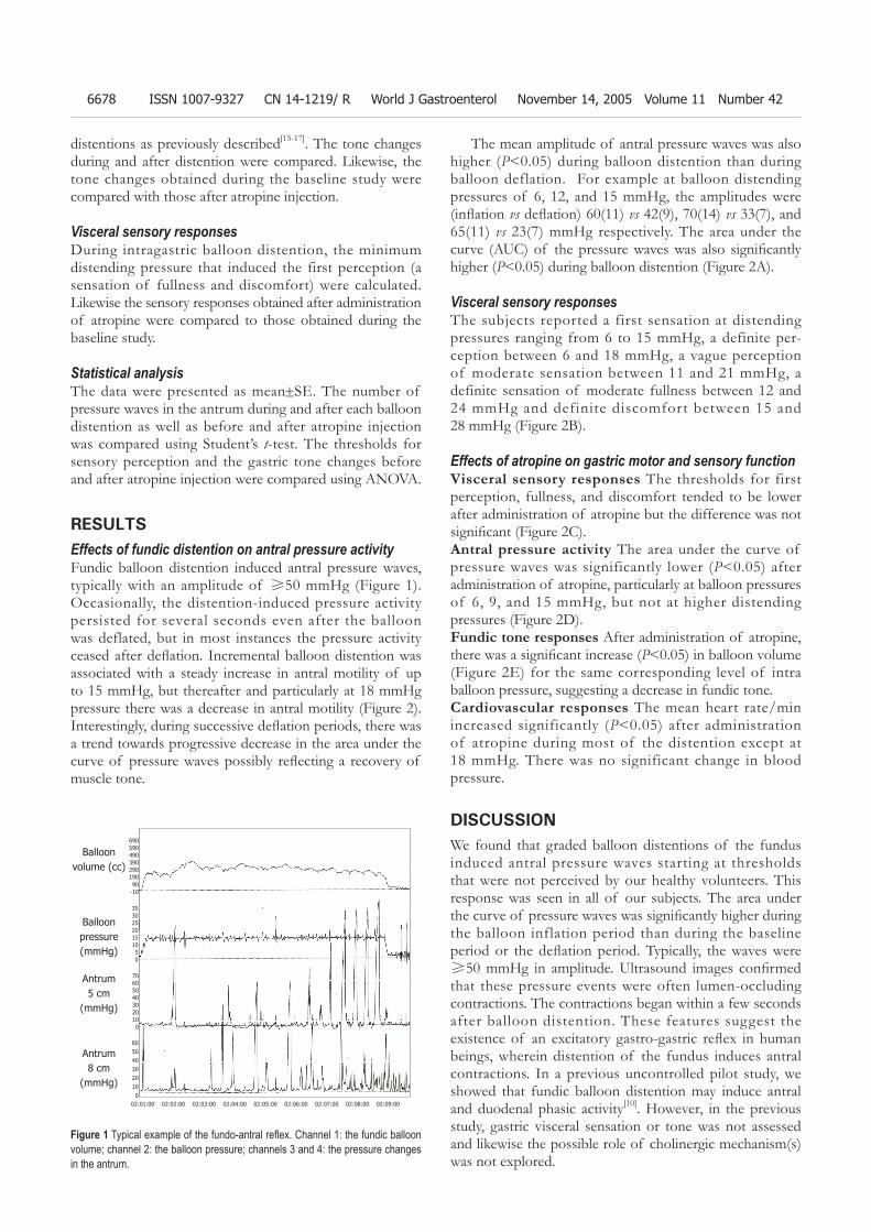
6678 ISSN 1007-9327 CN 14-1219/ R World J Gastroenterol November 14, 2005 Volume 11 Number 42
distentions as previously described[15-17]. The tone changes during and after distention were compared. Likewise, the tone changes obtained during the baseline study were compared with those after atropine injection.
Visceral sensory responsesDuring intragastric balloon distention, the minimum distending pressure that induced the first perception (a sensation of fullness and discomfort) were calculated. Likewise the sensory responses obtained after administration of atropine were compared to those obtained during the baseline study.
Statistical analysisThe data were presented as mean±SE. The number of pressure waves in the antrum during and after each balloon distention as well as before and after atropine injection was compared using Student’s t-test. The thresholds for sensory perception and the gastric tone changes before and after atropine injection were compared using ANOVA.
RESULTSEffects of fundic distention on antral pressure activityFundic balloon distention induced antral pressure waves, typically with an amplitude of ≥50 mmHg (Figure 1). Occasionally, the distention-induced pressure activity persisted for several seconds even after the balloon was deflated, but in most instances the pressure activity ceased after defl ation. Incremental balloon distention was associated with a steady increase in antral motility of up to 15 mmHg, but thereafter and particularly at 18 mmHg pressure there was a decrease in antral motility (Figure 2). Interestingly, during successive defl ation periods, there was a trend towards progressive decrease in the area under the curve of pressure waves possibly refl ecting a recovery of muscle tone.
The mean amplitude of antral pressure waves was also higher (P<0.05) during balloon distention than during balloon deflation. For example at balloon distending pressures of 6, 12, and 15 mmHg, the amplitudes were (infl ation vs defl ation) 60(11) vs 42(9), 70(14) vs 33(7), and 65(11) vs 23(7) mmHg respectively. The area under the curve (AUC) of the pressure waves was also signifi cantly higher (P<0.05) during balloon distention (Figure 2A).
Visceral sensory responsesThe subjects reported a first sensation at distending pressures ranging from 6 to 15 mmHg, a definite per-ception between 6 and 18 mmHg, a vague perception of moderate sensation between 11 and 21 mmHg, a definite sensation of moderate fullness between 12 and 24 mmHg and definite discomfort between 15 and 28 mmHg (Figure 2B).
Effects of atropine on gastric motor and sensory functionVisceral sensory responses The thresholds for first perception, fullness, and discomfort tended to be lower after administration of atropine but the difference was not signifi cant (Figure 2C).Antral pressure activity The area under the curve of pressure waves was significantly lower (P<0.05) after administration of atropine, particularly at balloon pressures of 6, 9, and 15 mmHg, but not at higher distending pressures (Figure 2D).Fundic tone responses After administration of atropine, there was a signifi cant increase (P<0.05) in balloon volume (Figure 2E) for the same corresponding level of intra balloon pressure, suggesting a decrease in fundic tone.Cardiovascular responses The mean heart rate/min increased significantly (P<0.05) after administration of atropine during most of the distention except at 18 mmHg. There was no significant change in blood pressure.
DISCUSSIONWe found that graded balloon distentions of the fundus induced antral pressure waves starting at thresholds that were not perceived by our healthy volunteers. This response was seen in all of our subjects. The area under the curve of pressure waves was signifi cantly higher during the balloon inflation period than during the baseline period or the defl ation period. Typically, the waves were ≥50 mmHg in amplitude. Ultrasound images confi rmed that these pressure events were often lumen-occluding contractions. The contractions began within a few seconds after balloon distention. These features suggest the existence of an excitatory gastro-gastric refl ex in human beings, wherein distention of the fundus induces antral contractions. In a previous uncontrolled pilot study, we showed that fundic balloon distention may induce antral and duodenal phasic activity[10]. However, in the previous study, gastric visceral sensation or tone was not assessed and likewise the possible role of cholinergic mechanism(s) was not explored.
Figure 1 Typical example of the fundo-antral refl ex. Channel 1: the fundic balloon volume; channel 2: the balloon pressure; channels 3 and 4: the pressure changes in the antrum.
Balloon volume (cc)
Balloon pressure (mmHg)
Antrum 5 cm
(mmHg)
Antrum 8 cm
(mmHg)
69059049039029019090
-10
35302520151050
706050403020100
6050403020100
02:01:00 02:02:00 02:03:00 02:04:00 02:05:00 02:06:00 02:07:00 02:08:00 02:09:00
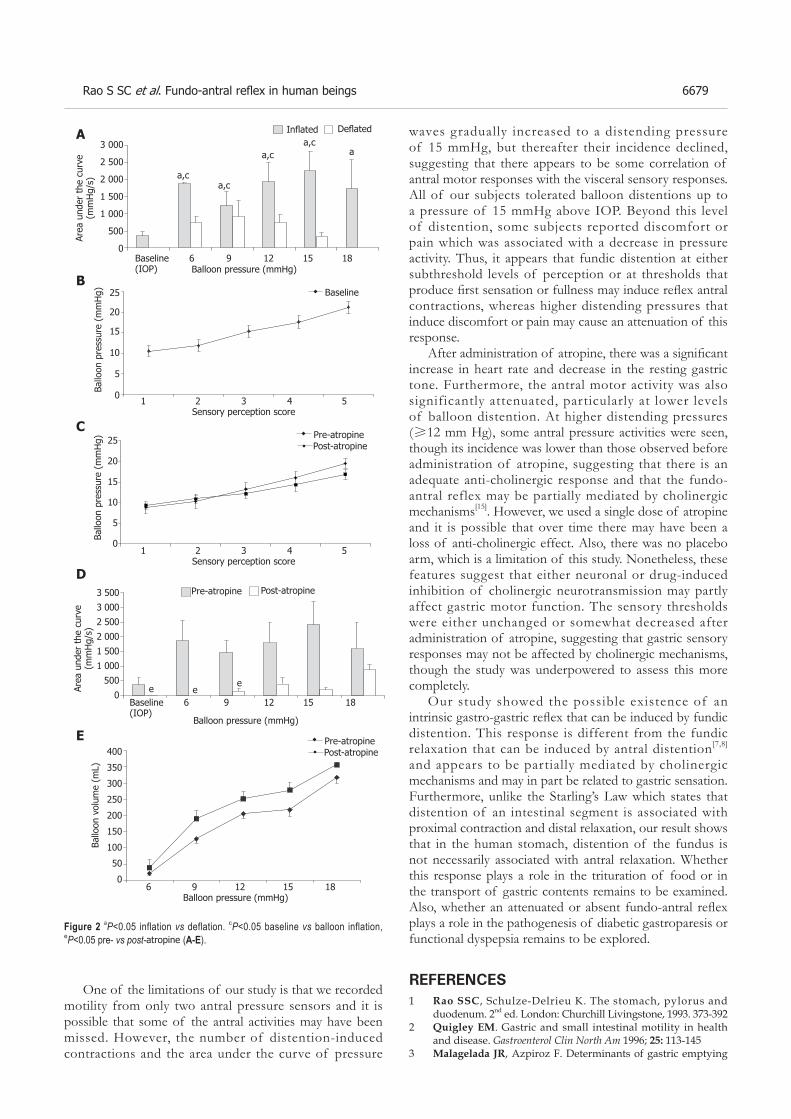
Rao S SC et al. Fundo-antral refl ex in human beings 6679
One of the limitations of our study is that we recorded motility from only two antral pressure sensors and it is possible that some of the antral activities may have been missed. However, the number of distention-induced contractions and the area under the curve of pressure
waves gradually increased to a distending pressure of 15 mmHg, but thereafter their incidence declined, suggesting that there appears to be some correlation of antral motor responses with the visceral sensory responses. All of our subjects tolerated balloon distentions up to a pressure of 15 mmHg above IOP. Beyond this level of distention, some subjects reported discomfort or pain which was associated with a decrease in pressure activity. Thus, it appears that fundic distention at either subthreshold levels of perception or at thresholds that produce fi rst sensation or fullness may induce refl ex antral contractions, whereas higher distending pressures that induce discomfort or pain may cause an attenuation of this response.
After administration of atropine, there was a signifi cant increase in heart rate and decrease in the resting gastric tone. Furthermore, the antral motor activity was also significantly attenuated, particularly at lower levels of balloon distention. At higher distending pressures (≥12 mm Hg), some antral pressure activities were seen, though its incidence was lower than those observed before administration of atropine, suggesting that there is an adequate anti-cholinergic response and that the fundo-antral reflex may be partially mediated by cholinergic mechanisms[15]. However, we used a single dose of atropine and it is possible that over time there may have been a loss of anti-cholinergic effect. Also, there was no placebo arm, which is a limitation of this study. Nonetheless, these features suggest that either neuronal or drug-induced inhibition of cholinergic neurotransmission may partly affect gastric motor function. The sensory thresholds were either unchanged or somewhat decreased after administration of atropine, suggesting that gastric sensory responses may not be affected by cholinergic mechanisms, though the study was underpowered to assess this more completely.
Our study showed the possible existence of an intrinsic gastro-gastric refl ex that can be induced by fundic distention. This response is different from the fundic relaxation that can be induced by antral distention[7,8] and appears to be partially mediated by cholinergic mechanisms and may in part be related to gastric sensation. Furthermore, unlike the Starling’s Law which states that distention of an intestinal segment is associated with proximal contraction and distal relaxation, our result shows that in the human stomach, distention of the fundus is not necessarily associated with antral relaxation. Whether this response plays a role in the trituration of food or in the transport of gastric contents remains to be examined. Also, whether an attenuated or absent fundo-antral refl ex plays a role in the pathogenesis of diabetic gastroparesis or functional dyspepsia remains to be explored.
REFERENCES1 Rao SSC, Schulze-Delrieu K. The stomach, pylorus and
duodenum. 2nd ed. London: Churchill Livingstone, 1993. 373-3922 Quigley EM. Gastric and small intestinal motility in health
and disease. Gastroenterol Clin North Am 1996; 25: 113-1453 Malagelada JR, Azpiroz F. Determinants of gastric emptying
3 000
2 500
2 000
1 500
1 000
500
0Baseline 6 9 12 15 18(IOP) Balloon pressure (mmHg)
Area
und
er t
he c
urve
(m
mH
g/s)
Infl ated Defl ated
Figure 2 aP<0.05 inflation vs deflation. cP<0.05 baseline vs balloon inflation, eP<0.05 pre- vs post-atropine (A-E).
a,ca,c
a,ca,c
a
25
20
15
10
5
01 2 3 4 5
Sensory perception score
Ballo
on p
ress
ure
(mm
Hg)
25
20
15
10
5
0
Ballo
on p
ress
ure
(mm
Hg)
3 500
3 000
2 500
2 000
1 500
1 000
500
0
Area
und
er t
he c
urve
(m
mH
g/s)
Baseline 6 9 12 15 18(IOP)
Balloon pressure (mmHg)
e
Pre-atropine Post-atropine
400
350
300
250
200
150
100
50
0
Ballo
on v
olum
e (m
L)
6 9 12 15 18Balloon pressure (mmHg)
1 2 3 4 5Sensory perception score
A
B
C
D
E
Baseline
Pre-atropinePost-atropine
Pre-atropinePost-atropine
ee

6680 ISSN 1007-9327 CN 14-1219/ R World J Gastroenterol November 14, 2005 Volume 11 Number 42
and transit in the small intestine. Bethesda (Maryland): American Physiological Society, 1989: 909-937
4 Andrews PL, Grundy D, Scratcherd T. Reflex excitation of antral motility induced by gastric distension in the ferret. J Physiol 1980; 298: 79-84
5 Grundy D, Hutson D, Scratcherd T. A permissive role for the vagus nerves in the genesis of antro-antral refl exes in the anaesthetized ferret. J Physiol 1986; 381: 377-384
6 Hennig GW, Brookes SJ, Costa M. Excitatory and inhibitory motor reflexes in the isolated guinea-pig stomach. J Physiol 1997; 501: 197-212
7 Stadaas J, Aune S, Haffner JF. Effects of proximal gastric vagotomy on intragastric pressure and adaptation in pigs. Scand J Gastroenterol 1974; 9; 479-485
8 Haffner JF, Stadaas J. Pressure responses to cholinergic and adrenergic agents in the fundus, corpus, and antrum of isolated rabbit stomachs. Acta Chir Scand 1972; 138: 713-719
9 Leclere PG, Lefebvre RA. Investigation of the interaction between cholinergic and nitrergic neurotransmission in the pig gastric fundus. Br J Pharmacol 1998; 125: 1779-1787
10 Rao SS, Vemuri S, Harris B, Schulze K. Fundic balloon distension stimulates antral and duodenal motility in man. Dig
Dis Sci 2002; 47: 1015-1019 11 Piessevaux H, Tack J, Geubel A, Janssens J. Infl uence of fundic
distension on fasting antro-duodenal manometric patterns in man. Gastroenterology 1998; 114: G3374
12 Tack J, Piessevaux H, Coulie B, Caenepeel P, Janssens J. Role of impaired gastric accommodation to a meal in functional dyspepsia. Gastroenterology 1998; 115: 1346-1352
13 Camilleri M, Malagelada JR. Abnormal intestinal motility in diabetics with the gastroparesis syndrome. Eur J Clin Invest 1984; 14: 420-427
14 Azpiroz F, Malagelada JR. Perception and reflex relaxation of the stomach in response to gut distention. Gastroenterology 1990; 98: 1193-1198
15 Azpiroz F, Malagelada JR. Gastric tone measured by an electronic barostat in health and postsurgical gastroparesis. Gastroenterology 1987; 92: 934-943
16 Sarnelli G, Vos R, Cuomo R, Janssens J, Tack J. Reproducibility of gastric barostat studies in healthy controls and in dyspeptic patients. Am J Gastroenterol 2001; 96: 1047-1053
17 Lidums I, Hebbard GS, Holloway RH. Effect of atropine on proximal gastric motor and sensory function in normal subjects. Gut 2000; 47: 30-36
Science Editor Wang XL and Guo SY Language Editor Elsevier HK

PO Box 2345, Beijing 100023, China World J Gastroenterol 2005;11(42):6681-6683www.wjgnet.com World Journal of Gastroenterology ISSN [email protected] © 2005 The WJG Press and Elsevier Inc. All rights reserved.E L S E V I E R
• BRIEF REPORTS •
INTRODUCTIONThere are a variety of methods available for the detection of Helicobacter pylori (H pylori), but many of these are invasive (such as biopsies for rapid urease test, culture, histology, and polymerase chain reaction) or require laboratories (such as urea breath test and serology). Recently, H pylori rapid test kits have become available. Rapid blood tests detect H pylori antibodies in whole or capillary blood, are easy to use, and yield results in a few minutes, making it a convenient point-of-care test for screening H pylori.
The 1997 Asia Pacific Consensus Conference on the management of H pylori infection[1] recommended that any blood test must be locally validated, with two or more alternative means of testing, before its widespread application.
Rapid blood test kits have not been widely validated in the Asian populations[2-5]. Local validation is important because the performance characteristics of blood test kits and population prevalence of H pylori vary in different populations. In Asian countries, the prevalence of H pylori infection is generally higher than in the developed Western nations (such as the United Kingdom, Australia, and France)[6]. In addition, the test performance of rapid blood kits may vary because local H pylori strains may be different[7,8].
Our prospective study aimed to validate four rapid blood test kits in the diagnosis of H pylori infection in a multi-ethnic Asian population. Amongst the Asian studies till date[2-5], none was conducted in a multi-ethnic population, and all were tested with less than four rapid blood test kits.
MATERIALS AND METHODSConsecutive patients who were referred from general practice or outpatient clinics and scheduled for endoscopy for initial evaluation of dyspepsia at the National University Hospital, Singapore, were included for the
Lee-Guan Lim, Khay-Guan Yeoh, Seng-Gee Lim, Department of Gastroenterology and Hepatology, National University Hospital, 5 Lower Kent Ridge Road, Singapore 119074, Republic of SingaporeBow Ho, Department of Microbiology, National University of Singapore, Lower Kent Ridge Road, Singapore 119260, Republic of SingaporeCorrespondence to: Associate Professor Khay-Guan Yeoh, Department of Gastroenterology and Hepatology, National University Hospital, 5 Lower Kent Ridge Road, Singapore 119074, Republic of Singapore. [email protected]: +65-67724353 Fax: +65-67794112Received: 2005-01-19 Accepted: 2005-04-26
AbstractAIM: To validate the accuracy of four rapid blood tests in the diagnosis of Helicobacter pylori .
METHODS: Consecutive dyspeptic patients scheduled for endoscopy at the National University Hospital, Singapore, were interviewed and had blood drawn for serology. The fi rst 109 patients were tested with BM-test (BM), Pyloriset Screen (PS) and QuickVue (QV), and the next 99 subjects were tested with PS and Unigold (UG). Endoscopies were performed blinded to rapid blood test results and biopsies were taken for culture and rapid urease test. Urea breath tests were performed after endoscopies. The rapid blood test results were compared with four reference tests (rapid urease test, culture, serology, and breath test).
RESULTS: The study population composed of 208 patients (mean age 43.1 years; range 18-73 years; 119 males; 174 Chinese). The number of evaluable patients for BM, QV, UG and PS were 102, 102, 95, and 197, respectively. The sensitivity and specifi city, respectively were: PS 80.2%, 95.8%; UG 55.9%, 100%; QV 43.3%, 100%; BM 67.2%, 97.1%.
CONCLUSION: The rapid blood test kits showed high specifi city and positive predictive value (97-100%), while sensitivity and negative predictive value ranged widely (43%-80% and 47%-73%, respectively). Among test kits, PS showed the best sensitivity (80%), best negative predictive value (73%) and best negative likelihood ratio (0.207). PS had a specifi city of 96%, positive predictive value of 97% and positive likelihood ratio of 19.1.
© 2005 The WJG Press and Elsevier Inc. All rights reserved.
Validation of four Helicobacter pylori rapid blood tests in a multi-ethnic Asian population
Lee-Guan Lim, Khay-Guan Yeoh, Bow Ho, Seng-Gee Lim
Key words: Helicobacter pylori ; Rapid blood test
Lim LG, Yeoh KG, Ho B, Lim SG. Val idat ion of four Helicobacter pylori rapid blood tests in a multi-ethnic Asian population. World J Gastroenterol 2005; 11(42): 6681-6683 http://www.wjgnet.com/1007-9327/11/6681.asp

6682 ISSN 1007-9327 CN 14-1219/ R World J Gastroenterol November 14, 2005 Volume 11 Number 42
study. Exclusion criteria included patients with known peptic ulcer or gastric cancer, subjects with prior H pylori treatment, and those who had taken antibiotics, bismuth or proton pump inhibitors in the previous one month.
At entry, patients were interviewed using a standard questionnaire. Ten cubic centimeter of blood was drawn from each patient for serology. Each of the first 109 patients were tested with BM-test (BM, Boehringer Mannheim, East Essex, UK), QuickVue (QV, Quidel, CA, USA) and Pyloriset Screen (PS, Orion Diagnostica, Espoo, Finland). The kit with the best sensitivity was retained for continued testing in the next 99 patients together with an additional kit, Unigold (UG, Trinity Biotech, NY, USA). Endoscopy was then performed in the routine fashion by experienced endoscopists blinded to earlier results and three antral biopsy specimens were taken from each patient. Two biopsy specimens were sent for culture and one specimen was sent for the rapid urease test.
A 13C urea breath test was performed directly after endoscopy. The technician doing the urea breath test was blinded to the results of the endoscopy. The results from the rapid blood test, rapid urease test, serology, culture, urea breath test and endoscopy were recorded on a standard data form.
The results of the rapid blood tests were compared with four reference tests: serology using HEL-p Test kit (AMRAD Operations Pty. Ltd, Australia), which had been validated locally[9], culture, rapid urease test, and urea breath test. H pylori infection was diagnosed, if any two reference tests were positive. If all the four reference tests were negative, it was assumed that infection was absent. Patients with a single positive test out of the four reference tests were classifi ed as having indeterminate results.
Sample size was estimated based on reference tables[10]. Based on sensitivity of 80% and specificity of 90%, absolute precision of 0.10 and confi dence interval of 95%, we needed a minimum of 62 H pylori-positive and 35 H pylori-negative patients.
The study was approved by the Research and Ethics Committee, National University Hospital, Singapore.
RESULTSThe characteristics of recruited patients are described in Table 1. One hundred and nine patients were tested with BM, QV, and PS (102 evaluable, 7 indeterminate results), and the next 99 subjects with PS and UG (95 evaluable, 4 indeterminate results).
Table 2 shows the sensitivity, specificity, predictive values and likelihood ratios of the respective rapid blood tests for H pylori. The rapid blood kits tested all showed specifi cities above 95% and very good positive predictive values exceeding 97%. There was a wide range in sensitivity between 43% and 80%, negative predictive value ranged from 48% to 73%, and negative likelihood ratios ranged from 0.207 to 0.567. PS had the best sensitivity of 80%, the best negative predictive value of 73%, and the best negative likelihood ratio of 0.207. PS had a high specifi city of 96%, a good positive predictive value of 97.1% and a high positive likelihood ratio of 19.1.
DISCUSSIONAmong the kits tested in our study, PS showed the best sensitivity (84%). Our study showed a wide range in the performance characteristics of the rapid tests. This may be attributable to the antigens used[11] or test kit designs.
The same rapid blood test kit might vary in per-formance between different populations. For example, QV’s sensitivity for H pylori was 43.3% in our Singapore population, compared with 81% in Europe[12] and 82% in America[13]. These factors make it important that kits are locally tested and validated before use. A meta-analysis had shown that rapid tests are less accurate than reference tests, with sensitivity and specifi city averaging 80-85% and 75-80%, respectively[14].
We conducted this study in an institution. For better evaluation of the potential of rapid blood test as a screening method in primary care, local studies conducted in general practice would be needed. Talley et al. reported that when used in general practice in Australia, rapid blood test had a sensitivity of 60% and specificity of 90%[15] Data on the performance characteristics of H pylori rapid blood test kits in general practice in the Asian population is lacking.
The Maastricht 2-2000 Consensus report[16] reco-mmended a ‘test and treat’ approach in the primary care for H pylori infection. However, there is a strong association between H pylori infection and gastric cancer, especially in the Asian population, which has a high incidence of gastric cancer. Therefore, the use of ‘test and treat’ approach in Asians remains controversial and awaits further study. PS had a good sensitivity and specificity for the detection of H pylori infection, with the positive likelihood ratio of above 10, providing convincing diagnostic evidence, and negative likelihood ratio of 0.2, giving a strong diagnostic evidence. PS might therefore be potentially useful for ‘test
Performance characteristics BM QV UG PS
Sensitivity (%) 67.2 43.3 55.9 80.2Specifi city (%) 97.1 99 99 95.8Positive predictive value (%) 97.8 98 99 97.1Negative predictive value (%) 60.7 47.9 58.1 73.1Positive likelihood ratio 23.2 31.2 41.3 19.1Negative likelihood ratio 0.338 0.567 0.441 0.207
Table 2 Sensitivity, specificity, predictive values, and likelihood ratios of rapid blood test for H pylori infection
Kits used PS, QV, BM PS, UG All
Number 109 99 208Male:female 56:53 63:36 119:89C:I:M:O1 91:12:4:2 83:7:6:3 174:19:10:5Mean age (range) 44.7 (18–73) 41.3 (20–68) 43.1 (18–73)H pylori positive 67 59 126H pylori negative 35 36 71Indeterminate results 7 4 11
Table 1 Characteristics of recruited patients
1C = Chinese; I = Indian; M = Malay; O = Others.
Lim LG et al. Validation of H pylori rapid blood tests 6683
and referral’ strategy in general practice. Our study, which validated the point-of-care rapid blood test kits in a multi-ethnic Asian population, is an important step for future studies in this area.
In conclusion, there was a wide range in the perfor-mance characteristics of rapid blood test, making it important for the kits to be tested and validated locally before being used. Of the rapid blood kits tested, the best sensitivity for H pylori detection was 80% (PS). The validation of rapid blood test kits in the local population facilitates future studies on the ‘test and treat’ or ‘test and referral’ approach in the Asian population.
ACKNOWLEDGMENTSWe would l ike to thank Mrs Lim Thiew Peng who performed the H pylori serology and our research nurse Ms Lai Shuet Ting, for their kind assistance.
REFERENCES1 Lam SK, Talley NJ. Report of the 1997 Asia Pacifi c Consensus
Conference on the management of Helicobacter pylori infection. J Gastroenterol Hepatol 1998; 13: 1-12
2 Leung WK, Chan FK, Falk MS, Suen R, Sung JJ. Comparison of two rapid whole-blood tests for Helicobacter pylori infection in Chinese patients. J Clin Microbiol 1998; 36: 3441-3442
3 Wong BC, Wong W, Tang VS, Lai K, Yuen S, Hu WH, Chan C, Lau GK, Lai C, Lam S. An evaluation of whole blood testing for Helicobacter pylori infection in the Chinese population. Aliment Pharmacol Ther 2000; 14: 331-335
4 Chen TS, Chang FY, Lee SD. No difference of accuracy between capillary and venous blood in rapid whole blood test for dia-gnosis of Helicobacter pylori infection. Dig Dis Sci 2002; 47: 2519-2522
5 Wong WM, Lam SK, Xia HH, Tang VS, Lai KC, Hu WH, Chan CK, Cheung KL, Wong BC. Accuracy of a new near patient test for the diagnosis of Helicobacter pylori infection in Chinese. J Gastroenterol Hepatol 2002; 17: 1272-1277
6 Graham DY. Helicobacter pylori: its epidemiology and its role in duodenal ulcer disease. J Gastroenterol Hepatol 1991; 6: 105-113
7 Hua J, Ng HC, Yeoh KG, Ho KY, Ho B. Characterization of clinical isolates of Helicobacter pylori in Singapore. Microbios 1998; 94: 71-81
8 Hook-Nikanne J, Perez-Perez GI, Blaser MJ. Antigenic characterization of Helicobacter pylori strains from di-fferent parts of the world. Clin Diagn Lab Immunol 1997; 4: 592-597
9 Kang JY, Yeoh KG, Ho KY, Guan R, Lim TP, Quak SH, Wee A, Teo D, Ong YW. Racial differences in Helicobacter pylori seroprevalence in Singapore: correlation with differences in peptic ulcer frequency. J Gastroenterol Hepatol 1997; 12: 655-659
10 Browner WS, Black D, Newman TB, Hulley SB. Estimating sample size and power. In: Hulley SB, Cummings SR, editors. Designing clinical research: an epidemiological approach. 1st ed. Williams and Wilkins, 1988: 139-150
11 Feldman RA, Evans SJ. Accuracy of diagnostic methods used for epidemiological studies of Helicobacter pylori. Aliment Pharmacol Ther 1995; 9 Suppl 2: 21-31
12 Hawthorne AB, Morgan S, Westmoreland D, Stenson R, Thomas GA, Newcombe RG. A comparison of two rapid whole blood tests and laboratory serology, in the diagnosis of Helicobacter pylori infection. Eur J Gastroenterol Hepatol 1999; 11: 863-865
13 Chey WD, Murthy U, Shaw S, Zawadski A, Montague J, Linscheer W, Laine L. A comparison of three fingerprick, whole blood antibody tests for Helicobacter pylori infection: A United States, multicenter trial. Am J Gastroenterol 1999; 94: 1512-1516
14 Glupczynski Y. Microbiological and serological diagnostic tests for Helicobacter pylori: an overview. Br Med Bull 1998; 54: 175-186
15 Talley NJ, Lambert JR, Howell S, Xia HH, Lin SK, Agreus L. An evaluation of whole blood testing for Helicobacter pylori in general practice. Aliment Pharmacol Ther 1998; 12: 641-645
16 Malfertheiner P, Megraud F, O'Morain C, Hungin AP, Jones R, Axon A, Graham DY, Tytgat G. Current concepts in the management of Helicobacter pylori infection--the Maastricht 2-2000 Consensus Report. Aliment Pharmacol Ther 2002; 16: 167-180
Science Editor Guo SY Language Editor Elsevier HK
PO Box 2345, Beijing 100023, China World J Gastroenterol 2005;11(42):6684-6688www.wjgnet.com World Journal of Gastroenterology ISSN [email protected] © 2005 The WJG Press and Elsevier Inc. All rights reserved.E L S E V I E R
• BRIEF REPORTS •
INTRODUCTIONCarbon tetrachloride (CCl4) is one of the oldest and most widely used toxins for experimental induction of liver fibrosis in laboratory animals[1]. This model has been used in various studies on examined the deposition of extracellular matrix in the fi brotic and cirrhotic liver[2,3].
CCl4 is a selective hepatotoxic chemical agent. CCl4-induced reactive free radicals initiate cell damage through two different mechanisms of covalent binding to the membrane proteins and cause lipid peroxidation. A number of investigators have utilized this chemical to produce liver cirrhosis in experimental animals[4]. Production of reactive oxygen species and lipid peroxidation induced by iron overload[5], cholestatic injury[6] and intoxication by ethanol[7] and CCl4[4] is associated with liver fibrosis and cirrhosis. These effects are partially prevented by antioxidant compounds including α-tocopherol[4,8], silymarin[9] and salvianolic acid[10].
The seed of Nigella sativa L (NS), an annual Ranun-culaceae herbaceous plant, has been used traditionally for centuries in the Middle East, Northern Africa, Far East and Asia for the treatment of asthma. NS contains more than 30 of a fixed oil and 0.40-0.45 w/w of a volatile oil. The volatile oil has been shown to contain 18.4-24% thymoquinone and 46% many monoterpenes such as p-cymene, and α-pinene[11]. Recently conducted clinical and experimental researches have shown many therapeutic effects of NS extracts such as immunomodulator[12], anti-infl ammatory[13] and anti-tumour agents[14].
Urtica dioica L (UD) is a plant belonging to the plant family Urticaceae. Its seeds are widely used in folk medicine in many parts of Turkey, especially in the therapy of advanced cancer patients. Polar extract of the UD contains lignans (+)-neoolivil, (-)-secoisolariciresinol, dehy-drodiconiferyl alcohol, isolariciresinol, pinoresinol, and 3,4-divanillyltetrahydrofuran, and has antiinflammatory
Mehmet Kanter, Omer Coskun, Department of Histology and Embryology, Faculty of Medicine, Trakya University, Edirne, TurkeyMustafa Budancamanak, Department of Rheumatology, Faculty of Medicine, Yuzuncu Yil University, Van, Turkey Correspondes to: Dr. Mehmet Kanter, Department of Histology and Embryology, Faculty of Medicine, Trakya University, Edirne, Turkey. [email protected]: +90-284-2357641 Fax: +90-284-2352730 Received: 2004-07-23 Accepted: 2004-09-27
AbstractAIM: To investigate the effects of Nigella sativa L (NS) and Urtica dioica L (UD) on lipid peroxidation, antioxidant enzyme systems and liver enzymes in CCl4-treated rats.
METHODS: Fifty-six healthy male Wistar albino rats were used in this study. The rats were randomly allotted into one of the four experimental groups: A (CCl4-only treated), B (CCl4+UD treated), C (CCl4+NS treated) and D (CCl4+UD+NS treated), each containing 14 animals. All groups received CCl4 (0.8 mL/kg of body weight, sc, twice a week for 60 d). In addition, B, C and D groups also received daily i.p. injections of 0.2 mL/kg NS or/and 2 mL/kg UD oils for 60 d. Group A, on the other hand, received only 2 mL/kg normal saline solution for 60 d. Blood samples for the biochemical analysis were taken by cardiac puncture from randomly chosen-seven rats in each treatment group at beginning and on the 60th d of the experiment.
RESULTS: The CCl4 treatment for 60 d increased the lipid peroxidation and liver enzymes, and also decreased the antioxidant enzyme levels. NS or UD treatment (alone or combination) for 60 d decreased the elevated lipid peroxidation and liver enzyme levels and also increased the reduced antioxidant enzyme levels. The weight of rats decreased in group A, and increased in groups B, C and D.
CONCLUSION: NS and UD decrease the lipid per-oxidation and liver enzymes, and increase the anti-oxidant defense system activity in the CCl4-treated rats.
© 2005 The WJG Press and Elsevier Inc. All rights reserved.
Key words: CCl4; Nigella sativa L.; Urtica dioica L.; Lipid
Hepatoprotective effects of Nigella sativa L and Urtica dioica L on lipid peroxidation, antioxidant enzyme systems and liver enzymes in carbon tetrachloride-treated rats
Mehmet Kanter, Omer Coskun, Mustafa Budancamanak
peroxidation; Antioxidant enzymes; Rat
Kanter M, Coskun O, Budancamanak M. Hepatoprotective effects of Nigella sativa L and Urtica dioica L on lipid per-oxidation, antioxidant enzyme systems and liver enzymes in carbon tetrachloride-treated rats. World J Gastroenterol 2005; 11(42): 6684-6688http://www.wjgnet.com/1007-9327/11/6684.asp
Kanter M et al. NS and UD on CCl4-treated rats 6685
effects[15] and stimulates the proliferation of human lymphocytes[16].
The present study aimed to investigate the preventive effects of NS and UD on lipid peroxidation, antioxidant enzyme systems and some liver enzymes in CCl4-treated rats.
MATERIALS AND METHODSPlant materials and extraction procedureThe NS and UD seeds were purchased from a local herb store, Zonguldak, Turkey. Voucher specimens were kept at the Department of Biochemistry, Zonguldak Karaelmas University, Zonguldak, Turkey for the future reference. The seeds of NS were powdered in a mixer, placed in a distillation fl ask and the volatile oil with 0.2 % yield was collected by a steam distillation. The fi xed oil of UD was extracted with the help of a rotary evaporator using diethyl ether as solvent.
Treatment of ratsFifty-six male Wistar albino rats, weighing 150-200 g, averaging 16 wk old, were used in this study. The rats were randomly allotted into one of the four experimental groups: A (CCl4-only treated), B (CCl4+UD treated), C (CCl4+NS treated) and D (CCl4+UD+NS treated), each containing 14 animals. All groups received CCl4 (Merck; 153.82 g/moL, 1.59 kg, Germany, 0.8 mL/kg of body weight, sc, twice a week for 60 d). In addition, B, C and D groups also received the daily ip injection of 0 .2 mL/kg NS or/and 2 mL/kg UD oils for 60 d. Group A, on the other hand, received only 2 mL/kg normal saline solution for 60 d. The animals were housed in macrolon cages under standard laboratory conditions (light period 7.00 a.m. to 7.00 p.m., 21±1 oC, rat chow and tap water freely available). All animals received human care according to the criteria outlined in the “Guide for the Care and Use of Laboratory Animals” prepared by the National Academy of Sciences and published by the National Institutes of Health. The experiment lasted for 60 d.
Biochemical analysisBlood samples for the biochemical analysis were taken from each treatment group at beginning and on the 60th d of the experiment. Rats from which blood samples were taken were excluded from the experiment to eliminate the haemorrhage- and stress-induced complications. At the end of the experiment, rats in all groups were starved overnight, and sacrificed under chloralhydrate (6 mL of 7% chloralhydrate/kg, Sigma, St. Louis, MO, USA) anaesthesia. Blood samples were collected by cardiac puncture using heparinised syringes. Leukocytes and plasma components were seperated by centrifugation of the blood. Erythrocytes were washed three times with 0.9% NaCl solution and packed, and then stored at -70 °C until study.
Blood MDA (mmol/L) was determined by the double
heating method of Draper and Hadley[17]. The principle of the method is spectrophotometric measurement of the colour produced during the reaction to thiobarbituric acid (TBA) with MDA. For this purpose, 2.5 mL of 100 g/L trichloroacetic acid solution was added to 0.5 mL erythrocytes in each centrifuge tube and placed in a boiling water bath for 15 min. After cooled in tap water, the mixture was centrifuged at 1 000 r/min for 10 min, and 2 mL of the supernatant was added to 1 mL of 6.7 g/L TBA solution in a test tube and placed in a boiling water bath for 15 min. The solution was then cooled in tap water and its absorbance was measured using a Shimadzu UV-1601 (Japan) spectrophotometer at 532 nm. The concentration of MDA was calculated by the absorbance coefficient of MDA-TBA complex 1.56×10⁵ /cm, and expressed in μmol/g Hb erythrocytes and μmol/g tissue protein.
Blood GSH concentration was measured by the method described by Beutler et al.[18]. Briefly, 200 μL of whole blood was added to 1.8 mL of distilled water. Three ml of the precipitating solution was mixed with the hemolysate. The mixture was allowed to stand for approximately 5 min and then filtered. Two milliliters of filtrate were taken and added into another tube, and then, 8 mL of the phosphate solution and 1 mL of the DTNB [5,5’dithiobis-(2-nitrobenzoic acid)] were added. A blank was prepared with 8 mL of the phosphate solution, 2 mL of the diluted precipitating solution (three parts to two parts of the distilled water), and 1 mL of the DTNB reagent. A standard solution of glutathione was prepared (40 mg/100 mL). The optical density was measured at 412 nm with a spectrophotometer.
Serum ceruloplasmin p-phenylenediamine (PPD) oxidase activity was measured according to Sunderman and Nomoto[19]. At pH 5.4, ceruloplasmin catalyze the oxidation of PPD to yield a colored product. The rate of formation of the colored oxidation product was proportional to the concentration of serum ceruloplasmin if a correction was made for nonenzymatic oxidation of PPD.
Vitamin E was analyzed color imetr ica l ly with 2,4,6-tripridyl-s-triazin and FeCl3 after the extraction with absolute ethanol and xylene[20]. Serum vitamin C level was deter mined after der ivat isat ion with 2,4-dinitrophenylhydrazine[21]. The levels of β-carotene at 425 nm and retinol at 325 nm were detected after the reaction of serum : ethanol : hexane at the ratio of 1 : 1 : 3 respectively[22].
Alanine aminotransferase (ALT), aspartate amino-transferase (AST) and alkaline phosphatase (ALP) levels were determined by an autoanalyzer (Roche-Hittachi, Japan) using commercial kits (Roche, Basel, Switzerland).
Statistical analysisThe data were expressed as mean±SD and analysed by repeated measures of variance. Tukey test was used to test for differences among means when ANOVA indicated a signifi cant (P≤0.05) F ratio.

6686 ISSN 1007-9327 CN 14-1219/ R World J Gastroenterol November 14, 2005 Volume 11 Number 42
RESULTSThe levels of blood MDA, antioxidants of all groups and the levels of serum liver enzymes are shown in Table 1-3, respectively. The CCl4 treatment for 60 d significantly (P<0.05) increased the MDA and liver enzymes, and also decreased (P<0.05) the antioxidant levels. NS or UD treatment (alone or in combination) for 60 d signifi cantly (P<0.05) decreased the elevated MDA and liver enzyme levels and also increased (P<0.05) the reduced antioxidant levels. The weight of rats decreased (P<0.05) in group A, and increased (P<0.05) in groups B, C, and D (Table 4).
DISCUSSION Treatment of animals with CCl4 is known to cause severe hepatic injury[23]. In our study, we showed that repeated CCl4 treatment for 60 d increased the lipid peroxidation and liver enzymes, and also decreased the antioxidant enzyme levels. It has been suggested that the lipid peroxidation may be a link between tissue injury and liver fibrosis by modulating collagen gene expression[24]. It was reported that CCl4 is suitable to induce lipid peroxidation in experimental animals within a few minutes after administration and its long-term use results in liver fi brosis and cirrhosis by lipid peroxidation pathway[25]. It is generally thought that CCl4 toxicity is due to reactive free radical (CCl3.), which is generated by its reductive metabolism by hepatic cytochrome P450. The reactive intermediate is believed to cause lipid peroxidation and
breakdown of cellular membranes[26].Recent experimental studies have investigated the role
of antioxidative vitamins, minerals, drugs and plant-derived compounds in the prevention and therapy of liver fi brosis. Parola et al.[4] showed that an increased liver content of vitamin E leads to a significant degree of protection against carbon tetrachloride-induced chronic liver damage and cirrhosis in rats. Ianas et al.[27] have described the all-round beneficial action of a selenium preparation upon the organism in rats exposed to CCl4 as well as a strong antioxidative effect, confirming the essential role of selenium in maintaining cellular integrity.
Several plant derived compounds such as colchicine (Colchicum dispert), silymarin (Slybum marianum), polyenylphosphatidyl choline (soy bean), ellagic acid (cruciferous vegetables), Gingko biloba composita and recently Sho-saiko-to (extract of seven herbs in Chinese folk medicine) have been proposed as antioxidants and antifi brotics in the treatment of chronic liver disease[28-30]. The antioxidative and hepatoprotective effects of chitosan against CCl4-induced liver toxicity in rats have been under investigation by measuring thiobarbituric acid reactive substances (TBARS) and antioxidant enzyme activities[31]. The antioxidant systems such as antioxidant vitamins (A, C, and E), superoxide dismutase (SOD), catalase, glutathione (GSH), ceruloplasmin and glutathione peroxidase (GSH-Px) protect the cells against lipid peroxidation, which is the base of many pathologic processes [32,33].
In our study, we found that NS or UD treatment (alone or combination) for 60 d decreased the elevated MDA and liver enzyme levels and also increased the reduced antioxidant enzyme levels in CCl4-treated rats. Previously performed clinical and experimental investigations have shown that NS has a protective effect against oxidative damage in isolated rat hepatocytes[34]. It was found that the fi xed oil of NS has both antioxidant and anti-eicosanoid effects greater than thymoquinone which is its active
Groups(d) 0 60
A B C D 1.23±0.09 1.18±0.14 1.21±0.12 1.25±0.03 2.81±0.05 1.35±0.07 1.41±0.09 1.28±0.12
Table 1 Blood MDA level (nmol/mL erythrocytes) in CCl4+NaCl-treated (A), CCl4+UD treated (B), CCl4+NS treated (C) and CCl4+UD+NS treated (D) rats (mean±SD)
Groups(d) A B C D
GSH
0 50.76±0.72 47.14±1.02 47.72±1.25 49.36±0.93 60 40.21±2.38 55.44±3.32 50.63±3.41 61.12±8.26
0 18.72±0.82 18.47±0.89 17.36±1.13 19.23±0.59 60 12.92±0.49 29.33±0.64 25.90±1.61 30.22±1.23
0 0.20±0.12 0.19±0.01 0.18±0.01 0.20±0.13 60 0.11±0.02 0.34±0.03 0.24±0.02 0.45±0.01
0 0.53±0.01 0.51±0.01 0.52±0.01 0.54±0.01 60 0.40±0.01 0.61±0.02 0.54±0.02 0.89±0.01
0 26.49±0.85 25.28±0.44 26.41±0.37 25.15±0.83 60 19.28±1.23 25.21±1.55 26.73±1.53 25.25±1.27
0 50.33±0.87 52.72±0.66 51.22±0.39 52.64±0.55 60 44.98±1.18 56.19±0.37 53.52±0.63 61.69±1.69
Ceruloplasmin
Vitamin E
Vitamin C
Beta-karoten
Retinol
Table 2 Serum antioxidant levels (mg/dL) in CCl4+NaCl-treated(A), CCl4+UD treated (B), CCl4+NS treated (C) and CCl4+UD+NS treated (D) rats (mean±SD)
Table 3 Serum liver enzyme levels (U/L) in CCl4+NaCl-treated (A), CCl4+UD treated (B), CCl4+NS treated (C) and CCl4+UD+NS treated (D) rats (mean±SD)
0 143.29±6.43 141.28±16.26 143.50±7.85 144.27±20.44 60 2342.25±34.06 554.23±14.83 1031.44±92.68 672.17±58.42
Groups(d) A B C D
ALP 0 957±23.62 961±16.64 987±13.64 1028±36.51 60 1354±32.58 692±24.21 637.82±31.21 772.57±91.45ALT
AST
0 89.3±4.86 78.21±17.35 80.14±20.12 83.17±11.21 60 2762±42.45 553±60.29 425±42.24 253±39.27
Groups(d) A B C D
0 189±6.21 181.43±12.21 187.21±4.33 189.63±3.21 60 171.73±3.90 204.02±12.09 214.76±12.72 217.50±11.52
Table 4 Weights (g) of rats in CCl4+NaCl-treated (A), CCl4+UD treated (B), CCl4+NS treated (C) and CCl4+UD+NS treated (D) rats (mean±SD)
Kanter M et al. NS and UD on CCl4-treated rats 6687
constituent[13]. Furthermore, NS has antioxidant activity by suppressing the chemiluminescence in phagocytes[35].
Recently, Turkdogan et al.[36] observed that NS has a significant hepatoprotective effect in CCl4-administrated rabbits, ans that hepatocellular degenerative and necrotic changes are slight without advanced fi brosis and cirrhotic process in NS-treated group. However, Turkdogan et al.[37] found that NS can prevent liver fibrosis and cirrhosis, suggesting that NS protects liver against fi brosis possibly through immunomodulator and antioxidant activities.
There are no comprehensive studies on the therapeutic effects of UD. Only one study reported that UD extract can inhibit in vitro prostate cancer cell proliferation[38]. It has been suggested that the extract of UD is effective in inducing glutathione S-transferase, SOD and catalase activity in the forestomach and SOD and CAT activity in the lung at both dose levels[39]. However, Turkdogan et al.[36] showed that NS and UD can significantly prevent CCl4-induced hepatotoxicity in rats. Our biochemical results demonstrated that NS and UD treatment prevented CCl4-induced hepatotoxicity in rats by decreasing the lipid peroxidation and increasing the antioxidant defense system activity. However, Kanter et al.[40] also showed the NS and UD increase the antioxidant defense system activity in experimentally CCl4-treated rats.
In conclusion, NS and UD decrease lipid peroxidation and liver enzymes, and increase antioxidant defense system activity in the CCl4-treated rats. They also prevent weight lose induced by the CCl4 treatment. Further studies are required to evaluate the possible hepatoprotective effect of NS and UD which are traditionally used as a medicine for many complaints including liver diseases.
REFERENCES1 Tsukamoto H, Matsuoka M, French SW. Experimental models
of hepatic fi brosis: a review. Seminar in Liver Disease 1990; 10: 56-65
2 Hernandez-Munoz R, Diaz-Munoz M, Chagoya de Sanchez V. Possible role of cell redox state on collagen metabolism in carbon tetrachloride-induced cirrhosis as evidenced by adenosine administration to rats. Biochim Biophys Acta 1994; 1200: 93-99
3 Muriel P. Nitric oxide protection of rat liver from lipid peroxidation, collagen accumulation, and liver damage induced by carbon tetrachloride. Biochemical Pharmacology 1998; 56: 773-779
4 Parola M, Leonarduzzi G, Biasi F, Albano E, Biocca ME, Poli G, Dianzani MU. Vitamin E dietary supplementation protects against carbon tetrachloride-induced chronic liver damage and cirrhosis. Hepatology 1992; 16: 1014-1021
5 Bacon BR, Britton RS. The pathology of hepatic iron overload: a free radical-mediated process? Hepatology 1990; 11: 127-137
6 Parola M, Leonarduzzi G, Robino G, Albano E, Poli G, Dianzani MU. On the role of lipid peroxidation in the pathogenesis of liver damage induced by long-standing cholestasis. Free Radic Biol Med 1996; 20: 351-359
7 Kamimura S, Gaal K, Britton RS, Bacon BR, Triadafi lopoulos G, Tsukamoto H. Increased 4-hydroxynonenal levels in experimental alcoholic liver disease: association of lipid peroxidation with liver fibrogenesis. Hepatology 1992; 16: 448-453
8 Halim AB, El-Ahmady O, Hassab-Allah S, Abdel-Galil F,
Hafez Y, Darwish A. Biochemical effect of antioxidants on lipids and liver function in experimentally-induced liver damage. Ann Clin Biochem 1997; 34 ( Pt 6): 656-663
9 Mourelle M, Muriel P, Favari L, Franco T. Prevention of CCl4-induced liver cirrhosis by silymarin. Fundam Clin Pharmacol 1989; 3: 183-191
10 Hu YY, Liu P, Liu C, Xu LM, Liu CH, Zhu DY, Huang MF. Actions of salvianolic acid A on CCl4-poisoned liver injury and fi brosis in rats. Zhongguo Yaoli Xuebao 1997; 18: 478-480
11 El Tahir KE, Ashour MM, al-Harbi MM. The respiratory effects of the volatile oil of the black seed (Nigella sativa) in guinea-pigs: elucidation of the mechanism(s) of action. Gen Pharmacol 1993; 24: 1115-1122
12 El-Kadi A, Kandil O. The black seed (Nigella sativa) and immunity: its effect on human T cell subset. Fed Proc 1987; 46: 1222
13 Houghton PJ, Zarka R, de las Heras B, Hoult JR. Fixed oil of Nigella sativa and derived thymoquinone inhibit eicosanoid generation in leukocytes and membrane lipid peroxidation. Planta Med 1995; 61: 33-36
14 El Daly ES. Protective effect of cysteine and vitamin E, Crocus sativus and Nigella sativa extracts on cisplatin-induced toxicity in rats. J Pharm Belg 1998; 53: 87-93; discussion 93-5
15 Riehemann K, Behnke B, Schulze-Osthoff K. Plant extracts from stinging nettle (Urtica dioica), an antirheumatic remedy, inhibit the proinfl ammatory transcription factor NF-kappaB. FEBS Lett 1999; 442: 89-94
16 Wagner H, Willer F, Kreher B. Biologically active compounds from the aqueous extract of Urtica dioica. Planta Med 1989; 55: 452-454
17 Draper HH, Hadley M. Malondialdehyde determination as index of lipid peroxidation. Methods Enzymol 1990; 186: 421-431
18 Buetler E, Dubon O, Kelly BM. Improved metod fot the determination of blood glutathione. J Lab Clin Med 1963; 61: 882-888
19 Sunderman FW Jr, Nomoto S. Measurement of human serum ceruloplasmin by its p-phenylenediamine oxidase activity. Clin Chem 1970; 16: 903-910
20 Martinek RG. Method for the determination of vjitamin E (total tocopherols) in serum. Clin Chem 1964; 10: 1078-1086
21 Omaye ST, Turnbull JD, Sauberlich HE.Selected methods for the determination of ascorbic acid in animal cells, tissues, and fl uids. Methods Enzymol 1979; 62: 3-11
22 Suzuki J , Katoh N. A simple and cheap methods for measuring serum vitamin A in cattle using only a spec-trophotometer. Nippon Juigaku Zasshi 1990; 52: 1281-1283
23 Terblanche J, Hickman R. Animal models of fulminant hepatic failure. Dig Dis Sci 1991; 36: 770-774
24 Parola M, Pinzani M, Casini A, Albano E, Poli G, Gentilini A, Gentilini P, Dianzani MU. Stimulation of lipid peroxidation or 4-hydroxynonenal treatment increases procollagen alpha 1 (I) gene expression in human liver fat-storing cells. Biochem Biophys Res Commun 1993; 194: 1044-1050
25 Sherlock S. [Drugs and the liver] G Clin Med 1970; 51: 753-759 26 De Groot H, Sies H. Cytochrome P-450, reductive metabolism,
and cell injury. Drug Metab Rev 1989; 20: 275-284 27 Ianas O, Olinescu R, Badescu I, Simionescu L, Popovici D. The
infl uence of "selenium organicum" upon the hepatic function of carbon tetrachloride poisoned rats. Rom J Intern Med 1995; 33: 113-120
28 Seitz HK, Pöschl G. Antioxidant drugs and colchicine in the treatment of alcoholic liver disease. In: Arroyo, V., Bosch, J., Rodes, J., eds. Treatments in Hepatology. First ed. Barcelone Masson 1995; 271-276
29 Schuppan D, Hahn EG. Clinical studies with silymarin: fi brosis progression is the end point. Hepatology 2001; 33: 483-484
30 Shimizu I. Sho-saiko-to: Japanese herbal medicine for pro-tection against hepatic fi brosis and carcinoma. J Gastroenterol Hepatol 2000; 15 Suppl: D84-90
31 Jeon TI, Hwang SG, Park NG, Jung YR, Shin SI, Choi SD,
6688 ISSN 1007-9327 CN 14-1219/ R World J Gastroenterol November 14, 2005 Volume 11 Number 42
Science Editor Wang XL and Guo SY Language Editor Elsevier HK
Park DK. Antioxidative effect of chitosan on chronic carbon tetrachloride induced hepatic injury in rats. Toxicology 2003; 187: 67-73
32 Williams RJ. Zinc: what is its role in biology? Endeavour 1984; 8: 65-70
33 Bray TM, Bettger WJ. The physiological role of zinc as an antioxidant. Free Radic Biol Med 1990; 8: 281-291
34 Daba MH, Abdel-Rahman MS. Hepatoprotective activity of thymoquinone in isolated rat hepatocytes. Toxicol Lett 1998; 95: 23-29
35 Haq A, Abdullatif M, Lobo PI, Khabar KS, Sheth KV, al-Sedairy ST. Nigella sativa: effect on human lymphocytes and polymorphonuclear leukocyte phagocytic activity. Immunopharmacology 1995; 30: 147-155
36 Turkdogan MK, Ozbek H, Yener Z, Tuncer I, Uygan I, Ceylan E. The role of Urtica dioica and Nigella sativa in the prevention of carbon tetrachloride-induced hepatotoxicity in rats. Phytother Res 2003; 17: 942-946
37 Turkdogan MK, Agaoglu Z, Yener Z, Sekeroglu R, Akkan HA, Avci ME. The role of antioxidant vitamins (C and E), selenium and Nigella sativa in the prevention of liver fi brosis and cirrhosis in rabbits: new hopes. Dtsch Tierarztl Wochenschr 2001; 108: 71-73
38 Konrad L, Muller HH, Lenz C, Laubinger H, Aumuller G, Lichius JJ. Antiproliferative effect on human prostate cancer cells by a stinging nettle root (Urtica dioica) extract. Planta Med 2000; 66: 44-47
39 Ozen T, Korkmaz H. Modulatory effect of Urtica dioica L. (Urticaceae) leaf extract on biotransformation enzyme systems, antioxidant enzymes, lactate dehydrogenase and lipid peroxidation in mice. Phytomedicine 2003; 10: 405-415
40 Kanter M, Meral I, Dede S, Gunduz H, Cemek M, Ozbek H, Uygan I. Effects of Nigella sativa L. and Urtica dioica L. on lipid peroxidation, antioxidant enzyme systems and some liver enzymes in CCl4-treated rats. J Vet Med A Physiol Pathol Clin Med 2003; 50: 264-268
PO Box 2345, Beijing 100023, China World J Gastroenterol 2005;11(42):6689-6693www.wjgnet.com World Journal of Gastroenterology ISSN [email protected] © 2005 The WJG Press and Elsevier Inc. All rights reserved.E L S E V I E R
• BRIEF REPORTS •
INTRODUCTIONImpaired immunity is well known to be correlated with the tumorigenesis and/or progressive behavior of human tumors[1-3]. Therefore, it is important to assess the immunologic dynamics of patients with mali-gnant tumors, especially esophageal carcinoma.
We have reported the significance of preoperative assessment of such immunological parameters as serum C-reactive protein concentration[4], prognostic nutritional index[5], and phytohemagglutinin (PHA) response test[6] as a prognostic indicator in esophageal carcinoma.
CD8+, cytotoxic T lymphocytes, plays an immunologic role as the specific tumor terminator and CD4+, helper T lymphocyte, serves the function of controlling CD8+ T-cell-dependent tumor termination[7]. However, only a few investigations are available on the clinicopathologic signifi cance of these lymphocytes in controlling esophageal carcinoma[8-10].
It was reported that lower CD4/CD8 ratio in per-ipheral blood can be used as an indicator for worse prognosis of patients with esophageal carcinoma[11]. In the current study, we investigated the clinical signifi cance of the serum values of CD4 and CD8, and the CD4/CD8 ratio in patients with esophageal squamous cell carcinoma (ESCC).
MATERIALS AND METHODSOne hundred and thirty-four patients (118 men and 16 women) with ESCC, who underwent esophageal resection and reconstruction of the digestive tract in our institute between 1990 and 1997, were enrolled in this study. The patients had a median age of 62 years (range, 41-82 years).
Follow-up was continued until their death. The interval of follow-up ranged from 29 d to 8 years and 9 mo averaged 2 years and 11 mo. Serum values of lymphocyte sub-populations, CD4 and CD8, were measured as previously described[11].
Pathological features were presented according to the guidelines for clinical and pathologic studies on carcinoma of the esophagus established by the Japanese Society for Esophageal Diseases[12], and clinical stages were determined by the TNM classifi cation of malignant tumors approved by the International Union Against Cancer[13]. Clinicopathologic features were compared between patients with lower and higher values of CD4 and
Tadahiro Nozoe, Department of Surgery, Fukuoka Higashi Medical Center, Koga, Japan Yoshihiko Maehara, Department of Surgery and Science, Graduate School of Medical Sciences, Kyushu University, Fukuoka, JapanKeizo Sugimachi, Kyushu Central Hospital Correspondence to: Tadahiro Nozoe, Department of Surgery, Fukuoka Higashi Medical Center, 1-1-1, Chidori, Koga 811-3195, Japan. [email protected]: +81-92-943-8775 Received: 2004-04-07 Accepted: 2005-02-15
AbstractAIM: To elucidate the immunologic parameters for the outcome of patients with malignant tumors, especially esophageal squamous cell carcinoma (ESCC) associated with high malignant potential.
METHODS: Clinicopathologic features were compared between patients with lower and higher CD4 and CD8 values as well as CD4/CD8 ratio in peripheral blood.
RESULTS: The survival rate of patients with higher CD4 value was signifi cantly better than that in patients with lower CD4 value (P = 0.039). The survival rate of patients with higher CD8 value was signifi cantly worse than that of patients with lower CD8 value (P = 0.026). Similarly, the survival rate of patients with higher CD4/CD8 ratio was signifi cantly better than that of patients with lower CD4/CD8 ratio (P = 0.042). Additionally, multivariate analysis demonstrated that lower CD8 and lower CD4/CD8 ratio were factors independently associated with worse prognosis of patients.
CONCLUSION: All the immunologic parameters can predict the outcome of patients with ESCC.
© 2005 The WJG Press and Elsevier Inc. All rights reserved.
Key words: Lymphocyte sub-population; Esophagus; Squamous cell carcinoma; Prognostic indicator
Nozoe T, Maehara Y, Sugimachi K. Preoperative sorting of circulating T lymphocytes in patients with esophageal squamous cell carcinoma: Its prognostic signifi cance. World J Gastroenterol 2005; 11(42): 6689-6693http://www.wjgnet.com/1007-9327/11/6689.asp
Preoperative sorting of circulating T lymphocytes in patients with esophageal squamous cell carcinoma: Its prognostic signifi cance
Tadahiro Nozoe, Yoshihiko Maehara, Keizo Sugimachi

6690 ISSN 1007-9327 CN 14-1219/ R World J Gastroenterol November 14, 2005 Volume 11 Number 42
CD8 as well as CD4/CD8 ratio.Chi-square test and Student’s t test were used to
compare the clinicopathologic data. The cumulative survival rates were calculated by the Kaplan-Meier method and the survival curves were tested by the Mantel-Cox method. P<0.05 was considered statistically signifi cant.
RESULTSAmong the clinicopathologic factors, the mean age of patients with higher CD4 value (group H-CD4) was signifi cantly lower than that of patients with lower CD4 value (group L-CD4, P = 0.046). However, no signifi cant difference was observed in other factors including tumor-related factors (Table 1). The 1-, 3-, and 5-year survival rates were 82.2%, 60.4% and 48.4%, respectively, in group H-CD4 and 70.8%, 36.9% and 32.7%, respectively, in group L-CD4 (P = 0.039, Figure 1A).
No significant difference was found in the clini-copathologic factors between patients with higher (group H-CD8) and lower CD8 value (group L-CD8, Table 2). The 1-, 3-, and 5-year survival rates were 69.7%, 38.5% and 30.0%, respectively, in group H-CD8 and 82.7%, 57.7%, and 49.0%, respectively, in group L-CD8 (P = 0.026, Figure 1B).
Significant difference between patients with higher (group H-CD4/8) and lower CD4/CD8 ratio (group L-CD4/8) was observed only in gender proportion (P = 0.036, Table 3). The 1-, 3-, and 5-year survival rates were 80.9%, 62.9%, and 52.9%, respectively, in group H-CD4/8 and 74.0%, 40.3% and 33.0%, respectively, in group L-CD4/8 (P = 0.042, Figure 1C).
Multivariate analysis demonstrated that lower CD8 (95%CI, 2.07, 1.26–3.38; P = 0.004) and lower CD4/CD8 ratio (95%CI, 1.73, 1.02-2.93; P = 0.043) were factors independently associated with worse prognosis of patients.
DISCUSSIONWith the development of monoclonal antibodies in detecting lymphocytes subpopulation[14], lymphocyte subtypes in peripheral blood were examined to investigate their functions in immune-surveillance. Among the subpopulations of lymphocytes, investigations of cancer immunology have been focused on CD8, suppressor/
Group H-CD4 Group L-CD4 P
Variables (n = 68) (n = 66)
Gender
Male 57 (83.8) 61 (92.4) 0.205
Female 11 (16.2) 5 (7.6)
Age 60.9±8.8 64.1+9.6 0.046
Location of tumor
Upper 12 (17.6) 11 (16.7) 0.845
Middle 38 (55.9) 40 (60.6)
Lower 18 (26.5) 15 (22.7)
Degree of differentiation
Well 4 (5.9) 6 (9.1) 0.620
Moderately 57 (83.8) 51 (77.3)
Poorly 7 (10.3) 9 (13.6)
Tumor size (cm) 5.3+2.7 5.4+2.5 0.970
Depth of tumor
Tis 2 (2.9) 2 (3.0) 0.051
T1a 6 (8.8) 5 (7.6)
T1b 15 (22.1) 9 (13.6)
T2 14 (20.6) 9 (13.6)
T3 15 (22.1) 33 (50.0)
T4 16 (23.5) 8 (12.2)
Lymph nodes metastasis
Positive 24 (35.3) 31 (53.0) 0.231
Negative 44 (64.7) 35 (47.0)
Lymphatic invasion
Positive 12 (17.6) 18 (27.3) 0.259
Negative 56 (82.4) 48 (72.7)
Venous invasion
Positive 5 (7.4) 11 (16.7) 0.163
Negative 63 (92.6) 55 (83.3)
TNM stage
0 2 (2.9) 2 (3.0) 0.749
I 17 (25.0) 11 (16.7)
IIA 16 (23.5) 18 (27.3)
IIB 10 (14.7) 8 (12.2)
III 23 (33.8) 27 (40.8)
Curability
Curative resection 52 (76.5) 56 (84.8) 0.314
Non curative resection 16 (23.5) 10 (15.2)
Table 1 Relationship between serum CD4 value and clinic-opathological features (mean±SD), n (%)
Well = well differentiated squamous cell carcinoma; Moderately = moderately differentiated squamous cell carcinoma; Poorly = poorly differentiated squamous cell carcinoma.
Figure 1 A: Survival rate in group H-CD4 (thick line) and group L-CD4 (thin line, P =0.039); B: Survival rate in group H-CD8 (thick line) and group L-CD8 (thin line, P =0.026); C: Survival rate in group H-CD4/8 (thick line) and group L-CD4/8 (thin line, P =0.042).
A B C100
80
60
40
20
0Surv
ival
rat
es (
%)
0 1 2 3 4 5Years after operation
0 1 2 3 4 5Years after operation
0 1 2 3 4 5Years after operation
100
80
60
40
20
0Surv
ival
rat
es (
%)
100
80
60
40
20
0Surv
ival
rat
es (
%)
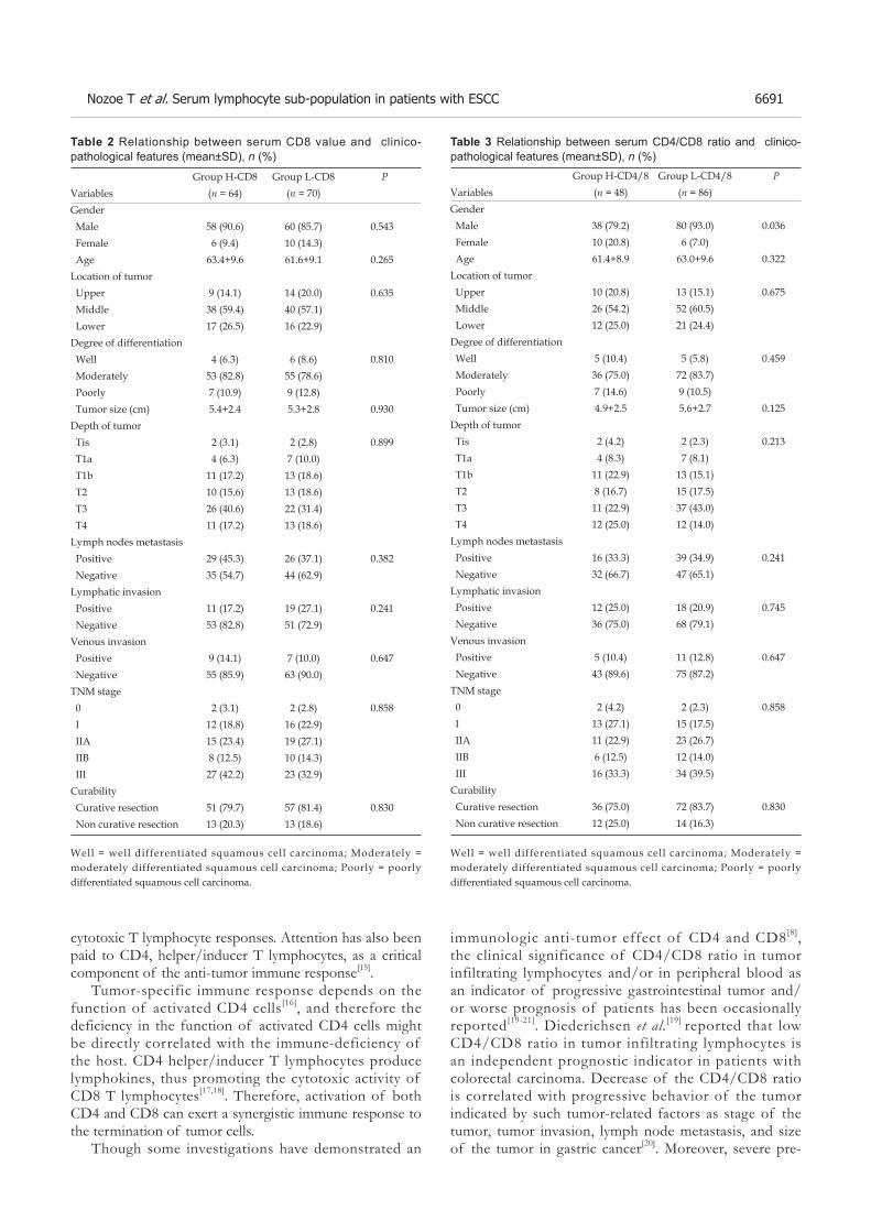
Nozoe T et al. Serum lymphocyte sub-population in patients with ESCC 6691
cytotoxic T lymphocyte responses. Attention has also been paid to CD4, helper/inducer T lymphocytes, as a critical component of the anti-tumor immune response[15].
Tumor-specific immune response depends on the function of activated CD4 cells[16], and therefore the deficiency in the function of activated CD4 cells might be directly correlated with the immune-deficiency of the host. CD4 helper/inducer T lymphocytes produce lymphokines, thus promoting the cytotoxic activity of CD8 T lymphocytes[17,18]. Therefore, activation of both CD4 and CD8 can exert a synergistic immune response to the termination of tumor cells.
Though some investigations have demonstrated an
immunologic anti-tumor effect of CD4 and CD8[8], the clinical significance of CD4/CD8 ratio in tumor infiltrating lymphocytes and/or in peripheral blood as an indicator of progressive gastrointestinal tumor and/or worse prognosis of patients has been occasionally reported[19-21]. Diederichsen et al.[19] reported that low CD4/CD8 ratio in tumor infiltrating lymphocytes is an independent prognostic indicator in patients with colorectal carcinoma. Decrease of the CD4/CD8 ratio is correlated with progressive behavior of the tumor indicated by such tumor-related factors as stage of the tumor, tumor invasion, lymph node metastasis, and size of the tumor in gastric cancer[20]. Moreover, severe pre-
Group H-CD8 Group L-CD8 P Variables (n = 64) (n = 70)Gender Male 58 (90.6) 60 (85.7) 0.543 Female 6 (9.4) 10 (14.3) Age 63.4+9.6 61.6+9.1 0.265Location of tumor Upper 9 (14.1) 14 (20.0) 0.635 Middle 38 (59.4) 40 (57.1) Lower 17 (26.5) 16 (22.9)Degree of differentiation Well 4 (6.3) 6 (8.6) 0.810 Moderately 53 (82.8) 55 (78.6) Poorly 7 (10.9) 9 (12.8) Tumor size (cm) 5.4+2.4 5.3+2.8 0.930Depth of tumor Tis 2 (3.1) 2 (2.8) 0.899 T1a 4 (6.3) 7 (10.0) T1b 11 (17.2) 13 (18.6) T2 10 (15.6) 13 (18.6) T3 26 (40.6) 22 (31.4) T4 11 (17.2) 13 (18.6)Lymph nodes metastasis Positive 29 (45.3) 26 (37.1) 0.382 Negative 35 (54.7) 44 (62.9)Lymphatic invasion Positive 11 (17.2) 19 (27.1) 0.241 Negative 53 (82.8) 51 (72.9)Venous invasion Positive 9 (14.1) 7 (10.0) 0.647 Negative 55 (85.9) 63 (90.0)TNM stage 0 2 (3.1) 2 (2.8) 0.858 I 12 (18.8) 16 (22.9) IIA 15 (23.4) 19 (27.1) IIB 8 (12.5) 10 (14.3) III 27 (42.2) 23 (32.9)Curability Curative resection 51 (79.7) 57 (81.4) 0.830 Non curative resection 13 (20.3) 13 (18.6)
Table 2 Relationship between serum CD8 value and clinico-pathological features (mean±SD), n (%)
Well = well differentiated squamous cell carcinoma; Moderately = moderately differentiated squamous cell carcinoma; Poorly = poorly differentiated squamous cell carcinoma.
Table 3 Relationship between serum CD4/CD8 ratio and clinico-pathological features (mean±SD), n (%)
Group H-CD4/8 Group L-CD4/8 P Variables (n = 48) (n = 86)Gender Male 38 (79.2) 80 (93.0) 0.036 Female 10 (20.8) 6 (7.0) Age 61.4+8.9 63.0+9.6 0.322Location of tumor Upper 10 (20.8) 13 (15.1) 0.675 Middle 26 (54.2) 52 (60.5) Lower 12 (25.0) 21 (24.4)Degree of differentiation Well 5 (10.4) 5 (5.8) 0.459 Moderately 36 (75.0) 72 (83.7) Poorly 7 (14.6) 9 (10.5) Tumor size (cm) 4.9+2.5 5.6+2.7 0.125Depth of tumor Tis 2 (4.2) 2 (2.3) 0.213 T1a 4 (8.3) 7 (8.1) T1b 11 (22.9) 13 (15.1) T2 8 (16.7) 15 (17.5) T3 11 (22.9) 37 (43.0) T4 12 (25.0) 12 (14.0)Lymph nodes metastasis Positive 16 (33.3) 39 (34.9) 0.241 Negative 32 (66.7) 47 (65.1)Lymphatic invasion Positive 12 (25.0) 18 (20.9) 0.745 Negative 36 (75.0) 68 (79.1)Venous invasion Positive 5 (10.4) 11 (12.8) 0.647 Negative 43 (89.6) 75 (87.2)TNM stage 0 2 (4.2) 2 (2.3) 0.858 I 13 (27.1) 15 (17.5) IIA 11 (22.9) 23 (26.7) IIB 6 (12.5) 12 (14.0) III 16 (33.3) 34 (39.5)Curability Curative resection 36 (75.0) 72 (83.7) 0.830 Non curative resection 12 (25.0) 14 (16.3)
Well = well differentiated squamous cell carcinoma; Moderately = moderately differentiated squamous cell carcinoma; Poorly = poorly differentiated squamous cell carcinoma.

6692 ISSN 1007-9327 CN 14-1219/ R World J Gastroenterol November 14, 2005 Volume 11 Number 42
operative cellular immune-suppression, where CD4/CD8 ratio was less than 1.0, is a predictive parameter for mor-tality in patients with gastric cancer[21].
CD8 expression in TIL in tumor tissue can serve the function of suppressing the proliferation of ESCC[9], and similarly CD8 infi ltration into the tumor is an independent prognostic indicator for ESCC[10]. Recently, increase of the number of CD4 and CD8 T lymphocytes in tumor nests and stroma has been found to be an independent indicator of favorable prognosis of patients with ESCC[8].
These results suggest that CD8 T-lymphocyte in-filtration, as have been investigated in some other tumors[22,23], plays a pivotal role in immune-potential against ESCCs.
However, it was reported that the prognosis of patients with lung carcinoma associated with more CD8 expressing T cells within cancer nests is significantly worse than that of patients with tumors of fewer CD8 expressing T cells[24]. High percentage of activated CD8-positive cells in postoperative peripheral blood is an indicator of worse prognosis for renal cell carcinoma[25].
The different methods used to evaluate the value or expression of CD8, histological type of the tumor, or balance between immunologic dynamics of the tumor and the host might explain this possible discrepancy in the signifi cance of CD8 T lymphocytes in anti-tumor immune.
In the current study, the decreased CD4/CD8 ratio as well as the increased CD8 and the decreased CD4 in peripheral blood could predict the worse prognosis in patients with ESCCs. Preoperative coexistence of impaired immunity could infl uence the postoperative complications[5]. The incidence of postoperative complications is an independent indicator of worse prognosis in patients with esophageal carcinoma[26]. Therefore, preoperative impaired immunity seems not to be negligible as the cause of death, other than esophageal carcinoma.
Assessment of preoperative immunity in patients seems to be of great importance in predicting the subsequent outcome of patients with ESCCs.
REFERENCES1 Neuner A, Schindel M, Wildenberg U, Muley T, Lahm H,
Fischer JR. Prognostic signifi cance of cytokine modulation in non-small cell lung cancer. Int J Cancer 2002; 101: 287-292
2 Takagi K, Yamamori H, Morishima Y, Toyoda Y, Nakajima N, Tashiro T. Preoperative immunosuppression: its relationship with high morbidity and mortality in patients receiving thoracic esophagectomy. Nutrition 2001; 17: 13-17
3 Eilber FR, Morton DL. Impaired immunologic reactivity and recurrence following cancer surgery. Cancer 1970; 25: 362-367
4 Nozoe T, Saeki H, Sugimachi K. Signifi cance of preoperative elevation of serum C-reactive protein as an indicator of prognosis in esophageal carcinoma. Am J Surg 2001; 182: 197-201
5 Nozoe T, Kimura Y, Ishida M, Saeki H, Korenaga D, Sugimachi K. Correlation of pre-operative nutritional condition with post-operative complications in surgical treatment for oesophageal carcinoma. Eur J Surg Oncol 2002; 28: 396-400
6 Nozoe T, Korenaga D, Ohga T, Futatsugi M, Maehara Y. Suppression of phytohemagglutinin response to lymphocytes
is an independent prognosticator in patients with squamous cell carcinoma of the esophagus. Ann Thorac Surg 2003; 76: 260-265
7 Shiku H. Importance of CD4+ helper T-cells in antitumor immunity. Int J Hematol 2003; 77: 435-438
8 Cho Y, Miyamoto M, Kato K, Fukunaga A, Shichinohe T, Kawarada Y, Hida Y, Oshikiri T, Kurokawa T, Suzuoki M, Nakakubo Y, Hiraoka K, Murakami S, Shinohara T, Itoh T, Okushiba S, Kondo S, Katoh H. CD4+ and CD8+ T cells cooperate to improve prognosis of patients with esophageal squamous cell carcinoma. Cancer Res 2003; 63: 1555-1559
9 Takeno S, Noguchi T, Kikuchi R, Wada S, Sato T, Uchida Y. Immunohistochemical study of leukocyte infiltration and expression of hsp70 in esophageal squamous cell carcinoma.Oncol Rep 2001; 8: 585-590
10 Schumacher K, Haensch W, Roefzaad C, Schlag PM. Prognostic signifi cance of activated CD8(+) T cell infi ltrations within esophageal carcinomas. Cancer Res 2001; 61: 3932-3936
11 Tsutsui S, Sonoda K, Sumiyoshi K, Kitamura K, Toh Y, Kitamura M, Kuwano H, Sugimachi K, Okamura S. Prognostic significance of immunological parameters in patients with esophageal cancer. Hepatogastroenterology 1996; 43: 501-509
12 Japanese Society for Esophageal Diseases. Guide lines for the clinical and pathological studies on carcinoma of the esophagus, 9th ed. Tokyo: Kanehara Company, 1999
13 LH Sobin, Wittekind C, editors. International Union Against Cancer. TNM classifi cation of malignant tumours, 5th ed. New York: Wiley-Liss, 1997: p 54-58
14 Reinherz EL, Kung PC, Goldstein G, Schlossman SF. Separation of functional subsets of human T cells by a monoclonal antibody. Proc Natl Acad Sci USA 1979; 76: 4061-4065
15 Pardoll DM, Topalian SL. The role of CD4+ T cell responses in antitumor immunity. Curr Opin Immunol 1998; 10: 588-594
16 Chen L, Linsley PS, Hellstrom KE. Costimulation of T cells for tumor immunity. Immunol Today 1993; 14: 483-486
17 Toes RE, Ossendorp F, Offringa R, Melief CJ. CD4 T cells and their role in antitumor immune responses. J Exp Med 1999; 189: 753-756
18 Qin Z, Richter G, Schuler T, Ibe S, Cao X, Blankenstein T. B cells inhibit induction of T cell-dependent tumor immunity. Nat Med 1998; 4: 627-630
19 Diederichsen AC, Hjelmborg JB, Christensen PB, Zeuthen J, Fenger C. Prognostic value of the CD4+/CD8+ ratio of tumour infiltrating lymphocytes in colorectal cancer and HLA-DR expression on tumour cells. Cancer Immunol Immunother 2003; 52: 423-428
20 Lee WJ, Chang KJ, Lee CS, Chen KM. Selective depression of T-lymphocyte subsets in gastric cancer patients: an implication of immunotherapy. J Surg Oncol 1994; 55: 165-169
21 Rey-Ferro M, Castano R, Orozco O, Serna A, Moreno A. Nutritional and immunologic evaluation of patients with gastric cancer before and after surgery. Nutrition 1997; 13: 878-881
22 Oshikiri T, Miyamoto M, Shichinohe T, Suzuoki M, Hiraoka K, Nakakubo Y, Shinohara T, Itoh T, Kondo S, Katoh H. Prognostic value of intratumoral CD8+ T lymphocyte in extrahepatic bile duct carcinoma as essential immune response. J Surg Oncol 2003; 84: 224-228
23 Naito Y, Saito K, Shiiba K, Ohuchi A, Saigenji K, Nagura H, Ohtani H. CD8+ T cells infi ltrated within cancer cell nests as a prognostic factor in human colorectal cancer. Cancer Res 1998; 58: 3491-3494
24 Wakabayashi O, Yamazaki K, Oizumi S, Hommura F, Kinoshita I, Ogura S, Dosaka-Akita H, Nishimura M. CD4(+) T cells in cancer stroma, not CD8(+) T cells in cancer cell nests, are associated with favorable prognosis in human non-small cell lung cancers. Cancer Sci 2003; 94: 1003-1009
25 Arima K, Nakagawa M, Yanagawa M, Sugimura Y, Tochigi

Nozoe T et al. Serum lymphocyte sub-population in patients with ESCC 6693
H, Kawamura J. Prognostic factors of peripheral blood lymphocyte subsets in patients with renal cell carcinoma. Urol Int 1996; 57: 5-10
Science Editor Wang XL and Guo SY Language Editor Elsevier HK
26 Nozoe T, Miyazaki M, Saeki H, Ohga T, Sugimachi K. Signifi cance of allogenic blood transfusion on decreased survival in patients with esophageal carcinoma. Cancer 2001; 92: 1913-1918
PO Box 2345, Beijing 100023, China World J Gastroenterol 2005;11(42):6694-6700www.wjgnet.com World Journal of Gastroenterology ISSN [email protected] © 2005 The WJG Press and Elsevier Inc. All rights reserved.E L S E V I E R
• BRIEF REPORTS •
Tumors, with increased DNA-binding activity, also showed a statistically significant increase in Ku70 and Ku86 nuclear expression, as determined by Western blot and immunohistochemical analyses (P<0.001). Cytoplasmic protein expression was found in pathological samples, but not in normal tissues either from tumor patients or from healthy subjects.
CONCLUSION: Overall, our DNA-binding activity and protein level are consistent with a substantial activation of the NHEJ pathway in colorectal tumors. Since the NHEJ is an error prone mechanism, its abnormal activation can result in chromosomal instability and ultimately lead to tumorigenesis.
© 2005 The WJG Press and Elsevier Inc. All rights reserved.
Key words: Colorectal cancer; Colon adenoma; DNA-dependent protein kinase; Ku70/80 heterodimer; Mismatch repair; Non-homologous end joining; Double strand break repair; Chromosomal instability
Mazzarelli P, Parrella P, Seripa D, Signori E, Perrone G, Rabitti C, Borzomati D, Gabbrielli A, Matera MG, Gravina C, Caricato M, Poeta ML, Rinaldi M, Valeri S, Coppola R, Fazio VM. DNA end binding activity and Ku70/80 heterodimer expression in human colorectal tumor. World J Gastroenterol 2005; 11(42): 6694-6700http://www.wjgnet.com/1007-9327/11/6694.asp
INTRODUCTIONColorectal cancer is a significant cause of morbidity and mortality in Western populations[1]. This cancer progresses through a series of defined histopathological stages, going from a small benign tumor (adenomatous polyps) to a malignant cancer (carcinoma)[2]. During tumor progression, a stepwise accumulation of genetic changes is observed, leading to inactivation of tumor suppressor genes (e.g. APC, p53) and activation of oncogenes (e.g. K-ras, β-catenin)[2]. The number of genomic alterations in cancer appears to exceed the level possibly due to the accumulation of mutations in cells with normal mutation rates. A number of intricate networks have evolved in eukaryotic cells to respond to exogenous and endogenous genotoxic stimuli[3]. Genes involved in these pathways play
DNA end binding activity and Ku70/80 heterodimer expression in human colorectal tumor
Paola Mazzarelli, Paola Parrella, Davide Seripa, Emanuela Signori, Giuseppe Perrone, Carla Rabitti, Domenico Borzomati, Armando Gabbrielli, Maria Giovanna Matera, Carolina Gravina, Marco Caricato, Maria Luana Poeta, Monica Rinaldi, Sergio Valeri, Roberto Coppola, Vito Michele Fazio
Paola Mazzarelli, Emanuela Signori, Maria Luana Poeta, Vito Michele Fazio, Laboratory of Molecular Medicine and Biotechnology, Interdisciplinary Center for Biomedical Research, Università Campus Bio-Medico, Rome 00155, ItalyPaola Parrella, Davide Seripa, Maria Giovanna Matera, Carolina Gravina, Vito Michele Fazio, Laboratory of Gene Therapy and Oncology, IRCCS “Casa Sollievo della Sofferenza” Hospital, San Giovanni Rotondo (FG) 71013, ItalyGiuseppe Perrone, Carla Rabitti, Service of Histopathology, Università Campus Bio-Medico, Rome 00155, ItalyDomenico Borzomati, Marco Caricato, Sergio Valeri, Roberto Coppola, Department of General Surgery, Università Campus Bio-Medico, Rome 00155, ItalyArmando Gabbriel l i , Department of Digestive Disease, Università Campus Bio-Medico, Rome 00155, ItalyEmanuela Signori, Monica Rinaldi, Vito Michele Fazio, CNR Gene-Medicine Division, Section of Molecular Medicine, Institute of Neurobiology and Molecular Medicine, Rome 00133, ItalySupported by Italian Ministero della Salute, IRCCS, RC0302TG13, and by Ministero dell’Istruzione, Università e Ricerca scientifi ca e tecnologica (MIUR), COFIN2002, to the Università Campus Bio-MedicoCorrespondence to: Professor Vito Michele Fazio, Laboratory of Molecular Medicine and Biotechnology, Università Campus Bio-Medico, Via Longoni, 83, Rome 00155, Italy. [email protected]: +39-06-22541780 Fax: +39-06-22541780Received: 2005-02-01 Accepted: 2005-04-26
AbstractAIM: To determine the DNA binding activity and protein levels of the Ku70/80 heterodimer, the functional mediator of the NHEJ activity, in human colorectal carcinogenesis.
METHODS: The Ku70/80 DNA-binding activity was determined by electrophoretic mobility shift assays in 20 colon adenoma and 15 colorectal cancer samples as well as matched normal colonic tissues. Nuclear and cytoplasmic protein expression was determined by immunohistochemistry and Western blot analysis.
RESULTS: A statistically significant difference was found in both adenomas and carcinomas as compared to matched normal colonic mucosa (P<0.00). However, changes in binding activity were not homogenous with approximately 50% of the tumors showing a clear increase in the binding activity, 30% displaying a modest increase and 15% showing a decrease of the activity.

Mazzarelli P et al. Non-homologous DNA end joining pathway in colorectal tumor 6695
a crucial role in maintaining DNA integrity and a defect in these processes may result in hypersensitivity to DNA damaging agents and genomic instability[4]. Two main forms of genetic instability are associated with tumors. One arises from the inactivation of DNA mismatch repair (MMR) genes[5], leading to instability at the nucleotide sequence level (microsatellite instability, MSI). The other results from a disruption of the pathways intending to protect the cells from chromosomal breakage (double-strand breaks, DSBs), which leads to gross chromosomal rearrangements (chromosomal instability, CIN)[6]. Of the many types of DNA damage, DSBs are the most dangerous, because of the intrinsic difficulty of their repair as compared to other types of DNA damage[6]. In physiological conditions, DNA-DSBs are generated by homologous recombination (HR) during meiosis and occur in other events, such as V(D)J recombination and immunoglobulin class switch[6]. In addition, DSBs can result from both exogenous agents such as ionizing radiation or chemotherapeutic agents and endogenously generated reactive oxygen species[6]. Erroneous rejoining of the broken DNA-DSBs may cause loss or amplifi cation of chromosomal material and even translocations, ultimately leading to tumorigenesis[4].
There are two distinct and complementary mechanisms for DNA-DSB repair: the NHEJ and the HR[4]. Recently, a caretaker role in preventing carcinogenesis has been proposed for the NHEJ pathway[7-9]. At present, five proteins involved in the NHEJ pathway have been identifi ed; namely, the ligase IV and its associated protein XRCC1, and the three components of the DNA-dependent protein kinase (DNA-PK) complex, Ku70, Ku86, and the catalytic subunit PKcs[10]. Mutational analysis has shown that activation of the DNA-PK well correlates with Ku protein heterodimerization and DNA-end binding[11]. Once anchored to the DNA, the Ku70/80 heterodimer translocates along the molecule and facilitates recruitment of the catalytic subunit to the site of the break, to form an activated DNA-PK complex[11]. Numerous studies have investigated the role of MMR pathway in colorectal carcinogenesis, but little is known about the involvement of the DSB repair pathway in the adenoma/carcinoma sequence[11-13]. In the attempt to better understand this role, we analyzed the DNA-binding activity of the Ku70/80 heterodimer and the protein expression of the two Ku subunits in colon adenomas, colorectal cancers and matched normal tissues. Ku70/80 DNA binding activity was increased in approximately 50% of adenoma and carcinoma samples, as compared to matched normal tissues. In tumors with increased DNA-binding activity, Ku70 and Ku86 protein expression correlated with the heterodimer binding activity.
MATERIALS AND METHODSPatients characteristics and tissue samplesTwenty patients with colon adenoma and 15 patients with colorectal carcinoma were recruited in the study. The
patients underwent endoscopic polypectomy or surgical resection between 1999 and 2002, at the Departments of Gastroenterology and General Surgery, Campus Bio-Medico University of Rome, Italy. None of the patients were affected by familial polyposis or HNPCC. Subjects included 20 males (57%) and 15 females (43%), with a mean age of 69.5±12 years (range 35-82 years). None had pre-operative chemotherapy or irradiation. All the patients gave informed consent for the study. Thirty-one lesions (89% including adenomas and carcinomas) were located in the colon and 4 (11%) in the rectum. Adenomas were classified according to the National Polyp Study Cohort and WHO recommendations on the basis of size and grade of dysplasia[14,15]. Clinical staging of colorectal cancer was assessed according to the Dukes’ classifi cation[13].
Preparation of cell and nuclear extractsFollowing surgical resection, tissue samples were im-mediately frozen at -80 °C. For each case, before protein extraction, one 3-μm hematoxylin-eosin stained slide was analyzed to ensure that tumor samples contained at least 70% cancer cells. Protein extraction was performed as previously described[16-18]. Briefly, frozen samples were mechanically fractionated to obtain a cellular suspension. Nuclear and cytoplasmic fractions were separated by centrifugation at 10 000 g and stored at -80 °C.
Protein content in nuclear and cytoplasmic extracts was determined in triplicate by Bradford assay (Bio-Rad Protein Assay, Bio-Rad Laboratories, Munchen).
Gel-shift assayElectrophoretic mobil i ty shift assay (EMSA) was performed as described previously[18,19]. Briefly, DNA binding reactions contained 50 000 cpm of the labeled probe, nuclear (2 μg) or cytoplasmic (5 μg) extracts with closed circular plasmid DNA pUC-19 (1 μg) as the unspecifi c competitor. For each sample, three single shift assays were performed. As controls, 33 normal human tissues from patients without colon tumor were analyzed (mammary gland n = 8, bladder mucosa n = 8, and skin n = 17).
To normalize all the samples, electrophoretic mobility shift assays were performed by incubating the nuclear extracts (3 μg) with 50 000 cpm/sample of 32P-end labeled Sp-1 oligonucleotide (Promega Corporation, Madison, WI, USA) in a binding buffer, with 1 μg of poly (dI-dC) as the unspecifi c competitor. The correction factor (CF) was calculated as follows: SP1 binding activity in the sample/mean SP1 binding activity. Data were normalized using the following formula: Mean Ku70/80 binding activity/CF[ 20].
For gel supershift experiments, goat polyclonal anti-Ku70 and anti-Ku86 antibodies (M-19, M-20: Santa Cruz Biotechnologies Inc., CA, USA) were incubated with protein extracts for 30 min at room temperature, before the other components were added to the binding react ion. Complexes were separated on 6% non-denaturing polyacrylamide gels and exposed to X-ray fi lms (Amersham-Pharmacia Biotech, England HP7 9NA).

6696 ISSN 1007-9327 CN 14-1219/ R World J Gastroenterol November 14, 2005 Volume 11 Number 42
The optical densities (OD) were obtained by scanning densitometry using colon carcinoma cell line CaCo-2 (ATCC) as internal control (OD = 10.7±6.4).
Supershift control experiments confirmed the specifi city of the results in each gelshift assay experiment.
ImmunohistochemistryParaffin sections from matched normal colonic mucosa were available for all the colon cancer cases entered in the study, whereas paraffin embedded blocks from matched normal tissues were available only for 10 adenomas. Paraffi n sections from fi ve subjects who showed normal colonic mucosa at colonoscopy were also analyzed. Consecutive two micron sections were immunostained for Ku70 and Ku86 following the streptavidin-biotin method, as described previously[20] . In brief, sections were deparaffi nized, rehydrated in decreasing alcohol and microwave treated. Endogenous peroxidase activity was quenched by treatment with 0.03% hydrogen peroxide in absolute methanol for 30 min at room temperature. The primary antibodies were goat polyclonal anti-Ku70 and anti-Ku86 antibodies specifically validated for immunohistochemical analysis of paraffin embedded tissues (M-19, M-20: Santa Cruz Biotechnology Inc., CA, USA), in a 1:200 dilution. Biotinylated swine antigoat/mouse/rabbit IgG (Dako A/S, Denmark) was used as secondary antibody. After washing, sections were treated with streptavidin-peroxidase reagent (Dakopatts A\S, Denmark), incubated with diaminobenzidine (DAB) and counterstained with hematoxylin. Slides were examined under a two-head microscope by two pathologists, unaware of the clinical data and molecular results. No discrepant results were identifi ed. Results of the nuclear immunostaining were expressed as percentage of positively stained cells. For cytoplasmic staining the microscopic analysis was not able to discriminate one cell from another. Thus the immunoreactivity was classifi ed into four staining levels (SL): 0 SL (no staining); 1 SL (1-33% of positively stained area); 2 SL (33-66% of the area), and 3 SL (66-99% of the area)[ 21].
Western blottingProtein extracts (10 μg) were separated in 10% SDS-PAGE, transferred to a PVDF membrane (Hybond-P, Amersham-Pharmacia Biotech, UK HP7 9NA) using an electroblotting apparatus, and incubated for 1 h at room temperature in 1% BSA, 1% skim milk (Difco Lab., Detroit, MI, USA), and 0.5% Tween 20 (USB, Cleveland, OH, USA). Membranes were stained with Ponceau S dye, to check for equal loading and homogeneous transfer. Immunodetection experiments were performed as described previously[17]. Filters were reprobed with anti β-actin (Sigma-Aldrich, St. Louis, MO 63103, USA) mouse IgG1 monoclonal antibody, to normalize the nuclear protein levels. Filters were washed and developed using an enhanced chemiluminescence system (ECL, Amersham-Pharmacia Biotech, UK HP7 9NA). The optical densities (OD) were obtained by scanning densitometric analysis of
the bands normalized for the β-actin levels, as reference protein.
Statistical analysis All values provided in the text and fi gures are means of three independent experiments±standard deviations (SD). Variation rates (VR) were defi ned by the following formula: [(pathological sample value–normal sample value)/pathological sample value]×100. Mean values were compared using the one- or two-tailed Student’s t-test, for independent samples. P<0.05 was considered statistically signifi cant.
RESULTSNuclear and cytoplasmic Ku70/80 DNA-binding activity EMSA were performed on nuclear extracts of colon tissues, to compare the DNA binding activity of the Ku70/80 heterodimer in adenoma and carcinoma samples as well as matched normal colonic mucosa samples (Figure 1A). We found that adenoma and carcinoma samples had an overall increase in DNA binding activity as compared to matched normal samples (P<0.01, Table 1). However, these variations were not homogeneous among the tumors that were tested. Since the overall increase in binding activity compared to normal was approximately 50% (Table 1), we set this value as the cut-off for better classifying our pathological samples. Of the 20 adenomas, 11 (55%) showed an increase in DNA binding activity higher than 50% (P = 0.000) (Figure 1A, Group I), 6 (30%) displayed an increase in DNA binding activity lower than 50% (P = NS) (Figure 2A, Group II), and 3 (15%) showed reduced levels of activity (P = 0.04) (Figure 2A, Group III). Of the 15 carcinomas, 7 (47%) displayed an increase in DNA binding activity higher than 50% (Figure 2B, Group I), 6 (40%) showed an increase in DNA binding activity lower than 50% (P<0.02) (Figure 2B, Group II), and 2 (13%) showed reduced levels of activity (P = ND) (Figure 2B, Group III).
No significant differences were found when the Ku70/80 DNA-binding activity was compared in adenoma and carcinoma samples (Table 1). We also determined, if functional Ku70/80 heterodimer was present in cytoplasmic protein extracts. Cytoplasmic Ku70/80 DNA binding activity was only found in three colorectal cancers, whereas adenomas did not show cytoplasmic activity (Figure 1B). No statistically significant correlation was found when results of nuclear and cytoplasmic Ku70/80 DNA-binding activity were compared with clinical parameters.
Colon Ku70/80 DNA binding activity Ku70 protein expression
Tissue Normal Tumor P Normal Tumor PAdenomas 2.66±2.32 5.01±3.15 <0.01 0.88±0.59 1.28±0.45 <0.01Carcinomas 2.99±2.84 5.35±2.75 <0.001 1.09±0.80 1.77±0.83 <0.01
Table 1 Statistical summary of Ku70/80 DNA binding activity and Ku70 protein expression in pathological samples and matched normal tissues (mean±SD)

Mazzarelli P et al. Non-homologous DNA end joining pathway in colorectal tumor 6697
Ku70 and Ku86 expression in nuclear and cytoplasmic protein extracts In adenomas and carcinomas with increased DNA-binding activity, the Ku70 nuclear levels were markedly increased as compared to matched normal samples (P<0.000) (Figures 3A and B, Group I), whereas protein level changes were not statistically signifi cant in tumors with minimal increases or reduced binding activity (Figures 3A and B, Groups II and III). Representative results are shown in Figure 4.
In all the samples, Ku86 protein levels were always lower than Ku70 and in most cases it was undetectable (data not shown). Analysis of cytoplasmic protein extracts showed barely detectable Ku70 protein in normal colon tissues and a significant increase in pathologic samples with increased DNA binding activity (0.22±0.3 OD and 0.80±0.2 OD, respectively, P = 0.001) (data not shown). Ku86 protein subunit was undetectable by Western blot analysis on cytoplasmic extracts.
Ku70 and Ku86 immunohistochemical analysis A signifi cant increase in the percentage of stained nuclei was found for Ku70 and Ku86 subunits in all adenoma and carcinoma samples, as compared to matched normal tissues (P<0.000) but no statistically signifi cant differences were found between adenoma and carcinoma samples (Table 3). In all cases, Ku subunits were not detected in the cytoplasm of normal colonic mucosa (n = 25) from patients or healthy donors (n = 5) (SL 0). For Ku70, a low level (SL 1) of cytoplasmic staining was detected in 11 of the 20 adenomas (55%), and in 3 of the 15 carcinomas
CaCo K1 K2 K3 K4 n t n t n t n t
A1 A2 A3 A4 A5 P n t n t n t n t n t
P K1 K2 K3 K4 K5 K6 A1 A2 A3 A4 A5 A6 A7 A8 A9 IgG Ku CaCo
←FP
A
B
Figure 1 Representative gel shift experiments on nuclear (A) and cytoplasmic (B) extracts from human colon tissues. Panel A: Normal (n) and pathologic (t) nuclear extracts, obtained from four colorectal cancer (K1-K4) and fi ve adenoma (A1-A5) patients, were analyzed by EMSA as described in "Materials and methods". The positions of Ku band-shifts and of the free probe (FP) are indicated. The binding activity in nuclear extracts from the CaCo-2 cell line (CaCo) has been used as a standard in order to compare the activity in the different gels. Lane P: free probe without protein extract. Panel B: EMSA was performed on pathologic cytoplasmic extracts from colorectal cancer (K1-K6) and adenoma (A1-A9) patients. Lane P: free probe without protein extract.
131211109876543210
A15 A14 A4 A9 A16 A2 A19 A7 A1 A10 A11 A17 A18 A5 A8 A12 A13 A3 A20 A6 K1 K5 K7 K6 K3 K8 K15 K10 K2 K9 K11 K13 K4 K14 K12
131211109876543210
OD
OD
A B
Figure 2 Changes in Ku70/Ku80 heterodimer DNA binding activity in adenoma (A) and carcinoma (B) compared to matched normal colonic mucosa. □ Normal; ■■ Tumor; vertical bars indicate standard deviation from the mean of three independent experiments. Adenomas (A) and carcinomas (B) samples characterize by: Group 1 more than 50% increase in DNA binding activity; Group 2 modest increase in binding activity; Group 3 reduced binding activity, 1P<0.000 vs group 1; 2P<0.04 vs group 3; 3P<0.02 vs group 2.
5
4
3
2
1
0K1 K5 K7 K6 K3 K8 K15 K10 K2 K9 K11 K13 K4 K14 K12
NKO
D
A15 A14 A4 A9 A16 A2 A19 A7 A1 A10 A11 A17 A18 A5 A8 A12 A13 A3 A20 A6
3
2.5
2
1.5
1
0.5
0
OD
Figure 3 Changes in Ku70 protein levels determined by Western blot analysis in adenoma (A) and carcinoma (B) compared to matched normal colonic mucosa. □ Normal; ■■Tumor; vertical bars indicate standard deviation from the mean of three independent experiments. Adenomas (A) and carcinomas (B) are grouped according to the criteria described in Figure 2, 1P<0.000 vs group 1.
A BGroup 1 Group 2 Group 3 Group 1
Group 2 Group 3
Group 1Group 2 Group 3Group 1 Group 2
Group 3
1 NS
2
13 NS
1 NS NS 1NS NS
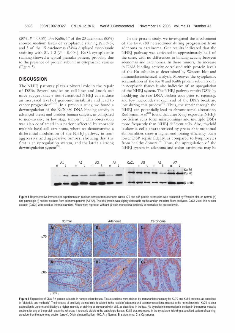
6698 ISSN 1007-9327 CN 14-1219/ R World J Gastroenterol November 14, 2005 Volume 11 Number 42
(20%, P = 0.089). For Ku86, 17 of the 20 adenomas (85%) showed medium levels of cytoplasmic staining (SL 2-3), and 5 of the 15 carcinomas (34%) displayed cytoplasmic staining with SL 1-2 (P = 0.004). Ku86 cytoplasmic staining showed a typical granular pattern, probably due to the presence of protein subunit in cytoplasmic vesicles (Figure 5).
DISCUSSIONThe NHEJ pathway plays a pivotal role in the repair of DSBs. Several studies on cell lines and knock-out mice suggest that a non-functional NHEJ can induce an increased level of genomic instability and lead to cancer progression[22-24]. In a previous study, we found a downregulation of the Ku70/80 DNA binding activity in advanced breast and bladder human cancers, as compared to non-invasive or low stage tumors[17]. This observation was also confirmed in a patient affected by sporadic multiple basal cell carcinoma, where we demonstrated a differential modulation of the NHEJ pathway in non-aggressive and aggressive tumors, showing that the first is an upregulation system, and the latter a strong downregulation system[20].
In the present study, we investigated the involvement of the ku70/80 heterodimer during progression from adenoma to carcinoma. Our results indicated that the NHEJ pathway was activated in approximately half of the cases, with no differences in binding activity between adenomas and carcinomas. In these tumors, the increase in DNA binding activity correlated with protein levels of the Ku subunits as determined by Western blot and immunohistochemical analysis. Moreover the cytoplasmic accumulation of the Ku70 and Ku86 protein subunits only in neoplastic tissues is also indicative of an upregulation of the NHEJ system. The NHEJ pathway repairs DSBs by modifying the two DNA broken ends prior to rejoining, and few nucleotides at each end of the DNA break are lost during this process[23]. Thus, the repair through the NHEJ can potentially lead to chromosomal alterations. Rothkamm et al.[25] found that after X-ray exposure, NHEJ-proficient cells form misrejoinings and multiple DSBs more frequently than NHEJ defi cient cells. Also, myeloid leukemia cells characterized by gross chromosomal abnormalities show a higher end-joining effi ciency but a lower DSB repair fidelity, as compared to lymphocytes from healthy donors[24]. Thus, the upregulation of the NHEJ system in adenoma and colon carcinoma may be
A1 A2 A3 A4 CaCo A5 A6 A7 n t n t n t n t n t n t n t
Ku 86Ku 70
β-actin
Figure 4 Representative immunoblot experiments on nuclear extracts from adenoma cases p70 and p86 protein expression was evaluated by Western blot, on normal (n) and pathologic (t) nuclear extracts from adenoma patients (A1-A7). The p86 protein was slightly detectable on this and on the other fi lters analyzed. CaCo-2 cell line nuclear extracts (CaCo) were used as internal standard. Filters were reprobed with anti-β-actin monoclonal antibody to normalize the protein levels.
Figure 5 Expression of DNA-PK protein subunits in human colon tissues. Tissue sections were stained by immunohistochemistry for Ku70 and Ku86 proteins, as described in “Materials and methods”. The increase of positively stained cells is evident in the nuclei of adenoma and carcinoma sections, respect to the normal controls. Ku70 nuclear expression is uniform and displays a higher intensity of staining as compared with p86, as described in the text. No cytoplasmic expression is evident in the normal mucosa sections for any of the protein subunits, whereas it is clearly visible in the pathologic tissues. Ku86 was expressed in the cytoplasm following a speckled pattern of staining, as evident on the adenoma section (arrow). Original magnifi cation ×400. A1-2: Normal; B1-2: Adenoma; C1-2: Carcinoma.
Normal Adenoma Carcinoma
p70
p86
5cm
↑
A1
A2
B1
B2
C1
C2
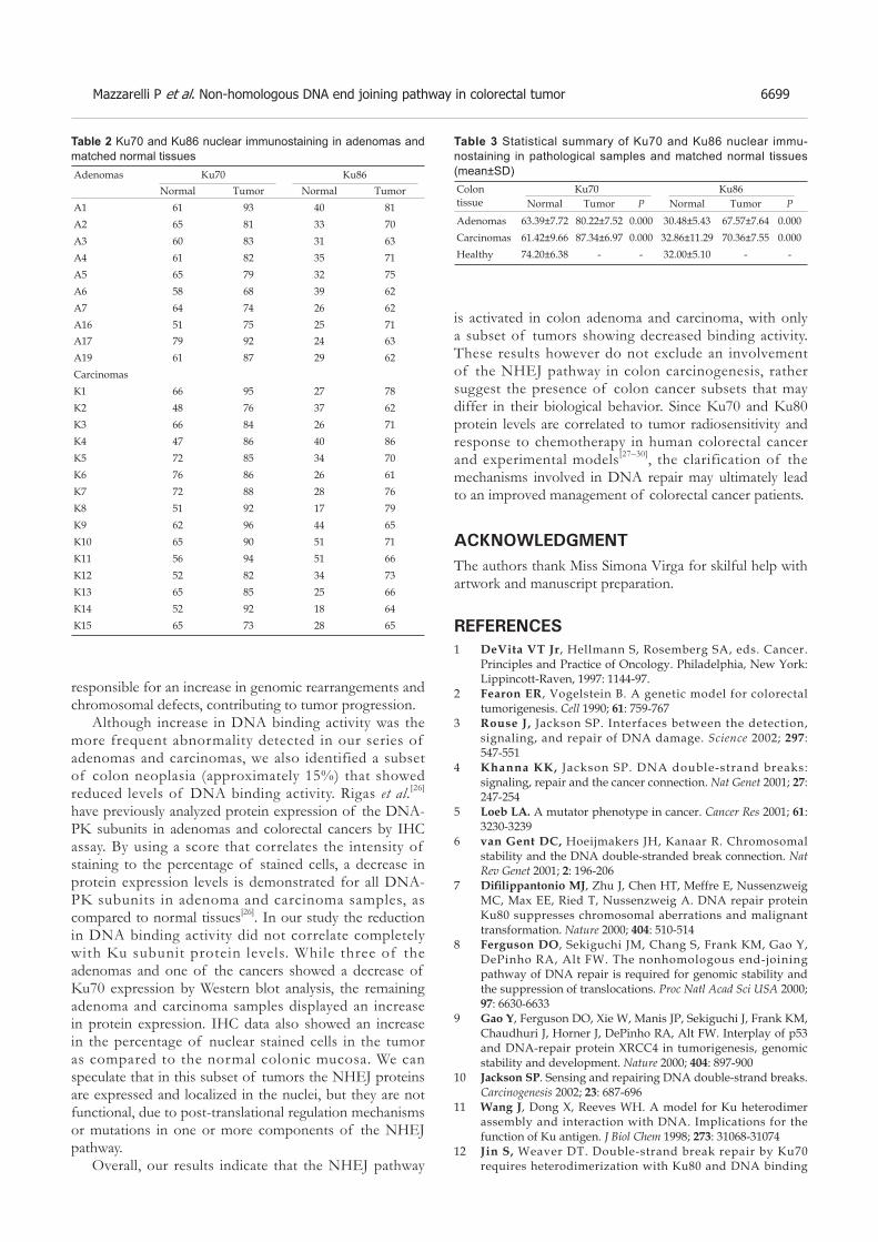
Mazzarelli P et al. Non-homologous DNA end joining pathway in colorectal tumor 6699
responsible for an increase in genomic rearrangements and chromosomal defects, contributing to tumor progression.
Although increase in DNA binding activity was the more frequent abnormality detected in our series of adenomas and carcinomas, we also identified a subset of colon neoplasia (approximately 15%) that showed reduced levels of DNA binding activity. Rigas et al.[26] have previously analyzed protein expression of the DNA-PK subunits in adenomas and colorectal cancers by IHC assay. By using a score that correlates the intensity of staining to the percentage of stained cells, a decrease in protein expression levels is demonstrated for all DNA-PK subunits in adenoma and carcinoma samples, as compared to normal tissues[26]. In our study the reduction in DNA binding activity did not correlate completely with Ku subunit protein levels. While three of the adenomas and one of the cancers showed a decrease of Ku70 expression by Western blot analysis, the remaining adenoma and carcinoma samples displayed an increase in protein expression. IHC data also showed an increase in the percentage of nuclear stained cells in the tumor as compared to the normal colonic mucosa. We can speculate that in this subset of tumors the NHEJ proteins are expressed and localized in the nuclei, but they are not functional, due to post-translational regulation mechanisms or mutations in one or more components of the NHEJ pathway.
Overall, our results indicate that the NHEJ pathway
is activated in colon adenoma and carcinoma, with only a subset of tumors showing decreased binding activity. These results however do not exclude an involvement of the NHEJ pathway in colon carcinogenesis, rather suggest the presence of colon cancer subsets that may differ in their biological behavior. Since Ku70 and Ku80 protein levels are correlated to tumor radiosensitivity and response to chemotherapy in human colorectal cancer and experimental models[27–30], the clarification of the mechanisms involved in DNA repair may ultimately lead to an improved management of colorectal cancer patients.
ACKNOWLEDGMENTThe authors thank Miss Simona Virga for skilful help with artwork and manuscript preparation.
REFERENCES 1 DeVita VT Jr, Hellmann S, Rosemberg SA, eds. Cancer.
Principles and Practice of Oncology. Philadelphia, New York: Lippincott-Raven, 1997: 1144-97.
2 Fearon ER, Vogelstein B. A genetic model for colorectal tumorigenesis. Cell 1990; 61: 759-767
3 Rouse J, Jackson SP. Interfaces between the detection, signaling, and repair of DNA damage. Science 2002; 297: 547-551
4 Khanna KK, Jackson SP. DNA double-strand breaks: signaling, repair and the cancer connection. Nat Genet 2001; 27: 247-254
5 Loeb LA. A mutator phenotype in cancer. Cancer Res 2001; 61: 3230-3239
6 van Gent DC, Hoeijmakers JH, Kanaar R. Chromosomal stability and the DNA double-stranded break connection. Nat Rev Genet 2001; 2: 196-206
7 Difi lippantonio MJ, Zhu J, Chen HT, Meffre E, Nussenzweig MC, Max EE, Ried T, Nussenzweig A. DNA repair protein Ku80 suppresses chromosomal aberrations and malignant transformation. Nature 2000; 404: 510-514
8 Ferguson DO, Sekiguchi JM, Chang S, Frank KM, Gao Y, DePinho RA, Alt FW. The nonhomologous end-joining pathway of DNA repair is required for genomic stability and the suppression of translocations. Proc Natl Acad Sci USA 2000; 97: 6630-6633
9 Gao Y, Ferguson DO, Xie W, Manis JP, Sekiguchi J, Frank KM, Chaudhuri J, Horner J, DePinho RA, Alt FW. Interplay of p53 and DNA-repair protein XRCC4 in tumorigenesis, genomic stability and development. Nature 2000; 404: 897-900
10 Jackson SP. Sensing and repairing DNA double-strand breaks. Carcinogenesis 2002; 23: 687-696
11 Wang J, Dong X, Reeves WH. A model for Ku heterodimer assembly and interaction with DNA. Implications for the function of Ku antigen. J Biol Chem 1998; 273: 31068-31074
12 Jin S, Weaver DT. Double-strand break repair by Ku70 requires heterodimerization with Ku80 and DNA binding
Adenomas Ku70 Ku86Normal Tumor Normal Tumor
A1 61 93 40 81A2 65 81 33 70A3 60 83 31 63A4 61 82 35 71A5 65 79 32 75A6 58 68 39 62A7 64 74 26 62A16 51 75 25 71A17 79 92 24 63A19 61 87 29 62CarcinomasK1 66 95 27 78K2 48 76 37 62K3 66 84 26 71K4 47 86 40 86K5 72 85 34 70K6 76 86 26 61K7 72 88 28 76K8 51 92 17 79K9 62 96 44 65K10 65 90 51 71K11 56 94 51 66K12 52 82 34 73K13 65 85 25 66K14 52 92 18 64K15 65 73 28 65
Table 2 Ku70 and Ku86 nuclear immunostaining in adenomas and matched normal tissues
Colontissue
Ku70 Ku86Normal Tumor P Normal Tumor P
Adenomas 63.39±7.72 80.22±7.52 0.000 30.48±5.43 67.57±7.64 0.000Carcinomas 61.42±9.66 87.34±6.97 0.000 32.86±11.29 70.36±7.55 0.000Healthy 74.20±6.38 - - 32.00±5.10 - -
Table 3 Statistical summary of Ku70 and Ku86 nuclear immu-nostaining in pathological samples and matched normal tissues (mean±SD)
6700 ISSN 1007-9327 CN 14-1219/ R World J Gastroenterol November 14, 2005 Volume 11 Number 42
Science Editor Wang XL and Guo SY Language Editor Elsevier HK
functions. EMBO J 1997; 16: 6874-6885 13 Li GC, He F, Shao X, Urano M, Shen L, Kim D, Borrelli M,
Leibel SA, Gutin PH, Ling CC. Adenovirus-mediated heat-activated antisense Ku70 expression radiosensitizes tumor cells in vitro and in vivo. Cancer Res 2003; 63: 3268-3274
14 O’Brien MJ, Winawer SJ, Zauber AG, Gottlieb LS, Sternberg SS, Diaz B, Dickersin GR, Ewing S, Geller S, Kasimian D. The National Polyp Study. Patient and polyp characteristics associated with high-grade dysplasia in colorectal adenomas. Gastroenterology 1990; 98: 371-379
15 Jass JR and Sobin LH. Histological typing of intestinal tumours: World Health Organization. 1989. Springer, Berlin.
16 Dignam JD, Lebovitz RM, Roeder RG. Accurate transcription initiation by RNA polymerase II in a soluble extract from isolated mammalian nuclei. Nucleic Acids Res 1983; 1: 1475-1489
17 Pucci S, Mazzarelli P, Rabitti C, Giai M, Gallucci M, Flammia G, Alcini A, Altomare V, Fazio VM. Tumor specifi c modulation of KU70/80 DNA binding activity in breast and bladder human tumor biopsies. Oncogene 2001; 20: 739-747
18 Lahiri DK, Ge Y. Electrophoretic mobility shift assay for the detection of specifi c DNA-protein complex in nuclear extracts from the cultured cells and frozen autopsy human brain tissue. Brain Res Brain Res Protoc 2000; 5: 257-265
19 Frasca D, Barattini P, Goso C, Pucci S, Rizzo G, Bartoloni C, Costanzo M, Errani A, Guidi L, Antico L, Tricerri A, Doria G. Cell proliferation and ku protein expression in ageing humans. Mech Ageing Dev 1998; 100: 197-208
20 Mazzarelli P, Rabitti C, Parrella P, Seripa D, Persichetti P, Marangi GF, Perrone G, Poeta ML, Delfino M, Fazio VM. Differential modulation of Ku70/80 DNA-binding activity in a patient with multiple basal cell carcinomas. J Invest Dermatol 2003; 121: 628-633
21 Kumagai I, Masuda T, Sato S, Ishikawa K. Immunoreactivity to monoclonal antibody, Hep Par 1, in human hepatocellular carcinomas according to histopathological grade and
histological pattern. Hepatol Res 2001; 20: 312-319 22 Lanuszewska J, Widlak P. The truncation of Ku86 in human
lymphocytes. Cancer Lett 2004; 205: 197-20523 Lieber MR, Ma Y, Pannicke U, Schwarz K. Mechanism and
regulation of human non-homologous DNA end-joining. Nat Rev Mol Cell Biol 2003; 4: 712-720
24 Choi EK, Lee YH, Choi YS, Kwon HM, Choi MS, Ro JY, Park SK, Yu E. Heterogeneous expression of Ku70 in human tissues is associated with morphological and functional alterations of the nucleus. J Pathol 2002; 198: 121-130
25 Rothkamm K, Lobrich M. Evidence for a lack of DNA double-strand break repair in human cells exposed to very low x-ray doses. Proc Natl Acad Sci U S A 2003; 100: 5057-5062
26 Rigas B, Borgo S, Elhosseiny A, Balatsos V, Manika Z, Shinya H, Kurihara N, Go M, Lipkin M. Decreased expression of DNA-dependent protein kinase, a DNA repair protein, during human colon carcinogenesis. Cancer Res 2001; 61: 8381-8384
27 Komuro Y, Watanabe T, Hosoi Y, Matsumoto Y, Nakagawa K, Tsuno N, Kazama S, Kitayama J, Suzuki N, Nagawa H. The expression pattern of Ku correlates with tumor radiosensitivity and disease free survival in patients with rectal carcinoma. Cancer 2002; 95: 1199-1205
28 Komuro Y, Watanabe T, Hosoi Y, Matsumoto Y, Nakagawa K, Saito S, Ishihara S, Kazama S, Tsuno N, Kitayama J, Suzuki N, Tsurita G, Muto T, Nagawa H. Prediction of tumor radiosensitivity in rectal carcinoma based on p53 and Ku70 expression. J Exp Clin Cancer Res 2003; 22: 223-228
29 Achanta G , Pelicano H, Feng L, Plunkett W, Huang P. Interaction of p53 and DNA-PK in response to nucleoside analogues: potential role as a sensor complex for DNA damage. Cancer Res 2001; 61: 8723-8729
30 Muller C, Christodoulopoulos G, Salles B, Panasci L. DNA-Dependent protein kinase activity correlates with clinical and in vitro sensitivity of chronic lymphocytic leukemia lymphocytes to nitrogen mustards. Blood 1998; 92: 2213-2219

PO Box 2345, Beijing 100023, China World J Gastroenterol 2005;11(42):6701-6706www.wjgnet.com World Journal of Gastroenterology ISSN [email protected] © 2005 The WJG Press and Elsevier Inc. All rights reserved.E L S E V I E R
• BRIEF REPORTS •
were associated with shorter overall survival. Multivariate analysis using the Cox regression model showed that the preoperative serum msAFP percentage (P = 0.022) and tumor cell differentiation status (P = 0.048) were independent prognostic indicators for patient overall survival.
CONCLUSION: Our results indicate that the presence of msAFP in blood circulation is associated with a decreased activity of ST6Gal I activity in HCC. Both tissue ST6Gal I and serum msAFP are potential prognostic markers for patients with operable HCC.
© 2005 The WJG Press and Elsevier Inc. All rights reserved.
Key words: Alpha-fetoprotein; Beta-galactoside alpha-2,6-sialyltransferase; Hepatocellular carcinoma; Patient survival; Cell differentiation
Poon T CW, Chiu C HS, Lai P BS, Mok T SK, Zee B, Chan A TC, Sung J JY, Johnson P J. Correlation and prognostic significance of beta-galactoside alpha-2,6-sialyltransferase and serum monosialylated alpha-fetoprotein in hepa-tocellular carcinoma. World J Gastroenterol 2005; 11(42): 6701-6706http://www.wjgnet.com/1007-9327/11/6701.asp
INTRODUCTIONBeta-galactoside alpha-2,6-sialyltransferase (ST6Gal I) is the key enzyme for the production of alpha-2,6-linked sialoglycoconjugates. Both ST6Gal I and alpha-2,6-linked sialoglycoconjugates have been suggested to play important roles in oncogenic transformation and metastasis[1-3]. Upregulated expression of ST6Gal I has been shown in colorectal cancer[4,5], breast cancer[6], cervical cancer[7], and choriocarcinoma[8]. However, elevated alpha-2,6 sialylation inhibited formation of glioma in vivo[9]. Expression of ST6Gal I may have different effects in different cancer types.
Serum alpha-fetoprotein (AFP) is a conventional marker for the diagnosis of hepatocellular carcinoma. Eighty to ninety percent of patients with HCC will have levels above the reference range[10-12]. A serum concentra-tion >500 μg/L, in an area with high incidence of HCC,
Correlation and prognostic significance of beta-galactoside alpha-2,6-sialyltransferase and serum monosialylated alpha-fetoprotein in hepatocellular carcinoma
Terence CW Poon, Clarissa HS Chiu, Paul BS Lai, Tony SK Mok, Benny Zee, Anthony TC Chan, Joseph JY Sung, Philip J Johnson
Terence CW Poon, Joseph JY Sung, Department of Medicine and Therapeutics, Sir YK Pao Cancer Centre, the Chinese University of Hong Kong, Prince of Wales Hospital, NT, Hong Kong, ChinaClarissa HS Chiu, Tony SK Mok, Benny Zee, Anthony TC Chan, Department of Clinical Oncology, Sir YK Pao Cancer Centre, the Chinese University of Hong Kong, Prince of Wales Hospital, NT, Hong Kong, ChinaPaul BS Lai, Department of Surgery, Sir YK Pao Cancer Centre, the Chinese University of Hong Kong, Prince of Wales Hospital, NT, Hong Kong, ChinaPhilip J Johnson, Cancer Research UK Institute for Cancer Studies, University of Birmingham, Vincent Drive, Edgbaston, Birmingham B15 2TT, United KingdomSupported by Central Allocation Grant CUHK 2/02C from the University Grants Committee of Hong Kong, and the Direct Grant for Research (2040750) from the Chinese University of Hong KongCo-fi rst authors: Terence CW Poon and Clarissa HS ChiuCorrespondence to: Terence CW Poon, PhD, Assistant Professor, Department of Medicine and Therapeutics, the Chinese University of Hong Kong, Prince of Wales Hospital, Shatin, Hong Kong, China. [email protected]: +852-2632-1205 Fax: +852-2648-8842Received: 2005-02-26 Accepted: 2005-06-02
AbstractAIM: To investigate the correlation between tissue ST6Gal I and serum msAFP in HCC patients, and to investigate their prognostic signifi cance.
METHODS: Preoperative sera, paired tumorous and non-tumorous tissues were collected from 19 consecutive patients who had undergone surgical resection of HCC. ST6Gal I activities in the tissues were measured by an in vitro microsomal enzyme activity assay. The percentages of tumor-specifi c msAFP in the sera were also estimated by an isoelectric focusing-immunoblotting assay.
RESULTS: The tumor ST6Gal I activity was negatively correlated with serum msAFP percentage (r = -0.53, P = 0.019). Both decreased tumor ST6Gal I activity and increased serum msAFP percentage were associated with poor tumor cell differentiation. Univariate analyses showed that both decreased tumor ST6Gal I activity (P = 0.028), increased serum msAFP percentage (P = 0.034) and poor tumor cell differentiation (P = 0.031)
6702 ISSN 1007-9327 CN 14-1219/ R World J Gastroenterol November 14, 2005 Volume 11 Number 42
and in the appropriate clinical setting, is usually con-sidered diagnostic of HCC. However, modestly raised levels of AFP (10-500 μg/L) are also common in non-malignant chronic liver disease, so that the specificity of the AFP test for HCC tends to be low[10,13-15]. Recent studies have shown that monosialylated AFP (msAFP), which is a hyposialylated isoform of alpha-fetoprotein (AFP), is specifi c to hepatocellular carcinoma (HCC)[16,17]. msAFP can be identified, measured quantitatively by isoelectrofocusing electrophoresis approach or by glycosylation immunosorbent assay[18]. msAFP percentage (msAFP%) relative to total AFP can be used as a serum marker to differentiate HCC patients with non-diagnostic total AFP from patients with chronic liver diseases[18].
Sialylation of AFP is mediated by cellular ST6Gal I. We hypothesize that downregulation of ST6Gal I plays an important role in the HCC pathogenesis, and causes the presence of msAFP in blood circulation of HCC patients. In the present study, we examined the enzyme activity and mRNA expression of ST6Gal I in the paired tumorous and non-tumorous tissues from patients with HCC. The correlation among ST6Gal I activity, serum msAFP percentage and their clinical implications were investigated.
MATERIALS AND METHODS Clinical materialsBetween December 1999 and January 2001, 19 consecutive patients who had undergone surgical resection of HCC, which contained viable tumor cells as shown by histological examination, and with serum AFP levels higher than 20 ng/mL were recruited into this study. Informed consent was obtained for using the resected tissues and sera for research studies. The patients were followed up for 2 years or until death. Preoperative sera, paired tumorous and non-tumorous tissues were collected. Sera were stored at -20 °C until further analysis. The tissues were cut into small pieces, snap frozen with liquid nitrogen, and stored at -70 °C. The AFP levels in the serum samples were quantified with a commercial sandwich-type ELISA (DAKO, Glostrup, Denmark). This study was approved by the Joint CUHK-NTEC Clinical Research Ethics Committee. Preparation of tissue microsomal fractionThe tissues were homogenized on ice in 1 mL of sodium phosphate buffer (PBS, 10 mmol/L NaPO4, 150 mmol/L NaCl, pH 7.4), containing 0.8 mmol/L protease inhibitor (Pefabloc® SC PLUS, Boehringer Mannheim, Roche Diagnostics, Germany), with a Polytron-Aggregate (Kinetica, Switzerland) using a 11-mm cutting probe at a speed setting of 5 for 15 s. After centrifugation at 4 °C for 10 min at 1 000 r/min, the supernatant fraction was saved and centrifuged again at 13 000 g for 30 min at 4 °C. The microsomal pellet obtained after centrifugation was then resuspended in Tris-HCl buffer (15 mmol/L Tris-HCl, pH 6.0) and stored at -70 °C until analyzed. The protein content was measured by the Bradford Coomassie Dye-
Binding Protein Assay.
ST6Gal I activity assayThe assay was similar to the method described by Pousset et al. and Halliday et al. It was based on the specifi c binding property of Sambucus nigra agglutinin (SNA, Calibiochem, San Diego, USA) that preferentially binds sialic acid residues attached to terminal Galβ1, 4GlcNAc units on N-glycan in alpha-2,6, but not 2,3 or 2,8 linkage[19,20].
Briefly, the microsomal preparation was used as a source of alpha-2,6-sialyltransferase, and the N-glycan on asialofetuin (ASF) (Sigma, St. Louis, USA) was used as a sialic acid acceptor. The ST6Gal I activity assay was carried out in a mixture containing 10 μg of tissue microsome preparation, 125 μg ASF, 125 μmol/L CMP-sialic acid and 0.25 μCi CMP-[3H]-sialic acid in reaction buffer (0.1% BSA, 0.1% Triton X-100, 45 mmol/L NaCl, 15 mmol/L Tris-HCl, pH 6.0) with a final volume of 20 μL. The mixture was added to 30 μL of SNA-sepharose (1 mg SNA covalently linked to 0.3 g of CNBr activated Sepharose 4B gel), mixed, and incubated at 37 °C overnight. After washing the gels four times with washing buffer (PBS containing 0.5% Tween 20), the molecules bound to the SNA-Sepharose were released by vortexing the gel in 300 μL of 1 mol/L H2SO4 for 30 s. Two hundred and eighty-fi ve microliters of the supernatant was transferred to a vial, and 5 mL scintillant solution was added. The amount of radiolabeled ASF was then measured with a liquid scintillation counter. Commercial rat ST6Gal I (Sigma) was used as the calibration standard. One unit of the commercial rat ST6Gal I is defi ned as the amount that will transfer 1.0 μmol of sialic acid from CMP-sialic acid to asialomucin per minute at pH 6.5.
Semi-quantitation of ST6Gal I mRNA by RT-PCR-ELISATen to thirty milligrams of frozen tumorous or non-tumorous liver tissue was disrupted and homogenized using a mortar and pestle (Kontes, Vineland, NJ, USA). Total RNA was extracted from the tissue sample using the RNeasy Mini Kit (Qiagen, GmbH, Germany), and reverse transcribed into cDNA by using a 1st strand cDNA Synthesis Kit (AMV, Roche Diagnostic, Boehringer Mannheim, Germany) with the ol igo-p(dT)15 primer provided. The cDNA was then used as the template for PCR amplifi cation with primers specifi c to ST6Gal I (forward: 5’-CCTGAACAATTCCAGCCT GCTCCTTT-3’ and reverse: 5’-GACGATGTTTCCAATCCCCTGTACCA-3’) or β-actin gene (forward, 5’-CTTCTACAATGAGCTGCGT-3’ and reverse: 5’-TCATGAGGTAGTCAGTCAG-3’). In the PCR for ST6Gal I, the cDNA, diluted in Tris–EDTA buffer (10 mmol/L Tris-HCl, 1 mmol/L EDTA, pH 8.0), was mixed with 1×PCR buffer, 2 mmol/L DIG-labeled dNTP, 2.5 mmol/L MgCl2, 1 μmol/L of each primer, 0.83 U Taq DNA polymerase, 0.022 μmol/L TaqStart Antibody, 0.8 U uracil glycosylase in a fi nal volume of 20 μL. The mixture was incubated at room temperature for 10 min. Then an initial denaturation step of 2 min at 94 °C was done, followed by repeating

Poon T CW et al. ST6Gal I and msAFP in hepatocellular carcinoma 6703
cycles of 1 min at 94 °C, 1 min at 63 °C for primer annealing, and 1 min at 72 °C for extension. A final extension run of 7 min at 72 °C was performed to ensure complete elongation of all amplicons. In the PCR for β-actin gene, 3.5 mmol/L of MgCl2 was used instead of 2.5 mmol/L, and the primer annealing temperature in each PCR cycle was 60 °C instead of 63 °C. In the initial attempts, 40 cycles of PCR were performed. The specifi city of the PCR amplication was checked by agarose gel electrophoresis to see if the sizes of the amplicons were the same as the expected values, 421 bp for ST6Gal I cDNA and 305 bp for β-actin gene. A commercially available cDNA preparation of human normal liver tissues, pooled from two male/female Caucasians, was used as positive control (Clontech Laboratories, Inc., CA, USA).
For the semi-quantitative assay, 25 cycles of PCR were performed in the presence of DIG-labeled dNTP. The concentrations of the DIG-labeled amplicons were measured with the PCR ELISA (DIG-detection) kit (Roche Diagnostics), according to the manufacturer’s instruction. Briefly, the DIG-labeled amplicons were first denatured, and hybridized with 7.5 pmol biotinylated DNA probe specific for the ST6Gal I (biotin-5’-TGCATTGGGC ACAATTGTAA-3’) or β-actin gene (biotin-5’-GTCCAGACGCAGGATGGCAT-3’). The DNA-probe hybrids were then captured onto an avidin-coated microplate. After washing, the amount of the DNA-probe hybrids bound to the microplate was determined by incubating with anti-DIG-HRP conjugate at 37 °C for 30 min, followed by adding the BM Blue peroxidase substrate (Roche Diagnostics). One hundred microliters of 1 mol/L H2SO4 was added to stop the colorimetric reaction. The optical density of the wells was determined at 450 nm with a reference wavelength of 690 nm. The ST6Gal I/β-actin mRNA ratios were calculated as the mRNA values of the tissue ST6Gal I.
Semi-quant i ta t ion of msAFP percentage by IEF-immunoblotting assayThe semi-quantitative analysis of msAFP was performed as previously reported by us[18]. AFP isoforms in the serum samples were separated by IEF on a polyacrylamide gel, which were pre-swollen with a solution containing 5 mol/L urea and 1:16 Pharmalyte 4.5-5.4 (Amersham Pharmacia Biotech, Uppsala, Sweden) in double distilled water. One microliter of pre-diluted serum samples or standard containing 5 or 10 ng/mL AFP. The total AFP was applied to the anode side of the gel after prefocusing (2 000 V, 2.0 mA, 3.5 W, 10 °C, 75 V.h). The sample was applied for 15 V.h (200 V, 2.0 mA, 3.5 W, 10 °C). The fi nal isoelectric separation step was done for 450 V.h (2 000 V, 5.0 mA, 3.5 W, 10 °C). The focused proteins were transferred to nitrocellulose membrane, and then incubated with polyclonal rabbit anti-human AFP (DAKO), followed by horseradish peroxidase conjugated polyclonal swine anti-rabbit immunoglobulin (DAKO). After washing, the enhanced chemiluminescence detection system (ECL, Amersham Pharmacia Biotech) was used to visualize the
AFP protein bands. The image of each band was scanned with a densitometer (GS-700, Bio-Rad, CA, USA), and the intensity of each band was expressed as a percentage of the total intensity of all AFP bands.
Statistical analysisThe Wilcoxon signed rank test was used to compare the differences between the paired tumor and non-tumor groups. The Mann–Whitney rank sum test and Fisher’s exact test were used to compare the differences between other study groups. Correlations between the study parameters were analyzed by the Spearman’s rank order correlation test. The log-rank test and Cox proportional hazards model were applied for survival analyses.
RESULTSTwo HCC subgroups with increased and decreased tumor ST6Gal I activitiesThe clinical features of the 19 patients with primary HCC are summarized in Table 1. The tumor ST6Gal I activity was compared to that of the paired non-tumorous tissue. Seven HCC patients fell into a group with decreased ST6Gal I activity in the tumorous tissue, whereas 12 HCC patients fell into a group with increased ST6Gal I activity in the tumorous tissue (Table 2). The tumor ST6Gal I activities in the decreased group were signifi cantly lower than the values in the increased group (P = 0.005).
Absence of correlation between ST6Gal I activity and mRNA level in HCCThe specificity of the RT-PCR reaction was checked by subjecting the RT-PCR product to agarose gel electrophoresis. In all cases including the positive control (normal human liver cDNA), the RT-PCR product appeared as a single DNA band with the expected size of 421 bp for ST6Gal I, or 305 bp for β-actin. The electrophoresis results indicated that the RT-PCR amplifications were specific. Our data also confirmed that ST6Gal I gene was expressed in all the HCC tissues and the non-tumorous liver tissues (Figure 1). Two cases were omitted for the RT-PCR ELISA to measure ST6Gal I mRNA level owing to insuffi cient tissue materials. The measured ST6Gal I mRNA level of individual tissues was normalized by expressing the data as a ratio of β-actin mRNA level. In the non-tumorous tissues, the ST6Gal I mRNA level positively correlated with the ST6Gal I activity (r = 0.49, P = 0.039). In the tumorous tissues, no significant correlation was found between the enzyme activity (r = -0.093, P = 0.72).
Correlation between tumor ST6Gal I enzyme activity and serum msAFP The percentages of tumor-specific msAFP (relative to total intensity of AFP isoforms) in the preoperative sera of the HCC patients were estimated, and the results of the patient groups with increased and decreased tumor ST6Gal I enzyme activity are shown in Table 2. Comparison of the

6704 ISSN 1007-9327 CN 14-1219/ R World J Gastroenterol November 14, 2005 Volume 11 Number 42
two groups shows that the percentage of msAFP in the patient group with decreased tumor enzyme activity was signifi cantly higher than those in the group with increased activity (P = 0.031), whereas the serum total AFP levels were not signifi cantly different. Furthermore, the msAFP percentage was negatively correlated with the relative tumor ST6Gal I enzyme activity (r = -0.53, P = 0.019).
Associations with poorly differentiated HCCThe relationships between ST6Gal I activity, msAFP percentage and various patient clinical parameters, including HBV status, tumor stage (ALTSG and AJCC), tumor size, differentiation status, vascular invasion, encapsulation, capsule invasion, metastasis and recurrent, were investigated. More cases with poorly differentiated tumor were found in the patient groups with decreased enzyme activity (P = 0.045, Figure 2). Similar to tumor ST6Gal I activity, but in an opposite manner, more cases with poorly differentiated tumor were found in the patient group with higher (>20%) msAFP percentage (P = 0.045, Figure 2).
Associations with poor overall survivalDuring the 2 years follow-up period, 8 out of the 19 patients died. The absolute/relative tumor ST6Gal I activity, serum msAFP percentage, serum AFP level and various patient clinical parameters were subjected to survival analyses. In univariate analyses using the Cox regression model, the preoperative serum msAFP percentage (P = 0.034), the relative tumor ST6Gal I activity (P = 0.028) and the tumor differentiation status (P = 0.031) were shown to be predictors for patient overall survival. The log-rank test results showed that the patient group with decreased tumor ST6Gal I activity had an overall survival (P = 0.0099, Figure 3A) shorter than the group with increased activity. When the cases were divided into two groups based on the preoperative serum msAFP percentage at a cut-off value of 20%, the log-rank test results showed that higher preoperative serum msAFP percentage was associated with poorer patient overall survival (P = 0.0031, Figure 3B). The log-rank test results also showed that patients with poorly differentiated tumor had poorer overall survival (P = 0.0063). In a multivariate analysis using the Cox regression model, among all the
Case Sex Age HBs Ag positivity Cirrhosis
Tumor stage Tumor size TumorEncapsulation Capsule
invasionVascular invasion
Preoperative serum
ALTSG AJCC (cm) differentiation AFP (ng/mL)
1 F 51 Yes Yes T2 T2 2.4 Moderate Yes Yes No 202 M 75 Yes Yes T2 T2 3.4 Moderate Yes Yes No 253 M 52 Yes No T3 T2 10 Moderate Yes No No 644 M 44 Yes Yes T3 T3 4 Well Yes Yes Yes 695 M 44 No Yes T2 T2 2.6 Poor Yes Yes No 736 M 53 Yes Yes T3 T2 8.3 Poor Uncertain Uncertain No 1007 M 65 No Yes T2 T2 4.4 Moderate Yes No No 1508 F 41 Yes Yes T3 T2 7 Well Yes Yes No 1519 M 57 No Yes T3 T2 6 Poor Yes Yes Yes 15210 M 46 Yes Yes T1 T1 1.2 Moderate Yes Yes No 36511 M 60 Yes Yes T2 T2 2.2 Poor Yes Yes No 61812 M 67 Yes Yes T2 T2 2.5 Moderate Yes Yes No 1 05713 M 48 Yes No T2 T2 4 Moderate Yes No No 1 42714 M 43 Yes No T2 T2 2.5 Moderate Yes Yes No 1 50015 F 31 Yes Yes T2 T2 2 Moderate Yes No No 2 72616 M 40 Yes No T2 T2 1.6 Moderate No No No 3 33817 M 35 Yes No T3 T2 8 Poor Yes No No 5 50518 M 42 Yes Yes T2 T2 3.4 Poor Yes Yes No 12 18519 M 72 Yes No T3 T3 6.5 Poor Yes Yes No 42 837
Group 1: decreased ST6Gal Group 2: increased ST6Gal P1
(n = 7) (n = 12)
Tumor ST6Gal I activity (mU/mg of protein) 0.57 (0.47, 0.37-0.59) 1.76 (1.21, 0.94-1.93) 0.005Relative tumor ST6Gal I activity (against non-tumor ST6Gal I activity) 0.46 (0.42, 0.25-0.71)2 2.04 (1.51, 1.35-1.88) <0.001Tumor ST6Gal I mRNA level3 1.78 (0.64, 0.24-1.7) 1.51 (1.08, 0.68-2.6) N.S4
Serum AFP level (ng/mL) 7687 (680, 246-145045) 739 (161, 72-1 197) N.S.Serum msAFP percentage (%) 35 (41, 23-45) 18 (13, 11-17) 0.031
Table 1 Clinical features of the studied subjects with operable HCC
Table 2 The average values of relative ST6Gal I activity, the relative ST6Gal I mRNA level, serum msAFP percentage in the patients with decreased (group 1) and increased (group 2) tumor ST6Gal I activity compared to that of non-tumorous tissue
1Mann–Whitney test; 2mean (median, 25th-75th percentile); 3group 1: n = 6, group 2: n = 11; 4N.S., not signifi cant.

Poon T CW et al. ST6Gal I and msAFP in hepatocellular carcinoma 6705
et al.’s observation. Compared to non-tumorous tissue, the HCC patients could be divided into two groups with increased and decreased ST6Gal I activity in the tumorous tissue. By immunohistochemical staining, Cao et al. showed that expression levels of ST6Gal were decreased in poorly differentiated HCC. This is also consistent to our observation that poorly differentiated HCCs had lower ST6Gal I activities.
The present study provided additional information about ST6Gal I in HCC. We showed that changes of ST6Gal I activity in HCC positively correlated with the presence of msAFP in blood circulation. As sialylation of AFP is mediated by ST6Gal I, probably the presence of hyposialyled AFP variants in blood circulation is caused by decreased ST6Gal I activity. Furthermore, our data suggests that tissue ST6Gal I and serum msAFP are potential prognostic markers for patients with operable HCC.
In the non-tumorous tissues, ST6Gal I mRNA level positively correlated with ST6Gal I activity. This result is consistent to the observation of Svensson et al. when studying normal liver[24]. This result therefore could be served as a positive control to confi rm the validity of our measurements of tissue ST6Gal I activity and mRNA level. No signifi cant correlation was found in the tumorous tissues. This suggests that ST6Gal I activity in HCC is regulated at post-transcriptional level.
The present study and the studies from Cao et al. and Dall’Olio et al. strongly indicate that the functional roles of ST6Gal I in HCC are different from those in colorec-tal cancer and in breast cancer. An in vitro antisense DNA experiment has shown that upregulation of ST6Gal I plays an important role in the invasive potential of human colon carcinoma HT29 cells[3]. Furthermore, high expression levels of ST6Gal I have been correlated with poor survival in colorectal cancer patients[25]. In breast cancers, high ST6Gal I expression was associated with histoprognostic grade III, and negatively correlated to progesterone receptor expression[26]. However, no ST6Gal I expression was found in malignant gliomas or in medulloblastomas[26]. It is worth noting that elevated alpha-2,6 sialylation
Posi
tive
cont
rol
Mar
ker
(bp)
305 bp421 bp
ST6Gal I β-actin
Posi
tive
cont
rol
←←
NT T NT T
1000500 300 150
50
Figure 1 Electrophoresis of RT-PCR products amplified from the total RNA preparations of the tumor (T) and non-tumor liver (NT) tissues from HCC patients. For both tumor and non-tumor total RNA preparations, the RT-PCR products corresponding to ST6Gal I and β-Actin DNA showing the expected sizes (421 bp and 305 bp respectively) were obtained. Commercially available human normal liver cDNA was used as the positive control. Similar DIG-labeled RT-PCR products with the same sizes were obtained when DIG-dNTPs were used.
Freq
uenc
y of
poo
rly
diffe
rent
iate
d tu
mor
s(%
of
tota
l)
100
75
50
25
0<1.0 >1.0
(n = 7) (n = 12)<20% >20%
(n = 12) (n = 7)relative ST6Gal I
activitymsAFP percentage
1 1
Figure 2 The frequency of poorly differentiated tumors among the 19 HCC patients with different relative tumor ST6Gal I activity or with different preoperative serum msAFP percentage. Differences between the study groups were tested by Fisher’s exact test. 1P = 0.045.
clinical features, only the preoperative serum msAFP percentage (P = 0.022) and tumor cell differentiation status (P = 0.048) were found to be independent prognostic indicators for patient overall survival.
DISCUSSIONBoth ST6Gal I and alpha-2,6-linked sialoglycoconjugates play important roles in oncogenic transformation and metastasis in various cancers[21]. However, information about the infl uence of ST6Gal I in human HCC tissue has been very limited. The results of the present study were consistent to the fi ndings reported by Cao et al. and Dall’Olio et al.[22,23]. Dalli’Olio et al. observed that both ST6Gal I activity can undergo up- or down-regulation in different HCC patients. The present study confirmed Dalli’Olio
100
75
50
25
0
100
75
50
25
0
Surv
ival
rat
e (%
)
Increased ST6Gal I (n = 12)
Decreased ST6Gal I (n = 7)
msAFP < 20% (n = 12)
msAFP > 20% (n = 7)
p = 0.0099
p = 0.0031
0 100 200 300 400 500 600 700 800
0 100 200 300 400 500 600 700 800Days of survival
Figure 3 Survival curves of 19 patients with operable hepatocellular carcinoma after surgical resection according to relative tumor ST6Gal I activity (A) and preoperative serum msAFP percentage (B). Censored cases ( ).
Surv
ival
rat
e (%
)
A
B

6706 ISSN 1007-9327 CN 14-1219/ R World J Gastroenterol November 14, 2005 Volume 11 Number 42
inhibited formation of glioma in vivo[9]. All these fi ndings indicate that expression of ST6Gal may have different effects in different cancer types.
In conclusion, HCC can be divided into two subtypes, one with decreased tumor ST6Gal I activity and one with increased tumor ST6Gal I activity. The ST6Gal I activity in HCC is not regulated at the transcription level. Downregulation of tumor ST6Gal I activity and an increase in msAFP percentage in preoperative serum are associated with poorly differentiated tumors and poor patient survival. Both ST6Gal I and msAFP percentage are potential prognostic markers for HCC. The presence of msAFP in the blood circulation probably refl ects the downregulation of ST6Gal I activity in the HCC tissue.
REFERENCES1 Collard JG, Schijven JF, Bikker A, La Riviere G, Bolscher JG,
Roos E. Cell surface sialic acid and the invasive and metastatic potential of T-cell hybridomas. Cancer Res 1986; 46: 3521-3527
2 Le Marer N, Laudet V, Svensson EC, Cazlaris H, Van Hille B, Lagrou C, Stehelin D, Montreuil J, Verbert A, Delannoy P. The c-Ha-ras oncogene induces increased expression of beta-galactoside alpha-2, 6-sialyltransferase in rat fi broblast (FR3T3) cells. Glycobiology 1992; 2: 49-56
3 Zhu Y, Srivatana U, Ullah A, Gagneja H, Berenson CS, Lance P. Suppression of a sialyltransferase by antisense DNA reduces invasiveness of human colon cancer cells in vitro. Biochim Biophys Acta 2001; 1536: 148-160
4 Dall’Olio F, Malagolini N, Serafi ni-Cessi F. Enhanced CMP-NeuAc:Gal beta 1,4GlcNAc-R alpha 2,6 sialyltransferase activity of human colon cancer xenografts in athymic nude mice and of xenograft-derived cell lines. Int J Cancer 1992; 50: 325-330
5 Bresalier RS, Ho SB, Schoeppner HL, Kim YS, Sleisenger MH, Brodt P, Byrd JV. Enhanced sialylation of mucin-associated carbohydrate structures in human colon cancer metastasis. Gastroenterology 1996; 110: 1354-1367
6 Recchi MA, Hebbar M, Hornez L, Harduin-Lepers A, Peyrat JP, Delannoy P. Multiplex reverse transcription polymerase chain reaction assessment of sialyltransferase expression in human breast cancer. Cancer Res 1998; 58: 4066-4070
7 Wang PH, Li YF, Juang CM, Lee YR, Chao HT, Tsai YC, Yuan CC. Altered mRNA expression of sialyltransferase in squamous cell carcinomas of the cervix. Gynecol Oncol 2001; 83: 121-127
8 Fukushima K, Hara-Kuge S, Seko A, Ikehara Y, Yamashita K. Elevation of alpha2-->6 sialyltransferase and alpha1-->2 fucosyltransferase activities in human choriocarcinoma. Cancer Res 1998; 58: 4301-4306
9 Yamamoto H, Oviedo A, Sweeley C, Saito T, Moskal JR. Alpha2,6-sialylation of cell-surface N-glycans inhibits glioma formation in vivo. Cancer Res 2001; 61: 6822-6829
10 Johnson PJ, Portmann B, Williams R. Alpha-fetoprotein concentrations measured by radioimmunoassay in diagnosing and excluding hepatocellular carcinoma. Br Med J 1978; 2: 661-663
11 Sheu JC, Sung JL, Chen DS, Yang PM, Lai MY, Lee CS, Hsu HC, Chuang CN, Yang PC, Wang TH. Growth rate
of asymptomatic hepatocellular carcinoma and its clinical implications. Gastroenterology 1985; 89: 259-266
12 Nomura F , Ohnishi K, Tanabe Y. Clinical features and prognosis of hepatocellular carcinoma with reference to serum alpha-fetoprotein levels. Analysis of 606 patients. Cancer 1989; 64: 1700-1707
13 Lok AS, Lai CL. alpha-Fetoprotein monitoring in Chinese patients with chronic hepatitis B virus infection: role in the early detection of hepatocellular carcinoma. Hepatology 1989; 9: 110-115
14 Okuda K. Early recognition of hepatocellular carcinoma. Hepatology 1986; 6: 729-738
15 Trevisani F, D’Intino PE, Morselli-Labate AM, Mazzella G, Accogli E, Caraceni P, Domenicali M, De Notariis S, Roda E, Bernardi M. Serum alpha-fetoprotein for diagnosis of hepatocellular carcinoma in patients with chronic liver disease: infl uence of HBsAg and anti-HCV status. J Hepatol 2001; 34: 570-575
16 Johnson PJ, Poon TCW, Hjelm NM, Ho CS, Ho SK, Welby C, Stevenson D, Patel T, Parekh R, Townsend RR. Glycan composition of serum alpha-fetoprotein in patients with hepatocellular carcinoma and non-seminomatous germ cell tumour. Br J Cancer 1999; 81: 1188-1195
17 Johnson PJ , Poon TC, Hjelm NM, Ho CS, Blake C, Ho SK. Structures of disease-specific serum alpha-fetoprotein isoforms. Br J Cancer 2000; 83: 1330-1337
18 Poon TC, Mok TS, Chan AT, Chan CM, Leong V, Tsui SH, Leung TW, Wong HT, Ho SK, Johnson PJ. Quantifi cation and utility of monosialylated alpha-fetoprotein in the diagnosis of hepatocellular carcinoma with nondiagnostic serum total alpha-fetoprotein. Clin Chem 2002; 48: 1021-1027
19 Pousset D, Piller V, Bureaud N, Monsigny M, Piller F. Increased alpha2,6 sialylation of N-glycans in a transgenic mouse model of hepatocellular carcinoma. Cancer Res 1997; 57: 4249-4256
20 Halliday JA, Franks AH, Ramsdale TE, Martin R, Palant E. A rapid, semi-automated method for detection of Galbeta1-4GlcNAc alpha2,6-sialyltransferase (EC 2.4.99.1) activity using the lectin Sambucus nigra agglutinin. Glycobiology 2001; 11: 557-564
21 Dall’Olio F, Chiricolo M. Sialyltransferases in cancer. Glycoconj J 2001; 18: 841-850
22 Cao Y, Merling A, Crocker PR, Keller R, Schwartz-Albiez R. Differential expression of beta-galactoside alpha2,6 sialyltransferase and sialoglycans in normal and cirrhotic liver and hepatocellular carcinoma. Lab Invest 2002; 82: 1515-1524
23 Dall’Olio F, Chiricolo M, D’Errico A, Gruppioni E, Altimari A, Fiorentino M, Grigioni WF. Expression of beta-galactoside alpha2,6 sialyltransferase and of alpha2,6-sialylated glyco-conjugates in normal human liver, hepatocarcinoma, and cirrhosis. Glycobiology 2004; 14: 39-49
24 Svensson EC, Conley PB, Paulson JC. Regulated expression of alpha 2,6-sialyltransferase by the liver-enriched transcription factors HNF-1, DBP, and LAP. J Biol Chem 1992; 267: 3466-3472
25 Lise M, Belluco C, Perera SP, Patel R, Thomas P, Ganguly A. Clinical correlations of alpha2,6-sialyltransferase expression in colorectal cancer patients. Hybridoma 2000; 19: 281-286
26 Kaneko Y, Yamamoto H, Kersey DS, Colley KJ, Leestma JE, Moskal JR. The expression of Gal beta 1,4GlcNAc alpha 2,6 sialyltransferase and alpha 2,6-linked sialoglycoconjugates in human brain tumors. Acta Neuropathol (Berl) 1996; 91: 284-292
Science Editor Guo SY Language Editor Elsevier HK
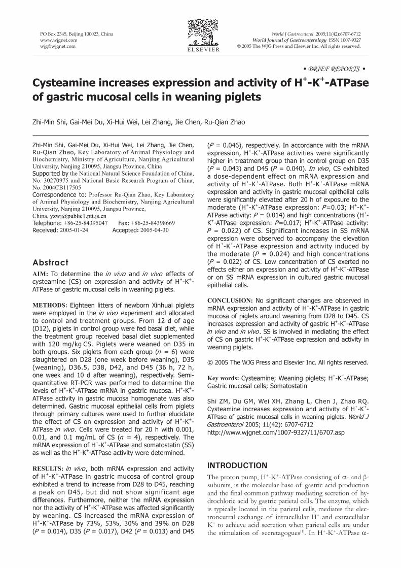
PO Box 2345, Beijing 100023, China World J Gastroenterol 2005;11(42):6707-6712www.wjgnet.com World Journal of Gastroenterology ISSN [email protected] © 2005 The WJG Press and Elsevier Inc. All rights reserved.E L S E V I E R
• BRIEF REPORTS •
(P = 0.046), respectively. In accordance with the mRNA expression, H+-K+-ATPase activities were significantly higher in treatment group than in control group on D35 (P = 0.043) and D45 (P = 0.040). In vivo , CS exhibited a dose-dependent effect on mRNA expression and activity of H+-K+-ATPase. Both H+-K+-ATPase mRNA expression and activity in gastric mucosal epithelial cells were signifi cantly elevated after 20 h of exposure to the moderate (H+-K+-ATPase expression: P =0.03; H+-K+-ATPase activity: P = 0.014) and high concentrations (H+-K+-ATPase expression: P=0.017; H+-K+-ATPase activity: P = 0.022) of CS. Significant increases in SS mRNA expression were observed to accompany the elevation of H+-K+-ATPase expression and activity induced by the moderate (P = 0.024) and high concentrations (P = 0.022) of CS. Low concentration of CS exerted no effects either on expression and activity of H+-K+-ATPase or on SS mRNA expression in cultured gastric mucosal epithelial cells.
CONCLUSION: No signifi cant changes are observed in mRNA expression and activity of H+-K+-ATPase in gastric mucosa of piglets around weaning from D28 to D45. CS increases expression and activity of gastric H+-K+-ATPase in vivo and in vivo . SS is involved in mediating the effect of CS on gastric H+-K+-ATPase expression and activity in weaning piglets.
© 2005 The WJG Press and Elsevier Inc. All rights reserved. Key words: Cysteamine; Weaning piglets; H+-K+-ATPase; Gastric mucosal cells; Somatostatin
Shi ZM, Du GM, Wei XH, Zhang L, Chen J, Zhao RQ. Cysteamine increases expression and activity of H+-K+-ATPase of gastric mucosal cells in weaning piglets. World J Gastroenterol 2005; 11(42): 6707-6712http://www.wjgnet.com/1007-9327/11/6707.asp
INTRODUCTIONThe proton pump, H+-K+-ATPase consisting of α- and β- subunits, is the molecular base of gastric acid production and the fi nal common pathway mediating secretion of hy-drochloric acid by gastric parietal cells. The enzyme, which is typically located in the parietal cells, mediates the elec-troneutral exchange of intracellular H+ and extracellular K+ to achieve acid secretion when parietal cells are under the stimulation of secretagogues[1]. In H+-K+-ATPase α-
Cysteamine increases expression and activity of H+-K+-ATPase of gastric mucosal cells in weaning piglets
Zhi-Min Shi, Gai-Mei Du, Xi-Hui Wei, Lei Zhang, Jie Chen, Ru-Qian Zhao
Zhi-Min Shi, Gai-Mei Du, Xi-Hui Wei, Lei Zhang, Jie Chen, Ru-Qian Zhao, Key Laboratory of Animal Physiology and Biochemistry, Ministry of Agriculture, Nanjing Agricultural University, Nanjing 210095, Jiangsu Province, ChinaSupported by the National Natural Science Foundation of China, No. 30270975 and National Basic Research Program of China, No. 2004CB117505Correspondence to: Professor Ru-Qian Zhao, Key Laboratory of Animal Physiology and Biochemistry, Nanjing Agricultural University, Nanjing 210095, Jiangsu Province, China. [email protected]: +86-25-84395047 Fax: +86-25-84398669 Received: 2005-01-24 Accepted: 2005-04-30
AbstractAIM: To determine the in vivo and in vivo effects of cysteamine (CS) on expression and activity of H+-K+-ATPase of gastric mucosal cells in weaning piglets.
METHODS: Eighteen litters of newborn Xinhuai piglets were employed in the in vivo experiment and allocated to control and treatment groups. From 12 d of age (D12), piglets in control group were fed basal diet, while the treatment group received basal diet supplemented with 120 mg/kg CS. Piglets were weaned on D35 in both groups. Six piglets from each group (n = 6) were slaughtered on D28 (one week before weaning), D35 (weaning), D36.5, D38, D42, and D45 (36 h, 72 h, one week and 10 d after weaning), respectively. Semi-quantitative RT-PCR was performed to determine the levels of H+-K+-ATPase mRNA in gastric mucosa. H+-K+-ATPase activity in gastric mucosa homogenate was also determined. Gastric mucosal epithelial cells from piglets through primary cultures were used to further elucidate the effect of CS on expression and activity of H+-K+-ATPase in vivo . Cells were treated for 20 h with 0.001, 0.01, and 0.1 mg/mL of CS (n = 4), respectively. The mRNA expression of H+-K+-ATPase and somatostatin (SS) as well as the H+-K+-ATPase activity were determined. RESULTS: in vivo , both mRNA expression and activity of H+-K+-ATPase in gastric mucosa of control group exhibited a trend to increase from D28 to D45, reaching a peak on D45, but did not show signif icant age differences. Furthermore, neither the mRNA expression nor the activity of H+-K+-ATPase was affected signifi cantly by weaning. CS increased the mRNA expression of H+-K+-ATPase by 73%, 53%, 30% and 39% on D28 (P = 0.014), D35 (P = 0.017), D42 (P = 0.013) and D45

6708 ISSN 1007-9327 CN 14-1219/ R World J Gastroenterol November 14, 2005 Volume 11 Number 42
or β-subunit defi ciency mice, achlorhydria and destruction of parietal cells have been observed[2,3]. The capability of gastric acid secretion is dependent on the gastric H+-K+-ATPase activity[4]. Therefore, H+-K+-ATPase activity can serve as an accurate indicator for evaluating the ability of gastric acid secretion from parietal cells.
Gastric acid secretion is regulated by stimulatory factors such as gastrin, histamine and acetylcholine, as well as inhibitory factors including somatostatin (SS). SS is a typical brain-gut-peptide releasing from D cells in the mucous membrane of stomach. Numerous publications reported that SS inhibits gastric acid secretion directly or indirectly by inhibiting the stimulatory effects of gastrin and histamine[5,6]. We found in our previous study that gastric expression of SS mRNA is upregulated in weaning piglets[7], and that gastric acid secretion is low in piglets[8]. Therefore, it is presumed that the increased inhibitory tone of SS is responsible for retarded gastric function development and insuffi cient gastric acid secretion which contribute, atleast partly, to diarrhea, poor growth and even death in newborn and early-weaning piglets.
Cysteamine (CS), which is able to deplete tissue SS, induces a profound loss of biological and immunological activities of SS both in vivo and in vitro[9,10]. CS is known to increase gastric acid secretion in rats, and is often used to produce the clinical model of gastric ulcer[11-13]. CS is approved to use as a feed additive in animal production to promote growth rate and improve feed efficiency[14]. However, CS application in pig production is mostly restricted to growing and fattening stages[15]. Up to now, the possible effect of CS on gastric acid secretion in weaning piglets remains unknown. Therefore, the present study was designed to examine the effect of CS on gastric acid secretion both in vivo and in vitro, the mRNA expression and activity of H+-K+-ATPase in gastric mucosa tissue and cultured mucosal epithelial cells were determined as response criteria. In addition, the change of SS mRNA expression in mucosal epithelial cells responding to CS exposure was also measured for elucidating the possible mechanisms underlying the CS action.
MATERIALS AND METHODSAnimals and samplingEighteen litters of newborn piglets from the 2nd or 3rd farrowing Xinhuai sows were employed in the in vivo experiment and allocated to control and treatment groups. From 12 d of age (D12), piglets in control group were fed basal diet, while the treatment group received basal diet supplemented with 120 mg/kg CS. The diet was formulated according to the requirement of piglets and provided ad libitum. Piglets were weaned on D35 in both groups. Six piglets from each group were slaughtered on D28 (one week before weaning), D35 (weaning), D36.5, D38, D42 and D45 (36 h, 72 h, one week and 10 d after weaning), respectively. Samples of the gastric fundic mucosa were frozen in liquid nitrogen immediately and then stored at -70 ºC until RNA extraction.
For in vitro experiment, four piglets at the age of D28 were killed to collect gastric mucosa for primary cell culture. DMEM (high glucose) and HEPES were products of Gibco, Hyclone, respectively. Trypsin was bought from Sigma and fetal bovine serum was purchased from Hangzhou Sijiqing Company, China.
Cells were dispersed from freshly obtained gastric mucosa of piglets as described previously[16], with minor modifications. Briefly, the gastric mucosa was washed in D-Hank’s solution containing 400 U/mL penicillin, 400 μg/mL streptomycin and dipped in D-Hank’s solution for 30 min. Then the tissues were dispersed by trypsin (0.15 mg/mL) at 37 °C for 1 h, fi ltrated and centrifuged (1 000 r/min, 5 min). Viability of the cells exceeded 95% as judged by trypan blue exclusion. Then cells at the density of 1×10⁶/mL were cultured (37 °C, 50 mL/L CO2) in a six-well plate containing DMEM (high glucose) with 10% fetal bovine serum, 15 mmol/L HEPES buffer, and 100 U/mL penicillin, 100 μg/mL streptomycin. After 24 h, the culture medium was refreshed by a new medium containing 0, 0.001, 0.01 and 0.1 mg/mL CS, respectively. The cells were continuously cultured for 20 h, and then collected for RNA extraction and H+-K+-ATPase activity determination.
The experiments were undertaken following the guidelines of the regional Animal Ethics Committee.
RNA extraction and analysisTotal RNA was extracted from the tissue samples with the single-step method of RNA extraction by acid guan-idinium thiocyanate-phenol-chloroform[17]. Total RNA concentration was then quantified by measuring the absorbance at 260 nm in a photometer (Eppendorf Biophotometer). Ratios of absorption (260/280 nm) of all preparations were between 1.8 and 2.0. Aliquots of RNA samples were subjected to electrophoresis through a 1.4% agarose-formaldehyde gel to verify their integrity.
Two micrograms of total RNA was reverse transcribed by incubation at 42 °C for 1 h in a 25 μL mixture consisting of 10 U avian myeloblastosis virus reverse transcriptase, 10 U RNase inhibitor, 12 μmol/L random primers, 50 mmol/L Tris-HCl (pH 8.3), 10 mmol/L MgCl2, 50 mmol/L KCl, 10 mmol/L DDT, 0.5 mmol/L spermidine and 0.8 mmol/L each dNTP. The reaction was terminated by heating at 95 °C for 5 min and quickly cooling on ice.
The primers for H+-K+-ATPase were designed according to the cDNA sequence published on GenBank (M22724): 5’-gagaaccaccacctacaag-3’ as sense primer, and 5’-caacagcgaactccaag-3’ as anti-sense primer, the predicted PCR product being 362 bp in size. The SS primers were designed according to the coding region of porcine SS genomic DNA sequence (GenBank, U36385): sense, 5’-agctgctgtctgaacccaac-3’ and anti-sense, 5’-gaaattcttgcagccagctt-3’, the expected PCR product being 161 bp in size. The PCR primers were designed using Primer Premier 5.0 and synthesized by Haojia Biotech. Ltd, China. The Quantum RNA 18S Internal Standards
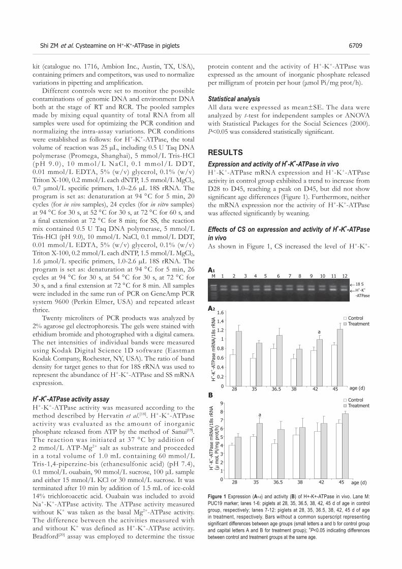
Shi ZM et al. Cysteamine on H+-K+-ATPase in piglets 6709
kit (catalogue no. 1716, Ambion Inc., Austin, TX, USA), containing primers and competitors, was used to normalize variations in pipetting and amplifi cation.
Different controls were set to monitor the possible contaminations of genomic DNA and environment DNA both at the stage of RT and RCR. The pooled samples made by mixing equal quantity of total RNA from all samples were used for optimizing the PCR condition and normalizing the intra-assay variations. PCR conditions were established as follows: for H+-K+-ATPase, the total volume of reaction was 25 μL, including 0.5 U Taq DNA polymerase (Promega, Shanghai), 5 mmol/L Tris-HCl (pH 9 .0 ) , 10 mmol/L NaCl , 0 .1 mmol/L DDT, 0.01 mmol/L EDTA, 5% (w/v) glycerol, 0.1% (w/v) Triton X-100, 0.2 mmol/L each dNTP, 1.5 mmol/L MgCl2, 0.7 μmol/L specifi c primers, 1.0–2.6 μL 18S rRNA. The program is set as: denaturation at 94 °C for 5 min, 20 cycles (for in vivo samples), 24 cycles (for in vitro samples) at 94 °C for 30 s, at 52 °C for 30 s, at 72 °C for 60 s, and a fi nal extension at 72 °C for 8 min; for SS, the reaction mix contained 0.5 U Taq DNA polymerase, 5 mmol/L Tris-HCl (pH 9.0), 10 mmol/L NaCl, 0.1 mmol/L DDT, 0.01 mmol/L EDTA, 5% (w/v) glycerol, 0.1% (w/v) Triton X-100, 0.2 mmol/L each dNTP, 1.5 mmol/L MgCl2, 1.6 μmol/L specifi c primers, 1.0-2.6 μL 18S rRNA. The program is set as: denaturation at 94 °C for 5 min, 26 cycles at 94 °C for 30 s, at 54 °C for 30 s, at 72 °C for 30 s, and a fi nal extension at 72 °C for 8 min. All samples were included in the same run of PCR on GeneAmp PCR system 9600 (Perkin Elmer, USA) and repeated atleast thrice.
Twenty microliters of PCR products was analyzed by 2% agarose gel electrophoresis. The gels were stained with ethidium bromide and photographed with a digital camera. The net intensities of individual bands were measured using Kodak Digital Science 1D software (Eastman Kodak Company, Rochester, NY, USA). The ratio of band density for target genes to that for 18S rRNA was used to represent the abundance of H+-K+-ATPase and SS mRNA expression.
H+-K+-ATPase activity assay H+-K+-ATPase activity was measured according to the method described by Hervatin et al.[18]. H+-K+-ATPase activity was evaluated as the amount of inorganic phosphate released from ATP by the method of Sanui[19]. The reaction was initiated at 37 °C by addition of 2 mmol/L ATP-Mg2+ salt as substrate and proceeded in a total volume of 1.0 mL containing 60 mmol/L Tris-1,4-piperzine-bis (ethanesulfonic acid) (pH 7.4), 0.1 mmol/L ouabain, 90 mmol/L sucrose, 100 μL sample and either 15 mmol/L KCl or 30 mmol/L sucrose. It was terminated after 10 min by addition of 1.5 mL of ice-cold 14% trichloroacetic acid. Ouabain was included to avoid Na+-K+-ATPase activity. The ATPase activity measured without K+ was taken as the basal Mg2+-ATPase activity. The difference between the activities measured with and without K+ was defined as H+-K+-ATPase activity. Bradford[20] assay was employed to determine the tissue
protein content and the activity of H+-K+-ATPase was expressed as the amount of inorganic phosphate released per milligram of protein per hour (μmol Pi/mg prot/h).
Statistical analysisAll data were expressed as mean±SE. The data were analyzed by t-test for independent samples or ANOVA with Statistical Packages for the Social Sciences (2000). P<0.05 was considered statistically signifi cant.
RESULTSExpression and activity of H+-K+-ATPase in vivoH+-K+-ATPase mRNA expression and H+-K+-ATPase activity in control group exhibited a trend to increase from D28 to D45, reaching a peak on D45, but did not show signifi cant age differences (Figure 1). Furthermore, neither the mRNA expression nor the activity of H+-K+-ATPase was affected signifi cantly by weaning.
Effects of CS on expression and activity of H+-K+-ATPase in vivoAs shown in Figure 1, CS increased the level of H+-K+-
1.6
1.4
1.2
1
0.8
0.6
0.4
0.2
0 28 35 36.5 38 42 45
H+-K
+-A
TPas
e m
RN
A/18
s rR
NA Control
Treatment
9
8
7
6
5
4
3
2
1
0 28 35 36.5 38 42 45
H+-K
+-A
TPas
e m
RN
A/18
s rR
NA
(μ m
oL P
i/mg
prot
/h)
a
Figure 1 Expression (A1-2) and activity (B) of H+-K+-ATPase in vivo. Lane M: PUC19 marker; lanes 1-6: piglets at 28, 35, 36.5, 38, 42, 45 d of age in control group, respectively; lanes 7-12: piglets at 28, 35, 36.5, 38, 42, 45 d of age in treatment, respectively. Bars without a common superscript representing signifi cant differences between age groups (small letters a and b for control group and capital letters A and B for treatment group); aP<0.05 indicating differences between control and treatment groups at the same age.
←←
18 SH+-K+
-ATPase
age (d)
age (d)
A1
A2
B
M 1 2 3 4 5 6 7 8 9 10 11 12
a
ControlTreatment

6710 ISSN 1007-9327 CN 14-1219/ R World J Gastroenterol November 14, 2005 Volume 11 Number 42
ATPase mRNA expression and the differences were significant on D28 (P = 0.014), D35 (P = 0.017), D42 (P = 0.013) and D45 (P = 0.046), respectively. Relative abundances of H+-K+-ATPase mRNA expression were significantly increased by 73%, 53%, 30%, and 39% in treatment group compared with control at the same age. CS supplementation increased markedly H+-K+-ATPase activity by 32.3% on D35 (P = 0.043) and 18.3% on D45 (P = 0.040), respectively, compared with that of the control counterparts.
Effects of CS on expression and activity of H+-K+-ATPase in vitroAs shown in Figure 2, low concentration of CS exhibited no effects on both mRNA expression and activity of H+-K+-ATPase, while moderate and high concentrations of CS markedly increased H+-K+-ATPase mRNA expression by 61% (P = 0.03) and 65% (P = 0.014), and H+-K+-ATPase activity by 48% (P = 0.017) and 50% (P = 0.022), respectively.
increased by 56% (P = 0.024) in moderate concentrations of CS and 64% (P = 0.022) in high concentrations of CS (Figure 3).
Effects of CS on SS mRNA expression in vitroLow concentration of CS had no infl uence on SS mRNA expression, whereas SS mRNA expression was markedly
DISCUSSIONThe developmental pattern of H+-K+-ATPase was found to be in agreement with that of gastric acid secretion capacity, both at the level of mRNA expression and enzyme activity. Yang et al.[21] reported that H+-K+-ATPase mRNA expression and activity in fundic gland keep increasing with age from gestational day 19.5 to week 18 of postnatal age in rats. The developmental pattern of human gastric H+-K+-ATPase from week 25 of gestation by Western blot analysis agrees with that of gastric pH recorded in preterm infants[22]. Furthermore, the H+-K+-ATPase mRNA content in rat fundus increases with age from one week to six weeks of age and the change parallels the developmental change of acid secretion capacity[23]. However, the developmental pattern of H+-K+-ATPase expression and activity in pigs has not been reported. In contrary to the reported data, the present study failed to show significant developmental change in both the expression and the activity of H+-K+-ATPase in piglets around weaning throughout the period of observation. It seems that the developmental pattern of H+-K+-ATPase is species-specifi c. In addition, it is documented that gastrin is involved in the regulation of H+-K+-ATPase expression. In ovine fetus, developmental pattern of gastrin mRNA agrees with that of H+-K+-ATPase expression[24]. Gastrin can stimulate H+-K+-ATPase expression[25] and improve its activity by increasing the intracellular Ca2+ concentration to initiate the phosphorization of H+-K+-ATPase in parietal cells[26]. In accordance with our fi ndings, gastrin contents in gastric tissues are stable from 4 to 6 wk of age in piglets[27].
M 1 2 3 4 5 6 7 8
←
←
18 S
H+-K+
-ATPase
1.4
1.2
1
0.8
0.6
0.4
0.2
00 0.001 0.01 0.1
a
H+-K
+-A
TPas
e m
RN
A/18
s rR
NA
a
concentration of CS (mg/mL)
3
2.5
2
1.5
1
0.5
00 0.001 0.01 0.1
concentration of CS (mg/mL)
a a
H+-K
+-A
TPas
e ac
tivity
(μ m
oL P
i/mg
prot
/h)
Figure 2 Effect of CS on expression (A1-2) and activity (B) of H+-K+-ATPase in vitro. Lane M: PUC19 marker, lanes 1-2: control; lanes 3-4: low concentration group; lanes 5-6: moderate concentration group; lanes 7-8: high concentration group. aP<0.05 vs 0 mg/mL of CS.
M 1 2 3 4 5 6 7 8
←
←
18 S
SS
1.4
1.2
1
0.8
0.6
0.4
0.2
00 0.001 0.01 0.1
concentration of CS (mg/mL)
SS m
RN
A/18
s rR
NA
aa
Figure 3 Effect of CS on SS mRNA expression in vitro. Lane M: PUC19 marker, lanes 1 and 2: control; lanes 3 and 4: low concentration group; lanes 5 and 6: moderate concentration group; lanes 7 and 8: high concentration group (A,B). aP<0.05 vs 0 mg/mL of CS.
A1
A2
B
B
A
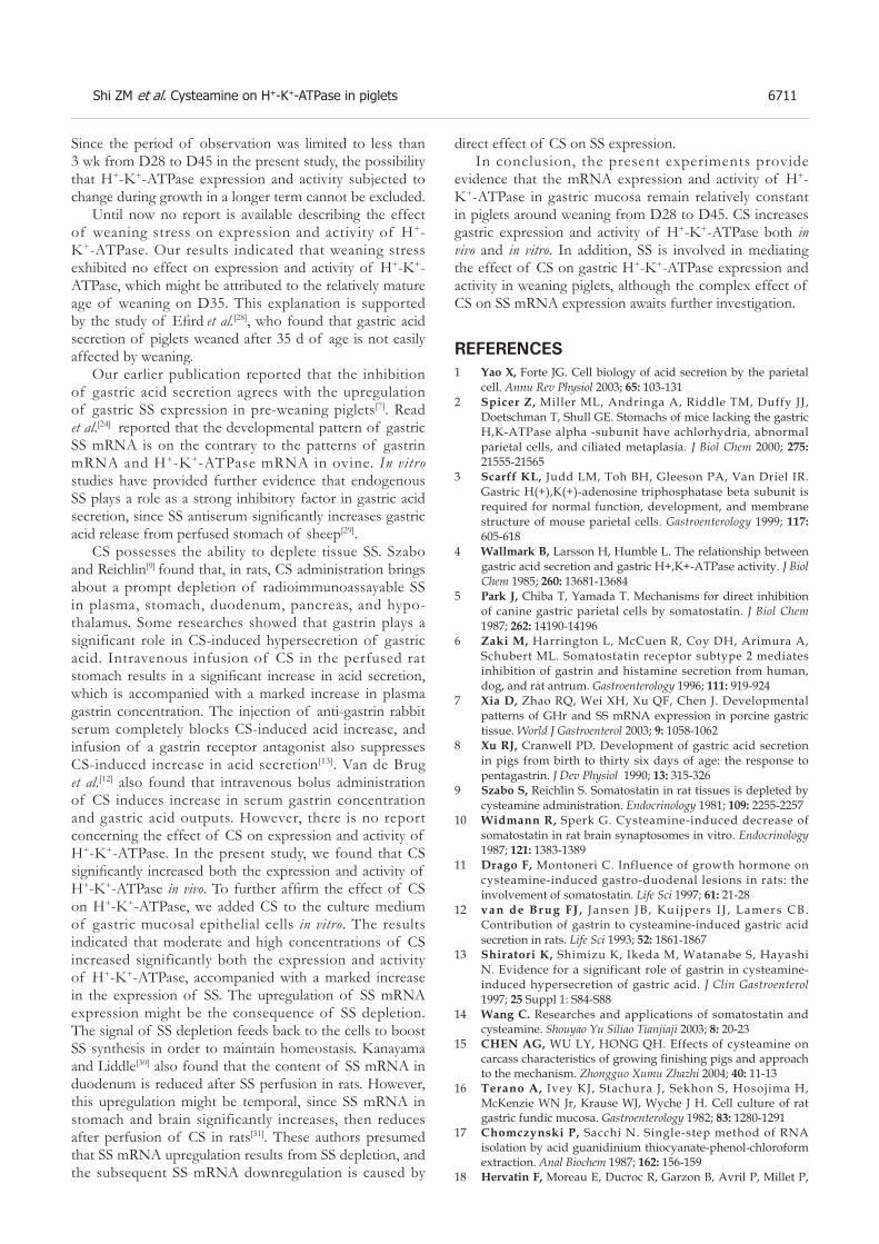
Shi ZM et al. Cysteamine on H+-K+-ATPase in piglets 6711
Since the period of observation was limited to less than 3 wk from D28 to D45 in the present study, the possibility that H+-K+-ATPase expression and activity subjected to change during growth in a longer term cannot be excluded.
Until now no report is available describing the effect of weaning stress on expression and activity of H+-K+-ATPase. Our results indicated that weaning stress exhibited no effect on expression and activity of H+-K+-ATPase, which might be attributed to the relatively mature age of weaning on D35. This explanation is supported by the study of Efi rd et al.[28], who found that gastric acid secretion of piglets weaned after 35 d of age is not easily affected by weaning.
Our earlier publication reported that the inhibition of gastric acid secretion agrees with the upregulation of gastric SS expression in pre-weaning piglets[7]. Read et al.[24] reported that the developmental pattern of gastric SS mRNA is on the contrary to the patterns of gastrin mRNA and H+-K+-ATPase mRNA in ovine. In vitro studies have provided further evidence that endogenous SS plays a role as a strong inhibitory factor in gastric acid secretion, since SS antiserum signifi cantly increases gastric acid release from perfused stomach of sheep[29].
CS possesses the ability to deplete tissue SS. Szabo and Reichlin[9] found that, in rats, CS administration brings about a prompt depletion of radioimmunoassayable SS in plasma, stomach, duodenum, pancreas, and hypo-thalamus. Some researches showed that gastrin plays a significant role in CS-induced hypersecretion of gastric acid. Intravenous infusion of CS in the perfused rat stomach results in a signifi cant increase in acid secretion, which is accompanied with a marked increase in plasma gastrin concentration. The injection of anti-gastrin rabbit serum completely blocks CS-induced acid increase, and infusion of a gastrin receptor antagonist also suppresses CS-induced increase in acid secretion[13]. Van de Brug et al.[12] also found that intravenous bolus administration of CS induces increase in serum gastrin concentration and gastric acid outputs. However, there is no report concerning the effect of CS on expression and activity of H+-K+-ATPase. In the present study, we found that CS signifi cantly increased both the expression and activity of H+-K+-ATPase in vivo. To further affi rm the effect of CS on H+-K+-ATPase, we added CS to the culture medium of gastric mucosal epithelial cells in vitro. The results indicated that moderate and high concentrations of CS increased significantly both the expression and activity of H+-K+-ATPase, accompanied with a marked increase in the expression of SS. The upregulation of SS mRNA expression might be the consequence of SS depletion. The signal of SS depletion feeds back to the cells to boost SS synthesis in order to maintain homeostasis. Kanayama and Liddle[30] also found that the content of SS mRNA in duodenum is reduced after SS perfusion in rats. However, this upregulation might be temporal, since SS mRNA in stomach and brain significantly increases, then reduces after perfusion of CS in rats[31]. These authors presumed that SS mRNA upregulation results from SS depletion, and the subsequent SS mRNA downregulation is caused by
direct effect of CS on SS expression. In conclusion, the present experiments provide
evidence that the mRNA expression and activity of H+-K+-ATPase in gastric mucosa remain relatively constant in piglets around weaning from D28 to D45. CS increases gastric expression and activity of H+-K+-ATPase both in vivo and in vitro. In addition, SS is involved in mediating the effect of CS on gastric H+-K+-ATPase expression and activity in weaning piglets, although the complex effect of CS on SS mRNA expression awaits further investigation.
REFERENCES1 Yao X, Forte JG. Cell biology of acid secretion by the parietal
cell. Annu Rev Physiol 2003; 65: 103-1312 Spicer Z, Miller ML, Andringa A, Riddle TM, Duffy JJ,
Doetschman T, Shull GE. Stomachs of mice lacking the gastric H,K-ATPase alpha -subunit have achlorhydria, abnormal parietal cells, and ciliated metaplasia. J Biol Chem 2000; 275: 21555-21565
3 Scarff KL, Judd LM, Toh BH, Gleeson PA, Van Driel IR. Gastric H(+),K(+)-adenosine triphosphatase beta subunit is required for normal function, development, and membrane structure of mouse parietal cells. Gastroenterology 1999; 117: 605-618
4 Wallmark B, Larsson H, Humble L. The relationship between gastric acid secretion and gastric H+,K+-ATPase activity. J Biol Chem 1985; 260: 13681-13684
5 Park J, Chiba T, Yamada T. Mechanisms for direct inhibition of canine gastric parietal cells by somatostatin. J Biol Chem 1987; 262: 14190-14196
6 Zaki M, Harrington L, McCuen R, Coy DH, Arimura A, Schubert ML. Somatostatin receptor subtype 2 mediates inhibition of gastrin and histamine secretion from human, dog, and rat antrum. Gastroenterology 1996; 111: 919-924
7 Xia D, Zhao RQ, Wei XH, Xu QF, Chen J. Developmental patterns of GHr and SS mRNA expression in porcine gastric tissue. World J Gastroenterol 2003; 9: 1058-1062
8 Xu RJ, Cranwell PD. Development of gastric acid secretion in pigs from birth to thirty six days of age: the response to pentagastrin. J Dev Physiol 1990; 13: 315-326
9 Szabo S, Reichlin S. Somatostatin in rat tissues is depleted by cysteamine administration. Endocrinology 1981; 109: 2255-2257
10 Widmann R, Sperk G. Cysteamine-induced decrease of somatostatin in rat brain synaptosomes in vitro. Endocrinology 1987; 121: 1383-1389
11 Drago F, Montoneri C. Influence of growth hormone on cysteamine-induced gastro-duodenal lesions in rats: the involvement of somatostatin. Life Sci 1997; 61: 21-28
12 van de Brug FJ , Jansen JB , Kui jpers I J , Lamers CB. Contribution of gastrin to cysteamine-induced gastric acid secretion in rats. Life Sci 1993; 52: 1861-1867
13 Shiratori K, Shimizu K, Ikeda M, Watanabe S, Hayashi N. Evidence for a significant role of gastrin in cysteamine-induced hypersecretion of gastric acid. J Clin Gastroenterol 1997; 25 Suppl 1: S84-S88
14 Wang C. Researches and applications of somatostatin and cysteamine. Shouyao Yu Siliao Tianjiaji 2003; 8: 20-23
15 CHEN AG, WU LY, HONG QH. Effects of cysteamine on carcass characteristics of growing fi nishing pigs and approach to the mechanism. Zhongguo Xumu Zhazhi 2004; 40: 11-13
16 Terano A, Ivey KJ, Stachura J, Sekhon S, Hosojima H, McKenzie WN Jr, Krause WJ, Wyche J H. Cell culture of rat gastric fundic mucosa. Gastroenterology 1982; 83: 1280-1291
17 Chomczynski P, Sacchi N. Single-step method of RNA isolation by acid guanidinium thiocyanate-phenol-chloroform extraction. Anal Biochem 1987; 162: 156-159
18 Hervatin F, Moreau E, Ducroc R, Garzon B, Avril P, Millet P,

6712 ISSN 1007-9327 CN 14-1219/ R World J Gastroenterol November 14, 2005 Volume 11 Number 42
Geloso JP. Ontogeny of rat gastric H+-K+-ATPase activity. Am J Physiol 1987; 252: G28-G32
19 Sanui H. Measurement of inorganic orthophosphate in biological materials: extraction properties of butyl acetate. Anal Biochem 1974; 60: 489-504
20 Bradford MM. A rapid and sensitive method for the quantitation of microgram quantities of protein utilizing the principle of protein-dye binding. Anal Biochem 1976; 72: 248-254
21 Yang DH, Tsuyama S, Murata F. The expression of gastric H+-K+-ATPase mRNA and protein in developing rat fundic gland. Histochem J 2001; 33: 159-166
22 Grahnquist L, Ruuska T, Finkel Y. Early development of human gastric H,K-adenosine triphosphatase. J Pediatr Gastroenterol Nutr 2000; 30: 533-537
23 Marino LR, Muglia BH, Yamada T. H(+)-K(+)-ATPase and carbonic anhydrase II gene expression in the developing rat fundus. Am J Physiol 1990; 259: G108-G115
24 Read MA, Chick P, Hardy KJ, Shulkes A. Ontogeny of gastrin, somatostatin, and the H+/K(+)-ATPase in the ovine fetus. Endocrinology 1992; 130: 1688-1697
25 Campbell VW, Yamada T. Acid secretagogue-induced
stimulation of gastric parietal cell gene expression. J Biol Chem 1989; 264: 11381-11386
26 Geibel J, Abraham R, Modlin I, Sachs G. Gastrin-stimulated changes in Ca2+ concentration in parietal cells depends on adenosine 3’,5’-cyclic monophosphate levels. Gastroenterology 1995; 109: 1060-1067
27 Xu RJ, Cranwell PD. Gastrin in fetal and neonatal pigs. Comp Biochem Physiol B 1991; 98: 615-621
28 Efi rd RC, Armstrong WD, Herman DL. The development of digestive capacity in young pigs: effects of age and weaning system. J Anim Sci 1982; 55: 1380-1387
29 Westbrook SL, McDowell GH, Hardy KJ, Shulkes A. Active immunization against somatostatin alters regulation of gastrin in response to gastric acid secretagogues. Am J Physiol 1998; 274: G751-G756
30 Kanayama S, Liddle RA. Somatostatin regulates duodenal cholecystokinin and somatostatin messenger RNA. Am J Physiol 1990; 258: G358-G364
31 Papachristou DN, Liu JL, Patel YC. Cysteamine-induced reduction in tissue somatostatin immunoreactivity is associated with alterations in somatostatin mRNA. Regul Pept 1994; 49: 237-247
Science Editor Wang XL and Guo SY Language Editor Elsevier HK
PO Box 2345, Beijing 100023, China World J Gastroenterol 2005;11(42):6713-6715www.wjgnet.com World Journal of Gastroenterology ISSN [email protected] © 2005 The WJG Press and Elsevier Inc. All rights reserved.E L S E V I E R
• BRIEF REPORTS •
http://www.wjgnet.com/1007-9327/11/6713.asp
INTRODUCTIONHepatitis E virus (HEV) is an unclassifi ed, non-enveloped RNA virus, a causative agent of acute hepatit is E transmitted principally via the fecal-oral route. The virus can cause large water-borne epidemics of the disease and sporadic cases as well. Hepatitis E occurs predominantly in developing countries usually affecting young adults with a fatality rate of 15-20% in pregnant women[1]. However, no effective treatment is currently available for hepatitis E and there are no commercial vaccines for hepatitis E in the world. Although at least four major genotypes of HEV have been identifi ed, only one serotype of HEV is recognized. DNA vaccine can synthesize viral proteins within the host cells and induce humoral and cellular immune responses[2,3]. In this study, we constructed eucaryotic expression plasmid containing HEV ORF2 fragment and full-length ORF3 (DNA vaccine) chimeric gene and inoculated it to BALB/c mice to detect the specifi c humoral and cellular immune responses in mice.
MATERIALS AND METHODSConstruction of plasmid All PCR primers were designed according to the nucleotide sequence of a Chinese HEV isolate (DDBJ accession number D11092)[4]. HEV mRNA was extracted from the feces of a patient with hepatitis E in Hangzhou, Zhejiang Province. The HEV ORF2 fragment and full-length ORF3 chimeric gene were amplifi ed by RT-PCR. The PCR product was inserted into an eucaryotic expression plasmid pcDNA3 to form a recombinant plasmid pcHEV23 (DNA vaccine) (Figure 1).
DNA inoculation protocolThirty-two female BALB/c mice (18-20 g) provided by Zhejiang Experimental Animal Center were used for immunization and divided into four groups: Group 1 was injected with 100 μL saline solution as control. Group 2 was injected with 100 μg/100 μL vector pcDNA3 as control. Group 3 was injected with 100 μg/100 μL pcHEV23 plasmid. Group 4 was injected with 200 μg/100 μL pcHEV23 plasmid. Three weeks after the fi rst injection, mice were bled and then boosted by same method. After another 3 wk, mice were boosted again for the second time (Table 1).
Hepatitis E virus chimeric DNA vaccine elicits immunologic response in mice
Yan Hong, Bing Ruan, Lian-Hua Yang, Yong Chen, Luo Jing, Yi-Ting Wang, Hua-Jun Hu
Yan Hong, Lian-Hua Yang, Yong Chen, Luo Jing, Yi-Ting Wang, Hua-Jun Hu, Institute of Bioengineering, Zhejiang Academy of Medical Sciences, Hangzhou 310013, Zhejiang Province, ChinaBing Ruan, Zhejiang University Medical School, Hangzhou 310003, Zhejiang Province, ChinaSupported by the Grants from the Natural Science Foundation of Zhejiang Province, No. RC01054, Science and Technology Department of Zhejiang Province, No. F11023 and Key Project of Health Bureau of Zhejiang ProvinceCorrespondence to: Dr. Yong Chen, Institute of Bioengineering, Zhejiang Academy of Medical Sciences, Hangzhou 310013, Zhejiang Province, China. [email protected]: +86-0571-88862228 Fax: +86-0571-88075447Received: 2005-03-12 Accepted: 2005-04-11
AbstractAIM: To construct the plasmid pcHEV23 containing fragments of HEV ORF2 and ORF3 chimeric gene and to assess its ability to elicit specifi c immunologic response in mice.
METHODS: The gene encoding the structural protein of HEV ORF2 fragment and full-length ORF3 was amplifi ed by PCR. The PCR products were cloned into an eucaryotic expression plasmid pcDNA3. The resulting plasmid pcHEV23 was used as a DNA vaccine to inoculate BALB/c mice intramuscularly thrice at a dose of 100 or 200 μg. Mice injected with empty pcDNA3 DNA or saline served as control and then specific immune responses in the mice were detected.
RESULTS: After 2-3 times of inoculation, all mice injected with pcHEV23 had anti-HEV IgG seroconversion and specifi c T lymphocyte proliferation. The lymphocyte stimulation index in the group immunized with pcHEV23 (3.1±0.49) was higher than that in the control group (0.787±0.12, P<0.01). None in the control group had a detectable level of anti-HEV IgG.
CONCLUSION: DNA vaccine containing HEV ORF2 and ORF3 chimeric gene can successfully induce specific humoral and cellular immune response in mice.
© 2005 The WJG Press and Elsevier Inc. All rights reserved.
Hong Y, Ruan B, Yang LH, Chen Y, Jing L, Wang YT, Hu HJ. Hepatitis E virus chimeric DNA vaccine elicits immunologic response in mice. World J Gastroenterol 2005; 11(42): 6713-6715

6714 ISSN 1007-9327 CN 14-1219/ R World J Gastroenterol November 14, 2005 Volume 11 Number 42
Serological testTwo weeks after the fi rst three injections, blood samples were collected. All serum specimens were tested for anti-HEV IgG by EIA. In brief, microwell plates (Nunc, Roskilde, Denmark) were coated overnight at 4 °C with purified HEV ORF23 proteins (expressed in E coli) at 1 μg/mL in carbonate–bicarbonate buffer (pH 9.6). The wells were washed thrice with 0.05% Tween 20 in PBS (PBS-T) and then blocked with 2% BSA in PBS-T at 37 °C for 1 h. Following three washes with PBS-T, serum samples diluted in 2% BSA were added to the plates and incubated at 37 °C for 1 h. Following fi ve washes, HRP-conjugated anti-human IgG (Sigma) diluted 2 000-fold in PBS-T was added to detect the bound antibodies. Following incubation at 37 °C for 1 h, the plates were washed as above and the substrate tetramethylbenzidine solution (Sigma) was added to the wells. After incubation
at room temperature for 15 min, color development was stopped by adding 2 mol/L H2SO4. Optical density (OD) at 450 nm was determined with an ELISA reader. The cut-off values were set for each test as 2.1 times the mean value of negative control samples.
T-lymphocyte proliferationAll the animals were killed 14 d after the last injection to analyze cellular immune responses. Single cell suspension of splenocytes was prepared for each individual animal. Splenocytes were immediately cultured in the presence of HEV ORF23 (20 μg/mL, expressed in E coli). Sixty-eighth day after culture, MTT was then added to the cells to measure antigen-specific proliferation. Lymphocyte stimulation index (SI) was measured according to formula: SI = A570 (antigen stimulation)/A570 (control).
RESULTSConstruction of pcHEV23 plasmidThe sequencing data indicated that the pcHEV23 construct (Figure 1) contained the correct orientation of the HEV ORF2 and ORF3 fragments (sequencing data not shown).
Analysis of anti-HEV response in pcHEV23-inoculated miceNone of the mice had seroconversion after the initial injection of pcHEV23. Following the third dose of pcHEV23, anti-HEV IgG titers in groups 3 and 4 was higher than those in groups 1 and 2 (P<0.01, Table 2).
T-lymphocyte proliferation assayThe lymphocyte SI in the g roup immunized with pcHEV23 was higher than that in saline group and pcDNA3 in control DNA group (P<0.01). Results are shown in Table 3.
DISCUSSIONDNA vaccines represent a new and potentially powerful approach for the development of subunit vaccines. DNA vaccines induce a broad range of immune responses due to efficient priming of T lymphocytes[5]. This novel approach to vaccination is attractive as it offers several
Group Mice (n) Immunogen Time of injection (wk)
1 8 Saline 0 3 62 8 pcDNA3(100 μg/100 μL) 0 3 63 8 pcHEV23(100 μg/100 μL) 0 3 64 8 pcHEV23(200 μg/100 μL) 0 3 6
Table 1 Protocol of DNA injection
Group 1 2 3 4
Mean±SD 0.116±0.009b 0.210±0.028b 0.353±0.085b 0.336±0.066b
Table 2 Anti-HEV IgG titers after the third injection (mean±SD)
bP<0.01 vs control.
Group 1 2 3 4
0.787±0.12b 1.54±0.25b 3.1±0.49b 2.85±0.59b
Table 3 Result of assay of T-cell proliferation (mean±SD)
bP<0.01 vs control.
Figure 1 Construction of pcHEV23 plasmid (pcDNA3-ORF23).
Extracted
Feces of HE patientExtracted
HEV mRNA
↓RT-PCR HEVORF2 (0.8 kb)
BamHI/EcoRI
Feces of HE patient
HEV mRNA
↓RT-PCRHEVORF3 (375 bp)
EcoRI/XhoI
↓
pcDNA35 400 bp
AmpicillinP CMV
BamHIXhoI
Neomycin
CoIE
SV40 poly A
EcoRI
Ampicillin
Ampicillin
CoIE1
SV40 poly A
Neomycin
XhoI
BamHI
pcDNA3-ORF236575 bp
ORF23
pcDNA3-ORF26200 bp
SV40 poly A
Neomycin
P CMV
BamHI
↓
Hong Y et al. HEV chimeric DNA vaccine 6715
desirable features. First, DNA is not infectious and does not replicate and encodes only the protein or proteins of interest. Second, DNA is stable and can be made inexpensively in large quantities at a high level of purity. Third, plasmid DNA does not contain any heterologous protein components compared to a recombinant virus vaccine. Fourth, DNA vaccine can induce both cell-mediated and humoral immunity. Final ly, antigen expression persists after DNA vaccination, promoting the induction of long-lived memory immune cells[6]. DNA immunization can be defined as a physical delivery of nucleic acids in vivo to express antigenic proteins and elicit specifi c immune responses. Direct naked DNA inoculation obviates the requirement of purifi ed antigens. Pathogenic antigens synthesized inside the inoculated host cells can be processed in a natural form to develop classes I and II-regulated immune responses, mimicking the aspects of live attenuated virus.
Epitope mapping demonstrates that there are at least 7 immunodominant epitopes in HEV ORF2 region and four epitopes in HEV ORF3 region[7,8]. In this study, the full length of HEV ORF3 cDNA and partial HEV ORF2 cDNA were combined and used as an antigen-coding sequence for construction of HEV DNA vaccine.
In conclusion, direct injection of pcHEV23 is able to induce specific anti-HEV IgG and T-lymphocyte proliferation. HEV DNA vaccine constructed by us can
successfully induce both humoral and cellular immune responses and appears to be a viable alternative to the recombinant protein subunit vaccine candidate.
REFERENCES1 Wang L, Zhuang H. Hepatitis E: an overview and recent
advances in vaccine research. World J Gastroenterol 2004; 10: 2157-2162
2 Tang DC, DeVit M, Johnston SA. Genetic immunization is a simple method for eliciting an immune response. Nature 1992; 356: 152-154
3 Wolff JA, Malone RW, Williams P, Chong W, Acsadi G, Jani A, Felgner PL. Direct gene transfer into mouse muscle in vivo. Science 1990; 247: 1465-1468
4 Bi SL, Purdy MA, McCaustland KA, Margolis HS, Bradley DW. The sequence of hepatitis E virus isolated directly from a single source during an outbreak in China. Virus Res 1993; 28: 233-247
5 Sarzotti M, Dean TA, Remington MP, Ly CD, Furth PA, Robbins DS. Induction of cytotoxic T cell responses in newborn mice by DNA immunization. Vaccine 1997; 15: 795-797
6 Pardoll DM, Beckerleg AM. Exposing the immunology of naked DNA vaccines. Immunity 1995; 3: 165-169
7 Khudyakov YE, Khudyakova NS, Fields HA, Jue D, Starling C, Favorov MO, Krawczynski K, Polish L, Mast E, Margolis H. Epitope mapping in proteins of hepatitis E virus. Virology 1993; 194: 89-96
8 Khudyakov YuE, Favorov MO, Jue DL, Hine TK, Fields HA. Immunodominant antigenic regions in a structural protein of the hepatitis E virus. Virology 1994; 198: 390-393
Science Editor Wang XL and Guo SY Language Editor Elsevier HK

PO Box 2345, Beijing 100023, China World J Gastroenterol 2005;11(42):6716-6721www.wjgnet.com World Journal of Gastroenterology ISSN [email protected] © 2005 The WJG Press and Elsevier Inc. All rights reserved.E L S E V I E R
• BRIEF REPORTS •
in carcinogenesis and progression of human gastric carcinoma in vivo , possibly via p27 proteolysis. PTEN may regulate the expression of p27 by negatively regulating Skp2 expression.
© 2005 The WJG Press and Elsevier Inc. All rights reserved.
Key words: Gastric carcinoma; Skp2; p27; PTEN
Ma XM, Liu Y, Guo JW, Liu JH, Zuo LF. Relation of overex-pression of S phase kinase-associated protein 2 with reduced expression of p27 and PTEN in human gastric carcinoma. World J Gastroenterol 2005; 11(42): 6716-6721 http://www.wjgnet.com/1007-9327/11/6716.asp
INTRODUCTIONDysregulation of the cell cycle is required for the for-mation of most malignant tumors. Progression of the cell cycle is controlled by interactions between cell cycle control proteins (cyclins) and their catalytically active cyclin-dependent kinase (CDKs). The activity of each cyclin-CDK complex is in turn regulated by several different mechanisms; the most important being negative regulation by CDK inhibitors[1]. p27 is an inhibitor of cyclinE-CDK2 and cyclinA-CDK2, which drive cells from G1 to S phase of the cell division cycle[2,3]. Loss of p27 function therefore accelerates cell cycle progression and predisposes cells to malignant transformation, as is well illustrated by the observation of increased tumor incidence in hemizygous and homozygous p27-deleted mutant mice after carcinogen exposure[4,5]. Many clinical studies also indicate that low levels of p27 are associated with high aggressiveness and poor prognosis in a large variety of malignant tumors[2,3], including breast carcinoma[6,7], colorectal carcinoma[8], lung cancer[9], prostate cancer[10] and gastric carcinoma[11].
The amount of p27 is mainly regulated by post-translational ubiquitin-proteasome-mediated proteolysis[12]. Cell cycle-dependent degradation of p27 is dependent on phosphorylation at Thr187 in late G1 phase by CDK2 under positive regulation by cyclinE. Thr187 phosphorylation is a necessary prerequisite for the sequential addition of ubiquitin molecules by an ubiquitin ligase complex, SCFskp2 composed of Skp1, Cull, Rbx1 and the F-box protein Skp2[3]. Polyubiquitination of p27 then targets p27 for degradation in proteasome, thus removing the p27 cell
Relation of overexpression of S phase kinase-associated protein 2 with reduced expression of p27 and PTEN in human gastric carcinoma
Xiu-Mei Ma, Ying Liu, Jian-Wen Guo, Jiang-Hui Liu, Lian-Fu Zuo
Xiu-Mei Ma, Ying Liu, Jian-Wen Guo, Jiang-Hui Liu, Lian-Fu Zuo. The Fourth Affi liated Hospital of Hebei Medical University, Shijiazhuang 050011, Hebei Province, ChinaCorrespondence to: Lian-Fu Zuo, Hebei Provincial Tumor Institute, the Fourth Affiliated Hospital of Hebei Medical University, Shijiazhuang 050011, Hebei Province, China. [email protected]: +86-0311-86033941-337 Received: 2005-01-24 Accepted: 2005-05-25
AbstractAIM: To investigate the signifi cance of S phase kinase-associated protein 2 (Skp2) expression in human gastric carcinoma and the relation between expressions of Skp2, p27 and PTEN.
METHODS: Immunohistochemica l analys is was performed on 138 gastric carcinoma specimens, their paired adjacent mucosa specimens, 102 paired lymphatic metastatic carcinoma tissue specimens, 30 dysplasia specimens, 30 intestinal metaplasia specimens, 10 chronic superficial gastritis specimens and 5 normal gastric mucosa specimens for Skp2 expression and on 138 gastric carcinoma specimens for p27 and PTEN expression.
RESULTS: Skp2 labeling frequency was significantly higher in intestinal metaplasia (12.68±0.86) and adjacent mucosa (19.32±1.22) than in normal gastric mucosa (0.53±0.13) and chronic superficial gastritis (0.47±0.19) (P = 0.000); in dysplasia (16.74±0.82) than in intestinal metaplasia (P = 0.000); in gastric primary carcinoma (31.34±2.17) than in dysplasia and adjacent mucosa (P = 0.000); in metastasis gastric carcinoma in lymph nodes (39.76±2.00) than in primary gastric carcinoma (P = 0.037), respectively. Skp2 labeling frequency was positively associated with differentiation degree (rho = 0.315, P = 0.000), vessel invasion (rho = 0.303, P = 0.000) and lymph node metastasis (rho = 0.254, P = 0.000) of gastric cancer. Expression o f S kp2 was nega t i ve l y a s so c i a t ed w i t h p27 (rho = -0.451, P = 0.000) and PTEN (rho = -0.480, P = 0.000) expression in gastric carcinoma. p27 ex-pression was positively associated with PTEN expression in gastric carcinoma (rho = 0.642, P = 0.000).
CONCLUSION: Skp2 overexpression may be involved

Ma XM et al. Skp2, p27 and PTEN expression in human gastric carcinoma 6717
cycle “brake” and allowing cells to transition from G1 to S phase[13].
Some investigations have shown that Skp2 is a specifi c substrate-recognition subunit of SCFskp2, expression of Skp2 is required for the ubiquitination and subsequent degradation of p27 in vitro[20-22], and Skp2 knock-out cells show high levels of p27[16]. The level of p27 has also been reported to be inversely related to that of Skp2 in lymphoma[24], oral squamous cell carcinoma[18,19] and colorectal carcinoma[20].
Recently, Mamillapalli et al.[21] reported that PTEN, a tumor suppressor, regulates the ubiquitin-dependent degradation of p27 through SCFSkp2. Yang et al . [22] suggested that induction of Skp2 may be causally linked with decreased levels of p27 in prostate cancer and implicate PTEN in the regulation of Skp2 expression in vivo.
Recent studies have shown that Skp2 has oncogenic potential in breast epithelial cells and is overexpressed in a subset of breast carcinomas (ER- and Her-2 negative)[23] and Skp2 can mediate transformation and is upregulated during oral epithelial carcinogenesis[18]. Recently, a line of evidence also indicates a possible relationship between Skp2 expression and the malignancy of tumors. Skp2 expression has been shown to be greatly increased in malignantly transformed cell lines including oral squamous cell carcinoma[18] and correlates directly with the grade of malignancy of lymphoma[17] and oral squamous cell carcinoma[18]. Kudo et al. [19] reported that high Skp2 expression is also correlated with poor prognosis in oral squamous cell carcinoma. Thus, Skp2 may have a great signifi cance in human carcinogenesis. However, few studies are available regarding the signifi cance of Skp2 expression in human gastric carcinomas in vivo and there are no studies regarding the relationship between Skp2, p27 and PTEN expression. We therefore investigated the signifi cance of Skp2 expression in human gastric carcinomas and the relationship between Skp2, p27 and PTEN expression in vivo.
MATERIALS AND METHODSPatients and gastric specimensOne hundred and thir ty-eight surgical ly resected gastric carcinoma specimens, paired adjacent mucosa specimens and paired 102 lymphatic gastric carcinoma tissue specimens metastatically selected from the Fourth Affi liated Hospital of Hebei Medical University were used in this study. All gastric carcinoma patients underwent total or subtotal gastrectomy and no patient received any treatment for cancer before surgery. All regional lymph nodes were removed. The patients comprised 111 males and 27 females with a mean age of 58.5 years and a median age of 60 years (from 36 to 78 years). After surgery, gastric specimens were fixed in 40 g/L neutral-buffered formaldehyde and embedded in paraffin. Additionally, 75 biopsy cases (5 cases of normal gastric mucosa, 10 cases of chronic superfi cial gastritis, 30 cases of intestinal
metaplasia and 30 cases of dysplasia) were included in this study as well. To avoid evaluator variability, all pathological diagnoses were done by two pathologists. Clinical stage was done according to the International Union Contrele Cancer criteria published in 1997.
ImmunohistochemistryA standard Non-Biotin HRP two-step immunohis-tochemical method (Zymed) was used. Four-micrometer-thick sections were deparaffinized and rehydrated. Endogenous peroxidase in sections was inactivated in 30 mL/L hydrogen peroxide (H2O2) in ethanol for 15 min at room temperature. The sections were washed thrice for 5 min with 0.01 mol/L phosphate-buffered saline (PBS) and then heated in a citrate buffer (0.01 mol/L, pH 6.4) for PTEN and p27 or in a EDTA buffer (1 mmol/L, pH 8.0) for Skp2 in an 800-W microwave oven for 12 min for antigen retrieval. The sections were incubated with mouse monoclonal antibody to Skp2 (1:50 dilution, Zymed), p27 (1:50 dilution, Santa Cruz) and PTEN (1:50 dilution, Zymed) overnight at 4 °C, washed thrice for 5 min with 0.01 mol/L PBS 3 and then incubated with common IgG (Fab fraction)-HRP complex for 30 min at room temperature. Immunoreactive products were visualized using 3,3’-diaminobenzidine/H2O2, and finally the sections were counterstained with hematoxylin. Some sections were incubated with PBS instead of primary antibody as negative control to verify the specifi city of the immunoreactions. Vascular endothelial cells showed strong PTEN expression with a nuclear predominance and served as an internal positive control for PTEN in this study. The positive breast adenocarcinoma served as a positive control for Skp2 and p27.
Immunohistochemical quantitationImmunostaining of nuclear Skp2, p27 and PTEN in each specimen was evaluated microscopically and recorded as the percentage of Skp2, p27, and PTEN-positive cells (labeling frequency), after at least 1 000 nuclei at the lesion site were calculated in at least fi ve high-power fi elds (×400). All specimens were evaluated without any knowledge of the patients' clinical information.
Statistical analysisThe differences in Skp2 labeling frequencies among specimens of normal gastric mucosa, chronic superficial gastr i t is, intest inal metaplas ia , dysplas ia , gastr ic adenocarcinoma, adjacent mucosa and lymphatic metastatic gastric carcinoma were compared by Mann-Whitney U test. The relation between Skp2 expression and the clinical and pathological variables was evaluated using the Spearman correlation coeffi cient. The Spearman’s correlation coeffi cient testing was also used to determine the relation between Skp2, PTEN and p27 as well as between PTEN and p27. P<0.05 was considered statistically signifi cant. All analyses were performed using SPSS 11.0 statistical software.

6718 ISSN 1007-9327 CN 14-1219/ R World J Gastroenterol November 14, 2005 Volume 11 Number 42
RESULTSExpression of Skp2 in human gastric specimenThe Skp2 immunoreactivity was predominantly localized in the nuclei of gastric cells (Figure 1). In 5% of the cancer specimens examined, a weak or moderate cytoplasmic immunoreactivity could be seen in addition to the predominant nuclear reactivity in cancer cells. Skp2 labeling frequency in normal gastric mucosa, intestinal metaplasia, dysplasia, primary gastric carcinoma and lymphatic metastatic gastric carcinoma was signifi cantly higher than in normal gastric mucosa and chronic superfi cial gastritis (P = 0.000); it was in dysplasia than in intestinal metaplasia (P = 0.000); in primary gastric carcinoma than in dysplasia (P = 0.000); in lymphatic metastatic gastric carcinoma than in primary gastric carcinoma (P = 0.037, Table 1).
Relation between Skp2 labeling frequency and clinico-pathological features in gastric carcinomaRe la t ion be tween Skp2 l abe l ing f r equency and clinicopathological variables including age, gender, histological differentiation, depth of invasion, vessel invasion, lymphatic metastasis, distant metastasis as well as clinical stage is summarized in Table 2. Skp2 labeling frequency was negatively associated with age, gender, depth of invasion, distant metastasis as well as clinical stage. A significant correlation was found between the
1,2 ,3 ,4Frequency was significantly higher than in normal gastric tissue and chronic superfi cial gastritis (P = 0.000,all the same; Mann-Whitney U test). 2Frequency was signifi cantly higher than in intestinal metaplasia (P = 0.000; Mann-Whitney U test). 3Frequency was signifi cantly higher than in intestinal metaplasia and dysplasia, (P = 0.000,all the same; Mann-Whitney U test). 5Frequency was Significantly higher than in primary tumors (P = 0.037; Mann-Whitney U test).
1, 2, 3Skp2 labeling frequency was positively correlated with differentiated degree (rho = 0.315, P = 0.000), vessel invasion (rho = 0.303, P = 0.000), and lymph node metastasis (rho = 0.254, P = 0.000) in gastric carcinoma, respectively. 4Because clinical staging could not be performed due to the lack of accurate records about lymph node in operation, data of 12 cases were cancelled in statistical analysis.
A B
C D
E F
Figure 1 Skp2, p27 and PTEN expression in different gastric tissues (DAB and hematoxylin stain). A: Positive Skp2 in nuclei of intestinal metaplasia and dysplasia cells (original magnification ×400); B: Positive Skp2 in nuclei of well-differentiated gastric carcinoma cells (original magnification ×400); C: Positive Skp2 in nuclei of poorly differentiated gastric carcinoma cells (original magnifi cation ×100); D: Positive Skp2 in lymphatic metastatic gastric carcinoma cells (original magnifi cation ×100); E: Positive p27 in nuclei of well-differentiated gastric carcinoma cells (original magnifi cation ×400); F: Positive PTEN in nuclei of well-differentiated gastric carcinoma cells (original magnifi cation ×400).
Specimens Skp2 labeling frequency(%)Tissue (n) Median Mean±SE
Normal gastric mucosa 5 0.00 0.47±0.19Chronic superfi cial gastritis 10 0.00 0.53±0.13Intestinal metaplasia 30 12.00 12.68±0.86Dysplasia 30 19.00 16.74±0.82Primary gastric carcinoma 138 29.00 31.34±2.17Adjacent mucosa 138 16.006 19.32±1.22Metastasis tumor tissue in lymph node
102 39.507 39.76±2.00
Table 1 Skp2 protein expression in human gastric tissues
Specimens Skp2 labeling frequency (%)
Clinicopathological feature
(n)
Mean±SE
rho PPatients 138 36.32±1.22Age (yr) 138 58.50 0.190 0.833 <60 66 34.25±3.58 ≥60 72 34.41±2.63Gender 0.119 0.179 Females 27 24.96±5.21 Males 111 32.90±2.37Differentiation grade 0.315 0.000 Well/Moderate 63 22.41±2.59 Poor 75 38.85±3.10Depth of invasion 0.001 0.986 Serosal 24 25.84±2.45 Outside soft tissue 114 29.25±4.87Vessel invasion 0.303 0.000 Absent 120 27.89±2.11 Present 18 54.39±6.84Lymph node metastasis 0.254 0.000 Absent 36 27.94±3.38 Present 102 37.14±2.69Distant metastasis 0.091 0.307 Absent 26 22.77±6.92 Present 12 31.79±2.38Clinical stage 0.069 0.4400 0Ia 0Ib 3 22.78±6.92II 36 29.81±3.14IIIa 12 44.67±6.78IIIb 66 34.50±3.55IV 9 53.00±0.57
Table 2 Skp2 protein expression in 138 human gastric cancers

Ma XM et al. Skp2, p27 and PTEN expression in human gastric carcinoma 6719
plays a fundamental role in the regulation of eukaryotic cell cycle. Proteolysis of G1 regulatory proteins is mediated by SCF ubiquitin ligase complex (SCFSkps) composed of four major subunits, Skp1, Cul1, Rbx1/Roc1 and one of the F-box proteins (Fbps)[24]. There are 11 Fbps in budding yeast, 22 in Drosophila[25] and 38 in human beings[26,27]. Skp2 is a Fbp first identified together with Skp1 as an interactor of the cyclinA-Cdk2 complex which drives cells from G1 to S phase, hence it is named as S phase kinase-associated protein 2 (Skp2)[24].
In this study, we found that Skp2 immunoreactivity was rare in normal gastric mucosa and chronic superfi cial gastritis, but Skp2 labeling frequency was significantly increased during the course of intestinal metaplasia, dysplasia and primary gastric carcinoma. From pathological point of view, development of gastric adenocarcinoma involves progression through a well-defined series of histological steps initiated by the change of normal mucosa to chronic superficial gastritis, followed by the appearance of atrophic gastritis and intestinal metaplasia, then dysplasia and fi nally adenocarcinoma[28]. These results suggest that Skp2 may have oncogenic potential in gastric carcinoma and is involved in gastric carcinogenesis. It may become a new biomarker in gastric carcinogenesis. Additionally, we found that Skp2 labeling frequency in adjacent mucosa of gastric carcinoma was significantly higher than that in normal gastric mucosa, suggesting that some adjacent mucosae of gastric carcinoma have normal histological structure, but the expression of some proteins is changed.
In this study, we found that Skp2 labeling frequency was positively correlated with differentiation degree, lymphatic metastasis and vessel invasion of gastric carcinoma. Poorly differentiated gastric carcinoma and gastric carcinoma with lymphatic metastasis and vessel invasion tended to have a higher Skp2 labeling frequency. Skp2 labeling frequency was higher in lymphatic metastatic gastric carcinoma than in primary gastric carcinoma. The overexpression of Skp2 could modulate the malignant phenotype of gastric carcinoma cells, suggesting that overexpression of Skp2 may be associated with metastasis
Skp2 labeling frequency and differentiation degree (rho = 0.315, P = 0.000), vessel invasion (rho = 0.303, P = 0.000) and lymphatic metastasis (rho = 0.254, P = 0.000). Poorly differentiated gastric carcinoma, gastric carcinoma with vessel invasion and gastric carcinoma with lymphatic metastasis tended to have a higher Skp2 labeling frequency.
Relation between Skp2, p27 and PTEN expression in gastric carcinomaGiven the biochemical link between Skp2 and p27, we asked whether there was a correlation between Skp2 and p27 levels in gastric carcinoma using the same set of gastric carcinoma specimens used for the Skp2 analysis. A statistically significant inverse relation between p27 and Skp2 labeling frequency was evident (rho = -0.451, P = 0.000; Figure 2A). In vitro study linking loss of PTEN with increased Skp2 levels led us[29] to also compare the expression of PTEN and Skp2 using the same set of gastric carcinoma specimens. Interestingly, we found that increased Skp2 expression was significantly correlated with loss and decrease of PTEN expression in gastric carcinoma (rho = -0.480, P = 0.000; Figure 2B). Additionally, a positive relation between p27 and PTEN expression was observed in gastric carcinoma (rho = 0.642, P = 0.000; Figure 3).
DISCUSSIONProtein degradation by the ubiquitin–proteasome pathway
836984969467689582
100
80
60
40
20
0
-20N= 76 23 22 17 0-24 25-49 50-74 75-100
P27 labeling frequency (%)
Skp2
labe
ling
freq
uenc
y (%
)
rho = -0.451P = 0.000
100
80
60
40
20
0
-20N= 68 13 20 37
0-24 25-49 50-74 75-100PTEN labeling frequency (%)
Skp2
labe
ling
freq
uenc
y (%
) rho = -0.480P = 0.000
A
B
Figure 2 Correlation of Skp2 with p27 (A) and PTEN (B) protein levels. Each box and the associated bars represent the values of middle 50% and the range of the data respectively. The dark line within a box denotes the median.
120
100
80
60
40
20
0
-20N= 76 23 22 17 0-24 25-49 50-74 75-100
P27 labeling frequency (%)
rho = 0.642P = 0.000
PTEN
labe
ling
freq
uenc
y (%
)
Figure 3 Correlation of p27 and PTEN expression in human gastric carcinoma. Each box and the associated bars represent the values of middle 50% and the range of the data respectively. The dark line within a box denotes the median.
6720 ISSN 1007-9327 CN 14-1219/ R World J Gastroenterol November 14, 2005 Volume 11 Number 42
potential of gastric carcinoma cells. Further studies need to be done to clarify their mechanism.
In the present study, we also found that Skp2 expression was inversely correlated with the expression of p27, which is consistent with the results reported in other studies[18,19,21]. These results suggest that Skp2 overexpression is associated with p27 protein deg-radation in cancer tissue, which may be a reason why Skp2 overexpression is involved in carcinogenesis and progression of cancer. Furthermore, Skp2 expression was also inversely correlated with PTEN expression and p27 expression was positively related with PTEN expression in gastric carcinoma. PTEN tumor suppressor acts as a phosphatase of phosphatidylinositol-3,4,5-trisphosphate (PIP3) and negatively controls the G1/S cell cycle transition and regulates the levels of p27[29]. Mamillapalli et al.[ 21] showed that PTEN defi ciency in mouse embryonic stem cells decreases p27 level while increases Skp2 level. Conversely, in human glioblastoma cells, ectopic PTEN expression leads to p27 accumulation accompanied with a reduction of Skp2 and ectopic expression of Skp2 alone is sufficient to reverse PTEN-induced p27 accumulation, restore the kinase activity of cyclin E/CDK2 and partially overcome the PTEN-induced G1
cell cycle arrest. Recombinant SCFSkp2 complex or Skp2 protein alone can rescue the defect in p27 ubiquitination in extracts from cells treated with a PI 3-kinase inhibitor. Yang et al.[30] showed that loss of expression or reduced expression of PTEN protein contributes to carcinogenesis and progression of gastric carcinoma. Myung et al.[31] reported that reduced expression of cyclin-dependent kinase inhibitor p27 is associated with advanced stage and invasiveness of gastric carcinoma. Additionally, Yang et al.[22] also found that elevated Skp2 protein expression in human prostate cancer is associated with loss of p27 and PTEN expression. Thus, all these findings suggest that PTEN functions as a negative regulator of the Skp2 pathway that is normally used to control S-phase entry through the regulation of p27 in gastric carcinoma and the effects of Skp2, p27 and PTEN together play an important role in carcinogenesis and progression of gastric carcinoma. These results also suggest that Skp2 functions as a critical component in the PTEN/PI3-kinase pathway for the regulation of p27.
In conclusion, alterations in the levels of proteins controlled by the ubiquitin–proteasome pathway during transformation are likely to be common. Further studies that defi ne how such posttranscriptional control pathways
are altered during transformation may provide additional biomarkers or facilitate the identification of novel therapeutic targets. The fi nding that Skp2 is induced in a number of different cancers suggests that drugs directed at this molecule may provide a more selective target for therapeutic development.
REFERENCES1 Sherr CJ. Cancer cell cycles. Science 1996; 274: 1672-16772 Philipp-Staheli J, Payne SR, Kemp CJ. p27(Kip1): regulation
and function of a haploinsuffi cient tumor suppressor and its misregulation in cancer. Exp Cell Res 2001; 264: 148-168
3 Slingerland J, Pagano M. Regulation of the cdk inhibitor p27 and its deregulation in cancer. J Cell Physiol 2000; 183: 10-17
4 Fero ML, Randel E, Gurley KE, Roberts JM, Kemp CJ. The murine gene p27Kip1 is haplo-insufficient for tumour suppression. Nature 1998; 396: 177-180
5 Nakayama K, Ishida N, Shirane M, Inomata A, Inoue T, Shishido N, Hori I, Loh DY, Nakayama K. Mice lacking p27(Kip1) display increased body size, multiple organ hyperplasia, retinal dysplasia, and pituitary tumors. Cell 1996; 85: 707-720
6 Tan P, Cady B, Wanner M, Worland P, Cukor B, Magi- Galluzzi C, Lavin P, Draetta G, Pagano M, Loda M. The cell cycle inhibitor p27 is an independent prognostic marker in small (T1a,b) invasive breast carcinomas. Cancer Res 1997; 57: 1259-1263
7 Catzavelos C, Bhattacharya N, Ung YC, Wilson JA, Roncari L, Sandhu C, Shaw P, Yeger H, Morava-Protzner I, Kapusta L, Franssen E, Pritchard KI, Slingerland JM. Decreased levels of the cell-cycle inhibitor p27Kip1 protein: prognostic implications in primary breast cancer. Nat Med 1997; 3: 227-230
8 Loda M, Cukor B, Tam SW, Lavin P, Fiorentino M, Draetta GF, Jessup JM, Pagano M. Increased proteasome-dependent degradation of the cyclin-dependent kinase inhibitor p27 in aggressive colorectal carcinomas. Nat Med 1997; 3: 231-234
9 Esposito V, Baldi A, De Luca A, Groger AM, Loda M, Giordano GG, Caputi M, Baldi F, Pagano M, Giordano A. Prognostic role of the cyclin-dependent kinase inhibitor p27 in non-small cell lung cancer. Cancer Res 1997; 57: 3381-3385
10 Tsihlias J, Kapusta LR, DeBoer G, Morava-Protzner I, Zbieranowski I, Bhattacharya N, Catzavelos GC, Klotz LH, Slingerland JM. Loss of cyclin-dependent kinase inhibitor p27Kip1 is a novel prognostic factor in localized human prostate adenocarcinoma. Cancer Res 1998; 58: 542-548
11 Ohtani M, Isozaki H, Fujii K, Nomura E, Niki M, Mabuchi H, Nishiguchi K, Toyoda M, Ishibashi T, Tanigawa N. Impact of the expression of cyclin-dependent kinase inhibitor p27Kip1 and apoptosis in tumor cells on the overall survival of patients with non-early stage gastric carcinoma. Cancer 1999; 85: 1711-1718
12 Pagano M, Tam SW, Theodoras AM, Beer-Romero P, Del Sal G, Chau V, Yew PR, Draetta GF, Rolfe M. Role of the ubiquitin-proteasome pathway in regulating abundance of the cyclin-dependent kinase inhibitor p27. Science 1995; 269: 682-685
13 Eguchi H, Herschenhous N, Kuzushita N, Moss SF. Helicobacter pylori increases proteasome-mediated degradation of p27(kip1) in gastric epithelial cells. Cancer Res 2003; 63: 4739-4746
14 Carrano AC, Eytan E, Hershko A, Pagano M. SKP2 is required for ubiquitin-mediated degradation of the CDK inhibitor p27. Nat Cell Biol 1999; 1: 193-199
15 Tsvetkov LM, Yeh KH, Lee SJ, Sun H, Zhang H. p27(Kip1) ubiquitination and degradation is regulated by the SCF(Skp2) complex through phosphorylated Thr187 in p27. Curr Biol 1999; 9: 661-664
16 Nakayama K, Nagahama H, Minamishima YA, Matsumoto M, Nakamichi I, Kitagawa K, Shirane M, Tsunematsu R, Tsukiyama T, Ishida N, Kitagawa M, Nakayama K, Hatakeyama S. Targeted disruption of Skp2 results in accumulation of cyclin E and p27(Kip1), polyploidy and centrosome overduplication. EMBO J 2000; 19: 2069-2081
17 Latres E, Chiarle R, Schulman BA, Pavletich NP, Pellicer A, Inghirami G, Pagano M. Role of the F-box protein Skp2 in lymphomagenesis. Proc Natl Acad Sci USA 2001; 98: 2515-2520
18 Gstaiger M, Jordan R, Lim M, Catzavelos C, Mestan J, Slingerland J, Krek W. Skp2 is oncogenic and overexpressed in human cancers. Proc Natl Acad Sci USA 2001; 98: 5043-5048
19 Kudo Y, Kitajima S, Sato S, Miyauchi M, Ogawa I, Takata T. High expression of S-phase kinase-interacting protein 2, human F-box protein, correlates with poor prognosis in oral squamous cell carcinomas. Cancer Res 2001; 61: 7044-7047
Ma XM et al. Skp2, p27 and PTEN expression in human gastric carcinoma 6721
20 Hershko D, Bornstein G, Ben-Izhak O, Carrano A, Pagano M, Krausz MM, Hershko A. Inverse relation between levels of p27(Kip1) and of its ubiquitin ligase subunit Skp2 in colorectal carcinomas. Cancer 2001; 91: 1745-1751
21 Mamillapalli R, Gavrilova N, Mihaylova VT, Tsvetkov LM, Wu H, Zhang H, Sun H. PTEN regulates the ubiquitin-dependent degradation of the CDK inhibitor p27(KIP1) through the ubiquitin E3 ligase SCF(SKP2). Curr Biol 2001; 11: 263-267
22 Yang G, Ayala G, De Marzo A, Tian W, Frolov A, Wheeler TM, Thompson TC, Harper JW. Elevated Skp2 protein expression in human prostate cancer: association with loss of the cyclin-dependent kinase inhibitor p27 and PTEN and with reduced recurrence-free survival. Clin Cancer Res 2002; 8: 3419-3426
23 Signoretti S, Di Marcotullio L, Richardson A, Ramaswamy S, Isaac B, Rue M, Monti F, Loda M, Pagano M. Oncogenic role of the ubiquitin ligase subunit Skp2 in human breast cancer. J Clin Invest 2002; 110: 633-641
24 Carrano AC, Pagano M. Role of the F-box protein Skp2 in adhesion-dependent cell cycle progression. J Cell Biol 2001; 153: 1381-1390
25 Rubin GM, Yandell MD, Wortman JR, Gabor Miklos GL, Nelson CR, Hariharan IK, Fortini ME, Li PW, Apweiler R, Fleischmann W, Cherry JM, Henikoff S, Skupski MP, Misra S, Ashburner M, Birney E, Boguski MS, Brody T, Brokstein P, Celniker SE, Chervitz SA, Coates D, Cravchik A, Gabrielian
A, Galle RF, Gelbart WM, George RA, Goldstein LS, Gong F, Guan P, Harris NL, Hay BA, Hoskins RA, Li J, Li Z, Hynes RO, Jones SJ, Kuehl PM, Lemaitre B, Littleton JT, Morrison DK, Mungall C, O’Farrell PH, Pickeral OK, Shue C, Vosshall LB, Zhang J, Zhao Q, Zheng XH, Lewis S. Comparative genomics of the eukaryotes. Science 2000; 287: 2204-2215
26 Cenciarelli C, Chiaur DS, Guardavaccaro D, Parks W, Vidal M, Pagano M. Identifi cation of a family of human F-box proteins. Curr Biol 1999; 9: 1177-1179
27 Winston JT, Koepp DM, Zhu C, Elledge SJ, Harper JW. A family of mammalian F-box proteins. Curr Biol 1999; 9: 1180-1182
28 Konturek PC, Kania J, Konturek JW, Nikiforuk A, Konturek SJ, Hahn EG. H.pylori infection, atrophic gastritis, cytokines, gastrin, COX-2, PPAR gamma and impaired apoptosis in gastric carcinogenesis. Med Sci Monit 2003; 9: SR53-SR66
29 Gottschalk AR, Basila D, Wong M, Dean NM, Brandts CH, Stokoe D, Haas-Kogan DA. p27Kip1 is required for PTEN-induced G1 growth arrest. Cancer Res 2001; 61: 2105-2111
30 Yang L, Kuang LG, Zheng HC, Li JY, Wu DY, Zhang SM, Xin Y. PTEN encoding product: a marker for tumorigenesis and progression of gastric carcinoma. World J Gastroenterol 2003; 9: 35-39
31 Myung N, Kim MR, Chung IP, Kim H, Jang JJ. Loss of p16 and p27 is associated with progression of human gastric cancer. Cancer Lett 2000; 153: 129-136
Science Editor Wang XL and Guo SY Language Editor Elsevier HK

PO Box 2345, Beijing 100023, China World J Gastroenterol 2005;11(42):6722-6724www.wjgnet.com World Journal of Gastroenterology ISSN [email protected] © 2005 The WJG Press and Elsevier Inc. All rights reserved.E L S E V I E R
• CASE REPORT •
common cancers worldwide[1]. HCC is developed mainly in patients with chronic hepatitis C or B virus infections and the screening of this high risk group makes it possible to detect HCC at the early stage. However, many patients are still diagnosed at the advanced stage and the prognosis is poor. Transcatheter chemoembolization or chemotherapies are performed in cases of advance HCC[1-3]. These treatments are sometimes very effective, but are harmful to the background liver and accelerate the progression of pre-existing chronic liver injury. Therefore, these therapies cannot be performed in patients with severe liver cirrhosis. In addition, severe side effects often impair the quality of life. Thus, the development of new low-invasive therapies is needed.
Vitamin K is a factor in blood coagulation and is used for the treatment of osteoporosis[4]. Vitamin K is also known to inhibit the growth of cancer cell lines, but its clinical usefulness in the treatment of human cancer remains controversial[5-7]. In this report, we present a case of advanced HCC in which marked regression during the administration of vitamin K has been documented.
CASE REPORTIn July 2003, an 85-year-old man with liver cirrhosis due to HCV and diabetes mellitus was admitted to our hospital because of giant liver tumors. The patient had no history of alcohol drinking or blood transfusion. He did not smoke and was not taking any medicine, but was under intermediate-acting insulin (12 U/d) injection. He had undergone surgery for prostate hypertrophy 3 years before the present admission. Routine laboratory studies showed Hb 13.8 g/L, platelet count 101 000/mm3, albumin 3.2 g/L, total bilirubin 0.6 mg/dL, aspartate aminotransferase and alanine aminotransferase 232 U/L and 102 U/L respectively, γ-glutamyl transpeptidase 93 IU/L, fasting blood glucose 126 mg/dL, HbA1c 11.1%. Serum alpha-fetoprotein (AFP), lentil lectin-A-reactive AFP (AFP-L3), and des-γ-carboxy prothrombin (DCP) were 1 212 ng/mL, 76.9%, and 51 300 mAU/mL, respectively. The patient was positive for HCV antibody and hepatitis B virus core antigen but negative for hepatitis B virus surface antibody. There were no esophageal or gastric varices observed during gastro-intestinal endoscopy. Although the patient did not complain of abdominal pain, gastric ulcer was detected by endoscopy and administration of the proton pump inhibitor was started.
Regression of hepatocellular carcinoma during vitamin K administration
Kazuhiro Nouso, Shuji Uematsu, Kunihiro Shiraga, Ryoichi Okamoto, Ryo Harada, Shoko Takayama, Wakako Kawai, Shigeru Kimura, Toru Ueki, Nobuaki Okano, Masahiro Nakagawa, Motowo Mizuno, Yasuyuki Araki, Yasushi Shiratori
Kazuhiro Nouso, Shuji Uematsu, Kunihiro Shiraga, Ryoichi Okamoto, Ryo Harada, Shoko Takayama, Wakako Kawai, Shigeru Kimura, Toru Ueki, Nobuaki Okano, Masahiro Na-kagawa, Motowo Mizuno, Yasuyuki Araki, Department of In-ternal Medicine, Hiroshima City Hospital, Hiroshima 730-8518, JapanYasushi Shiratori, Department of Medicine and Medical Science, Okayama University Graduate School of Medicine and Dentistry, Okayama 700-8558, JapanCorrespondence to: Kazuhiro Nouso, MD, PhD, Department of Internal Medicine, Hiroshima City Hospital, 7-33 Motomachi, Naka-ku, Hiroshima-city, Hiroshima 730-8518, Japan. [email protected]: +81-82-221-2291 Fax: +81-82-223-1447Received: 2005-01-24 Accepted: 2005-02-18
AbstractAn 85-year-old man with HCV infection and diabetes mellitus was diagnosed as having hepatocellular carci-noma (HCC, 13 cm in diameter) based on high serum alpha-fetoprotein (AFP), AFP-L3, and des-γ-carboxy pro-thrombin levels as well as typical enhancement pattern on contrast-enhanced CT. The patient did not receive any interventional treatments because of advanced age and the advanced stage of HCC. He chose to take vitamin K, which was reported to suppress the growth of HCC in vitro . Three months after starting vitamin K, all three tumor markers were normalized and HCC was markedly regressed, showing no enhancement in the early arterial phase on CT. Here we present the report describing the regression of HCC during the administration of vitamin K. © 2005 The WJG Press and Elsevier Inc. All rights reserved.
Key words: Hepatocellular carcinoma; Vitamin K; Reg-ression
Nouso K, Uematsu S, Shiraga K, Okamoto R, Harada R, Takayama S, Kawai W, Kimura S, Ueki T, Okano N, Nakaga-wa M, Mizuno M, Araki Y, Shiratori Y. Regression of hepato-cellular carcinoma during vitamin K administration. World J Gastroenterol 2005; 11(42): 6722-6724http://www.wjgnet.com/1007-9327/11/6722.asp
INTRODUCTIONHepatocellular carcinoma (HCC) is one of the most
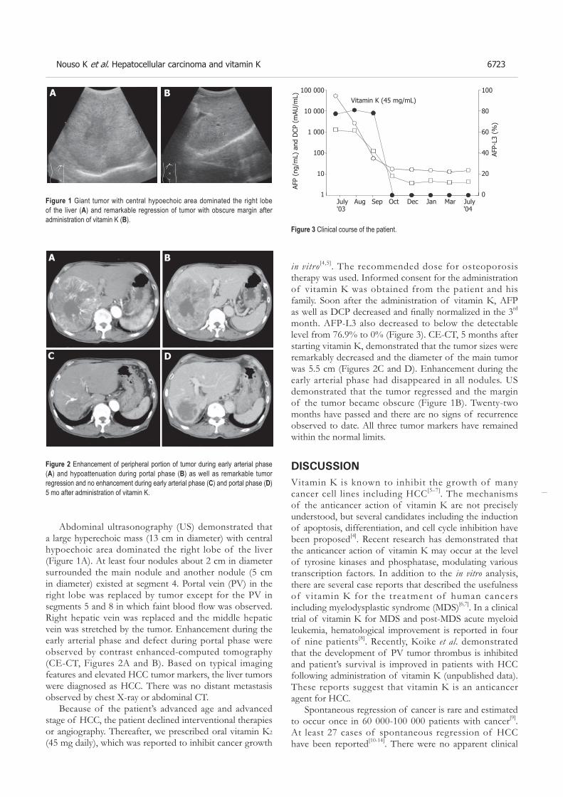
Nouso K et al. Hepatocellular carcinoma and vitamin K 6723
Abdominal ultrasonography (US) demonstrated that a large hyperechoic mass (13 cm in diameter) with central hypoechoic area dominated the right lobe of the liver (Figure 1A). At least four nodules about 2 cm in diameter surrounded the main nodule and another nodule (5 cm in diameter) existed at segment 4. Portal vein (PV) in the right lobe was replaced by tumor except for the PV in segments 5 and 8 in which faint blood fl ow was observed. Right hepatic vein was replaced and the middle hepatic vein was stretched by the tumor. Enhancement during the early arterial phase and defect during portal phase were observed by contrast enhanced-computed tomography (CE-CT, Figures 2A and B). Based on typical imaging features and elevated HCC tumor markers, the liver tumors were diagnosed as HCC. There was no distant metastasis observed by chest X-ray or abdominal CT.
Because of the patient’s advanced age and advanced stage of HCC, the patient declined interventional therapies or angiography. Thereafter, we prescribed oral vitamin K2 (45 mg daily), which was reported to inhibit cancer growth
in vitro [4,5]. The recommended dose for osteoporosis therapy was used. Informed consent for the administration of vitamin K was obtained from the patient and his family. Soon after the administration of vitamin K, AFP as well as DCP decreased and fi nally normalized in the 3rd month. AFP-L3 also decreased to below the detectable level from 76.9% to 0% (Figure 3). CE-CT, 5 months after starting vitamin K, demonstrated that the tumor sizes were remarkably decreased and the diameter of the main tumor was 5.5 cm (Figures 2C and D). Enhancement during the early arterial phase had disappeared in all nodules. US demonstrated that the tumor regressed and the margin of the tumor became obscure (Figure 1B). Twenty-two months have passed and there are no signs of recurrence observed to date. All three tumor markers have remained within the normal limits.
DISCUSSIONVitamin K is known to inhibit the growth of many cancer cell lines including HCC[5–7]. The mechanisms of the anticancer action of vitamin K are not precisely understood, but several candidates including the induction of apoptosis, differentiation, and cell cycle inhibition have been proposed[4]. Recent research has demonstrated that the anticancer action of vitamin K may occur at the level of tyrosine kinases and phosphatase, modulating various transcription factors. In addition to the in vitro analysis, there are several case reports that described the usefulness of vitamin K for the treatment of human cancers including myelodysplastic syndrome (MDS)[6,7]. In a clinical trial of vitamin K for MDS and post-MDS acute myeloid leukemia, hematological improvement is reported in four of nine patients[8]. Recently, Koike et al. demonstrated that the development of PV tumor thrombus is inhibited and patient’s survival is improved in patients with HCC following administration of vitamin K (unpublished data). These reports suggest that vitamin K is an anticancer agent for HCC.
Spontaneous regression of cancer is rare and estimated to occur once in 60 000-100 000 patients with cancer[9]. At least 27 cases of spontaneous regression of HCC have been reported[10-14]. There were no apparent clinical
A B
Figure 1 Giant tumor with central hypoechoic area dominated the right lobe of the liver (A) and remarkable regression of tumor with obscure margin after administration of vitamin K (B).
A B
C D
Figure 2 Enhancement of peripheral portion of tumor during early arterial phase (A) and hypoattenuation during portal phase (B) as well as remarkable tumor regression and no enhancement during early arterial phase (C) and portal phase (D) 5 mo after administration of vitamin K.
Figure 3 Clinical course of the patient.
100 000
10 000
1 000
100
10
1
100
80
60
40
20
0July Aug Sep Oct Dec Jan Mar July'03 '04
AFP
(ng/
mL)
and
DCP
(m
AU/m
L)
AFP-
L3 (
%)
Vitamin K (45 mg/mL)

6724 ISSN 1007-9327 CN 14-1219/ R World J Gastroenterol November 14, 2005 Volume 11 Number 42
characteristics discriminating the patient with spontaneous regression of HCC from others and various causes of regression have been documented (e.g. lack of blood supply due to arterial infarction or rapid growth of HCC, withdrawal from steroids, abstinence of alcohol consumption, administration of herbal medicines, and immunological activation due to high fever or sepsis)[10,15]. Hepatic angiography was performed in some cases and blood shortage due to the intimal damage induced by micro catheters could also be the cause of regression. In this report, the speed of tumor growth was uncertain because the patient was not followed up before admission. Other than that, he did not have any of the background factors reported previously. Therefore, possible reasons for regression are limited in this case; spontaneous regression due to blood shortage induced by rapid tumor growth or regression due to vitamin K administration, which was reported to inhibit tumor growth.
Although we could not conclude that administration of vitamin K was the cause of the regression in this case, this is a case report documenting regression of HCC during the administration of vitamin K. Further prospective study is needed to confirm the efficacy of vitamin K for the treatment of HCC.
REFERENCES1 Llovet JM, Burroughs A, Bruix J. Hepatocellular carcinoma.
Lancet 2003; 362: 1907-19172 Leung TW, Johnson PJ. Systemic therapy for hepatocellular
carcinoma. Semin Oncol 2001; 28: 514-5203 Anthony PP . Hepatocellular carcinoma: an overview.
Histopathology 2001; 39: 109-118 4 Lamson DW, Plaza SM. The anticancer effects of vitamin K.
Altern Med Rev 2003; 8: 303-3185 Wang Z, Wang M, Finn F, Carr BI. The growth inhibitory
effects of vitamins K and their actions on gene expression. Hepatology 1995; 22: 876-882
6 Yaguchi M, Miyazawa K, Otawa M, Katagiri T, Nishimaki J, Uchida Y, Iwase O, Gotoh A. Kawanishi Y, Toyama K. Vitamin K2 selectively induces apoptosis of blastic cells in myelodysplastic syndrome: flow cytometric detection of apoptotic cells using APO2.7 monoclonal antibody. Leukemia 1998; 12: 1392-1397
7 Nishimaki J, Miyazawa K, Yaguchi M, Katagiri T, Kawanishi Y, Toyama K, Ohyashiki K, Hashimoto S, Nakaya K, Takiguchi T. Vitamin K2 induces apoptosis of a novel cell line established from a patient with myelodysplastic syndrome in blastic transformation. Leukemia 1999; 13: 1399-1405
8 Miyazawa K, Nishimaki J, Ohyashiki K, Enomoto S, Kuriya S, Fukuda R, Hotta T, Teramura M, Mizoguchi H, Uchiyama T. Omine M. Vitamin K2 therapy for myelodysplastic syndromes (MDS) and post-MDS acute myeloid leukemia: information through a questionnaire survey of multi-center pilot studies in Japan. Leukemia 2000; 14: 1156-1157
9 WH C. Efforts to explain spontaneous regression of cancer. J Surg Oncol 1981; 17: 201-209
10 Lin TJ, Liao LY, Lin CL, Shih LS, Chang TA, Tu HY, Chen RC, Wang CS. Spontaneous regression of hepatocellular carcinoma: a case report and literature review. Hepatogastroenterology 2004; 51: 579-582
11 Iiai T , Sato Y, Nabatame N, Yamamoto S, Makino S, Hatakeyama K. Spontaneous complete regression of hepa-tocellular carcinoma with portal vein tumor thrombus. Hepatogastroenterology 2003; 50: 1628-1630
12 Morimoto Y, Tanaka Y, Itoh T, Yamamoto S, Mizuno H, Fushimi H. Spontaneous necrosis of hepatocellular carcinoma: a case report. Dig Surg 2002; 19: 413-418
13 Abiru S, Kato Y, Hamasaki K, Nakao K, Nakata K, Eguchi K. Spontaneous regression of hepatocellular carcinoma associated with elevated levels of interleukin 18. Am J Gastroenterol 2002; 97: 774-775
14 Ikeda M , Okada S, Ueno H, Okusaka T, Kuriyama H. Spontaneous regression of hepatocellular carcinoma with multiple lung metastases: a case report. Jpn J Clin Oncol 2001; 31: 454-458
15 Takeda Y , Togashi H, Shinzawa H, Miyano S, Ishii R, Karasawa T, Saito T, Saito K, Haga H, Matsuo T, Aoki M, Mitsuhashi H, Watanabe H, Takahashi T. Spontaneous regression of hepatocellular carcinoma and review of literature. J Gastroenterol Hepatol 2000; 15: 1079-1086
Science Editor Wang XL and Guo SY Language Editor Elsevier HK

PO Box 2345, Beijing 100023, China World J Gastroenterol 2005;11(42):6725-6727www.wjgnet.com World Journal of Gastroenterology ISSN [email protected] © 2005 The WJG Press and Elsevier Inc. All rights reserved.E L S E V I E R
• CASE REPORT •
as indolent benign disease in young adults with a median age of approximately 35 years and is equally distributed between males and females[2,3]. A subset of patients with plasma cell variant may have systemic symptoms. UCD in the abdomen and pelvis has been only described in the radiology literature as a focal enhancing mass of varying locations, including the retroperitoneum, mesentery, and hepatic porta[4].
CASE REPORTA 53-year-old man was well until one week before admission with renal colic with an onset of pain strongest at the right paravertebral region and radiating in the right iliac crest. Excretion urography performed in another hospital showed a slight pyelectasis of the right kidney and a left renal duplication with ureter bifidus, while, incidentally, abdominal ultrasound showed a calcifi ed mass (3.4 cm × 3.4 cm) in the left hilar region of the kidney. Then the patient was subsequently referred to our tertiary care centre for further evaluation and treatment . On physical examination the patient was afebrile and appeared well. The pulse was 80 bpm and the blood pressure 130/70 mm Hg. No lymphadenopathy was found. A non-tender liver edge descended 2 to 3 cm below the right costal margin; the spleen was not felt. Peripheral blood counts, liver function tests, cancer antigen (CA 19-9), carcinoembryonic antigen (CEA) and β-2 microglobulin were within normal levels. Immunologic tests for hepatitis A, B, C were negative. An ELISA for human immunodefi ciency virus antibodies was negative. A postoperative ELISA (assay against viral latency-associated nuclear antigen) and a polymerase-chain-reaction test for herpes simplex virus 8 was negative and the serum interleukin 6 level was normal. The patient had undergone a thorough systematic investigation, which included abdominal ultrasonography, computed tomography (CT), endoscopic ultrasound with fine-needle aspiration and endoscopic retrograde pancreatog raphy (ERP) . A sp i ra l abdomina l CT examination showed a well-circumscribed (5.5 cm × 4.5 cm × 4.5 cm) hypervascularized mass of soft tissue density with focal calcifi cation in the tail of the pancreas without evidence of invasion of surrounding structures or vessels (Figure 1A). A thoracic and neck CT was normal. Endoscopic ultrasound revealed a solid, well defi ned, slightly heterogeneous, hypoechogenic soft-tissue mass with central calcification. Local lymph nodes were not enlarged (Figure 1B). The cytological evaluation of
Oliver Goetze, Matthias Banasch, Wolfgang E. Schmidt, Christian Szymanski, Department of Medicine, St. Josef-Hospital, Ruhr-University Bochum, GermanyKlaus Junker, Institute of Pathology, Bergmannsheil University Hospital, Ruhr-University, Bochum, GermanyCorrespondence to: Oliver Goetze, MD, Department of Medicine, St. Josef-Hospital, Ruhr-University, Bochum, GermanyGudrunstr, 5644791 Bochum, Germany. [email protected]: +49-234-509-1 Fax: +49-234-509-2309Received: 2005-01-29 Accepted: 2005-04-18
AbstractUnicentric Castleman’s disease of the pancreas is extremely rare, with only six cases described in the worldwide literature. An asymptomatic case of unicentric, hyaline, vascular-type Castleman’s disease (UCD) localized to the tail of the pancreas with central calcification imitating a primary neoplasm of the pancreas is presented. This is the first description of endosonographic and endoscopic retrograde pancreatographic findings of pancreatic UCD. Additionally, computed tomography, histological and serologic fi ndings are reported.
© 2005 The WJG Press and Elsevier Inc. All rights reserved.
Key words: Unicentric Castleman’s disease; Calcifi cation; Pancreas; Endoscopic ultrasound
Goetze O, Banasch M, Junker K, Schmidt WE, Szymanski C.Unicentric Castleman’s disease of the pancreas with massive central calcification. World J Gastroenterol 2005; 11(42): 6725-6727http://www.wjgnet.com/1007-9327/11/6725.asp
Unicentric Castleman’s disease of the pancreas with massive central calcifi cation
Oliver Goetze, Matthias Banasch, Klaus Junker, Wolfgang E. Schmidt, Christian Szymanski
INTRODUCTIONUnicentric Castleman’s disease (UCD), also called giant lymph node hyperplasia, follicular lymphoreticuloma or an-giofollicular mediastinal lymph node hyperplasia, is a rare lymphoproliferative disorder of unknown aetiology. UCD was fi rst described and defi ned by Benjamin Castleman in 1956 in tumors of the thymus with asymptomatic medi-astinal lymph node hyperplasia, which could be separated histologically from thymomas and other neoplastic lymph node disorders[1]. Flendrig and Schillings distinguished two basic pathologic types and one mixed variant[2], while Keller et al. designated hyaline-vascular, plasma cell, and hyaline-vascular plasma cell type[3]. UCD typically presents
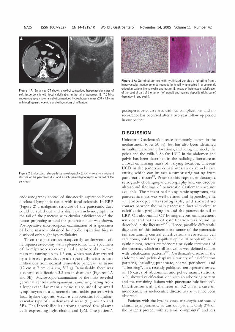
6726 ISSN 1007-9327 CN 14-1219/ R World J Gastroenterol November 14, 2005 Volume 11 Number 42
A A BB
100 μm100 μm
endosonography controlled fi ne-needle aspiration biopsy disclosed lymphatic tissue with focal sclerosis. In ERP (Figure 2) a malignant stricture of the pancreatic duct could be ruled out and a slight parenchymography in the tail of the pancreas with circular calcifi cation of the tumor projecting around the pancreatic duct was shown. Postoperative microscopical examination of a specimen of bone marrow obtained by needle aspiration biopsy disclosed only slight hypercellularity. Then the pat ient subsequently underwent left hemipancreatectomy with splenectomy. The specimen of hemipancreatectomy had a spheroidal tumor mass measuring up to 4.6 cm, which was demarcated by a f ibrous pseudocapsule (par tial ly with tumor infi ltration) from normal tumor-free pancreas tail tissue (12 cm × 7 cm × 4 cm, 367 g). Remarkably, there was a central calcification 3.2 cm in diameter (Figures 1A and 3B). Microscopic examination of the mass revealed germinal centres with hyalinized venules originating from a hypervascular mantle zone surrounded by small lymphocytes in a concentric onionskin pattern with focal hyaline deposits, which is characteristic for hyaline-vascular type of Castleman’s disease (Figures 3A and 3B). The interfollicular stroma showed few plasma cells expressing light chains and IgM. The patient’s
postoperative course was without complications and no recurrence has occurred after a two year follow up period in our patient.
DISCUSSIONUnicentric Castleman’s disease commonly occurs in the mediastinum (over 50 %), but has also been identified in multiple anatomic locations, including the neck, the pelvis and the axilla[5]. So far, UCD in the abdomen and pelvis has been described in the radiology literature as a focal enhancing mass of varying location, whereas UCD in the pancreas constitutes an extremely rare entity, which can imitate a tumor originating from pancreatic tissue[4]. Prior to this report, endoscopic retrograde cholangiopancreatography and endoscopic ultrasound findings of pancreatic Castleman’s are not available. The patient had no systemic symptoms, the pancreatic mass was well defined and hypoechogenic on endoscopic u l t rasonog raphy and showed no contact between the main pancreatic duct with circular calcification projecting around the pancreatic tail at ERP. On abdominal CT homogeneous enhancement with central pattern of calcification was found, as described in the literature[4,6,7]. Hence, possible differential diagnoses of this indeterminate tumor of the pancreatic tail containing central calcifications were acinar cell carcinoma, solid and papillary epithelial neoplasm, solid cystic tumor, serous cystadenoma or cystic teratomas of the pancreas, which are all known as well defi ned tumors with calcification patterns[8,9]. Castleman’s disease in the abdomen and pelvis displays a variety of calcification patterns, including punctuate, coarse, peripheral, and “arborizing”. In a recently published retrospective review of 16 cases of abdominal and pelvic manifestations, 31% showed calcifi cation, one with an arborizing pattern and the remaining lesions with punctuate calcifi cation[4].Calcification with a diameter of 3.2 cm in a case of monocentric or multicentric disease has as yet not been observed. Patients with the hyaline-vascular subtype are usually clinical asymptomatic, as was our patient. Only 3% of the patients present with systemic complaints[3] and less
Figure 1 A: Enhanced CT shows a well-circumscribed hypervascular mass of soft tissue density with focal calcification in the tail of pancreas; B: 7.5 MHz endosonography shows a well-circumscribed hypoechogenic mass (2.8 x 4.9 cm) with focal hyperechogenicity and without signs of infi ltration.
Figure 2 Endoscopic retrograde pancreatography (ERP) shows no malignant stricture of the pancreatic duct and a slight parenchymography in the tail of the pancreas.
Figure 3 A: Germinal centers with hyalinized venules originating from a hypervascular mantle zone surrounded by small lymphocytes in a concentric onionskin pattern (hematoxylin and eosin); B: Areas of heterotopic calcifi cation of the central part of the tumor (left panel) and hyaline deposits (right panel) (hematoxylin and eosin).
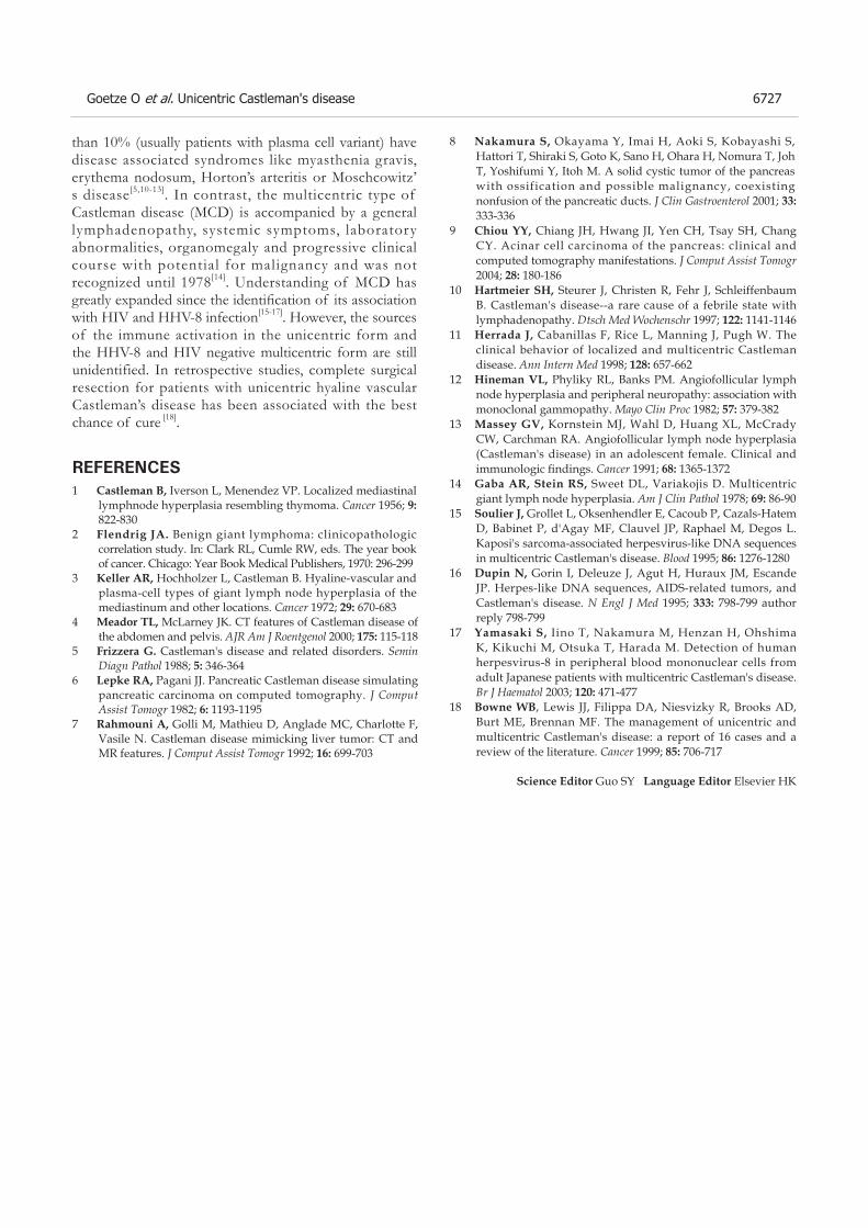
Goetze O et al. Unicentric Castleman's disease 6727
than 10% (usually patients with plasma cell variant) have disease associated syndromes like myasthenia gravis, erythema nodosum, Horton’s arteritis or Moschcowitz’s disease[5,10-13]. In contrast, the multicentric type of Castleman disease (MCD) is accompanied by a general lymphadenopathy, systemic symptoms, laboratory abnormalities, organomegaly and progressive clinical course with potential for malignancy and was not recognized until 1978[14]. Understanding of MCD has greatly expanded since the identifi cation of its association with HIV and HHV-8 infection[15-17]. However, the sources of the immune activation in the unicentric form and the HHV-8 and HIV negative multicentric form are still unidentified. In retrospective studies, complete surgical resection for patients with unicentric hyaline vascular Castleman’s disease has been associated with the best chance of cure [18].
REFERENCES1 Castleman B, Iverson L, Menendez VP. Localized mediastinal
lymphnode hyperplasia resembling thymoma. Cancer 1956; 9: 822-830
2 Flendrig JA. Benign giant lymphoma: clinicopathologic correlation study. In: Clark RL, Cumle RW, eds. The year book of cancer. Chicago: Year Book Medical Publishers, 1970: 296-299
3 Keller AR, Hochholzer L, Castleman B. Hyaline-vascular and plasma-cell types of giant lymph node hyperplasia of the mediastinum and other locations. Cancer 1972; 29: 670-683
4 Meador TL, McLarney JK. CT features of Castleman disease of the abdomen and pelvis. AJR Am J Roentgenol 2000; 175: 115-118
5 Frizzera G. Castleman's disease and related disorders. Semin Diagn Pathol 1988; 5: 346-364
6 Lepke RA, Pagani JJ. Pancreatic Castleman disease simulating pancreatic carcinoma on computed tomography. J Comput Assist Tomogr 1982; 6: 1193-1195
7 Rahmouni A, Golli M, Mathieu D, Anglade MC, Charlotte F, Vasile N. Castleman disease mimicking liver tumor: CT and MR features. J Comput Assist Tomogr 1992; 16: 699-703
8 Nakamura S, Okayama Y, Imai H, Aoki S, Kobayashi S, Hattori T, Shiraki S, Goto K, Sano H, Ohara H, Nomura T, Joh T, Yoshifumi Y, Itoh M. A solid cystic tumor of the pancreas with ossification and possible malignancy, coexisting nonfusion of the pancreatic ducts. J Clin Gastroenterol 2001; 33: 333-336
9 Chiou YY, Chiang JH, Hwang JI, Yen CH, Tsay SH, Chang CY. Acinar cell carcinoma of the pancreas: clinical and computed tomography manifestations. J Comput Assist Tomogr 2004; 28: 180-186
10 Hartmeier SH, Steurer J, Christen R, Fehr J, Schleiffenbaum B. Castleman's disease--a rare cause of a febrile state with lymphadenopathy. Dtsch Med Wochenschr 1997; 122: 1141-1146
11 Herrada J, Cabanillas F, Rice L, Manning J, Pugh W. The clinical behavior of localized and multicentric Castleman disease. Ann Intern Med 1998; 128: 657-662
12 Hineman VL, Phyliky RL, Banks PM. Angiofollicular lymph node hyperplasia and peripheral neuropathy: association with monoclonal gammopathy. Mayo Clin Proc 1982; 57: 379-382
13 Massey GV, Kornstein MJ, Wahl D, Huang XL, McCrady CW, Carchman RA. Angiofollicular lymph node hyperplasia (Castleman's disease) in an adolescent female. Clinical and immunologic fi ndings. Cancer 1991; 68: 1365-1372
14 Gaba AR, Stein RS, Sweet DL, Variakojis D. Multicentric giant lymph node hyperplasia. Am J Clin Pathol 1978; 69: 86-90
15 Soulier J, Grollet L, Oksenhendler E, Cacoub P, Cazals-Hatem D, Babinet P, d'Agay MF, Clauvel JP, Raphael M, Degos L. Kaposi's sarcoma-associated herpesvirus-like DNA sequences in multicentric Castleman's disease. Blood 1995; 86: 1276-1280
16 Dupin N, Gorin I, Deleuze J, Agut H, Huraux JM, Escande JP. Herpes-like DNA sequences, AIDS-related tumors, and Castleman's disease. N Engl J Med 1995; 333: 798-799 author reply 798-799
17 Yamasaki S, Iino T, Nakamura M, Henzan H, Ohshima K, Kikuchi M, Otsuka T, Harada M. Detection of human herpesvirus-8 in peripheral blood mononuclear cells from adult Japanese patients with multicentric Castleman's disease. Br J Haematol 2003; 120: 471-477
18 Bowne WB, Lewis JJ, Filippa DA, Niesvizky R, Brooks AD, Burt ME, Brennan MF. The management of unicentric and multicentric Castleman's disease: a report of 16 cases and a review of the literature. Cancer 1999; 85: 706-717
Science Editor Guo SY Language Editor Elsevier HK

PO Box 2345, Beijing 100023, China World J Gastroenterol 2005;11(42):6728-6729www.wjgnet.com World Journal of Gastroenterology ISSN [email protected] © 2005 The WJG Press and Elsevier Inc. All rights reserved.E L S E V I E R
• CASE REPORTS •
presented with a 12-h history of upper abdominal pain, nausea and vomiting. He was shocked (BP 80/40, pulse 120/min) and clinical examination revealed guarding, ten-derness with rebound in the upper abdomen and absence of bowel sounds. A diagnosis of peritonitis was made and he was appropriately resuscitated. He responded well to the treatment and became hemodynamically stable. His hematological blood profi le as well as his urea and elec-trolytes were unremarkable. He had conjugated hyperbili-rubinemia with raised alkaline phosphatase and alanine transaminase. Plain radiology (erect CXR and abdominal fi lms) did not demonstrate any pneumoperitoneum.
When the patient was optimized, he underwent surgery. At laparotomy, there was no evidence of gastrointestinal perforation; however, free intra-peritoneal bile was found. The extra hepatic biliary system was grossly dilated due to obvious obstruction at the distal common bile duct by an impacted calculus. The gall bladder contained calculi and was thick walled. There was a perforated superfi cial biliary radicle in the left lobe of the liver, which was the source of the free bile in the peritoneal cavity.
A cholecystectomy was performed and the CBD was explored. The calculus in the distal CBD needed to be extracted through a trans-duodenal sphincterotomy. Post explorator y choledochoscopy and on-table cholangiography confirmed clearance of the bile ducts. The common bile duct was drained with a T-tube and at the end of the operation the ruptured biliary radicle stopped leaking bile. It was however reinforced with two interrupted 3/0 prolene sutures. The right sub-hepatic space was drained and the abdomen was closed as per standard.
The patient was transferred to the high dependency unit for immediate care. He made a slow, but steady post-operative recovery. T-tube cholangiogram performed on day 14 (post-op) confi rmed free passage of contrast into the duodenum and absence of residual CBD stones; thus allowing its removal. The patient was discharged 34 days following surgery and he is still healthy with normal liver function tests when last reviewed 2 years following surgery.
DISCUSSION Peritonitis requiring surgical intervention is caused by perforated peptic ulcer in about 40% cases (duodenum:gastric: 3:1), appendicitis in 20%, gangrene of the small bowel or gall bladder in 15%, post-operative complications in 10% and miscellaneous causes in 15% cases[5]. Most commonly, peritonitis in the clinical setting is due to microorganisms, though the initial insult is usually chemical as in peptic ulcer perforation where bile, pancreatic
Bile peritonitis due to intra-hepatic bile duct rupture
R Lochan, BV Joypaul
R Lochan, BV Joypaul, Department of Surgery, South Tyneside District General Hospital, South Shields, NE34 0PL United KingdomCorrespondence to: R Lochan, Research Fellow, HPB Unit, Department of Surgery (level 4 Secretaries Office), Freeman Hospital, Newcastle upon Tyne NE7 7DN, United Kingdom. [email protected]: +44-191-2448427 Fax: +44-191-2231483Received: 2004-08-13 Accepted: 2004-12-14
AbstractGeneralized biliary peritonitis is a serious intra-abdominal emergency. Most of them occur due to duodenal ulcer perforation and rapidly evolve into bacterial peritonitis due to contamination by gut organisms and food. In this situation, recognition of the pathology and its treatment is straightforward and is usually associated with a good outcome. There are a few unusual causes of biliary peritonitis, of which rupture of the biliary tree is one. We describe a rare case of biliary peritonitis due to rupture of an intra-hepatic biliary radical. Unusual causes of peritonitis do interrupt our daily routine emergency surgical experience. Rapid recognition of the presence of peritonitis, adequate resuscitation, recognition of operative fi ndings, establishment of biliary anatomy, and performance of a meticulous surgical procedure resulted in a good outcome.
© 2005 The WJG Press and Elsevier Inc. All rights reserved.
Key words: Biliary peritonitis; Intra-hepatic biliary radicle; Rupture
Lochan R, Joypaul BV. Bile peritonitis due to intra-hepatic bile duct rupture. World J Gastroenterol 2005; 11(42): 6728-6729http://www.wjgnet.com/1007-9327/11/6728.asp
INTRODUCTIONRupture of an intra-hepatic biliary duct leading to biliary peritonitis is a rare occurrence, with only few cases reported in the literature[1-4]. This case report reinforces the necessity of complete and meticulous operative assessment of the biliary system in every case of bile peritonitis.
CASE REPORTA 78-year-old male with severe spondyloarthropathy
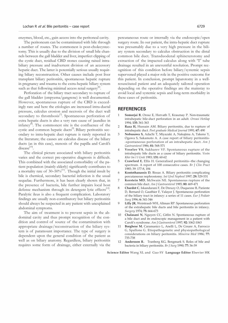
Lochan R et al. Bile peritonitis – case report 6729
enzymes, blood, etc., gain access into the peritoneal cavity. The peritoneum can be contaminated with bile through
a number of routes. The commonest is post-cholecystec-tomy. This is usually due to the division of small bile chan-nels between the gall bladder and liver, imperfect clipping of the cystic duct, residual CBD stones causing raised intra-biliary pressure and inadvertent division of an accessory hepatic duct. The latter is potentially serious usually requir-ing biliary reconstruction. Other causes include post liver transplant biliary peritonitis, spontaneous hepatic rupture in pregnancy and trauma to the extra-hepatic biliary system such as that following minimal access renal surgery[6].
Perforation of the biliary tract secondary to rupture of the gall bladder (empyema/gangrene) is well documented. However, spontaneous rupture of the CBD is exceed-ingly rare and here the etiologies are increased intra-ductal pressure, calculus erosion and necrosis of the duct wall secondary to thrombosis[7]. Spontaneous perforation of extra hepatic ducts is also a very rare cause of jaundice in infancy[8]. The commonest site is the confluence of the cystic and common hepatic ducts[9]. Biliary peritonitis sec-ondary to intra-hepatic duct rupture is rarely reported in the literature; the causes are calculus disease of the biliary ducts (as in this case), stenosis of the papilla and Caroli’s disease[10].
The clinical picture associated with biliary peritonitis varies and the correct pre-operative diagnosis is diffi cult. This combined with the associated comorbidity of the pa-tient population (mainly elderly) signifi cantly contributes to a mortality rate of 30–50%[11]. Though the initial insult by bile is chemical, secondary bacterial infection is the usual sequelae. Furthermore, it has been clearly shown that, in the presence of bacteria, bile further impairs local host defense mechanism through its detergent lytic effects[12].
Paralytic ileus is also a frequent complication. Laboratory fi ndings are usually non-contributory but biliary peritonitis should always be suspected in any patient with unexplained abdominal symptoms.
The aim of treatment is to prevent sepsis in the ab-dominal cavity and thus prompt recognition of the con-dition and control of source of the contamination with appropriate drainage/reconstruction of the biliary sys-tem is of paramount importance. The type of surgery is dependent upon the general condition of the patient as well as on biliary anatomy. Regardless, biliary peritonitis requires some form of drainage, either externally via the
percutaneous route or internally via the endoscopic/open surgery route. In our patient, the intra-hepatic duct rupture was presumably due to a very high pressure in the bili-ary system secondary to calculus obstruction in the distal common bile duct. Transduodenal sphincterotomy and extraction of the impacted calculus along with ‘T’ tube drainage resulted in an uneventful resolution. Prompt rec-ognition of this condition before biliary/systemic sepsis supervened played a major role in the positive outcome for this patient. In conclusion, prompt laparotomy in a well-resuscitated patient and an adequately tailored operation depending on the operative findings are the mainstay to avoid local and systemic sepsis and long-term morbidity in these cases of peritonitis.
REFERENCES1 Somorjai B, Orosz E, Horvath T, Krasznay P. Non-traumatic
intrahepatic bile-duct perforation in an adult. Orvasi Hetilap 1994; 135: 361-363
2 Raza H, Hussain AM. Biliary peritonitis, due to rupture of intrahepatic duct. Post graduate Medical Journal 1991; 67: 490
3 Nobusawa S, Adachi T, Miyazaki A, Nakajima A, Takeno Y, Ogawa S, Sakamoto A. A case report of biliary peritonitis--spontaneous perforation of an intrahepatic duct. Am J Gastroenterol 1986; 81: 568-571
4 Veselov VS, Sukharev VF. Spontaneous rupture of the intrahepatic bile ducts as a cause of biliary peritonitis. Vestn Khir Im I I Grek 1983; 131: 60-62
5 Crawford E, Ellis H. Generalised peritonitis--the changing spectrum. A report of 100 consecutive cases. Br J Clin Pract 1985; 39: 177-8, 184
6 Kontothanassis D, Bissas A. Biliary peritonitis complicating percutaneous nephrostomy. Int Urol Nephrol 1997; 29: 529-531
7 Kerstein MD, McSwain NE. Spontaneous rupture of the common bile duct. Am J Gastroenterol 1985; 80: 469-471
8 Chardot C, Iskandarani F, De Dreuzy O, Duquesne B, Pariente D, Bernard O, Gauthier F, Valayer J. Spontaneous perforation of the biliary tract in infancy: a series of 11 cases. Eur J Pediatr Surg 1996; 6: 341-346
9 Lilly JR, Weintraub WH, Altman RP. Spontaneous perforation of the extrahepatic bile ducts and bile peritonitis in infancy. Surgery 1974; 75: 664-673
10 Chalasani N, Nguyen CC, Gitlin N. Spontaneous rupture of a bile duct and its endoscopic management in a patient with Caroli’s syndrome. Am J Gastroenterol 1997; 92: 1062-1063
11 Borghese M, Caramanico L, Anelli L, De Cesare A, Farrocco G, Spallone G. Etiopathogenetic and physiopathological considerations on biliary peritonitis. Minerva Med 1986; 77: 735-738
12 Andersson R, Tranberg KG, Bengmark S. Roles of bile and bacteria in biliary peritonitis. Br J Surg 1990; 77: 36-39
Science Editor Wang XL and Guo SY Language Editor Elsevier HK
Related Documents