REVIEW ARTICLE Immune response patterns in non-communicable inflammatory skin diseases K. Eyerich, 1, * S. Eyerich 2 1 Department of Dermatology and Allergy, Technical University of Munich, Munich, Germany 2 ZAUM – Center of Allergy and Environment, Technical University and Helmholtz Center Munich, Munich, Germany *Correspondence: K. Eyerich. E-mail: [email protected] Abstract Non-communicable inflammatory skin diseases (ncISD) such as psoriasis or atopic eczema are a major cause of global disease burden. Due to their impact and complexity, ncISD represent a major challenge of modern medicine. Dermatol- ogy textbooks describe more than 100 different ncISD based on clinical phenotype and histological architecture. In the last decades, this historical description was complemented by increasing molecular knowledge – and this knowledge is now being translated into specific therapeutics. Combining the enormous advances made in lymphocyte immunology and molecular genetics with clinical and histological phenotyping reveals six immune response patterns of the skin – type I immune cells cause the lichenoid pattern characterized by immune-mediated cell death of keratinocytes; type II immune cells underlie the eczematous pattern with impaired epidermal barrier, infection and eosinophils as well as the bullous pattern with loss of epithelial integrity; Th17 cells and ILC3 mediate the psoriatic pattern characterized by acan- thosis, high metabolic activity and neutrophils; dysbalance of regulatory T cells causes either the fibrogenic pattern with rarefication of cells and dermal thickening or the granulomatous pattern defined by formation of granulomas. With more and more specific therapeutic agents approved, classifying ncISD also according to their immune response pattern will become highly relevant. This review defines the six immune response patterns of ncISD and highlights therapeutic strategies targeting key lymphocyte mediators. Received: 31 August 2017; Accepted: 19 October 2017 Conflicts of interest None declared. Funding sources None declared. An immunologic view at inflammatory skin diseases Non-communicable inflammatory skin diseases (ncISD) are fre- quent, affected individuals suffer from a devastating loss of qual- ity of life, and socio-economic costs are enormous. The complex pathogenesis of ncISD is based on genetic predisposition and environmental influences that result in impaired epithelial func- tion and altered immunity. Historically, disease classification in dermatology relies on precise clinical description in combination with histological description of microscopic tissue alterations and infiltrating immune cells. This classification is complex, and at times misleading. At the same time, insights into mechanisms how distinct lymphocyte subsets terminally orchestrate the inflammatory response and how these lymphocytes interact with resident skin cells 1 resulted in a translational revolution leading to more and more specific therapeutics. 2 To acknowledge these recent advances made in design and approval of specific immune-mediating therapeutics, a classification of ncISD according to their immune response patterns is required (Fig. 1, Tables 1 and 2). This review summarizes what is known about immunology, histopathology and clinical phenotype for each of the immune response patterns. It further describes limitations of the classification, early pathogenic events, and focuses on thera- peutic consequences and future developments. Lichenoid pattern (pattern 1) The major physiologic role of the lichenoid pattern is disposal of keratinocytes that are potentially infected with intracellular microbes or are (pre-)carcinogenic due to DNA damages beyond repair. It is characterized by a cytotoxic immune response against keratinocytes of the basal layer (‘interface dermatitis’) that is mediated by killer T cells (Tc1), Th1 cells, ILC1, NKT and © 2017 The Authors. Journal of the European Academy of Dermatology and Venereology published by John Wiley & Sons Ltd on behalf of European Academy of Dermatology and Venereology. JEADV 2018, 32, 692–703 This is an open access article under the terms of the Creative Commons Attribution-NonCommercial-NoDerivs License, which permits use and distribution in any medium, provided the original work is properly cited, the use is non-commercial and no modifications or adaptations are made. DOI: 10.1111/jdv.14673 JEADV

Welcome message from author
This document is posted to help you gain knowledge. Please leave a comment to let me know what you think about it! Share it to your friends and learn new things together.
Transcript
REVIEW ARTICLE
Immune response patterns in non-communicableinflammatory skin diseasesK. Eyerich,1,* S. Eyerich2
1Department of Dermatology and Allergy, Technical University of Munich, Munich, Germany2ZAUM – Center of Allergy and Environment, Technical University and Helmholtz Center Munich, Munich, Germany
*Correspondence: K. Eyerich. E-mail: [email protected]
AbstractNon-communicable inflammatory skin diseases (ncISD) such as psoriasis or atopic eczema are a major cause of global
disease burden. Due to their impact and complexity, ncISD represent a major challenge of modern medicine. Dermatol-
ogy textbooks describe more than 100 different ncISD based on clinical phenotype and histological architecture. In the
last decades, this historical description was complemented by increasing molecular knowledge – and this knowledge is
now being translated into specific therapeutics. Combining the enormous advances made in lymphocyte immunology
and molecular genetics with clinical and histological phenotyping reveals six immune response patterns of the skin –
type I immune cells cause the lichenoid pattern characterized by immune-mediated cell death of keratinocytes; type II
immune cells underlie the eczematous pattern with impaired epidermal barrier, infection and eosinophils as well as the
bullous pattern with loss of epithelial integrity; Th17 cells and ILC3 mediate the psoriatic pattern characterized by acan-
thosis, high metabolic activity and neutrophils; dysbalance of regulatory T cells causes either the fibrogenic pattern with
rarefication of cells and dermal thickening or the granulomatous pattern defined by formation of granulomas. With more
and more specific therapeutic agents approved, classifying ncISD also according to their immune response pattern will
become highly relevant. This review defines the six immune response patterns of ncISD and highlights therapeutic
strategies targeting key lymphocyte mediators.
Received: 31 August 2017; Accepted: 19 October 2017
Conflicts of interestNone declared.
Funding sourcesNone declared.
An immunologic view at inflammatory skindiseasesNon-communicable inflammatory skin diseases (ncISD) are fre-
quent, affected individuals suffer from a devastating loss of qual-
ity of life, and socio-economic costs are enormous. The complex
pathogenesis of ncISD is based on genetic predisposition and
environmental influences that result in impaired epithelial func-
tion and altered immunity. Historically, disease classification in
dermatology relies on precise clinical description in combination
with histological description of microscopic tissue alterations
and infiltrating immune cells. This classification is complex, and
at times misleading. At the same time, insights into mechanisms
how distinct lymphocyte subsets terminally orchestrate the
inflammatory response and how these lymphocytes interact with
resident skin cells1 resulted in a translational revolution leading
to more and more specific therapeutics.2 To acknowledge these
recent advances made in design and approval of specific
immune-mediating therapeutics, a classification of ncISD
according to their immune response patterns is required (Fig. 1,
Tables 1 and 2). This review summarizes what is known about
immunology, histopathology and clinical phenotype for each of
the immune response patterns. It further describes limitations of
the classification, early pathogenic events, and focuses on thera-
peutic consequences and future developments.
Lichenoid pattern (pattern 1)The major physiologic role of the lichenoid pattern is disposal of
keratinocytes that are potentially infected with intracellular
microbes or are (pre-)carcinogenic due to DNA damages beyond
repair. It is characterized by a cytotoxic immune response
against keratinocytes of the basal layer (‘interface dermatitis’)
that is mediated by killer T cells (Tc1), Th1 cells, ILC1, NKT and
© 2017 The Authors. Journal of the European Academy of Dermatology and Venereology published by John Wiley & Sons Ltdon behalf of European Academy of Dermatology and Venereology.
JEADV 2018, 32, 692–703
This is an open access article under the terms of the Creative Commons Attribution-NonCommercial-NoDerivs License, which permits use anddistribution in any medium, provided the original work is properly cited, the use is non-commercial and no modifications or adaptations are made.
DOI: 10.1111/jdv.14673 JEADV
Lichenoid
a) Eczematous
b) Blistering
Psoriatic
a) Fibrogenic
b) Granulomatous
Physiological role: limitation
Physiological role: Barrier Homeostasis/
Wound Healing
Physiological role: parasites/humoral immunity
Physiological role: cellular immunity
PAT
TE
RN
1PA
TT
ER
N 2
PAT
TE
RN
4PA
TT
ER
N 3
IFN-γTNF-α
IL-12
TBET
IL-4IL-5IL-13
IL-4
GATA3
IL-1ßIL-6IL-23TGF-β
RORc2
IL-22TNF-α
IL-6TNF-α
AHR
IL-10
TGF-βIL-2TGF-β
FOXP3
IL-17IL-21IL-22
Th1/ILC1
Th2/ILC2
Th17/ILC3
Th22
iTreg
Precursor
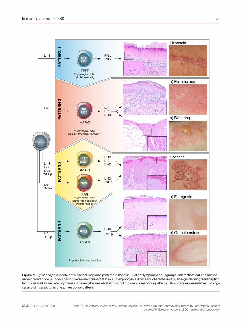
Figure 1 Lymphocyte subsets drive distinct response patterns in the skin. Distinct lymphocyte subgroups differentiate out of commonna€ıve precursor cells under specific micro-environmental stimuli. Lymphocyte subsets are characterized by lineage-defining transcriptionfactors as well as secreted cytokines. These cytokines elicit six distinct cutaneous response patterns. Shown are representative histologi-cal and clinical pictures of each response pattern.
© 2017 The Authors. Journal of the European Academy of Dermatology and Venereology published by John Wiley & Sons Ltdon behalf of European Academy of Dermatology and Venereology.
JEADV 2018, 32, 692–703
Immune patterns in ncISD 693

Tab
le1
Hallm
arks
ofim
mun
eresp
onse
patternsin
ncISD
12a
2b3
4a4b
Lich
enoid
Ecz
ematous
Bullous
Pso
riatic
Fibrog
enic
Granu
lomatou
s
Clinical
phen
otyp
ePolyg
onal
papu
les,
sharply
demarca
tedlivid
plaq
ues,
fine
andsh
inyde
squa
mation
Ves
icles,
papu
les,
erythe
ma,
eros
ion,
crus
ts,d
esqu
amation,
sebo
stas
is
Bullaewith
surrou
nding
erythe
ma,
eros
ions
,crus
ts
Pus
tules,
thick
desq
uamation,
sharply
demarca
tedplaq
ues
Skinthicke
ning
,epide
rmal
atroph
y,telang
iectas
ia,
papu
leswith
out
desq
uamation
Brownish
/yellowish
papu
les,
with
out
desq
uamation
Histologica
lph
enotyp
eInterfac
ede
rmatitis,
hype
rgranu
losis,
lymph
ocyte
infiltrationtilld
eepe
rlay
ers,
cytoid
bodies
Spo
ngiosis,
serum
crus
ts,e
osinop
hils,
oede
ma
Aca
ntho
lysis/
epidermolysiswith
cellularinfi
ltration
(micro)-ab
sces
s/ne
utroph
ils,
regu
lara
cantho
sis,
dilated
capillarie
s
Prese
nceof
muc
in/a
myloid,
thicke
ning
offibres
,cellular
rarefica
tion,
norm
alor
atroph
icep
idermis
Prese
nceof
Granu
lomas
,normal
oratroph
icep
idermis
Patho
-mec
hanism
/molec
ular
phen
otyp
e
Apo
ptos
is,n
ecroptos
isDow
nreg
ulationof
epith
elialinn
ate
immun
ity,
Epithelialb
arrie
rim
pairm
ent,
Eos
inop
hilrec
ruitm
ent,
mas
tcella
ctivation
Dire
ctlysisof
antib
ody,
Ops
onization
Rec
ruitm
ento
fneu
trop
hils,
Activationof
epith
elialinn
ate
immun
ity,
Migratio
nof
epith
elialcells,
Dow
nreg
ulationof
epith
elial
diffe
rentiatio
n,va
scularization
Extrace
llulard
epos
itof
peptides
/pep
tidog
lyca
ns/
muc
ins,
grow
thfactors
Granu
lomaform
ation
Major
cytokine
sIFN-c
IL-4,IL-5,
IL-13,
IL-31
IL-4,IL-5
IL-17A
,IL-17
F,IL-21
,IL-22
TGF-b,
IL-10
IL-10,
TNF-a
(non
-Treg)
Biomarke
rsSkin:
CXCL1
0,RIP-3,F
as/
Fas
L,Cas
pase
3Blood
andskin:C
CL1
7,CCL2
2Blood
andskin:S
pecific
antib
odyleve
lsBlood
:HBD-2
Skin:
IL-36,
NOS2
Skin:
Fox
p3,C
OMP
Skin:
Adipo
philin7
4
© 2017 The Authors. Journal of the European Academy of Dermatology and Venereology published by John Wiley & Sons Ltdon behalf of European Academy of Dermatology and Venereology.
JEADV 2018, 32, 692–703
694 Eyerich and Eyerich

Tab
le2
ncISDgrou
ped
into
immun
eresp
onse
patterns
12a
2b3
4a4b
Lich
enoid
Ecz
ematous
Bullous
Pso
riatic
Fibrog
enic
Granu
lomatou
s
Alope
ciaarea
taAtopicec
zema/
derm
atitis
Adu
ltlinea
rIgA
bullous
derm
atos
isAcn
evu
lgaris
Amyloido
sis(Ear
amyloid;
nodu
lar)
Actinicgran
ulom
a
Ash
yde
rmatos
is(Erythem
ady
schron
icum
perstans
)Childho
odgran
ulom
atou
spe
riorifi
cial
derm
atitis†
Bruns
ting-Perry
cica
tricial
pemph
igoid
Acn
eke
loidalis(Folliculitis
keloidalisnu
chae
)Atrop
hode
rma(Pierin
i-Pas
ini)
Ann
ular
elas
tolytic
gian
tcell
gran
ulom
a
Ben
ignliche
noid
keratosis
Chron
icurticaria
(cho
linergic,
idiopa
thic,p
hysica
l)Bullous
pemph
igoid(IgG
,IgE
type
)Acn
efulm
inan
sEos
inop
hilic
fasciitis(Shu
lman
)Che
ilitis
gran
ulom
atos
is(M
iesche
r/Melke
rsso
n-Ros
enthal)
Con
tact
derm
atitis†
,allergic/
photo-allergic/p
hoto-tox
ic/
irrita
nt/s
ystemic
Chron
icac
tinicde
rmatitis
Chron
icbu
llous
derm
atos
isof
childho
odAcn
einve
rsa(H
idrade
nitis
supp
urativa)
Graft-vs.-ho
stdise
ase,
sclerode
rmatou
s†Childho
odgran
ulom
atou
spe
riorifi
cial
derm
atitis†
Dermatom
yositis
Chron
icsu
perficial
derm
atitis/
smallp
laqu
epa
raps
oriasis†
Cicatric
ialp
emph
igoid
Acrod
ermatitisco
ntinua
supp
urativa(H
allope
au)
Lich
enam
yloido
sis
Drugreac
tion,
interstitial
gran
ulom
atou
s
Drugerup
tion(lich
enoid,
fixe
d)Con
tact
derm
atitis†
,allergic/
photo-allergic/p
hoto-tox
ic/
irrita
nt/s
ystemic
Dermatitishe
rpetifo
rmis
(Duh
ring)
Acu
tefebrile
neutroph
ilic
derm
atos
is(Swee
t)Hya
linos
iscu
tiset
muc
osae
(Urbac
h-Wiethe)
Fac
iala
septicgran
ulom
a
Erythem
amultiforme
DRESSsynd
rome
Epide
rmolysisbu
llosa
acqu
isita
Acu
tege
neralized
exan
them
atou
spu
stulos
isKeloid
Foreign
body
gran
ulom
a
Graft-vs.-ho
stdise
ase,
liche
noid
Dyshidroticec
zema
Lich
enplan
uspe
mph
igoide
s†Acu
tege
neralized
pustular
bacterid
(And
rews)
Lich
enmyxed
ematos
usGranu
lomaan
nulare
Graft-vs.-ho
stdise
ase,
sclerode
rmatou
s†Drugerup
tion,
spon
giotic
Pem
phigoidge
stationis
(Herpe
sge
stationis)
Chron
icsu
perficial
derm
atitis/
smallp
laqu
epa
raps
oriasis†
Lich
ensclerosu
set
atroph
icus
Interstitialg
ranu
lomatou
sde
rmatitis
Graha
m–L
ittle–P
icca
rdi–
Lasseu
rsyn
drom
eEos
inop
hilic
cellulitis(W
ells
synd
rome)
Pem
phigus
foliace
usDisse
ctingce
llulitisof
thescalp
Morph
ea/s
clerod
erma(line
ar/
profun
da)
Nec
robios
islipoidica
Keratos
isliche
noides
chronica
†
Eos
inop
hilic
annu
lare
rythem
aPem
phigus
erythe
matos
us(Sen
ear-Ush
er)
Drugerup
tion,
psoriasiform
Muc
inos
is(acral
persistent
popu
lar;po
pular)
Palisad
edne
utroph
ilic
gran
ulom
atou
sde
rmatitis
Lich
ennitid
usEos
inop
hilic
follicu
litis(O
fuji)
Pem
phigus
herpetifo
rmis
Folliculitisde
calvan
sNep
hrog
enicfibros
ing
derm
opathy
Ros
acea
†
Lich
enstria
tus
Erythem
atoxicu
mne
onatorum
Pem
phigus
,IgA
type
Impe
tigohe
rpetifo
rmis
Parry-R
ombe
rgsynd
rome
Sarco
idos
is
Lich
en(planu
s,plan
opilaris
)Giano
tti-C
rostisyn
drom
ePem
phigoidve
getans
Infantile
acropu
stulos
isPretib
ialm
yxed
ema
Lich
enplan
uspe
mph
igoide
s†Granu
lomaglutea
leinfantum
Pem
phigus
vulgaris
Keratos
isliche
noides
chronica
†Reticular
erythe
matou
smuc
inos
is(R
EM)
Lupu
serythe
matos
us(disco
id,s
ubac
ute,
chilblain,
tumid)
Ichthy
osis,a
cquired
Palmop
lantar
pustulos
isSclerom
yxed
ema
Lymph
ocyticinfiltration
(Jes
sner-Kan
of)
Lich
ensimplex
chronicu
sPAPAsynd
rome
Striaedisten
sae
Pityria
sisliche
noides
etva
rioliformisac
utaMuc
ha-
Hab
erman
n
Num
mular
ecze
ma/
derm
atitis
Pityria
sisrubrapilaris
Systemicsclerosis
Pityria
sisliche
noides
chronica
Patch
ypityria
siform
liche
noid
ecze
ma
Prurig
opigm
entosa
†
© 2017 The Authors. Journal of the European Academy of Dermatology and Venereology published by John Wiley & Sons Ltdon behalf of European Academy of Dermatology and Venereology.
JEADV 2018, 32, 692–703
Immune patterns in ncISD 695

NK cells (type 1 lymphocytes). This cytotoxic reaction is driven
by the master regulator of type 1 lymphocytes, IFN-c and cyto-
toxic granules such as granulysin,3 perforin,4 granzyme B5 and
Fas/FasL.6 In line with that observation, transcriptional network
comparison of lesional lichen planus and lupus erythematosus
with non-interface skin diseases revealed that differentially
expressed genes are attributable to type 1 lymphocytes as well
as to the effect of IFN-c on keratinocytes, including apoptosis
and necroptosis (unpublished data). Furthermore, interface
dermatitis is induced in murine models of xenotransplantation
or adoptive transfer of keratinocyte-reactive cytotoxic T cells.7
In cell culture models, FasL induces the characteristic hyper-
granulosis while IFN-c causes keratinocyte apoptosis with
cytoid body formation, and ICAM-1 expression.8 Increasing
evidence suggests an additional and important role for plasma-
cytoid dendritic cells and IFN-a in the pathogenesis of liche-
noid diseases, possibly via recruitment and amplification of
interface dermatitis.9
These molecular alterations have direct consequences that can
be observed histologically: type 1 lymphocytes form a band
along the basal membrane that is called ‘lichenoid infiltrate’.
Keratinocytes show signs of cell death, and cytoid bodies are pre-
sent. Clinically, this results in flattened, polygonal papules with
shiny desquamation; maximum clinical variants are erosions or
bullae.
Eczematous pattern (pattern 2a)The major physiologic role of the eczematous pattern is defence
against extracellular parasites. Furthermore, recent evidence sug-
gests a role in protection against toxins.10 Skin lesions are domi-
nated by Th2 and ILC2 cells (type 2 lymphocytes) secreting IL-4,
IL-5, IL-13 and IL-31. These cytokines affect the epidermis in
two ways: IL-4 and IL-13 downregulate genes of the epidermal
differentiation complex, thus impairing the epidermal barrier
and resulting in dry skin.11 Furthermore, IL-4 and IL-13 inhibit
cutaneous innate immunity12,13 which explains why most
patients affected by eczematous diseases suffer from skin colo-
nization with Staphylococcus aureus or other microbials.14 Th2-
derived IL-31 also impacts epidermal barrier and is a critical
mediator of itch, a leading symptom of most diseases grouped
into the eczematous pattern.15,16 IL-5 is a strong activator of
eosinophil and basophil granulocytes as well as mast cells.17 The
release of a plethora of mediators from these cells leads to
oedema and influx of further immune cells into the skin.
The type 2 immune deviation results in histological hallmarks
such as spongiosis, serum crusts, and a mixed cellular infiltrate
composed of lymphocytes and eosinophil granulocytes in the
acute phase and irregular acanthosis in the chronic phase char-
acterize the eczematous pattern. Clinically, the phenotype
eczema presents as epidermo-dermatitis with co-occurrence of
vesicles, papules, erythema, erosions and desquamation as well
as dry skin.Tab
le2
Con
tinue
d
12a
2b3
4a4b
Lich
enoid
Ecz
ematou
sBullous
Pso
riatic
Fibrog
enic
Granu
lomatou
s
Polym
orph
iclight
erup
tion†
Periorald
ermatitis
Pso
riasis(plaqu
etype
,inv
erse
,pa
lmop
antar,gu
ttate)
Pos
tmen
opau
salfrontal
fibros
ingalop
ecia
(Kos
sard)
Pityria
sisalba
Pso
riasispu
stulos
a(palmop
lantar,g
eneralized
)
Tox
icep
idermal
necrolysis
Polym
orph
icerup
tionof
preg
nanc
yReiter’s
synd
rome
Vitiligo
Polym
orph
iclight
erup
tion†
Ros
acea
†
Prurig
ono
dularis
SAPHO
synd
rome
Prurig
opigm
entosa
†Seb
opso
riasis
Seb
orrhoe
icde
rmatitis
Stasisde
rmatitis(eczem
acraq
uel� e)
Zoo
n’sba
lanitis
†Morethan
onepa
ttern,d
ominan
tpattern
unreso
lved
.
© 2017 The Authors. Journal of the European Academy of Dermatology and Venereology published by John Wiley & Sons Ltdon behalf of European Academy of Dermatology and Venereology.
JEADV 2018, 32, 692–703
696 Eyerich and Eyerich
Bullous pattern (pattern 2b)A distinct pathology mediated by type 2 lymphocytes results in
the bullous pattern, whose physiologic role is neutralization of
extracellular microbes. Type 2 lymphocytes instruct B cells and
plasma cells to form the antibody subclasses IgE, IgG1 and IgG4
via secretion of IL-4 and IgA via secretion of IL-5. The contribu-
tion of other lymphocytes such as follicular helper T cells to
pathogenic antibody formation in bullous skin diseases is
currently under debate.18 IgG, IgA or IgE19 antibodies directed
against structural proteins of the skin elicit the bullous pattern.
They may either directly lead to keratinocyte apoptosis and loss
of cellular adhesion, a concept called apoptolysis,20 or bind to
their target and cause secondary inflammation via opsoniza-
tion.21
Histological hallmark of type 2 lymphocyte-mediated auto-
antibody formation is destruction of the skin integrity as a result
TNF-α
IL-12 (p40/p23)
IL-4Ra
IgE
CD20
IL-17
TGF-β
bLys
IL-1
IL-6
1 2a 2b 3 4a 4b
LichenAtopic eczema
Pemphigoid PsoriasisHidradenitis suppurativa
Sclero-dermaLupus Urticaria Sarcoidosis
n.d.
n.d.
n.d.
n.d.
n.d.
n.d.
n.d.
n.d.
n.d.
n.d.
n.d.
n.d.
n.d.
n.d.
n.d.
n.d.
n.d.
n.d.
n.d.
n.d.
n.d.
n.d.
n.d.
n.d.
n.d.
n.d.
n.d.
n.d.
n.d.
n.d.
n.d.
n.d.
n.d.
n.d.
n.d.
n.d.
n.d.
n.d.
n.d.
n.d.
n.d.
n.d.
n.d.
Very high efficacy(>90% improve >90%)
High efficacy(>75% improve >75%)
Good efficacy(>50% improve >50%)
Moderate efficacy(<50% improve <50%)
No / conflicting evidence/ negative effect
Drug approved, in clinical use
Double-blind, randomized trials exist
Case series orcase reports
Col
our:
effi
cacy
Siz
e: le
vel o
f evi
denc
e
Target
Pattern
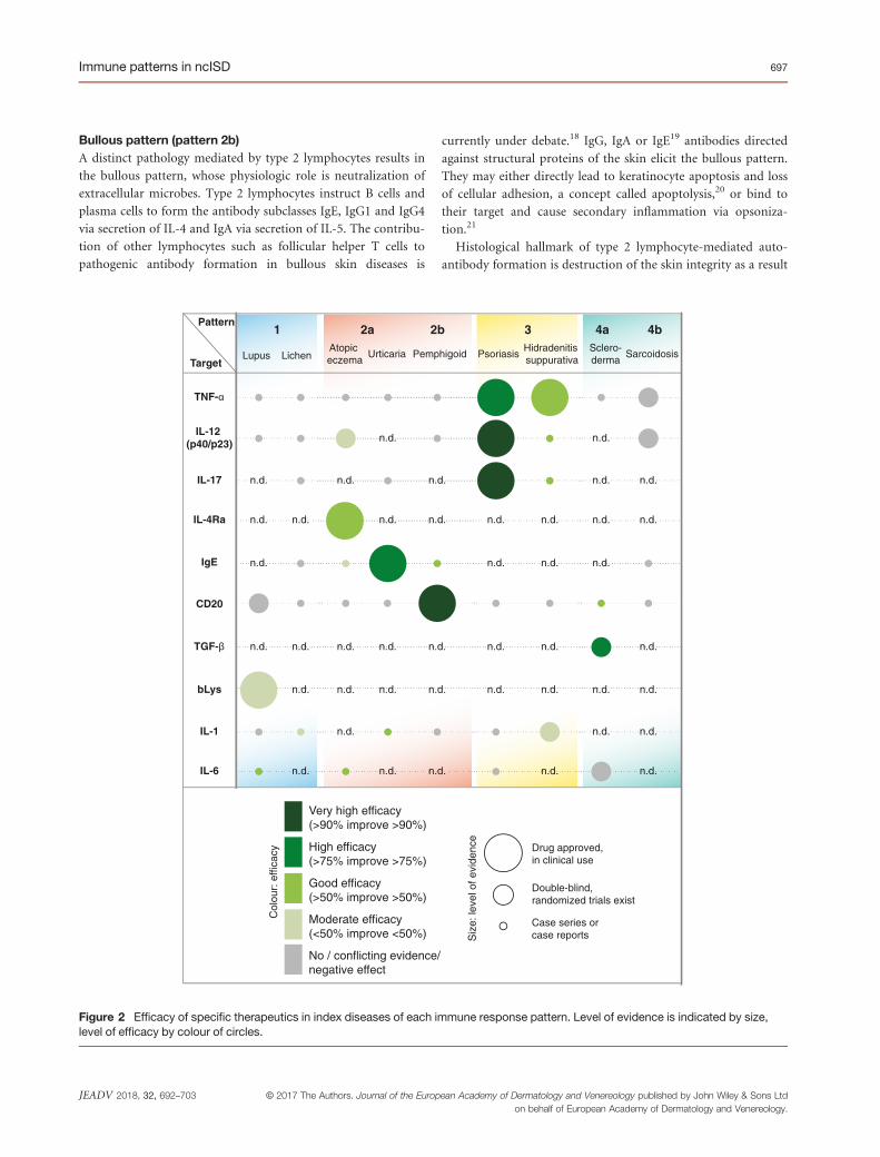
Figure 2 Efficacy of specific therapeutics in index diseases of each immune response pattern. Level of evidence is indicated by size,level of efficacy by colour of circles.
© 2017 The Authors. Journal of the European Academy of Dermatology and Venereology published by John Wiley & Sons Ltdon behalf of European Academy of Dermatology and Venereology.
JEADV 2018, 32, 692–703
Immune patterns in ncISD 697

of acantholysis, a gap between epidermis and dermis, or dermal
split. An inflammatory infiltrate composed of lymphocytes, eosi-
nophil or neutrophil granulocytes is always observed. Using
immune-fluorescence, antibody deposits of distinct patterns are
disease-defining. Clinically, the primary resulting lesion is a blis-
ter with surrounding erythema; depending on the thickness of
the epidermal roof and manipulation, also erosions and crusts
are frequently observed. Circulating specific antibodies are typi-
cal and represent biomarkers of bullous skin diseases.22 Of note,
diseases of the lichenoid or eczematous pattern may show a bul-
lous clinical variant; those variants are not regarded as bullous
pattern diseases, but rather as maximal variants of interface der-
matitis or spongiosis, respectively, due to their distinct primary
pathology.
Psoriatic pattern (pattern 3)The psoriatic pattern is mediated by a group of lymphocytes
comprised of Th17, Tc17, ILC3 and Th22 cells (type 3 lympho-
cytes) that share the physiologic role to warrant homeostasis at
barrier organs such as the skin and mucous membranes of lung
and gastrointestinal tract.23 The pattern is caused by increased
epidermal metabolism as well as by activation of innate immune
signals. IL-21 and IL-22 increase keratinocyte proliferation and
migration and inhibit their differentiation, thus contributing to
acanthosis and parakeratosis.24,25 IL-17A and IL-17F induce ker-
atinocyte secretion of several antimicrobial peptides as well as of
CXCL8, a chemokine recruiting neutrophils to the epidermis,
and VEGF that stimulates vascularization.23,26
Collectively, this results in histological hallmarks such as
regular acanthosis with hyperparakeratosis, (micro)-abscesses
in the upper layers of the epidermis, dilated dermal capillar-
ies and a lymphocytic dermal infiltrate. Clinically, a type 3
lymphocyte response is reflected by sharply demarcated pla-
ques with thick desquamation. Sterile pustules are a further
hallmark of the psoriatic pattern. IL-36 proteins and induci-
ble nitric oxidase (NOS2)27 in the skin and the antimicrobial
peptide HBD-2 in the serum28 are valid biomarkers of the
psoriatic pattern.
Fibrogenic pattern (pattern 4a)The fibrogenic pattern is a consequence of prolonged lympho-
cyte anti-inflammatory activity, usually a counter-regulation of a
preceding inflammatory response. Lead cytokines of causative
regulatory T cells (Tregs) such as iTreg, Th3 and Tr1 (type 4
lymphocytes) are IL-10 and TGF-b. The fibrogenic pattern is
mediated via TGF-b that induces numerous pro-fibrotic genes
in distinct tissue cells.29 Furthermore, it is central in endothelial-
to-mesenchymal transition to pro-fibrotic myofibroblasts.30 The
consequence is excessive extracellular matrix production, depo-
sition and tissue remodelling (fibrosis).
Alterations in the regulatory T-cell department histologically
lead to fibrosis that is observed as thickened collagen bundles
and diminished number of cells. The lymphoid infiltrate is typi-
cally mild and located in deeper skin layers. The epidermis is
normal or atrophic. This is reflected by clinical hallmarks such
as well-demarcated thickening of the whole skin and a shiny,
atrophic epidermis that may be surrounded by erythema in
active lesions.
Granulomatous pattern (pattern 4b)Granuloma formation is a general mechanism of the immune
system after identification of a potentially harmful molecule that
cannot be eliminated. In the skin, such molecules may be of
infectious nature or degenerated extracellular matrix.31 Recently,
the term ‘Immunocompromised districts’ (ICD) has been pro-
posed for a localized immune dysbalance in the skin after
trauma. Interestingly, granulomatous skin diseases occur fre-
quently in ICD predilection sites.32 As compared to the other
patterns, level of evidence for a dominating role of a single lym-
phocyte subset is low for the granulomatous pattern. Both pro-
inflammatory and regulatory T cells33 are involved. The balance
of TNF-a and type 4 lymphocyte-derived IL-10 expression seems
to be critical for granuloma development and sustainability.34
Interestingly, Tregs decrease after therapy with TNF-a blocking
antibodies, indicating a functional link of Tregs and Th1/Th17
cells via TNF receptor 2.35
The histological architecture of a granuloma is comprised of a
centre of epitheloid cells and histiocytes that may melt to giant
cells or die and leave a cell-free mass (caseating granuloma). This
centre is surrounded by lymphocytes to a varying degree. In the
skin, granulomas develop in the dermis, the epidermis is typi-
cally non-involved or atrophic. Clinically, granulomatous dis-
eases present as brownish papules of sharp demarcation with or
without epidermal desquamation. Figurated or annular manifes-
tation is regularly observed.
Concept limitationsThe pattern principle deciphers only inflammatory skin diseases
with a marked interaction of epithelia and inflammatory infil-
trate. This excludes inflammation at deeper layers of the skin
such as panniculitis and vasculitis, and it excludes also primary
dyskeratotic diseases without marked inflammation such as
monogenetic keratinization disorders (ichthyosis), acantholytic
dyskeratosis or keratosis pilaris. Furthermore, the current con-
cept is focused on terminal lymphocyte-mediated molecular
events, because these are shared by different ncISD and they can
be targeted therapeutically. The concept does not integrate the
more heterogeneous early pathogenic events mediated by non-
lymphoid immunity, although innate signals may influence the
clinical course of ncISD. Typical examples are type 1 interferons
that mediate lichenoid diseases36 and psoriasis,37 alterations in
the inflammasome causing autoinflammatory diseases,38 and
Toll-like receptor-induced activation of acute phase proteins
that alter eczematous diseases.39
© 2017 The Authors. Journal of the European Academy of Dermatology and Venereology published by John Wiley & Sons Ltdon behalf of European Academy of Dermatology and Venereology.
JEADV 2018, 32, 692–703
698 Eyerich and Eyerich

Pattern interactionsncISD are usually dominated by one immune response pattern,
but their complexity and heterogeneity may be reflected by a
mixture of patterns, especially in chronic disease situations.
This holds, for example, true for atopic eczema, where type 2
lymphocytes are causative despite a mixed infiltrate of lympho-
cytes reflected by morphologic changes in the course of the dis-
ease.14 Also contact dermatitis is not exclusively mediated by
type 2 immunity, even though it is clinically and histologically
to be attributed to the eczematous pattern. Other examples are
bullous variants of lichenoid or eczematous diseases or granu-
loma formation that may occur in the course of several ncISD
such as lichen planus, lichen nitidus or lichen striatus. Further-
more, early lichenoid pattern responses may ultimately trans-
form into the fibrogenic pattern, as frequently observed in
scleroderma.
Evidence for the relevance of a lymphocyte subset balance is
given by side-effects observed after therapeutic intervention.
Specific treatment of one lymphocyte subset causing an
immune response pattern might cause imbalance towards
another immune response pattern. The most evident example
is treatment of psoriatic pattern diseases with molecules
inhibiting TNF-a. A side-effect is dryness of the skin and
eosinophilia40 – hallmarks of the eczematous pattern. In gen-
eral, so-called paradoxical effects after treatment with biologics
acting specifically on lymphocyte subsets comprises two phe-
nomena. On the one hand, a spatial shift of lymphocytes, for
example from the gastrointestinal system to the skin, results in
development of psoriasis-like skin inflammation in patients
treated for inflammatory bowel diseases. On the other hand, a
shift in immune response patterns might result in lupus-like,
lichenoid, eczematous or granulomatous cutaneous immune
responses.41
Trigger factors and early eventsThe concept of lymphocyte-driven inflammatory patterns in the
skin is further supported by insights into the biochemistry of
antigens and mechanisms by which they stimulate lymphocytes.
Although for the majority of ncISD, the primary antigen remains
unknown, recent evidence suggests that different types of anti-
gens exist. A first group consists of common self-antigens in the
skin such as DNA, collagens, antimicrobial peptides and desmo-
somal components. Several of these antigens are proposed to
play a role in psoriasis, namely the antimicrobial peptide LL-
37,42 the melanocytic protease ADAMTSL543 and the phospholi-
pase PLA2G4D.44 Depending on the underlying lymphocyte
reaction, self-antigens cause different immune response patterns.
Desmoglein 3 (Dsg3) may stand exemplary: Dsg3-specific type 2
lymphocytes are causative for pemphigus vulgaris,45 but a type 1
dominated immune response results in interface dermatitis46
and type 3 lymphocytes specific for Dsg3 cause a psoriasis-like
inflammation in mice.47
In contrast to self-antigens, exogenous antigens frequently
influence the resulting immune response in the skin. One exam-
ple is birch or grass pollen that carry lipid mediators (PALMs)
inducing a type 2 immune response.48 In line with that observa-
tion, lymphocytes reacting to common aeroallergens in early
patch test reactions are almost exclusively Th2 cells.13 In con-
trast, microbial antigens derived from candida or staphylococci
preferentially induce Th17 cells.49 Guttate psoriasis is induced
by molecular mimicry after infection with streptococci.50 Lichen
planus is associated with HCV infection.51
Lessons learned for specific therapyThe current complex disease classification of ncISD results in
the fact that clinical studies leading to drug approval are under-
taken only in a small minority of diseases, while for most dis-
eases, an off-label use of biologics is common practice.52
Grouping ncISD according to their molecular pathogenesis
gives a rationale for the use of specific therapies (Fig. 2 and
Table 2). One example is the rare disease pityriasis rubra pilaris
(PRP) that is grouped in the psoriatic pattern. Despite missing
approval, biologics used for psoriasis are also effective in
PRP.53 Specific therapeutics targeting type 3 lymphocytes,
more recently type 2 lymphocytes and finally first evidences for
type 1 or type 4 targeting molecules, strengthen the concept of
immune response patterns in the skin.
No satisfying specific therapy is available for lichenoid (pat-
tern 1) skin diseases (Figure 2). Despite the fact that belimumab,
a monoclonal antibody targeting the B lymphocyte stimulator
bLys, is approved for systemic lupus erythematosus,54 efficacy at
cutaneous lesions is limited. Also for lichen planus, established
biologics failed.55 Thus, there is a high unmet medical need to
define cutaneous endpoints in skin autoimmune diseases, and to
identify new therapeutics.56 In line with the pathogenic concept
of the lichenoid pattern, early studies investigating antibodies
targeting either IFN-a or IFN-c are encouraging.57
More advanced are therapeutics targeting type 2 lymphocytes
mediating the eczematous (pattern 2a) and the bullous (pattern
2b) patterns. Dupilumab inhibits effects of IL-4 and IL-13 via
targeting the IL-4 receptor a. Phase III studies in atopic eczema
show a clinical efficacy superior to all previous therapeutic
attempts.58 Neutralizing the IL-4-induced antibody subtype IgE
using omalizumab is an approved and efficient therapy for
chronic urticaria.59 In contrast to type 2-targeted therapies, con-
flicting evidence exists regarding efficacy of TNF inhibitors or
ustekinumab in eczematous diseases. While some case series are
encouraging,12,60 others report lack of long-term evidence61 or
paradoxical eczematous reactions after therapy with TNF inhibi-
tors.62
An established therapy for diseases following the bullous pat-
tern (pattern 2b) is rituximab that eliminates B cells by targeting
CD20.63 Furthermore, it is speculated that blocking of IL-4
might be effective in bullous diseases such as pemphigus.64
© 2017 The Authors. Journal of the European Academy of Dermatology and Venereology published by John Wiley & Sons Ltdon behalf of European Academy of Dermatology and Venereology.
JEADV 2018, 32, 692–703
Immune patterns in ncISD 699

Figure 3 Immune response patterns in non-communicable inflammatory skin diseases (ncISD). The pathogenesis of most ncISD isbased on the interaction of lymphocytes and epithelial cells in the skin. Depending on the dominating lymphocyte subset, these interac-tions might be characterized by cytotoxic events (pattern I: lichenoid); reduced antimicrobial peptides, impaired skin barrier, and eosino-phils (pattern II: eczematous); antibody deposits and blistering (pattern IIb: bullous); enhanced metabolism and neutrophils (pattern III:psoriatic); rarefication of cells and deposit of extracellular matrix (pattern IVa: fibrogenic); or granuloma formation (pattern IVb: granuloma-tous). [Correction added on 09 February after online publication: Figure 3 was missed out in previous version and has been added in thisversion].
© 2017 The Authors. Journal of the European Academy of Dermatology and Venereology published by John Wiley & Sons Ltdon behalf of European Academy of Dermatology and Venereology.
JEADV 2018, 32, 692–703
700 Eyerich and Eyerich

The translational revolution in ncISD started when therapies
specifically inhibiting type 3 lymphocytes and the psoriatic pat-
tern (pattern 3) became available (Figure 2). Today, several anti-
bodies targeting TNF-a (infliximab, adalimumab, golimumab,
etanercept), IL-12p40 (ustekinumab), and IL-17A (secuk-
inumab, ixekizumab) are approved for psoriasis with enormous
clinical efficacy.65 Recently, adalimumab was also approved for
hidradenitis suppurativa (HS).66 Also ustekinumab seems to be
effective in HS.67 A lot of evidence exists for efficacy of type 3
targeting therapies in other diseases grouped in the psoriatic pat-
tern, for example PRP.68
Therapies neutralizing regulatory T cells and with that the fibro-
genic (pattern 4a) or eventually the granulomatous (pattern 4b)
pattern are in early clinical studies. Namely, fresolimumab, an anti-
body targeting TGF-b, had positive effects in a small clinical study
with patients affected by sclerosis.69 Other specific therapies did
not significantly improve skin symptoms in scleroderma, including
a recently published study on tocilizumab, an antibody targeting
IL-670 (Table 2, Figure 3). [Correction added on 09 February
after online publication: the figure citation was previously incorrect
and table citation has been updated in this version]
For the granulomatous reaction pattern, best evidence exists
for therapies targeting TNF-a or IL-12p40. While case series
report conflicting evidence on efficacy,71 TNF-a inhibitors may
also induce granulomas in a paradoxical manner.72 A similar situ-
ation is reported for rituximab.73 Thus, no fully convincing thera-
peutic option to treat granulomatous skin diseases exists to date.
Technological advances drive both a better understanding of
lymphocyte-mediated downstream events in the pathogenesis of
ncISD as well as development of therapeutics specifically inter-
fering with lymphocyte subpopulations. That is why a down-
stream-oriented molecular classification of ncISD as proposed in
this review is reasonable and why the current classification based
on clinical picture and histology needs revision. A challenge of
the future will be to standardize diagnostics of ncISD and to
define adequate endpoints for clinical studies beyond the dis-
eases psoriasis and atopic eczema. Although it may not be obvi-
ous at first glance, grouping ncISD according to their immune
response pattern is the first step towards individualized (also
called precision) medicine. It may be speculated that the future
of defining and treating ncISD will be a combination of the
immune response pattern at disease-level with early pathogenic
triggers at the individual patient’s level. Specifically, an individ-
ual patient will be classified into one of the six immune response
patterns to determine the ideal symptomatic therapy, and in par-
allel specific early events – e.g. environmental trigger factors,
stress, or infections – will be identified to combine the symp-
tomatic therapy with individualized disease prevention.
AcknowledgementsThe authors wish to thank Heidrun Behrendt and Johannes Ring
for their critical review of the manuscript. K.E. was supported by
grants of the German Research Foundation (EY07/3-1) and the
European Research Council (IMCIS 676858). S.E. was supported
by the Helmholtz Association.
References1 Eyerich S, Zielinski CE. Defining Th-cell subsets in a classical and tissue-
specific manner: examples from the skin. Eur J Immunol 2014; 44: 3475–3483.
2 Noda S, Krueger JG, Guttman-Yassky E. The translational revolution and
use of biologics in patients with inflammatory skin diseases. J Allergy Clin
Immunol 2015; 135: 324–336.3 Chung WH, Hung SI, Yang JY et al. Granulysin is a key mediator for dis-
seminated keratinocyte death in Stevens-Johnson syndrome and toxic
epidermal necrolysis. Nat Med 2008; 14: 1343–1350.4 Prpic Massari L, Kastelan M, Gruber F et al. Perforin expression in
peripheral blood lymphocytes and skin-infiltrating cells in patients with
lichen planus. Br J Dermatol 2004; 151: 433–439.5 Grassi M, Capello F, Bertolino L, Seia Z, Pippione M. Identification of
granzyme B-expressing CD-8-positive T cells in lymphocytic inflamma-
tory infiltrate in cutaneous lupus erythematosus and in dermatomyositis.
Clin Exp Dermatol 2009; 34: 910–914.6 Viard I, Wehrli P, Bullani R et al. Inhibition of toxic epidermal necrolysis
by blockade of CD95 with human intravenous immunoglobulin. Science
1998; 282: 490–493.7 Okiyama N, Fujimoto M. Clinical perspectives and murine models of
lichenoid tissue reaction/interface dermatitis. J Dermatol Sci 2015; 78:
167–172.8 Farley SM, Wood LJ, Iordanov MS. An epidermotypic model of interface
dermatitis reveals individual functions of fas ligand and gamma inter-
feron in hypergranulosis, cytoid body formation, and gene expression.
Am J Dermatopathol 2011; 33: 244–250.9 Saadeh D, Kurban M, Abbas O. Update on the role of plasmacytoid den-
dritic cells in inflammatory/autoimmune skin diseases. Exp Dermatol
2016; 25: 415–421.10 Galli SJ. The mast cell-IgE paradox: from homeostasis to anaphylaxis. Am
J Pathol 2016; 186: 212–224.11 Howell MD, Kim BE, Gao P et al. Cytokine modulation of atopic der-
matitis filaggrin skin expression. J Allergy Clin Immunol 2009; 124(3
Suppl 2): R7–R12.12 Eyerich S, Onken AT, Weidinger S et al.Mutual antagonism of T cells
causing psoriasis and atopic eczema. N Engl J Med 2011; 365: 231–238.13 Eyerich K, Pennino D, Scarponi C et al. IL-17 in atopic eczema: linking
allergen-specific adaptive and microbial-triggered innate immune
response. J Allergy Clin Immunol 2009; 123: 59–66 e4.14 Eyerich K, Eyerich S, Biedermann T. The multi-modal immune pathogen-
esis of atopic eczema. Trends Immunol 2015; 36: 788–801.15 Dillon SR, Sprecher C, Hammond A et al. Interleukin 31, a cytokine pro-
duced by activated T cells, induces dermatitis in mice. Nat Immunol 2004;
5: 752–760.16 Singh B, Jegga AG, Shanmukhappa KS et al. IL-31-driven skin remodel-
ing involves epidermal cell proliferation and thickening that lead to
impaired skin-barrier function. PLoS One 2016; 11: e0161877.
17 Molfino NA. Targeting of eosinophils in asthma. Expert Opin Biol Ther
2012; 12: 807–809.18 Hennerici T, Pollmann R, Schmidt T et al. Increased frequency of t follic-
ular helper cells and elevated interleukin-27 plasma levels in patients with
pemphigus. PLoS One 2016; 11: e0148919.
19 van Beek N, Schulze FS, Zillikens D, Schmidt E. IgE-mediated mecha-
nisms in bullous pemphigoid and other autoimmune bullous diseases.
Exp Rev Clin Immunol 2016; 12: 267–277.20 Grando SA, Bystryn JC, Chernyavsky AI et al. Apoptolysis: a novel mech-
anism of skin blistering in pemphigus vulgaris linking the apoptotic path-
ways to basal cell shrinkage and suprabasal acantholysis. Exp Dermatol
2009; 18: 764–770.
© 2017 The Authors. Journal of the European Academy of Dermatology and Venereology published by John Wiley & Sons Ltdon behalf of European Academy of Dermatology and Venereology.
JEADV 2018, 32, 692–703
Immune patterns in ncISD 701

21 Nousari HC, Anhalt GJ. Pemphigus and bullous pemphigoid. Lancet
1999; 354: 667–672.22 Otten JV, Hashimoto T, Hertl M, Payne AS, Sitaru C. Molecular diagnosis
in autoimmune skin blistering conditions. Curr Mol Med 2014; 14: 69–95.23 Eyerich S, Eyerich K, Cavani A, Schmidt-Weber C. IL-17 and IL-22: sib-
lings, not twins. Trends Immunol 2010; 31: 354–361.24 Caruso R, Botti E, Sarra M et al. Involvement of interleukin-21 in the epi-
dermal hyperplasia of psoriasis. Nat Med 2009; 15: 1013–1015.25 Zheng Y, Danilenko DM, Valdez P et al. Interleukin-22, a T(H)17 cyto-
kine, mediates IL-23-induced dermal inflammation and acanthosis. Nat-
ure 2007; 445: 648–651.26 Wolk K, Haugen HS, Xu W et al. IL-22 and IL-20 are key mediators of
the epidermal alterations in psoriasis while IL-17 and IFN-gamma are
not. J Mol Med 2009; 87: 523–536.27 Quaranta M, Knapp B, Garzorz N et al. Intraindividual genome expres-
sion analysis reveals a specific molecular signature of psoriasis and
eczema. Sci Transl Med 2014; 6: 244ra90.
28 Kolbinger F, Loesche C, Valentin MA et al. beta-Defensin 2 is a respon-
sive biomarker of IL-17A-driven skin pathology in patients with psoriasis.
J Allergy Clin Immunol 2017; 139: 923–932.e829 Blobe GC, Schiemann WP, Lodish HF. Role of transforming growth fac-
tor beta in human disease. N Engl J Med 2000; 342: 1350–1358.30 Wermuth PJ, Li Z, Mendoza FA, Jimenez SA. Stimulation of transforming
growth factor-beta1-induced endothelial-to-mesenchymal transition and
tissue fibrosis by endothelin-1 (ET-1): a novel profibrotic effect of ET-1.
PLoS One 2016; 11: e0161988.
31 Gunes P, Goktay F, Mansur AT, Koker F, Erfan G. Collagen-elastic tissue
changes and vascular involvement in granuloma annulare: a review of 35
cases. J Cutan Pathol 2009; 36: 838–844.32 Lo Schiavo A, Ruocco E, Gambardella A, O’Leary RE, Gee S. Granuloma-
tous dysimmune reactions (sarcoidosis, granuloma annulare, and others)
on differently injured skin areas. Clin Dermatol 2014; 32: 646–653.33 Rappl G, Pabst S, Riemann D et al. Regulatory T cells with reduced
repressor capacities are extensively amplified in pulmonary sarcoid lesions
and sustain granuloma formation. Clin Immunol 2011; 140: 71–83.34 Cilfone NA, Perry CR, Kirschner DE, Linderman JJ. Multi-scale modeling
predicts a balance of tumor necrosis factor-alpha and interleukin-10 con-
trols the granuloma environment during Mycobacterium tuberculosis
infection. PLoS One 2013; 8: e68680.
35 Verwoerd A, Hijdra D, Vorselaars AD et al. Infliximab therapy balances
regulatory T cells, tumour necrosis factor receptor 2 (TNFR2) expression
and soluble TNFR2 in sarcoidosis. Clin Exp Immunol 2016; 185: 263–270.36 Banchereau J, Pascual V. Type I interferon in systemic lupus erythemato-
sus and other autoimmune diseases. Immunity 2006; 25: 383–392.37 Nestle FO, Conrad C, Tun-Kyi A et al. Plasmacytoid predendritic cells
initiate psoriasis through interferon-alpha production. J Exp Med 2005;
202: 135–143.38 de Jesus AA, Canna SW, Liu Y, Goldbach-Mansky R. Molecular mecha-
nisms in genetically defined autoinflammatory diseases: disorders of
amplified danger signaling. Annu Rev Immunol 2015; 33: 823–874.39 Skabytska Y, Kaesler S, Volz T, Biedermann T. The role of innate immune
signaling in the pathogenesis of atopic dermatitis and consequences for
treatments. Semin Immunopathol 2016; 38: 29–43.40 Malisiewicz B, Murer C, Pachlopnik Schmid J, French LE, Schmid-Gren-
delmeier P, Navarini AA. Eosinophilia during psoriasis treatment with
TNF antagonists. Dermatology 2011; 223: 311–315.41 Her M, Kavanaugh A. Alterations in immune function with biologic ther-
apies for autoimmune disease. J Allergy Clin Immunol 2016; 137: 19–27.42 Lande R, Botti E, Jandus C et al. The antimicrobial peptide LL37 is a T-
cell autoantigen in psoriasis. Nat Commun 2014; 5: 5621.
43 Arakawa A, Siewert K, Stohr J et al.Melanocyte antigen triggers autoim-
munity in human psoriasis. J Exp Med 2015; 212: 2203–2212.44 Cheung KL, Jarrett R, Subramaniam S et al. Psoriatic T cells recognize
neolipid antigens generated by mast cell phospholipase delivered by exo-
somes and presented by CD1a. J Exp Med 2016; 213: 2399–2412.
45 Zhu H, Chen Y, Zhou Y, Wang Y, Zheng J, Pan M. Cognate Th2-B cell
interaction is essential for the autoantibody production in pemphigus
vulgaris. J Clin Immunol 2012; 32: 114–123.46 Takahashi H, Kouno M, Nagao K et al. Desmoglein 3-specific CD4+ T
cells induce pemphigus vulgaris and interface dermatitis in mice. J Clin
Invest 2011; 121: 3677–3688.47 Nishimoto S, Kotani H, Tsuruta S et al. Th17 cells carrying TCR recog-
nizing epidermal autoantigen induce psoriasis-like skin inflammation. J
Immunol 2013; 191: 3065–3072.48 Gilles S, Mariani V, Bryce M et al. Pollen allergens do not come alone:
pollen associated lipid mediators (PALMS) shift the human immue sys-
tems towards a T(H)2-dominated response. Allergy Asthma Clin Immunol
2009; 5: 3.
49 Zielinski CE, Mele F, Aschenbrenner D et al. Pathogen-induced human
TH17 cells produce IFN-gamma or IL-10 and are regulated by IL-1beta.
Nature 2012; 484: 514–518.50 Ruiz-Romeu E, Ferran M, Sagrista M et al. Streptococcus pyogenes-
induced cutaneous lymphocyte antigen-positive T cell-dependent epider-
mal cell activation triggers TH17 responses in patients with guttate psori-
asis. J Allergy Clin Immunol 2016; 138: 491–499e6.51 Nagao Y, Nishida N, Toyo-Oka L et al. Genome-wide association study
identifies risk variants for lichen planus in patients with hepatitis C virus
infection. Clin Gastroenterol Hepatol 2017; 15: 937–944.e552 Han G. Biologics in dermatology beyond psoriasis. Cutis 2014; 93: E21–
E27.
53 Feldmeyer L, Mylonas A, Demaria O et al. Interleukin 23-helper T cell 17
Axis as a treatment target for pityriasis rubra pilaris. JAMA Dermatol
2017; 153: 304–308.54 Navarra SV, Guzman RM, Gallacher AE et al. Efficacy and safety of beli-
mumab in patients with active systemic lupus erythematosus: a ran-
domised, placebo-controlled, phase 3 trial. Lancet 2011; 377: 721–731.55 Atzmony L, Reiter O, Hodak E, Gdalevich M, Mimouni D. Treatments
for cutaneous lichen planus: a systematic review and meta-analysis. Am J
Clin Dermatol 2016; 17: 11–22.56 Kuhn A, Landmann A, Bonsmann G. The skin in autoimmune diseases-
Unmet needs. Autoimmun Rev 2016; 15: 948–954.57 Mathian A, Hie M, Cohen-Aubart F, Amoura Z. Targeting interferons in
systemic lupus erythematosus: current and future prospects. Drugs 2015;
75: 835–846.58 Beck LA, Thaci D, Hamilton JD et al. Dupilumab treatment in adults
with moderate-to-severe atopic dermatitis. N Engl J Med 2014; 371: 130–139.
59 Maurer M, Rosen K, Hsieh HJ et al. Omalizumab for the treatment of
chronic idiopathic or spontaneous urticaria. N Engl J Med 2013; 368:
924–935.60 Khattri S, Brunner PM, Garcet S et al. Efficacy and safety of ustekinumab
treatment in adults with moderate-to-severe atopic dermatitis. Exp Der-
matol 2017; 26: 28–35.61 Samorano LP, Hanifin JM, Simpson EL, Leshem YA. Inadequate response
to ustekinumab in atopic dermatitis – a report of two patients. J Eur Acad
Dermatol Venereol 2016; 30: 522–523.62 Wilson LH, Eliason MJ, Leiferman KM, Hull CM, Powell DL. Treatment
of refractory chronic urticaria with tumor necrosis factor-alfa inhibitors. J
Am Acad Dermatol 2011; 64: 1221–1222.63 Joly P, Mouquet H, Roujeau JC et al. A single cycle of rituximab for the
treatment of severe pemphigus. N Engl J Med 2007; 357: 545–552.64 Tavakolpour S, Tavakolpour V. Interleukin 4 inhibition as a potential
therapeutic in pemphigus. Cytokine 2016; 77: 189–195.65 Boehncke WH, Schon MP. Psoriasis. Lancet 2015; 386: 983–994.66 Kimball AB, Okun MM, Williams DA et al. Two phase 3 trials of adali-
mumab for hidradenitis suppurativa. N Engl J Med 2016; 375: 422–434.
67 Blok JL, Li K, Brodmerkel C, Horvatovich P, Jonkman MF, Horvath B.
Ustekinumab in hidradenitis suppurativa: clinical results and a search for
potential biomarkers in serum. Br J Dermatol 2016; 174: 839–846.
© 2017 The Authors. Journal of the European Academy of Dermatology and Venereology published by John Wiley & Sons Ltdon behalf of European Academy of Dermatology and Venereology.
JEADV 2018, 32, 692–703
702 Eyerich and Eyerich

68 Petrof G, Almaani N, Archer CB, Griffiths WA, Smith CH. A systematic
review of the literature on the treatment of pityriasis rubra pilaris type 1
with TNF-antagonists. J Eur Acad Dermatol Venereol 2013; 27: e131–e135.
69 Rice LM, Padilla CM, McLaughlin SR et al. Fresolimumab treatment
decreases biomarkers and improves clinical symptoms in systemic sclero-
sis patients. J Clin Invest 2015; 125: 2795–2807.70 Khanna D, Denton CP, Jahreis A et al. Safety and efficacy of
subcutaneous tocilizumab in adults with systemic sclerosis (faSSci-
nate): a phase 2, randomised, controlled trial. Lancet 2016; 387: 2630–2640.
71 Judson MA, Baughman RP, Costabel U et al. Safety and efficacy of ustek-
inumab or golimumab in patients with chronic sarcoidosis. Eur Respir J
2014; 44: 1296–1307.72 Amber KT, Bloom R, Mrowietz U, Hertl M. TNF-alpha: a treatment target
or cause of sarcoidosis? J Eur Acad Dermatol Venereol 2015; 29: 2104–2111.73 Galimberti F, Fernandez AP. Sarcoidosis following successful treatment of
pemphigus vulgaris with rituximab: a rituximab-induced reaction further
supporting B-cell contribution to sarcoidosis pathogenesis? Clin Exp Der-
matol 2016; 41: 413–416.74 Schulman JM, LeBoit PE. Adipophilin expression in necrobiosis lipoidica,
granuloma annulare, and sarcoidosis. Am J Dermatopathol 2015; 37: 203–209.
© 2017 The Authors. Journal of the European Academy of Dermatology and Venereology published by John Wiley & Sons Ltdon behalf of European Academy of Dermatology and Venereology.
JEADV 2018, 32, 692–703
Immune patterns in ncISD 703
Related Documents