Cancer Immunol Irnmunother (1987) 24:25-36 ancer L I immunology [J mmunotherapy © Springer-Verlag 1987 Immune reactivity in SL2 lymphoma-bearing mice compared with SL2-immunized mice Roel A. De Weger, Bert Wilbrink, Roel M. P. Moberts, Dennis Mans, Ralph Oskam, and Willem Den Otter Experimental Pathology, Pathologisch Instituut, Rijksuniversiteit Utrecht, Pasteurstraat 2, 3511 HX Utrecht, The Netherlands Summary. We have studied the rather paradoxical pheno- menon of the growth of an antigenic tumor in an immu- nocomponent host. This phenomenon was studied by com- paring (a) the lymphocyte reactivity, and (b) the macro- phage cytotoxicity, during SL2 growth in DBA/2 mice (SL2-bearing mice) and in DBA/2 mice immunized against SL2 tumor cells (SL2-immune mice). Immune mice rejected a challenge of tumor cells. The immune T-lym- phocytes rendered macrophages cytotoxic (arming) and were able to transfer tumor resistance to naive animals. Nonimmunized mice did not reject a challenge of SL2 cells. In these tumor-bearing mice various forms of im- mune reactivity were tested. Lymphocytes with the capaci- ty to arm macrophages could not be found in the lym- phoid organs. However, lymphocytes isolated from the tis- sue directly surrounding the subcutaneous SL2 tumor could arm macrophages in vitro. Shortly after subcutaneous tumor grafting cytotoxic macrophages were found in the peritoneal cavity. In the serum macrophage arming factors were detected that ren- dered macrophages cytotoxic in vitro. This cytotoxicity of the peritoneal macrophages and the presence of macro- phage arming factors in the serum showed a similar bipha- sic pattern. The first phase of cytotoxicity between day 3 and 8 after tumor grafting was tumor (SL2) specific. The second phase from day 12 and onwards was not tumor specific. During the first 4 days after SL2 grafting the DBA/2 mice expressed a specific concomitant immunity to a second tumor graft. Then 7 or more days after grafting the first SL2 tumor, the concomitant immunity was nonspecific as the growth of a second SL2 tumor graft and a L5178Y (DBA/2) tumor graft were inhibited. In addi- tion, the immune suppressive activity of serum and lym- phocytes was tested. Neither serum nor lymphocytes from SL2-bearing mice suppressed the macrophage arming ca- pacity of SL2 immune lymphocytes. Lymphocytes from tu- mor-bearing mice did not inhibit the capacity of SL2-im- mune lymphocytes to transfer resistance to naive animals. On the contrary, lymphocytes obtained from SL2-bearing mice 14 days after SL2 grafting transfered tumor resistance in a Winn-type assay. These data suggest that the growth of an antigenic tumor is due to the inability of the immune system to mount an effective antitumor effector cell popu- lation during tumor growth, rather than an immune sup- Offprint requests to: R. A. De Weger pression of the antitumor reactivity, as a limited immune reactivity could be detected in tumor-bearing mice, where- as immune suppression could not be detected. Introduction We have previously described the SL2 lymphosarcoma as a rapidly metastasizing T-cell tumor, that can kill DBA/2 mice after transplantation of 104 cells within 30 days [10]. Still, this tumor is antigenic as injection of irradiated SL2 ceils leads to an effective immunization of DBA/2 mice. T-lymphocytes obtained from these immunized animals can transfer tumor resistance to tumor-bearing mice [27, 29] and induce specifically cytotoxic macrophages in vitro [7]. This indicated that the SL2 cells can be recognized im- munologically by the DBA/2 mouse. In the SL2-DBA/2 murine tumor system [27, 28] and in other murine tumor systems [2, 22] the resistance against the tumor has been shown to be dependent on cooperation between T-lymphocytes and macrophages. The T-lympho- cytes are important for the immunological recognition of the tumor cells, whereas, the macrophages probably, after activation by the lymphocytes, act as the major tumoricid- al cell [5]. In this paper we have studied the reactivity of lympho- cytes from SL2 tumor-bearing mice, to find out why the DBA/2 mouse cannot resist the growth of the antigenic SL2 tumor. In addition, we have studied the presence of cytotoxic macrophages and macrophage arming factors during tumor growth. To account for the low lymphocyte reactivity during tumor growth it was investigated whether the antitumor reactivity was inhibited by suppressor cells or suppressor factors in the serum, as has been described in other murine tumor systems [17, 18, 25, 26]. We also ex- amined the alternative possibility, namely that the tumor- bearing animals are not able to mount an effective antitu- mor effector cell population. We have approached these questions by studying the ineffective T-cell reactivity and macrophage cytotoxicity in tumor-bearing mice in com- parison with the effective T-cell reactivity and macrophage cytotoxicity in immunized mice. The data obtained in the SL2 tumor-bearing DBA/2 mouse system favor an immune reactivity against the SL2 cells that does not lead to an effective effector cell popula- tion against the tumor, rather than suppression of the anti- tumor immune response.

Welcome message from author
This document is posted to help you gain knowledge. Please leave a comment to let me know what you think about it! Share it to your friends and learn new things together.
Transcript
Cancer Immunol Irnmunother (1987) 24:25-36 ancer L I immunology [J mmunotherapy © Springer-Verlag 1987
Immune reactivity in SL2 lymphoma-bearing mice compared with SL2-immunized mice
Roel A. De Weger, Bert Wilbrink, Roel M. P. Moberts, Dennis Mans, Ralph Oskam, and Willem Den Otter
Experimental Pathology, Pathologisch Instituut, Rijksuniversiteit Utrecht, Pasteurstraat 2, 3511 HX Utrecht, The Netherlands
Summary. We have studied the rather paradoxical pheno- menon of the growth of an antigenic tumor in an immu- nocomponent host. This phenomenon was studied by com- paring (a) the lymphocyte reactivity, and (b) the macro- phage cytotoxicity, during SL2 growth in DBA/2 mice (SL2-bearing mice) and in DBA/2 mice immunized against SL2 tumor cells (SL2-immune mice). Immune mice rejected a challenge of tumor cells. The immune T-lym- phocytes rendered macrophages cytotoxic (arming) and were able to transfer tumor resistance to naive animals. Nonimmunized mice did not reject a challenge of SL2 cells. In these tumor-bearing mice various forms of im- mune reactivity were tested. Lymphocytes with the capaci- ty to arm macrophages could not be found in the lym- phoid organs. However, lymphocytes isolated from the tis- sue directly surrounding the subcutaneous SL2 tumor could arm macrophages in vitro.
Shortly after subcutaneous tumor grafting cytotoxic macrophages were found in the peritoneal cavity. In the serum macrophage arming factors were detected that ren- dered macrophages cytotoxic in vitro. This cytotoxicity of the peritoneal macrophages and the presence of macro- phage arming factors in the serum showed a similar bipha- sic pattern. The first phase of cytotoxicity between day 3 and 8 after tumor grafting was tumor (SL2) specific. The second phase from day 12 and onwards was not tumor specific. During the first 4 days after SL2 grafting the DBA/2 mice expressed a specific concomitant immunity to a second tumor graft. Then 7 or more days after grafting the first SL2 tumor, the concomitant immunity was nonspecific as the growth of a second SL2 tumor graft and a L5178Y (DBA/2) tumor graft were inhibited. In addi- tion, the immune suppressive activity of serum and lym- phocytes was tested. Neither serum nor lymphocytes from SL2-bearing mice suppressed the macrophage arming ca- pacity of SL2 immune lymphocytes. Lymphocytes from tu- mor-bearing mice did not inhibit the capacity of SL2-im- mune lymphocytes to transfer resistance to naive animals. On the contrary, lymphocytes obtained from SL2-bearing mice 14 days after SL2 grafting transfered tumor resistance in a Winn-type assay. These data suggest that the growth of an antigenic tumor is due to the inability of the immune system to mount an effective antitumor effector cell popu- lation during tumor growth, rather than an immune sup-
Offprint requests to: R. A. De Weger
pression of the antitumor reactivity, as a limited immune reactivity could be detected in tumor-bearing mice, where- as immune suppression could not be detected.
Introduction
We have previously described the SL2 lymphosarcoma as a rapidly metastasizing T-cell tumor, that can kill DBA/2 mice after transplantation of 104 cells within 30 days [10]. Still, this tumor is antigenic as injection of irradiated SL2 ceils leads to an effective immunization of DBA/2 mice. T-lymphocytes obtained from these immunized animals can transfer tumor resistance to tumor-bearing mice [27, 29] and induce specifically cytotoxic macrophages in vitro [7]. This indicated that the SL2 cells can be recognized im- munologically by the DBA/2 mouse.
In the SL2-DBA/2 murine tumor system [27, 28] and in other murine tumor systems [2, 22] the resistance against the tumor has been shown to be dependent on cooperation between T-lymphocytes and macrophages. The T-lympho- cytes are important for the immunological recognition of the tumor cells, whereas, the macrophages probably, after activation by the lymphocytes, act as the major tumoricid- al cell [5].
In this paper we have studied the reactivity of lympho- cytes from SL2 tumor-bearing mice, to find out why the DBA/2 mouse cannot resist the growth of the antigenic SL2 tumor. In addition, we have studied the presence of cytotoxic macrophages and macrophage arming factors during tumor growth. To account for the low lymphocyte reactivity during tumor growth it was investigated whether the antitumor reactivity was inhibited by suppressor cells or suppressor factors in the serum, as has been described in other murine tumor systems [17, 18, 25, 26]. We also ex- amined the alternative possibility, namely that the tumor- bearing animals are not able to mount an effective antitu- mor effector cell population. We have approached these questions by studying the ineffective T-cell reactivity and macrophage cytotoxicity in tumor-bearing mice in com- parison with the effective T-cell reactivity and macrophage cytotoxicity in immunized mice.
The data obtained in the SL2 tumor-bearing DBA/2 mouse system favor an immune reactivity against the SL2 cells that does not lead to an effective effector cell popula- tion against the tumor, rather than suppression of the anti- tumor immune response.
26
Materials and methods
Animals
Inbred DBA/2 and BALB/c mice (6-10 weeks) were ob- tained from CPB-TNO, Zeist, The Netherlands. Inbred C57BL/10ScCR mice (6-10 weeks) were purchased from Bomholtgfird.
Tumors
The DBA/2 tumors used were the SL2 lymphoma, which arose spontaneously and the chemically induced lympho- ma L5178Y. The BALB/c tumor used was the plasmacyto- ma MPC11. The C57BL tumor was the EL4 lymphoma which was chemically induced. All tumors grew as ascitic tumors in the peritoneal cavity. The tumors were main- tained by weekly i.p. passage. In vitro SL2, L5178Y, and EL4 had a mean generation time of 16-20 h; the mean generation time of MPC11 was 24-32 h. The tumors were free of Mycoplasma and cross-reacting viruses.
Tumor immunization and tumor grafting
DBA/2 mice were immunized by 2 injections of 107 irradi- ated (5,000 rad) SL2 cells i.p. on days - 2 0 and - 10. After this immunization 95% of the mice could resist a lethal dose of 5 x 10 6 nonirradiated SL2 cells injected i.p. on day 0. Peritoneal lymphocytes used to arm macrophages were harvested on day 7 (after injection of non irradiated cells [7]. Spleen lymphocytes used for transfer experiments were harvested on day 12 [27]. To obtain tumor-bearing ani- mals, DBA/2 mice were injected s.c. with 1 0 4 SL2 cells in the flank.
To test concomitant immunity, SL2-bearing DBA/2 mice were injected with 104 tumor cells in the groin. At this site tumors developed as ellipsoids and the tumor mass was calculated using the formula 4/3 HAZB, where A is the length of the short axis and B is the length of the long axis of the tumor mass, as measured with calipers.
Histology and cytology
Subcutaneous tumors and the skin covering the tumor were removed, fixed in 4% formaldehyde, and embedded in paraffin. Tissue sections were stained with hematoxylin eosin. Cytocentrifuge preparations were made from cell suspensions to determine the cell composition. The macro- phage content in cell suspensions was assayed with nonspecific esterase using a-naphthyl acetate as substrate [301.
Cell cultures
A. Tumor cell cultures. Tumor cells were harvested from the peritoneal cavity of mice 7-11 days after transplanta- tion, washed with Fischer's medium and suspended at a concentration of 1 .5-2x 10⁵ cells/ml growth medium (Fischer's medium supplemented with 10% fetal bovine serum (FBS)).
B. Lymphocyte cultures. Lymph nodes or spleens were squeezed through a metal sieve in Fischer's medium. The cell suspension was centrifuged and the cell pellet resu- spended. After removal of dead cells and cell debris by glass wool filtration the lymphocytes were resuspended in growth medium (2 x 1 0 6 cells/ml). The suspension was cul-
tured for 2 h at 37 °C in glass flasks before use, to remove most of the adherent cells.
Peritoneal lymphocytes were obtained from peritoneal (exudate) cells. The macrophages were removed from the suspension by glass adherence (culturing twice for 1 h at 37 °C in glass flasks). After this procedure the cell suspen- sions contained less than 3% nonspecific esterase positive macrophages. The lymphocytes were suspended in growth medium (2 x 10⁶ cells/ml).
Lymphocytes surrounding the subcutaneous SL2 tu- mor were isolated from the skin. Between day 3 and 10 af- ter tumor grafting the skin above and about 3 mm around the tumor (injection site) was shaved and excised. Tumors were 0.5-6 mm in diameter at that time and only loosely attached to the skin, so the skin could easily be removed from the tumor. After rinsing in Fischer's medium, the skins of 10 mice were cut in 2 mm 2 pieces and incubated for 60 min at 4 °C in 10 ml Fischer's medium containing 10% FBS and 0.2% collagenase. Subsequently, this suspen- sion was incubated for 75 min at 37 °C with constant stir- ring. The cell suspension was filtered over glass wool to re- move cell debris and washed twice in Fisher's medium to remove the collagenase. To remove the adherent cells (macrophages and fibroblasts) from the cell suspension the cells were incubated for 1 h at 37 °C in a glass flask. The final cell suspension was used at a cell concentration of 2x 10⁶ lymphocytes/ml. Only lymphocyte suspensions containing less than 5% tumor cells were used.
C. Macrophage monolayers. Peritoneal cell suspensions containing 8 x 10⁵ or 4 x 10⁵ macrophages were seeded into 24 or 48-well culture dishes respectively (Costar, O 16 mm or 11.3 ram). The macrophages were allowed to adhere at 37 °C. After 1-2 h of incubation the cultures were washed carefully with jets of medium from a pipet to remove non- adhering cells. The adhering cells formed a confluent monolayer as observed by phase contrast microscopy. At least 95% of the cells were characterized as macrophages as described previously [21]. The peritoneal macrophages from normal mice used did not display natural cytotoxici- ty against tumor cells [20].
Direct arming of macrophages by lymphocytes
Macrophages were rendered specifically cytotoxic by di- rect arming with lymphocytes as described previously [7, 8]. Briefly: unless stated otherwise, macrophage monolay- ers were incubated with lymphocytes (either from immune animals or tumor-bearing animals) and SL2 cells at a mac- rophage:lymphocyte:tumor cell ratio of 10:20:1 for 24 h at 37 °C (arming). After incubation, the lymphocytes and tumor cells were washed from the macrophage monolayer and the macrophages were challenged with a suspension of 105 SL2 tumor cells/ml (macrophage:tumor cell ratio of 10:I).
Macrophages from SL2-immunized mice and SL2-bearing mice
DBA/2 mice immunized i.p. with 107 irradiated (5,000 rad) SL2 cells on days - 2 0 : a n d -10 , received an addi- tional injection on day 0 of 5 x 10⁶ nonirradiated cells. On different days after the last injection, peritoneal cells were collected. Peritoneal cells from tumor-bearing mice were harvested on different days after s.c. tumor injection. The
peritoneal cells were seeded in flat bottomed culture di- shes. The macrophages were allowed to adhere for 2 h at 37 °C. Subsequently the macrophage monolayers were washed to remove nonadherent cells and the cytotoxicity was determined. As control, peritoneal macrophages from normal mice were used.
Collection of serum
Serum from normal and tumor-bearing animals was ob- tained by puncture of the retro orbital plexus with a fine glass pipet. The sera were used immediately or stored at - 2 0 °C.
Macrophage cytotoxicity induced by serum
Serum from tumor-bearing and normal mice was collect- ed. Monolayers from normal peritoneal macrophages were incubated for 4 h with serum or a serum dilution from tu- mor-bearing mice. Subsequently, the monolayers were washed and the cytotoxicity determined. As control, peri- toneal macrophages were incubated with serum from nor- mal mice.
Cytotoxicity
Cytotoxicity was assessed after 24 h comparing the growth of tumor cells in the test system with the growth of tumor cells in the control [9]. In control experiments for armed macrophages, normal macrophages incubated with normal lymphocytes and SL2 cells were used. The growth of tu- mor cells on control macrophages was no different from the growth of SL2 cells only or the control macrophages were slightly growth stimulating (less than 10%). Before counting, the cultures were incubated with 10 txl Indian ink (1/10 diluted) for 30 rain. Macrophages phagocytose Indian ink in contrast to tumor cells. Only viable tumor cells were counted as judged by trypan blue exclusion. Cytotoxicity was expressed as: CI = (1-T/N) x 100, where CI is the cytotoxicity index, N is the number of tumor cells in controls, and T is the number of cells in the test system.
Resul t s
SL2-1mmune DBA/2 Mice
Macrophage arming lymphocytes in SL2-immunized DBA/2 mice. DBA/2 mice can be immunized against SL2 cells. These mice resisted a challenge of 5 x l 0 6 nonirradiated SL2 cells. This indicated that DBA/2 mice recognized the SL2 tumor cells as "foreign". Lymphocytes obtained from these mice rendered macrophages specifically cytotoxic (Table 1). The capacity of lymphocytes to render macro- phages cytotoxic was abrogated by treatment of the lym- phocytes with anti-Thy 1 serum and complement but not with anti-murine Ig (K + L) serum and complement. Treat- ment of monolayers of armed macrophages with anti-Thy 1 or anti-murine Ig sera and complement did not alter the cytotoxicity (data not shown [7]). This indicated that the cytotoxicity was induced by T-cells, but that the cytotoxi- city itself was not due to T-cells and not due to antibody dependent cell mediated cytotoxicity but expressed by the armed macrophages. The specificity for SL2 cells was not due to a higher sensitivity of SL2 cells for macrophage cy- totoxicity, as in other systems (allogeneic and syngeneic) other target specificity spectra were shown [7].
27
Table I. Specific cytotoxicity of macrophages armed by SL2- immune lymphocytes ~'
Target cell Strain of origin H-2 type Cytotoxicity index b
SL2 DBA/2 d 60_+ 7 L5178Y DBA/2 d 27+9 c MPC11 BALB/c d 20_+ 5 c EL4 C57BL b 19 _+ 3 c
~, DBA/2 mice were immunized i.p. with 107 irradiated SL2 cells on days -20 and - 10. On day 0 the mice received an i.p. injec- tion of 5 x 10⁶ nonirradiated SL2 cells. On day 7 the peritoneal lymphocytes were harvested. These SL2-immune lymphocytes were incubated with a monolayer of normal macrophages and SL2 cells. After 24 h the lymphocytes and SL2 cells were removed and the cytotoxicity of the armed macrophages was determined against various target cells [7]
b Mean + SEM of 4 experiments performed in triplicate c Cytotoxicity significantly different from the cytotoxicity against
SL2 cells (P < 0.05)
Macrophage cytotoxicity in SL2-immunized DBA/2 mice
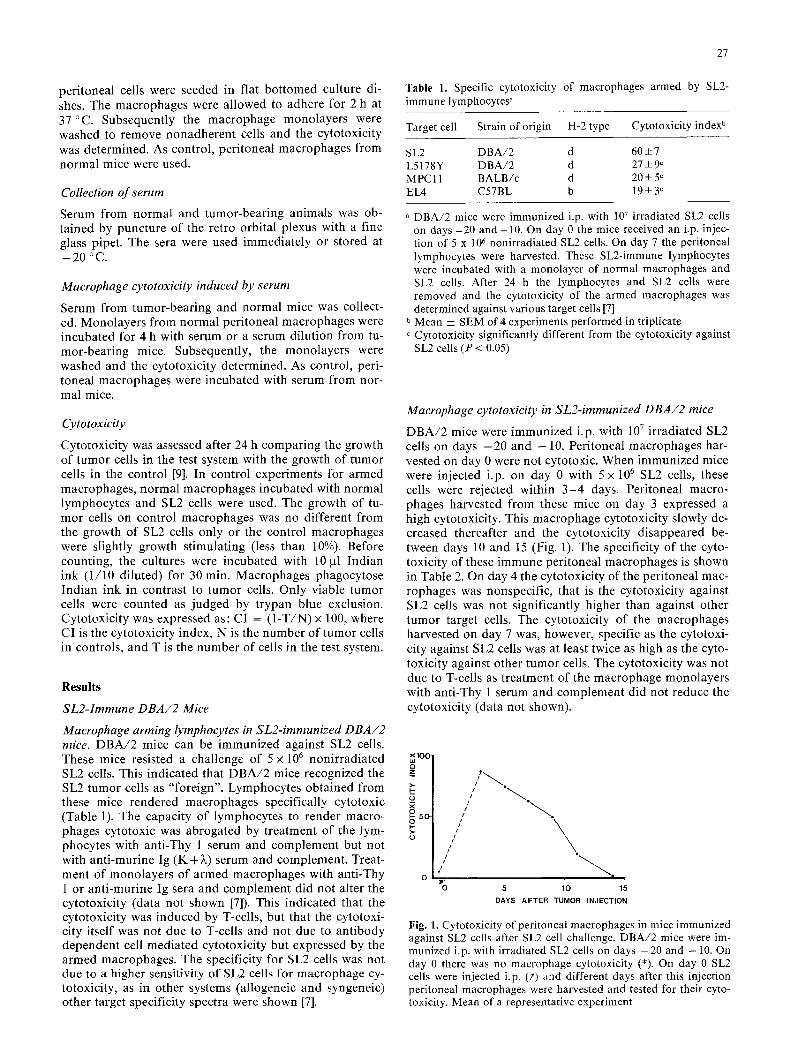
DBA/2 mice were immunized i.p. with 10 7 irradiated SL2 cells on days - 2 0 and - 1 0 . Peritoneal macrophages har- vested on day 0 were not cytotoxic. When immunized mice were injected i.p. on day 0 with 5 x 10 6 SL2 cells, these cells were rejecte d within 3 - 4 days. Peritoneal macro- phages harvested from these mice on day 3 expressed a high cytotoxicity. This macrophage cytotoxicity slowly de- creased thereafter and the cytotoxicity disappeared be- tween days 10 and 15 (Fig. 1). The specificity of the cyto- toxicity of these immune peritoneal macrophages is shown in Table 2. On day 4 the cytotoxicity of the peritoneal mac- rophages was nonspecific, that is the cytotoxicity against SL2 cells was not significantly higher than against other tumor target cells. The cytotoxicity of the macrophages harvested on day 7 was, however, specific as the cytotoxi- city against SL2 cells was at least twice as high as the cyto- toxicity against other tumor cells. The cytotoxicity was not due to T-cells as treatment of the macrophage monolayers with anti-Thy 1 serum and complement did not reduce the cytotoxicity (data not shown).
x100-
£ 50 O
o
I1'= ~ , iii ~ =
tl I ~ . //Ill
i I */ ° ~ t
O 5 10 15
DAYS AFTER TUMOR INJECTION
Fig. 1. Cytotoxicity of peritoneal macrophages in mice immunized against SL2 cells after SL2 cell challenge. DBA/2 mice were im- munized i.p. with irradiated SL2 cells on days - 2 0 and -10. On day 0 there was no macrophage cytotoxicity (*). On day 0 SL2 cells were injected i.p. (~) and different days after this injection peritoneal macrophages were harvested and tested for their cyto- toxicity. Mean of a representative experiment
28
Table 2. Specificity of the cytotoxicity of peritoneal macrophages from mice immunized with SL2 cells a
Target Strain H-2 Cytotoxicity index b cell origin type
Day 4 ° Day 7 d
SL2 DBA/2 d 69 +_ 8 46_+ 4 L5178Y DBA/2 d 64+5 12_+ 10 e MPCll BALB/c d 59-+8 18+ 8 e EL4 C57BL b 62_+4 14+ 7 e
a DBA/2mice were immunized i.p. with 107 irradiated SL2 cells on days -20 and - 10. On day 0 the mice were injected with 5 x 10⁶ SL2 cells. On day 4 or 7 the peritoneal cells were harvested and cultured for 2 h in flat bottomed wells. The nonadherent cells were washed off and the macrophage cytotoxicity was tested against various target cells
b Cytotoxicity was determined after 24 h. Normal peritoneal macrophages were used as controls
° Mean _+ SEM of 2 experiments performed in triplicate Mean _+ SEM of 5 experiments performed in triplicate
e Cytotoxicity significantly (P < 0.05) different from the cytotoxi- city against SL2 cells
SL2-Bearing DBA/2 mice
Macrophage arming lymphocytes in SL2-bearing DBA/2 mice. To test whether lymphocytes from SL2-bearing ani- mals (s. c. SL2 tumor in the f lank) were able to induce mac- rophage cytotoxicity, lymphocytes were collected from draining lymph nodes, spleen, and per i toneal cavity on different days after tumor grafting. None of these lympho- cytes showed a significant induct ion of macrophage cyto- toxicity (data are par t ly given in Table 3).
It was then tested whether injection of a second SL2 tu- mor (104 cel ls /mouse) in the opposi te f lank could attract or induce reactive lymphocytes to the draining lymph nodes or the spleen. In one group, mice were injected with the first SL2 tumor graft on day 0. Part of this group re- ceived a second tumor in the opposi te f lank on day 4. On day 5 the lymph node lymphocytes draining the first and second SL2 tumor and the spleen lymphocytes were tested for their macrophage arming capacity. Both the draining lymph node lymphocytes from the first tumor and spleen
lymphocytes showed a capaci ty to induce macrophage cy- totoxicity, however, only when a second tumor was inject- ed on day 4. Lymphocytes from the lymph node draining the second tumor graft were not able to arm macrophages (Table 3).
In a second group of D B A / 2 mice the first tumor was injected on day 0. This group of mice was divided in three subgroups. The first subgroup received no second tumor. The second subgroup received a second tumor in the op- posite f lank on day 4, and the third subgroup received a second tumor on day 11. On day 12 the lymph node lym- phocytes draining either the first or the second tumor or spleen lymphocytes from all three subgroups were tested for their macrophage arming capacity. Only when a sec- ond tumor graft was injected either on day 4 or day 11, did the lymph node lymphocytes draining the pr imary tumor show a low macrophage arming capacity. No macrophage arming activity was found in the draining lymph node of the second tumor. The spleen lymphocytes were only able to arm macrophages in vitro when the second tumor was injected 1 day before the lymphocytes were collected (Table 3). None of the lymphocytes were able to render macrophages cytotoxic if SL2 cells were absent during the arming of the macrophages. This indicated that the lym- phocytes had to be tr iggered in vitro before they arm mac- rophages.
In summary, these data showed that no arming lym- phocytes were present in the lymphoid organs of tumor- bear ing mice. After injection of a second tumor graft, ar- ming lymphocytes appeared in the draining lymph node of the first tumor, and for a short per iod of t ime in the spleen.
Localization of SL2-sensitized lymphocytes at the site of the tumor
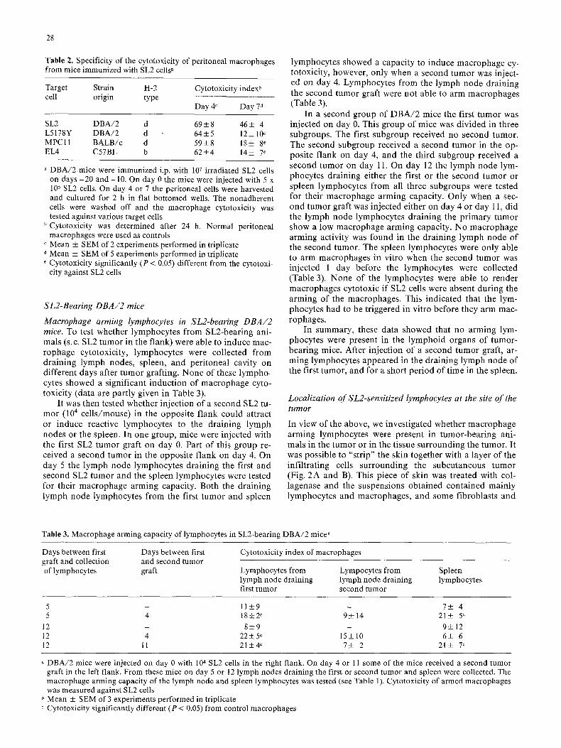
In view of the above, we investigated whether macrophage arming lymphocytes were present in tumor-bear ing ani- mals in the tumor or in the tissue surrounding the tumor. It was possible to "strip" the skin together with a layer of the infil trating ceils surrounding the subcutaneous tumor (Fig. 2 A and B). This piece of skin was treated with col- lagenase and the suspensions obta ined contained mainly lymphocytes and macrophages , and some fibroblasts and
Table 3. Macrophage arming capacity of lyrnphocytes in SL2-bearing DBA/2 mice a
Days between first graft and collection of lymphocytes
Days between first and second tumor graft
Cytotoxicity index of macrophages
Lymphocytes from Lympocytes from Spleen lymph node draining lymph node draining lymphocytes first tumor second tumor
5 - 11+9 - 7+ 4 5 4 18+2 c 9+14 21-t- 5 c
12 - 8_+9 - 9+12 12 4 22_+5 c 15+10 6_+ 6 12 11 21+_4 ~ 7-+ 2 21-+ 7 c
DBA/2 mice were injected on day 0 with 104 SL2 cells in the right flank. On day 4 or 11 some of the mice received a second tumor graft in the left flank. From these mice on day 5 or 12 lymph nodes draining the first or second tumor and spleen were collected. The macrophage arming capacity of the lymph node and spleen lymphocytes was tested (see Table 1). Cytotoxicity of armed macrophages was measured against SL2 cells
~' Mean _+ SEM of 3 experiments performed in triplicate Cytotoxicity significantly different (P < 0.05) from control macrophages
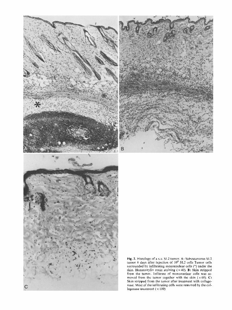
Fig. 2. Histology of a s.c. SL2 tumor. A: Subcutaneous SL2 tumor 8 days after injection of 10 4 SL2 cells Tumor cells surrounded by infiltrating mononuclear cells (*) under the skin. Hematoxylin eosin staining (x40) . B: Skin stripped from the tumor. Infiltrate of mononuclear cells was re- moved from the tumor together with the skin (x60) . C: Skin stripped from the tumor after treatment with collage- nase. Most of the infiltrating cells were removed by the col- lagenase treatment ( x 100)
30
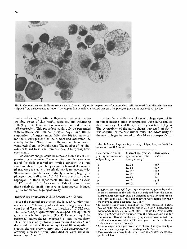
Fig. 3. Mononuclear cell infiltrate from a s.c. SL2 tumor. Cytospin preparation of mononuclear cells removed from the skin that was stripped from a subcutaneous tumor. The preparation contained macrophages (M), lymphocytes (L), and tumor cells (T) ( x 800)
tumor cells (Fig. 3). After collagenase t reatment the re- maining pieces of skin hardly contained any infil trating cells (Fig. 2C). These pieces of skin were removed from the cell suspension. This procedure could only be per formed with relatively small tumors (between days 3 and 10). In suspensions of larger tumors (after day 10) too many tu- mor cells were present, as the tumors had infi l trated the skin by that time. These tumor cells could not be separated complete ly from the lymphocytes . The number of lympho- cytes obtained from small tumors (days 3 to 5) was, how- ever, small.
Most macrophages could be removed from the cell sus- pension by adherence. The remaining lymphocytes were tested for their macrophage arming capacity. As only small numbers of lymphocytes were obta ined the macro- phages were armed with relatively few lymphocytes. With SL2-immune lymphocytes routinely a macrophage : lym- phocy te : tumor cell ratio of 10:20:1 was used to arm mac- rophages. In these experiments the ratio was between 10 : 12 : 1 and 10 : 1 : 1. As shown in Table 4 in most cases these relatively small numbers of lymphocytes induced significant macrophage cytotoxicity.
Macrophage cytotoxicity in SL2-bearing DBA/2 mice
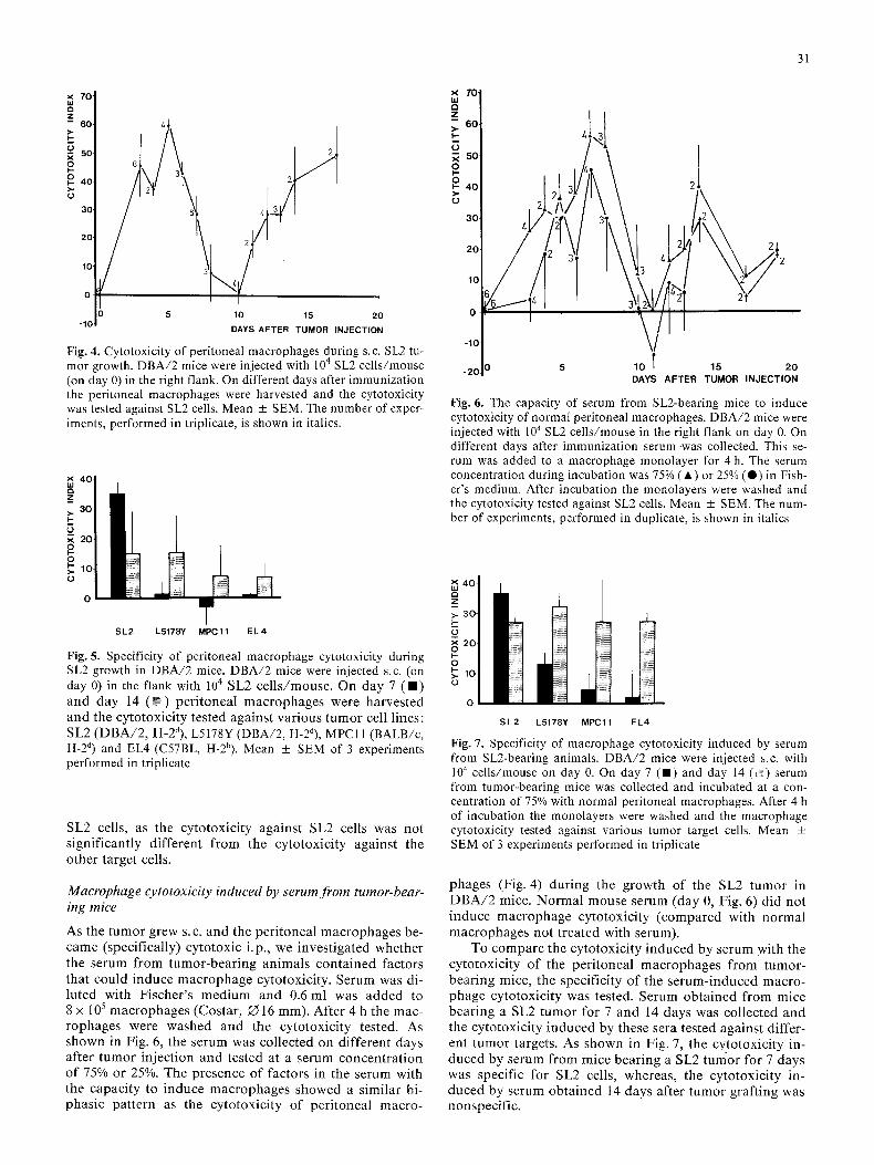
To test the macrophage cytotoxici ty in D B A / 2 mice bear- ing a s. c. SL2 tumor, per i toneal macrophages were har- vested on different days after s.c. injection of 104 SL2 cells. The macrophage cytotoxici ty developed during tumor growth in a biphasic pat tern (Fig. 4). Even on day 3 the per i toneal macrophages expressed a high cytotoxicity. This first phase of cytotoxici ty was maximal on day 5 and decreased thereafter. Between days 8 and l0 no significant cytotoxici ty was present. After day 10 the macrophage cyt- otoxicity increased again. Mice died or were kil led be- tween days 17 and 20.
To test the specificity of the macrophage cytotoxici ty in tumor-bear ing mice, macrophages were harvested on day 7 and day 14, and the cytotoxici ty was tested (Fig. 5). The cytotoxicity of the macrophages harvested on day 7 was specific for the SL2 tumor cells. The cytotoxici ty of the macrophages harvested on day 14 was nonspecif ic for
Table 4. Macrophage arming capacity of lymphocytes around a subcutaneous SL2 tumor .
Days between tumor Macrophage :lympho- Cytotoxicity grafting and collection cyte :tumor cell ratio index c of lymphocytes during arming b
3 10:6:1 20 d 5 10:3:1 24 d 7 10:10:1 38 d 7 10:10:1 47 d 7 10:6:1 10 9 10:10:1 37 d
10 10:12:1 6
a Lymphocytes removed from the subcutaneous tumor by colla- genase treatment of the skin that was stripped from the tumor. Lymphocytes were harvested on different days after tumor injec- tion (10 4 cells s.c.). These lymphocytes were tested for their macrophage arming capacity (see Table 1)
b In routine experiments lymphocytes were incubated during arming with macrophages and tumor cells at a macrophages: lymphocyte:tumor cell ratio of 10:20:1 (Table 1). Often insuffi- cient lymphocytes were obtained from the pieces of skin and for this reason different numbers of lymphocytes were added to a confluent monolayer of 4 x 10⁵ macrophages in flat bottomed dishes to arm the macrophages
° Mean of experiments performed in triplicate. The cytotoxicity of the armed macrophages was tested against SL2 cells Cytotoxicity significantly different from the control macropha- ges (P < 0.05)
7 0
Z - - 60.
0 ,~ 50. o
o 4o
3O
O ,
5 10 15 20 -10
DAYS A F T E R T U M O R INJECTION
Fig. 4. Cytotoxicity of peritoneal macrophages during s.c. SL2 tu- mor growth. DBA/2 mice were injected with 104 SL2 cells/mouse (on day 0) in the right flank. On different days after immunization the peritoneal macrophages were harvested and the cytotoxicity was tested against SL2 cells. Mean _+ SEM. The number of exper- iments, performed in triplicate, is shown in italics.
X 4 0
30 >- F-
× 2 0
0
S L 2 L5178Y MPC11 E L 4
Fig. 5. Specificity of peritoneal macrophage cytotoxicity during SL2 growth in DBA/2 mice. DBA/2 mice were injected s.c. (on day 0) in the flank with l04 SL2 cells/mouse. On day 7 (B) and day 14 ( ~ ) peritoneal macrophages were harvested and the cytotoxicity tested against various tumor cell lines: SL2 (DBA/2, H-2d), L5178Y (DBA/2, H-2d), MPCll (BALB/c, H-2 d) and EL4 (C57BL, H-2b). Mean _+ SEM of 3 experiments performed in triplicate
SL2 cells, as the cytotoxicity against SL2 cells was not significantly different from the cytotoxicity against the other target cells.
Macrophage cytotoxicity induced by serum from tumor-bear- ing mice
As the tumor grew s.c. and the peritoneal macrophages be- came (specifically) cytotoxic i.p., we investigated whether the serum from tumor-bearing animals contained factors that could induce macrophage cytotoxicity. Serum was di- luted with Fischer's medium and 0.6 ml was added to 8 x 10⁵ macrophages (Costar, O 16 mm). After 4 h the mac- rophages were washed and the cytotoxicity tested. As shown in Fig. 6, the serum was collected on different days after tumor injection and tested at a serum concentrat ion of 75% or 25%. The presence of factors in the serum with the capacity to induce macrophages showed a similar bi- phasic pattern as the cytotoxicity of peritoneal macro-
31
X 70 u.I Q Z >. 60 I--
~ so 0 k- 0 I-- 4 0
30-
20
10 ̧
0
-10 ̧
-20
21 , / '/~L*2
10 T 15 20 DAYS AFTER TUMOR INJECTION
Fig. 6. The capacity of serum from SL2-bearing mice to induce cytotoxicity of normal peritoneal macrophages. DBA/2 mice were injected with 104 SL2 cells/mouse in the right flank on day 0. On different days after immunization serum .was collected. This se- rum was added to a macrophage monolayer for 4 h. The serum concentration during incubation was 75% ( • ) or 25% ( • ) in Fish- er's medium. After incubation the monolayers were washed and the cytotoxicity tested against SL2 cells. Mean _+ SEM. The num- ber of experiments, performed in duplicate, is shown in italics
X ,
Z
(9 X O
O
S L 2 L5178Y MPC11 EL4
Fig. 7. Specificity of macrophage cytotoxicity induced by serum from SL2-bearing animals. DBA/2 mice were injected s.c. with l04 cells/mouse on day 0. On day 7 (B) and day 14 ('~;) serum from tumor-bearing mice was collected and incubated at a con- centration of 75% with normal peritoneal macrophages. After 4 h of incubation the monolayers were washed and the macrophage cytotoxicity tested against various tumor target cells. Mean _+ SEM of 3 experiments performed in triplicate
phages (Fig. 4) during the growth of the SL2 tumor in DBA/2 mice. Normal mouse serum (day 0, Fig. 6) did not induce macrophage cytotoxicity (compared with normal macrophages not treated with serum).
To compare the cytotoxicity induced by serum with the cytotoxicity of the peritoneal macrophages from tumor- bearing mice, the specificity of the serum-induced macro- phage cytotoxicity was tested. Serum obtained from mice bearing a SL2 tumor for 7 and 14 days was collected and the cytotoxicity induced by these sera tested against differ- ent tumor targets. As shown in Fig. 7, the cvtotoxicity in- duced by serum from mice bearing a SL2 tumor for 7 days was specific for SL2 cells, whereas, the cytotoxicity in- duced by serum obtained 14 days after tumor grafting was nonspecific.
6-
E 1 2 -
uJ N
gC
~o I..,-
4-
2-
r lo 1'3 17 21 DAYS AFTER SECOND TUMOR I N J E C T I O N
32
Fig. 8. Concomitant immunity in SL2-bearing DBA/2 mice against a second SL2 tumor graft. DBA/2 mice were injected with 104 SL2 cells in the right flank. On different days after this first tumor graft, a second SL2 tumor graft was given in the groin (104 cells/mouse). The growth of the second tumor graft was meas- ured. The second tumor graft was given on the same day as the first SL2 graft (O) or 1 day (111), 4 days (A) or 7 days (A) after the first graft. In the control only a tumor in the groin was given (0) . Mean tumor size of 10 animals per group
~E 10- O
I,l N
o 8
F-
CO b~
~ 6
4
2-
O-
-/ 10 1'3 1 '5 1'7 2'0 DAYS AFTER SECOND TUMOR INJECTION
Fig. 9. Concomitant immunity in SL2-bearing DBA/2 mice against a L5178Y tumor graft. DBA/2 mice were injected with 10 4
SL2 cells in the right flank. On the same day a L5178Y tumor (104 cells/mouse) was injected into the groin (O) or the L5178Y tumor was injected 1 day (11), 4 days ( 0 ) or 7 days (A) after the SL2 tumor. In the control only a L5178Y tumor was given in the groin ( A ). The growth of the L5178Y tumor was measured. Mean tumor size of 10 animals per group
Concomitant immunity in SL2-bearing DBA/2 mice
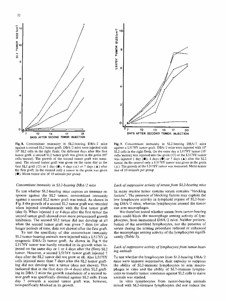
To test whether SL2-bearing mice express an immune re- sponse against the SL2 tumor, concomitant immuni ty against a second SL2 tumor graft was tested. As shown in Fig. 8 the growth of a second SL2 tumor graft was re tarded when injected s imultaneously with the first tumor graft (day 0). When injected 1 or 4 days after the first tumor the second tumor graft showed even more p ronounced growth inhibit ion. The second SL2 tumor did not develop at all when the second tumor graft was given 7 days (or after longer per iods of time, da ta not shown) after the first graft.
To test the specificity of this concomitant immuni ty SL2 tumor-bear ing animals were injected with a L5178Y (a syngeneic D B A / 2 ) tumor graft. As shown in Fig. 9 the L5178Y tumor was hardly re tarded in its growth when in- jected on the same day or 1 or 4 days after the (first) SL2 tumor. However, a second L5178Y tumor graft injected 7 days after the SL2 tumor did not grow at all. Also L5178Y cells injected more than 7 days after the SL2 tumor graft- ing did not develop into a tumor (data not shown). This indica ted that in the first days ( 0 - 4 days) after SL2 graft- ing in D B A / 2 mice the growth re tardat ion of a second tu- mor graft was specifically directed against SL2 cells. F rom day 7 onwards a second tumor graft was, however, nonspecif ical ly b locked in its growth.
Lack of suppressive activity of serum from SL2-bearing mice
In many murine tumor systems serum contains "blocking factors". The presence of blocking factors may explain the low lymphocyte activity in lymphoid organs of SL2-bear- ing D B A / 2 mice, whereas lymphocytes a round the tumor can arm macrophages.
We therefore tested whether serum from tumor-bear ing mice could block the macrophage arming activity of lym- phocytes, from immunized D B A / 2 mice. Neither preincu- ba t ion of the sensitized lymphocytes , nor the presence of serum during the arming procedure reduced or enhanced the macrophage arming activity of the lymphocytes signifi- cantly (Table 5).
Lack of suppressive activity of lymphocytes from tumor-bear- ing animals
To test whether the lymphocytes from SL2-bearing D B A / 2 mice were immune suppressive, their capaci ty to suppress the abil i ty of SL2-immune lymphocytes to arm macro- phages in vitro and the abil i ty of SL2-immune lympho- cytes to transfer tumor resistance against SL2 cells to naive animals was studied.
In vitro lymphocytes from tumor-bear ing animals mixed with SL2-immune lymphocytes did not reduce the
33
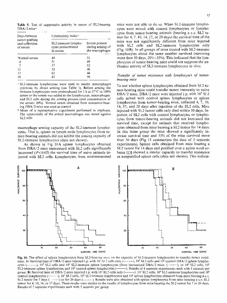
Table 5. Test of suppressive activity in serum of SL2-bearing DBA/2 mice a
Days between tumor grafting and collection of serum
Cytotoxicity index b
SL2-immune lympho- Serum present cytes preincubated during arming of in serum the macrophages
Normalserum 44 51 4 51 49 7 57 52
11 53 46 13 61 44 16 41 50
SL2-immune lymphocytes were used to render macrophages cytotoxic by direct arming (see Table 1). Before arming the immune lymphocytes were preincubated for 1 h at 37°C in 100% serum or the serum was added to the lymphocytes, macrophages, and SL2 cells during the arming process (end concentration of the serum: 20%). Normal serum obtained from nontumor-bear- ing DBA/2 mice was used as control
b Mean of a representative experiment performed in triplicate. The cytotoxicity of the armed macrophages was tested against SL2 cells
macrophage arming capaci ty of the SL2-immune lympho- cytes. That is, spleen or lymph node lymphocytes from tu- mor-bear ing animals did not inhibit the arming capaci ty of SL2-immune lymphocytes (data not shown).
As shown in Fig. 10A spleen lymphocytes obta ined from D B A / 2 mice immunized with SL2 cells significantly increased ( P < 0.05) the survival t ime of naive animals in- jected with SL2 cells. Lymphocytes from non immunized
mice were not able to do so. When SL2-immune lympho- cytes were mixed with control lymphocytes or lympho- cytes from tumor-bear ing animals (bearing a s.c. SL2 tu- mor for 4, 7, 10, 14, 17, or 20 days) the survival t ime of the mice was not significantly different from mice injected with SL2 cells and SL2-immune lymphocytes only (Fig. 10B). In all groups of mice treated with SL2-immune lymphocytes about the same number survived (surviving more then 50 days, 20%-35%). This indica ted that the lym- phocytes of tumor-bear ing mice could not suppress the an- t i tumor activity of SL2-immune lymphocytes in vivo.
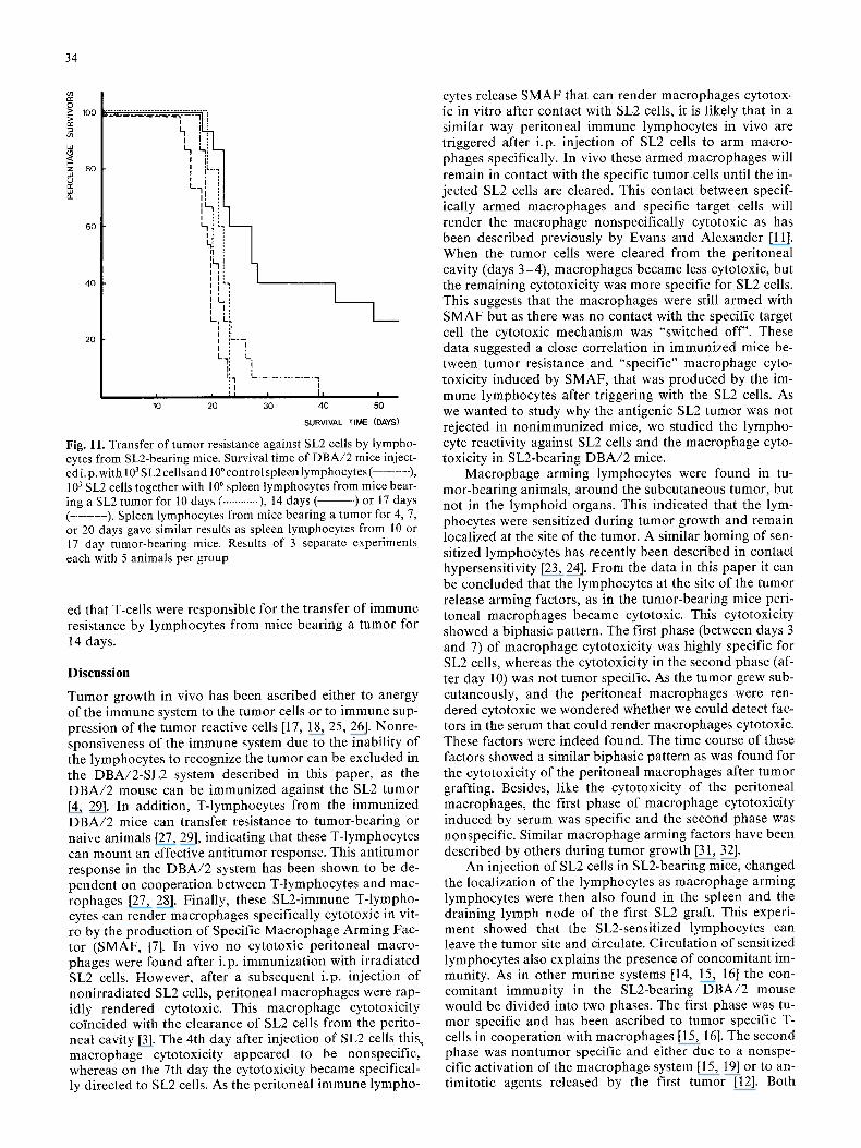
Transfer of tumor resistance with lymphocytes of tumor- bearing mice
To test whether spleen lymphocytes obtained from SL2 tu- mor-bear ing mice could transfer tumor immuni ty to naive D B A / 2 mice, D B A / 2 mice were injected i.p. with 103 SL2 ceils mixed with control spleen lymphocytes or spleen lymphocytes from tumor-bear ing mice, collected 4, 7, 10, 14, 17, and 20 days after injection of the SL2 cells. Mice injected with SL2 tumor ceils only died within 30 days. In- ject ion of SL2 cells with control lymphocytes or lympho- cytes from tumor-bear ing animals did not increased the survival time, except for animals that received lympho- cytes obta ined from mice bear ing a SL2 tumor for 14 days. In this latter group the mice showed a significantly in- crease survival t ime and 33% of the mice survived more than 50 days (Fig. 11 summarizes the da ta of 3 separate experiments). Spleen cells obta ined from mice bearing a SL2 tumor for 14 days and purif ied over a nylon wool co- lumn [13] showed a similar capaci ty to transfer resistance as nonpur i f ied spleen cells (data not shown). This indicat-
0 >_
w
,z,, o=
A 100 ! -'m! " -~- '-'~- -" --~----" --~2--~- - "-- o ], ] .~L~.I7 ~
8o i!4~, :l
6o [
4 "I._,
40 LI :I L . ~
I i "-'r7 I ]
I [ k . . . . . -1 - - 20 I L 1 I
kL, 1 l 4, "'"7 " '7
I m • . . . . . . . . . . . . . . . . . . . . ~ I - - - - I
I I I I [ I i ' 10 20 30 40 50
SURVIVAL TIME (DAYS)
0 >_ 100
if) w
,,z 8o
B ':~'~"~'-=-'~: ~"~e'-%i"7 7
t L.-~
I : l _ .
' r ' I
i ! L 7
:LL. 6 0 I . . . . . . . ]
I L I I
40 t_! ; . . . . . . . .
20 L- -I . . . . . . . . . . . . . . . . . . . . . . . I ,,
I I ] I I I 10 20 30 40 50
SURVIVAL TIME (DAYS)
Fig. 10. The effect of spleen lymphocytes from SL2-bearing mice on the capacity of SL2-immune tymphocytes to transfer tumor resist- ance. A: Survival time of DBA/2 mice injected i.p. with 103 SL2 cells only ( . . . . . ), 103 SL2 cells and l06 control DBA/2 spleen lympho- cytes ( ............. ), 103 SL2 cells and 10 6 SL2-immune spleen lymphocytes (from immunized DBA/2 mice; (- . . . . . . . ), or 103 SL2 cells, l 0 6
SL2-immune spleen lymphocytes and l 0 6 control spleen lymphocytes ( ). Results of 4 separate experiments each with 5 animals per group. B: Survival time of DBA/2 mice injected i.p. with 103 SL2 cells only (- . . . . ), 103 SL2 cells, l06 SL2-immune lymphocytes and l 0 6
control lymphocytes ( ), 103 SL2 ceils, 106 SL2-immune lymphocytes and 106 spleen lymphocytes obtained from mice bearing a s.c. SL2 tumor for 7 days (- . . . . . ) or for 20 days (. ............ ). Results were also obtained with spleen lymphocytes from mice bearing a s.c. SL2 tumor for 4, 10, 14, or 17 days. These results were similar to the results of lymphocytes from mice bearing the SL2 tumor for 7 or 20 days. Results of 2 separate experiments each with 5 animals per group
34
O -> 100 =>
z 80
,,=,
60
40
20
;'--'=-'-'-~-'.:=-'-~-"..'.-=_:2-:',=--:':'"".' ' ] !
I ' L I "3
L - 1 , i
I L I : I , :
-T] t I :
L~ I :
L. I L:,
I!L-- i I : L.li L,
I: ! t~. I L . . . . . . . . . . .
I I
I 30 40 5 0 I0 20 I i I
SURVIVAL TIME (DAYS)
Fig. ll. Transfer of tumor resistance against SL2 cells by lympho- cytes from SL2-bearing mice. Survival time of DBA/2 mice inject- ed i. p. with 103 S L2 cells and 106 control spleen lympho cytes ( . . . . . . . ), 103 SL2 cells together with 106 spleen lymphocytes from mice bear- ing a SL2 tumor for 10 days ( ............ ), 14 days ( ) or 17 days ( . . . . . . . . ). Spleen lymphocytes from mice bearing a tumor for 4, 7, or 20 days gave similar results as spleen lymphocytes from 10 or 17 day tumor-bearing mice. Results of 3 separate experiments each with 5 animals per group
ed that T-cells were responsible for the transfer of immune resistance by lymphocytes from mice bearing a tumor for 14 days.
Discussion
Tumor growth in vivo has been ascribed either to anergy of the immune system to the tumor cells or to immune sup- pression of the tumor reactive cells [17, 18, 25, 26]. Nonre- sponsiveness of the immune system due to the inability of the lymphocytes to recognize the tumor can be excluded in the DBA/2-SL2 system described in this paper, as the DBA/2 mouse can be immunized against the SL2 tumor [4, 29]. In addition, T-lymphocytes from the immunized DBA/2 mice can transfer resistance to tumor-bearing or naive animals [27, 29], indicating that these T-lymphocytes can mount an effective antitumor response. This antitumor response in the DBA/2 system has been shown to be de- pendent on cooperation between T-lymphocytes and mac- rophages [27, 28]. Finally, these SL2-immune T-lympho- cytes can render macrophages specifically cytotoxic in vit- ro by the production of Specific Macrophage Arming Fac- tor (SMAF, [7]. In vivo no cytotoxic peritoneal macro- phages were found after i.p. immunization with irradiated SL2 cells. However, after a subsequent i.p. injection of nonirradiated SL2 cells, peritoneal macrophages were rap- idly rendered cytotoxic. This macrophage cytotoxicity coincided with the clearance of SL2 cells from the perito- neal cavity [3]. The 4th day after injection of SL2 cells this macrophage cytotoxicity appeared to be nonspecific, whereas on the 7th day the cytotoxicity became specifical- ly directed to SL2 cells. As the peritoneal immune lympho-
cytes release SMAF that can render macrophages cytotox- ic in vitro after contact with SL2 cells, it is likely that in a similar way peritoneal immune lymphocytes in vivo are triggered after i.p. injection of SL2 cells to arm macro- phages specifically. In vivo these armed macrophages will remain in contact with the specific tumor cells until the in- jected SL2 cells are cleared. This contact between specif- ically armed macrophages and specific target cells will render the macrophage nonspecifically cytotoxic as has been described previously by Evans and Alexander [11]. When the tumor cells were cleared from the peritoneal cavity (days 3-4), macrophages became less cytotoxic, but the remaining cytotoxicity was more specific for SL2 cells. This suggests that the macrophages were still armed with SMAF but as there was no contact with the specific target cell the cytotoxic mechanism was "switched off''. These data suggested a close correlation in immunized mice be- tween tumor resistance and "specific" macrophage cyto- toxicity induced by SMAF, that was produced by the im- mune lymphocytes after triggering with the SL2 cells. As we wanted to study why the antigenic SL2 tumor was not rejected in nonimmunized mice, we studied the lympho- cyte reactivity against SL2 cells and the macrophage cyto- toxicity in SL2-bearing DBA/2 mice.
Macrophage arming lymphocytes were found in tu- mor-bearing animals, around the subcutaneous tumor, but not in the lymphoid organs. This indicated that the lym- phocytes were sensitized during tumor growth and remain localized at the site of the tumor. A similar homing of sen- sitized lymphocytes has recently been described in contact hypersensitivity [23, 24]. From the data in this paper it can be concluded that the lymphocytes at the site of the tumor release arming factors, as in the tumor-bearing mice peri- toneal macrophages became cytotoxic. This cytotoxicity showed a biphasic pattern. The first phase (between days 3 and 7) of macrophage cytotoxicity was highly specific for SL2 cells, whereas the cytotoxicity in the second phase (af- ter day 10) was not tumor specific. As the tumor grew sub- cutaneously, and the peritoneal macrophages were ren- dered cytotoxic we wondered whether we could detect fac- tors in the serum that could render macrophages cytotoxic. These factors were indeed found. The time course of these factors showed a similar biphasic pattern as was found for the cytotoxicity of the peritoneal macrophages after tumor grafting. Besides, like the cytotoxicity of the peritoneal macrophages, the first phase of macrophage cytotoxicity induced by serum was specific and the second phase was nonspecific. Similar macrophage arming factors have been described by others during tumor growth [31, 32].
An injection of SL2 cells in SL2-bearing mice, changed the localization of the lymphocytes as macrophage arming lymphocytes were then also found in the spleen and the draining lymph node of the first SL2 graft. This experi- ment showed that the SL2-sensitized lymphocytes can leave the tumor site and circulate. Circulation of sensitized lymphocytes also explains the presence of concomitant im- munity. As in other murine systems [14, 15, 16] the con- comitant immunity in the SL2-bearing DBA/2 mouse would be divided into two phases. The first phase was tu- mor specific and has been ascribed to tumor specific T- cells in cooperation with macrophages [15, 16]. The second phase was nontumor specific and either due to a nonspe- cific activation of the macrophage system [15, 19] or to an- timitotic agents released by the first tumor [12]. Both
35
phases seemed to occur in the DBA/2-SL2 system as well. The two phases of specific and nonspecific concomitant immunity closely paralleled the two phases of macrophage cytotoxicity. This suggests that the specific phase of con- comitant immunity (between days 1 and 6) might be caused by sensitized lymphocytes and specifically armed macrophages, whereas, the nonspecific phase (after day 6) of concomitant immunity is caused by the nonspecific cy- totoxicity of macrophages. This is in agreement with data from North et al. [19].
The similarity in concomitant immunity between this DBA/2-SL2 system and other nonlymphoid tumor murine systems (e.g., sarcomas, [14, 17] indicates that the immune reactions occurring in the DBA/2-SL2 system are not ne- cessarily limited to lymphomas.
So, sensitization of lymphocytes to SL2 cells occurrs in SL2-bearing mice. Why then, is the tumor not rejected? We first studied whether the SL2-immune lymphocytes can be suppressed in their macrophage arming activity in vitro by factors in the serum from tumor-bearing mice or by lymphocytes from tumor-bearing mice. No inhibition or suppression by serum or lymphocytes from tumor-bearing mice on the macrophage arming activity of immune lym- phocytes was found, suggesting that no suppressive mech- anism for macrophage arming activity of the lymphocytes was present. This is in line with the finding that macro- phage arming lymphocytes were present in the tumor- bearing mice as well.
The second possibility is that lymphocytes from tumor- bearing animals suppressed another activity required for tumor resistance. We therefore tested whether lymphocytes from tumor-bearing mice could suppress the capacity of immune lymphocytes to transfer tumor resistance to naive animals. However, spleen lymphocytes from tumor-bear- ing animals did not suppress the transfer of tumor resist- ance by SL2-immune spleen lymphocytes in a Winn-type assay. On the contrary, it was found that T-lymphocytes from DBA/2 mice bearing a SL2 tumor for 14 days, trans- fered resistance to naive animals as well. Lymphocytes ob- tained from DBA/2 mice injected 10 days or 17 days after injection of the SL2 tumor did not transfer tumor resist- ance. This was found in three separate experiments (Fig. 11). Why only spleen lymphocytes from day 14 trans- fered resistance is as yet unknown.
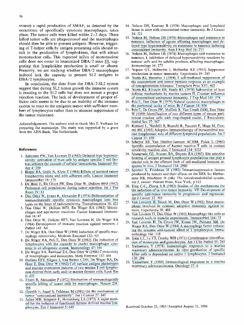
We have recently hypothesized that the immune re- sponse against tumor cells can be divided into an induc- tion phase and an effector cell phase (Fig. 12, [6]). Accord- ing to this hypothesis antigens on tumor cells can be recog- nized directly by T-cells, as was shown previously [8]. This T-cell-tumor cell interaction enables the T-cells to react with the release of a specific T-cell factor (e.g., SMAF). The factor can adhere to macrophages (or other cells). The specific factor can function as a bridge between the specif- ic tumor cells and the macrophages (or other cells). The contact triggers the macrophage to kill the tumor cells. Subsequently, the macrophage will phagoeytose the tumor cell debris and present tumor antigens. This will lead to the induction of helper T-cells and consequently to the fur- ther development of the immune response (the effector cell phase, Fig. 12). Other cells armed with the early specific T- cell factors might facilitate the development of the immune response as well. For instance, armed mast cells release se- rotonin after contact with the antigen. This facilitates the entry of lymphocytes and macrophages to the site of the
INDUCTION
PHASE
EFFECTORpHAsECELL I
T-cell + Tumor cell
SMAF production
Macrophage Arming
Tumor cell killing
~'~ APC + T-helper cell
T Lymphokine production
(IL2, MAF, MIF, CF)
Y Attraction of
mononuclear cells
Y Phagocytosis of ~ ~ Induction of
tumor cell debris CTL and cytotoxic
macrophages
Fig. 12. Hypothesis for the immune response against tumor cells. The immune reaction is divided into two phases. An initiation phase in which the T-cells react directly with tumor cells, leading to the production of SMAF. Macrophages armed by SMAF can kill tumor cells. The tumor cell debris can be phagocytosed by an- tigen presenting cells (APC). These APC can induce the second phase of the response, the effector cell phase, by activating T- helper cells. These T-cells will produce lymphokines such as in- terleukin-2 (IL-2), Macrophage Activating Factor (MAF), Migra- tion Inhibitory Factor (MIF) and Chemotactic Factor (CF). These lymphokines will attract mononuclear cells to the site of the tumor and activate cytotoxic T-cells (CTL) and macrophages
tumor [1]. This latter suggestion of a role for a specific T- cell factor and mast cells in the initiation of an immune re- sponse, was recently put forward by Askenase and Van Loveren [1].
It is of interest to compare the data described in this paper on SL2-bearing DBA/2 mice with the hypothesis de- picted in Fig. 12. Apparently the induction phase is not af- fected in the tumor-bearing animal, as macrophage arming lymphocytes are present, SMAF is present in the serum, and specifically cytotoxic macrophages are present in the peritoneal cavity. This suggests that the induction phase develops in contrast to the effector cell phase. This may be due to two reasons. In the first place it is possible that agents produced by the tumor cells or activated macro- phages inhibit the induction of helper T-cells by the anti- gen presenting cells, which will prevent the development of the effector cell phase. In the second place it is possible that macrophages in syngeneic tumor systems cannot pres- ent an antigenic moiety from killed tumor cells. In this lat- ter case resistance to the tumor only occurs when the pro- duction of specific T-cell factors and the subsequent ar- ming of macrophages (initiation phase) is strong enough to reject a challenge of tumor cells directly. This situation might occur after hyperimmunization with irradiated tu- mor cells and subsequent challenge with nonirradiated cells. In the DBA/2-SL2 system it has been shown that two subsequent immunizations with irradiated ceils are re- quired to induce immunity to SL2 cells, as after one injec- tion with irradiated SL2 cells DBA/2 mice were unable to reject a challenge with nonirradiated SL2 cells [7]. When the mice, immunized twice i.p. with 1 0 7 irradiated cells, were challenged i.p. with nonirradiated cells (5 x 10 6 cells/
36
mouse) a rapid product ion of SMAF, as detected by the occurrence of specifically cytotoxic macrophages, takes place. The tumor cells were killed within 2 - 3 days. These killed tumor cells are phagocytosed and the macrophages should then be able to present antigens. However, trigger- ing of T-helper ceils by antigen presenting cells should re- sult in the production of lymphokines, that will attract mononuclear cells. This expected influx of mononuclear cells does not occur in immunized DBA/2 mice [3], sug- gesting that lymphokine product ion is small or absent. Presently, we are studying whether DBA/2 macrophages indeeed lack the capacity to present SL2 antigens to DBA/2 lymphocytes.
In conclusion, the data from the DBA/2-SL2 system suggest that during SL2 tumor growth the immune system is reacting to the SL2 cells but does not mount a proper rejection reaction. The lack of development of proper ef- fector cells seems to be due to an inability of the immune system to react to the antigenic tumor with sufficient num- bers of lymphocytes rather than to immune suppression of the tumor resistance.
Acknowledgements. The authors wish to thank Mrs E. Verbaan for preparing the manuscript. The study was supported by a grant from the ABN-Bank, The Netherlands.
References
1. Askenase PW, Van Loveren H (1983) Delayed-type hypersen- sitivity: activation of mast cells by antigen specific T cell fac- tors initiates the cascade of cellular interactions. Immunol To- day 4:259
2. Blazar BA, Galik N, Klein E (1984) Effects of isolated tumor lymphocytes alone and with adherent cells. Cancer Immunol Immunother 18:179
3. De Heer E, De Groot JW, Den Otter W, Dullens HFJ (1982) Peritoneal cell populations during tumor rejection. Int J Tiss React IV: 81
4. Den Otter W, Evans R, Alexander P (1974) Differentiation of immunologically specific cytotoxic macrophages into two types on the basis of radiosensitivity. Transplantation 18:421
5. Den Otter W, Dullens HFJ, De Weger RA (1983) Macro- phages and anti-tumor reactions. Cancer Immunol Immuno- ther 16:67
6. Den Otter W, Dullens HFJ, Van Loveren H, De Weger RA (1984) Development of immune reaction against tumours. J Pathol 143:A4
7. De Weger RA, Den Otter W (1986) Induction of specific mac- rophage cytotoxicity. Methods Enzymol 132:531
8. De Weger RA, Pels E, Den Otter W (1982) The induction of lymphocytes with the capacity to render macrophages cyto- toxic in an allogeneic system. Immunology 47:541
9. De Weger RA, Runhaar EA, Den Otter W (1986) Cytotoxicity of macrophages and monocytes. Meth Enzymol 132:458
10. Dullens HFJ, Hilgers J, Van Basten CDH, De Weger RA, De Heer E, Den Otter W (1982) Cell surface antigen phenotypes and enzyme expression patterns of two murine T cell lympho- mas derived from early and/or mature thymus cells. Leuk Res 6: 425
11. Evans R, Alexander P (1972) Mechanism of immunologically specific killing of tumor cells by macrophages. Nature 236: 168
12. Gorelik E, Segal S, Feldman M (1981) On the mechanism of tumor "concomitant immunity". Int J Cancer 27:847
13. Julius MH, Simpson E, Herzenberg LA (1973) A rapid meth- od for the isolation of functional thymus derived murine lym- phocytes. Eur J Immunol 3:645
14. Nelson DS, Kearney R (1976) Macrophages and lymphoid tissue in mice with concomitant tumor immunity. Br J Cancer 34:221
15. Nelson M, Nelson DS (1978) Macrophages and resistance to tumours. Influence of agents affecting macrophages and de- layed type hypersensitivity on resistance to tumours inducing concomitant immunity. Aust J Exp Biol 56:211
16. Nelson M, Nelson DS (1978) Macrophages and resistance to tumours. I. Inhibition of delayed hypersensitivity reactions by tumour cells and by soluble products affecting macrophages. Immunology 34:277
17. Nepom GT, Hellstr6m I, Hellstr6m KE (1983) Suppressor mechanism in tumor immunity. Experientia 39:243
18. North RJ, Bursaker I (1984) T cell-mediated suppression of the concomitant anti tumor immune response as an example of transplantation tolerance. Transplant Proc XVI: 463
19. North RJ, Kirstein DJ, Tuttle RL (1976) Subversion of host defense mechanisms by murine tumors II. Counter influence of concomitant antitumour immunity. J Exp Med 143:574
20. Pels E, Den Otter W (1979) Natural cytotoxic macrophages in the peritoneal cavity of mice. Br J Cancer 34:856
21. Pels E, De Groot JW, Mullink R, Van Unnik JAM, Den Otter W (1980) Identification of two different types of mouse peri- toneal exudate cells with ring-shaped nuclei. J Reticuloen- dothel Soc 27:367
22. Romani L, Nardelli B, Bianchi R, Puccetti P, Mage M, Fior- etti MC (1985) Adoptive immunotherapy of intracerebral mu- rine lymphomas: role of different lymphoid populations. Int J Cancer 35:659
23. Scheper RJ, Van Dinther-Janssen ACHM, Polak L (1985) Specific accumulation of hapten reactive T cells in contact sensitivity reaction sites. J Immunol 134:1333
24. Spangrude GJ, Araneo BA, Daynes RA (1985) Site selective homing of antigen primed lymphocyte populations can play a crucial role in the efferent limb of cell-mediated immune re- sponse in vivo. J Immunol 134:2900
25. Spector S, Friedman H (1983) Immunosuppressive factors produced by tumors and their effects on the RES. In: Herber- man RB, Friedman H (eds) The reticuloendothelial system, vol 5, cancer. Plenum Press, New York, p 315
26. Ting C-C, Zhang S-R (1983) Studies of the mechanisms for the induction of in vivo tumor immunity. VII Development of specific anti-tumor immunity in progressors and regressors. Int J Cancer 32:385
27. Van Loveren H, Snoek M, Den Otter W (1982) Host macro- phage involved in systemic adoptive immunity against tu- mors. Experientia 38:488
28. Van Loveren H, Den Otter W (1983) Macrophage like cells as research tools in transfer experiments. Immunobiol 164:23
29. Van Loveren H, De Groot JW, Koten JW, Piersma AH, De Weger RA, Den Otter W (1984) A macrophage factor enhanc- ing the systemic anti-tumour effect of T lymphocytes. Immu- nobiology 166:118
30. Yam LT, Ly CI, Crosby WH (1971) Cytochemical identifica- tion of monocytes and granulocytes. Am J Clin Pathol 55 : 283
31. Yamamura Y (1978) Immunologic responses to a murine mammary adenocarcinoma: In vitro production of specific killer cells is dependent on active T lymphocytes. J Immunol 120:286
32. Yamamura Y (1980) Immunological responses to a murine mammary adenocarcinoma. Oncology 37:6
Received October 25, 1985/Accepted August 21, 1986
Related Documents


